Clinical Study Effect of Gender on Coagulation Functions ...
6
Clinical Study Effect of Gender on Coagulation Functions: A Study in Metastatic Colorectal Cancer Patients Treated with Bevacizumab, Irinotecan, 5-Fluorouracil, and Leucovorin Cemil Bilir, Hüseyin Engin, and Yasemin Bakkal Temi B¨ ulent Ecevit University School of Medicine, Department of Internal Medicine, Division of Medical Oncology, 67100 Zonguldak, Turkey Correspondence should be addressed to Cemil Bilir; [email protected] Received 12 June 2014; Revised 22 July 2014; Accepted 6 August 2014; Published 21 August 2014 Academic Editor: Aldo Roccaro Copyright © 2014 Cemil Bilir et al. is is an open access article distributed under the Creative Commons Attribution License, which permits unrestricted use, distribution, and reproduction in any medium, provided the original work is properly cited. Introduction. We designed this study to evaluate how coagulation parameters are changed in metastatic colorectal cancer (mCRC) patients treated with bevacizumab, irinotecan, 5-fluorouracil, and leucovorin (FOLFIRI). Methods. A total of 48 mCRC patients who initially received bevacizumab with FOLFIRI were eligible for this study. irty-four patients were analyzed at baseline and on the 4th, 8th, and 12th cycles of chemotherapy. Results. ere were 19 male and 15 female patients. Baseline characteristics of the groups were similar, but women had better overall survival than men (14 months versus 12 months, = 0.044). D-dimer levels decreased significantly aſter the 12th cycle compared with baseline in men but not in women. Men and women had increased levels of serum fibrinogen at the early cycles, but these increased fibrinogen levels continued aſter the 4th cycle of chemotherapy only in women. In addition, serum fibrinogen levels did not significantly change, but aPTT levels decreased in men. Discussion. e major finding of this study is that bevacizumab-FOLFIRI chemotherapy does not promote changes in the coagulation system. If chemotherapy treatment and the possible side effects of FOLFIRI-bevacizumab treatment are well managed, then alterations of the coagulation cascade will not have an impact on overall survival and mortality. 1. Introduction Bevacizumab is typically used in combination with fluoropy- rimidine-based chemotherapy for the treatment of patients with metastatic colorectal cancer (mCRC) [1]. In a pivotal phase III trial of first-line mCRC, bevacizumab in com- bination with standard irinotecan/fluorouracil (FOLFIRI) chemotherapy increased tumor response rate by 10% and significantly lengthened the overall survival [1, 2]. In the bevacizumab arm, the patients had an increased incidence of thrombotic events compared with the control arm (19.4% versus 16.2%, resp.), but this result was not significant [1]. In a meta-analysis, a total of 7956 patients diagnosed with many types of solid tumors from 15 randomized trials were identified. e authors revealed that patients treated with bevacizumab had a statistically significant increased risk of venous thromboembolic events (VTEs), with an RR of 1.33 (95% CI, 1.13–1.56, < 0.001) compared with the controls [3]. Hurwitz et al. reported a meta-analysis of 6,055 patients in 10 randomized studies [4]. is study concluded that the addition of bevacizumab to chemotherapy did not significantly increase the risk of VTEs versus chemotherapy alone. e risk for VTEs was attributed to tumor and host factors [4]. Epistaxis, hemoptysis, hematemesis, gastroin- testinal bleeding, vaginal bleeding, and brain hemorrhage have been reported as hemorrhagic toxicities associated with bevacizumab [5]. ese controversial findings open the possibility that factors other than bevacizumab, such as drugs, cancer, age, gender, or comorbidities, can cause VTEs. erefore, we designed this study to evaluate how coagulation parameters, such as the international normalized ratio (INR), activated partial thromboplastin time (aPTT), and fibrinogen and D-dimer levels, can be altered in mCRC patients treated with FOLFIRI-bevacizumab chemotherapy. Hindawi Publishing Corporation Advances in Hematology Volume 2014, Article ID 473482, 5 pages http://dx.doi.org/10.1155/2014/473482
Transcript of Clinical Study Effect of Gender on Coagulation Functions ...

Clinical StudyEffect of Gender on Coagulation Functions: A Study inMetastatic Colorectal Cancer Patients Treated withBevacizumab, Irinotecan, 5-Fluorouracil, and Leucovorin
Cemil Bilir, Hüseyin Engin, and Yasemin Bakkal Temi
Bulent Ecevit University School ofMedicine, Department of InternalMedicine, Division ofMedical Oncology, 67100 Zonguldak, Turkey
Correspondence should be addressed to Cemil Bilir; [email protected]
Received 12 June 2014; Revised 22 July 2014; Accepted 6 August 2014; Published 21 August 2014
Academic Editor: Aldo Roccaro
Copyright © 2014 Cemil Bilir et al. This is an open access article distributed under the Creative Commons Attribution License,which permits unrestricted use, distribution, and reproduction in any medium, provided the original work is properly cited.
Introduction.We designed this study to evaluate how coagulation parameters are changed in metastatic colorectal cancer (mCRC)patients treated with bevacizumab, irinotecan, 5-fluorouracil, and leucovorin (FOLFIRI). Methods. A total of 48 mCRC patientswho initially received bevacizumab with FOLFIRI were eligible for this study. Thirty-four patients were analyzed at baseline andon the 4th, 8th, and 12th cycles of chemotherapy. Results.There were 19 male and 15 female patients. Baseline characteristics of thegroups were similar, but women had better overall survival than men (14 months versus 12 months, 𝑃 = 0.044). D-dimer levelsdecreased significantly after the 12th cycle compared with baseline in men but not in women. Men and women had increased levelsof serum fibrinogen at the early cycles, but these increased fibrinogen levels continued after the 4th cycle of chemotherapy onlyin women. In addition, serum fibrinogen levels did not significantly change, but aPTT levels decreased in men. Discussion. Themajor finding of this study is that bevacizumab-FOLFIRI chemotherapy does not promote changes in the coagulation system. Ifchemotherapy treatment and the possible side effects of FOLFIRI-bevacizumab treatment are well managed, then alterations of thecoagulation cascade will not have an impact on overall survival and mortality.
1. Introduction
Bevacizumab is typically used in combination with fluoropy-rimidine-based chemotherapy for the treatment of patientswith metastatic colorectal cancer (mCRC) [1]. In a pivotalphase III trial of first-line mCRC, bevacizumab in com-bination with standard irinotecan/fluorouracil (FOLFIRI)chemotherapy increased tumor response rate by 10% andsignificantly lengthened the overall survival [1, 2]. In thebevacizumab arm, the patients had an increased incidenceof thrombotic events compared with the control arm (19.4%versus 16.2%, resp.), but this result was not significant [1].In a meta-analysis, a total of 7956 patients diagnosed withmany types of solid tumors from 15 randomized trials wereidentified. The authors revealed that patients treated withbevacizumab had a statistically significant increased riskof venous thromboembolic events (VTEs), with an RR of
1.33 (95% CI, 1.13–1.56, 𝑃 < 0.001) compared with thecontrols [3]. Hurwitz et al. reported a meta-analysis of 6,055patients in 10 randomized studies [4]. This study concludedthat the addition of bevacizumab to chemotherapy did notsignificantly increase the risk of VTEs versus chemotherapyalone. The risk for VTEs was attributed to tumor and hostfactors [4]. Epistaxis, hemoptysis, hematemesis, gastroin-testinal bleeding, vaginal bleeding, and brain hemorrhagehave been reported as hemorrhagic toxicities associatedwith bevacizumab [5]. These controversial findings openthe possibility that factors other than bevacizumab, such asdrugs, cancer, age, gender, or comorbidities, can cause VTEs.Therefore, we designed this study to evaluate how coagulationparameters, such as the international normalized ratio (INR),activated partial thromboplastin time (aPTT), and fibrinogenand D-dimer levels, can be altered in mCRC patients treatedwith FOLFIRI-bevacizumab chemotherapy.
Hindawi Publishing CorporationAdvances in HematologyVolume 2014, Article ID 473482, 5 pageshttp://dx.doi.org/10.1155/2014/473482
2 Advances in Hematology
2. Materials and Methods
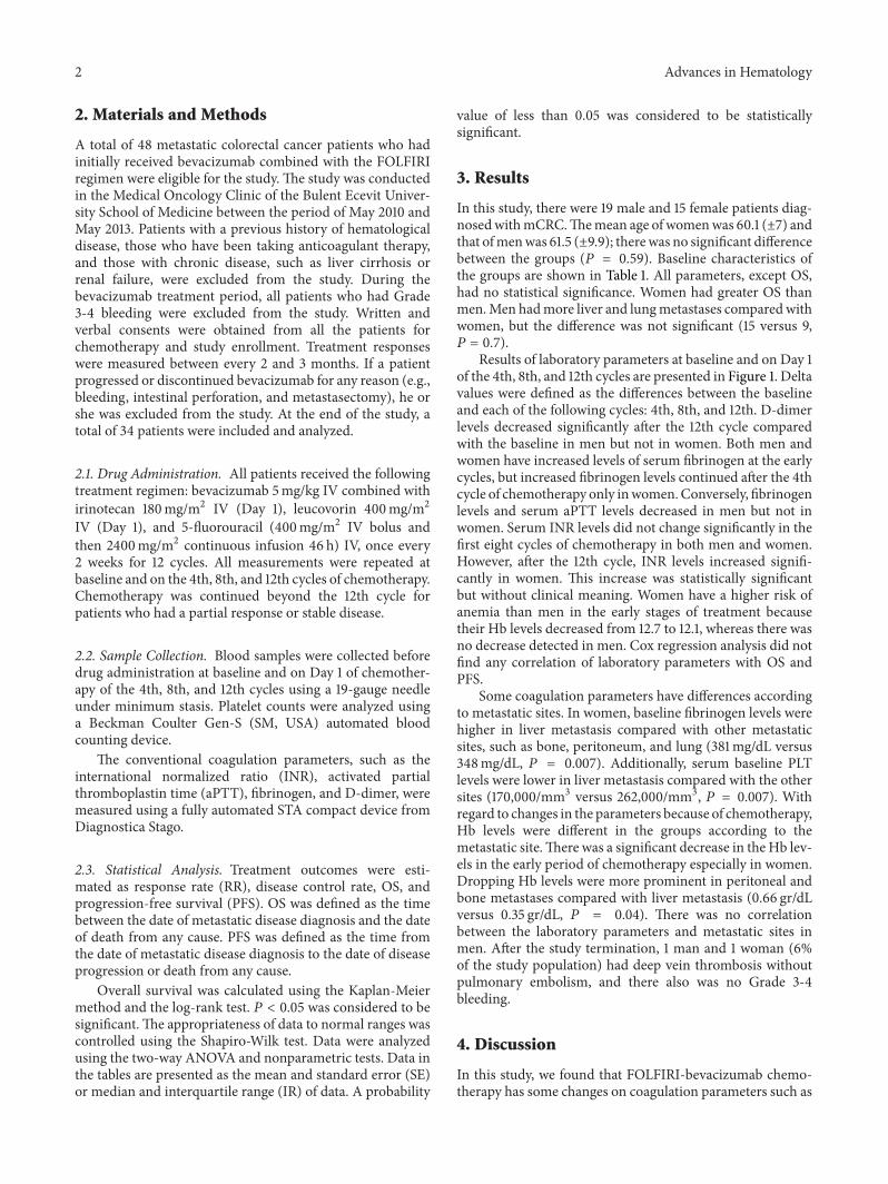
A total of 48 metastatic colorectal cancer patients who hadinitially received bevacizumab combined with the FOLFIRIregimen were eligible for the study. The study was conductedin the Medical Oncology Clinic of the Bulent Ecevit Univer-sity School of Medicine between the period of May 2010 andMay 2013. Patients with a previous history of hematologicaldisease, those who have been taking anticoagulant therapy,and those with chronic disease, such as liver cirrhosis orrenal failure, were excluded from the study. During thebevacizumab treatment period, all patients who had Grade3-4 bleeding were excluded from the study. Written andverbal consents were obtained from all the patients forchemotherapy and study enrollment. Treatment responseswere measured between every 2 and 3 months. If a patientprogressed or discontinued bevacizumab for any reason (e.g.,bleeding, intestinal perforation, and metastasectomy), he orshe was excluded from the study. At the end of the study, atotal of 34 patients were included and analyzed.
2.1. Drug Administration. All patients received the followingtreatment regimen: bevacizumab 5mg/kg IV combined withirinotecan 180mg/m2 IV (Day 1), leucovorin 400mg/m2IV (Day 1), and 5-fluorouracil (400mg/m2 IV bolus andthen 2400mg/m2 continuous infusion 46 h) IV, once every2 weeks for 12 cycles. All measurements were repeated atbaseline and on the 4th, 8th, and 12th cycles of chemotherapy.Chemotherapy was continued beyond the 12th cycle forpatients who had a partial response or stable disease.
2.2. Sample Collection. Blood samples were collected beforedrug administration at baseline and on Day 1 of chemother-apy of the 4th, 8th, and 12th cycles using a 19-gauge needleunder minimum stasis. Platelet counts were analyzed usinga Beckman Coulter Gen-S (SM, USA) automated bloodcounting device.
The conventional coagulation parameters, such as theinternational normalized ratio (INR), activated partialthromboplastin time (aPTT), fibrinogen, and D-dimer, weremeasured using a fully automated STA compact device fromDiagnostica Stago.
2.3. Statistical Analysis. Treatment outcomes were esti-mated as response rate (RR), disease control rate, OS, andprogression-free survival (PFS). OS was defined as the timebetween the date of metastatic disease diagnosis and the dateof death from any cause. PFS was defined as the time fromthe date of metastatic disease diagnosis to the date of diseaseprogression or death from any cause.
Overall survival was calculated using the Kaplan-Meiermethod and the log-rank test. 𝑃 < 0.05 was considered to besignificant.The appropriateness of data to normal ranges wascontrolled using the Shapiro-Wilk test. Data were analyzedusing the two-way ANOVA and nonparametric tests. Data inthe tables are presented as the mean and standard error (SE)or median and interquartile range (IR) of data. A probability
value of less than 0.05 was considered to be statisticallysignificant.
3. Results
In this study, there were 19 male and 15 female patients diag-nosedwithmCRC.Themean age of womenwas 60.1 (±7) andthat ofmenwas 61.5 (±9.9); therewas no significant differencebetween the groups (𝑃 = 0.59). Baseline characteristics ofthe groups are shown in Table 1. All parameters, except OS,had no statistical significance. Women had greater OS thanmen.Men hadmore liver and lungmetastases comparedwithwomen, but the difference was not significant (15 versus 9,𝑃 = 0.7).
Results of laboratory parameters at baseline and on Day 1of the 4th, 8th, and 12th cycles are presented in Figure 1. Deltavalues were defined as the differences between the baselineand each of the following cycles: 4th, 8th, and 12th. D-dimerlevels decreased significantly after the 12th cycle comparedwith the baseline in men but not in women. Both men andwomen have increased levels of serum fibrinogen at the earlycycles, but increased fibrinogen levels continued after the 4thcycle of chemotherapy only inwomen.Conversely, fibrinogenlevels and serum aPTT levels decreased in men but not inwomen. Serum INR levels did not change significantly in thefirst eight cycles of chemotherapy in both men and women.However, after the 12th cycle, INR levels increased signifi-cantly in women. This increase was statistically significantbut without clinical meaning. Women have a higher risk ofanemia than men in the early stages of treatment becausetheir Hb levels decreased from 12.7 to 12.1, whereas there wasno decrease detected in men. Cox regression analysis did notfind any correlation of laboratory parameters with OS andPFS.
Some coagulation parameters have differences accordingto metastatic sites. In women, baseline fibrinogen levels werehigher in liver metastasis compared with other metastaticsites, such as bone, peritoneum, and lung (381mg/dL versus348mg/dL, 𝑃 = 0.007). Additionally, serum baseline PLTlevels were lower in liver metastasis compared with the othersites (170,000/mm3 versus 262,000/mm3, 𝑃 = 0.007). Withregard to changes in the parameters because of chemotherapy,Hb levels were different in the groups according to themetastatic site.There was a significant decrease in the Hb lev-els in the early period of chemotherapy especially in women.Dropping Hb levels were more prominent in peritoneal andbone metastases compared with liver metastasis (0.66 gr/dLversus 0.35 gr/dL, 𝑃 = 0.04). There was no correlationbetween the laboratory parameters and metastatic sites inmen. After the study termination, 1 man and 1 woman (6%of the study population) had deep vein thrombosis withoutpulmonary embolism, and there also was no Grade 3-4bleeding.
4. Discussion
In this study, we found that FOLFIRI-bevacizumab chemo-therapy has some changes on coagulation parameters such as

Advances in Hematology 3
Table 1: Baseline characteristics of the groups.
Women (minimum–maximum) 𝑛 = 15 Men (minimum–maximum) 𝑛 = 19 𝑃 valueAge, years 60 61.5 0.85Colon cancer 12 14 —Rectal cancer 3 5 —Adenocarcinoma 15 19Grades 1-2 5 4Grade 3 10 15 0.4Metastases site
Liver 8 12Lung 1 3Bone 3 4 0.8Diabetes 3 4Hypertension 2 5 1D-dimer (mg/dL) 883 (106–2233) 794 (206–2760) 0.88
Fibrinogen (mg/dL) 382 (330–415) 348 (250–630) 0.73aPTT (sec) 24 (19–29) 26 (19–29) 0.26INR 0.93 (0.8–1.06) 1.0 (0.8–1.1) 0.44Platelet count (×1000mm3) 185 (160–274) 274 (165–468) 0.057Hemoglobin, gr/dL 12.7 (11.9–13.3) 12.7 (10.7–16) 0.98Creatinine, mg/dL 0.9 (0.7–1.1) 0.8 (0.5–1.1) 0.17White blood cell, mm3 5600 (4400–13500) 6600 (3900–11000) 0.46PFS, months 8 (6–11) 9 (4–14) 0.7OS, months 14 (10–33) 12 (6–28) 0.044aPTT: activated partial thromboplastin time, INR: international normalized ratio, PFS: progression-free survival, and OS: overall survival.
794457 650
273
883900 556
450
Baseline 4th 8th 12th
D-dimer, (lg/dL) womenD-dimer, (lg/dL) men
26 25 24 24
24 25 24 25
Baseline 4th 8th 12th
aPTT, (sec.) menaPTT, (sec.) women
348 398 389 356
382 389 400 402
Baseline 4th 8th 12th
Fibrinogen, (mg/dL) womenFibrinogen, (mg/dL) men
1 1 0.9 0.96
0.93 0.9 1 1.05
Baseline 4th 8th 12th
INR menINR women
274 253 252 252
185 221 240 210
Baseline 4th 8th 12th
12.7 13 12.8 13.2
12.7 12.1 12.2 12.5
Baseline 4th 8th 12th
Hemoglobin, (g/dL) womenHemoglobin, (g/dL) men
PLT, (×1000/mm3) womenPLT, (×1000/mm3) men
Figure 1: Results of laboratory parameters at baseline and on Day 1 of cycles 4, 8, and 12.

4 Advances in Hematology
decreased D-dimer levels in men, increased fibrinogen levelsin both men and women in the early stage of treatment, andincreased INR in women. Despite these changes, none ofthese variables had an effect on OS and/or PFS.
A few small studies have reported changes in coagulationsystem markers in response to breast cancer chemother-apy, and these reports supported the development of achemotherapy-induced hypercoagulable state [6]. Kirwanet al. also reported a well-designed prospective study on themarkers of hemostasis (thrombin-antithrombin (TAT), fib-rinogen, D-dimer, and platelet count) and functional clottingassays (prothrombin time (PT) and aPTT and procoagulantstissue factor (TF), cancer procoagulant (CP), and plasmavascular endothelial growth factor (pVEGF)), which weremeasured before chemotherapy and at 24 h, 4 days, 8 days,and 3 months after chemotherapy in patients with breastcancer. The authors revealed that the coagulation markersand procoagulants were increased before chemotherapy inpatients who subsequently developed VTE [7]. Additionally,the authors revealed that aPTT showed a marked decreasewithin 24 h of chemotherapy; however, it was more pro-nounced in patients who subsequently developed VTE. Thereduction in aPTT was maintained for up to 3 months[7], whereas a marked prolongation of PT was detectedat 8 days and occurred only in patients who subsequentlydeveloped VTE [7]. In a lung cancer study, patients receivingchemotherapy revealed an early reduction of aPTT (atDays 2,5, and 7 after treatment) and a slightly later decrease of PT (atDays 5, 7, and 14 after treatment) [8]. According to these find-ings, we speculate that there have been some alterations in thecoagulation systems in patients who received chemotherapy.In colorectal cancer, there is only one small study publishedby Ustuner et al. They examined changes in coagulationparameters in 18metastatic colorectal cancer patients and didnot find any significant changes in platelet count, PT, andaPTT at baseline and in the subsequent chemotherapy cycles[5]; however, their study population was small and had nogender differentiation [5]. In our study, we found a shortenedaPTT after the 8th cycle in men but not in women. However,this decrease remains within normal limits, and there was noclinical finding, such as VTE, in any patients for the durationof our study. Additionally, similar to previous breast cancerstudies, we found minimal INR prolonged from the 0th cycleof the chemotherapy to the 12th cycle in women; only twowomen had Grade 1-2 bleeding during the study, and INRprolongation was detected after the 12th cycle. We may havebeen able to show more INR prolongation if we followed thepatients beyond the 12th cycle, but we did not have prolongedfollow-up results.
In this study, women had greater OS compared withmen,and this finding may be a reason for men to have more liverand lung metastases compared with women, so men had ahigher metastatic tumor burden.
In patients with cancer, the major causes are directmyelotoxicity from chemotherapeutic drugs and cytokine-mediated inhibition of erythropoiesis [9]. We showed adrop in Hb after the 4th cycle of FOLFIRI-bevacizumabchemotherapy in women but not in men. After the 4th cycle,we did not find any significant change in both men and
women, but we must consider that, during the course oftreatment, especially after the 4th to 6th cycles, some patientsreceived blood transfusions.
Ustuner et al. did not find any significant changes in D-dimer and fibrinogen levels after the FOLFIRI-bevacizumabtreatment [5], but we found a nonsignificant reduction inD-dimer levels in women and a significant decrease afterthe 12th cycle in men. This result is interesting becauseit reveals that D-dimer has a prognostic value in patientswith colorectal and lung cancers [10]. In addition, Altiayet al. reported a significant correlation between response tochemotherapy and D-dimer levels in patients with both localand advanced lung cancer [11]. In our study, we showed asignificant decrease inD-dimer levels inmen; this reasonmaybe the cause of higher metastatic tumor burden in men, andthus, after cancer chemotherapy, tumor burden and D-dimercan decrease; however, there was no significant correlationwith overall survival. We also found a significant increasein fibrinogen levels in women, but there was no significantcorrelation with overall survival.
The major limitation of our study was having limitednumber of patients. We have only 34 patients with colorectalcancer, and we could not show the long-term outcomes suchas VTE and/or bleeding complications. Only two patientshad deep vein thrombosis after the chemotherapy; thus, alimited number of these patients avoid the statistical analyses.To our knowledge, there are some reports similar to ourstudy that revealed the changes of coagulation parameterswith cancer chemotherapy, but our study initially reportedthat these changes did not have any negative impact on OSin patients with colorectal cancer.
In conclusion, FOLFIRI-bevacizumab chemotherapyaltered the coagulation functions according to gender.Although FOLFIRI-bevacizumab chemotherapy has someeffects, these changes do not have any negative impact onoverall survival and progression-free survival. If chemother-apy treatment and the possible side effects of FOLFIRI-bevacizumab are well managed, then alterations of the coag-ulation cascade will not have any impact on overall survivaland mortality.
Conflict of Interests
All authors have no conflict of interests, and they did notreceive any financial support for this study.
References
[1] H. Hurwitz, L. Fehrenbacher, W. Novotny et al., “Bevacizumabplus irinotecan, fluorouracil, and leucovorin for metastaticcolorectal cancer,” The New England Journal of Medicine, vol.350, no. 23, pp. 2335–2342, 2004.
[2] P. Ferroni, V. Formica, M. Roselli, and F. Guadagni, “Throm-boembolic events in patients treated with anti-angiogenicdrugs,”Current Vascular Pharmacology, vol. 8, no. 1, pp. 102–113,2010.
[3] S. R. Nalluri, D. Chu, R. Keresztes, X. Zhu, and S. Wu, “Riskof venous thromboembolism with the angiogenesis inhibitor

Advances in Hematology 5
bevacizumab in cancer patients: a meta-analysis,” JAMA—Journal of the AmericanMedical Association, vol. 300, no. 19, pp.2277–2285, 2008.
[4] H. I. Hurwitz, L. B. Saltz, E. van Cutsem et al., “Venousthromboembolic events with chemotherapy plus bevacizumab:a pooled analysis of patients in randomized phase II and IIIstudies,” Journal of Clinical Oncology, vol. 29, no. 13, pp. 1757–1764, 2011.
[5] Z.Ustuner, O.M.Akay,M.Keskin, E. Kus, C. Bal, andZ.Gulbas,“Evaluating coagulation disorders in the use of bevacizumab formetastatic colorectal cancer by thrombelastography,” MedicalOncology, vol. 29, no. 5, pp. 3125–3128, 2012.
[6] D. Pectasides, D. Tsavdaridis, C. Aggouridaki et al., “Effectson blood coagulation of adjuvant CNF (cyclophosphamide,novantrone, 5-fluorouracil) chemotherapy in stage II breastcancer patients,” Anticancer Research, vol. 19, no. 4, pp. 3521–3526, 1999.
[7] C. C. Kirwan, G. McDowell, C. N. McCollum, S. Kumar, and G.J. Byrne, “Early changes in the haemostatic and procoagulantsystems after chemotherapy for breast cancer,” British Journal ofCancer, vol. 99, no. 7, pp. 1000–1006, 2008.
[8] E. C. Gabazza, O. Taguchi, T. Yamakami et al., “Coagulation-fibrinolysis system and markers of collagen metabolism in lungcancer,” Cancer, vol. 70, pp. 2631–2636, 1992.
[9] J. L. Spivak, “The anaemia of cancer: death by a thousand cuts,”Nature Reviews Cancer, vol. 5, no. 7, pp. 543–555, 2005.
[10] B. Komurcuoglu, S. Ulusoy, M. Gayaf, A. Guler, and E. Ozden,“Prognostic value of plasma D-dimer levels in lung carcinoma,”Tumori, vol. 97, no. 6, pp. 743–748, 2011.
[11] G. Altiay, A. Ciftci, M. Demir et al., “High plasma d-dimerlevel is associated with decreased survival in patients with lungcancer,” Clinical Oncology, vol. 19, no. 7, pp. 494–498, 2007.

Submit your manuscripts athttp://www.hindawi.com
Stem CellsInternational
Hindawi Publishing Corporationhttp://www.hindawi.com Volume 2014
Hindawi Publishing Corporationhttp://www.hindawi.com Volume 2014
MEDIATORSINFLAMMATION
of
Hindawi Publishing Corporationhttp://www.hindawi.com Volume 2014
Behavioural Neurology
EndocrinologyInternational Journal of
Hindawi Publishing Corporationhttp://www.hindawi.com Volume 2014
Hindawi Publishing Corporationhttp://www.hindawi.com Volume 2014
Disease Markers
Hindawi Publishing Corporationhttp://www.hindawi.com Volume 2014
BioMed Research International
OncologyJournal of
Hindawi Publishing Corporationhttp://www.hindawi.com Volume 2014
Hindawi Publishing Corporationhttp://www.hindawi.com Volume 2014
Oxidative Medicine and Cellular Longevity
Hindawi Publishing Corporationhttp://www.hindawi.com Volume 2014
PPAR Research
The Scientific World JournalHindawi Publishing Corporation http://www.hindawi.com Volume 2014
Immunology ResearchHindawi Publishing Corporationhttp://www.hindawi.com Volume 2014
Journal of
ObesityJournal of
Hindawi Publishing Corporationhttp://www.hindawi.com Volume 2014
Hindawi Publishing Corporationhttp://www.hindawi.com Volume 2014
Computational and Mathematical Methods in Medicine
OphthalmologyJournal of
Hindawi Publishing Corporationhttp://www.hindawi.com Volume 2014
Diabetes ResearchJournal of
Hindawi Publishing Corporationhttp://www.hindawi.com Volume 2014
Hindawi Publishing Corporationhttp://www.hindawi.com Volume 2014
Research and TreatmentAIDS
Hindawi Publishing Corporationhttp://www.hindawi.com Volume 2014
Gastroenterology Research and Practice
Hindawi Publishing Corporationhttp://www.hindawi.com Volume 2014
Parkinson’s Disease
Evidence-Based Complementary and Alternative Medicine
Volume 2014Hindawi Publishing Corporationhttp://www.hindawi.com