Clinical Neurology and Neurosurgery - andrei-irimia.com · 5/20/2013 · Whereas standard...
7
Clinical Neurology and Neurosurgery 115 (2013) 2159–2165 Contents lists available at ScienceDirect Clinical Neurology and Neurosurgery jou rn al h om epage: www.elsevier.com/locate/clineuro Electroencephalographic inverse localization of brain activity in acute traumatic brain injury as a guide to surgery, monitoring and treatment Andrei Irimia a , S.-Y. Matthew Goh a , Carinna M. Torgerson a , Nathan R. Stein b , Micah C. Chambers c , Paul M. Vespa b , John D. Van Horn a,∗ a The Institute for Neuroimaging and Informatics, Keck School of Medicine, University of Southern California, Los Angeles, USA b Brain Injury Research Center, Department of Neurosurgery, University of California, Los Angeles, USA c Department of Neurology, University of California, Los Angeles, USA a r t i c l e i n f o Article history: Received 20 May 2013 Received in revised form 24 July 2013 Accepted 4 August 2013 Available online 12 August 2013 Keywords: Electroencephalography Traumatic brain injury Localization Monitoring Outcome Epilepsy a b s t r a c t Objective: To inverse-localize epileptiform cortical electrical activity recorded from severe traumatic brain injury (TBI) patients using electroencephalography (EEG). Methods: Three acute TBI cases were imaged using computed tomography (CT) and multimodal magnetic resonance imaging (MRI). Semi-automatic segmentation was performed to partition the complete TBI head into 25 distinct tissue types, including 6 tissue types accounting for pathology. Segmentations were employed to generate a finite element method model of the head, and EEG activity generators were modeled as dipolar currents distributed over the cortical surface. Results: We demonstrate anatomically faithful localization of EEG generators responsible for epileptiform discharges in severe TBI. By accounting for injury-related tissue conductivity changes, our work offers the most realistic implementation currently available for the inverse estimation of cortical activity in TBI. Conclusion: Whereas standard localization techniques are available for electrical activity mapping in uninjured brains, they are rarely applied to acute TBI. Modern models of TBI-induced pathology can inform the localization of epileptogenic foci, improve surgical efficacy, contribute to the improvement of critical care monitoring and provide guidance for patient-tailored treatment. With approaches such as this, neurosurgeons and neurologists can study brain activity in acute TBI and obtain insights regarding injury effects upon brain metabolism and clinical outcome. Published by Elsevier B.V. 1. Introduction Electroencephalography (EEG) plays an important role in the treatment of critically ill patients [1,2], in the monitoring of acute traumatic brain injury (TBI) [3,4] and in the preoperative plan- ning of epileptogenic focus removal [5–7]. The use of continuous EEG (cEEG) is particularly important in the neurointensive care treatment of patients with TBI and with status epilepticus, where cEEG can allow clinicians to determine treatment effectiveness in patients undergoing continuous infusion of antiseizure drugs [8]. The Neurocritical Care Society has suggested that cEEG, rather than serum drug levels, should guide therapy of refractory status epilep- ticus [8], which highlights the importance of this method in the acute care of patients with epileptic seizures. ∗ Corresponding author at: The Institute for Neuroimaging and Informatics, Keck School of Medicine, University of Southern California, 2001 North Soto Street, Room 102, MC 9232, Los Angeles CA 90089-9235, USA. Tel.: +1 323 442 7246; fax: +1 323 442 7246. E-mail address: [email protected] (J.D. Van Horn). Recent research on acute TBI pathophysiology has led to renewed interest in the potential use of cEEG to improve TBI outcomes [3,9,10]. When combined with physiologically driven decision making via multimodal brain monitoring, EEG can aid neu- rointensivists to determine when the brain is at risk for injury and whether clinical intervention is warranted to prevent permanent brain damage [11]. Unfortunately, though scalp EEG can provide much clinically useful information, its spatial resolution is too low for the task of resolving the detailed spatial patterns of electric activity in the hours and days following brain trauma. This makes it difficult to determine which specific brain locations exhibit TBI- related pathophysiology, which largely precludes the integration of EEG with structural neuroimaging methods such as magnetic reso- nance imaging (MRI) and diffusion tensor imaging (DTI) to improve clinical decision making. In the context of the present article, inverse localization involves the process of computationally estimating the locations, orienta- tions and strengths of the electric currents in the brain which generate EEG signals. As a consequence of being a noninvasive method for identifying the sources of brain activity, EEG inverse localization has been used extensively in the past to identify and 0303-8467/$ – see front matter. Published by Elsevier B.V. http://dx.doi.org/10.1016/j.clineuro.2013.08.003
Transcript of Clinical Neurology and Neurosurgery - andrei-irimia.com · 5/20/2013 · Whereas standard...

Et
AMa
b
c
a
ARRAA
KETLMOE
1
ttnEtcpTsta
S1f
0h
Clinical Neurology and Neurosurgery 115 (2013) 2159– 2165
Contents lists available at ScienceDirect
Clinical Neurology and Neurosurgery
jou rn al h om epage: www.elsev ier .com/ locate /c l ineuro
lectroencephalographic inverse localization of brain activity in acuteraumatic brain injury as a guide to surgery, monitoring and treatment
ndrei Irimiaa, S.-Y. Matthew Goha, Carinna M. Torgersona, Nathan R. Steinb,icah C. Chambersc, Paul M. Vespab, John D. Van Horna,∗
The Institute for Neuroimaging and Informatics, Keck School of Medicine, University of Southern California, Los Angeles, USABrain Injury Research Center, Department of Neurosurgery, University of California, Los Angeles, USADepartment of Neurology, University of California, Los Angeles, USA
r t i c l e i n f o
rticle history:eceived 20 May 2013eceived in revised form 24 July 2013ccepted 4 August 2013vailable online 12 August 2013
eywords:lectroencephalographyraumatic brain injuryocalizationonitoring
a b s t r a c t
Objective: To inverse-localize epileptiform cortical electrical activity recorded from severe traumatic braininjury (TBI) patients using electroencephalography (EEG).Methods: Three acute TBI cases were imaged using computed tomography (CT) and multimodal magneticresonance imaging (MRI). Semi-automatic segmentation was performed to partition the complete TBIhead into 25 distinct tissue types, including 6 tissue types accounting for pathology. Segmentations wereemployed to generate a finite element method model of the head, and EEG activity generators weremodeled as dipolar currents distributed over the cortical surface.Results: We demonstrate anatomically faithful localization of EEG generators responsible for epileptiformdischarges in severe TBI. By accounting for injury-related tissue conductivity changes, our work offersthe most realistic implementation currently available for the inverse estimation of cortical activity in TBI.
utcomepilepsy
Conclusion: Whereas standard localization techniques are available for electrical activity mapping inuninjured brains, they are rarely applied to acute TBI. Modern models of TBI-induced pathology caninform the localization of epileptogenic foci, improve surgical efficacy, contribute to the improvement ofcritical care monitoring and provide guidance for patient-tailored treatment. With approaches such asthis, neurosurgeons and neurologists can study brain activity in acute TBI and obtain insights regardinginjury effects upon brain metabolism and clinical outcome.
. Introduction
Electroencephalography (EEG) plays an important role in thereatment of critically ill patients [1,2], in the monitoring of acuteraumatic brain injury (TBI) [3,4] and in the preoperative plan-ing of epileptogenic focus removal [5–7]. The use of continuousEG (cEEG) is particularly important in the neurointensive carereatment of patients with TBI and with status epilepticus, whereEEG can allow clinicians to determine treatment effectiveness inatients undergoing continuous infusion of antiseizure drugs [8].he Neurocritical Care Society has suggested that cEEG, rather than
erum drug levels, should guide therapy of refractory status epilep-icus [8], which highlights the importance of this method in thecute care of patients with epileptic seizures.∗ Corresponding author at: The Institute for Neuroimaging and Informatics, Keckchool of Medicine, University of Southern California, 2001 North Soto Street, Room02, MC 9232, Los Angeles CA 90089-9235, USA. Tel.: +1 323 442 7246;ax: +1 323 442 7246.
E-mail address: [email protected] (J.D. Van Horn).
303-8467/$ – see front matter. Published by Elsevier B.V.ttp://dx.doi.org/10.1016/j.clineuro.2013.08.003
Published by Elsevier B.V.
Recent research on acute TBI pathophysiology has led torenewed interest in the potential use of cEEG to improve TBIoutcomes [3,9,10]. When combined with physiologically drivendecision making via multimodal brain monitoring, EEG can aid neu-rointensivists to determine when the brain is at risk for injury andwhether clinical intervention is warranted to prevent permanentbrain damage [11]. Unfortunately, though scalp EEG can providemuch clinically useful information, its spatial resolution is too lowfor the task of resolving the detailed spatial patterns of electricactivity in the hours and days following brain trauma. This makesit difficult to determine which specific brain locations exhibit TBI-related pathophysiology, which largely precludes the integration ofEEG with structural neuroimaging methods such as magnetic reso-nance imaging (MRI) and diffusion tensor imaging (DTI) to improveclinical decision making.
In the context of the present article, inverse localization involvesthe process of computationally estimating the locations, orienta-
tions and strengths of the electric currents in the brain whichgenerate EEG signals. As a consequence of being a noninvasivemethod for identifying the sources of brain activity, EEG inverselocalization has been used extensively in the past to identify and
2 nd Ne
ts
agmamobchbht
ceusis
FFdCp
160 A. Irimia et al. / Clinical Neurology a
o study the neurophysiological correlates of phenomena such asleep, cognition and affect [12,13].
Given the past and present usefulness of EEG in the context ofcute TBI clinical care, the absence of neurological and neurosur-ical insights derived from EEG inverse localization may equate toissed opportunities to track acute injury evolution both spatially
nd temporally, with possibly negative consequences upon the for-ulation of treatment decisions for TBI patients. Thus far, the use
f inverse localization methods in TBI has been extremely limitedecause standard source localization techniques can generate inac-urate results in the presence of pathology. The anatomy of the TBIead and the spatial variations in its conductivity have previouslyeen challenging to take into account, and EEG inverse localizationas been used very seldom in the TBI research community, let alonehe neurointensive care setting.
In this paper, we demonstrate the use of anatomically pre-ise models derived from multimodal MRI to localize epileptiformlectrical activity recorded noninvasively from severe TBI patients
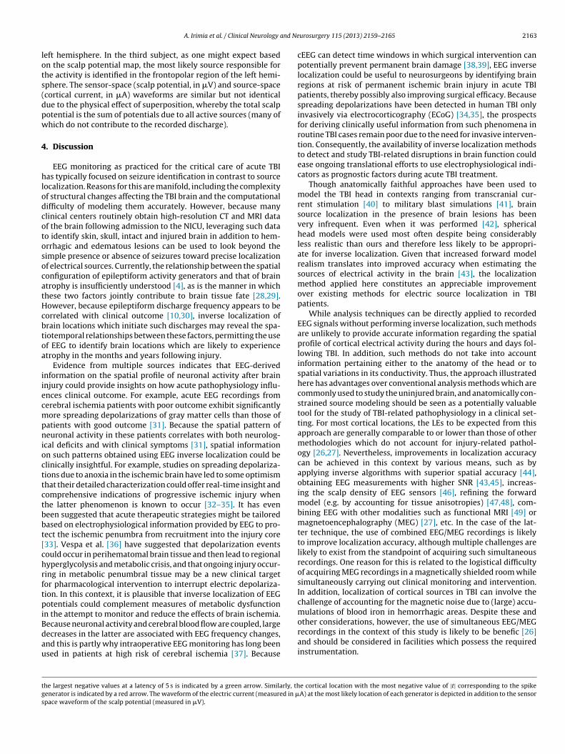
sing scalp EEG. Our contribution illustrates a realistic, patient-pecific approach to TBI source localization and the investigationtself can be appropriately conceptualized as a proof-of-concepttudy to assess the feasibility of the implemented method.ig. 1. Three-dimensional models of representative tissue types in three sample TBI patieor each subject, the full model is shown in the first column (A), cross-sections throughisplayed in the third column (C). Each row corresponds to a patient. In (B), MRI T1 imagSF and pathology are also shown available. Skin is omitted for convenience. Bone is showurple, CSF in blue, edema in cyan and hemorrhage in red.
urosurgery 115 (2013) 2159– 2165
2. Materials and methods
Participants included three males of ages 31, 25 and 45, respec-tively, from whom MRI volumes were acquired at 3.0 Tesla (1 mm3
voxel size, Siemens Trio TIM Scanner, Erlangen, Germany) within72 h after injury. Although the Glasgow coma scale (GCS) scoresof the three patients upon admission to the neurointensive careunit (NICU) were 9, 14 and 14, respectively, their Glasgow out-come scale (GOS) score upon transfer from the NICU was 3 forall patients, reflecting the severity of their injuries as well as thedecline of their clinical condition subsequent to hospital admis-sion. The study was approved by the Institutional Review Board ofthe School of Medicine at the University of California, Los Ange-les, and signed informed consent was obtained from the patients’legally authorized representatives prior to the performance ofany procedure (UCLA IRB approval #10-000929 dated 11/8/2012).The three subjects are examples of TBI patients with progressivelesion loads and were selected for the study based on (1) the
type, location and spatial extent of their lesions, as well as (2)the presence of epileptiform discharges in their cEEG recordings,as identified in the EEG recordings subsequent to their acquisition(see below).nts. Models were generated in 3D Slicer [16] based on MRI volume segmentations. the head are shown in the second column (B), and TBI-related brain pathology ises are superposed onto each FEM model, and 3D models of subcortical structures,n in light brown, eyes in dark brown, gray matter in lilac, subcortical structures in

nd Neurosurgery 115 (2013) 2159– 2165 2161
enTteiwsNmbFeiGmEsa[
fstoetdeheemmrwmsltt[
uhliwmestisimaimtclcop
Fig. 2. Sample 60-s EEG recordings for three representative sensors (FZ, CZ and PZ)
A. Irimia et al. / Clinical Neurology a
The TBI neuroimaging protocol is described extensivelylsewhere [14]. Briefly, acquired MRI sequences included mag-etization prepared rapid acquisition gradient echo (MP-RAGE)1-weighted imaging, fluid attenuated inversion recovery (FLAIR),urbo spin echo (TSE) T2-weighted imaging, gradient-recalledcho (GRE) T2-weighted imaging, and susceptibility weightedmaging (SWI). Conventional computed tomography (CT) scans
ere also acquired. Image alignment, bias field correction andkull stripping were performed using the LONI (Laboratory ofeuro Imaging) Pipeline (http://pipeline.loni.ucla.edu/). Whiteatter (WM), gray matter (GM), cerebrospinal fluid (CSF), cere-
ellar WM/GM and subcortical structures were segmented inreeSurfer (FS) [15], and manual correction of tissue labelingrrors was performed by three experienced users with train-ng in neuroanatomy. TBI-related lesions were segmented fromRE/SWI/FLAIR volumes as detailed elsewhere [14], skin was seg-ented from T1 MRI, and hard bone was segmented from CT.
yes, muscle, cartilage, mucus, nerves, teeth, and ventriculostomyhunts were segmented from T1/T2 MRI. 3D models and visu-lizations were created using 3D Slicer (http://www.slicer.org/)16].
Acquisition of cEEG recordings from each patient was per-ormed in the NICU at 250 Hz over three consecutive days using atandard referential electrode montage. Because scalp EEG poten-ials are due to electrical currents within the apical dendritesf cortical pyramidal neurons [17], EEG generators were mod-led as dipolar currents oriented perpendicular with respect tohe cortical surface. A total of 25 tissue types with distinct con-uctivity values were modeled, including healthy-appearing anddematous skin, fat, hard and soft bone, cerebrospinal fluid (CSF),ealthy-appearing and edematous GM, healthy-appearing anddematous WM, cerebellum, spinal cord, subcortical structures,pidural hemorrhages, connective tissue, muscle, eyes, cartilages,ucus, nerves, teeth, silicone polyurethane (the manufacturingaterial of the ventriculostomy shunts), and sinus air. After co-
egistering the head and all sensor locations, each head volumeas discretized into volume elements from which finite ele-ent method (FEM) models were generated [18,19]. For each
ubject, a regular grid-based mesh (∼400,000 nodes, ∼450,000inear elements, ∼2 mm average edge length) was created andhe so-called forward matrix (the values of the electric poten-ial at each sensor due to every cortical source) was computed18].
The inverse localization technique employed has been widelysed for the study of epilepsy [20–22] and its technical detailsave been comprehensively explored elsewhere [23,24], particu-
arly in our previous publication [25]. Briefly, source localizations performed using a minimum-norm inverse linear operator [26]
hich seeks to minimize the expected difference between the esti-ated and the true inverse solution. The localization accuracy of
ach model can be quantified using the localization error (LE) mea-ure, defined as the distance from the estimated source location tohe true source location [27]. Previous results on our TBI-specificmplementation [25] indicate that the latter can localize corticalources in the presence of brain injury with an approximate LEn the range of ∼0.5–1.5 cm for relatively superficial sources (i.e.
ost gyri and sulci), and ∼1.5–2.5 cm for deep sources (e.g. insuland cingulate cortex). To visualize the results of the source local-zation process, the inverse estimate of the cortical activity can be
apped onto the surface of a reference brain using t scores, suchhat the magnitude of t associated with some cortical location indi-ates the likelihood for that location to be electrically active. The
ocations most likely to be active have |t| > 4. The sign of t indi-ates whether the localized electric current is oriented out of (t > 0)r into (t < 0) the cortex (see Figs. 2 and 3 for details and exam-les).in each of the three patients denoted as P1, P2, and P3, respectively. Note, in eachcase, the aperiodic epileptiform spikes and their large magnitudes compared to therest of the recording.
3. Results
For each subject, each row of Fig. 1 displays anatomical 3D mod-els of the appropriate subject included in the study. The first column(A) displays the full model (except skin), while the second column(B) shows cross-sections through the head with MRI T1 superposedonto each FEM model. Also included in (B) are 3D models of sub-cortical structures, CSF and pathology where available. The thirdcolumn (C) only displays brain pathology visible in the MR scans(edematous and hemorrhagic GM/WM, as segmented from GRET2 and SWI imaging, see Methods section). As Fig. 1 shows, eachselected patient exhibits variable lesion loads and types of pathol-ogy; whereas fronto-temporal lesions and a large craniotomy areboth visible in the right hemisphere of Patient 1, Patient 2 exhibits acomparably low lesion load. Patient 3, by contrast, has large lesionsover both frontal and temporal cortices.
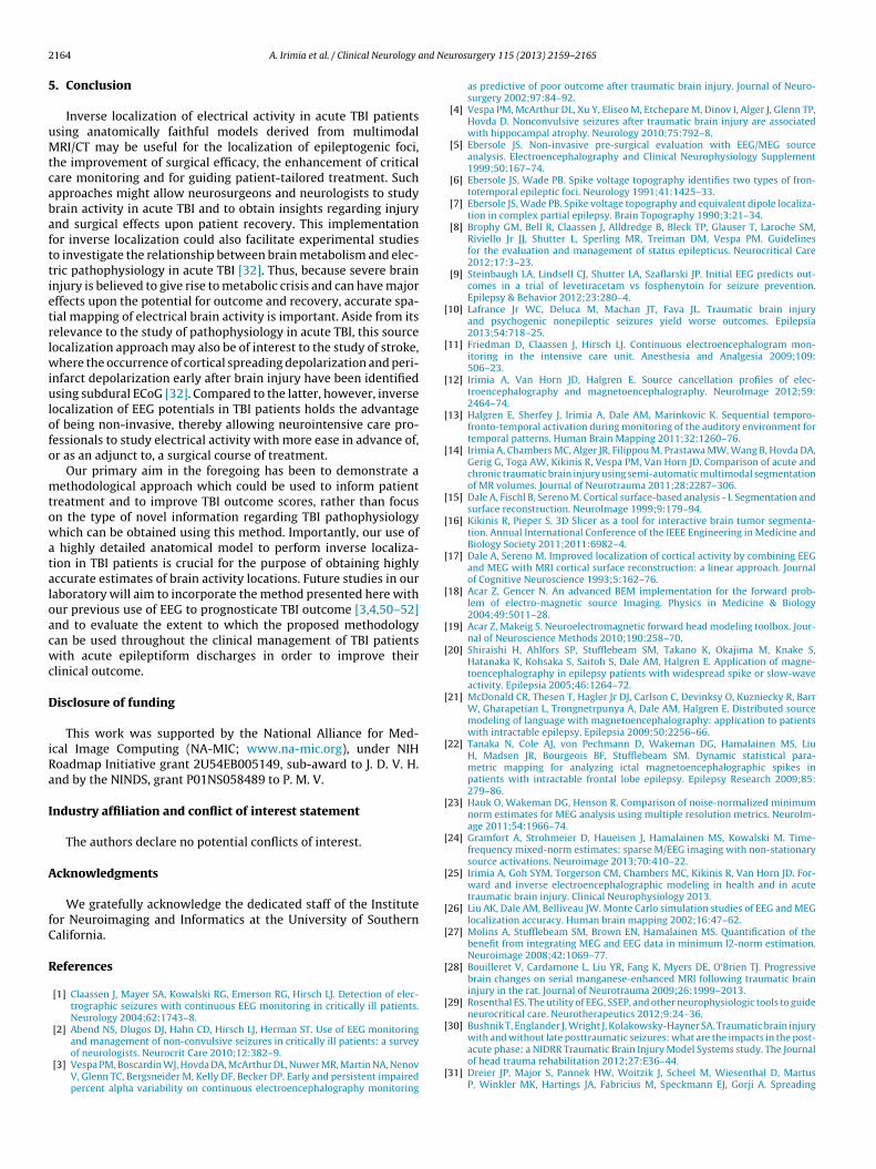
Fig. 2 illustrates 60 s of EEG recordings from each patient, con-taining aperiodic epileptiform spikes, and Fig. 3 shows examples ofinverse localization for the three selected patients. Each subject’sillustration demonstrates the localization of the cortical sourceresponsible for the generation of an interictal epileptiform dis-charge. In each patient, the EEG signal (i.e. sensor-space) waveformassociated with the localized activity is shown, and the EEG poten-tials recorded at the time of the discharge are mapped over thescalp using the interpolated values of the potentials measured ateach sensor (see figure caption for details). The inverse estimateof the cortical activity responsible for the epileptiform activity ismapped onto the surface of a reference brain as Student’s t scores,such that the magnitude of t associated with some cortical locationindicates the likelihood for that location to be electrically active atthe time of the spike. Thus, the locations most likely to be activehave |t| > 4. The sign of t indicates whether the electric current atthe location in question is oriented out of (t > 0) or into (t < 0) thecortex.
Comparing EEG topographic maps to cortical localization plotsreveals that, for every patient, the epileptiform discharge waslocalized to the same cortical region below the scalp wherethe negative deflection in electric potential had been identi-fied in the topographic map. Additionally, however, epileptiform
activity generators were localized to specific gyri or sulci at previ-ously unavailable resolution. In the first subject, the epileptiformgenerator is localized to the left middle temporal gyrus, whereasin the second subject it is localized to the precentral sulcus of the
2162 A. Irimia et al. / Clinical Neurology and Neurosurgery 115 (2013) 2159– 2165
Fig. 3. Examples of inverse localization in three acute TBI patients. The localization technique is used to identify the cortical source responsible for the generation of anEEG waveform containing a large-magnitude deflection (often referred to as a “graphoelement” in the EEG literature). For each subject, the waveform of the EEG potential,�, being localized is shown for an interval of 10 s. Localization is illustrated at the time point with a latency of 5 s with respect to the beginning of the waveform. The EEGpotentials recorded at this time point are mapped over the scalp using the interpolated values of the potentials measured at each sensor. The amplitudes of the potential� are measured in �V and different ranges are used for the topographic map of each subject to emphasize scalp differences in potential which are specific to each subject.The inverse estimate of the cortical activity responsible for the graphoelement is mapped onto the surface of a reference brain as t scores (see Methods section). Thelocations most likely to be active have |t| > 4. The sign of t indicates whether the localized electric current is oriented out of (t > 0, red hues) or into (t < 0, blue hues) the cor-tex. A single color map is used for t in all subjects, and color intensity indicates likelihood for the presence of electrical activity. In each subject, the scalp location which exhibits
nd Ne
lots(dpw
4
hlodcotosocatHcbtoa
iiecmpniocttctbbt[chrftpiBdau
tgs
A. Irimia et al. / Clinical Neurology a
eft hemisphere. In the third subject, as one might expect basedn the scalp potential map, the most likely source responsible forhe activity is identified in the frontopolar region of the left hemi-phere. The sensor-space (scalp potential, in �V) and source-spacecortical current, in �A) waveforms are similar but not identicalue to the physical effect of superposition, whereby the total scalpotential is the sum of potentials due to all active sources (many ofhich do not contribute to the recorded discharge).
. Discussion
EEG monitoring as practiced for the critical care of acute TBIas typically focused on seizure identification in contrast to source
ocalization. Reasons for this are manifold, including the complexityf structural changes affecting the TBI brain and the computationalifficulty of modeling them accurately. However, because manylinical centers routinely obtain high-resolution CT and MRI dataf the brain following admission to the NICU, leveraging such datao identify skin, skull, intact and injured brain in addition to hem-rrhagic and edematous lesions can be used to look beyond theimple presence or absence of seizures toward precise localizationf electrical sources. Currently, the relationship between the spatialonfiguration of epileptiform activity generators and that of braintrophy is insufficiently understood [4], as is the manner in whichhese two factors jointly contribute to brain tissue fate [28,29].owever, because epileptiform discharge frequency appears to beorrelated with clinical outcome [10,30], inverse localization ofrain locations which initiate such discharges may reveal the spa-iotemporal relationships between these factors, permitting the usef EEG to identify brain locations which are likely to experiencetrophy in the months and years following injury.
Evidence from multiple sources indicates that EEG-derivednformation on the spatial profile of neuronal activity after brainnjury could provide insights on how acute pathophysiology influ-nces clinical outcome. For example, acute EEG recordings fromerebral ischemia patients with poor outcome exhibit significantlyore spreading depolarizations of gray matter cells than those of
atients with good outcome [31]. Because the spatial pattern ofeuronal activity in these patients correlates with both neurolog-
cal deficits and with clinical symptoms [31], spatial informationn such patterns obtained using EEG inverse localization could belinically insightful. For example, studies on spreading depolariza-ions due to anoxia in the ischemic brain have led to some optimismhat their detailed characterization could offer real-time insight andomprehensive indications of progressive ischemic injury whenhe latter phenomenon is known to occur [32–35]. It has eveneen suggested that acute therapeutic strategies might be tailoredased on electrophysiological information provided by EEG to pro-ect the ischemic penumbra from recruitment into the injury core33]. Vespa et al. [36] have suggested that depolarization eventsould occur in perihematomal brain tissue and then lead to regionalyperglycolysis and metabolic crisis, and that ongoing injury occur-ing in metabolic penumbral tissue may be a new clinical targetor pharmacological intervention to interrupt electric depolariza-ion. In this context, it is plausible that inverse localization of EEGotentials could complement measures of metabolic dysfunction
n the attempt to monitor and reduce the effects of brain ischemia.
ecause neuronal activity and cerebral blood flow are coupled, largeecreases in the latter are associated with EEG frequency changes,nd this is partly why intraoperative EEG monitoring has long beensed in patients at high risk of cerebral ischemia [37]. Becausehe largest negative values at a latency of 5 s is indicated by a green arrow. Similarly, thenerator is indicated by a red arrow. The waveform of the electric current (measured in �pace waveform of the scalp potential (measured in �V).
urosurgery 115 (2013) 2159– 2165 2163
cEEG can detect time windows in which surgical intervention canpotentially prevent permanent brain damage [38,39], EEG inverselocalization could be useful to neurosurgeons by identifying brainregions at risk of permanent ischemic brain injury in acute TBIpatients, thereby possibly also improving surgical efficacy. Becausespreading depolarizations have been detected in human TBI onlyinvasively via electrocorticography (ECoG) [34,35], the prospectsfor deriving clinically useful information from such phenomena inroutine TBI cases remain poor due to the need for invasive interven-tion. Consequently, the availability of inverse localization methodsto detect and study TBI-related disruptions in brain function couldease ongoing translational efforts to use electrophysiological indi-cators as prognostic factors during acute TBI treatment.
Though anatomically faithful approaches have been used tomodel the TBI head in contexts ranging from transcranial cur-rent stimulation [40] to military blast simulations [41], brainsource localization in the presence of brain lesions has beenvery infrequent. Even when it was performed [42], sphericalhead models were used most often despite being considerablyless realistic than ours and therefore less likely to be appropri-ate for inverse localization. Given that increased forward modelrealism translates into improved accuracy when estimating thesources of electrical activity in the brain [43], the localizationmethod applied here constitutes an appreciable improvementover existing methods for electric source localization in TBIpatients.
While analysis techniques can be directly applied to recordedEEG signals without performing inverse localization, such methodsare unlikely to provide accurate information regarding the spatialprofile of cortical electrical activity during the hours and days fol-lowing TBI. In addition, such methods do not take into accountinformation pertaining either to the anatomy of the head or tospatial variations in its conductivity. Thus, the approach illustratedhere has advantages over conventional analysis methods which arecommonly used to study the uninjured brain, and anatomically con-strained source modeling should be seen as a potentially valuabletool for the study of TBI-related pathophysiology in a clinical set-ting. For most cortical locations, the LEs to be expected from thisapproach are generally comparable to or lower than those of othermethodologies which do not account for injury-related pathol-ogy [26,27]. Nevertheless, improvements in localization accuracycan be achieved in this context by various means, such as byapplying inverse algorithms with superior spatial accuracy [44],obtaining EEG measurements with higher SNR [43,45], increas-ing the scalp density of EEG sensors [46], refining the forwardmodel (e.g. by accounting for tissue anisotropies) [47,48], com-bining EEG with other modalities such as functional MRI [49] ormagnetoencephalography (MEG) [27], etc. In the case of the lat-ter technique, the use of combined EEG/MEG recordings is likelyto improve localization accuracy, although multiple challenges arelikely to exist from the standpoint of acquiring such simultaneousrecordings. One reason for this is related to the logistical difficultyof acquiring MEG recordings in a magnetically shielded room whilesimultaneously carrying out clinical monitoring and intervention.In addition, localization of cortical sources in TBI can involve thechallenge of accounting for the magnetic noise due to (large) accu-mulations of blood iron in hemorrhagic areas. Despite these and
other considerations, however, the use of simultaneous EEG/MEGrecordings in the context of this study is likely to be benefic [26]and should be considered in facilities which possess the requiredinstrumentation.e cortical location with the most negative value of |t| corresponding to the spikeA) at the most likely location of each generator is depicted in addition to the sensor
2 nd Ne
5
uMtcabafttietrlwiulofo
mtowataloacwc
D
iRa
I
A
fC
R
[
[
[
[
[
[
[
[
[
[
[
[
[
[
[
[
[
[
[
[
[
164 A. Irimia et al. / Clinical Neurology a
. Conclusion
Inverse localization of electrical activity in acute TBI patientssing anatomically faithful models derived from multimodalRI/CT may be useful for the localization of epileptogenic foci,
he improvement of surgical efficacy, the enhancement of criticalare monitoring and for guiding patient-tailored treatment. Suchpproaches might allow neurosurgeons and neurologists to studyrain activity in acute TBI and to obtain insights regarding injurynd surgical effects upon patient recovery. This implementationor inverse localization could also facilitate experimental studieso investigate the relationship between brain metabolism and elec-ric pathophysiology in acute TBI [32]. Thus, because severe brainnjury is believed to give rise to metabolic crisis and can have majorffects upon the potential for outcome and recovery, accurate spa-ial mapping of electrical brain activity is important. Aside from itselevance to the study of pathophysiology in acute TBI, this sourceocalization approach may also be of interest to the study of stroke,
here the occurrence of cortical spreading depolarization and peri-nfarct depolarization early after brain injury have been identifiedsing subdural ECoG [32]. Compared to the latter, however, inverse
ocalization of EEG potentials in TBI patients holds the advantagef being non-invasive, thereby allowing neurointensive care pro-essionals to study electrical activity with more ease in advance of,r as an adjunct to, a surgical course of treatment.
Our primary aim in the foregoing has been to demonstrate aethodological approach which could be used to inform patient
reatment and to improve TBI outcome scores, rather than focusn the type of novel information regarding TBI pathophysiologyhich can be obtained using this method. Importantly, our use of
highly detailed anatomical model to perform inverse localiza-ion in TBI patients is crucial for the purpose of obtaining highlyccurate estimates of brain activity locations. Future studies in ouraboratory will aim to incorporate the method presented here withur previous use of EEG to prognosticate TBI outcome [3,4,50–52]nd to evaluate the extent to which the proposed methodologyan be used throughout the clinical management of TBI patientsith acute epileptiform discharges in order to improve their
linical outcome.
isclosure of funding
This work was supported by the National Alliance for Med-cal Image Computing (NA-MIC; www.na-mic.org), under NIHoadmap Initiative grant 2U54EB005149, sub-award to J. D. V. H.nd by the NINDS, grant P01NS058489 to P. M. V.
ndustry affiliation and conflict of interest statement
The authors declare no potential conflicts of interest.
cknowledgments
We gratefully acknowledge the dedicated staff of the Instituteor Neuroimaging and Informatics at the University of Southernalifornia.
eferences
[1] Claassen J, Mayer SA, Kowalski RG, Emerson RG, Hirsch LJ. Detection of elec-trographic seizures with continuous EEG monitoring in critically ill patients.Neurology 2004;62:1743–8.
[2] Abend NS, Dlugos DJ, Hahn CD, Hirsch LJ, Herman ST. Use of EEG monitoring
and management of non-convulsive seizures in critically ill patients: a surveyof neurologists. Neurocrit Care 2010;12:382–9.[3] Vespa PM, Boscardin WJ, Hovda DA, McArthur DL, Nuwer MR, Martin NA, NenovV, Glenn TC, Bergsneider M, Kelly DF, Becker DP. Early and persistent impairedpercent alpha variability on continuous electroencephalography monitoring
[
urosurgery 115 (2013) 2159– 2165
as predictive of poor outcome after traumatic brain injury. Journal of Neuro-surgery 2002;97:84–92.
[4] Vespa PM, McArthur DL, Xu Y, Eliseo M, Etchepare M, Dinov I, Alger J, Glenn TP,Hovda D. Nonconvulsive seizures after traumatic brain injury are associatedwith hippocampal atrophy. Neurology 2010;75:792–8.
[5] Ebersole JS. Non-invasive pre-surgical evaluation with EEG/MEG sourceanalysis. Electroencephalography and Clinical Neurophysiology Supplement1999;50:167–74.
[6] Ebersole JS, Wade PB. Spike voltage topography identifies two types of fron-totemporal epileptic foci. Neurology 1991;41:1425–33.
[7] Ebersole JS, Wade PB. Spike voltage topography and equivalent dipole localiza-tion in complex partial epilepsy. Brain Topography 1990;3:21–34.
[8] Brophy GM, Bell R, Claassen J, Alldredge B, Bleck TP, Glauser T, Laroche SM,Riviello Jr JJ, Shutter L, Sperling MR, Treiman DM, Vespa PM. Guidelinesfor the evaluation and management of status epilepticus. Neurocritical Care2012;17:3–23.
[9] Steinbaugh LA, Lindsell CJ, Shutter LA, Szaflarski JP. Initial EEG predicts out-comes in a trial of levetiracetam vs fosphenytoin for seizure prevention.Epilepsy & Behavior 2012;23:280–4.
10] Lafrance Jr WC, Deluca M, Machan JT, Fava JL. Traumatic brain injuryand psychogenic nonepileptic seizures yield worse outcomes. Epilepsia2013;54:718–25.
11] Friedman D, Claassen J, Hirsch LJ. Continuous electroencephalogram mon-itoring in the intensive care unit. Anesthesia and Analgesia 2009;109:506–23.
12] Irimia A, Van Horn JD, Halgren E. Source cancellation profiles of elec-troencephalography and magnetoencephalography. NeuroImage 2012;59:2464–74.
13] Halgren E, Sherfey J, Irimia A, Dale AM, Marinkovic K. Sequential temporo-fronto-temporal activation during monitoring of the auditory environment fortemporal patterns. Human Brain Mapping 2011;32:1260–76.
14] Irimia A, Chambers MC, Alger JR, Filippou M, Prastawa MW, Wang B, Hovda DA,Gerig G, Toga AW, Kikinis R, Vespa PM, Van Horn JD. Comparison of acute andchronic traumatic brain injury using semi-automatic multimodal segmentationof MR volumes. Journal of Neurotrauma 2011;28:2287–306.
15] Dale A, Fischl B, Sereno M. Cortical surface-based analysis - I. Segmentation andsurface reconstruction. NeuroImage 1999;9:179–94.
16] Kikinis R, Pieper S. 3D Slicer as a tool for interactive brain tumor segmenta-tion. Annual International Conference of the IEEE Engineering in Medicine andBiology Society 2011;2011:6982–4.
17] Dale A, Sereno M. Improved localization of cortical activity by combining EEGand MEG with MRI cortical surface reconstruction: a linear approach. Journalof Cognitive Neuroscience 1993;5:162–76.
18] Acar Z, Gencer N. An advanced BEM implementation for the forward prob-lem of electro-magnetic source Imaging. Physics in Medicine & Biology2004;49:5011–28.
19] Acar Z, Makeig S. Neuroelectromagnetic forward head modeling toolbox. Jour-nal of Neuroscience Methods 2010;190:258–70.
20] Shiraishi H, Ahlfors SP, Stufflebeam SM, Takano K, Okajima M, Knake S,Hatanaka K, Kohsaka S, Saitoh S, Dale AM, Halgren E. Application of magne-toencephalography in epilepsy patients with widespread spike or slow-waveactivity. Epilepsia 2005;46:1264–72.
21] McDonald CR, Thesen T, Hagler Jr DJ, Carlson C, Devinksy O, Kuzniecky R, BarrW, Gharapetian L, Trongnetrpunya A, Dale AM, Halgren E. Distributed sourcemodeling of language with magnetoencephalography: application to patientswith intractable epilepsy. Epilepsia 2009;50:2256–66.
22] Tanaka N, Cole AJ, von Pechmann D, Wakeman DG, Hamalainen MS, LiuH, Madsen JR, Bourgeois BF, Stufflebeam SM. Dynamic statistical para-metric mapping for analyzing ictal magnetoencephalographic spikes inpatients with intractable frontal lobe epilepsy. Epilepsy Research 2009;85:279–86.
23] Hauk O, Wakeman DG, Henson R. Comparison of noise-normalized minimumnorm estimates for MEG analysis using multiple resolution metrics. NeuroIm-age 2011;54:1966–74.
24] Gramfort A, Strohmeier D, Haueisen J, Hamalainen MS, Kowalski M. Time-frequency mixed-norm estimates: sparse M/EEG imaging with non-stationarysource activations. Neuroimage 2013;70:410–22.
25] Irimia A, Goh SYM, Torgerson CM, Chambers MC, Kikinis R, Van Horn JD. For-ward and inverse electroencephalographic modeling in health and in acutetraumatic brain injury. Clinical Neurophysiology 2013.
26] Liu AK, Dale AM, Belliveau JW. Monte Carlo simulation studies of EEG and MEGlocalization accuracy. Human brain mapping 2002;16:47–62.
27] Molins A, Stufflebeam SM, Brown EN, Hamalainen MS. Quantification of thebenefit from integrating MEG and EEG data in minimum l2-norm estimation.Neuroimage 2008;42:1069–77.
28] Bouilleret V, Cardamone L, Liu YR, Fang K, Myers DE, O‘Brien TJ. Progressivebrain changes on serial manganese-enhanced MRI following traumatic braininjury in the rat. Journal of Neurotrauma 2009;26:1999–2013.
29] Rosenthal ES. The utility of EEG, SSEP, and other neurophysiologic tools to guideneurocritical care. Neurotherapeutics 2012;9:24–36.
30] Bushnik T, Englander J, Wright J, Kolakowsky-Hayner SA. Traumatic brain injury
with and without late posttraumatic seizures: what are the impacts in the post-acute phase: a NIDRR Traumatic Brain Injury Model Systems study. The Journalof head trauma rehabilitation 2012;27:E36–44.31] Dreier JP, Major S, Pannek HW, Woitzik J, Scheel M, Wiesenthal D, MartusP, Winkler MK, Hartings JA, Fabricius M, Speckmann EJ, Gorji A. Spreading

nd Ne
[
[
[
[
[
[
[
[
[
[
[
[
[
[
[
[
[
[
[
[
A. Irimia et al. / Clinical Neurology a
convulsions, spreading depolarization and epileptogenesis in human cerebralcortex. Brain 2012;135:259–75.
32] Dohmen C, Sakowitz OW, Fabricius M, Bosche B, Reithmeier T, Ernestus RI,Brinker G, Dreier JP, Woitzik J, Strong AJ, Graf R. Spreading depolarizationsoccur in human ischemic stroke with high incidence. Annals of Neurology2008;63:720–8.
33] Dreier JP, Woitzik J, Fabricius M, Bhatia R, Major S, Drenckhahn C, Lehmann TN,Sarrafzadeh A, Willumsen L, Hartings JA, Sakowitz OW, Seemann JH, Thieme A,Lauritzen M, Strong AJ. Delayed ischaemic neurological deficits after subarach-noid haemorrhage are associated with clusters of spreading depolarizations.Brain 2006;129:3224–37.
34] Hartings JA, Strong AJ, Fabricius M, Manning A, Bhatia R, Dreier JP, Mazzeo AT,Tortella FC, Bullock MR. Spreading depolarizations and late secondary insultsafter traumatic brain injury. Journal of Neurotrauma 2009;26:1857–66.
35] Hartings JA, Watanabe T, Bullock MR, Okonkwo DO, Fabricius M, Woitzik J,Dreier JP, Puccio A, Shutter LA, Pahl C, Strong AJ. Spreading depolarizationshave prolonged direct current shifts and are associated with poor outcome inbrain trauma. Brain 2011;134:1529–40.
36] Vespa PM. Metabolic penumbra in intracerebral hemorrhage. Stroke A Journalof Cerebral Circulation 2009;40:1547–8.
37] Astrup J, Siesjo BK, Symon L. Thresholds in cerebral ischemia – the ischemicpenumbra. Stroke A Journal of Cerebral Circulation 1981;12:723–5.
38] Jordan KG. Emergency EEG and continuous EEG monitoring in acute ischemicstroke. Journal of Clinical Neurophysiology: Official Publication of the AmericanElectroencephalographic Society 2004;21:341–52.
39] Jordan KG. Continuous EEG monitoring in the neuroscience intensivecare unit and emergency department. Journal of Clinical Neurophysiology1999;16:14–39.
40] Datta A, Bikson M, Fregni F. Transcranial direct current stimulation inpatients with skull defects and skull plates: high-resolution computational
FEM study of factors altering cortical current flow. NeuroImage 2010;52:1268–78.41] Chafi MS, Karami G, Ziejewski M. Biomechanical assessment of brain dynamicresponses due to blast pressure waves. Annals of Biomedical Engineering2010;38:490–504.
[
urosurgery 115 (2013) 2159– 2165 2165
42] Vatta F, Bruno P, Inchingolo P. Improving lesion conductivity estimate by meansof EEG source localization sensitivity to model parameter. Journal of ClinicalNeurophysiology 2002;19:1–15.
43] Ramon C, Schimpf PH, Haueisen J. Influence of head models on EEG simulationsand inverse source localizations. BioMedical Engineering OnLine 2006;5:10.
44] Michel CM, Murray MM, Lantz G, Gonzalez S, Spinelli L, Grave de Peralta R. EEGsource imaging. Clinical Neurophysiology 2004;115:2195–222.
45] Zumer JM, Attias HT, Sekihara K, Nagarajan SS. A probabilistic algorithm inte-grating source localization and noise suppression for MEG and EEG data.NeuroImage 2007;37:102–15.
46] Lantz G, Grave de Peralta R, Spinelli L, Seeck M, Michel CM. Epileptic sourcelocalization with high density EEG: how many electrodes are needed. ClinicalNeurophysiology 2003;114:63–9.
47] Wolters CH, Anwander A, Tricoche X, Weinstein D, Koch MA, MacLeod RS. Influ-ence of tissue conductivity anisotropy on EEG/MEG field and return currentcomputation in a realistic head model: a simulation and visualization studyusing high-resolution finite element modeling. NeuroImage 2006;30:813–26.
48] Haueisen J, Tuch DS, Ramon C, Schimpf PH, Wedeen VJ, George JS, Belliveau JW.The influence of brain tissue anisotropy on human EEG and MEG. NeuroImage2002;15:159–66.
49] Babiloni F, Babiloni C, Carducci F, Romani GL, Rossini PM, Angelone LM, Cin-cotti F. Multimodal integration of high-resolution EEG and functional magneticresonance imaging data: a simulation study. NeuroImage 2003;19:1–15.
50] Vespa PM, Nuwer MR, Nenov V, Ronne-Engstrom E, Hovda DA, BergsneiderM, Kelly DF, Martin NA, Becker DP. Increased incidence and impact of non-convulsive and convulsive seizures after traumatic brain injury as detectedby continuous electroencephalographic monitoring. Journal of Neurosurgery1999;91:750–60.
51] Vespa PM, Miller C, McArthur D, Eliseo M, Etchepare M, Hirt D, Glenn TC, MartinN, Hovda D. Nonconvulsive electrographic seizures after traumatic brain injury
result in a delayed, prolonged increase in intracranial pressure and metaboliccrisis. Critical Care Medicine 2007;35:2830–6.52] Vespa PM, Nenov V, Nuwer MR. Continuous EEG monitoring in the intensivecare unit: early findings and clinical efficacy. Journal of Clinical Neuro-physiology 1999;16:1–13.