Clinical Implementation of a New Ultrasound …chapter.aapm.org/rockymtn/images/sarkar.pdf ·...
36
Clinical Implementation of a New Ultrasound Guidance System Vikren Sarkar Bill Salter Martin Szegedi
Transcript of Clinical Implementation of a New Ultrasound …chapter.aapm.org/rockymtn/images/sarkar.pdf ·...
Clinical Implementation of a New
Ultrasound Guidance System
Vikren Sarkar
Bill Salter
Martin Szegedi
∗ The University of Utah has research agreements with Elekta
Disclosure

∗ Historical Review
∗ Trans-Abdominal Ultrasound
∗ Trans-Perineal Ultrasound
∗ Lessons Learned
Agenda
∗ IMRT was introduced as a novel method for conformal dose distributions.
∗ Prostate RT was one of the first major application.
∗ But we had a new problem to deal with… motion of the prostate within the pelvis (not an issue with box fields)
∗ Balter et al. (IJROBP 1993) – 12 mm (95% CI)
∗ Roach et al. (IJROBP 1994) – 7.5 to 22 mm
A long long time ago (late 90s)

∗ Pin the prostate between the bladder and rectum using a rectal balloon∗ Not as comfortable for the patient.
∗ But it works (Still used by some proton therapy centers).
∗ Perform daily IGRT on the prostate∗ Using markers (they can migrate, need invasive procedure,
extra radiation dose).
∗ With CBCT (prostate not really visible, extra radiation dose).
∗ With ultrasound (non invasive, no radiation dose, needs training).
Solutions

∗ Available since the late 90s.
∗ An IR camera is used to track a probe within the linacvault.
BAT
∗ A calibration means the cameras understand the location of isocenter (laser defined).
∗ This also allows the images to be localized in space.∗ This allows for the transfer of contours from the treatment
plan onto the ultrasound images obtained using the probe. ∗ That process shows the “expected” position of the various
structures (as they were on the simulation CT relative to where isocenter was planned).
∗ If they are not aligned with what the ultrasound images show, they have moved and so we can shift.
∗ If we obtain axial and sagittal images, we can shift in all three dimensions.
BAT

COUCH MOVEMENTS: Right 0.300cm; Up 0.883cm; In 0.928cm
BAT ExamplePRE ALIGNMENT POST ALIGNMENT
COUCH MOVEMENTS: Left 0.160cm; Down 0.761cm; Out: 0.410cm
∗ Depends on whom you ask∗ Early inter-modality studies showed large inter-user
variability and assumed any “error” observed between the two image-sets were solely attributable to the ultrasound system.
∗ Multiple studies since have shown that, when alignments were done by ultrasound-trained technologists, the quality of the alignments were comparable to other IGRT methods.
Is BAT accurate?
∗ System introduced at the U of U in 2006
∗ As of 2008, BAT became our standard approach for localization of prostate patients
∗ In fact, we even started localizing some of our liver and pancreas patients using the system.
BAT at the University of Utah

∗ There was still the question of what happens to the prostate once we have localized it.
∗ Fluoro studies (with implanted markers) as well as MRI cine studies show large prostate intra-fractional motion.
∗ So, while we optimize the prostate position at the start of treatment, we should also track the prostate and make sure its position does not deviate too much during the time it takes to deliver the treatment.
Intra-fractional motion

∗ Uses electromagnetic beacons to track the prostate prior to and during treatment.
∗ Can gate the treatment and stop the beam when excursions are greater than certain pre-determined limits for a given amount of time.
∗ Unfortunately, the approach does not work for all patients∗ Patients on blood thinners
∗ Patients with large AP separations
∗ Patients with prostheses
∗ Non-deal placement of beacons
Calypso
∗ System developed by Resonant Medical and now part of Elekta.
∗ Uses an ultrasound dataset obtained at the time of simulation for comparison∗ This allows for an intra-modality comparison to be done
at the time of treatment (instead of an ultrasound with CT-based contours).
∗ Given our previous experience with the BAT system, we should just be able to use it within our clinic… or should we?
Clarity
∗ The clarity system uses a full volumetric 3D dataset instead of 2 “orthogonal” 2D images.
∗ The dataset has to be acquired by angling and rocking the ultrasound probe on the patient’s belly.
∗ There is a large change in workflow from BAT
∗ Whole new system with a very different UI
∗ For comparison sake, we kept using BAT as a backup after Clarity and used BAT as our gold standard.
Clarity – Trans-Abdominal approach
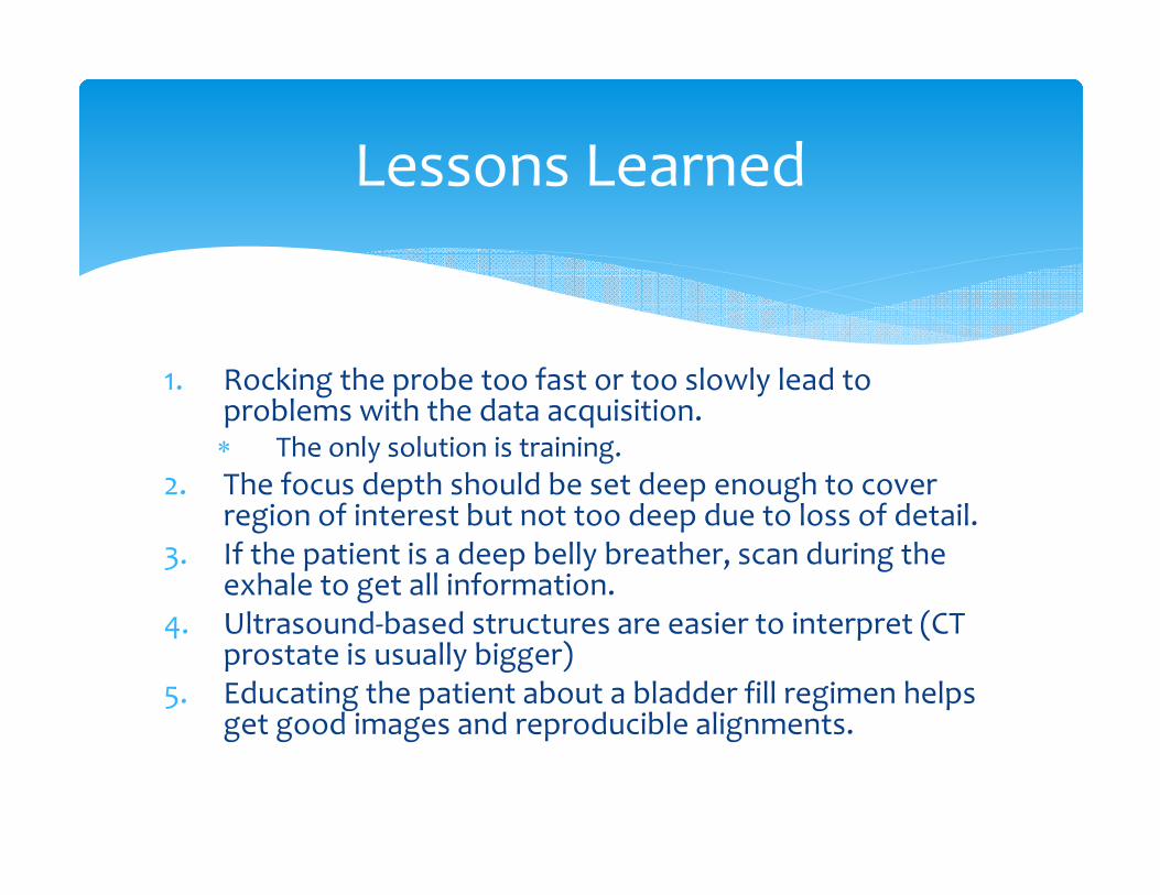
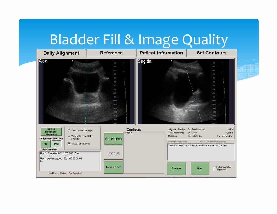
1. Rocking the probe too fast or too slowly lead to problems with the data acquisition. ∗ The only solution is training.
2. The focus depth should be set deep enough to cover region of interest but not too deep due to loss of detail.
3. If the patient is a deep belly breather, scan during the exhale to get all information.
4. Ultrasound-based structures are easier to interpret (CT prostate is usually bigger)
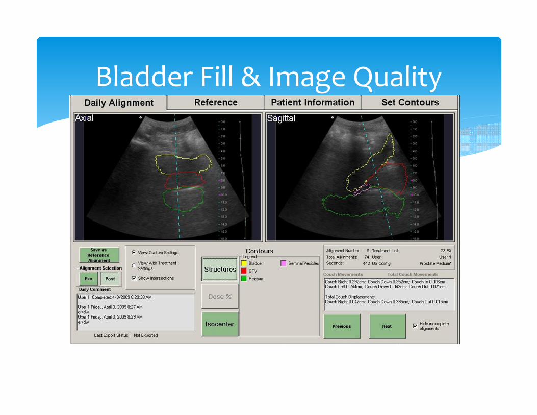
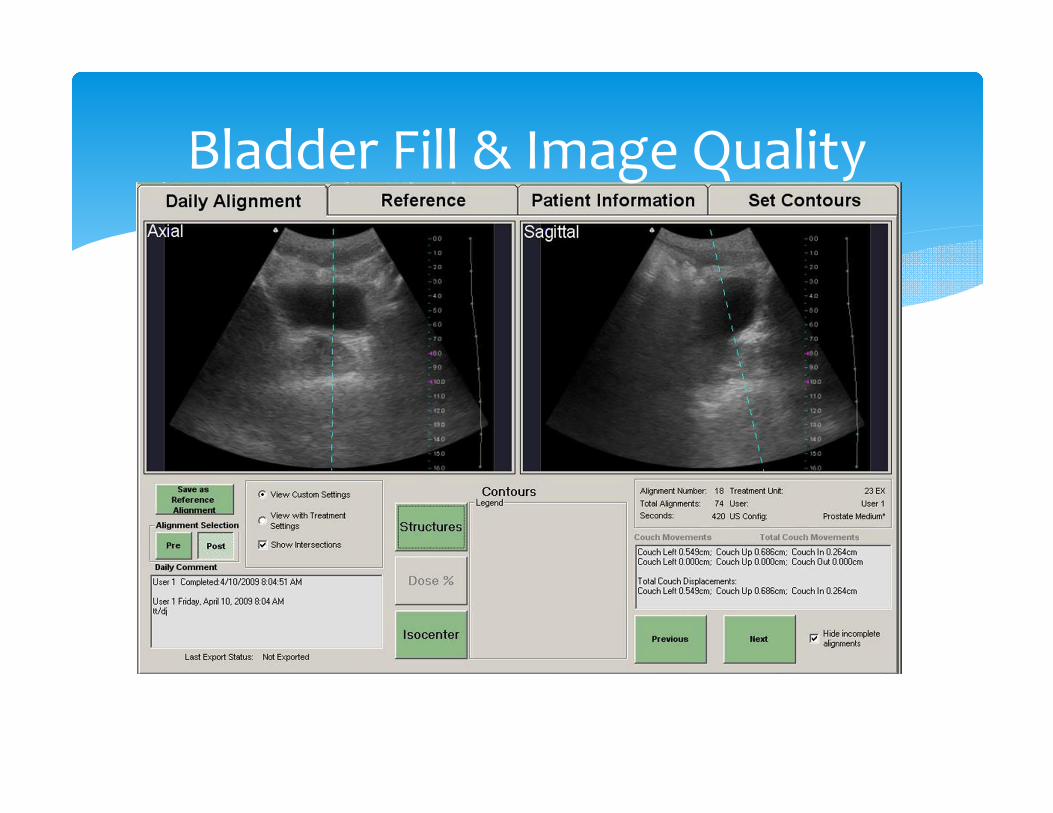
5. Educating the patient about a bladder fill regimen helps get good images and reproducible alignments.
Lessons Learned
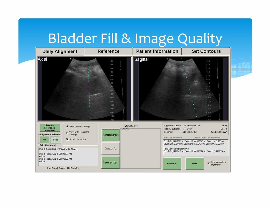
Bladder Fill & Image Quality
Bladder Fill & Image Quality

Bladder Fill & Image Quality
Bladder Fill & Image Quality
Bladder Fill & Image Quality
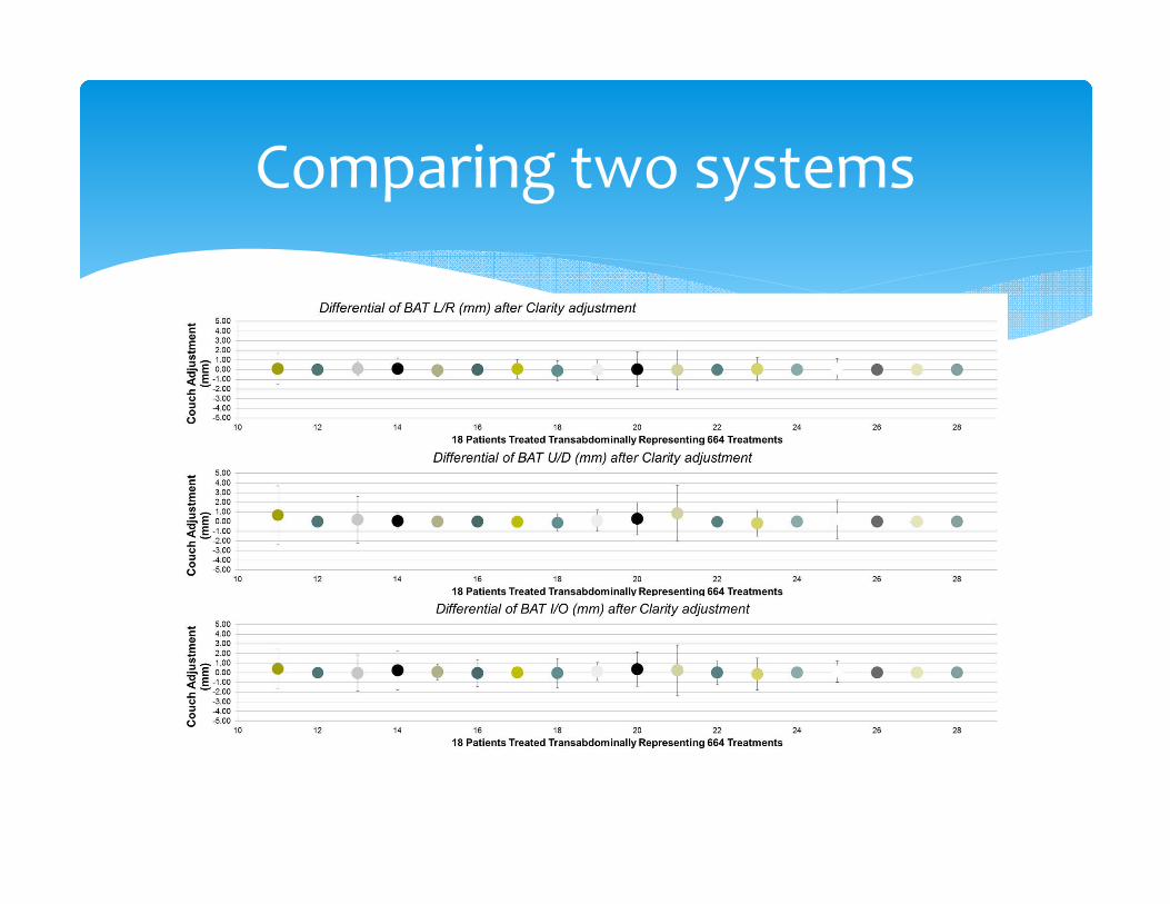
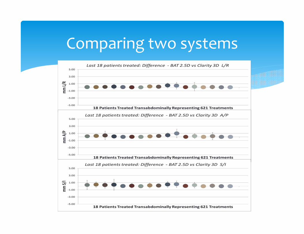
∗ Since we have a lot of experience with BAT, we wanted to know whether BAT and Clarity gives us the same information.
∗ For about two years, we started our patients with Clarity for IGRT and followed by doing a BAT alignment to verify (expecting ~zero shift between systems)
Comparing two systems
Comparing two systems
Comparing two systems

∗ We have shown that BAT and TA clarity give the same information.
∗ But what about tracking? That would not be possible with a TA approach.
∗ What if we could image trans-perineally?
∗ We could leave the probe in place during treatment and even think of tracking the prostate.
Trans-Perineal Approach
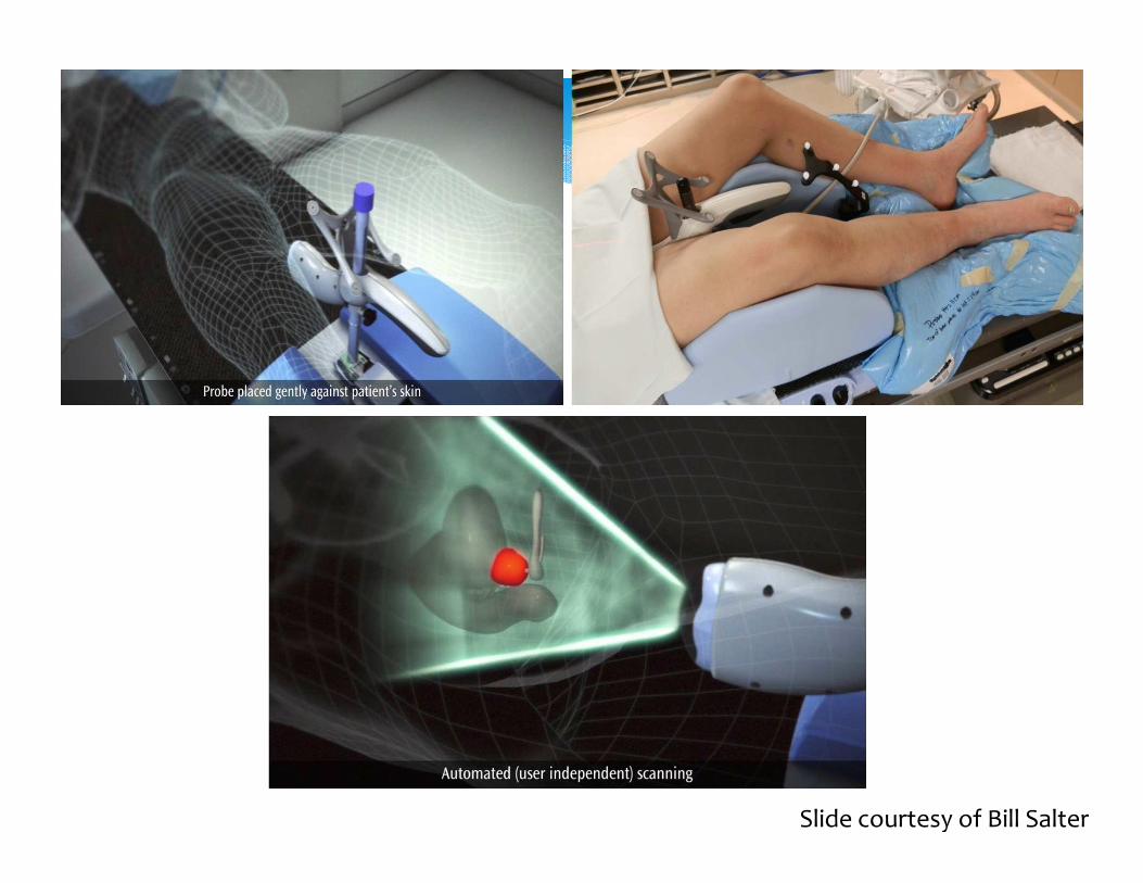
Slide courtesy of Bill Salter

∗ More learning to do
1. To interpret images that look different - primarily sagittal
2. A new workflow
3. A new probe
4. A new setup (Sim and Treatment)
5. We need to think of the timing of our scans (Ultrasound and CT)
6. We need to think of what structures we line up to.
Trans-Perineal Approach
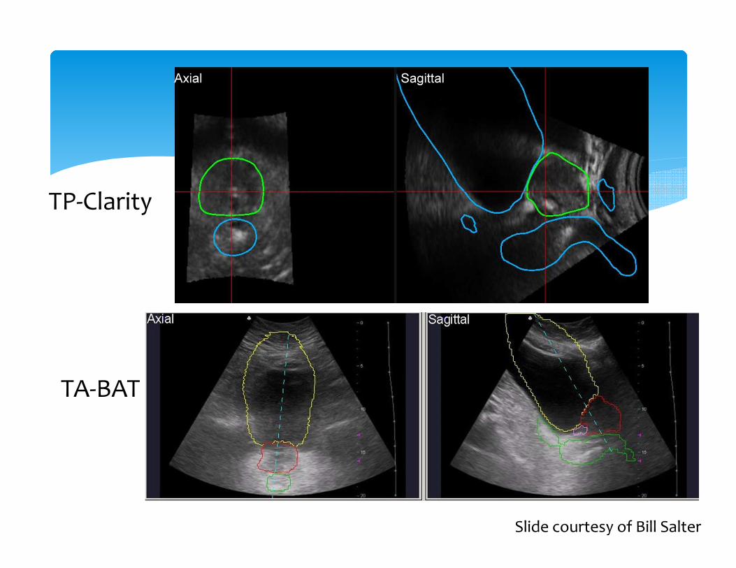
TP-Clarity
TA-BAT
SagittalAxial
Slide courtesy of Bill Salter

Tracking
1 m 16 s

∗ Advantages:
∗ Direct visualization of the prostate
∗ Can be applied to any patient
∗ No radiation dose
∗ Allows for tracking of volume of interest throughout treatment
Trans-Perineal Approach
∗ We should aim for the same setup every day
∗ Worked with Elekta to change the post holding the probe and the “knee cushion”.
∗ Slightly raising the feet makes the pelvis a little more flat making
1. Patient more stable
2. General alignment easier
∗ Certain structures may help with alignment when using the TP approach (penile bulb, sphincter, seminal vessicles etc)
Lessons Learned
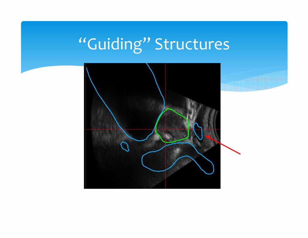
“Guiding” Structures
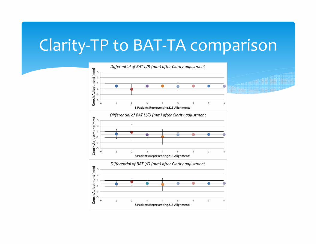
Clarity-TP to BAT-TA comparison

∗ Ultrasound guidance, especially for prostate treatments, provides several advantages over other IGRT methods.
∗ Just as for any system, there is a learning curve to correctly use the system.
∗ At our center, we have a very strict training program before anyone is able to independently use the system∗ Core team of ultrasound therapists who went through at least
200 alignments as part of their training
∗ A core team of “ultrasound physicists” who can address questions or any new problems that arise
∗ Biweekly ultrasound review (analogous to chart rounds) with therapists, physicists and physicians.
Take Away Messages

∗ We started this process with a known requirement for training and workflow adjustment in mind.
∗ We had a constant feedback loop with Elekta and helped them make changes to both the hardware and software components of the system.
∗ We are now using clarity in 2 vaults (and planning on a third).
∗ Clarity-TP is used for all of our prostate SBRT patients (program started in 2013)
∗ All three of our GU physicians regularly use the system and have come to depend on it.
Final Thought

∗ My thanks to the following people who helped with this presentation:
1. Bill Salter, Ph.D.
2. Martin Szegedi, Ph.D.
3. Brennan Esch, RTT
Acknowledgements

Thank you
http://samluce.com/2014/09/answer-kids-direct-questions-direct-answers/