CLINICAL EVALUATION OF CAST AND PREFABRICATED METAL POST ... · clinical evaluation of cast and...
61
1 CLINICAL EVALUATION OF CAST AND PREFABRICATED METAL POST: A 5-YEAR RETROSPECTIVE STUDY By HASSAN MOUSAWI A THESIS PRESENTED TO THE GRADUATE SCHOOL OF THE UNIVERSITY OF FLORIDA IN PARTIAL FULFILLMENT OF THE REQUIREMENTS FOR THE DEGREE OF MASTER OF SCIENCE UNIVERSITY OF FLORIDA 2009
Transcript of CLINICAL EVALUATION OF CAST AND PREFABRICATED METAL POST ... · clinical evaluation of cast and...
1
CLINICAL EVALUATION OF CAST AND PREFABRICATED METAL POST: A 5-YEAR RETROSPECTIVE STUDY
By
HASSAN MOUSAWI
A THESIS PRESENTED TO THE GRADUATE SCHOOL OF THE UNIVERSITY OF FLORIDA IN PARTIAL FULFILLMENT
OF THE REQUIREMENTS FOR THE DEGREE OF MASTER OF SCIENCE
UNIVERSITY OF FLORIDA
2009

2
© 2009 Hassan Mousawi

3
To my mother; the pillar of my strength

4
ACKNOWLEDGMENTS
I would like to express my appreciation to Dr. Josephine Esquivel, my supervisory
committee chair, for all of her support and guidance. I would also like to thank Dr. Buddy Clark,
my supervisory committee cochair, for his time and effort. Special thanks go to Dr. Edgar
O’Neill and Dr. Lucius Battle for their insight, support, and inspiration. Thanks also go to Dr.
Olin Tyler II for his help with reviewing the dental charts used in this study. To my precious
mother, I express much gratitude and appreciation for all the support and love she has provided
to me.

5
TABLE OF CONTENTS page
ACKNOWLEDGMENTS.................................................................................................................... 4
LIST OF TABLES................................................................................................................................ 6
LIST OF FIGURES .............................................................................................................................. 7
ABSTRACT .......................................................................................................................................... 8
CHAPTER
1 INTRODUCTION AND LITERATURE REVIEW................................................................. 10
Historical Overview of Dental Posts and Cores ........................................................................ 10 Treatment Planning ..................................................................................................................... 13 Considerations and Restorations ................................................................................................ 14 Anterior Teeth ............................................................................................................................. 15 Posterior Teeth ............................................................................................................................ 16 Best Time for Restoration ........................................................................................................... 17 Custom Casts vs. Prefabricated Posts ........................................................................................ 18 Post Space Preparation ................................................................................................................ 20 Post Cementation......................................................................................................................... 22 Objective of Study....................................................................................................................... 25
2 MATERIALS AND METHODS ............................................................................................... 27
Study Population ......................................................................................................................... 27 Grading Codes ............................................................................................................................. 28 Statistical Approach .................................................................................................................... 28
3 RESULTS AND DISCUSSION ................................................................................................ 30
Results .......................................................................................................................................... 30 Discussion .................................................................................................................................... 34
4 SUMMARY AND CONCLUSION........................................................................................... 54
LIST OF REFERENCES ................................................................................................................... 55
BIOGRAPHICAL SKETCH ............................................................................................................. 61

6
LIST OF TABLES
Table page 3-1 One-way ANOVA for failure by age. ................................................................................... 41
3-2 Means of age for failure ......................................................................................................... 41
3-3 Failure by gender .................................................................................................................... 42
3-4 Failure by post type. ............................................................................................................... 43
3-5 Failure by post length............................................................................................................. 44
3-6 Failure by percentage of root in bone. .................................................................................. 45
3-7 Failure by type of restoration’s material. .............................................................................. 46
3-8 Failure by cement. .................................................................................................................. 47
3-9 Failure by tooth position. ....................................................................................................... 48
3-10 Failure by type of prosthetic treatment. ................................................................................ 49
3-11 Failure by type opposing occlusion. ..................................................................................... 50
3-12 Type III tests of the significant variables. ............................................................................ 52
3-13 Post length least square means. ............................................................................................. 52
3-14 Percentage of root in bone least square means. .................................................................... 53
3-15 Type of final prosthetic treatment least square means. ........................................................ 53

7
LIST OF FIGURES
Figure page 3-1 Percentage of failure by gender. ............................................................................................ 42
3-2 Percentage of failure by post type. ........................................................................................ 43
3-3 Percentage of failure by post length. ..................................................................................... 44
3-4 Percentage of failure by % of root in bone. .......................................................................... 45
3-5 Percentage of post failure by type of restoration’s material. ............................................... 46
3-6 Percentage of failure by cement. ........................................................................................... 47
3-7 Percentage of failure by tooth position. ................................................................................ 48
3-8 Percentage of failure by type of tooth treatment. ................................................................. 49
3-9 Percentage of failure by type of opposing dentition. ........................................................... 50
3-10 Percentage of total failure of all post types. ......................................................................... 51

8
Abstract of Thesis Presented to the Graduate School of the University of Florida in Partial Fulfillment of the
Requirements for the Degree of Master of Science
CLINICAL EVALUATION OF CAST AND PREFABRICATED METAL POST: A 5-YEAR RETROSPECTIVE STUDY
By
Hassan Mousawi
May 2009
Chair: Josephine Esquivel Cochair: Buddy Clark Major: Dental Sciences While an abundance of in vitro studies on different aspects of custom cast and
prefabricated posts has been reported and discussed in the literature, few studies compared the
success of clinically meaningful restorative approaches, and the materials used. It is therefore
still difficult to justify a preference for cast or prefabricated post and core restorations based on in
vitro studies alone. Very little clinical data are available on post and core treatment that are
performed on a daily basis.
The objective of this study was to evaluate the clinical survival rate of custom-fabricated
cast post and cores, and prefabricated post and cores used in dental practices, and to see if there
is any significant difference in their performance and longevity as influenced by the age and
gender of the population, type of post material used, length of the post, amount of alveolar bone
tissue supporting the roots, location of the tooth in the dental arch, the type of cement used, the
effect of opposing occlusion, and the type of final prosthetic treatment received.
This retrospective analysis will aim to prove that one of the methods for fabrication of
post and cores is more predictable with a higher survival rate based on a large patient group over
a five-year period.

9
The study population for this study was patients who had been treated at the
Undergraduate Student Clinic for Fixed and Removable Prosthodontics at the University of
Florida with custom fabricated cast posts or prefabricated posts, from 2003 till 2007, and whom
have been treated by third or fourth year dental student, were analyzed. And information were
gathered manually and recorded for statistical analysis.
Out of all the variables evaluated in this study, and their correlations to the survival of
custom-fabricated cast posts and prefabricated metal posts, age of patients, post length, amount of
alveolar bone supporting the root(s), and the type of final prosthetic treatment endodontically
treated teeth have received, were found to be significant.
Both treatment modalities can be recommended if they are applied within indications and
with the necessary caution. Metallic posts continue to be the standard for most situations
because they have stood the test of time.
10
CHAPTER 1 INTRODUCTION AND LITERATURE REVIEW
Historical Overview of Dental Posts and Cores
The restoration of endodontically treated teeth is an important aspect of dental practice that
involves a range of treatment options of varying complexity. An endodontically treated tooth
should have a good prognosis. It can resume full function and serve satisfactorily as an abutment
for a fixed dental prosthesis or a removable partial dental prosthesis. However, special
techniques are needed to restore such a tooth. Usually a considerable amount of tooth structure
has been lost because of caries, trauma, endodontic treatment, and the placement of previous
restorations. The loss of tooth structure makes retention of subsequent restorations more
problematic and increases the likelihood of fracture during function. The challenge may be
complicated by substantial loss of coronal tooth structure and the ability to predict restorative
success. Cast posts and cores are often used to provide retention and stability for final
restorations of endodontically treated teeth (Robbins 1990).
The use of posts in the root canal space to retain an overlaying restoration has a history of
at least 300 years (Ring 1985). In the 1700s Fauchard inserted wooden dowels in canals of teeth
to aid in crown retention (Fauchard 1746). Over time the wood would expand in the moist
environment to enhance retention of the dowel unit, unfortunately, the root would often fracture
vertically (Shillingburg 1997). In 1871 Harris Chapin recommended a post or a “ pivot” to retain
an artificial crown in a root with an extirpated pulp (Harris C 1871). Additional efforts to
develop crowns retained with posts or dowels in the 1800s were limited by the failure of the
endodontic therapy of that era. Several of the 19th century versions of dowels also used wooden
pivots, but some dentists reported the use of metal posts favored by Black (Black 1869) in which
a porcelain-faced crown was secured by a screw passing into gold-lined root canal.

11
The Richmond crown was introduced in 1878 and incorporated a threaded tube in the canal
with a screw-retained crown (Richmond 1878). The glossary of prosthodontic terms 8th edition
defined the Richmond crown as “ an artificial crown consisting of a metal base that fits the
prepared abutment of the natural tooth and carries a post or pivot for insertion into the
endodonticaly treated root canal: a porcelain facing reinforces the metal backing” (Glossary of
Prosthodontics 2005). The Richmond crown was later modified to eliminate the threaded tube
and was redesigned as a 1-piece dowel and crown (Hampson 1958, Demas 1957). In 1911 the
Davis crown was introduced; a dental restoration supported by a dowel in root canal over which
was cemented a porcelain tube tooth in direct contact with the root face of the tooth (Davis
1916). A later modification of the Davis crown involved a gold casting that improved the fit
between the root and artificial tooth (Davis 1916). One-piece dowel and crown became
unpopular because they were not practical. This was evident when divergent paths of insertion
of the post-space and remaining tooth structure existed, especially for abutments to fixed dental
prosthesis. One-piece dowel restorations also presented problems when the crown or fixed
partial denture required removal and replacement. These difficulties led to the development of a
post-and-core restoration as a separate entity with an artificial crown cemented over core and
remaining tooth structure (Morgano 1999).
With the major advances in endodontic therapy that occurred in this century, the challenges
increased for restorative dentistry. Teeth that were commonly extracted without hesitation were
successfully treated with predictable endodontic therapy, and a satisfactory restorative solution
was necessary, especially for teeth with severe damage. Cast post and cores became routine
methods for restoration of endodontically treated teeth (Morgano 1999).

12
The development of cast dowel cores was a logical evolution from the Richmond and
Davis crowns. An alternative method using prefabricated metal posts and composite resin or
amalgam as a core material was introduced around the 1970s and has been used ever since on a
large scale (Baraban 1972, Spalten 1971). Individually cast posts and cores are normally cast
from metal alloy (gold alloy type III, type IV). As for prefabricated posts, these are either
metallic posts such as stainless steel, titanium alloy, and non-metallic posts such as posts of
zirconia and carbon fiber or glass fiber reinforced resin composite.
Traditional thought has gone from one extreme to another and back again. While Fauchard
in late 1700s, used wooden posts to retain crowns (Fauchard 1746). Radke and Eismann
suggested in 1991 that one function of the post is to provide reinforcement of the tooth (Cohen
1991). A cast restoration that extended at least 2 mm apical to the junction of the core and the
remaining tooth structure was recommended. It was suggested that encirclement of the root with
this “ ferrule effect” would protect the pulpless tooth against fracture by counteracting spreading
forces generated by the post. The most current literature, however, seems to dispute the
reinforcement potential of posts.
Trope et al. evaluated fracture resistance of restored endodontically treated teeth; they
found that preparation of post space significantly weakened endodontically treated teeth and that
a post did not significantly strengthened treated teeth (Trope 1985).
Posts have one purpose, or one main indication and that is to retain the core material that
can be used to support the final restoration. The decision regarding post placement should be
made based on the amount of coronal remaining tooth structure. Thus, if adequate retention for
the core can be derived from the use of natural undercuts in the pulp chamber and canal
13
entrances, a post is not indicated. Cast post and core can help to change axial inclination of
crowns to improve alignment.
Post placement requires the removal of additional tooth structure, and this will likely
weaken the tooth further and create an area of stress concentration at the terminus of the post
channel (Whitwortth 2002). There is compelling evidence that they do not strengthen teeth
(Trope 1985, Sorensen 1984, Guzy 1979, Assif 1993) and a post is not necessary when
substantial tooth structure is present after a tooth has been prepared. In actuality, placing a post
can predispose a tooth to fracture. The use of certain post designs can predispose them to
catastrophic failure, as shown by Sorensen and Engelman (Sorensen 1984).
In response to the discovery that posts do not strengthen teeth; they only serve to retain the
core, research into design, shape, diameter, and length of posts now focuses on issues of
retention.
Treatment Planning
When a decision is made to treat the tooth endodontically, consideration must have been
given to its subsequent restoration. Before being restored, teeth that have been endodontically
treated must be carefully evaluated for the following: good apical seal, no sensitivity to pressure,
no exudates, no fistula, no apical sensitivity, and no active inflammation (Rosenstiel 2001).
Using a post system to retain a core, over which a crown can be placed, is often necessary when
inadequate coronal tooth structure remains. A unique balance exists between maximizing
retention of the post and maintaining resistance to root fracture. Resistance to root fracture is
directly related to he thickness of remaining dentin walls (Stockton 1999). The amount of
alteration, the location of the tooth in the dental arch, its current morphology, the opposing
14
contacts, and the manner in which it is restored, all will affect the degree to which dentin is
susceptible to fracture (Hunter 1989).
Considerations and Restorations
Multiple factors must be considered in choosing a final restoration. Essential considerations
include the amount of remaining sound tooth structure, occlusal function, opposing dentition,
and position of the tooth in the arch, as well as length, width and curvature of the root(s).
It is also important to understand that changes occur in the dentin of endodontically
treated teeth; affect its function under stress. In an in vitro study with matched teeth pairs,
Sedgley and Messer were able to show that vital dentin is harder that dentin from contralateral
endodontically treated teeth, but there was no significant biomechanical change that would
indicate that the endodontically treated teeth had become more brittle (Sedgley 1992). This result
was supported by another study by Papa et al, which showed that there was no significant
difference in the moisture content between endodontically treated teeth and vital teeth (Papa
1994). It appears that the remaining amount of tooth hard tissue influences stability. Where as
the preparation of pulpal access only reduces structural stability by about 5%, loss of
circumferencial integrity by mesio-occlusodistal cavities reduces the stability by about 63%
(Reeh 1989). The weakness is primarily caused by loss of tooth structure due to caries, previous
restorations, fractures, or endodontic access procedures. Therefore, the strongest tooth is the one
in which the most sound dentin and enamel can be retained and used to rebuild the tooth. The
use of posts, however, does not increase the fracture resistance significantly. This was shown in
several comparative in vitro studies (Guzy 1979, Baratieri 2000, Mcdonald 1990)

15
Anterior Teeth
Crowns placed on anterior teeth do not make teeth inherently stronger (Sorensen 1984,
Sidoli 1997). Laboratory testing demonstrated a comparable resistance to fracture between sound
and endodontically treated anterior teeth (Trabert 1978). Placement of a lingual or palatal dentin-
bonded composite resin is the treatment of choice for anterior teeth with intact marginal ridges,
cingulum and incisal edges.
Placement of a crown on an anterior tooth is indicated when there is extensive coronal
destruction or the need for occlusal change, or for esthetic reasons. In such situations, the
mechanical and esthetic properties of all ceramic, metal-ceramic, or modified resin crowns offer
advantages over large composites (McLean 1998).
Some anterior teeth may require complete coronal coverage along with posts and cores.
This is common when large proximal restorations are present, caries has undermined the
remaining marginal ridges, or the majority of the incisal edge has been lost due to trauma.
Current research indicates that when an enamel-bonded porcelain veneer is being placed
on an endodontically treated tooth, there is no need for post (Baratieri 2000).
Because the maxillary lateral incisor and the mandibular incisors are smaller teeth, a post
is commonly indicated before crown placement (Morgano 1999). In maxillary central incisor
and canine teeth, however, the decision should be made after crown preparation. If the dentist
believes there is adequate remaining tooth structure to provide adequate resistance to fracture, a
bonded composite is placed in the access preparation. If, in the judgment of the dentist, there is
insufficient remaining coronal tooth structure to resist the functional forces, a post is placed
(Robbins 2002).
16
Posterior Teeth
Posterior teeth present a different set of restorative needs due to their structure and the
occlusal forces placed on them during function. Posterior teeth receive predominantly vertical
rather than shear forces. Contemporary thought, in both research and clinical practice, supports
the placement of a protective restoration with full cuspal coverage on these teeth (Sorensen 1984,
Hoag 1982). This is easily accomplished with a crown or onlay when sufficient tooth structure
remains. Full coverage restorations prevent the fractures that can result from occlusal forces
separating cusp tips during function.
Many endodontically treated molars do not require a post because they have more tooth
substance and a larger pulp chamber to retain a core buildup (Kane 1991). When a post is
required as a result of extensive loss of natural tooth substance, it should be placed in the largest
and straightest canal to avoid weakening the root too much during post space preparation and
root perforation in curved canals. The distal canal of mandibular molars and the palatal canal of
maxillary molars usually are the best canals for post placement. When core retention still is
insufficient after a single post is inserted, placement of pins can be considered for additional
retention (Kane 1991).
Unless a large percentage of coronal tooth structure is missing, posts are rarely required
in endodontically treated molars (Robbins 2002). More conservative methods of core retention
can be used.
Premolars have less tooth substance and smaller pulp chambers to retain a core buildup
after endodontic treatment than do molars, and posts are required more often in premolars. In
addition to root taper and curvature, many pre-molar roots are thin mesiodistally, and some have
proximal root invaginations. Furthermore, the clinical crown of the mandibular first premolar

17
often is inclined lingually in relation to its root. These anatomical characteristics must be
considered carefully during post space preparation to avoid perforating the root.
However, complete coronal coverage may not always be necessary in cases of posterior
teeth opposing partial or complete dentures. In these cases, the forces of mastication and cuspal
interdigitation may be significantly reduced, thus minimizing chance of fracture (Shillingburg
1997).
Best Time for Restoration
Because modern endodontic therapy achieves a predictably high success rate, postponing
restoration for extended periods of time to be certain of endodontic success is unnecessary and
could place the tooth at risk.
Bishop and Biggs (Bishop 1995) reiterated the need for prompt restoration immediately
following completion of endodontic therapy to protect the treated tooth from microbial
contamination (Safavi 1987, Vire 1991). In addition, when immediate preparation of the post
space after the endodontic filling was compared to delayed preparation (after at least 24 hours),
neither method proved to be consistently superior (Portell 1982).
Ideally, post space preparation is completed at the appointment when the root canal is filled
(Whitworth 2002). At this time, the clinician is most familiar with the canal system and reference
points. He/she is also able to prepare the post space with the rubber dam in place to minimize
microbial entry, and can further condense the apical segment of the root filling after the coronal
gutta percha has been removed (Abramovitz 2000).
When a tooth had a periradicular lesion, some practitioners commonly waited months for
radiographic evidence of healing prior to restoration. If a final restoration cannot be placed
within a few weeks of endodontic treatment, a strong, leak-resistant, protective, provisional

18
restoration is indicated. A well-processed temporary crown or bridge, glass ionomer, or acid
etched composite build-up may be considered for the minimum time possible, as can a properly
fitted and cemented orthodontic band (Abramovitz 2000).
Custom Casts vs. Prefabricated Posts
Custom cast post and core restorations have had a long history of successful use in
restorative dentistry, especially when a coronal ferrule is provided. Its advantages include
rigidity, better fit and more uniform thickness of cement.
One six-year retrospective study reported a success rate of 90.6 percent using a cast post and
core as a foundation restoration (Bergman 1989). Cast gold alloy type III or IV is an inert
material with modulus of elasticity (stiffness of 14.5 x 10⁶ psi) and coefficient of thermal
expansion ( 15 [C–1] x 106) similar to those of dentin, and yet it has good compressive strength
that can withstand normal occlusal forces (Cheung 2005) .
The main disadvantage of the cast post and core placement procedure is that it requires two
visits and laboratory fabrication.
In general, custom cast post and core restorations are indicated in teeth with elliptical or
excessively flared canals. It is also indicated where alignment of the proposed crown is
significantly different from the inclination of the canal, which is often the case with anterior
teeth. With most anterior, and some bicuspid teeth, there is also inadequate room for sufficient
bulk of build-up material around the post to provide a solid unit. Thus for most anterior teeth and
small bicuspid teeth requiring a post, the choice is a cast post core design.
When used, a cast post core should utilize a high-strength type III or IV gold alloy or a
similar high-strength non-precious alloy.
The main disadvantage of the cast post and core placement procedure is that it requires two

19
visits and laboratory fabrication.
Preformed posts with an amalgam build-up are often more conservative of tooth structure
than cast gold especially in posterior teeth. They are generally less expensive and quicker and
easier to fabricate.
In recent years, there has been a considerable increase in the number of post systems
available. An alternative to the custom cast post is a prefabricated post that can be adjusted and
inserted in a single visit. Many types of prefabricated posts (in terms of shape, design, material)
are available. Stainless steel, titanium and titanium alloys, gold-plated brass, ceramic and fiber-
reinforced polymers have been used as materials for prefabricated posts. The ideal post and core
material should have physical properties—such as modulus of elasticity, compressive strength
and coefficient of thermal expansion—that are similar to those of dentin (cheung 2005). In
addition, prefabricated posts should not be corrosive and should bond easily and strongly to
dentin inside the root using suitable cement so that the entire assembly of a post and core
resembles the original tooth.
Stainless steel has been used for a long time in prefabricated posts. However, it contains
nickel, and nickel sensitivity is a concern, especially among female patients. Stainless steel and
brass have problems with corrosion. Pure titanium has slightly lower physical properties such as
compressive and flexural strength than alloys, but it is the least corrosive and most biocompatible
material (Monaghan 1992). Titanium posts, however, have low fracture strength and tend to
break more easily compared with stainless steel posts during removal in re-treatment cases
(Cheung 2005). Furthermore, most titanium alloys used in posts have a density similar to that of
gutta-percha when seen on radiographs, which makes them more difficult to detect.

20
In general, prefabricated posts are indicated with small circular canals. The prefabricated
post and core remains the most widely used system. Prefabricated posts with a direct build-up
work very well in posterior teeth where there is room for sufficient bulk of build-up material.
Canal angulations are infrequently a problem. Custom cast posts are indicated when a
prefabricated post cannot be properly fitted (Shillingburg 1997).
In regard to conservation of tooth structure, the use of tapered posts requires removing less
dentin because root canal spaces are cleaned and shaped in a tapered fashion. Although parallel
posts and screw posts are more retentive in the root canal, more dentin removal is required in
their post space preparation. This can be undesirable, especially in post space preparation for
parallel posts, as more dentin is removed from the thinner apical and middle aspects of the root
canal walls. From the point view of the conservation of tooth structure alone, it seems that the
use of anatomical custom posts would provide for a stronger tooth than would prefabricated
posts, which require removal of additional tooth structure to adapt the canal space to the post.
Post Space Preparation
Knowing the root anatomy of different teeth is important before attempting to prepare any
canal space for post installation. Clinicians must be aware that root diameter may differ in the
facial-lingual and mesio-distal dimensions. To determine the appropriate post length and width
to avoid root perforation, clinicians must consider conditions such as root taper, proximal root
invaginations, root curvatures and angle of the crown to the root during the mechanical
preparation of a post space (Cohen1991). Gutmann gave a good review of anatomical and
biological considerations in restoring endodontically treated teeth (Gutmann 1992).
Studies have shown that as the post length increases, so does retention (Ruemping 1979,
Kurer 1977, Standlee 1978). While longer posts demonstrate increased retention, their position

21
in the root may lead to clinical problems. In thin or curved roots, long posts can cause
perforations or fractures. In short roots, they may disrupt the apical seal (Sorensen and Martinoff
1984).
Many formulae for recommended lengths have been proposed. It is rational to prepare a
post channel as long as it is consistent with anatomical limitations while maintaining 4 to 5 mm
of apical gutta percha seal (Kvist 1989).
Acceptable guidelines for determining the post length include the following:
– The post length should be equal to the clinical crown length (Rosen 1961, Silverstein 1962).
– The post length should be equal to one-half to two-thirds of the length of the remaining root
(Baraban 1967, Bartlett 1968).
– The post should extend to one-half the length of the root that is supported by bone (Stern 1973).
Clinical success rates support post length equal to or greater than the crown length of the
tooth. In a study of 1,273 teeth restored a minimum of one year, Sorensen and Martinoff showed
a 97% success rate for any post crown restoration in which the post length was equal or exceeded
the crown length. Another recommendation was that the post length should be between one half
and three quarters the length of the root (Sorensen and Martinoff 1984).
As was stated previously, root anatomy varies from tooth to tooth and even within the
same tooth in different patients. Clinicians must consider these variations along with the
guidelines. Each clinical situation is unique, so the preparation of the post space must be
evaluated carefully and planned for accordingly.
Most endodontic texts and researchers advocate maintaining a 4-5 mm apical seal
(Mattison1984). However, if a post is shorter than the coronal height of the clinical crown, the
prognosis is considered unfavorable, because stress is distributed over a smaller surface area,

22
thereby increasing the probability of radicular fracture. A Short root and tall clinical crown
present the clinician with the dilemma of having to compromise the mechanics, apical seal or
both. Under such circumstances, an apical seal of 3 mm is considered acceptable (Rosenstiel
2001).
It is accepted widely that the post diameter makes little difference in the retention of the
post. An increase in the post’s width, on the other hand, will increase the risk of root fracture
(Standlee 1978, Caputo 1987). In general, the post width should not exceed one-third of the root
width at its narrowest dimension, and clinicians should bear in mind that most roots are not
perfectly rounded (Morgano 1996). An experimental impact testing with cemented posts of
different diameters showed that teeth with thicker 1.8mm posts fractured more easily than those
with thinner 1.3mm ones (Helfer 1972). A minimum of 1 mm of sound dentin should be
maintained circumferentially, especially in the apical area where the root surface usually
becomes narrower and functional stresses are concentrated (Caputo 1976). In choosing a post
size, the practitioner must consider that root diameter decreases apically and that concavities in
the root can be invisible radiographically. These anatomical factors can contribute to thin
dentinal walls that are subject to fracture during the initial post cementation or during occlusion
if the post is too wide. The cleaning and shaping procedures used in modern endodontic
treatment are aggressive in the removal of dentin within the root canal space; therefore, removal
of more dentin from the canal wall in the preparation of the post space should be kept to a
minimum to preserve tooth substance and minimize root fracture.
Post Cementation
Dental cements lute the post to radicular dentin and properties such as compressive
strength, tensile strength, and adhesion of the cement are commonly described as predictors for

23
success of a cemented post. Other factors such as potential for plastic deformation,
microleakage, water imbibition, behavior of cement during the setting process, and handling
characteristics cab also influence the survival rate of a cemented post.
Cements for posts and core restorations have been investigated extensively (Chapman
1985, Young 1985, Radke 1989, Burgess 1992).
All posts, whether cast or prefabricated, are cemented inside the root canal. The cementing
medium enhances retention, aids in stress distribution, and, ideally, seals microgaps between the
tooth and the post.
Among the most commonly used dental cements are zinc phosphate, polycarboxylate, glass
ionomer cement, resin-based composite and the hybrid of resin and ionomer cements. Zinc
phosphate has had the longest history of success, and remains the standard of comparison.
Historically, zinc phosphate was the cement of choice, yielding higher retentive values than
polycarboxylate or standard resin cements. In addition to having an extended working time, it is
compatible with zinc oxide eugenol (ZOE), which is contained in most root canal sealers. In the
case of an endodontic failure, a metal post that is cemented in the canal space with zinc
phosphate is easier to remove and has a lower risk of root fracture compared with a metal post
that is bonded strongly with a resin-based composite cement in the root canal space (Cheung
2005).
Both zinc phosphate and glass ionomer have similar properties and are commonly used
because of their ease of use, coupled with their history of clinical success (Radke 1989, Ertugrul
2005). Zinc phosphate, and resin modified glass ionomer cements such as vitremer luting, offer
adequate retention and resistance to leakage and simplify post removal. Pure glass ionomer
cements should work as well but are sensitive to moisture or the lack of it in a canal when

24
setting. The use of resin cements should be reserved for cases outside of these criteria where
adequate post length and retention are not available.
Many current in vitro studies have shown more favorable results with adhesive cements
(Balbosh 2005, Mendoza 1994, Tjan 1987, Cohen 2000). These studies have shown a significant
increase in post retention and increase in fracture resistance with adhesive resin cements
compared with other cements.
However, it must always be borne in mind that, despite improved retention in some
laboratory studies, especially if the post has a poor fit within the canal (Mendoza 1994), none of
the cements can overcome the inadequacies of a poorly designed post, and, ultimately, the choice
of luting agent seems to have little effect on post retention (Chapman 1985) or the fracture
resistance of dentine (Dreissen 1997).
With regard to cements, the practitioner must keep in mind that coronal leakage is a major
factor in endodontic failure. All contemporary cements are susceptible to dissolution in the
presence of saliva. Therefore, the importance of close marginal adaptation of crown to tooth for
protection of the cementing medium cannot be over emphasized.
Venting is a means for cement to escape must always be provided to reduce the
intraradicular hydrostatic pressure created during cementation of the post. This factor is of
profound importance especially with the custom cast post (Gross 1983). Most prefabricated
posts have a venting mechanism incorporated in their design. A vent may be incorporated in the
custom cast post with a bur prior to cementation or it may be incorporated in the wax pattern
before.
Methods of cementation include placement of the cement with the post, or cement
placement with a lentulo spiral, a paper point, or an endodontic explorer. Investigations of these

25
methods have shown that the lentulo spiral is the superior instrument for cement placement
(Goldman 1984, Nathanson 1993, Goldstein 1986). Another method for cement placement is
using a needle tube, taking care to insert the tip of the tube all the way to the bottom of the canal
space and provided that cement extrudes from the tip as it slowly is removed from the canal.
After cement placement, the post is coated with the cement and is inserted (Schwartz 1996).
Objective of Study
While an abundance of in vitro studies on different aspects of custom cast and
prefabricated posts has been reported and discussed in the literature, few studies compared the
success of clinically meaningful restorative approaches, and the materials used. It is therefore
still difficult to justify a preference for cast or prefabricated post and core restorations based on
in vitro studies alone. Very little clinical data are available on post and core treatment that are
performed on a daily basis.
Based on these facts, the objective of this study was to evaluate the clinical survival rate of
custom-fabricated cast post and cores, and prefabricated post and cores used in dental practices,
and to see if there is any significant difference in their performance and longevity as influenced
by the age and gender of the population, type of post material used, length of the post, amount of
bone supporting the roots, location of the tooth in the arch, the type of cement used, the effect of
opposing occlusion, and the type of final prosthetic treatment received. The following specific
aims are proposed:
1. To test the hypothesis that age and gender has no effect on the clinical survival rate of custom fabricated cast posts, or prefabricated posts.
2. To test the hypothesis that cast posts made of Type III gold alloy will exhibit higher clinical survival rates than prefabricated posts made of titanium alloy (Parapost XH Whaledent USA).

26
3. To test the hypothesis that a longer post will exhibit higher clinical survival rates in cast and prefabricated metal posts.
4. To test the hypothesis that clinical survival rates for cast and prefabricated posts are affected by the amount of bone supporting the roots.
5. To test the hypothesis that dental posts used in endodontically treated teeth that performed as abutments for removable or fixed partial dentures will exhibit a lower survival rate than those performed as abutments for single crowns.
6. To test the hypothesis that endodontically treated tooth’s position in the arch affects the survival rate of custom fabricated cast posts and prefabricated metal posts.
7. To test the hypothesis that metal posts cemented with resin reinforced cements will exhibit a higher survival rate.
8. To the test the hypothesis that opposing occlusion is related to survival rates in custom fabricated cast posts and prefabricated metal posts.
This retrospective analysis will aim to prove that one of the methods for fabrication of
post and cores is more predictable with a higher survival rate based on a large patient group over
a five-year period.

27
CHAPTER 2 MATERIALS AND METHODS
Study Population
This retrospective study was approved by the Institutional Review Board at the University
of Florida (approval number: 390-2007), to use six hundreds and thirty nine patient files who had
received a cast fabricated post and core buildup or a prefabricated post and either a composite or
amalgam buildup at the Undergraduate Student Clinic for Fixed and Removable Prosthodontics
at the University of Florida, between January 2003 and December 2007, to be evaluated and
acquire data relevant for the results of the study. These patients had been treated by the third and
fourth year dental students with either cast (gold type III alloy) fabricated posts, or prefabricated
(titanium parapost XH, Whaledent) posts.
A computer query was done using Quick Recovery and Medical Manager Programs to
identify the number of cast and prefabricated post and cores made at the University of Florida-
College of Dentistry between 2003 -2007. The codes D2952 was entered for cast post and cores
and D2954 for prefabricated post and cores. This allowed us with the identification of the
number of cast and prefabricated post and cores along with the chart numbers associated with the
procedures.
A pilot study consisted of ten charts that were selected randomly, two charts from each
year, starting with 2003 and ending with 2007, was conducted in order to calibrate the
investigators that were participating in the study.
The patient files were analyzed using grading codes, which helped collecting the needed
information to be recorded for statistical analysis.
A manual search of the charts was conducted. Data were gathered mainly from the student-
doctors treatment notes, and from examining patient’s dental radiographs attached in their charts.
28
Grading Codes
Grading codes were carried out for all patients’ files fulfilling the needed information as follows: 1) Age 2) Gender
Female = 0 Male = 1
3) Post Type Prefab = 0 Cast = 1
4) Post Length up to 1/2 root = 0 more than 1/2 root = 1
5) % Root in Bone < 50% = 0 50 – 75% = 1 > 75% = 2
6) Type of Restoration Build up = 0 PFM = 1 Gold = 2 Temporary = 3
7) Cement Zinc Phosphate = 0 Other = 1 Not available = 2
8) Tooth Position Anterior tooth = 0 Posterior tooth = 1
9) Free Standing ( not an abutment for FPD or RPD ) Yes = 0 FPD abutment = 1 RPD abutment = 2
10) Opposing Dentition Natural = 0 FPD = 1 RPD or CD = 2
11) Failure Yes = 0 No = 1
Statistical Approach
A spreadsheet was made to identify the birth year of the patients, the gender, post type
(prefabricated or cast), post length, percentage root in bone, the type of restoration, cement used,
tooth position within the arch, whether the tooth is free standing (not an abutment for the fixed
29
partial denture or removable partial denture, the opposing dentition, failure, and if there is
inadequate information to determine any of the preceding data.
Data and information were analyzed using the Statistical Analysis System (SAS), and were
subjected to determine the survival rate for both cast and prefabricated post and cores. To
ascertain if ten of the possible variables are correlated to post failure a two-step process was
performed. In the first, unviariate tests of correlation were performed to test for correlation
between post failure and the variables. Since nine of the variables were categorical, a Chi
squared test was performed and the one-way ANOVA was used for the one continuous variable
age (Weaver 2008). A P-value less than 0.05 was considered as significant.
The second step was to look at the multivariate correlation between significant variables
and failure. Because failure is a binary variable, logistic regression was used (Weaver 2008).
The process to find the best model was to start with all significant variables and their first order
interactions, and then remove parameters till only significant variables at the 0.05 level
remained.

30
CHAPTER 3 RESULTS AND DISCUSSION
Results
The one-way ANOVA for the continuous variable age is shown in Table 3-1 with the
probability level set at 0.05 for statistical significance, and its corresponding means in Table 3-2.
The study shows that age is significant and the mean age for failure for female showed to be 59
years of age, while the mean age for failure for males showed to be 52 years of age.
Ten pair wise tables and their corresponding Chi squared test results are shown in Table 3-
3 through 3-11, and also, ten plots with their corresponding percent of row frequencies are
shown in Figure 3-1 through 3-9.
Table 3-3 and Figure 3-1 show that male patients treated with metal posts performed
slightly better than female patients. Failure was recorded in 24 patients out of total 261 female
patients, compared to 29 failed cases out of 378 male patients treated with post and core in the
clinic. This difference was not statistically significant with P>0.05. Figure 3-1 illustrates 90%
survival rate of metal posts for female patients and 91% survival rate of metal posts for male
patients. Therefore, gender had no statistical significance with post failures.
Table 3-4 and Figure 3-2 showed the effect of post type used and if it had any significance
with the failure rate of post treatment. From the table we found 9 failed cases out 129
prefabricated post treatment and 44 failed cases out of 510 custom fabricated cast post treatment.
Figure 3-2 illustrated the percentages of the survival rate of both types of posts used in the
undergraduate clinic with 92% survival rate for prefabricated posts, and 91.5% for custom cast
posts. The difference was not statistically significant to the study P>0.05.
Table 3-5 and Figure 3-3 indicated that post length had a significant influence on the
survival probability of the post and core treatment. Shorter posts demonstrated larger number of
31
failure than longer posts. Post spaces prepared to more than half of the root length had higher
survival rate than post spaces prepared up to or less than half the length of the root, with failure
rates of 5% and 16.5%, respectively. 26 failed posts were found in 159 patients treated with
prefabricated metal posts, and 27 failed cases were documented in 480 patients treated with
custom fabricated cast posts. The Chi-square test resulted in a statistical significance of post
length related to metal posts survivability P<0.05.
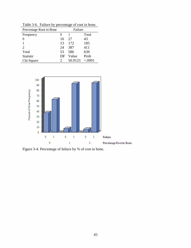
Table 3-6 and Figure 3-4 illustrated the effect of the amount of bone remained supporting
roots of endodontically treated teeth with metal prefabricated or custom fabricated posts. A total
of 43 patients were treated with metal posts in teeth that had less than 50% of roots embedded by
bone, 16 posts failure were documented. The other group was 185 teeth that had 50-75% of
bone supporting roots of the treated teeth. Thirteen cases of this group had failed. The last
group of this category of testing was teeth that had more than 75% of bone supporting roots of
the treated teeth. There were 24 cases documented as failure out of total 411 teeth treated with
metal posts. In Figure 3-4 there was shown how poorly the treatment outcome was for teeth that
had less than 50% bone supporting the roots with percentage of failure of 38%. Teeth with more
than 75% of bone supporting their roots had only 6% of failure. Statistically this variable has
shown to be very significant on how well metal posts performed and survived P<0.05.
Table 3-7 and Figure 3-5 illustrated the influence of material of restorations were used to
treat teeth that had metal posts performed on them. Different type of restorations was used and
varied from build up restorations, PFM materials, gold, and temporary restorations. Statistical
tests have shown a little influence between each group in the survival probability of metal posts,
however there was not a statistical significant difference of the type of material used to restore
endodontically treated teeth with percentage of failure recorded.

32
Table 3-8 and Figure 3-6 showed the influence of different type of cement used on the
survival probability of teeth treated with metal posts. The statistical tests compared the use of
zinc phosphate cements to other type of cements used in the Undergraduate Clinic for
Removable and Fixed Prosthodontics at the University of Florida. No statistical significant
difference P>0.05 was found with different type of cements used to the percentage of survival of
metal posts in endodontically treated teeth. Over 90% survival of metal posts were recorded
with all type of cements documented.
In Table 3-9 and Figure 3-7 an illustration of the importance of tooth position in the dental
arch to the survival probability of teeth treated with metal posts was tested. It was shown that no
statistically significant differences were ascertained between anterior and posterior
endodontically treated teeth in the dental arch with the percentages of survival of metal posts.
Table 3-10 and Figure 3-8 demonstrated the influence of final treatment type of the
endodontically treated teeth to their probability of survival of metal posts used. It was recorded
that endodontically treated teeth with metal posts that were not splinted by other teeth as part of a
fixed partial denture prosthesis or a removable partial denture prosthesis had a larger percentages
of survival probability of metal posts used. Figure 3-8 illustrated the percentages of failure for
teeth that performed as an abutment for removable partial denture prosthesis to be 18%, while it
was 9% for teeth that performed as an abutment for fixed partial denture prosthesis, and 6% for
teeth that performed as un-splinted single crowns. This variable appeared to be of a great
statistical significance to the probability of survival of metal posts P<0.05.
Table 3-11 and Figure 3-9 represent the influence of opposing dentition on the survival
probability of metal posts in endodontically treated teeth. From the data collected it was made
clear that teeth with metal posts performed a little better when opposed natural teeth with 8%

33
failure of row frequency. Teeth that opposed fixed and removable prostheses performed
relatively well in comparison. No statistically significant differences were determined between
the different types of opposing occlusion on the survival probability of metal posts in
endodontically treated teeth P>0.05.
Figure 3-10 demonstrates the total percentage of failure of both types of metal posts
documented in this study. It exhibited 92% survival probability of prefabricated and custom-
fabricate metal posts combined.
At a significance level of 0.05, four variables were shown to be correlated with failure,
they are age, post length, percentages of root in the bone, and whether the tooth treated with post
was a free standing crown or an abutment for fixed or removable partial denture prosthesis.
The final model of significant variables was shown to be models of post length, percentage
of roots in bone, and the type of final prosthetic treatment. All these variables were significant at
< 0.001. The type III tests for these effects are shown in table 12. To illustrate how these
variables affect failure the adjusted least square means are shown in Table 13-15. The mean
represents the average probability that a patient with metal post treatment would experience a
failure.
Table 3-13 illustrates the probability of survival of metal posts in relation to the post
length. It was found out that when metal posts are prepared to more than half the length of the
roots to have a probability of survival of 91%. While the probability of survival dropped to 72%
when posts were to be prepared to less than half of the length of the roots.
Table 3-14 represents the probability of survival of metal posts in relation to the amount of
bone remained supporting the roots. It was found out that when teeth were supported 50-75% or
more by bone tissue, had a survival probability of 90%, 92% respectively. When endodontically
34
treated teeth had less than 50% of bone supporting the roots, the survival probability of metal
posts treatment was only 56.8%.
Table 3-15 brings clear the relationship between the survival probability of metal posts
performed in endodontically treated teeth and the type of final treatment associated with those
teeth. It showed that the probability of survival of metal posts performed in teeth that are un-
splinted single crowns, or teeth that presented as abutments for fix partial denture prostheses,
was 89%. The probability of survival of metal posts dropped significantly in teeth that acted as
abutments for removable partial denture prosthesis to 69%.
Discussion
The design of this study was specifically meant to assess variables that might influence the
survival of 2 different metal post systems that were used for endodontically treated teeth in the
Undergraduate Clinic of Fixed and Removable Prosthodontics at the University of Florida.
As regards age, older patients were reported to have less survival of metal posts than
younger patients. A significant difference was recorded and illustrated in Table 3.1. Our
findings could be attributed to the fact that elderly patients take various medications that might
cause xerostomia, which could be a major cause of developing root caries. Another significant
correlation was the fact that teeth that have been treated with metal posts were abutments for
some type of removable partial denture prostheses. Many geriatric studies showed that
alterations in nature of pulp sensitivity, higher risk of developing root caries, and poor oral
hygiene, had a significant influence on the survival of metal posts. Pulp and dentin, like other
connective tissues, undergoes changes with time. Some of these changes are natural, whereas
others may be a result of injury such as caries, periodontal disease, trauma, or restorative dental
procedures. Regardless of the cause changes in pulp and dentin appearance and function do
occur (Waton R 2002). A deeply entrenched clinical perception persists that root canal treated

35
teeth become more brittle, assumingly, losing resilience as the moisture content of dentin
declines after pulp loss. This perception is unsupported experimentally (Walton 2002). Few
studies have compared physical properties of endodontically treated versus non-treated human
teeth with vital pulp. The moisture content of endodontically treated teeth was not reduced, even
after 10 years (Papa 1994). Also, a comparison of these two groups revealed no significant
differences in strength, toughness and hardness of dentin (Huang 1992, Sedgley 1992). Thus,
susceptibility to failure cannot be reliably attributed to structural changes in dentin after loss of
pulp vitality or after root canal treatment (Walton 2002). It is more attributed to the type of final
treatment the endodontically treated teeth are receiving and other factors mentioned previously.
Post length was shown to have a significant influence on the survival of both types of
metal post systems evaluated. In this clinical study it was illustrated that endodontically treated
teeth that had longer posts; extending to more than half of the root length while maintaining the
necessary apical seal; to have significantly greater survival than teeth treated with shorter metal
posts. Table 3.13 shows a clear presentation of the importance of post length. It was shown that
post spaces that were prepared up to half the length of the roots had a survival probability of
72.8%. While posts spaces that were prepared for more than half of the root length had a
survival probability of 91.5%. An abundance of reports and documentations support our
findings (Santos-Filho 2008, Asmussen 2005, Caputo 1987, Colley 1968, Cooney 1986, Holmes
1996, Sorensen 1984). Fernandes concluded in his study, that posts needed to be long enough to
prevent excessive internal stresses in the roots (Fernandes 2001). The length of metal post plays
an important role in its retention. Various investigators have demonstrated a significant
relationship between vertical resistance to displacement and length of the post. Posts should be
as long as possible with 3-5 mm of root filling left at the apex for seal (Baraban 1988, Colman

36
1979). Research suggests that longer the post, greater is the retention and less is the stress
(Standlee 1978).
One of the significant variables that were investigated in this study was the amount of bone
present to support the roots of the endodontically treated teeth. It was cleared out in this
examination that the survival of metal posts was significantly reduced in endodontically treated
teeth that had less than 50% of bone tissue surrounding their roots. It was exhibited in Table
3.14 that when endodontically treated teeth had more than 75% of bone tissue support present,
the survival probability of metal posts treatment was 92%. While the survival probability had
significantly dropped to 56.8% when less than half of the roots were supported by alveolar bone
tissue. Metal post should extend to at least half the length of the root contained in the remaining
alveolar bone (Jacoby 1976). It has demonstrated in this clinical investigation the importance of
proper treatment planning prior to endodontic therapy of teeth. The amount of alveolar bone
present with respect of the necessary apical seal should be always taken into consideration. It
was apparent that teeth with good overall prognosis and adequate periodontal support as
measured clinically and radiographically, would have illustrated higher survival probability of
metal posts treatment assuming the proper maintenance by the clinician and patient (Walton
2002).
Another important variable examined was the different types of final prosthetic
restorations that endodontically treated teeth with metal posts have received. It was brought
clear in Table 3.15 that teeth that were used as abutments for single crowns, and abutments for
fixed partial denture prostheses have demonstrated the highest survival probabilities when
compared to teeth that were used as abutments for removable partial dentures prostheses, with
survival probabilities of 89.4%, 89.1%, and 69.3%, recorded respectively. Many reports support
37
our findings. A retrospective clinical study by Wegner aimed to evaluate the survival rate of
teeth that were endodontically treated and restored with endodontic posts and prosthodontic
restorations. In her study, the calculated survival rates of the abutments were found to be
significantly different for fixed partial dentures and for removable partial dentures with survival
rate of 92.7% and 51% respectively (Wegner 2006). Endodontically treated teeth used as
removable partial denture abutments have a five times greater failure than single teeth (Sorensen
1990). Endodontically treated teeth that serve as abutments for fixed or removable prostheses
have been reported to be most prone to failure (Palmqvist 1994). A study by Kantor
recommended the use of cast metal post and core for restoring endodontically treated teeth that
are used as abutments for removable partial dentures (Kantor 1977).
One of our main objectives in this study was to compare clinically the cumulative survival
rate of custom fabricated cast posts to prefabricate metal post used in endodontically treated
teeth. Figure 3.10 demonstrated the cumulative survival rate of both types of posts used and was
found to be 92% during the observation period of 5 years. 510 custom fabricated posts were
documented and exhibited 91% survival, and 129 prefabricated posts were used with a survival
rate of 92% reported. Few in vivo studies illustrated similar results, but did not evaluate large
patient collectives and possible covariates that may affect the risk of failure. Bergman examined
the success rate of cast post and cores over 6 years with 96 posts cases. The failure rate was
established in relation to the type of prosthetic restoration (crown, bridge), type of tooth
(anterior, premolar, molar), the jaw (maxillary, Mandibular). There was a 10% failure rate after 6
years (Bergman 1989). Ellner et al. in his prospective study of 50 posts in 31 patients recorded a
success rate of 100% for the group with custom-fabricated post and cores with an excellent
success probability in the observation period of 10 years. The patient collective was, however,

38
highly selective and had only been treated with single crowns (Ellner 2003). Ferrari et al.
compared in their study custom-fabricated post and cores with fiber posts. After 4 years in
service, custom-fabricated post and cores showed a failure rate of 14% (Ferrari 2000).
Hatzikyriakos et al. examined the failure rate with 154 post and cores involving prefabricated,
screw-retained, custom-fabricated, cemented post and cores under crowns, bridges and
removable dentures. The cumulative failure rate was 9.1%for custom-fabricated post and cores
after a period of 3 years. The number of cases in each group was, however, too small to draw
any further conclusions from findings (Hatzikyriakos 1992). Sorensen and Martinoff examined
the failure rate with 1273 root-filled teeth in relation to the postendodontic treatment (no post and
core versus different post systems). In this study, the majority of teeth 65.4% had not been
treated with a post and core and only 19.2% had been treated with a cast post and core. The
failure rate recorded for the latter group was 12.7%, but no information was provided about the
time in situ (Sorensen 1985). Torbjorner et al examined the success rate of two different post
designs paraposts versus custom-fabricated cast posts in a 6- years study. The 456 cast posts and
cores exhibited a failure rate of 10.5 % during the observation period (Torbjorner 2004).
Balkenhol et al. examined the survival time of custom-fabricated cast post and core, and
evaluated different variables, which influenced the risk of failure over a period of 10 years. They
have concluded that custom-fabricated post and core have a good long-term prognosis, and
reported a cumulative failure rate of 11.2%, and an average survival time of 7.3 years (Balkenhol
2007).
In this study gender was found not to have a significant influence on the survival of neither
of the 2 metal post systems used. Figure 3.1 demonstrated clearly the effect of gender on the
survival of metal posts and showed relatively close percentages. A study conducted in Denmark

39
have illustrated different results (Peutzfeldt 2007); where they found males to have less survival
probability of metal posts, and they have related their findings to the fact that men exerted
greater bite forces than women.
It was shown in this retrospective study that there were no significant differences between
types of materials used for the coronal coverage of endodontically treated teeth. It was reported
that most endodontically treated posterior teeth had full coronal coverage, metal or porcelain
fused to metal coverage. Both type of materials performed relatively equally as demonstrated in
Figure 3.5. Anterior teeth on the other hand, were found to be treated either with composite
restorations, or porcelain fused to metal crowns. A slight advantage of anterior teeth that were
treated with full coronal coverage had been recorded over teeth that had only filling restorations.
However the difference in survival of both types of metal posts with different types of materials
of coronal coverage used in anterior teeth was not significant.
Type of cements used in the clinic for retaining metal posts was another variable that has
been evaluated and found to be of no significance to survival of metal posts in endodontically
treated teeth. The majority of cases examined have documented the use of zinc phosphate with a
survival rate greater than 90%. However, metal posts that were retained using other type of
cements available in the clinic also reported relatively similar values. There was no significant
difference in the survival rate of metal posts retained by different type of permenant cements
considering the proper post space preparation, amount of osseos bone present, and the type of
final prosthetic restoration.
Neither tooth position in the arch, nor the type of opposing occlusion showed to have a
significant influence on the survival probability of metal posts with endodontically treated teeth
in this study. Other studies also produced this result (Bergman 1989, Dammaschke 2003). On
40
the other hand, a few studies recorded a prevalence of failures in the upper jaw, generally in the
anterior region (Mentink 1993, Torbjorner 1995).
The data of the study were acquired using a retrospective, longitudinal study design. A
typical problem with retrospective studies is the availability of analyzable, consistent data. This
did not, however, pose a problem with this retrospective study, as the clinical findings had been
recorded in the department of prosthodontics since the beginning of 2003 according to a
standardised procedure. Operators’ lack of completed training was not likely to be a
disadvantage, because there was extensive treatment plan and treatments were closely
supervised. It can therefore be assumed that the recorded data are representative and
comparable. It would have been more practical, if the average observation period had been
much longer than 5 years.
Dentists should evaluate the root length. If the root is too short, or the crown-to-root ratio
is unfavorable, the tooth may be unsuitable as an abutment for removable partial denture
prostheses. If the osseos support and the root length are inadequate, the dentist should relate to
the patient that the prognosis is poor. These considerations, in addition to minimal coronal tooth
structure, make the prognosis questionable for a compromised tooth. Pulpless teeth are
commonly avoided as abutments for removable partial dentures, especially if the terminal
abutment is for a distal extension (Sorensen 1990).
Limitations of the study varied from sometimes lack of documentation of all investigated
variables, the uncertainty of amount of ferrule remained and number of dentinal walls
maintained, and unavailability of documenting posts’ width preparations.
Future clinical studies should focus on evaluating different prefabricated non-metallic
posts to custom fabricated posts, and for a longer period of observation time.

41
Table 3-1. One-way ANOVA for failure by age. Source DF Sum of Squares Mean Square F Value Pr > F Model 1 2117.7758 2117.7758 9.22 0.0025 Error 637 146364.1678 229.7711 Corrected Total 638 148481.9437 Table 3-2. Means of age for failure Failure Age LSMEAN 0 59.3773585 1 52.7764505

42
Table 3-3. Failure by gender Gender Failure Frequency 0 1 Total 0 24 237 261 1 29 349 378 Total 53 586 639 Statistic DF Value Prob Chi-Square 1 0.4711 0.4925
Figure 3-1. Percentage of failure by gender.

43
Table 3-4. Failure by post type. Post Type Failure Frequency 0 1 Total 0 9 120 129 1 44 466 510 Total 53 586 639 Statistic DF Value Prob Chi-Square 1 0.3688 0.5436
Figure 3-2. Percentage of failure by post type.

44
Table 3-5. Failure by post length. Post Length Failure Frequency 0 1 Total 0 26 133 159 1 27 453 480 Total 53 586 639 Statistic DF Value Prob Chi-Square 1 18.0692 <.0001
Figure 3-3. Percentage of failure by post length.
45
Table 3-6. Failure by percentage of root in bone. Percentage Root in Bone Failure Frequency 0 1 Total 0 16 27 43 1 13 172 185 2 24 387 411 Total 53 586 639 Statistic DF Value Prob Chi-Square 2 50.9123 <.0001
Figure 3-4. Percentage of failure by % of root in bone.

46
Table 3-7. Failure by type of restoration’s material. Restoration Failure Frequency 0 1 Total 0 3 51 54 1 38 419 457 2 12 90 102 3 0 26 26 Total 53 586 639 Statistic DF Value Prob Chi-Square 3 4.4994 0.2123
Figure 3-5. Percentage of post failure by type of restoration’s material.

47
Table 3-8. Failure by cement. Cement Failure Frequency 0 1 Total 0 36 412 448 1 13 120 133 2 4 54 58 Total 53 586 639 Statistic DF Value Prob Chi-Square 2 0.5714 0.7515
Figure 3-6. Percentage of failure by cement.

48
Table 3-9. Failure by tooth position. Tooth Position Failure Frequency 0 1 Total 0 30 393 423 1 23 193 216 Total 53 586 639 Statistic DF Value Prob Chi-Square 1 2.3770 0.1231
Figure 3-7. Percentage of failure by tooth position.

49
Table 3-10. Failure by type of prosthetic treatment. Final Treatment Failure Frequency 0 1 Total 0 22 367 389 1 11 123 134 2 20 96 116 Total 53 586 639 Statistic DF Value Prob Chi-Square 2 15.7705 0.0004
Figure 3-8. Percentage of failure by type of tooth treatment.

50
Table 3-11. Failure by type opposing occlusion. Opposing Dentition Failure Frequency 0 1 Total 0 28 373 401 1 12 75 87 2 13 138 151 Total 53 586 639 Statistic DF Value Prob Chi-Square 2 4.3853 0.1116
Figure 3-9. Percentage of failure by type of opposing dentition.

51
Figure 3-10. Percentage of total failure of all post types.

52
Table 3-12. Type III tests of the significant variables. Effect Num DF Den DF F Value Pr > F Percentage Root in Bone 2 633 15.76 <.0001 Post Length 1 633 18.79 <.0001 Free Standing 2 633 7.68 0.0005 Table 3-13. Post length least square means. Post Length Mean Standard Error Mean 0 0.7288 0.04844 1 0.9154 0.01732

53
Table 3-14. Percentage of root in bone least square means. Percentage Root in Bone Mean Standard ErrorMean 0 0.5683 0.08235 1 0.9086 0.02468 2 0.9230 0.01638 Table 3-15 Type of final prosthetic treatment least square means. Type of Final Treatment Mean Standard Error Mean 0 0.8942 0.02295 1 0.8915 0.03330 2 0.6932 0.05960

54
CHAPTER 4 SUMMARY AND CONCLUSION
Within the limitations of this clinical study, multiple variables have been evaluated for
their influence on survival probability of custom-fabricated cast post and prefabricated metal
posts used in endodontically treated teeth. Multiple hypotheses were fabricated and the
conclusions were drawn to be:
1. Age of patients did influence the survival rate of both treatment modalities, and was found that older patients exhibited less survival rate than younger patients.
2. Metal posts that were prepared for more than half of the root length while maintaining the necessary apical seal shown to have greater survival probability.
3. The percentage of alveolar bone remain to support the endodontically treated teeth was found to influence the survival probability of both treatment modalities significantly.
4. The type of final prosthetic restoration fitted has a significant effect on the survival probability. Posts under single crowns and fixed partial denture prostheses have the highest survival probability.
5. The clinical survival of custom-fabricated cast posts and prefabricated metal posts were not affected by patients’ gender, type of cements used to retain posts, types of opposing occlusion, and the position of the endodonticaly treated teeth in the dental arches.
Very little clinical data are available on metal posts treatments that are performed on a
daily basis. This fact, combined with the inconsistency of the clinical data that have been
published we could not conclude the preference of custom-fabricated cast posts over
prefabricated metal posts. Both treatment modalities can be recommended if they are applied
within indications and with the necessary caution. Metallic posts continue to be the standard for
most situations because they have stood the test of time.
55
LIST OF REFERENCES
Abramovitz I, Tagger M, Tamse A, Metzger Z. The effect of immediate vs. delayed post space preparation on the apical seal of root canal filling: a study in an increased-sensitivity –pressure driven system. J Endod 2000;26:435-439.
Asmussen E, Peutzfeldt A, Sahafi A. Finite element analysis of stresses in endodontically treated, dowel-restored teeth. J Prosthet Dent 2005;94:321–329
Assif D, Bitenski A, Pilo R, Oren E. Effect of post design on resistance to fracture of endodontically treated teeth with complete crowns. J Prosthet Dent 1993;69:36-40.
Balbosh A, Ludwig K, Kern M. Comparison of titanium dowel retention using four different luting agents. J Prosthet Dent 2005;94:227-233.
Balkenhol M, Wostmann B, Rein C, Ferger P. Survival time of cast post and cores: A 10-year retrospective study. Journal of dentistry 2007;35:50-58.
Baraban D. Immediate restoration of pulpless teeth. J Prothet Dent 1972;28:607-612.
Baraban D. The restoration of endodontically treated teeth-An update. J Prosthet Dent 1988;59:553-558.
Baratieri L, De Andrada M, Arcari G, Ritter A. Influence of post placement in the fracture resistance of endodontically treated incisors veneered with direct composite. J Prosthet Dent 2000;84:180-184.
Bartlett S. Construction of detached core crowns for pulpless teeth in only two sittings. JADA 1968;77:843–845.
Bergman B, Lundquist P, Sjögren U, Sundquist G. Restorative and endodontic results after treatment with cast posts and cores. J Prosthet Dent 1989;61:10–5
Bishop K. and Biggs P. Endodontic failure-A problem from top to bottom. Br Dent J 1995; 179: 35-36.
Black G. A method of grafting artificial crowns on roots of teeth. Mo Dent J 1869;1:233-236.
Burgess J, Summitt J, Robbins J. The resistance to tensile, compression, and torsional forces provided by four post systems. J Prosthet Dent 1992;68:899-903.
Caputo A, Standlee J. Pins and posts: why, when and how. Dent Clin North Am 1976;20:299–311.
Caputo A, Standlee J. Restoration of endodontically involved teeth. In: Biomechanics in clinical dentistry. Chicago: Quintessence 1987;P:185.
Chapman K, Worley J, von Fraunhofer J. Retention of prefabricated posts by cements and resins. J Prosthet Dent 1985;54:649-652.

56
Chapman K, Worley J, von Fraunhofer J. Retention of prefabricated posts by cements and resins. J Prosthet Dent 1985;54:649-652.
Cheung W. A review of the management of endodontically treated teeth. J Am Dent 2005;6:611-619.
Cohen B, Pagnillo M, Newman I, Musikant B, Deutsch AS. Retention of four endodontic posts cemented with composite resin. Gen Dent 2000;48:320-324.
Cohen S, Burn RC. Pathway of The Pulp, ed 6. St. Louis, MO:CV Mosby, 1991:641.
Colley I, Hampson E, Lehman M. Retention of post crowns. An assessment of the relative efficiency of posts of different shapes and sizes. Br Dent J 1968;124:63–69.
Colman H. Restoration of endodontically treated teeth. Dent Clin North Am 1979;23:647-662.
Cooney J, Caputo A, Trabert K. Retention and stress distribution of tapered-end endodontic posts. J Prosthet Dent 1986;55:540–546.
Dammaschke T, Steven D, Kaup M, Ott K. Long-term survival of root-canal-treated teeth: a retrospective study over 10 years. Journal of Endodontics 2003;29:638–643.
Davis W. Essentials of Operative Dentistry, ed 2. St. Louis, MO:CV Mosby, 1916:23.
Demas N. Direct impression for cast Richmond crown using acetate crown forms. Dental Dig 1957;63:258-259.
Driessen C, Marais J, de Villers D, Nel J. The effect of bonded and non-bonded posts on the fracture resistance of dentine. J Dent Assoc S Afr 1997;52:393-398.
Ellner S, Bergendal T, Bergman B. Four post-and-core combinations as abutments for fixed single crowns: a prospective up to 10-year study. International Journal of Prosthodontics 2003;16:249–254.
Ertugul Z, Ismail Y. An in vitro comparison of cast metal dowel retention using various luting agents and tensile loading. J Prosthet Dent 2005;93:546-451.
Fauchard P. The Surgeon Dentist or Treatise on The Teeth, ed 2. London:Butterworth, 1746:31.
Fernandes A, Rodrigues S, SarDessai G, Mahta A. Retention of endodontic post-A review. Endodontology 2001;13: 11-18.
Ferrari M, Vichi A, Garcia-Godoy F. Clinical evaluation of fiber-reinforced epoxy resin posts and cast post and cores. American Journal of Dentistry 2000;13:8B–15B.
Goldman M, DeVitre R, Tenca J. Cement distribution and bond strength in cemented posts. J Dent Res 1984;63:1392-1395

57
Goldstein G, Hudis S, Weintraub D. Comparison of four techniques for cementation of posts. J Prosthet Dent 1986;55:209-211.
Gross M, Turner C. Intra-radicular hydrostatic pressure changes during the cementation of post-retained crowns. J Oral Rehabil 1983;10:237-249.
Gutmann J. The dentin-root complex: anatomic and biologic considerations in restoring endodontically treated teeth. J Prosthet Dent 1992;67:458–67.
Guzy G, Nicholls J. In vitro comparison of intact endodontically treated teeth with and without endo-post reinforcement. J Prosthet Dent 1979;42:39-44.
Hampson E, Clark J. The post-retained crown. Dent Pract Dent Rec 1958;8:130-135.
Harris C. The Principles and Practice of Dentistry, ed 2. Philadelphia:Lindsay and Blackiston, 1871:87.
Hatzikyriakos AH, Reisis GI, Tsingos N. A 3-year postoperative clinical evaluation of posts and cores beneath existing crowns. J Prosthet Dent 1992;67:454–458.
Helfer A, Melnick S, Schilder H. Determination of moisture content of vital and pulpless teeth. Oral Surg Oral Med Oral Pathol 1972;34: 661-670.
Hoag E, Dwyer T. Comperative evaluation of three post and core techniques, J Prosthet Dent 1982;47:177-181.
Holmes D, Diaz-Arnold A, Leary J. Influence of post dimension on stress distribution in dentin. J Prosthet Dent 1996;75:140–147.
Huang T, Schilder H, Nathanson D. Effects of moisture content and endodontic treatment on some mechanical properties of human dentin. J Endod 1992;18:209.
Hunter A, Feiglin B, Williams J. Effects of post placement on endodontically treated teeth. J Prosthet Dent 1989;62:166-72.
Jacoby W. Practical technique for the fabrication of a direct pattern for a post-core restoration. J Prosthet Dent 1976;35:357-360.
Kane J, Burgess J. Modification of the resistance form of amalgam coronal-radicular restorations. J Prosthet Dent 1991;65:470–474.
Kantor M, Pines M. A comparative study of restorative technique for pulpless teeth. J Prosthet Dent 1977;38:405-412.
Kurer H, Combe E, Grant A. Factors influencing the retention of dowels. J Prosthet Dent 1977;38:515-525.
Kvist T, Rydin E, Reit C. The relative frequency of periapical lesions in teeth with root canal–retained posts. J Endod 1989;15:578–580

58
Mattison G, Delivanis P, Thacker R, Hassell K. Effect of post preparation on the apical seal. J Prosthet Dent 1984;51:785-789.
Mcdonald A, King P, Setchell D. In vitro study to compare impact fracture resistance of intact root-treated teeth. J Endodont 1990;23:304-312.
McLean A. Predictably restoring endodontically treated teeth. J Can Dent Assoc 1998;64:782-787.
Mendoza D, Eakle W. Retention of posts cemented with various dentinal bonding cements. J Prosthet Dent 1994;72:591-594.
Mentink AG, Meeuwissen R, Kayser AF, Mulder J. Survival rate and failure characteristics of the all metal post and core restoration. Journal of Oral Rehabilitation 1993;20:455–461.
Monaghan P, Roh L, Kim J. Corrosion behaviour of selected implant alloys. J Dent Res 1992;71:253.
Morgano S, Brackett SE. Foundation restorations in fixed prosthodontics: current knowledge and future needs. J Prosthet Dent 1999;82:643–657.
Morgano S. Restoration of pulpless teeth: application of traditional principles in present and future contexts. J Prosthet Dent 1996;75:375–380.
Nathanson D. New views on restoring the endodontically treated tooth. Den Econ 1993;83:48-50.
Organization participating in the Eighth Edition of The Glossary of Prosthodontic Terms. J Prosthet Dent 2005;94:71.
Palmqvist S, Soderfeldt B. Multivariate analyses of factors influencing the longevity of fixed partial dentures, retainers, and abutments. J Prosthet Dent 1994;71:245-250.
Papa J, Chain C, Messer H. Moisture content of vital vs. endodontically treated teeth. Endod Dent Traumatol 1994;10:91-93.
Peutzfeldt A, Sahafi A, Asmussen E. A survery of failed post-retained restorations. Clin Oral Invest 2008;12:37-44.
Portell F, Bernier W, Lorton L, and Peters D. The effect of immediate versus delayed dowel space preparation on the integrity of the apical seal. J Endod 1982;8:154-160.
Radke R, Barkordar R, Podesta R. Retention of cast endodontic posts: Comparison of cementing agents. J Prosthet Dent 1988;59:318-320.
Reeh E, Douglas W, Messer H. Stiffness of endodontically treated teeth related to restoration technique. J Dent Res 1989;68:1540-1544.

59
Richmond C. New method of attaching gold crowns to natural roots of teeth. Am J Dent Sci 1878;12:425.
Ring M. Dentistry: An Illustrated History, ed 2. St Louis, MO:CV Mosby, 1985:91.
Robbins J. Guidelines for the restoration of endodontically treated teeth. J Am Dent Assoc 1990;120:558-566.
Robbins J. Restoration of the endodontically treated teeth. Dent Clin N AM 2002;46:367-387.
Rosen H. Operative procedures on mutilated endodontically treated teeth. J Prosthet Dent 1961;11:973–986.
Rosenstiel S, Land M, Fujimoto J, Contemporary Fixed Prosthodontics, ed 2. St. Louis, MO:CV Mosby, 2001:255.
Ruemping D, Lund M, Schnell R. Retention of dowel subjected to tensile and torsional forces. J Prosthet Dent 1979;41:159-162.
Safavi K, Dowden W, Langeland K. Influence of delayed coronal permanent restoration on endodontic prognosis. Endod Dent Traumatol 1987;3:187-191.
Santos-Filho P, Castro C, Silva G, Campos R, Soares C. Effects of post system and ength on the strain and fracture resistance of root filled bovine teeth. Intern Endod J 2008;41:493-501.
Schwartz R, Summitt J, Robbins J. Fundamental of Operative Dentistry: A Contemporary Approach, ed 2. Chicago:Quintessence, 1996:321.
Sedgley C, Messer H. Are endodontically treated teeth more brittle? J Endod 1992;18:332-335.
Shillingburg H, Keesler J. Restoration of The Endodontically Treated Tooth, ed 1. Chicago:Quintessence, 1982:13.
Sidoli G, King P, Setchell D. An in vitro evaluation of a carbon fiber-based post and core system. J Prosthet Dent 1997;78:5-9.
Silverstein W. The reinforcement of weakened pulpless teeth. J Prosthet Dent 1964;14:372–381.
Sorensen J, Engelman M. Ferrule design and fracture resistance of endodontically treated teeth. J Prosthet Dent 1990;63:529-536.
Sorensen J, Martinoff J. Clinically significant factors in dowel design. J Prosthet Dent 1984;52; 28-35.
Sorensen J, Martinoff J. Intracoronal reinforcement and coronal coverage: A study of endodontically treated teeth. J Prosthet Dent 1984;51:780-4.
Spalten R. Composite resins to restore mutilated teeth. Oper Dent 1971;25:323-326.

60
Standlee J, Caputo A, Hanson EC. Retention of endodontic dowels: Effects of cement, dowel length, diameter, and design. J Prosthet Dent 1978;39:400-405.
Stern N, Hirschfeld Z. Principles of preparing endodontic treated teeth for dowel and core restorations. J Prosthet Dent 1973;30:162–165.
Stockton L. Factors affecting retention of post systems: a literature review. J Prosthet Dent 1999;4:380-385.
Tjan A, Greive J. Effects of various cementation methods on retention of prefabricated posts. J Prosthet Dent 1987;58:309-313.
Torbjorner A, Karlsson S, Odman PA. Survival rate and failure characteristics for two post designs. J Prosthet Dent 1995;73:439–444.
Trabert K, Caput A and Abou-Rass M. Tooth fracture: a comparison of endodontic and restorative treatment. J Endodont 1978;4: 341.
Trope M, Maltz DO, Tronstad L. Resistance to fracture of restored endodontically treated teeth. Endod Dent Traumatol 1985;1:108-111.
Vire D. Failure of endodontically treated teeth: classification and evaluation. J Endod 1991;17:338-342.
Walton R, Torabinejad M. Principles and Practice of Endodontics, ed 3. Philadelphia:Saunders, 2002:130.
Weaver A, Kremenak N. Let’s talk about statistics and prosthodontics research:part 3, presentation of results. J Prosthet Dent 2008;17:686-687.
Wegner P, Freitag S, Kern M. Survival rate of endodontically treated teeth with posts after prosthetic restoration. J Endod 2006;32:928-931.
Whitworth J, Walls A, Wassell R. Crowns and extra-coronal restorations: Endodontic considerations: the pulp, the root-treated tooth and the crown. Br Dent J 2002;192:315-327.
Young H, Shen C, Maryniuk G. Retention of cast posts relative to cement selection. Quintessence Int 1985;16:357-360.
61
BIOGRAPHICAL SKETCH
The author, Hassan Mosuawi, was born in 1978 in Malta. He grew up in Kuwait City,
Kuwait. Graduated from Salah Aldeen High School in Kuwait City in June1996. In August of
1996, he enrolled at the University of Missouri-Kansas-City as a dental student where he joined
the six-year-combined program of Bachelor of Art and Science. In 2003, Mousawi graduated
from the University of Missouri-Kansas-City School of Dentistry, with a Doctor of Dental
Surgery degree. The author returned home to Kuwait and worked for the public health system of
Kuwait from 2003 to 2006 before deciding to pursue a dental specialty degree in the United
States. In July 2006, he enrolled in the Graduate prosthodontics program at the University of
Florida. After graduation with a Master of Science in dental sciences with a specialization in
prosthodontics from the University of Florida in May of 2009, he plans to return to Kuwait and
work for the public health system for the betterment of the dental service of his beloved country.
This is the beginning of the rest of his life.