
Clinical Effectiveness of Direct Class II Restorations – A ... · PDF fileClinical...
25
Vol 14, No 5, 2012 407 Clinical Effectiveness of Direct Class II Restorations – A Meta-Analysis Siegward D. Heintze a / Valentin Rousson b Purpose: More than five hundred million direct dental restorations are placed each year worldwide. In about 55% of the cases, resin composites or compomers are used, and in 45% amalgam. The longevity of posterior resin restorations is well documented. However, data on resin composites that are placed without enamel/dentin con- ditioning and resin composites placed with self-etching adhesive systems are missing. Material and Methods: The database SCOPUS was searched for clinical trials on posterior resin composites without restricting the search to the year of publication. The inclusion criteria were: (1) prospective clinical trial with at least 2 years of observation; (2) minimum number of restorations at last recall = 20; (3) report on drop- out rate; (4) report of operative technique and materials used; (5) utilization of Ryge or modified Ryge evaluation criteria. For amalgam, only those studies were included that directly compared composite resin restorations with amalgam. For the statistical analysis, a linear mixed model was used with random effects to account for the het- erogeneity between the studies. P-values under 0.05 were considered significant. Results: Of the 373 clinical trials, 59 studies met the inclusion criteria. In 70% of the studies, Class II and Class I restorations had been placed. The overall success rate of composite resin restorations was about 90% after 10 years, which was not different from that of amalgam. Restorations with compomers had a significantly lower lon- gevity. The main reason for replacement were bulk fractures and caries adjacent to restorations. Both of these inci- dents were infrequent in most studies and accounted only for about 6% of all replaced restorations after 10 years. Restorations with macrofilled composites and compomer suffered significantly more loss of anatomical form than restorations with other types of material. Restorations that were placed without enamel acid etching and a dentin bonding agent showed significantly more marginal staining and detectable margins compared to those restorations placed using the enamel-etch or etch-and-rinse technique; restorations with self-etching systems were between the other groups. Restorations with compomer suffered significantly more chippings (reparable fracture) than restora- tions with other materials, which did not statistically differ among each other. Restorations that were placed with a rubber-dam showed significantly fewer material fractures that needed replacement, and this also had a significant effect on the overall longevity. Conclusion: Restorations with hybrid and microfilled composites that were placed with the enamel-etching tech- nique and rubber-dam showed the best overall performance; the longevity of these restorations was similar to amalgam restorations. Compomer restorations, restorations placed with macrofilled composites, and resin res- torations with no-etching or self-etching adhesives demonstrated significant shortcomings and shorter longevity. J Adhes Dent 2012;14:407–431. Submitted for publication: 01.12.11; accepted for publication: 20.05.12 doi: 10.3290/j.jad.a28390 a Head of Preclinical Research, Research and Development, Ivoclar Vivadent AG, Schaan, Liechtenstein. Wrote manuscript. b Statistician, Biostatistics Unit, Institute for Social and Preventive Medicine, University of Lausanne, Switzerland. Performed statistical evaluation. Correspondence: Dr. Siegward D. Heintze, Research and Development, Ivoclar Vivadent, Preclinical Research, Bendererstrasse 2, FL-9494 Schaan, Liechten- stein. Tel: +423-235-3570, Fax: +423-233-1279 e-mail: [email protected] proximal surfaces of posterior teeth. 67 Although mini- mally invasive operative techniques and instruments are available, the majority of dental practitioners still opt for the traditional Class II preparation design based on the guidelines that were established by G.V. Black in 1910. 9 In industrialized countries, the most frequently used materials to restore posterior lesions are compos- ite resins of various kinds, which are placed according to the adhesive technique involving the conditioning of both dentin and enamel. The usage of composite resins has surpassed the us- age of amalgam over the last 10 years, but amalgam is still widely used in many countries. 68 Based on the market volume and materials sold, it can be calculated W orldwide, multisurface restorations in permanent premolars and molars are the most frequent type of dental restorations. This is due to the localization of caries, which primarily occurs on the occlusal and
Transcript of Clinical Effectiveness of Direct Class II Restorations – A ... · PDF fileClinical...

Vol 14, No 5, 2012 407
Clinical Effectiveness of Direct Class II Restorations –
A Meta-Analysis
Siegward D. Heintzea / Valentin Roussonb
Purpose: More than five hundred million direct dental restorations are placed each year worldwide. In about 55% of the cases, resin composites or compomers are used, and in 45% amalgam. The longevity of posterior resin restorations is well documented. However, data on resin composites that are placed without enamel/dentin con-ditioning and resin composites placed with self-etching adhesive systems are missing.
Material and Methods: The database SCOPUS was searched for clinical trials on posterior resin composites without restricting the search to the year of publication. The inclusion criteria were: (1) prospective clinical trial with at least 2 years of observation; (2) minimum number of restorations at last recall = 20; (3) report on drop-out rate; (4) report of operative technique and materials used; (5) utilization of Ryge or modified Ryge evaluation criteria. For amalgam, only those studies were included that directly compared composite resin restorations with amalgam. For the statistical analysis, a linear mixed model was used with random effects to account for the het-erogeneity between the studies. P-values under 0.05 were considered significant.
Results: Of the 373 clinical trials, 59 studies met the inclusion criteria. In 70% of the studies, Class II and Class I restorations had been placed. The overall success rate of composite resin restorations was about 90% after 10 years, which was not different from that of amalgam. Restorations with compomers had a significantly lower lon-gevity. The main reason for replacement were bulk fractures and caries adjacent to restorations. Both of these inci-dents were infrequent in most studies and accounted only for about 6% of all replaced restorations after 10 years. Restorations with macrofilled composites and compomer suffered significantly more loss of anatomical form than restorations with other types of material. Restorations that were placed without enamel acid etching and a dentin bonding agent showed significantly more marginal staining and detectable margins compared to those restorations placed using the enamel-etch or etch-and-rinse technique; restorations with self-etching systems were between the other groups. Restorations with compomer suffered significantly more chippings (reparable fracture) than restora-tions with other materials, which did not statistically differ among each other. Restorations that were placed with a rubber-dam showed significantly fewer material fractures that needed replacement, and this also had a significant effect on the overall longevity.
Conclusion: Restorations with hybrid and microfilled composites that were placed with the enamel-etching tech-nique and rubber-dam showed the best overall performance; the longevity of these restorations was similar to amalgam restorations. Compomer restorations, restorations placed with macrofilled composites, and resin res-torations with no-etching or self-etching adhesives demonstrated significant shortcomings and shorter longevity.J Adhes Dent 2012;14:407–431. Submitted for publication: 01.12.11; accepted for publication: 20.05.12doi: 10.3290/j.jad.a28390
a Head of Preclinical Research, Research and Development, Ivoclar Vivadent AG, Schaan, Liechtenstein. Wrote manuscript.
b Statistician, Biostatistics Unit, Institute for Social and Preventive Medicine, University of Lausanne, Switzerland. Performed statistical evaluation.
Correspondence: Dr. Siegward D. Heintze, Research and Development, Ivoclar Vivadent, Preclinical Research, Bendererstrasse 2, FL-9494 Schaan, Liechten-stein. Tel: +423-235-3570, Fax: +423-233-1279 e-mail: [email protected]
proximal surfaces of posterior teeth.67 Although mini-mally invasive operative techniques and instruments are available, the majority of dental practitioners still opt for the traditional Class II preparation design based on the guidelines that were established by G.V. Black in 1910.9 In industrialized countries, the most frequently used materials to restore posterior lesions are compos-ite resins of various kinds, which are placed according to the adhesive technique involving the conditioning of both dentin and enamel.
The usage of composite resins has surpassed the us-age of amalgam over the last 10 years, but amalgam is still widely used in many countries.68 Based on the market volume and materials sold, it can be calculated
Worldwide, multisurface restorations in permanent premolars and molars are the most frequent type
of dental restorations. This is due to the localization of caries, which primarily occurs on the occlusal and

408 The Journal of Adhesive Dentistry
Heintze and Rousson
that more than 500 million direct dental restorations are placed each year in the world. Of these. about 261 mil-lion are direct composite resin restorations, followed by 236 million amalgam restorations and about 26 million compomer restorations.115 These numbers mean that every 10th person on earth receives one restoration per year on average. The composite and compomer restora-tions also include anterior restorations in both primary and permanent teeth. However, the distribution shows strong regional differences. Almost no amalgam restora-tions are placed in Scandinavian countries, whereas in central Europe and the US, more teeth are restored with composite than with amalgam, and in southern and east-ern European countries as well as in developing countries, it is vice versa. The above-mentioned estimates suggest that the direct placement of a dental restoration repre-sents one of the most prevalent medical interventions in the human body worldwide. Therefore, there should be great interest in the efficacy of this type of medical/dental treatment. Different groups that are involved in this type of intervention should look for valid data, such as the academics who teach dentistry, the dental professionals who place the restorations, insurance companies or other third parties which pay for them in some countries, and last but not least, the patient who receives the restoration and in many countries must also pay for it.
Data from the last 10 to 20 years indicate that – as far as the longevity is concerned – there is no significant dif-ference between posterior composite resin and amalgam restorations in controlled clinical trials at universities.62 A large randomized clinical trial of 1748 restorations placed in 8- to 12-year-olds at a dental faculty showed a more than twofold higher failure rate for composite than for amalgam restorations after 7 years (15% vs 6%), mainly due to marginal caries.8 Such a difference in longevity between composite and amalgam restorations is normally only found in studies that involved general practitioners.2,73 The difference in longevity between dental faculty and general practitioners may be explained by the overall inferior quality of the restorations placed by general practitioners compared to those placed at universities or dental institutes. Another reason is the premature replacement of posterior resin restorations for reasons that contradict the scientific evidence. The most frequent reason for replacement given by general practitioners is caries adjacent to the restorative margin, which is also known as secondary or recurrent caries.72 However, as most dentists confuse marginal staining with marginal caries, most restorations are replaced prematurely. Economic reasons may also explain the premature replacement of restorations. The frequent replacement of restorations, however, leads to larger cavities, and large restorations have a reduced longev-ity compared to small restorations.106 Eventually, large restorations are replaced by crowns or extracted due to complications resulting from endodontic treatment.
As early as in the 1970s, composite resins were placed in posterior teeth. Those resins were macrofilled peroxide-initiated curing composites that were placed in bulk. In those days, the enamel was not etched with 36% phos-
phoric acid and the cavities were drilled in the same way as for amalgam restorations.64,93 Mostly calcium hydrox-ide or glass-ionomer materials were placed as liners under the composite resin restorations. As there was only a limited number of shades available and as the peroxide-initiated curing properties led to a shift of color, the color match of those restorations was not good.
In the 1980s, enamel etching became integrated into the operative procedure and it became common practice to use an unfilled, hydrophobic, low-viscosity bonding ma-terial between resin and dental tissue. The resins were at first light cured with UV light units and later with halogen lamps. In addition to the macrofilled composites, micro-filled composites appeared on the market. In the late 1980s, the first dentin bonding agents were developed, but these materials still required separate etching of enamel. This method was later replaced by the etch-and-rinse technique, which involves the simultaneous etching of both enamel and dentin. In 1999, the first self-etching enamel-dentin adhesive systems were released on the market. Since then, these systems – either one- or two-step – have gained popularity among dental practitioners because they shorten and simplify the operational proce-dures. Self-etching adhesive systems account for about 50% of the market share of all adhesive systems.91
Other strategies for streamlining the restorative proce-dures include the reduction of the number of composite layers applied, the so-called bulk-filling technique, as well as the reduction of curing time of the composite. Another step forward in reducing operational steps and time are self-adhering composite resins, which aim to incorporate adhesive properties into the resin composite and there-fore eliminate the need for applying a separate adhesive system. The first such material, Vertise, a flowable self-adhering composite for small Class I and II restorations, was introduced on the market in 2010.108
The question arises as to how and to what extent the different tooth conditioning systems and composite resins affect the quality and longevity of posterior resin restorations. Four systematic reviews13,16,46,62 and one meta-analysis28 on Class II resin restorations have been published in the last 20 years. A short summary of the first systematic review has been published in two papers.17,25 Two studies focused exclusively on direct composite resin restorations,13,28 whereas the other studies included amalgam restorations and/or ceramic, gold restorations, and indirect composite restorations. However, all sys-tematic reviews invariably included only those studies in which the restorative materials were applied in con-junction with the enamel etching technique. Furthermore, only one study28 included a detailed analysis of various outcome variables, such as color match, marginal discol-oration, etc, as well as the performance of two specific composite resin materials. The other systematic reviews only focused on the overall longevity of posterior restora-tions and did not pay attention to the specific reason for failures and other outcome variables. One review calcu-lated a mean annual failure rate of direct composite resin restorations in Class II cavities of 2.3%, which was equal to that of amalgam restorations.62
Vol 14, No 5, 2012 409
Heintze and Rousson
Early studies involving operational procedures without etching and bonding were never systematically evalu-ated and compared to studies with enamel etching and enamel/dentin bonding. Furthermore, no systematic re-view which includes composite resins that are applied with self-etching adhesive resins has been published since 2003/2004.
The aim of this review was to systematically evaluate prospective clinical trials on multisurface resin composite restorations without restricting the search to the year of publication or the type of resin or adhesive system used.
The following factors in the clinical outcome were spe-cifically evaluated: type of enamel/dentin conditioning; type of resin composite; operative technique: bevelling of enamel, absolute vs relative isolation, number of com-posite layers.
These factors were assessed by the following outcome criteria: time elapsed until replacement and reason for replacement (marginal caries, fracture of restoration, reten-tion loss, endodontic treatment, etc); marginal integrity and marginal staining; color match and surface texture; anatom-ical shape; chipping and fracture; postoperative sensitivity.
The following hypotheses were examined: � Etching of the enamel with phosphoric acid reduces
the number of restorations that develop caries adja-cent to restorations, marginal discoloration, defective marginal integrity, material chipping, and postoperative sensitivity compared to no etching and no bonding.
� Etching of the enamel with phosphoric acid reduces the number of restorations that show marginal discol-oration and defective marginal integrity compared to self-etching systems.
� The type of isolation, bevelling of the enamel, or the number of layers does not influence the clinical out-come.
� Hybrid and microfilled composites show better color match than macrofilled composites.
� Hybrid composites demonstrate a better retention of their anatomical shape than microfilled composites and compomers.
� The type of composite resin used to fabricate the res-toration does not influence the overall longevity.
� Compomer restorations have a reduced longevity compared to composite resin restorations.
� The longevity of composite resin restorations is simi-lar to that of amalgam and does not depend on the type of resin composite.
MATERIALS AND METHODS
Selection of Clinical Trials on Class II RestorationsProspective clinical studies on Class II restorations in permanent teeth were searched in SCOPUS (search period 1966-2011, search time April 2011). The search words were “Class II” or “posterior” and “clinical”. The inclusion criteria were as follows: � Prospective clinical trial of direct Class II restorations
or in Class I and Class II restorations in permanent teeth.
� Minimal duration of 2 years. � Minimal sample size at last recall: 20 restorations
per material group. � The study had to report about the following out-
come variables: marginal discoloration, marginal integrity, marginal caries, material fractures, color match, and anatomical shape. The variables “sur-face texture”, “surface staining”, “post-operative sensitivity” and “endodontic treatment” were op-tional variables.
� The study had to report on the applied materials and conditioning technique of hard tissues (etching of enamel with phosphoric acid yes/no, dentin/enamel bonding agent).
� The study had to report on the operative technique (bevelling of enamel, preparation, isolation technique, type of curing, layering technique).
If a study evaluated indirect and direct resin composite restorations, only the results of the direct resin restora-tions were included. Studies with experimental materi-als that were never launched on the market were not taken into account. The results of studies that evalu-ated minimally invasive procedures for proximal caries (slot preparations, cavity preparations with oscillating instruments, tunnel preparations, etc) or the repair of existing Class II composite resin restorations were also not included. There was no restriction with regard to the publication year.
With regard to the materials, studies with polyacid-mod-ified resin composites (compomers) were also included because these materials do not differ very much from conventional resin composites. If a composite was placed in conjunction with a resin-modified glass-ionomer cement (placed on the gingival floor of Class II cavities) in what is known as the open-sandwich restoration technique, these studies were also included, as the major part of these restorations consists of conventional composite. However, studies with ion-releasing materials, known as “smart composites”, were excluded.
The restorative materials (RM) and adhesive systems (AS) were grouped as follows:
Restorative material
1 = macrofiller2 = microfiller3 = hybrid4 = polyacid-modified resin composite (compomer)5 = amalgam
Adhesive systems (AS)
1 = enamel etch-and-rinse (selective enamel etch-and-rinse + enamel bonding)
2 = enamel and dentin etch-and-rinse – 3 steps3 = enamel and dentin etch-and-rinse – 2 steps 4 = self-etching – 2 steps5 = self-etching – 1 step6 = no etching + no bond

410 The Journal of Adhesive Dentistry
Heintze and Rousson
To further reduce the number of categories and to increase the statistical power, three adhesive classes (groups) were defined:1 = etch-and-rinse (enamel etch-and-rinse and enamel
and dentin etch-and-rinse)2 = self-etching3 = no etching + no bond
The following binary variables (two gradings) were con-sidered, where the percentage of the category given in brackets will be analyzed below:1. MD: marginal discoloration (not visible)2. MI: marginal integrity (no clinically detectable margins
with explorers)3. CAR: caries adjacent to restorations (no caries) 4. F: material fracture (no chipping, no bulk fracture; al-
ternatively with slight chipping or fracture)5. AF: anatomical shape (good or very good)6. C: color match (good or very good)7. ST: surface texture (good or very good)8. R: retention (retained restoration)9. PHS: postoperative hypersensitivity (no)
For most of these variables (MD, MI, F, C, ST, and AF), the data were originally graded into three categories (1 = good or very good, corresponds to Ryge criterion “Alpha”; 2 = acceptable or repairable, corresponds to Ryge criteria “Beta” or “Charlie”; 3 = inacceptable and needs replacement, corresponds to Ryge criterion “Delta”), but since category 3 occurred only rarely, the variables were dichotomized for the analysis, as given above. However, category 3 was taken into account for some variables, particularly when defining and analyzing the longevity of a restoration and calculating the percent-age of restorations still in function, referring to those restorations which did not need replacement for one (or more) of the following reasons: 1. CAR = caries adja-cent to restorations (secondary caries); 2. F = material fracture; 3. R = loss or partial loss of restoration; 4. C = inacceptable color match; 5. MI = inacceptable marginal integrity; 6. AF = inacceptable anatomical shape.
Statistical AnalysisAll the clinical outcomes could be expressed as per-centages of restorations retaining a given property for the duration of the given experiment, for example, the percentage of restorations without a visible marginal discoloration, the percentage of restorations with a good or a very good anatomical form, or the percentage of restorations which did not need replacement, as defined above. To enable a comparison of the rate of deteriora-tion among the various experiments, the percentages ob-served at the different points in time were divided by the percentage observed at baseline for those experiments where the latter was below 100% (which happened for some experiments with respect to the outcomes in the categories C, ST, AF, and PHS).
Let Y(t) be a percentage measured at time t (expressed in years). To model the rate of deterioration, we were looking for a model where Y(t) is a decreasing function of
t ranging from Y(0) = 100% down to 0% for large values of t. A linear model of the form Y(t) = 100-beta x t would for example not be convenient, since it would have become negative for large values of t, which did not make sense in our context. We considered instead a deterioration model of the form Y = 100 x exp(-lambda x t^alpha) with positive values of alpha and lambda, which is equivalent to stating that Log(-Log Y/100) = beta+alpha x Log(t), with beta = Log(lambda).
To study how the deterioration process depends on a given factor of interest, we then considered the following statistical model for our empirical percentages Y(t):
Log(-Log(Y(t)/100)) = beta_j*X+alpha*Log(t)+study_effect+experiment_effect+random error.
In this model, beta_j is a fixed parameter characterizing the rate of deterioration for the level j of the factor of in-terest, such that the higher the parameter, the faster the deterioration (eg, a value of beta_j = -2 indicates a faster deterioration than a value of beta_j = -3). The parameter alpha characterizes the shape of the deterioration which does not depend on the factor of interest. A random exper-iment effect has been included to account for the obvious dependencies among the repeated percentages observed in the same experiment along time, while a random study effect has been included to account for the fact that the patients involved in different experiments from the same study were partly the same (split-mouth design).
In our model, the deterioration curve is thus assumed to be different from study to study and from experiment to experiment. Figures 1 to 14 show some of our fitted mod-els as Y = 100*exp(-lambda_j*t^alpha), with lambda_j = exp(beta_j), which can be interpreted as a median de-terioration curve for the level j of the factor of interest (estimated over all studies and experiments).
Such a linear mixed model could be fitted using the restricted maximum likelihood method implemented in the routine lme which can be found in the package nlme from the statistical software R. In this routine, it was also possible to weight each empirical percentage by the cor-responding number of restorations (the denominator of the percentage). To test for the statistical significance of the factor of interest, a maximum likelihood ratio test was used, with the number of levels of the factor of interest minus one as number of degrees of freedom. P-values smaller than 0.05 were considered to be significant.
RESULTS
Study SearchThe initial search revealed 373 clinical studies on Class II or posterior composite or compomer resin res-torations. However, only 59 studies could be included in the review (see Appendix). The most frequent rea-sons for exclusion were (in descending order according to frequency): observation period less than 2 years, retrospective study, evaluation of restorations in pri-mary teeth, pooling of data across different restorative materials, indirect composite restorations, specific outcome variables not related to the pre-defined ones,

Vol 14, No 5, 2012 411
Heintze and Rousson
specific preparation designs or other operational proce-dures (eg, repair), and application of an experimental material.
The results of 6 studies included in the evaluation were reported in more than one publication (usually in two). One material (Occlusin, ICI Dental; Macclesfield, UK) was tested in a multicenter trial. The various centers reported on their results separately. However, in this review, only the results of the publication that summed the results of all individual trials were included.
Structure of Included StudiesThe 59 studies included contain 132 in vivo experiments with 58 different composites, 38 different adhesive systems, and 63 different combinations of adhesives/composites. The type of adhesive and composite/re-storative material is listed in the table of the Appendix. Eighty-four percent of the experiments had an observation period of up to 5 years. In 55 experiments, only Class II cavities had been filled with composite resin, and in 75 experiments, both Class I and Class II cavities had been treated. Only in two experiments were the results pub-lished separately for Class I and Class II restorations. For
84 experiments, data on the ratio of premolar vs molar restorations were reported; the mean ratio was 46% (± 24) with a range from 0% to 100%, which means that all teeth treated were either premolars or molars. In Tables 1 and 2, the frequency of studies as well as the number of restorations at baseline in relation to the hard tissue conditioning method and the different groups of restorative materials are listed.
Out of the 132 experiments, the occlusal (and some-times proximal) enamel was bevelled in 18 experiments, and absolute isolation (rubber-dam) was applied in 78 experiments (Tables 1 and 2). For 8 experiments, no data were published for enamel bevelling, and for 26 experi-ments, there was no indication as to what type of isolation was applied (Tables 1 and 2).
In 10 experiments a so-called packable resin com-posite was used, in one experiment a so-called Ormocer composite, and in 3 experiments a resin-modified glass-ionomer cement was placed on the gingival floor of the proximal box in conjunction with a hybrid composite resin. Due to the low number of experiments, these experiments were added to the category of “hybrid com-posite”.
Table 1 Number of experiments and sample size in relation to the tooth conditioning technique
Hard tissue conditioning class Number of experiments
Number of restorations at baseline
Rubber-dam* Bevelling of enamel*
yes no yes no
Selective enamel etch-and-rinse + enamel bonding
41 3152 25 0 11 29
Enamel and dentin etch-and-rinse – 3 steps 15 1442 8 4 1 11
Enamel and dentin etch-and-rinse – 2 steps 34 1769 18 11 4 24
Self-etching – 2 steps 4 292 4 0 1 2
Self-etching – 1 step 9 394 6 3 1 8
No etching + no bond 13 709 6 6 0 13
*number of experiments
Table 2 Number of experiments and sample size in relation to the type of restorative material
Composite class
Number of experiments
Number of restorations at baseline
Rubber-dam* Bevelling of enamel*
yes no yes no
Amalgam 16 925 11 2 0 15
Macrofiller 18 1389 9 5 0 18
Microfiller 10 412 6 2 2 8
Hybrid 83 6155 50 14 16 57
Compomer 5 293 2 3 0 4
*number of experiments

412 The Journal of Adhesive Dentistry
Heintze and Rousson
Outcome VariablesThe curves presented in the figures below refer to the estimated median percentage of deterioration (across studies and experiments) for the binary outcomes in relation to time and to various other factors of interest. Curves are plotted for the longest observation time of the corresponding factor levels. The parameters alpha and beta_j, as well as the number of experiments (nexp) and the number of observed percentages (nobs) were also provided for each factor level, together with a p-value from a maximum likelihood ratio test. As usual in a statistical study, the result might not be statistically significant despite large differences among the curves, due to a high between-studies variability and/or to the small number of studies involved. On the other hand, statistical significance might be achieved despite a seemingly small difference among the curves in case of a low between-studies variability. The length of the curves corresponds to the observation time of the inves-tigated materials or hard tissue conditioning systems.
The decrease of restorations with good or very good color match was dependent on the type of composite material. Macrofilled composites showed the worst de-terioration and hybrid composites and compomers the least (Fig 1). However, the difference was not statistically significant.
As far as surface texture is concerned, there was no statistically significant difference between the different types of materials (Fig 2). However, for studies which in-
cluded more than 50% molars compared to premolars and for those that included more than 50% Class II in relation to Class I restorations, the statistical analysis revealed significantly fewer restorations with good surface texture compared to those studies with fewer molar restorations (p = 0.04) and Class II restorations (p = 0.004).
The loss of anatomical form was material dependent. Restorations with macrofillers and compomer showed a significantly greater increase of restorations with sub-optimal anatomical form than those that were restored with other restorative materials (Fig 3). Restorations with amalgam showed the least decrease. For most of the other materials, the decrease was statistically significant, with the exception of the microfilled composites (post-hoc test amalgam vs macrofiller p < 0.001, amalgam vs hy-brid filler p = 0.021, amalgam vs compomer p = 0.014). Macrofilled composites showed a statistically significantly higher decrease of anatomical wear than hybrid and mi-crofilled composites (post-hoc test p < 0.001). There was no difference between microfillers and hybrid composites (post-hoc test p = 0.467). The variables “bevelling of enamel”, “rubber-dam”, “ratio of Class I/Class II restora-tions” or “ratio of premolar/molar restorations” did not influence the results.
Restorations with compomers suffered more chipping (repairable fracture) than restorations with other materi-als; however, the difference was not statistically signifi-cant (post-hoc test p = 0.144) (Fig 4). Median frequencies of chipping were estimated at 9% vs 3% after 4 years for
0 2 4 6 8 10
020
4060
8010
0
time (years)
colo
ur m
atch
(%
gra
de 1
)
������������ ���������������������� ����������� ������������ �����������������!!��� �!�������� �"���#���� ������������������$��� �%���������� compomer (alpha=0.53 beta_j=�3.5� �e�p=5 �ob�=��
df= 3 p= 0.409
Fig 1 Estimated median percentage of restorations across the studies and experiments with good or very good color match in relation to the type of restorative material and to the observation time.
0 2 4 6 8 10
020
4060
8010
0
time (years)
surf
ace
text
ure
(% g
rade
1)
���'������ ������!��������������� ��������� ������������ ������!�������)*��� ��������� ������������ ������!�����������!��� ��������� �"���#���� ������!�����������$��� ������������ c�mp�mer (alpha=0.48 beta_j=��.56 �e�p=5 ��b�=��
df= 3 p= 0.975
Fig 2 Estimated median percentage of restorations across the studies and experiments with good or very good surface texture in relation to the type of restorative material and to the observation time.

Vol 14, No 5, 2012 413
Heintze and Rousson
0 2 4 6 8 10
020
4060
8010
0
time (years)
mat
eria
l fra
ctur
e (%
gra
de 1
)
amalgam (alpha=0.52 beta_j=�4.46 nexp=15 nobs=31)macrofiller (alpha=0.52 beta_j=�4.46 nexp=18 nobs=46)microfiller (alpha=0.52 beta_j=�4.2 nexp=9 nobs=17)hybrid (alpha=0.52 beta_j=�4.37 nexp=67 nobs=159)compomer (alpha=0.52 beta_j=�3.11 nexp=4 nobs=11)
df= 4 p= 0.144
Fig 3 Estimated median percentage of restorations across the studies and experiments with adequate anatomical form (shape) in relation to the type of restorative material and to the observation time.
0 2 4 6 8 10
020
4060
8010
0
time (years)
anat
omic
al fo
rm (
% g
rade
1)
amalgam (alpha=1 beta_j=�4.44 nexp=12 nobs=28)macrofiller (alpha=1 beta_j=�2.19 nexp=16 nobs=41)microfiller (alpha=1 beta_j=�3.82 nexp=10 nobs=20)hybrid (alpha=1 beta_j=�3.58 nexp=76 nobs=169)compomer (alpha=1 beta_j=�2.45 nexp=5 nobs=12)
df= 4 p= 0
Fig 4 Estimated median percentage of restorations across the studies and experiments without material chipping/fracture to the restoration in relation to the type of restorative material.
compomer restorations vs restorations made with the other materials. As far as material fractures that led to the replacement of restorations are concerned, there was also no statistically significant difference between the materials (Fig 5). There were, however, significantly more material fractures in those resin restorations which were applied without rubber-dam (see also Fig 14). The variables “bevelling of enamel”, “ratio of Class I/Class II restorations” or “ratio of premolar/molar restorations” did not significantly influence the results.
The frequency of caries adjacent to restorations (CAR) was low in most studies, with a median prevalence of about 3% after 10 years (Fig 6). The occurrence was not dependent on the type of material (Fig 6) or the type of tooth conditioning (Fig 7).
The decrease of restorations with no marginal staining was dependent on the tooth conditioning technique. When enamel was not acid etched with phosphoric acid and when no adhesive system was applied, the decline was rapid, and after 4 years already 58% of the restorations had marginal staining. In contrast, marginal discolora-tion was found in only 11% of the restorations when the enamel was phosphoric-acid etched and in 21% when a self-etching system was used (Figs 8 and 9). The dif-ference was statistically significant for phosphoric-acid etched enamel vs no etching (post-hoc test p = 0.001), enamel etching vs self-etching (post-hoc test p = 0.036), as well as for self-etching vs no etching (post-hoc test p = 0.037). The variables “bevelling of enamel”, “rubber-
dam”, “ratio of Class I/Class II restorations” or “ratio of premolar/molar restorations” did not significantly influ-ence the results.
For the outcome variable marginal integrity, an average of 27% of the no etch/no bond restorations had detecta-ble margins after 4 years, 32% for the self-etching restora-tions and 13% for the etch-and-rinse restorations (Fig 10). The difference was statistically significant only for enamel etch-and-rinse vs self-etching restorations (post-hoc test p = 0.001) but not for etch-and-rinse vs no etch/no bond. The variables “bevelling of enamel”, “rubber-dam”, “ratio of Class I/Class II restorations” or “ratio of premolar/mo-lar restorations” did not significantly influence the results.
Post-operative sensitivity was infrequent and there was no significant difference between restorations placed with etch-and-rinse adhesives and those placed with self-etch-ing adhesives (Fig 11).
The reasons for restoration replacement were pre-dominantly bulk fractures and caries at the restorative margins. A very small number of restorations were re-placed due to retention loss, inacceptable color match, inacceptable marginal integrity, endodontic treatment, or cusp fracture. The replacement rate for restorations with compomer was higher compared to the other materials (Fig 12), but the difference was not statistically signifi-cant. As far as the dental-tissue conditioning method is concerned (Fig 13), restorations that were placed with self-etching adhesive systems were statistically more often replaced than those that were placed with the

414 The Journal of Adhesive Dentistry
Heintze and Rousson
etch-and-rinse technique applied to enamel and dentin or to enamel separately (post-hoc test p = 0.037). The me-dian success rate of composite restorations (excluding compomer) was about 92% after 10 years and was simi-lar to that of amalgam restorations (94%). Restorations that were placed with rubber-dam showed a statistically significantly higher longevity than restorations that were placed without rubber-dam (p = 0.003). This is probably mainly due to the fact that restorations that were placed with rubber-dam had statistically significantly fewer ma-terial fractures than restorations that were placed with-out rubber-dam (Fig 14).
In Table 3, the clinical performance of the three major conditioning methods in relation to certain outcome vari-ables is summarized. In Table 4, the same is done for the 5 different groups of restorative materials.
For seven composite resin materials, data were avail-able from at least 4 studies (Table 5). A separate analysis was performed for these materials. As far as the overall longevity is concerned, there was no statistically signifi-cant difference between the 7 materials. However, there were differences according to certain clinical parameters, such as color match, surface texture, anatomical form, material fractures, and marginal integrity. The macrofilled materials Adaptic and Concise as well as the hybrid ma-terial P30 showed a significantly worse performance for these parameters compared to Prisma TPH, Tetric Ceram, SureFil, and Ful-Fil (Table 5).
DISCUSSION
Meta-analyses are considered a valid method to com-bine the results from clinical trials that were selected according to predefined criteria and to extract data in order to draw conclusions about the efficacy of a thera-peutic intervention – in this case, the longevity of artifi-cial materials and their operative technique to restore defective teeth in the posterior region. However, there is no consensus on the applied statistical method for a meta-analysis. In the present study, we considered a linear mixed model with random effects to account for both the heterogeneity between the studies and the repeated observations across time within an experiment to be the most appropriate approach.
This is the first meta-analysis that evaluated the effects of the hard tissue conditioning method and the effects of the composite material on specified outcome variables, including the longevity of restorations. The review included studies that tested composite resins placed in cavities without enamel etching and enamel bonding; these stud-ies comprised more than 700 Class II restorations at baseline. It was surprising that 7 studies were found in the literature of the 1970s and early 1980s that applied composite resins in the way described before. The qual-ity of the study design and reporting of the results were adequate and could be compared with studies that were published 10 to 20 years later. In 6 of the 7 studies,
0 2 4 6 8 10
020
4060
8010
0
time (years)
���
����
���
����
� �
���
��
����
��
amalgam (alpha=0.48 beta_j=�4.46 nexp=15 nobs=31)macrofiller (alpha=0.48 beta_j=�4.46 nexp=18 nobs=46)microfiller (alpha=0.48 beta_j=�4.23 nexp=9 nobs=17)hybrid (alpha=0.48 beta_j=�4.5 nexp=70 nobs=162)compomer (alpha=0.48 beta_j=�4.02 nexp=4 nobs=11)
����������
Fig 5 Estimated median percentage of restorations across the studies and experiments without bulk fractures in relation to the type of restorative material and to the observation time.
0 2 4 6 8 10
020
4060
8010
0
time (years)
no c
arie
s ad
jace
nt to
res
tora
tions
(%
)
amalgam (alpha=0.36 beta_j=�4.49 nexp=16 nobs=32)macrofiller (alpha=0.36 beta_j=�4.3 nexp=18 nobs=57)������������ ������%�������������� ����������� �"���#���� ������%��������������� �$!�������!$ ��� ������� ������%����������%���� ����������
df= 4 p= 0.747
Fig 6 Estimated median percentage of restorations across the studies and experiments without caries adjacent to the res-toration in relation to the type of restorative material.

Vol 14, No 5, 2012 415
Heintze and Rousson
0 2 4 6 8 10
020
4060
8010
0
time (years)
no c
arie
s ad
jace
nt to
res
tora
tions
(%
)
���,�������� ������%��������������� �!���������! ���������� ������%��������������� ����������� �������-�������# �� ������%��������������� �����������
df= 2 p= 0.127
Fig 7 Estimated median percentage of restorations across the studies and experiments without caries adjacent to the res-toration in relation to the type of adhesive system.
0 2 4 6 8 10
020
4060
8010
0
time (years)
mar
gina
l dis
colo
ratio
n (%
gra
de 1
)
enamel etch&rinse �� �����!��������������� ����������$ ������#�#��������,����������� �
���� �����!��������������� ����������� ������#�#��������,����������� �
���� �����!�������������� ���������$� ������������ � ���� �����!�����������%��� ��������� ������������ ���� �����!����������$���� ���������� �������-�������# ���� �����!�����������%��� �$��������
df= 5 p= 0.008
Fig 8 Estimated median percentage of restorations across the studies and experiments without marginal staining in rela-tion to the adhesive technique and adhesive system and to the observation time.
0 2 4 6 8 10
020
4060
8010
0
time (years)
mar
gina
l dis
colo
ratio
n (%
gra
de 1
)
���,�������� �����!��������������� �!$��������� ���������� �����!��������������� ����������� �������-�������#���� �����!�����������%��� �$��������
df= 2 p= 0.001
Fig 9 Estimated median percentage of restorations across the studies and experiments without marginal staining in rela-tion to the adhesive technique and adhesive system and to the observation time.
0 2 4 6 8 10
020
4060
8010
0
time (years)
mar
gina
l int
egrit
y (%
gra
de 1
)
���,�������� �����%����������$���� �$��������$% ���������� �����%����������$!��� ����������� �������-�������#���� �����%������������ ����������
df= 2 p= 0.002
Fig 10 Estimated median percentage of restorations across the studies and experiments without detectable margins in relation to the adhesive technique and to the observation time.

416 The Journal of Adhesive Dentistry
Heintze and Rousson
0 2 4 6 8 10
020
4060
8010
0
time (years)
���
����
����
���!
�"#
����
����
��!��#
�
�
���,�������� ������$��������������� ����������� ���������� ������$��������������� �$�������� �������-�������#���� ������$�������)*��� ���������
df= 1 p= 0.415
Fig 11 Estimated median percentage of restorations across the studies and experiments without post-operative sensitivity in relation to the adhesive technique and to the observation time.
0 2 4 6 8 10
020
4060
8010
0
time (years)
rest
orat
ions
stil
l in
func
tion
(%)
������������ �����$$��������������� ����������� ������������ �����$$����������%%��� ��������� �"���#���� �����$$�������������� ����������� ��� ������� �����$$��������������� ����������
df= 3 p= 0.528
Fig 12 Estimated median percentage of restorations across the studies and experiments that were not replaced in relation to the type of restorative material.
0 2 4 6 8 10
020
4060
8010
0
time (years)
rest
orat
ions
stil
l in
func
tion
(%)
���,�������� �����$$��������������� ������������ ���������� �����$$����������%���� ���������! �������-�������#���� �����$$��������������� �!��������
df= 2 p= 0.096
Fig 13 Estimated median percentage of restorations across the studies and experiments that were not replaced in relation to the adhesive technique and to the observation time.
0 2 4 6 8 10
020
4060
8010
0
time (years)
rest
orat
ions
stil
l in
func
tion
(%)
�:���#������ �����$$�������������� ��$������$� ����:���#������ �����$$��������������� �����������
df= 1 p= 0.003
Fig 14 Estimated median percentage of restorations across the studies and experiments that were not replaced in relation to the application of rubber-dam.

Vol 14, No 5, 2012 417
Heintze and Rousson
the control material was amalgam and the allocation of test and control material was carried out in a split-mouth design. Five of these 7 studies included more than 1 composite resin and 1 study (n = 66) had an observation time of 5 years.
The best overall performance (good color match, small amount of fractures) was achieved with restorations based on hybrid and microfilled composites; the overall longevity was similar to that of amalgam restorations. The performance of so-called packable composite materials (SureFil, Alert, Prodigy Condensable, Tetric Ceram HB, Solitaire) was similar to that of hybrid composites, which
is in line with the review by Brunthaler et al.13 Macrofilled composites exhibited a significantly less favourable color match and a significantly higher loss of anatomical form. Compomer restorations suffered more material fractures than any other type of material. The systematic review by Brunthaler et al13 also revealed a higher failure rate for conventional (macrofilled) than hybrid composites.
As far as specific resin materials are concerned, the two macrofilled materials Adaptic and Concise as well as the hybrid material P30 showed a significantly worse performance compared to the other hybrid materials Tetric Ceram, Prisma TPH, SureFil, and Ful-Fil. Of these materi-
Table 3 Clinical performance of composite resin restorations in relation to the tooth conditioning method
Hard tissue conditioning class Number of experiments
Marginal staining Marginal integrity
Caries adjacent to restorations
Material fracture
Etch-and-rinse 16 + + + +
Self-etching 18 - +/- + +
No etching + no bond 10 -- +/- +/- +
++ = very good, + = good, +/- acceptable, - = bad, -- = very bad.
Table 4 Clinical performance of posterior restorations in relation to the restorative material
Composite resin Number of experiments
Color match Surface texture Anatomical form (shape) Material fracture
Amalgam 16 + ++ +
Macrofiller 18 - + - +
Microfiller 10 + + + +
Hybrid 83 + + + +
Compomer 5 + + - -
++ = very good, + = good, +/- acceptable, - = bad, -- = very bad.
Table 5 Clinical performance of 7 composite resin materials for which data were present from at least 4 studies
Composite resin Number of experiments
Color match Surface texture
Anatomical form (shape)
Material fracture
Caries at margin
Adaptic 10 - + - + +
Concise 4 - + - + +
Tetric Ceram 6 ++ + + + +
Prisma TPH 4 + + ++ + +
SureFil 4 ++ + + + +
P30 5 - -- + + +
Ful-Fil 7 + - + + +
++ = very good, + = good, +/- acceptable, - = bad, -- = very bad.

418 The Journal of Adhesive Dentistry
Heintze and Rousson
als, only SureFil and Prisma TPH are still available on the market. Interestingly, the meta-analysis by El Mowafy et al28 which did a separate analysis for Ful-Fil in 1994, resulted in similar numbers with regard to the anatomical form and marginal adaptation but not with regard to the color match.
Some of the clinical phenomena can be explained by material-inherent properties. The rapid deterioration of the color match in the macrofilled peroxide-initiated cur-ing materials (eg, Concise and Adaptic) is related first to the initiators of the peroxide-initiated curing mechanism (eg, amines), which are not very color stable, and second to the higher amount of monomers compared to hybrid and microfilled composites.19,102 The increased loss of anatomical form is related to the size of the filler: the larger the diameter of the main filler, the higher the wear rate of the composite.43 However, the flexural strength of the self-curing macrofilled composite was comparable to that of contemporary composites ( > 100 MPa).48,87 and explains the similar chipping and fracture frequency compared to hybrid composites. If the flexural strength is below 80 MPa, which is the minimum value required by the ISO standard for posterior restorations,49 Class II restorations exhibit more chipping and bulk fractures. This was shown for the packable material Solitaire, which was put on the market in 1998. At the time, its flexural strength was only 57 MPa.1 In prospective clinical trials with this material, more than 20% of the Class II restora-tions exhibited fractures in the area of the marginal ridge and margins after only 2 years31,55 (appendix: study no. 33). The manufacturer altered the material, which then possessed a flexural strength of 120 MPa.1 The subsequent clinical studies showed that Class II restora-tions with Solitaire 2 exhibited much fewer restoration fractures after 2 years.14,35 Compomers (eg, Dyract, Dyract AP) have a flexural strength that is lower (< 80 MPa) than that of contemporary resin composites (eg, Z100);27 other physical properties, such as compres-sive strength and microhardness, are also significantly lower.27 In one laboratory study, the flexural strength of Dyract AP dropped to 40 MPa after storage for 6 months in artificial saliva.79 The low flexural strength of Dyract and Dyract AP explains the higher chipping and fracture rate of these materials.
Marginal discoloration and detectable margins are the only clinical measurable signs for the evaluation of the marginal seal of direct restorations. As 80% of the margins and 100% of the visible margin of Class II res-torations is located in enamel, the bonding to enamel is crucial for the prevention of marginal discoloration and for a good seal. Enamel etching with 36% phosphoric acid is the best method to establish a microretentive pattern that allows favorable bonding to cut enamel: the bond strength to cut enamel conditioned with a phosphoric-acid etching system is superior to cut enamel conditioned with a self-etching system.21 Besides the conditioning method, the orientation of the enamel prisms is important. A study showed that the microtensile bond strength was higher if the enamel was cut parallel rather than perpendicular to the prism orientation.47 The bevelling of the enamel
margin prior to conditioning, however, did not significantly influence the occurrence of marginal discoloration in the present meta-analysis.
Based on the mean annual failure rate of 2.3% of Class II restorations published by Manhart et al,62 the failure rate after 10 years would amount to about 23%. This could not be confirmed in the present systematic review, as the median failure rate after 10 years was about 8% for resin composites (without compomers). This discrepancy may be explained by three facts: (1) Manhart et al62 assumed a linear progession of the failure or success rate, which is not true and has not been confirmed by the present review; (2) Manhart et al did not analyze the data with a linear mixed model; (3) Manhart et al included some stud-ies with higher failure rates which, however, did not fulfil the inclusion criteria of the present review.
Another interesting and unexpected result of this review was that the overall longevity of multisurface composite resin restorations did not significantly depend on the type of enamel and dentin conditioning system, at least not over a period of 5 years. If, however, the enamel was not etched with either phosphoric acid or a self-etching primer, there was a rapid increase in the number of restorations with marginal staining. As far as the marginal integrity is concerned, the difference between restorations that were placed with the etch-and-rinse technique was less pronounced compared to those restorations that were placed with the no etch/no bond technique; the difference between these two techniques was still statistically sig-nificant, while the difference between the no-etching and self-etching methods was not statistically different.
The low number of restorations with caries at the margins compared to those that showed marginal stain-ing confirms the conclusions based on other studies, ie, marginal staining as such is not indicative of marginal caries, nor is it an indication of a defective margin.69 In cross-sectional investigations performed in general practices, marginal caries was usually cited as the most frequent reason for replacing a restoration, irrespective of the restorative material used and the type of prepar-ation and location in the mouth.18,34,70,72,74-76,82,110,114 In these studies, data were collected from general prac-titioners by questionnaires and they were asked to give the reasons for the replacement of direct restorations. The frequency of replaced restorations reported by the general practitioners was about 50%; 40% of these res-torations were replaced due to caries at the margins, which means that in general practices, about 20% of restorations are replaced due to suspected caries at the margins. However, the present systematic review, which involved only prospective clinical trials, showed a low incidence of both replaced restorations and restora-tions that were replaced due to caries adjacent to res-torations. The variability across studies was small and the frequency was independent of the type of composite material. Two reasons for these contradictory results are possible: (1) the quality of restorations fabricated by general practitioners is considerably worse compared to the quality achieved at dental faculties, and (2) general practitioners often replace restorations unnecessarily.

Vol 14, No 5, 2012 419
Heintze and Rousson
The question is whether restorations whose quality does not comply with the quality standards established by academia are directly correlated with reduced longev-ity. According to the systematic review by Brunthaler et al,13 the type of operator (general practitioner, university dentist) had no significant influence on the longevity of direct posterior resin restorations evaluated in prospec-tive clinical trials. This result suggests that most practi-tioners replace restorations unnecessarily, which may be explained either by economic reasons and/or ignorance about evidence-based reasons for the replacement of restorations, which also explains the great variability among dentists with regard to diagnosis and treatment options.4,39 As far as caries at the restorative margins is concerned, it is evident from a number of investigations that general practitioners often relate marginal staining to caries at the margins and replace the restorations because of suspected caries.96
In restorative and operative dentistry, most clinical studies published in peer-reviewed journals were carried out and are still carried out at dental faculties. Few stud-ies have been conducted with the help of general practi-tioners. Only recently has practice-based research gained importance,23 and longitudinal clinical trials have shown that the repair of defective restorations is equivalent to or even better than the replacement of restorations.40
The low frequency of caries adjacent to the restorative margins confirms the results of an earlier review on dif-ferent composite restorations with an observation period between 3 and 17 years.13 In most of the studies, the earliest time at which marginal caries appeared was 2 years after the placement of the restoration. Material-dependent differences in the frequency of caries at the margins were observed in studies involving different ma-terials.18,90 However, it is doubtful that these results were directly related to the materials. The incidence rate of sec-ondary caries was higher in test subjects with higher car-ies activity.52 The same applies to the studies which were conducted in general practices.16 One study in which 912 amalgam and 1955 composite restorations were placed between 1990 and 1997 by two general practitioners and re-examined in 2002 showed a prevalence rate of mar-ginal caries of 5% to 6% – irrespective of the restoration material used.80 As far as the location of caries at the restorative margin of posterior restorations is concerned, a clinical trial revealed that caries is about 8 times more frequent at the gingival floor than at the occlusal margin, of Class II composite restorations and about 10 times more frequent at the gingival floor of amalgam restora-tions compared to the occlusal margin.71 This can be explained by the fact that the biofilm is easily removed from occlusal and axio-proximal margins by tooth brush-ing, saliva, and mastication. However, the same does not apply to the cervico-proximal margins, where the biofilm can grow almost unchecked.
One drawback to making the above conclusion is the relatively short duration of the clinical trials which did not etch the enamel or apply a bonding agent (between 2 and 5 years). It is possible that with an observation period of more than 5 years, the number of restorations
with caries at the restorative margins might have been higher. In one study, the authors wrote that the number of restorations showing caries at the margins of or beneath the restoration significantly increased over time, but this was not substantiated by the data concerning the materi-als Concise and Adaptic. With these two resin materials, the number of restorations that developed caries was not significantly different from the number of amalgam resto-rations with caries. However, in all these studies, some sort of liner was used, either calcium hydroxide or glass-ionomer cement; this may have reduced the risk for caries and also postoperative hypersensitivity. Restorations with another resin material (Epoxydent), which was carved with hand instruments during the setting process, developed caries adjacent to restorations very rapidly. As this was the only trial that included this type of material, it was excluded from the systematic review. The sculptability of this material should have given it amalgam-like handling properties, but instead it resulted in wide marginal open-ings which promoted the development of caries. The low incidence of caries at the restorative margins of those restorations whose enamel was not etched indicates that a gap per se is not a prerequisite for the formation of car-ies. As the volumetric shrinkage of the peroxide-initiated curing materials Adaptic and Concise were in the range of 2%,38 which is similar to that of contemporary compos-ites, the width of the resulting gap may be in a range that did not promote caries beyond an incidence of 3% after 5 years.
The duration time of the clinical trials was short, not only for the trials without enamel etching and bonding, but also for the trials with self-etching adhesive systems. The results of long-term clinical trials are only available for the enamel and dentin etch-and-rinse technique as well as the selective enamel etch-and-rinse technique. There was a tendency for restorations with self-etching primers to have a higher prevalence of marginal staining than restorations with phosphoric-acid enamel etching. Although there was no significant difference between etch-and-rinse systems and self-etching as far as marginal caries is concerned, general practitioners are advised to use etch-and-rinse systems, as the occurrence of marginal staining tends to be lower with this technique. Marginal discoloration is usually linked to irregularities in the margin, such as gaps, fractures, etc. Therefore, it is more appropriate to clinically evaluate marginal fractures, gaps, etc than marginal adaptation, as considerable differences exist be-tween the assessments of marginal adaptation by differ-ent evaluators.99 Besides the properties of the margins, factors specific to the patient, such as eating habits and oral hygiene regimes, most probably play an important part. In a prospective study on inlays cemented with the adhesive technique, the restorations were clinically ex-amined every year over a period of 8 years. At the same time, the marginal quality was evaluated on sub-samples by means of replicas.41 The examinations revealed that the existence of marginal irregularities closely correlated with the clinical diagnosis of “marginal discoloration”. The appearance of marginal imperfections preceded clinically visible marginal discolorations by about 1 to 2 years.

420 The Journal of Adhesive Dentistry
Heintze and Rousson
Nevertheless, it is still unclear in which state (fractured margins, marginal gap of which width, etc) the margin is more susceptible to discoloration.
A clinical study in which composite restorations in pos-terior teeth were annually examined over a period of 10 years showed that the percentage of restorations with mar-ginal discoloration increased linearly over a period of 0 to 5 years, with an annual increase of 6% to 10%. After the 5th year, there was only a slight increase.36 In some studies which examined various composite materials, differences in the frequency of marginal discoloration dependending on the material were recorded.18,61,90,100 Besides possible effects of the composite material itself, clinical results can be influenced by the specific operative technique used as well as the operator or factors related to the patient. However, the factor which most strongly influences mar-ginal discoloration is certainly the type of enamel condi-tioning. Among the composite restorations placed without any enamel etching or bonding, the frequency of marginal discoloration increased very rapidly, with about 40% of res-torations showing stained margins already after 2 years. In contrast, if the enamel was etched with phosphoric acid, the mean number of restorations with stained margins was only about 10% after 3 years, increasing to about 20% after 10 years. Composite restorations that are placed with self-etching adhesive systems showed a somewhat higher frequency of stained margins compared to those that were placed with enamel etching.
As the prevalence of marginal discoloration is about 6 to 7 times higher than that of marginal caries and as the existence of marginal gaps or imperfections is usually necessary for the development of marginal discoloration, this is an indication that (1) marginal gaps per se are not responsible for causing marginal caries and (2) marginal staining is not indicative of marginal caries. Nevertheless, practitioners should try to reduce the risk of marginal staining. This can only be achieved by etching the enamel with 37% phosphoric acid. This operative procedure con-tributes to less premature replacement of restorations, as general practitioners often associate marginal staining with caries at the margins.96
Studies have shown that large Class II restorations exhibit more stained margins than small restorations and they appear more often along axio-proximal margins than along occlusal margins.111 The present study revealed that bevelling of the coronal (and proximal) enamel did not reduce the number of restorations with marginal staining. A meta-analysis on cervical restorations (Class V) came to the same conclusion:42 bevelling of the coronal enamel did not reduce the occurrence of marginal staining. How-ever, it must be mentioned that in the present review the enamel was bevelled in only 18 of the 116 experiments (= 16%) involving composite resins.
The application in bulk vs layering technique had no significant influence on any of the clinical outcome param-eters. However, the application of rubber-dam (absolute isolation) compared to cotton rolls and suction (relative isolation) significantly reduced the occurrence of material fractures and therefore promoted the overall success of the restorations. This is in contrast to the previous sys-
tematic review on Class II restorations by Brunthaler et al13 – a study which found that rubber-dam application did not influence the longevity of posterior resin restorations. The reason for the increased frequency of material chipping can only be speculative. It is possible that without rubber-dam, the polymerization of the composite may be inferior compared to that with rubber-dam due to moisture, and the material is therefore more prone to material chipping. No laboratory study has been found in the literature that has evaluated the flexural strength of composite materials that were submitted to moisture or artificial saliva during polymerization. Another possible explanation could be that moisture impairs a good bond between different layers of composite and/or to the conditioned dental tissue, thus compromising the stability of the entire restoration.
The number of Class II restorations relative to that of Class I restorations or the number of molar restorations relative to the number of premolar restorations did not influence the median longevity rate of the restorations in the studies and was not significant for any of the other variables except for surface texture. A lower number of restorations with good surface texture was observed when more molars than premolars were restored and when more Class II than Class I cavities were present. This may be explained by the size of the restoration. Molar restorations have a larger surface that can disin-tegrate than do premolar restorations and so do Class II compared to Class I restorations. One review found significantly more failures for Class II than Class I resin restorations,13 but no difference between premolar and molar restorations.
CONCLUSION
For clinicians and general practitioners, the implications of the present meta-analysis are as follows:1. Adhesive system: To achieve best results, the dentist
should prefer an adhesive system which includes enamel conditioning with 37% phosphoric acid. This reduces the occurrence of marginal discoloration, which in turn may reduce the temptation to prema-turely replace restorations due to the confusion be-tween stained margins and caries at the margin.
2. Material: Hybrid and microfiller composites were equal to amalgam (except for color match). Macro-filled composites and compomers demonstrated more shortcomings (wear, fractures).
3. Operative procedure: The additional bevelling of the enamel did not result in reduced marginal discolor-ation. If the clinical situation allows it, absolute isola-tion with rubber-dam is preferable.
REFERENCES
1. Adabo GL, dos Santos Cruz CA, Fonseca RG, Vaz LG. The volumetric fraction of inorganic particles and the flexural strength of composites for posterior teeth. J Dent 2003;31:353-359.
2. Antony K, Genser D, Hiebinger C, Windisch F. Longevity of dental amalgam in comparison to composite materials. GMS Health Technol Assess 2008;4:Doc12.

Vol 14, No 5, 2012 421
Heintze and Rousson
3. Arhun N, Celik C, Yamanel K. Clinical evaluation of resin-based composites in posterior restorations: two-year results. Oper Dent 2010;35:397-404.
4. Bader JD, Shugars DA. Agreement among dentists’ recommendations for restorative treatment. J Dent Res 1993;72:891-896.
5. Baratieri LN, Ritter AV. Four-year clinical evaluation of posterior resin-based composite restorations placed using the total-etch technique. J Esthet Restor Dent 2001;13:50-57.
6. Barnes DM, Blank LW, Thompson VP, Holston AM, Gingell JC. A 5- and 8-year clinical evaluation of a posterior composite resin. Quintessence Int 1991;22:143-151.
7. Bekes K, Boeckler L, Gernhardt CR, Schaller HG. Clinical performance of a self-etching and a total-etch adhesive system - 2-year results. J Oral Rehabil 2007;34:855-861.
8. Bernardo M, Luis H, Martin MD, Leroux BG, Rue T, Leitão J, DeRouen TA. Survival and reasons for failure of amalgam vs composite posterior restorations placed in a randomized clinical trial. J Am Dent Assoc 2007;138:775-783.
9. Black GV. Konservierende Zahnheilkunde. Berlin: Meusser, 1914. 10. Boksman L, Jordan RE, Suzuki M, Charles DH. A visible light-cured
posterior composite resin: results of a 3-year clinical evaluation. J Am Dent Assoc 1986;112:627-631.
11. Bottenberg P, Jacquet W, Alaerts M, Keulemans F. A prospective ran-domized clinical trial of one bis-GMA-based and two ormocer-based composite restorative systems in class II cavities: Five-year results. J Dent 2009;37:198-203.
12. Brunson WD, Bayne SC, Shurdevant JR, Roberson TM, Wilder AD, Tay-lor DF. Three-year clinical evaluation of a self-cured posterior compos-ite resin. Dent Mater 1989;5:127-132.
13. Brunthaler A, König F, Lucas T, Sperr W, Schedle A. Longevity of direct resin composite restorations in posterior teeth. Clin Oral Investig 2003;7:63-70.
14. Burke FJ, Crisp RJ, Balkenhol M, Bell TJ, Lamb JJ, McDermott K, Siddons C, Weller B. Two-year evaluation of restorations of a pack-able composite placed in UK general dental practices. Br Dent J 2005;199:293-296.
15. Busato AL, Loguercio AD, Reis A, de Oliveira Carrilho MR. Clinical evaluation of posterior composite restorations: 6-year results. Am J Dent 2001;14:304-308.
16. Chadwick B, Dummer P, Dunstan F. A systematic review of the longev-ity of dental restorations. York: NHS Centre for Reviews and Dissemi-nation, University of York, 1999.
17. Chadwick BL, Dummer PM, Dunstan FD, Gilmour AS, Jones RJ, Phillips CJ, Rees J, Richmond S, Stevens J, Treasure ET. What type of filling? Best practice in dental restorations. Qual Health Care 1999;8:202-207.
18. Collins CJ, Bryant RW, Hodge KL. A clinical evaluation of posterior com-posite resin restorations: 8-year findings. J Dent 1998;26:311-317.
19. Cook WD, Chong MP. Colour stability and visual perception of dimeth-acrylate based dental composite resins. Biomaterials 1985;6:257-264.
20. Cunningham J, Mair LH, Foster MA, Ireland RS. Clinical evaluation of three posterior composite and two amalgam restorative materials: 3-year results. Br Dent J 1990;169:319-323.
21. De Munck J, Van Meerbeek B, Satoshi I, Vargas M, Yoshida Y, Arm-strong S, Lambrechts P, Vanherle G. Microtensile bond strengths of one- and two-step self-etch adhesives to bur-cut enamel and dentin. Am J Dent 2003;16:414-420.
22. Demarco FF, Cenci MS, Lima FG, Donassollo TA, Andre Dde A, Leida FL. Class II composite restorations with metallic and translucent matri-ces: 2-year follow-up findings. J Dent 2007;35:231-237.
23. DeRouen TA, Cunha-Cruz J, Hilton TJ, Ferracane J, Berg J, Zhou L, Rothen M. What’s in a dental practice-based research network? Char-acteristics of Northwest PRECEDENT dentists, their patients and office visits. J Am Dent Assoc 2011;141:889-899.
24. Dietschi D, Holz J. A clinical trial of four light-curing posterior compos-ite resins: two-year report. Quintessence Int 1990;21:965-975.
25. Downer MC, Azli NA, Bedi R, Moles DR, Setchell DJ. How long do routine dental restorations last? A systematic review. Br Dent J 1999;187:432-439.
26. Eames WB, Strain JD, Weitman RT, Williams AK. Clinical comparison of composite, amalgam, and silicate restorations. J Am Dent Assoc 1974;89:1111-1117.
27. el-Kalla IH, Garcia-Godoy F. Mechanical properties of compomer restor-ative materials. Oper Dent 1999;24:2-8.
28. El Mowafy OM, Lewis DW, Benmergui C, Levinton C. Meta-analysis on long-term clinical performance of posterior composite restorations. J Dent 1994;22:33-43.
29. Ermis RB, Kam O, Celik EU, Temel UB. Clinical evaluation of a two-step etch&rinse and a two-step self-etch adhesive system in Class II resto-rations: two-year results. Oper Dent 2009;34:656-663.
30. Ernst CP, Brandenbusch M, Meyer G, Canbek K, Gottschalk F, Wil-lershausen B. Two-year clinical performance of a nanofiller vs a fine-particle hybrid resin composite. Clin Oral Investig 2006;10:119-125.
31. Ernst CP, Martin M, Stuff S, Willershausen B. Clinical performance of a packable resin composite for posterior teeth after 3 years. Clin Oral Investig 2001;5:148-155.
32. Fagundes TC, Barata TJ, Carvalho CA, Franco EB, van Dijken JW, Na-varro MF. Clinical evaluation of two packable posterior composites: a five-year follow-up. J Am Dent Assoc 2009;140:447-454.
33. Freilich MA, Goldberg AJ, Gilpatrick RO, Simonsen RJ. Direct and indi-rect evaluation of posterior composite restorations at three years. Dent Mater 1992;8:60-64.
34. Friedl KH, Hiller KA, Schmalz G. Placement and replacement of com-posite restorations in Germany. Oper Dent 1995;20:34-38.
35. Gallo JR, Burgess JO, Ripps AH, Walker RS, Winkler MM, Mercante DE, Davidson JM. Two-year clinical evaluation of a posterior resin com-posite using a fourth- and fifth-generation bonding agent. Oper Dent 2005;30:290-296.
36. Gängler P, Hoyer I, Montag R. Clinical evaluation of posterior compos-ite restorations: the 10-year report. J Adhes Dent 2001;3:185-194.
37. Gibson GB, Richardson AS, Patton RE, Waldman R. A clinical evalua-tion of occlusal composite and amalgam restorations: one- and two-year results. J Am Dent Assoc 1982;104:335-337.
38. Goldman M. Polymerization shrinkage of resin-based restorative materi-als. Aust Dent J 1983;28:156-161.
39. Gordan VV, Garvan CW, Blaser PK, Mondragon E, Mjör IA. A long-term evaluation of alternative treatments to replacement of resin-based composite restorations: results of a seven-year study. J Am Dent Assoc 2009;140:1476-1484.
40. Gordan VV, Riley JL, 3rd, Blaser PK, Mondragon E, Garvan CW, Mjör IA. Alternative treatments to replacement of defective amalgam res-torations: Results of a seven-year clinical study. J Am Dent Assoc 2011;142:842-849.
41. Hayashi M, Tsuchitani Y, Kawamura Y, Miura M, Takeshige F, Ebisu S. Eight-year clinical evaluation of fired ceramic inlays. Oper Dent 2000;25:473-481.
42. Heintze SD, Ruffieux C, Rousson V. Clinical performance of cervical restorations--a meta-analysis. Dent Mater 2010;26:993-1000.
43. Heintze SD, Zellweger G, Zappini G. The relationship between physcal parameters and wear of dental composites. Wear 2007;263:1138-1146.
44. Helbig EB, Klimm W, Haufe E, Richter G. Klinische Fünfjahresstudie zum Feinpartikelhybird P-50 in Kombination mit Scotchbond 2. Schweiz Monatsschr Zahnheilk 1998;3:171-177.
45. Hendriks FH, Letzel H, Vrijhoef MM. Composite vs amalgam restora-tions. A three-year clinical evaluation. J Oral Rehabil 1986;13:401-411.
46. Hickel R, Manhart J. Longevity of restorations in posterior teeth and reasons for failure. J Adhes Dent 2001;3:45-64.
47. Ikeda T, Uno S, Tanaka T, Kawakami S, Komatsu H, Sano H. Relation of enamel prism orientation to microtensile bond strength. Am J Dent 2002;15:109-113.
48. Ilie N, Hickel R. Investigations on mechanical behaviour of dental com-posites. Clin Oral Investig 2009;13:427-438.
49. ISO. Dentistry - Polymer-based filling, restorative and luting materials. International Standard 2009;No.4049:1-27.
50. Johnson GH, Bales DJ, Gordon GE, Powell LV. Clinical perfor-mance of posterior composite resin restorations. Quintessence Int 1992;23:705-711.
51. Kiremitci A, Alpaslan T, Gurgan S. Six-year clinical evaluation of pack-able composite restorations. Oper Dent 2009;34:11-17.
52. Köhler B, Rasmusson CG, Odman P. A five-year clinical evaluation of Class II composite resin restorations. J Dent 2000;28:111-116.
53. Krämer N, Garcia-Godoy F, Reinelt C, Feilzer AJ, Frankenberger R. Nanohybrid vs. fine hybrid composite in extended Class II cavities after six years. Dent Mater 2011;27:455-464.
54. Krämer N, Garcia-Godoy F, Reinelt C, Frankenberger R. Clinical perfor-mance of posterior compomer restorations over 4 years. Am J Dent 2006;19:61-66.
55. Krämer N, Garcia Godoy F, Frankenberger R. Evaluation of resin compos-ite materials. Part II: in vivo investigations. Am J Dent 2005;18:75-81.
56. Leinfelder KF, Sluder TB, Sockwell CL, Strickland WD, Wall JT. Clinical evaluation of composite resins as anterior and posterior restorative materials. J Prosthet Dent 1975;33:407-416.
422 The Journal of Adhesive Dentistry
Heintze and Rousson
57. Lindberg A, van Dijken JW, Lindberg M. Nine-year evaluation of a polyacid-modified resin composite/resin composite open sandwich technique in Class II cavities. J Dent 2007;35:124-129.
58. Lopes LG, Cefaly DF, Franco EB, Mondelli RF, Lauris JR, Navarro MF. Clinical evaluation of two “packable” posterior composite resins: two-year results. Clin Oral Investig 2003;7:123-128.
59. Lundin SA, Rasmusson CG. Clinical evaluation of a resin composite and bonding agent in Class I and II restorations: 2-year results. Quin-tessence Int 2004;35:758-762.
60. Luo Y, Lo EC, Fang DT, Smales RJ, Wei SH. Clinical evaluation of Dyract AP restorative in permanent molars: 2-year results. Am J Dent 2002;15:403-406.
61. Mair LH. Ten-year clinical assessment of three posterior resin compos-ites and two amalgams. Quintessence Int 1998;29:483-490.
62. Manhart J, Chen H, Hamm G, Hickel R. Buonocore Memorial Lecture. Review of the clinical survival of direct and indirect restorations in pos-terior teeth of the permanent dentition. Oper Dent 2004;29:481-508.
63. Manhart J, Chen HY, Hickel R. Clinical evaluation of the posterior com-posite Quixfil in class I and II cavities: 4-year follow-up of a randomized controlled trial. J Adhes Dent 2010;12:237-243.
64. Mannerberg F, Birkhed D, Göran E. Kunststoff-Füllungen im Seit-enzahnbereich Fünf Jahre klinische Erfahrungen mit Isocap und drei verschiedenen konventionellen Composits (I). Die Quintessenz 1983;5:911-917.
65. Mannerberg F, Birkhed D, Göran E. Kunststoff-Füllungen im Seit-enzahnbereich Fünf Jahre klinische Erfahrungen mit Isocap und drei verschiedenen konventionellen Composits (II) Die Quintessenz 1983:1135-1144.
66. Mannerberg F, Birkhed D, Göran E. Kunststoff-Füllungen im Seit-enzahnbereich Fünf Jahre klinische Erfahrungen mit Isocap und drei verschiedenen konventionellen Composits (III) Die Quintessenz 1983;7:1319-1323.
67. Mejàre I, Källestal C, Stenlund H, Johansson H. Caries development fromm 11 to 22 years of age: A prospective radiographic study. Caries Res 1998;32:10-16.
68. Mitchell RJ, Koike M, Okabe T. Posterior amalgam restorations--usage, regulation, and longevity. Dent Clin North Am 2007;51:573-589, v.
69. Mjör IA. Clinical diagnosis of recurrent caries. J Am Dent Assoc 2005;136:1426-1433.
70. Mjör IA. Glass-ionomer cement restorations and secondary caries: a preliminary report. Quintessence Int 1996;27:171-174.
71. Mjör IA. The location of clinically diagnosed secondary caries. Quintes-sence Int 1998;29:313-317.
72. Mjör IA. The reasons for replacement and the age of failed restorations in general dental practice. Acta Odontol Scand 1997;55:58-63.
73. Mjör IA, Dahl JE, Moorhead JE. Age of restorations at replacement in permanent teeth in general dental practice. Acta Odontol Scand 2000;58:97-101.
74. Mjör IA, Medina JE. Reasons for placement, replacement, and age of gold restorations in selected practices. Oper Dent 1993;18:82-87.
75. Mjör IA, Pakhomov GN. Dental amalgam and alternative direct restor-ative materials. Geneva: World Health Organization, 1997.
76. Mjör IA, Toffenetti F. Placement and replacement of resin-based com-posite restorations in Italy. Oper Dent 1992;17:82-85.
77. Monteiro PM, Manso MC, Gavinha S, Melo P. Two-year clinical evalu-ation of packable and nanostructured resin-based composites placed with two techniques. J Am Dent Assoc 2010;141:319-329.
78. Morris CF, Garman T, Kinzer R, Binon P. Clinical evaluation of an ex-perimental composite in Class II restorations. A three year report. J Dent Res 1977;56:Spec Issue B, B180, Abstract No 515.
79. Musanje L, Shu M, Darvell BW. Water sorption and mechanical behav-iour of cosmetic direct restorative materials in artificial saliva. Dent Mater 2001;17:394-401.
80. Opdam NJ, Bronkhorst EM, Roeters JM, Loomans BA. A retrospective clinical study on longevity of posterior composite and amalgam restora-tions. Dent Mater 2006;14:14.
81. Osborne JW, Gale EN, Ferguson GW. One-yer and two-year clini-cal evaluation of a composite resin vs. amalgam. J Prosthet Dent 1973;30:795-800.
82. Özer L, Thylstrup A. What is known about caries in relation to res-torations as a reason for replacement ? A review. Adv Dent Res 1995;9:394-402.
83. Pallesen U, Qvist V. Composite resin fillings and inlays. An 11-year evaluation. Clin Oral Investig 2003;7:71-79.
84. Perdigão J, Dutra-Correa M, Anauate-Netto C, Castilhos N, Carmo AR, Lewgoy HR, Amore R, Cordeiro HJ. Two-year clinical evaluation of self-etching adhesives in posterior restorations. J Adhes Dent 2009;11:149-159.
85. Perry RD, Kugel G. Two-year clinical evaluation of a high-density posterior restorative material. Compend Contin Educ Dent 2000;21: 1067-1072.
86. Perry RD, Kugel G, Habib CM, McGarry P, Settembrini L. A two-year clinical evaluation of TPH for restoration of Class II carious lesions in permanent teeth. Gen Dent 1997;45:344-349.
87. Peutzfeldt A, Asmussen E. Modulus of resilience as predictor for clini-cal wear of restorative resins. Dent Mater 1992;8:146-148.
88. Phillips RW, Avery DR, Mehra R, Swartz ML, McCune RJ. Observations on a composite resin for Class II restorations: three-year report. J Pros-thet Dent 1973;30:891-897.
89. Prati C, Montanari G. Three-year clinical study of two composite resins and one non-gamma 2 conventional amalgam in posterior teeth. Sch-weiz Monatsschr Zahnmed 1988;98:120-125.
90. Rasmusson CG, Lundin SA. Class II restorations in six different posterior composite resins: five-year results. Swed Dent J 1995;19: 173-182.
91. Research IV-M. Global dental market - adhesive systems. 2011. 92. Richardson AS, Derkson GD. Clinical evaluation of light-cured and
auto-cured composite resin restorations. J Can Dent Assoc 1987;53: 681-683.
93. Roulet JF, Mettler P, Friedrich U. Ein klinischer Vergleich dreier Kom-posits mit Amalgam für Klasse-II-Füllungen unter besonderer Berück-sichtigung der Abrasion. Resultate nach 2 Jahren. Schweiz Mschr Zahnheilk 1980;90:19-30.
94. Roulet JF, Mettler P, Friedrichs U. Studie über die Abrasion von Kom-posits im Seitenzahnberich- Resultate nach 3 Jahren. Deutsch Zahn-ärzt Z 1980;35:433-497.
95. Sachdeo A, Gray GB, Sulieman MA, Jagger DC. Comparison of wear and clinical performance between amalgam, composite and open sandwich restorations: 2-year results. Eur J Prosthodont Restor Dent 2004;12:15-20.
96. Sarrett DC. Prediction of clinical outcomes of a restoration based on in vivo marginal quality evaluation. J Adhes Dent 2007;9 (Supplement 1): 117-120.
97. Sarrett DC, Brooks CN, Rose JT. Clinical performance evaluation of a packable posterior composite in bulk-cured restorations. J Am Dent Assoc 2006;137:71-80.
98. Sturdevant JR, Lundeen TF, Sluder TB, Wilder AD, Taylor DF. Five-year study of two light-cured posterior composite resins. Dent Mater 1988;4:105-110.
99. Tobi H, Groen HJ, Kreulen CM, van Amerongen WE. Observer variation in the assessment of resin composite. Dent Mater 1998;14:1-5.
100. Türkün LS, Aktener BO, Ates M. Clinical evaluation of different pos-terior resin composite materials: a 7-year report. Quintessence Int 2003;34:418-426.
101. Türkün LS, Türkün M, Ozata F. Clinical performance of a packable resin composite for a period of 3 years. Quintessence Int 2005;36:365-372.
102. Tyas MJ. Colour stability of composite resins: a clinical comparison. Aust Dent J 1992;37:88-90.
103. Van Dijken JW. Direct resin composite inlays/onlays: an 11 year follow-up. J Dent 2000;28:299-306.
104. Van Dijken JW, Lindberg A. Clinical effectiveness of a low-shrinkage resin composite: a five-year evaluation. J Adhes Dent 2009;11: 143-148.
105. van Dijken JW, Pallesen U. Four-year clinical evaluation of Class II nano-hybrid resin composite restorations bonded with a one-step self-etch and a two-step etch-and-rinse adhesive. J Dent 2011;39:16-25.
106. Van Nieuwenhuysen JP, D’Hoore W, Carvalho J, Qvist V. Long-term evaluation of extensive restorations in permanent teeth. J Dent 2003;31:395-405.
107. Wassell RW, Walls AW, McCabe JF. Direct composite inlays vs conven-tional composite restorations: 5-year follow-up. J Dent 2000;28:375-382.
108. Wei YJ, Silikas N, Zhang ZT, Watts DC. Hygroscopic dimensional changes of self-adhering and new resin-matrix composites during water sorption/desorption cycles. Dent Mater 2011;27:259-266.
109. Wilder AD, Bayne SC, May KN, Leinfelder KF, Taylor DF. Five-year clinical study of u.v.-polymerized posterior composites. J Dent 1991;19:214-220.
110. Wilson NH, Burke FJ, Mjör IA. Reasons for placement and replacement of restorations of direct restorative materials by a selected group of practi-tioners in the United Kingdom. Quintessence Int 1997;28:245-248.

Vol 14, No 5, 2012 423
Heintze and Rousson
111. Wilson NH, Gordan VV, Brunton PA, Wilson MA, Crisp RJ, Mjör IA. Two-centre evaluation of a resin composite/ self-etching restorative system: three-year findings. J Adhes Dent 2006;8:47-51.
112. Wilson NH, Wilson MA, Offtell DG, Smith GA. Performance of occlusin in butt-joint and bevel-edged preparations: five-year results. 4. Dent Mater 1991;7:92-98.
113. Wucher M, Grobler SR, Senekal PJ. A 3-year clinical evaluation of a compomer, a composite and a compomer/composite (sandwich) in class II restorations. Am J Dent 2002;15:274-278.
114. York AK, Arthur JS. Reasons for placement and replacement of den-tal restorations in the United States Navy Dental Corps. Oper Dent 1993;18:203-208.
115. Zöchbauer H. Number of dental restorations worldwide. Market Re-search Ivoclar Vivadent 2011.
Clinical relevance: The results of the meta-analysis on posterior composite restorations showed that under university-clinic conditions, only a few resin restorations needed replacement after 10 years of clinical service and that longevity did not differ from that of amalgam restorations. The best clinical performance can be achieved if hybrid composites are applied with rubber-dam in conjunction with etch-and-rinse adhesive sys-tems. However, longevity of composite resin restorations was not dependent on the type of adhesive system.

424 The Journal of Adhesive Dentistry
Heintze and Rousson
APPENDIX
First author Reference
no.
Publication
year
Year when
study started
Black class Premolar: molar ratio
(%)
Class I: Class II ratio
(%)
Bevelling Rubber-
dam
Phillips 88 1973 1969 Class II - 0 no yes
Phillips 88 1973 1969 Class II - 0 no yes
Eames 26 1974 1969 Class I - 100 no yes
Eames 26 1974 1969 Class II - 0 no yes
Eames 26 1974 1969 Class I - 100 no yes
Eames 26 1974 1969 Class II - 0 no yes
Osborne 81 1973 1970 Class II - 0 no yes
Osborne 81 1973 1970 Class II - 0 no yes
Leinfelder 56 1975 1972 Class I + Class II
- 77 no yes
Leinfelder 56 1975 1972 Class I + Class II
- 72 no yes
Leinfelder 56 1975 1972 Class I + Class II
- 48 no yes
Morris 78 1977 1973 Class II - 0 no -
Morris 78 1977 1973 Class II - 0 no -
Roulet 93, 94 1980 1976 Class II - 0 no no
Roulet 93, 94 1980 1976 Class II - 0 no no
Roulet 93, 94 1980 1976 Class II - 0 no no
Mannerberg 64-66 1983 1977 Class II - 0 no no
Mannerberg 64-66 1983 1977 Class II - 0 no no
Mannerberg 64-66 1983 1977 Class II - 0 no no
Mannerberg 64-66 1983 1977 Class II - 0 no no
Gibson 37 1982 1979 Class I 100 no yes
Gibson 37 1982 1979 Class I 100 no yes
Hendriks 45 1986 1982 Class I + Class II
- 3 no yes
Hendriks 45 1986 1982 Class I + Class II
- 30 no yes
Hendriks 45 1986 1982 Class I + Class II
- 9 no yes
Hendriks 45 1986 1982 Class I + Class II
- 9 no yes
Hendriks 45 1986 1982 Class I + Class II
- 3 no yes
Hendriks 45 1986 1982 Class I + Class II
- 26 no yes
Sturdevant 98 1988 1982 Class I + Class II
40 43 no -
Barnes 6 1991 1982 Class I + Class II
30 76 yes yes
Richardson 92 1987 1982 Class I + Class II
- 70 yes yes
Richardson 92 1987 1982 Class I + Class II
- 77 yes yes
Boksman 10 1986 1982 Class I + Class II
50 44 no -
Wilson 112 1991 1984 Class I + Class II
50 24 3 -
Wilson 112 1991 1984 Class I + Class II
50 24 3 -
Prati 89 1988 1984 Class I + Class II
60 60 no yes
Prati 89 1988 1984 Class I + Class II
62 52 no yes
Prati 89 1988 1984 Class I + Class II
54 53 no yes
Cunningham 20 1990 1985 Class I + Class II
- 17 no -

Vol 14, No 5, 2012 425
Heintze and Rousson
Curing of composite Adhesive
system
Adhesive class Restorative
material
Material class Observation
period (years)
Number of restorations
baseline
peroxide-initiated curing no etch, no bond Adaptic macrofiller 3 109
no etch, no bond Amalgam amalgam 3 109
peroxide-initiated curing no etch, no bond Adaptic macrofiller 4 35
peroxide-initiated curing no etch, no bond Adaptic macrofiller 3 43
peroxide-initiated curing no etch, no bond Amalgam amalgam 4 36
peroxide-initiated curing no etch, no bond Amalgam amalgam 3 43
peroxide-initiated curing no etch, no bond Concise Macrofiller 2 103
no etch, no bond Amalgam amalgam 2 103
peroxide-initiated curing no etch, no bond Adaptic macrofiller 2 79
peroxide-initiated curing no etch, no bond Concise macrofiller 2 86
no etch, no bond Amalgam amalgam 2 141
peroxide-initiated curing no etch, no bond Adaptic macrofiller 3 77
no etch, no bond Amalgam amalgam 3 77
peroxide-initiated curing no etch, no bond Adaptic macrofiller 3 41
peroxide-initiated curing no etch, no bond Concise macrofiller 3 41
no etch, no bond Amalgam amalgam 3 41
peroxide-initiated curing no etch, no bond Adaptic macrofiller 5 22
peroxide-initiated curing no etch, no bond Compocap macrofiller 5 23
peroxide-initiated curing no etch, no bond Concise macrofiller 5 21
peroxide-initiated curing no etch, no bond Isocap microfiller 3 29
peroxide-initiated curing enamel etch+enamel bond Adaptic macrofiller 2 84
enamel etch+enamel bond Amalgam amalgam 2 84
peroxide-initiated curing enamel etch+enamel bond Adaptic macrofiller 3 37
peroxide-initiated curing enamel etch+enamel bond Adaptic macrofiller 3 23
no etch, no bond Amalgam amalgam 3 22
peroxide-initiated curing enamel etch+enamel bond Profile hybrid 3 34
peroxide-initiated curing enamel etch+enamel bond Estic microfiller 3 31
peroxide-initiated curing enamel etch+enamel bond Estic microfiller 3 27
light curing enamel etch+enamel bond Ful-Fil hybrid 4 65
light curing Prisma Bond enamel etch+enamel bond Ful-Fil hybrid 7 33
light curing enamel etch+enamel bond Ful-Fil hybrid 4 116
peroxide-initiated curing enamel etch+enamel bond P-10 hybrid 4 121
light curing Prisma Bond enamel etch+enamel bond Ful-Fil hybrid 3 98
light curing enamel etch+enamel bond Occlusin hybrid 4 958
- Amalgam amalgam 3 232
peroxide-initiated curing Concise enamel bond
enamel etch+enamel bond Silar microfiller 3 45
peroxide-initiated curing Concise enamel bond
enamel etch+enamel bond P-10 hybrid 3 48
Amalgam amalgam 3 55
peroxide-initiated curing Clearfil Bonding enamel/dentin etch-and-rinse -2 steps
Clearfil Posterior hybrid 10 121

426 The Journal of Adhesive Dentistry
Heintze and Rousson
First author Reference
no.
Publication
year
Year when
study started
Black class Premolar: molar ratio
(%)
Class I: Class II ratio
(%)
Bevelling Rubber-
dam
Cunningham 20 1990 1985 Class I + Class II
- 10 no -
Cunningham 20 1990 1985 Class I + Class II
- 17 yes -
Cunningham 20 1990 1985 Class I + Class II
- 19 no -
Brunson 12 1989 1985 Class I + Class II
47 44 yes yes
Wilder 109 1991 1985 Class I + Class II
24 72 no -
Wilder 109 1991 1985 Class I + Class II
32 83 no -
Wilder 109 1991 1985 Class I + Class II
58 58 no -
Wilder 109 1991 1985 Class I + Class II
44 76 no -
Collins 18 1998 1986 Class I + Class II
10 90 no yes
Collins 18 1998 1986 Class I + Class II
10 90 no yes
Gängler 36 2001 1987 Class I + Class II
44 59 yes yes
Dietschi 24 1990 1987 Class I + Class II
- 21 yes yes
Dietschi 24 1990 1987 Class I + Class II
- 10 yes yes
Dietschi 24 1990 1987 Class I + Class II
- 20 yes yes
Dietschi 24 1990 1987 Class I + Class II
- 14 yes yes
van Dijken 103 2000 1988 Class II 56 0 no yes
Johnson 50 1992 1988 Class I + Class II
21 54 yes yes
Johnson 50 1992 1988 Class I + Class II
28 58 yes yes
Johnson 50 1992 1988 Class I + Class II
20 75 no yes
Freilich 33 1992 1988 Class I + Class II
- - no -
Freilich 33 1992 1988 Class I + Class II
- - no -
Rasmussen 90 1995 1989 Class II 100 0 no -
Rasmussen 90 1995 1989 Class II 100 0 no -
Rasmussen 90 1995 1989 Class II 100 0 no -
Rasmussen 90 1995 1989 Class II 100 0 no -
Rasmussen 90 1995 1989 Class II 76 0 no -
Rasmussen 90 1995 1989 Class II 100 0 no -
Pallesen 83 2003 1991 Class II 63 0 no no
Pallesen 83 2003 1991 Class II 63 0 no no
Helbig 44 1998 1992 Class I + Class II
63 19 yes yes
Busato 15 2001 1993 Class I + Class II
45 79 3 yes
Busato 15 2001 1993 Class I + Class II
48 69 3 yes
Busato 15 2001 1993 Class I + Class II
47 75 3 yes
Perry 86 1997 1994 Class II 22 0 no yes
Wassell 107 2000 1994 Class II 25 0 no yes
Türkün 100 2003 1995 Class I + Class II
22 73 no -

Vol 14, No 5, 2012 427
Heintze and Rousson
Curing of composite
(ref. no.)
Adhesive
system
Adhesive class Restorative
material
Material class Observation
period (years)
Number of restorations
baseline
light curing Occlusin Bond enamel etch+enamel bond Occlusin hybrid 10 122
light curing Scotchbond 1 enamel/dentin etch-and-rinse-2 steps
P-30 hybrid 10 119
peroxide-initiated curing Amalgam amalgam 10 243
peroxide-initiated curing - enamel etch-and-rinse P-10 hybrid 3 89
light curing - enamel etch-and-rinse Estlilux hybrid 5 29
light curing - enamel etch-and-rinse Nuva-Fil macrofiller 5 41
light curing - enamel etch-and-rinse Nuva-Fil PA macrofiller 5 26
light curing - enamel etch-and-rinse Uvio-Fil macrofiller 5 34
light curing - enamel etch-and-rinse Heliomolar microfiller 6 83
- - no etch, no bond Amalgam amalgam 6 82
light curing Universalbond enamel etch-and-rinse Visio-Molar radi-opaque
hybrid 6 194
light curing - enamel etch-and-rinse P-30 hybrid 2 19
light curing - enamel etch-and-rinse Ful-Fil hybrid 2 20
light curing - enamel etch-and-rinse Heliomolar microfiller 2 20
light curing - enamel etch-and-rinse Estilux post. hybrid 2 21
light curing - enamel etch-and-rinse Ful-Fil hybrid 11 34
light curing - enamel etch-and-rinse Bisfil-P hybrid 3 48
light curing Scotchbond L/C enamel etch-and-rinse P-30 hybrid 3 40
peroxide-initiated curing - Amalgam amalgam 3 40
light curing - enamel etch-and-rinse Adaptic II hybrid 3 46
light curing - enamel etch-and-rinse Marathon hybrid 3 32
light curing - enamel etch-and-rinse Occlusin hybrid 5 47
light curing - enamel etch-and-rinse P-30 hybrid 5 49
light curing - enamel etch-and-rinse Ful-Fil hybrid 5 62
light curing - enamel etch-and-rinse Profile hybrid 5 32
light curing - enamel etch-and-rinse Heliomolar microfiller 5 30
light curing - enamel etch-and-rinse Distalite microfiller 5 27
light curing Gluma/Clearfil Bond
enamel/dentin etch-and-rinse 3 steps
Brilliant hybrid 11 28
light curing Gluma/Clearfil Bond
enamel/dentin etch-and-rinse 3 steps
Estilux post. hybrid 11 28
light curing Scotchbond2 enamel/dentin etch-and-rinse 3 steps
P-50 microfiller 5 27
light curing Scotchbond Multi-purpose
enamel/dentin etch-and-rinse 3 steps
Z100 hybrid 6 34
light curing Scotchbond Multi-purpose
enamel/dentin etch-and-rinse 3 steps
Tetric hybrid 6 34
light curing Scotchbond Multi-purpose
enamel/dentin etch-and-rinse 3 steps
Charisma hybrid 6 35
light curing ProBond enamel/dentin etch-and-rinse 2 steps
Prisma TPH hybrid 2 50
light curing Duo Cure Bond enamel etch-and-rinse Brilliant hybrid 5 100
light curing - enamel/dentin etch-and-rinse 2 steps
Z100 hybrid 5 40

428 The Journal of Adhesive Dentistry
Heintze and Rousson
First author Reference
no.
Publication
year
Year when
study started
Black class Premolar: molar ratio
(%)
Class I: Class II ratio
(%)
Bevelling Rubber-
dam
Türkün 100 2003 1995 Class I + Class II
22 73 no -
Türkün 100 2003 1995 Class I + Class II
22 73 no -
Baratieri 5 2001 1996 Class I + Class II
34 35 no yes
Perry 85 2000 1997 Class II 0 0 no -
Lindberg 57 2007 1997 Class II 45 0 no yes
Lindberg 57 2007 1997 Class II 45 0 no yes
Ernst 31 2001 1997 Class I + Class II
49 28 no no
Krämer 54 2006 1998 Class I + Class II
56 30 no no
Krämer 54 2006 1998 Class I + Class II
56 30 no no
Wucher 113 2002 1998 Class II 43 0 no no
Wucher 113 2002 1998 Class II 43 0 no no
Wucher 113 2002 1998 Class II 43 0 no no
Luo 60 2002 1999 Class I + Class II
43 45 3 yes
Lopes 58 2003 2000 Class I + Class II
34 45 no yes
Lopes 58 2003 2000 Class I + Class II
45 48 no yes
Fagundes 32 2007 2001 Class I + Class II
51 55 3 yes
Fagundes 32 2007 2001 Class I + Class II
55 36 3 yes
Sachdeo 95 2004 2001 Class II 0 0 no no
Sachdeo 95 2004 2001 Class II 0 0 no no
Sachdeo 95 2004 2001 Class II 0 0 no no
Türkün 101 2005 2001 Class I + Class II
38 29 no no
Lundin 59 2004 2001 Class I + Class II
54 18 - yes
Wilson 111 2006 2002 Class I + Class II
10 33 no yes
Sarrett 97 2006 2002 Class II 57 0 no yes
van Dijken 104 2009 2003 Class II - 0 no no
van Dijken 104 2009 2003 Class II - 0 no no
Bottenberg 11 2009 2003 Class II 59 0 yes yes
Bottenberg 11 2009 2003 Class II 58 0 yes yes
Bottenberg 11 2009 2003 Class II 68 0 yes yes
Ernst 30 2006 2003 Class II 72 0 no yes
Ernst 30 2006 2003 Class II 66 0 no yes
Gallo 35 2005 2003 Class I + Class II
- 40 no yes
Gallo 35 2005 2003 Class I + Class II
- 40 no yes
Bekes 7 2007 2004 Class I + Class II
40 27 yes yes

Vol 14, No 5, 2012 429
Heintze and Rousson
Curing of composite Adhesive system Adhesive class Restorative
material
Material class Observation
period (years)
Number of restorations
baseline
light curing - enamel/dentin etch-and-rinse 2 steps
Clearfil Posterior hybrid 5 40
light curing - enamel/dentin etch-and-rinse 2 steps
Prisma TPH hybrid 5 40
light curing Scotchbond Multi-purpose
enamel/dentin etch-and-rinse 3 steps
Z100 hybrid 4 726
light curing Prime&Bond 2.1 enamel/dentin etch-and-rinse 2 steps
SureFil hybrid 2 24
light curing Prime&Bond 2.1 enamel/dentin etch-and-rinse 2 steps
Dyract/Prisma TPH
compomer 9 75
light curing Prime&Bond 2.1 total etch-2 steps Prisma TPH hybrid 9 75
light curing Solid Bond enamel/dentin etch-and-rinse 2 steps
Solitaire hybrid 3 250
light curing OSB enamel/dentin etch-and-rinse 2 steps
Hytac compomer 4 38
light curing Prime&Bond 2.1 enamel/dentin etch-and-rinse 2 steps
Dyract AP/TPH Spectrum
compomer 4 33
light curing Prime&Bond 2.1 enamel/dentin etch-and-rinse 2 steps
Prisma TPH hybrid 3 23
light curing Prime&Bond 2.1 enamel/dentin etch-and-rinse 2 steps
Dyract compomer 3 23
light curing Prime&Bond 2.1 enamel/dentin etch-and-rinse 2 steps
Dyract AP compomer 3 23
light curing NRC/Prime&Bond NT
self-etch 2 steps Dyract AP compomer 2 91
light curing OptiBond Solo enamel/dentin etch-and-rinse 2 steps
Prodigy Condens-able
hybrid 2 38
light curing Etch&Prime 3.0 self-etch 1 step Definite hybrid 2 40
light curing Bond1 enamel/dentin etch-and-rinse 2 steps
SureFil hybrid 5 33
light curing Prime&Bond NT enamel/dentin etch-and-rinse 2 steps
Alert hybrid 5 33
- Amalgam amalgam 2 45
light curing Syntac Sprint enamel/dentin etch-and-rinse 2 steps
Tetric Ceram hybrid 2 35
light curing Prime&Bond 2.1 enamel/dentin etch-and-rinse 2 steps
Dyract AP/TPH Spectrum
compomer 2 53
light curing Prime&Bond NT enamel/dentin etch-and-rinse 2 steps
SureFil hybrid 3 55
light curing Syntac Sprint enamel/dentin etch-and-rinse 2 steps
Tetric Ceram hybrid 2 148
light curing FL Primer/Bond self-etch 2 steps Beautiful compomer 3 108
light curing OptiBond Solo enamel/dentin etch-and-rinse 2 steps
Prodigy Condens-able
hybrid 3 53
light curing Excite enamel/dentin etch-and-rinse 2 steps
InTen-S hybrid 5 53
light curing OptiBond Solo enamel/dentin etch-and-rinse 2 steps
Point 4 hybrid 5 53
light curing Admira Bond enamel/dentin etch-and-rinse 2 steps
Admira hybrid 5 44
light curing Etch&Prime 3.0 self-etch 1 step Definite hybrid 5 43
light curing Syntac Sprint enamel/dentin etch-and-rinse 2 steps
Tetric Ceram hybrid 5 41
light curing Scotchbond 1 enamel/dentin etch-and-rinse 2 steps
Filtek Supreme hybrid 2 56
light curing Scotchbond 1 enamel/dentin etch-and-rinse 2 steps
Tetric Ceram hybrid 2 56
light curing Comfort Bond enamel/dentin etch-and-rinse 2 steps
Solitaire2 hybrid 2 31
light curing Solid Bond enamel/dentin etch-and-rinse 2 steps
Solitaire2 hybrid 2 31
light curing AdheSE self-etch 2 steps Tetric Ceram HB hybrid 2 50
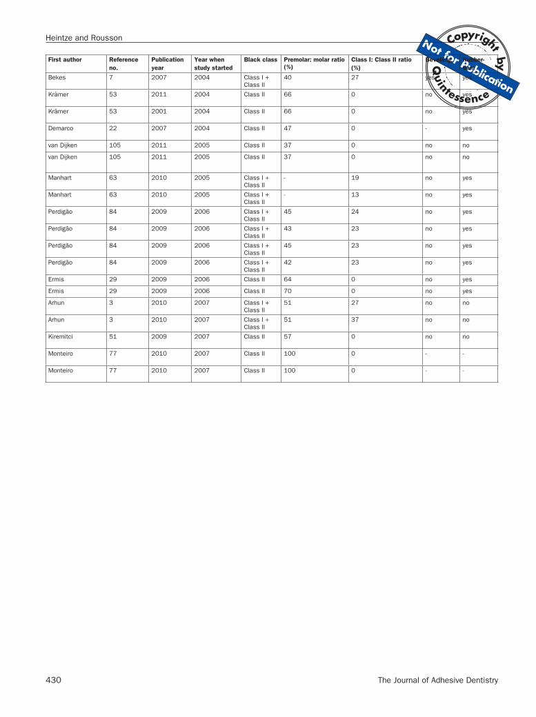
430 The Journal of Adhesive Dentistry
Heintze and Rousson
First author Reference
no.
Publication
year
Year when
study started
Black class Premolar: molar ratio
(%)
Class I: Class II ratio
(%)
Bevelling Rubber-
dam
Bekes 7 2007 2004 Class I + Class II
40 27 yes yes
Krämer 53 2011 2004 Class II 66 0 no yes
Krämer 53 2001 2004 Class II 66 0 no yes
Demarco 22 2007 2004 Class II 47 0 - yes
van Dijken 105 2011 2005 Class II 37 0 no no
van Dijken 105 2011 2005 Class II 37 0 no no
Manhart 63 2010 2005 Class I + Class II
- 19 no yes
Manhart 63 2010 2005 Class I + Class II
- 13 no yes
Perdigão 84 2009 2006 Class I + Class II
45 24 no yes
Perdigão 84 2009 2006 Class I + Class II
43 23 no yes
Perdigão 84 2009 2006 Class I + Class II
45 23 no yes
Perdigão 84 2009 2006 Class I + Class II
42 23 no yes
Ermis 29 2009 2006 Class II 64 0 no yes
Ermis 29 2009 2006 Class II 70 0 no yes
Arhun 3 2010 2007 Class I + Class II
51 27 no no
Arhun 3 2010 2007 Class I + Class II
51 37 no no
Kiremitci 51 2009 2007 Class II 57 0 no no
Monteiro 77 2010 2007 Class II 100 0 - -
Monteiro 77 2010 2007 Class II 100 0 - -
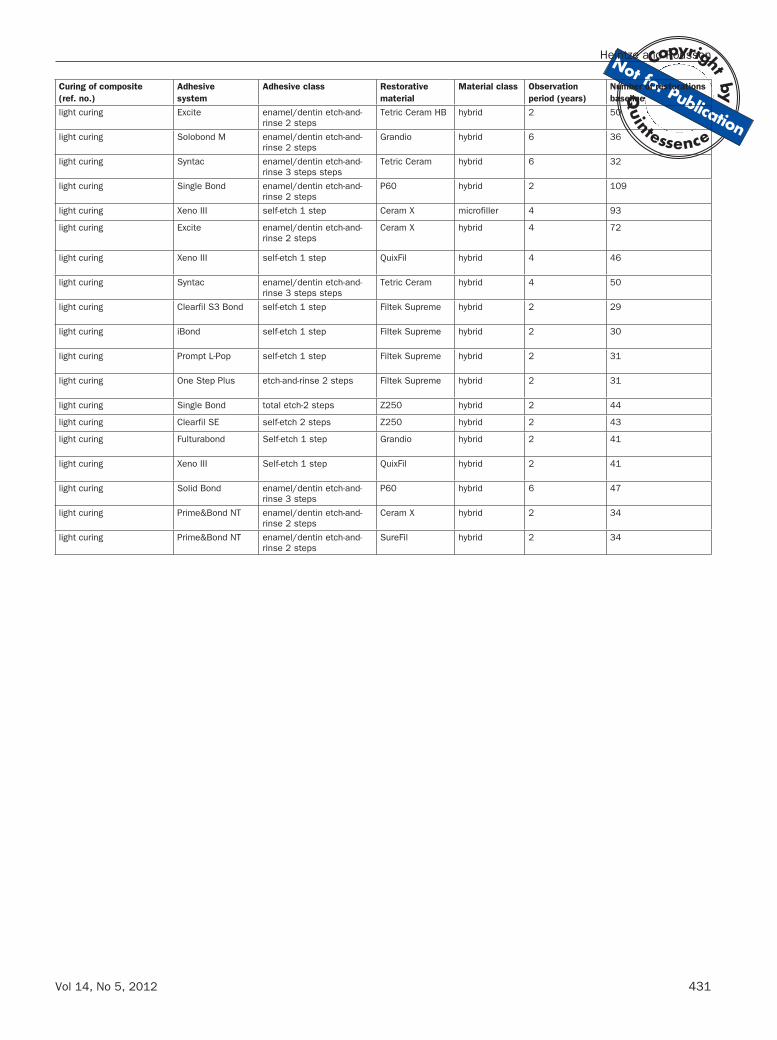
Vol 14, No 5, 2012 431
Heintze and Rousson
Curing of composite
(ref. no.)
Adhesive
system
Adhesive class Restorative
material
Material class Observation
period (years)
Number of restorations
baseline
light curing Excite enamel/dentin etch-and-rinse 2 steps
Tetric Ceram HB hybrid 2 50
light curing Solobond M enamel/dentin etch-and-rinse 2 steps
Grandio hybrid 6 36
light curing Syntac enamel/dentin etch-and-rinse 3 steps steps
Tetric Ceram hybrid 6 32
light curing Single Bond enamel/dentin etch-and-rinse 2 steps
P60 hybrid 2 109
light curing Xeno III self-etch 1 step Ceram X microfiller 4 93
light curing Excite enamel/dentin etch-and-rinse 2 steps
Ceram X hybrid 4 72
light curing Xeno III self-etch 1 step QuixFil hybrid 4 46
light curing Syntac enamel/dentin etch-and-rinse 3 steps steps
Tetric Ceram hybrid 4 50
light curing Clearfil S3 Bond self-etch 1 step Filtek Supreme hybrid 2 29
light curing iBond self-etch 1 step Filtek Supreme hybrid 2 30
light curing Prompt L-Pop self-etch 1 step Filtek Supreme hybrid 2 31
light curing One Step Plus etch-and-rinse 2 steps Filtek Supreme hybrid 2 31
light curing Single Bond total etch-2 steps Z250 hybrid 2 44
light curing Clearfil SE self-etch 2 steps Z250 hybrid 2 43
light curing Fulturabond Self-etch 1 step Grandio hybrid 2 41
light curing Xeno III Self-etch 1 step QuixFil hybrid 2 41
light curing Solid Bond enamel/dentin etch-and-rinse 3 steps
P60 hybrid 6 47
light curing Prime&Bond NT enamel/dentin etch-and-rinse 2 steps
Ceram X hybrid 2 34
light curing Prime&Bond NT enamel/dentin etch-and-rinse 2 steps
SureFil hybrid 2 34


