Clinical detection of transverse skeletal discrepancies ...
20
Clinical detection of transverse skeletal discrepancies derived from mandibular first molar buccal-lingual inclinations measured from CBCTs. Dr. Warren D. Woods Dr. Leslie Will Boston University Goldman School of Dental Medicine Science Day 2021
Transcript of Clinical detection of transverse skeletal discrepancies ...
Clinical detection of transverseskeletal discrepancies derived from
mandibular first molar buccal-lingual inclinations measured from CBCTs.
Dr. Warren D. WoodsDr. Leslie Will
Boston University Goldman School of Dental MedicineScience Day 2021
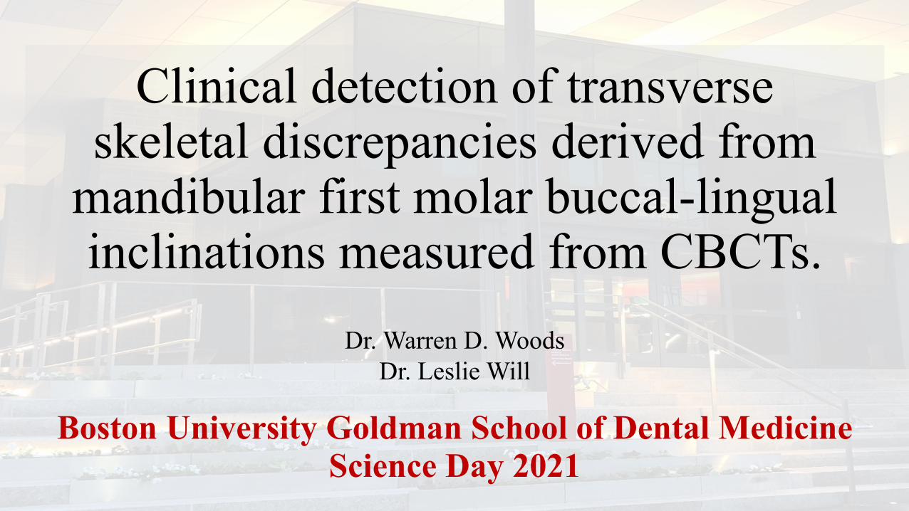
BackgroundPost treatment, American Board of Orthodontics defines an optimum curve of Wilson at a deviation of no more than a 1mm discrepancy between the buccal and lingual cusps of the posterior teeth.
The optimum curve of Wilson is thought to exist for two purposes (Tong et al., 2012) : Optimal masticatory muscle loading Ease of food manipulation during function.
Deviation from the optimum curve of Wilson might be an indicator of adental compensation for a transverse skeletal discrepancy
between the maxillary and mandibular arches.
PurposeThe first part of this study will determine if there is a statistically significant correlation between a deviation from the ideal curve of Wilson and a transverse skeletal discrepancy using a CBCT.
“One of the advantages of using CBCT is the ability to visualize the whole tooth, thus removing some of the uncertainty in long-axis inclination that can result from using casts
with uneven cusp wear or tooth morphology.”(Alkhatib & Chung, 2017)
If a statistically significant positive correlation can be determined between first molar angulation (curve of Wilson) and a presence or absence of a skeletal transverse discrepancy, then these findings can be used to assess the use of molar angulation in a clinical setting to quickly visually assess a patient for skeletal transverse discrepancy without/prior to taking full diagnostic records (CBCT).
Methods & MaterialsCBCT images will be acquired from the CBCT repository of the Department of Orthodontics and Dentofacial Orthopedics Repository (H-32515) of Boston University Henry M. Goldman School of Dental Medicine.
Males and females ages 6-10 with mandibular first molars erupted to at least the same occlusal plane level as the adjacent primary second molar.
No patients with gross anomalies, crossbites, gross restorations, gross cuspal wear of the mandibular first molars, or prior orthodontic treatment will be included in the study.
Evaluation and analysis of CBCT images will be performed using Dolphin. Specific measurements will be recorded, including the buccal lingual inclination of the mandibular first molars (Curve of Wilson) and the skeletal transverse width.
Methods & Materials
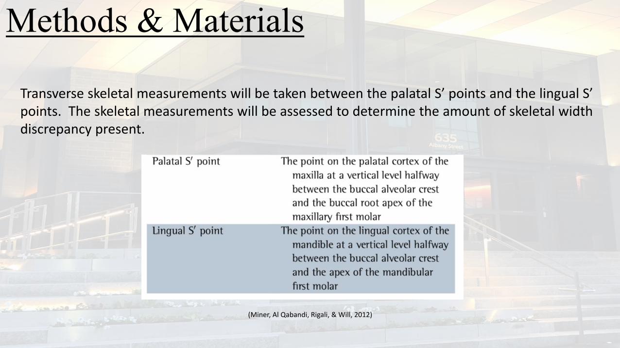
Transverse skeletal measurements will be taken between the palatal S’ points and the lingual S’ points. The skeletal measurements will be assessed to determine the amount of skeletal width discrepancy present.
(Miner, Al Qabandi, Rigali, & Will, 2012)
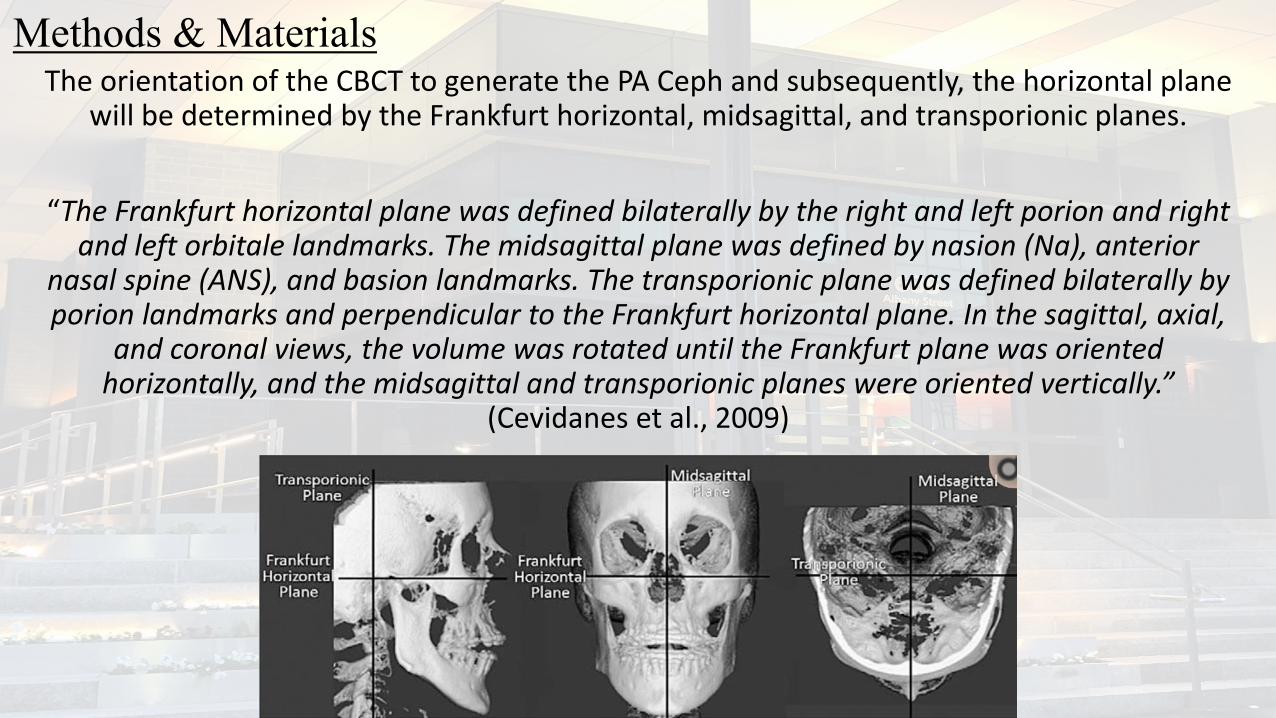
Methods & MaterialsThe orientation of the CBCT to generate the PA Ceph and subsequently, the horizontal plane
will be determined by the Frankfurt horizontal, midsagittal, and transporionic planes.
“The Frankfurt horizontal plane was defined bilaterally by the right and left porion and right and left orbitale landmarks. The midsagittal plane was defined by nasion (Na), anterior
nasal spine (ANS), and basion landmarks. The transporionic plane was defined bilaterally by porion landmarks and perpendicular to the Frankfurt horizontal plane. In the sagittal, axial,
and coronal views, the volume was rotated until the Frankfurt plane was oriented horizontally, and the midsagittal and transporionic planes were oriented vertically.”
(Cevidanes et al., 2009)
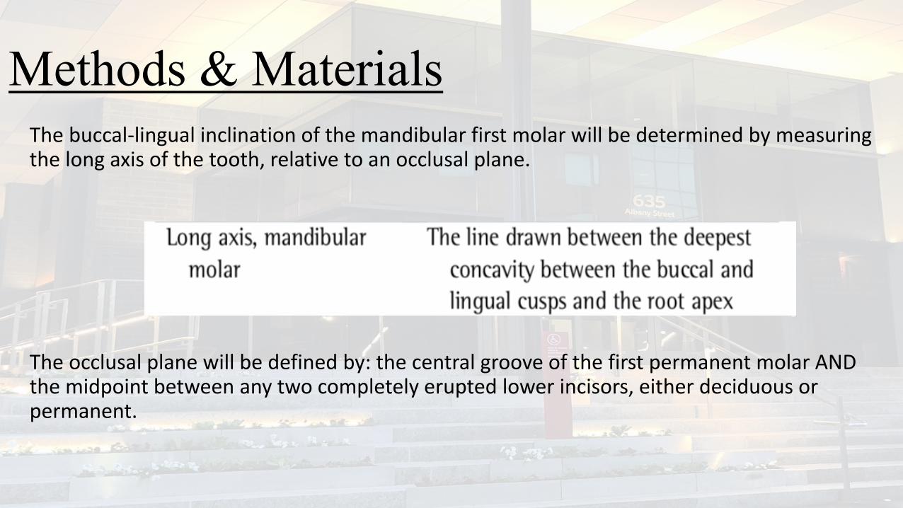
Methods & MaterialsThe buccal-lingual inclination of the mandibular first molar will be determined by measuring the long axis of the tooth, relative to an occlusal plane.
The occlusal plane will be defined by: the central groove of the first permanent molar AND the midpoint between any two completely erupted lower incisors, either deciduous or permanent.
Methods & Materials
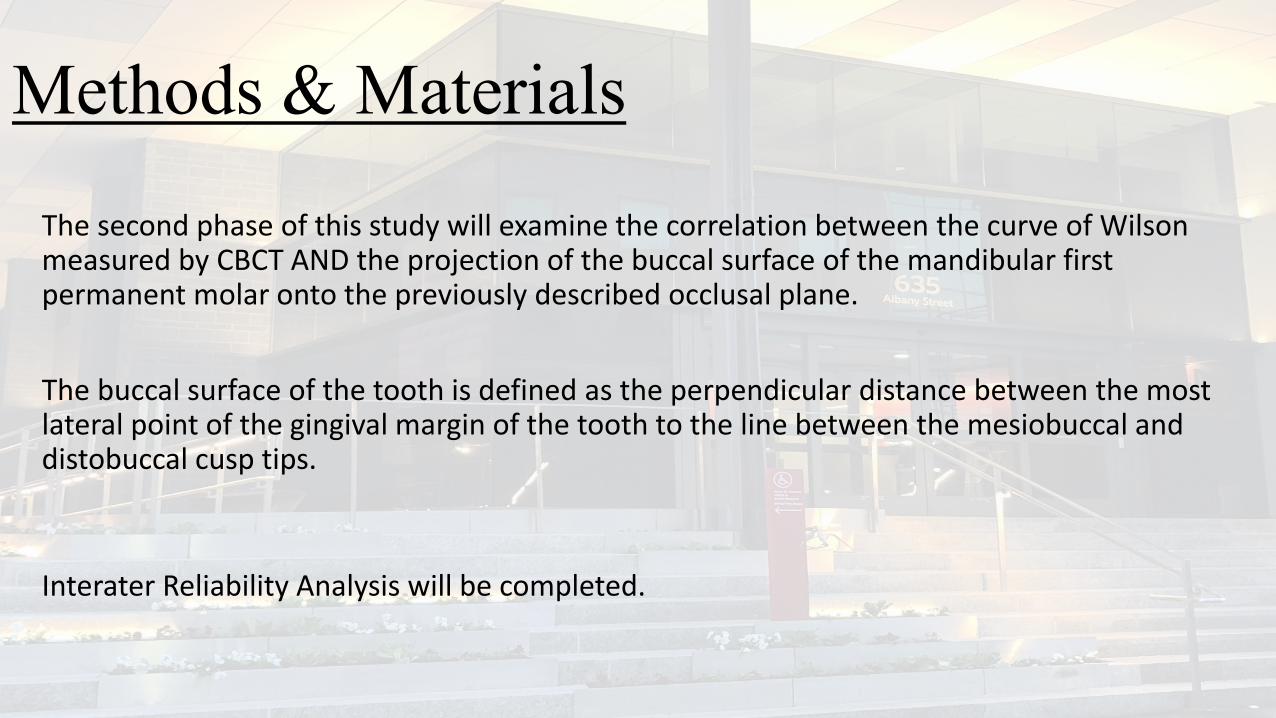
The second phase of this study will examine the correlation between the curve of Wilson measured by CBCT AND the projection of the buccal surface of the mandibular first permanent molar onto the previously described occlusal plane.
The buccal surface of the tooth is defined as the perpendicular distance between the most lateral point of the gingival margin of the tooth to the line between the mesiobuccal and distobuccal cusp tips.
Interater Reliability Analysis will be completed.
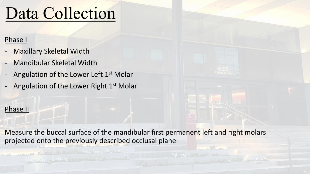
Data CollectionPhase I- Maxillary Skeletal Width- Mandibular Skeletal Width- Angulation of the Lower Left 1st Molar- Angulation of the Lower Right 1st Molar
Phase II
Measure the buccal surface of the mandibular first permanent left and right molars projected onto the previously described occlusal plane
Statistical Analysis
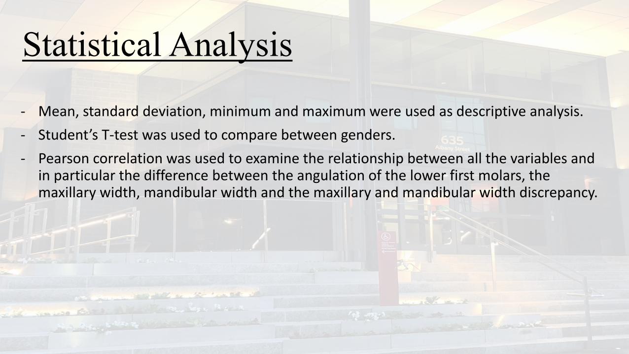
- Mean, standard deviation, minimum and maximum were used as descriptive analysis.- Student’s T-test was used to compare between genders.- Pearson correlation was used to examine the relationship between all the variables and
in particular the difference between the angulation of the lower first molars, the maxillary width, mandibular width and the maxillary and mandibular width discrepancy.

Results – Phase I
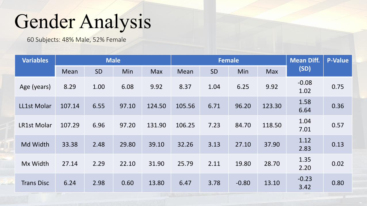
Gender Analysis
Variables Male Female Mean Diff.(SD)
P-Value
Mean SD Min Max Mean SD Min Max
Age (years) 8.29 1.00 6.08 9.92 8.37 1.04 6.25 9.92 -0.081.02 0.75
LL1st Molar 107.14 6.55 97.10 124.50 105.56 6.71 96.20 123.30 1.586.64 0.36
LR1st Molar 107.29 6.96 97.20 131.90 106.25 7.23 84.70 118.50 1.047.01 0.57
Md Width 33.38 2.48 29.80 39.10 32.26 3.13 27.10 37.90 1.122.83 0.13
Mx Width 27.14 2.29 22.10 31.90 25.79 2.11 19.80 28.70 1.352.20 0.02
Trans Disc 6.24 2.98 0.60 13.80 6.47 3.78 -0.80 13.10 -0.233.42 0.80
60 Subjects: 48% Male, 52% Female
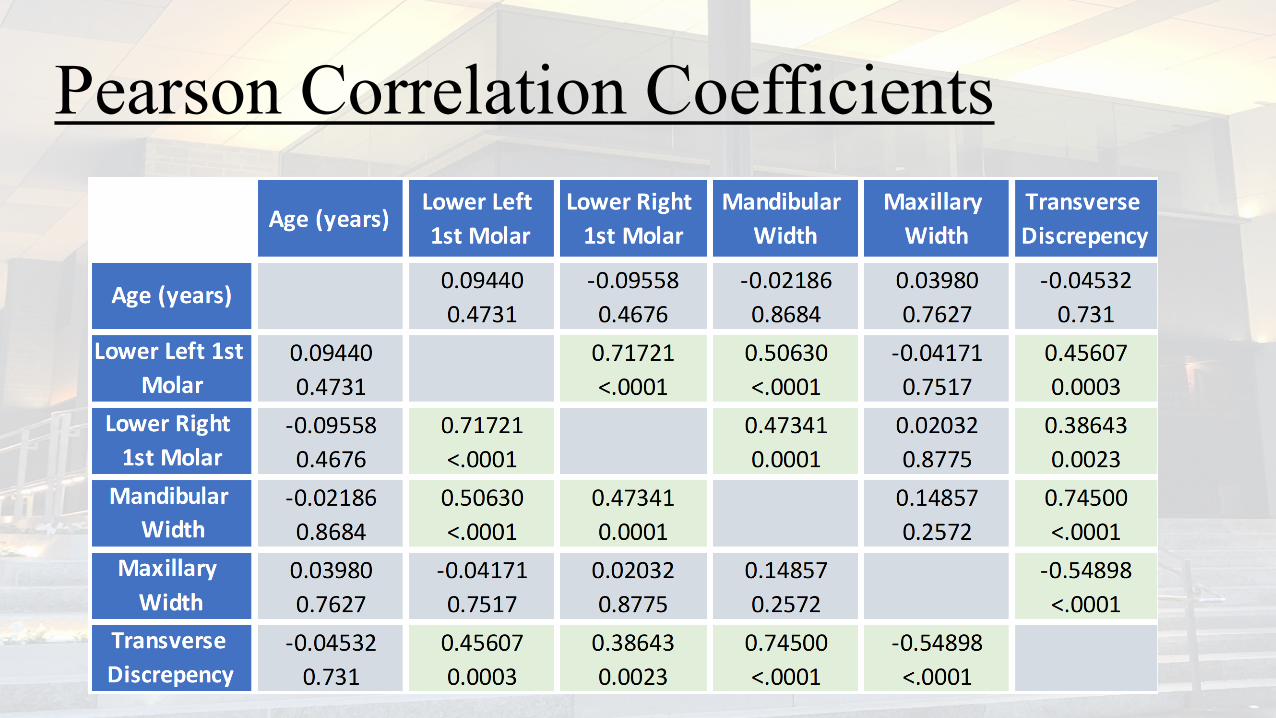
Pearson Correlation Coefficients
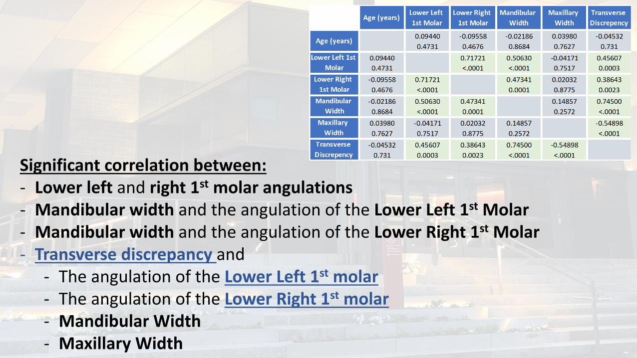
Significant correlation between:- Lower left and right 1st molar angulations- Mandibular width and the angulation of the Lower Left 1st Molar- Mandibular width and the angulation of the Lower Right 1st Molar- Transverse discrepancy and
- The angulation of the Lower Left 1st molar- The angulation of the Lower Right 1st molar- Mandibular Width- Maxillary Width
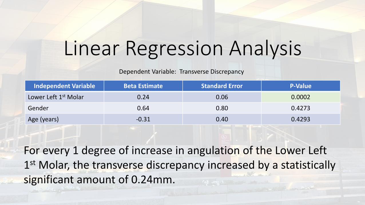
Linear Regression AnalysisIndependent Variable Beta Estimate Standard Error P-Value
Lower Left 1st Molar 0.24 0.06 0.0002
Gender 0.64 0.80 0.4273
Age (years) -0.31 0.40 0.4293
Dependent Variable: Transverse Discrepancy
For every 1 degree of increase in angulation of the Lower Left 1st Molar, the transverse discrepancy increased by a statistically significant amount of 0.24mm.
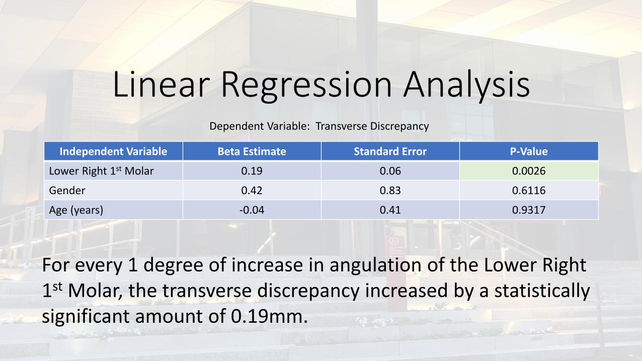
Linear Regression AnalysisIndependent Variable Beta Estimate Standard Error P-Value
Lower Right 1st Molar 0.19 0.06 0.0026
Gender 0.42 0.83 0.6116
Age (years) -0.04 0.41 0.9317
Dependent Variable: Transverse Discrepancy
For every 1 degree of increase in angulation of the Lower Right 1st Molar, the transverse discrepancy increased by a statistically significant amount of 0.19mm.

Results – Phase IICurrently PendingStatistical Analysis
Conclusions:It is possible to determine the degree of skeletal discrepancy between the maxilla and mandible from the measurement of the angulation of the mandibular first molars measured from the long axis of the molar.
Phase II [Data currently in analysis]: Is it possible to determine the degree of skeletal discrepancy between the maxilla and mandible from the measurement of the buccal surface of the lower first molar?

Thank you!Contact: [email protected]
Thank you to Dr. Leslie Will and Dr. Melih Motro for the support that they have given me on this study.
Thank you to Dr. Ahmed Alsulaiman for his statistical analysis.

ReferencesAlkhatib, R., & Chung, C. H. (2017). Buccolingual inclination of first molars in untreated adults: A CBCT study. Angle Orthodontist, 87(4), 598–602. https://doi.org/10.2319/110116-786.1Cevidanes, L., Oliveira, A. E. F., Motta, A., Phillips, C., Burke, B., & Tyndall, D. (2009). Head orientation in CBCT-generated cephalograms. The Angle Orthodontist, 79(5), 971–977. https://doi.org/10.2319/090208-460.1McNamara, J. A. (2000). Maxillary transverse deficiency. American Journal of Orthodontics and Dentofacial Orthopedics : Official Publication of the American Association of Orthodontists, Its Constituent Societies, and the American Board of Orthodontics, 117(5), 567–570. https://doi.org/10.1016/S0889-5406(00)70202-2Miner, R. M., Al Qabandi, S., Rigali, P. H., & Will, L. A. (2012). Cone-beam computed tomography transverse analysis. Part I: Normative data. American Journal of Orthodontics and Dentofacial Orthopedics, 142(3), 300–307. https://doi.org/10.1016/j.ajodo.2012.04.014Tong, H., Kwon, D., Shi, J., Sakai, N., Enciso, R., & Sameshima, G. T. (2012). Mesiodistal angulation and faciolingual inclination of each whole tooth in 3-dimensional space in patients with near-normal occlusion. American Journal of Orthodontics and Dentofacial Orthopedics, 141(5), 604–617. https://doi.org/10.1016/j.ajodo.2011.12.018
[Additional Articles reviewed but not cited directly can be found cited in the Mendeley file “WoodsBib.bib”]