CLINICAL CYTOLOGY 09888888;r - University of … class2014-2015...Diagnostic or clinical cytology...
45
09888888;r CLINICAL CYTOLOGY Dr. Issraa Ali Hussein
Transcript of CLINICAL CYTOLOGY 09888888;r - University of … class2014-2015...Diagnostic or clinical cytology...
09888888;rCLINICAL CYTOLOGY
Dr. Issraa Ali Hussein
objectives
Define diagnostic cytology
(clinical cytology).
Explain the differences between
histopathology and cytopathology.
Recognize the methods for collection of the
materials for cytology.
objectives
Explain FNA.
Discuss the advantages of cytologic
examination
Identify the criteria for the dignosis of
cellular malignancy.
WHAT IS DIAGNOSTIC
CYTOLOGY ?
Diagnostic or clinical cytology
is the study of the normal and diseased altered cells obtained from various sites of the body
i.e., through the detection of abnormal morphologic characteristics of the examined dissociated human cells.
CYTOLOGY
Is the science of cell structure. Compared to histology, the diagnostic criteria are few and depend solely on nuclear and cytoplasmic features of cells.
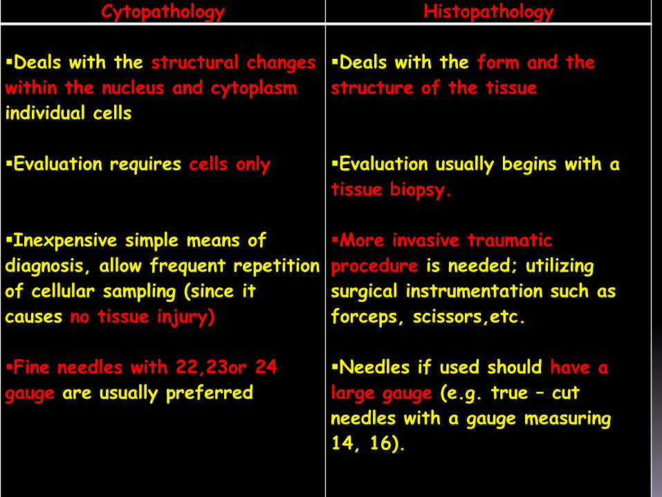
HistopathologyCytopathology
Deals with the form and the
structure of the tissue
Evaluation usually begins with a
tissue biopsy.
More invasive traumatic
procedure is needed; utilizing
surgical instrumentation such as
forceps, scissors,etc.
Needles if used should have a
large gauge (e.g. true – cut
needles with a gauge measuring
14, 16).
Deals with the structural changes
within the nucleus and cytoplasm
individual cells
Evaluation requires cells only
Inexpensive simple means of
diagnosis, allow frequent repetition
of cellular sampling (since it
causes no tissue injury)
Fine needles with 22,23or 24
gauge are usually preferred
HistopathologyCytopathology
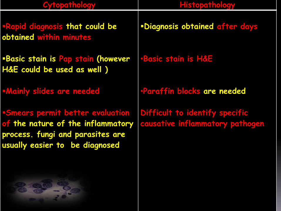
Diagnosis obtained after days
•Basic stain is H&E
•Paraffin blocks are needed
Difficult to identify specific
causative inflammatory pathogen
Rapid diagnosis that could be
obtained within minutes
Basic stain is Pap stain (however
H&E could be used as well )
Mainly slides are needed
Smears permit better evaluation
of the nature of the inflammatory
process. fungi and parasites are
usually easier to be diagnosed
Indications (the Advantages )for CytopathologyDetection of inflammation and certain types
of pathogenic agents
Differentiation between benign and malignant Diagnosis of premalignant diseases lesions
Diagnosis of the type of Malignancy(primary,metastatic or recurrent tumors)
Study of hormonal patterns
Monitoring of response to therapy and Follow-up of irradiation & chemotherapy
Study of tumour markers
In general diagnostic cytology is based upon
three basic sampling techniques:
1- The collection of exfoliated cells.
2- The collection of cells removed by brushingor similar abrasive techniques.
3- The aspiration biopsy. F.N.A. biopsy.
EXFOLIATIVE CYTOLOGY:
From normal (physiological)desquamation products:Is based on a spontaneous shedding of cells derived from a lining of an organ into a cavity, where they can be removed by non abrasive means.Examples:
Vaginal smears: cells removed from the posterior fornix of the vagina (squamous epithelium, endocervical cells, endometrialcells).
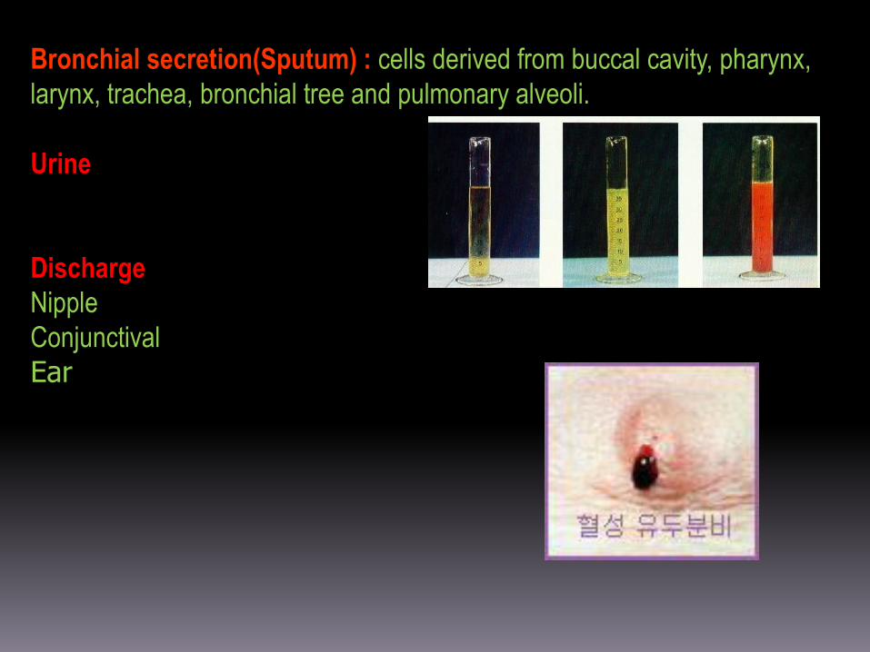
Bronchial secretion(Sputum) : cells derived from buccal cavity, pharynx,
larynx, trachea, bronchial tree and pulmonary alveoli.
Urine
Discharge
Nipple
Conjunctival
Ear
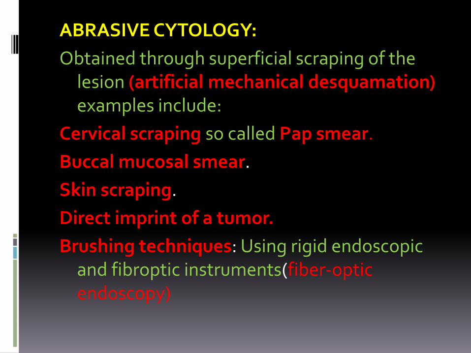
ABRASIVE CYTOLOGY:
Obtained through superficial scraping of the lesion (artificial mechanical desquamation) examples include:
Cervical scraping so called Pap smear.
Buccal mucosal smear.
Skin scraping.
Direct imprint of a tumor.
Brushing techniques: Using rigid endoscopic and fibroptic instruments(fiber-optic endoscopy)

Cervico-vaginal PAP smears
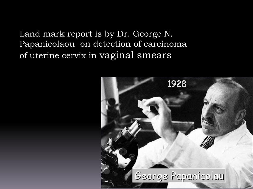
Land mark report is by Dr. George N.
Papanicolaou on detection of carcinoma
of uterine cervix in vaginal smears
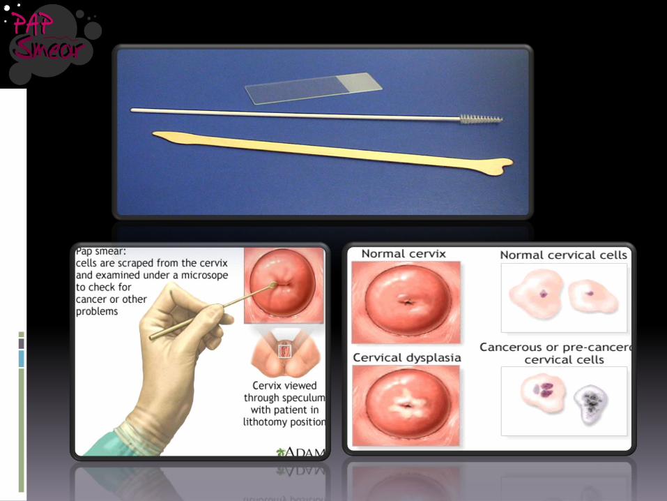
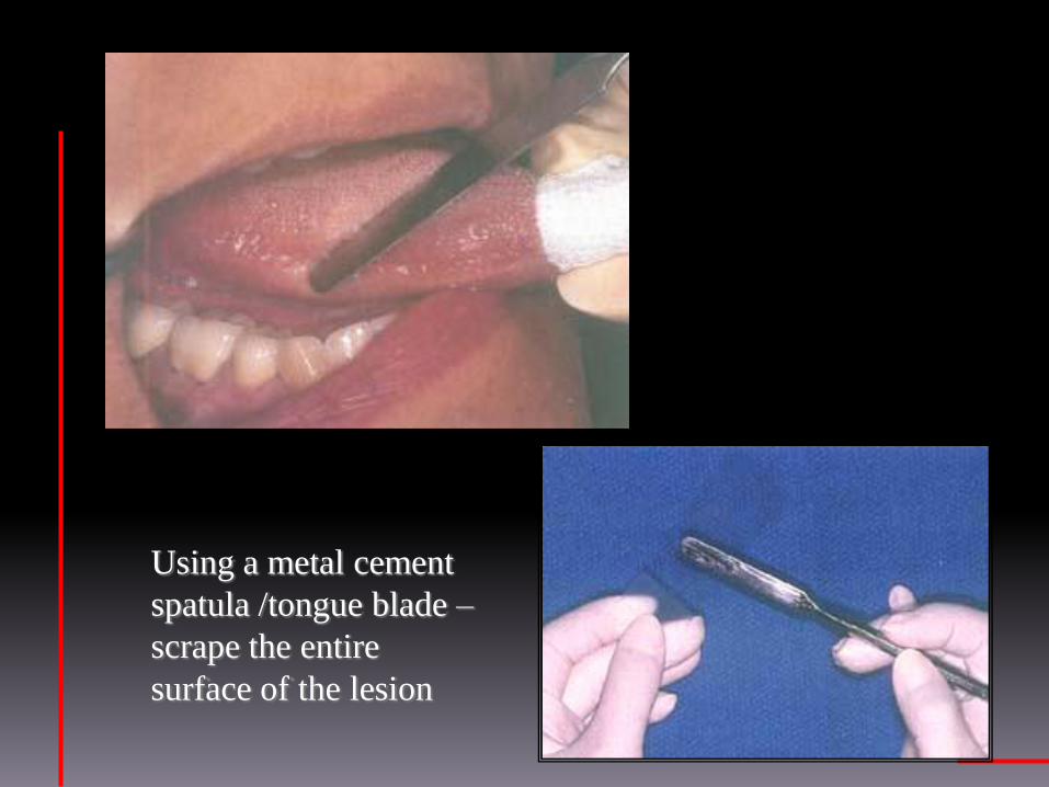
Using a metal cement
spatula /tongue blade –
scrape the entire
surface of the lesion
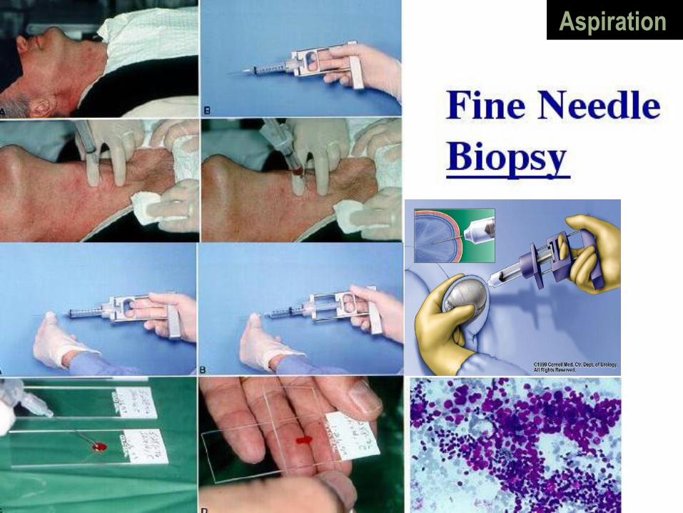
Aspiration
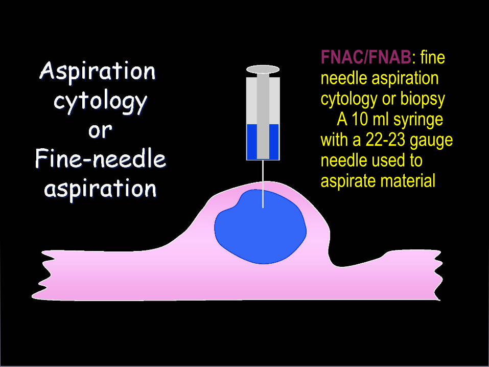
FNAC/FNAB: fine needle aspiration cytology or biopsy
A 10 ml syringe with a 22-23 gauge needle used to aspirate material
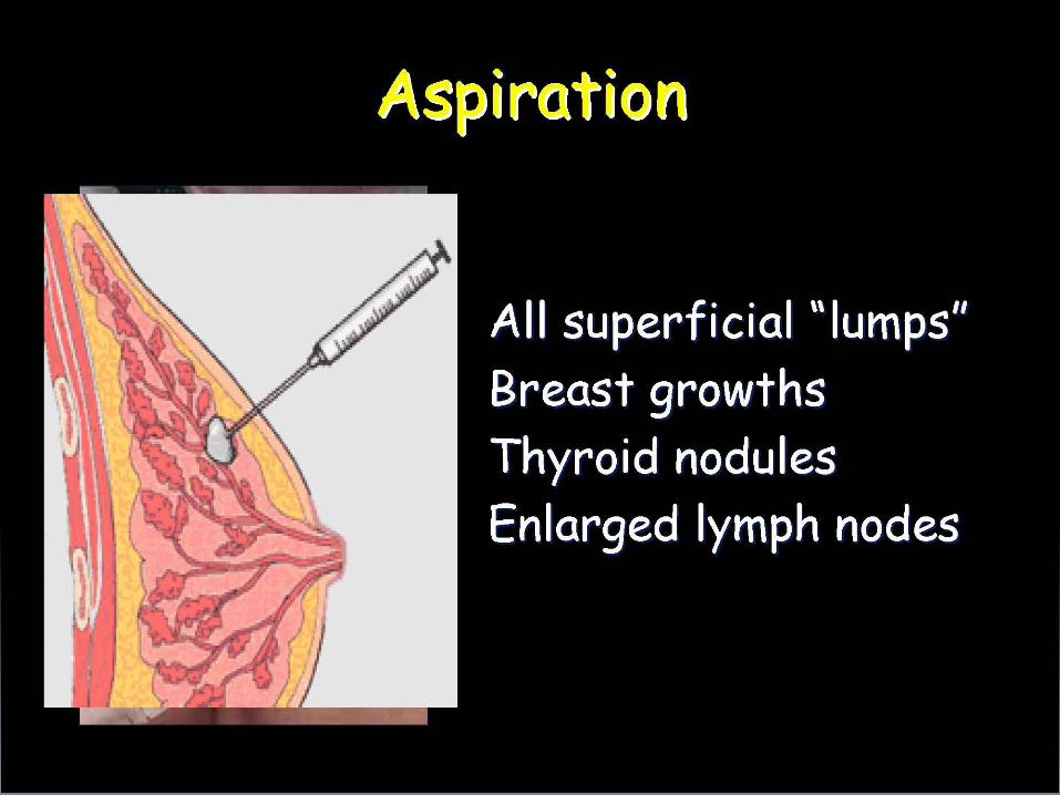

Deep organs (Ultrasound guided) : Liver –kidney –pancreas -retroperitoneal - prostate etc.......
Fine Needle aspiration Cytology
In general, the definitive diagnosis of any mass can be established by:
Open biopsy,Tissue core needle (Tru-cut) biopsy, Fine needle aspiration biopsy.
Compared to FNA, Tru-cut biopsy is a more traumatic procedure which should be performed under local anaesthesia. It requires more time and special equipment that are more expensive. Pain, discomfort and bleeding are common complications.
Fine Needle aspiration Cytology
FNAC, on the other hand, provides many advantages to the surgeons:
It is an easy, reliable, cost effective diagnostic technique which can give rapid results.
The procedure could be performed in an office setting without anaesthesia. It is usually notmore painful than a venipuncture and can be repeated immediately if the acquired material is inadequate.
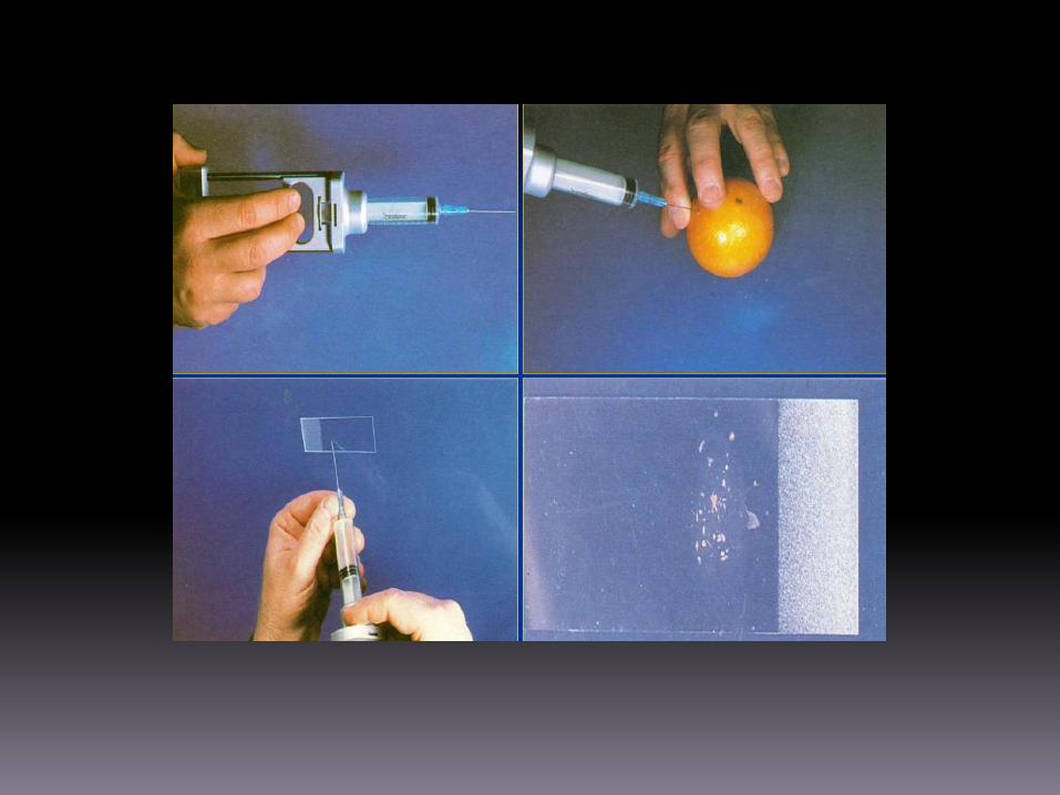
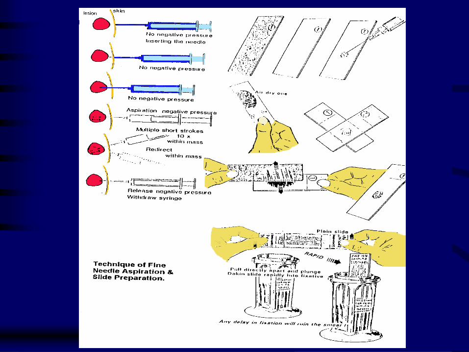
Equipments and Procedure of FNAC:
When reduced to its simplest terms, FNA consists of:
- Using a needle and syringe to remove
material from a mass.
- Smearing it on a glass slide.
- Applying a routine stain.
- Examining it under the microscope.

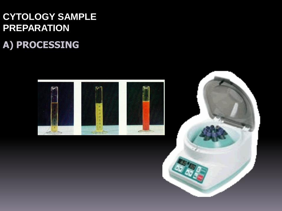
CYTOLOGY SAMPLE
PREPARATION
A) PROCESSING
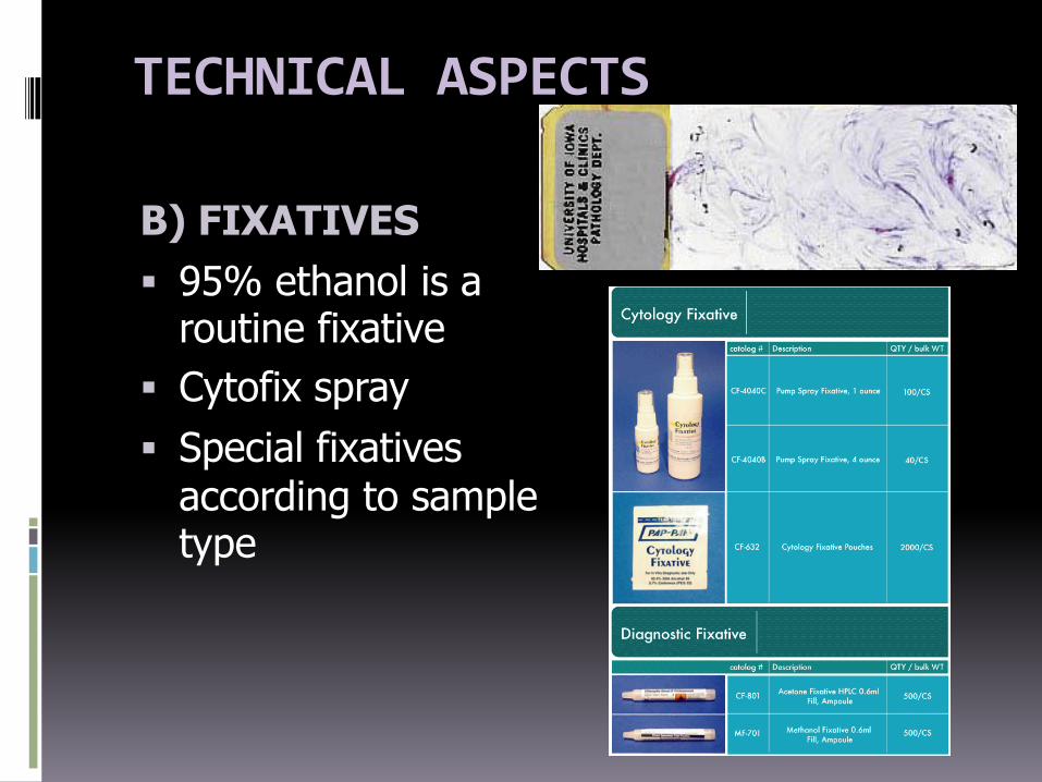
TECHNICAL ASPECTS
B) FIXATIVES
95% ethanol is a routine fixative
Cytofix spray
Special fixatives according to sample type
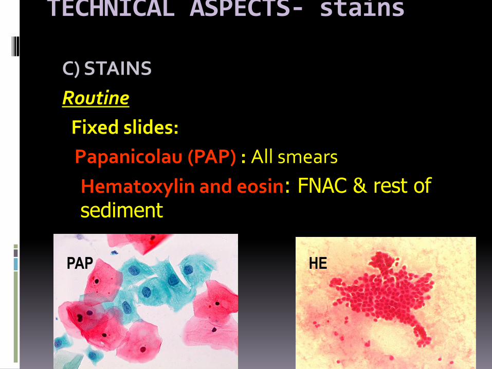
TECHNICAL ASPECTS- stains
C) STAINS
Routine
Fixed slides:
Papanicolau (PAP) : All smears
Hematoxylin and eosin: FNAC & rest of sediment
PAP HE
TECHNICAL ASPECTS- stains
C) STAINS
PAP Stain
- 3 colours (pink-blue-orange)+different shades
- colour & shade determines degree of keratinization
- nuclear details clearer than HE

PAP smear showing normal
Intermediate & superficial Sq.cells
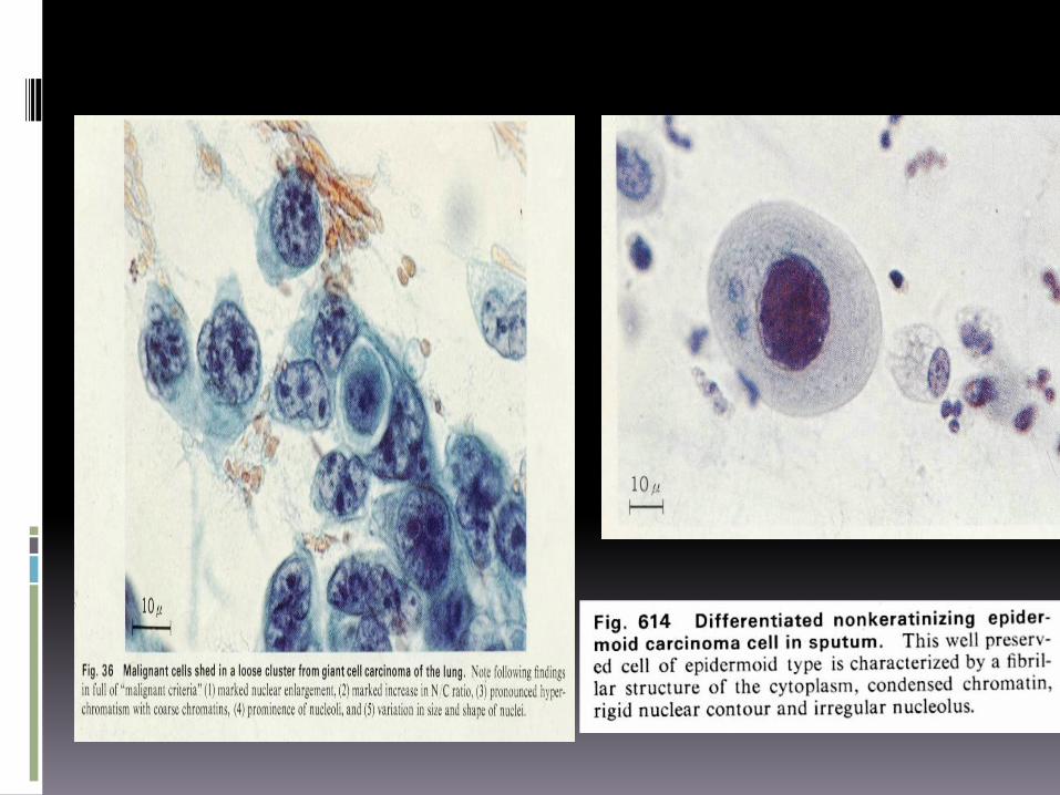
Pleomorphism ;
Irregular N & C outlineCancerous Cell

TECHNICAL ASPECTS-stains
Unfixed slides
Giemsa
Toluidene blue or Diff Quick
TECHNICAL ASPECTS-stains
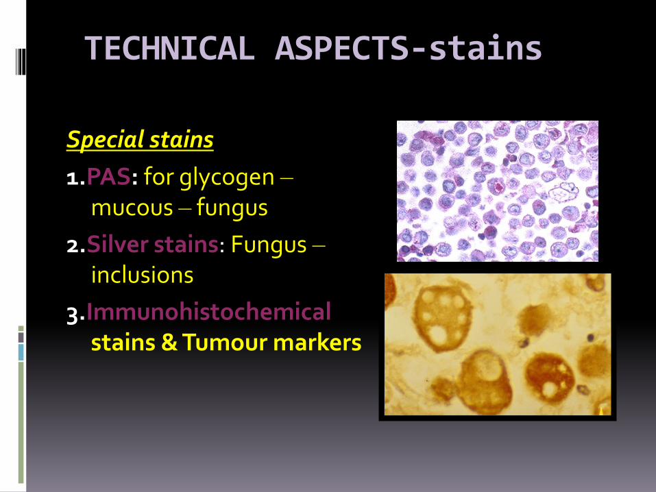
Special stains
1.PAS: for glycogen –mucous – fungus
2.Silver stains: Fungus –inclusions
3.Immunohistochemicalstains & Tumour markers

EXAMINATION OF SAMPLE & DIAGNOSIS
CYTOLOGIST
1) The cytologist examines the gross appearance of the sample and describes it: color, volume and whether sample is clear or turbid.
2) The cytologist then performs the microscopic examination & diagnosis:
EXAMINATION OF SAMPLE & DIAGNOSIS
Low power is important for:
· Determination of adequacy ( are there enough cells & are the cells representative of the tissue being examined?)
· Pattern & background
· Cell types

EXAMINATION OF SAMPLE & DIAGNOSISHigh power + Oil immersion are important for: The determination of the benign or malignant
nature of cells examined depending on cytoplasmic features & nuclear details:
1-N/C ratio2-Cell & nuclear shape and size3- Cell & nuclear membranes4- Nucleoli-mitotic figures-chromatin pattern5-Others: Inclusion bodies
Bacteria/fungi/parasitesArtifacts
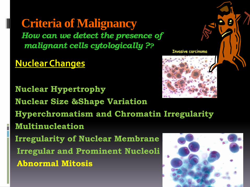
Criteria of MalignancyHow can we detect the presence of
malignant cells cytologically ??
Nuclear Changes
Nuclear Hypertrophy
Nuclear Size &Shape Variation
Hyperchromatism and Chromatin Irregularity
Multinucleation
Irregularity of Nuclear Membrane
Irregular and Prominent Nucleoli
Abnormal Mitosis
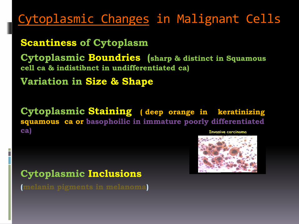
in Malignant CellsCytoplasmic Changes
Scantiness of Cytoplasm
Cytoplasmic Boundries (sharp & distinct in Squamous
cell ca & indistibnct in undifferentiated ca)
Variation in Size & Shape
Cytoplasmic Staining ( deep orange in keratinizing
squamous ca or basophoilic in immature poorly differentiated
ca)
Cytoplasmic Inclusions
(melanin pigments in melanoma)
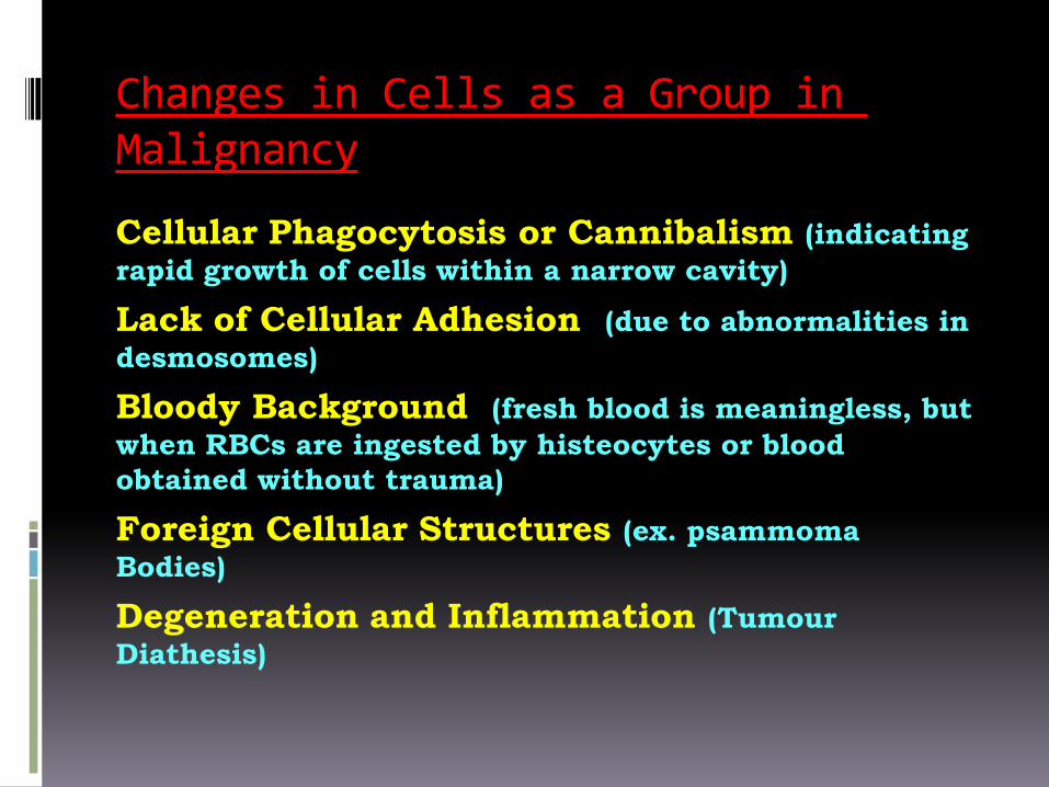
Changes in Cells as a Group in Malignancy
Cellular Phagocytosis or Cannibalism (indicating
rapid growth of cells within a narrow cavity)
Lack of Cellular Adhesion (due to abnormalities in
desmosomes)
Bloody Background (fresh blood is meaningless, but
when RBCs are ingested by histeocytes or blood
obtained without trauma)
Foreign Cellular Structures (ex. psammoma
Bodies)
Degeneration and Inflammation (Tumour
Diathesis)
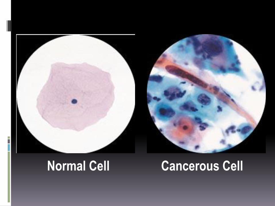
Normal Cell Cancerous Cell

BENIGN MALIGNANT

Ca bladder
High nuclear-cytoplasmic ratio and pleomorphism
CYTOLOGYLOVE IT OR LEAVE IT
Thank you