Clinical Characterisation of Cardiac Involvement in ...discovery.ucl.ac.uk/1528776/7/Shah_Clinical...
159
1 Clinical Characterisation of Cardiac Involvement in Anderson-Fabry Disease. Jaymin Shantilal Shah MBBS, MRCP A thesis submitted to University College London for the degree of Doctor of Medicine (Research) Cardiomyopathy (Inherited Cardiovascular Disease Unit) 16-18 Westmoreland Street London W1G 8PH
Transcript of Clinical Characterisation of Cardiac Involvement in ...discovery.ucl.ac.uk/1528776/7/Shah_Clinical...

1
Clinical Characterisation of Cardiac
Involvement in Anderson-Fabry Disease.
Jaymin Shantilal Shah
MBBS, MRCP
A thesis submitted to University College London for the
degree of Doctor of Medicine (Research)
Cardiomyopathy (Inherited Cardiovascular Disease Unit)
16-18 Westmoreland Street
London W1G 8PH
2
Declaration
I, Jaymin Shantilal Shah confirm that the work presented in this thesis is my own.
The data presented represents my own clinical assessment of the patients, data
collection, management and analysis. The thesis presents my own original ideas
and hypothesis. This work also reflects my efforts in coordinating the
collaboration with multiple different centers to present a systematic clinical
evaluation of Anderson – Fabry disease related cardiomyopathy. Where
information has been derived from other sources, I confirm that this has been
indicated in the thesis.
Dr Jaymin S Shah
3
Abstract
Background
Anderson – Fabry Disease (AFD) is an X-linked lysosomal storage disorder that results
in a deficiency in lysosomal -galactosidase A. This leads to an accumulation of
glycosphingolipids in multiple cell types resulting in the characteristic angiokeratomata,
acroparesthesiae, hypohidrosis, and corneal opacities of classical AFD. Although
patients with AFD have a reduced life span, the pathogenesis and impact of cardiac
involvement on prognosis remain unclear. The aim of this thesis was to clinically
characterise cardiac involvement in AFD using a systematic cardiac assessment of a
consecutive cohort of patients with AFD.
Methods
A cardiac assessment protocol including history and examination, 12 lead ECG, 24 hour
ambulatory ECG monitoring, echocardiography and cardiopulmonary exercise testing
were performed in a cohort of 122 consecutive patients. Subgroups had coronary flow
reserve and evaluation of serum levels of MMP-9, TIMP-1 and TIMP-2 to assess
coronary microvascular function and extracellular matrix (ECM) turnover, respectively.
Results
The main results of the thesis are as follows: (1) Tachy and brady arrhythmias are
common and may contribute to premature death; (2) Left ventricular systolic
impairment is common and decline in systolic function may represent a measure of
disease severity; (3) objective evidence for exercise limitation is common and is
4
related to overall disease severity as assessed with a validated disease severity score;
(4) patients with AFD have markedly abnormal coronary microvascular function which
does not improve with ERT; (5) patients with AFD have abnormal ECM turnover
associated with overall disease severity.
Conclusion
Patients with AFD related cardiomyopathy have progressive disease characterised by
heart failure and arrhythmia. It is likely that myocardial ischaemia and abnormal
interstitial turnover are related in the pathogenesis of cardiac disease and
complications. Patients with AFD need regular and continued cardiac follow up for
early identification and treatment of cardiac complications.
5
Preface
Anderson – Fabry Disease (AFD) is an X-linked lysosomal storage disorder that results
in a deficiency in the activity of lysosomal -galactosidase A, leading to an accumulation
of glycosphingolipids in multiple different cell types. This in turn causes the
characteristic angiokeratomata, acroparesthesiae, hypohidrosis, and corneal opacities
of classical AFD. AFD related heart disease is common, but its impact on prognosis
remains unclear.
When I started my work in AFD, the evidence for cardiac involvement in AFD was in the
form of case reports and case series. There had been few systematic studies to look at
the prevalence of left ventricular failure, arrhythmia and exercise limitation in patients
with AFD. Our collaboration with The Lysosmal Disorders Unit, The Charles Dent
Metabolic unit and our own tertiary referral population of patients with unexplained
left ventricular hypertrophy provided a unique opportunity to characterise cardiac
involvement in AFD. This thesis is a systematic and comprehensive clinical cardiac
evaluation of patients with AFD. The data presented are contemporary with my period
as a research fellow. Since completion of my work numerous studies on AFD related
heart disease have been published. I put my own work into the context of these data
in the discussion.

6
Acknowledgements
The work in this thesis was undertaken during my period as a clinical research fellow at
The Heart Hospital, University College London Hospital. The following have greatly
assisted in its completion:
Professor Perry M Elliott: He introduced me to the field of hypertrophic
cardiomyopathy. His support and supervision in the planning, development and writing
of this thesis and manuscripts related to the research has been invaluable. His
unstoppable drive, passion and vision have been an inspiration. He was not only a
supervisor and mentor, but also a friend. I am honoured to have had the opportunity
to have worked with him.
Professor William McKenna: for his supervision and constructive criticisms of the
project.
The Research Team at The Heart Hospital: for their continued support throughout my
time as a research fellow. They all have been invaluable sources of wisdom and have
provided a steadying influence through all the phases of this project.
The staff of The Lysosomal Disease Unit, The Royal Free Hospital and The Charles Dent
Metabolic Unit, The National Hospital for Neurology and Neurosurgery: For their
continued support throughout my time as a research fellow and beyond.

7
Colleagues at City Hospital, Birmingham: For their expertise in the extra cellular
matrix.
Colleagues at MRC Clinical Sciences Centre, Imperial College, London: For their
expertise in coronary flow reserve
TKT-5S (Now Shire Human Genetic Therapies) and Sheila Bone: For funding my
research and providing me with the opportunities that have resulted in this work.
My wife and family: For their support and forgiveness for ‘bringing work home’.
8
Table of contents
Abstract p3
Preface p5
Acknowledgement p6
Contents p8
List of figures p9
List of table’s p11
List of abbreviations p12
1: Introduction p14
2: Characterisation of cardiac arrhythmia p38
3: The natural history of left ventricular systolic performance p57
4: Exercise capacity p69
5: Coronary microvascular function, its relationship to p85
symptoms and response to enzyme replacement therapy
6: Extracellular matrix turnover in Anderson-Fabry disease p100
7: Summary of findings and current literature p115
8: References p129
9: Publications arising from dissertation p158

9
List of figures
Figure 1.1: Disease course in classical AFD p17
Figure 1.2: Johannes Fabry and William Anderson p18
Figure 1.3: Angiokeratomata p27
Figure 1.4: Cornea Verticillata p28
Figure 1.5: Anderson Fabry disease cardiomyopathy histology p30
Figure 1.6: Fibrosis in Anderson Fabry disease cardiomyopathy p31
Figure 2.1: Complete heart block p47
Figure 2.2a: Age adjusted prevalence of arrhythmia in Anderson-Fabry p50
Disease in Males
Figure 2.2b: Age adjusted prevalence of arrhythmia in Anderson-Fabry p51
Disease in Females
Figure 2.3: Non-Sustained Ventricular Tachycardia p53

10
Figure 3.1: The Change in Left Ventricular End Systolic Diameter p65
and Left Ventricular End Diastolic Diameter During Follow Up
Figure 3.2: The Change in Fractional Shortening During Follow Up p66
Figure 4.1: Correlation between percentage predicted VO2 max and MSSI p81
Score
Figure 5.1: Myocardial Blood Flow at Rest and During Adenosine-Induced p95
Hyperaemia in Patients with Anderson-Fabry Disease and Controls
Figure 5.2: Myocardial Blood Flow at Rest and During Adenosine-Induced p96
Hyperaemia Pre- and Post-Enzyme Replacement Therapy (FabrazymeTM)
Figure 6.1: Difference in Matrix Metalloproteinase–9 between Patients and p108
Controls
Figure 6.2: Correlation between Metalloproteinase–9 and Disease Severity p109
Figure 6.3: Correlation between Metalloproteinase–9 and Fractional p110
Shortening

11
List of tables
Table 1.1: Studies of prevalence of AFD p21
Table 1.2 Signs and symptoms of Fabry Disease p23
Table 2.1: Symptoms at Baseline Evaluation in Males and Females p46
Table 2.2: Baseline Evaluation Indices in Males and Females p49
Table 3.1: Anderson-Fabry Disease Symptoms and Baseline p63
Echocardiographic Findings
Table 4.1: Mainz Severity Score Index p73
Table 4.2: Anderson - Fabry Disease Symptoms and Signs Stratified by Gender p77
Table 4.3: ECG and Echocardiographic Data Stratified by Gender p79
Table 4.4: Exercise Data Stratified by Gender p80
Table 5.1: Patient characteristics p93
Table 6.1: Baseline Characteristics in Males and Females p106
12
List of abbreviations
AFD Anderson Fabry Disease
ECG Electrocardiogram
MMP Matrix metalloproteinase
TIMP Tissue inhibitors of metalloproteinase
ECM Extracellular matrix turnover
LSD Lysosomal storage disorders
Gb3 Globotriaosylceramide
A-Gal α – galactosidase A
FOS Fabry outcome survey
AF Atrial fibrillation
PAF Paroxysmal atrial fibrillation
NSVT Non-sustained ventricular tachycardia
NYHA New York Heart Association
ERT Enzyme replacement therapy
RE Romhilt-Estes score
LVed End diastolic left ventricular internal cavity diameter
LVes End systolic left ventricular internal cavity diameter
IVS Interventricular septum
PW Posterior wall
RWT Relative wall thickness
FS Fractional shortening
LV Left ventricular
LVH Left ventricular hypertrophy
13
HCM Hypertrophic cardiomyopathy
CPET Cardiopulmonary exercise testing
pVO2 Oxygen consumption at peak exercise
%VO2max Per cent of predicted oxygen consumption at peak exercise
MSSI Mainz severity score index
VCO2 Carbon dioxide output
RQ Respiratory quotient
AT Anaerobic threshold
O2P Oxygen pulse
SV Stroke volume
A-V systemic arteriovenous oxygen difference
CFR Coronary flow reserve
MBF Myocardial blood flow
PET Positron emission tomography
ARSAC Administration of radioactive substances advisory committee
RPP Rate pressure product

14
Chapter 1
Introduction
15
Introduction
Lysosomal storage disorders (LSD) are a distinct group of diseases that are the result of
genetic defects in the genes encoding lysosomal enzymes or structural proteins1.
Absent or reduced enzyme levels result in the accumulation of lysosomal storage
products that would normally be degraded by the affected enzyme. The diseases are
categorised according to the type of storage product that accumulates (e.g.
mucopolysaccharidoses and glycoproteinoses2).
LSD are characterised by a progressive disease course and premature death. They are
individually rare with population incidences ranging from 1 in 57 000 live births for
Gaucher’s disease and 1 in 4.2 million live births for Sialidosis3; however as a group their
incidence has been estimated as 1 in 7000-8000 live births3, 4.
The lysosome
In the late 1950s and 60s, Christian De Duve, using cell fractionation techniques,
cytological studies and biochemical analysis, characterised the lysosome as the
intracellular organelle responsible for the degradation and recycling of
macromolecules5-7. This scientific breakthrough led to the physiological understanding
of LSDs, with Pompe’s disease being the first to be identified in 1963 by Hers and
colleagues8, 9. Much subsequent research has focused on diagnosis (both prenatal and
postnatal), carrier identification and the development of specific therapies.
16
Anderson – Fabry disease
AFD is an X-linked lysosomal storage disorder resulting in systemic accumulation of
lysosomal globotriaosylceramide (Gb3) due to a deficiency in lysosomal α-galactosidase
A (A-Gal) activity1. AFD is unlike most other X-linked conditions as it can affect both
genders although the course of the disease in females may be delayed in presentation1.
The disease is characterised by progressive clinical manifestation of multiple organ
system involvement, with symptoms usually presenting in childhood, and premature
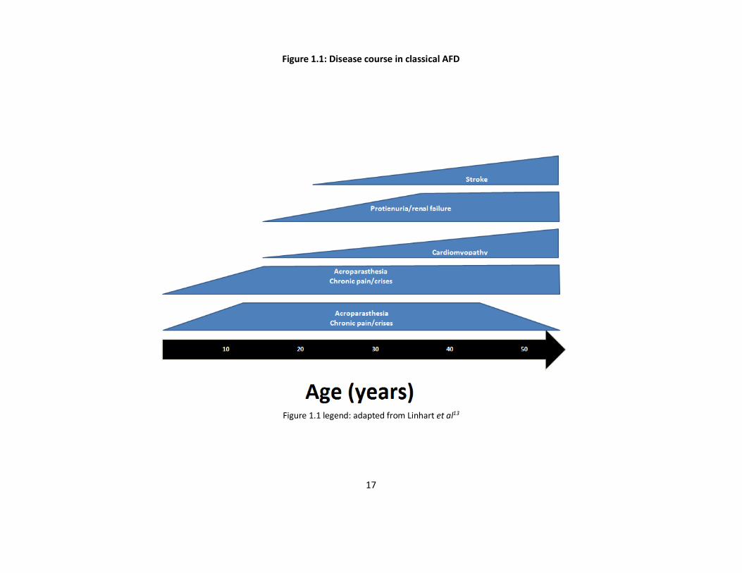
death from stroke, renal or cardiac involvement10 (Figure 1.1).
The history of Anderson-Fabry disease
The first descriptions of AFD were made simultaneously by two independently working
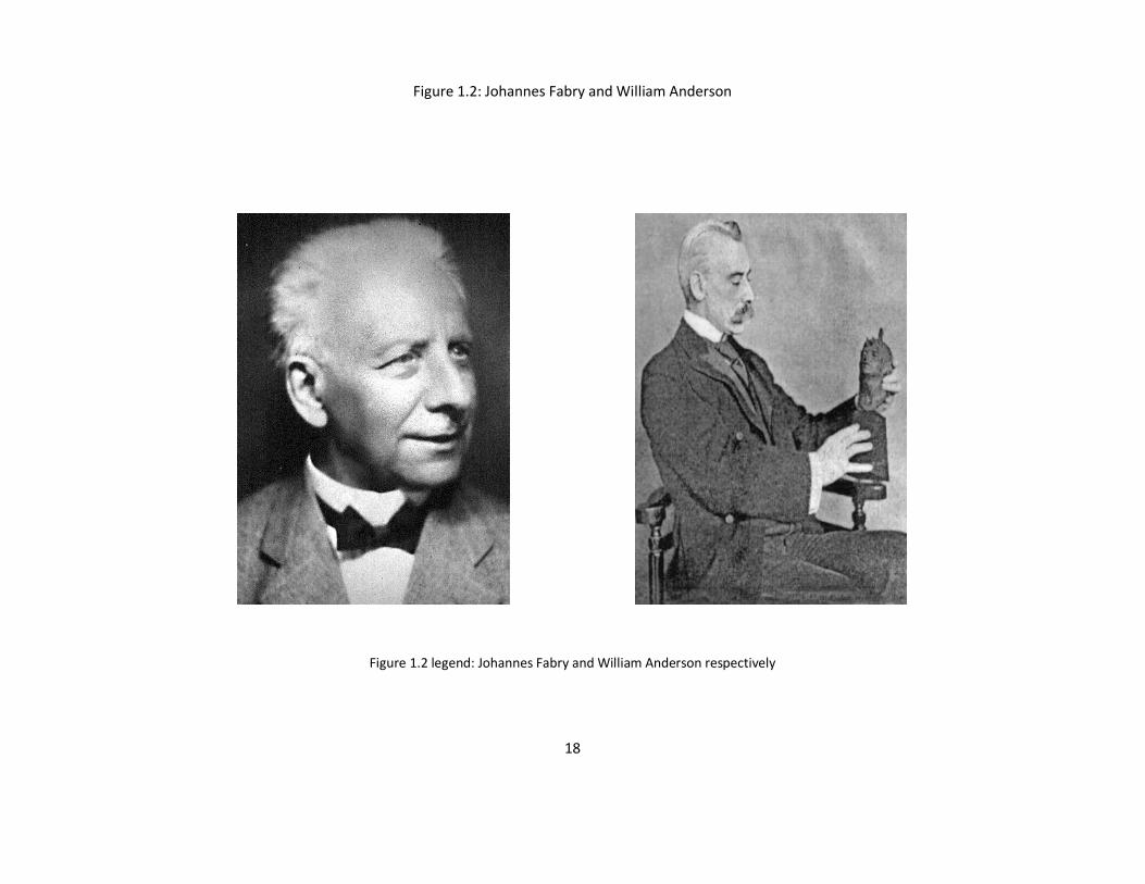
physicians in 1898. William Anderson and Johannes Fabry described ‘angiokeratoma
corporis diffusum’, the characteristic cutaneous manifestation of AFD (Figure 1.2) 11, 12.
William Anderson, who trained at St Thomas’s Hospital, saw his first patient with the
rash, without commenting on the underlying cause. Johannes Fabry was a
dermatologist at the University of Bonn. He described a 13 year old boy who had
previously developed an eruption starting at the back of his left knee and spreading to
his thigh. He thought that the disease may be a form of naevus or represent a
developmental defect. Both these physicians saw their patients in 1897 and in
recognition of this the disease is sometimes referred to as Anderson – Fabry disease.
17
Figure 1.1: Disease course in classical AFD
Figure 1.1 legend: adapted from Linhart et al13
18
Figure 1.2: Johannes Fabry and William Anderson
Figure 1.2 legend: Johannes Fabry and William Anderson respectively
19
Between 1909 and 1939 many physicians described the multi-organ involvement in
AFD with a suspicion that there was a common cause behind all the symptoms14-16. In
1947, an autopsy demonstrated changes in the entire vascular system with
accumulation of a peptide like substance17. These findings were confirmed in another
autopsy in 1950 where birefringent Maltese-cross-like structures under polarized light
were noted as a typical pathological feature and characterised as phosphoglycolipid18.
It was until the late 50s that it was felt that AFD only affected males but in 1959 Colley
reported possible involvement in females19. This was contrary to prevalent literature
at the time20. In 1964 de Groot published on a single family of 45 individuals spanning
4 generations (including females with minimal symptoms) and characterised AFD as a
dominant hereditary disturbance of lipid metabolism. He commented on the
discrepancy between severity of symptoms in females as opposed to males was
incongruous with it being a dominant hereditary disturbance21.
With the use of an electron microscope, Hashimoto et al identified the lysosome in a
patient with AFD and commented on the lysosomes being ‘extremely overcrowded’22.
In this study he concluded that the cause of AFD was a genetic disturbance in a
lysosomal enzyme. In the same year it was realised that AFD was a sex linked genetic
enzyme deficiency. It was noted that there was occasional penetrance in heterozygous
females and constant penetrance in hemizygous males23. Additionally in the same year
the genetic deficit was isolated to the long arm of the X chromosome (locus Xg)24.
In 1967 the enzyme deficiency of ceramidetrihexosidase and subsequent substrate
accumulation was identified as the underlying cause of AFD25. In 1972, it was reported
20
that AFD was corrected with kidney transplantation26, subsequently the treatment of a
patient with purified ceramidetrihexosidase was described in 197327.
The epidemiology of AFD
Reported prevalence of AFD ranges from 1 in 476,000 to 1 in 117,000 in the general
population3, 4, based on the total number of cases identified in a particular period
divided by the total number of births in that same period. These figures have important
limitations due to the nature of the studies, the failure to detect asymptomatic carriers
(particularly women) and misdiagnosed cases resulting in significantly under estimated
figures. Table 1.1 demonstrates that systematic new born screening reveals a higher
incidence than thought previously. The consistently high incidence of AFD in these
systematic new born screening studies suggests that AFD is pan ethnic28-32 and suggest
that the cardiac predominant genotypes may be more common than previously
estimated29, 33.
The tests used in these studies, for new born screening (flourometry, flow-injection
tandem mass spectrometry or digital micro fluidics) have a positive predictive value
ranging from 3.8 to 54% with appreciable false positive rates34. There is a lack of
prospective new born screening and treatment studies in AFD and uncertainty
regarding effectiveness of and immunologic response to treatment make this kind of
screening controversial.

21
Table 1.1: Studies of prevalence of AFD
Type of study Study period Number per 100,000 Country
Birth prevalence 1980-1996 0.85 Austrailia3
Birth prevalence 1970-1996 0.21 Netherlands4
Prevalence of obligate carriers (females) 1980-1995 0.29 UK35
Prevalence 1980-1995 0.27 UK10
Birth prevalence 1997-2002 0.015 Turkey30
Birth prevalence 1982-2001 0.12 Portugal31
Neonatal screening 2004-2006 30 Italy32
Neonatal screening 2006-2008 80 Taiwan33
Neonatal screening 2007-2010 14 Japan28
Neonatal screening 2013 (6 months) 34 Missouri29
Table 1.1 legend: Table adapted from Germain36
22
Molecular genetics of AFD
AFD results from mutations in the galactosidase gene (GLA), located on the long arm
of the X-chromosome (Xq22.1) which encodes a 101 kDa homodimeric glycoprotein
that exists in a number of natural forms resulting from variations in sialic acid residues
on carbohydrate chains1. Over 600 mutations have been described in all seven gene
exons, the majority (57%) being mis-sense point mutations with some premature stop
codons, splice site mutations and insertion/deletions1, 37-42. Reduced enzyme activity
occurs by several mechanisms including abnormal/unstable protein folding,
perturbation to active binding site, and defects in enzyme tracking to the lysosome1, 40.
Despite this condition being described as an X-linked recessive disease, female carriers
also develop symptoms and signs of the disease. This is thought to be related to the
mechanism of non-random X chromosome inactivation (Lyonisation)43
Clinical presentation
Patients with AFD can present with non-specific symptoms, resulting in a delay in
diagnosis. Data from the Fabry Outcome Survey (FOS), an International prospective
registry study, suggest that there is a delay of 13.7 years for males and 16.3 years for
females from the onset of symptoms to diagnosis44. Overall, 25% of patients in FOS
have reported a previous misdiagnosis44.
In the FOS study the most frequently reported signs and symptoms were neurological44.
Both male and female patients had a high prevalence of neuropathic pain (76% and
64%, respectively) occurring with earlier age of onset in males than in females.
Angiokeratomata occurred in 78% of males and 50% of females.
23
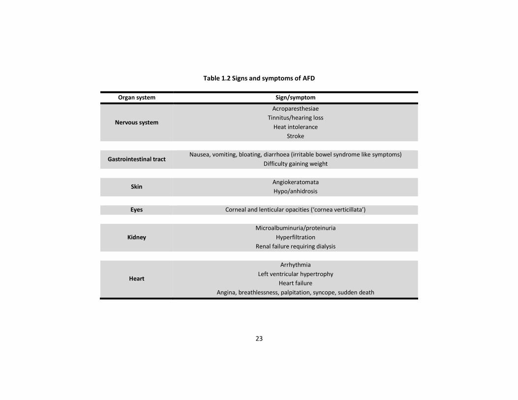
Table 1.2 Signs and symptoms of AFD
Organ system Sign/symptom
Nervous system
Acroparesthesiae
Tinnitus/hearing loss
Heat intolerance
Stroke
Gastrointestinal tract Nausea, vomiting, bloating, diarrhoea (irritable bowel syndrome like symptoms)
Difficulty gaining weight
Skin Angiokeratomata
Hypo/anhidrosis
Eyes Corneal and lenticular opacities (‘cornea verticillata’)
Kidney
Microalbuminuria/proteinuria
Hyperfiltration
Renal failure requiring dialysis
Heart
Arrhythmia
Left ventricular hypertrophy
Heart failure
Angina, breathlessness, palpitation, syncope, sudden death
24
Fifty percent of patients with AFD had signs and symptoms of renal disease, the
commonest being proteinuria observed in 44% of males and 33% of females. End-stage
renal failure was present in 17% of males and 1% of females. Women (27%) had a
higher prevalence of cerebrovascular events than men (17%), with the events occurring
at a younger age in males than in females. Auditory symptoms, such as tinnitus and
hearing loss, were present in more than 50% of patients and ocular signs in just less
than 60% at presentation. Gastrointestinal symptoms, including abdominal pain and
diarrhoea were present in approximately 50% of patients with AFD. Fatigue was
reported as a major symptom in approximately 20% of patients44. Table 1.2 describes
the signs and symptoms of AFD stratified by organ system.
Neurology
Early neural damage affects the small nerve fibres of the peripheral somatic and
autonomic nerve systems resulting in the characteristic acroparasthesiae, hypohidrosis
and gastrointestinal symptoms45, 46. Patients describe two types of pain:
(a) ‘Fabry crisis’ – these are episodic crises of agonizing burning pains in the
extremities radiating inward to the limbs and other parts of the body. These crises
may be precipitated by fever, fatigue, exercise and rapid changes in temperature47.
Crises are often accompanied with a raised erythrocyte sedimentation rate, resulting
in misdiagnosis such as rheumatoid arthritis and Raynaud’s phenomenon; (b) Chronic
pain characterised by chronic burning and tingling paraesthesia46. Both types of pain
often wane in adulthood and so it is important to question patients about childhood
symptoms.
25
Cerebrovascular involvement in AFD can lead to a variety of symptoms and signs
including headache, vertigo/dizziness, transient ischaemic attacks, ischaemic strokes
and rarely vascular dementia48, 49. The potential pathophysiology behind
cerebrovascular disease in AFD includes multifocal involvement of small vessels,
increased thrombotic potential (measurable activation of the endothelium and
leukocytes), dilative arteriopathy in the vertibrobasilar circulation and changes in
regional cerebral hyperperfusion50-52. In one large study of patients with AFD, the
prevalence of cryptogenic stroke was 6.9% in males and 4.3% in females with the
median age at presentation of 39 in males and 46 in females53.
Auditory and vestibular involvement results in a range of symptoms from hearing loss
(progressive or sudden), tinnitus and vertigo44. The mechanism of these symptoms is
likely to be related to both neuropathy and vasculopathy.
Gastrointestinal
The gastrointestinal symptoms are under-appreciated, they include post prandial
abdominal pain, diarrhoea, vomiting and failure to gain weight45. These symptoms are
caused by deposition of Gb3 in the autonomic ganglia of the bowel and mesenteric
blood vessels54.
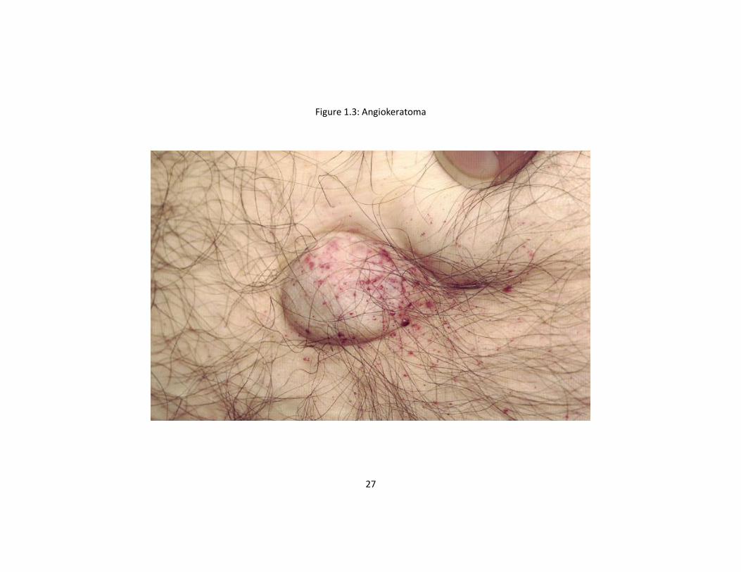
Skin
The most visible clinical feature of AFD is angiokeratomata consisting of clusters of
small reddish purple, and raised skin lesions (Figure 1.3). These are typically found on
26
the buttocks, groin, umbilicus and upper thighs and sometimes on mucous
membranes10, 55, 56. Histologically, the skin lesions are small superficial angiomas
caused by cumulative damage of the vascular endothelial cells of the skin with vessel
dilatation in the dermis that increase in number and size with age and can occur singly
or in groups55, 56. Anhidrosis and hypohidrosis are attributable to selective peripheral
nerve damage or to intracytoplasmic lipid deposits in the sweat glands57. This results
in exercise and heat intolerance46.
Eye changes
Patients frequently have corneal changes (‘cornea verticillata’), these are readily
detectable on slit lamp examination, but do not have any impact on visual acuity (Figure
1.4)58. There are also descriptions of increased retinal vascular tortuosity58.
Renal involvement
Renal involvement is a major cause of morbidity and mortality in AFD44. Like several
aspects of AFD, renal involvement is progressive and increases in severity with age
(Figure 1.1)13. Renal impairment is thought to be a direct consequence of GB3
deposition in glomerular endothelial, mesangial, interstitial cells and podocytes59.
Initially patients develop microalbuminuria and proteinuria in the 2nd and 3rd decades
of life60.
27
Figure 1.3: Angiokeratoma

28
Figure 1.4: Cornea Verticillata
29
Progressive disease results in worsening proteinuria and a reduction in glomerular
filtration, generally in the 3rd to 5th decade of life60. At this stage fibrosis, sclerosis and
tubular atrophy predominate on histology with end stage renal disease occurring in 4th-
5th decade of life59, 60.
Cardiac disease in AFD
Cardiac involvement in AFD begins early (Figure 1.1), with the average age for clinically
overt symptoms being 32 years in the male and 40 years in the female 44. Cardiac
disease in AFD, as with other organ systems is associated with progressive Gb3
accumulation in all cellular components of the heart. Histological studies have
identified GB3 in cardiomyocytes, conduction system cells, valvular fibroblasts,
endothelial cells and vascular smooth muscle cells 61, 62.
Cardiomyocytes are vacuolated and hypertrophied, but unlike familial hypertrophic
cardiomyopathy, myofibrillar disarray is not prominent 62. Lysosomal inclusions are
present within myofibrils and vascular structures; fibrosis is evident within the mid-
myocardial layers and the posterolateral segments of the left ventricle (Figures 1.5 and
1.6) 63. While Gb3 accumulation is the most prominent feature histologically, it
represents only 1–2% of the total cardiac mass64. Therefore, it is likely that disease in
the heart results from activation of other signalling pathways that lead to hypertrophy,
apoptosis, necrosis and fibrosis. These progressive changes correlate with
observations of relatively mild diastolic dysfunction in early stages of the disease
progressing to systolic and severe diastolic ventricular impairment in advanced
disease64.
30
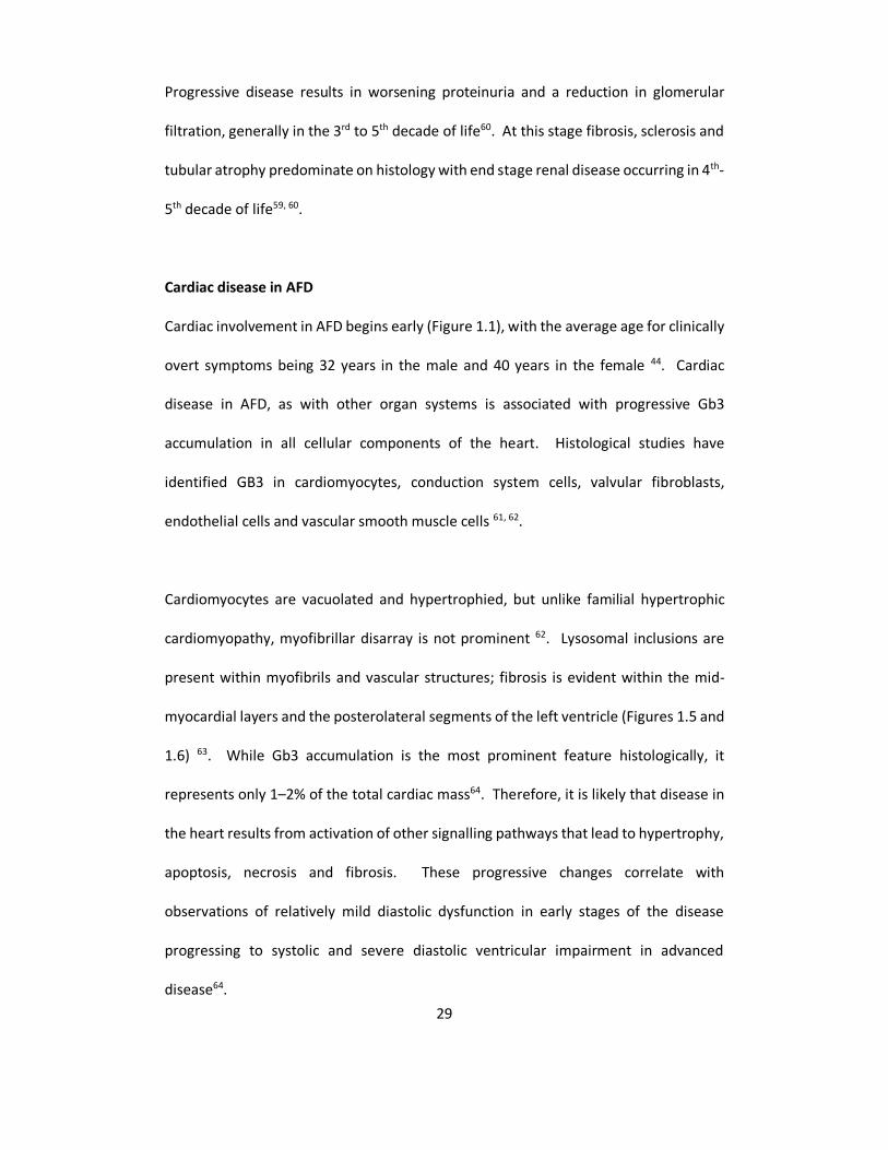
Figure 1.5: AFD cardiomyopathy histology
Figure 1.5 Legend: Haematoxylin and eosin stain (x100) demonstrating myocyte hypertrophy and vacuolization from GB3 accumulation within the cell.
31
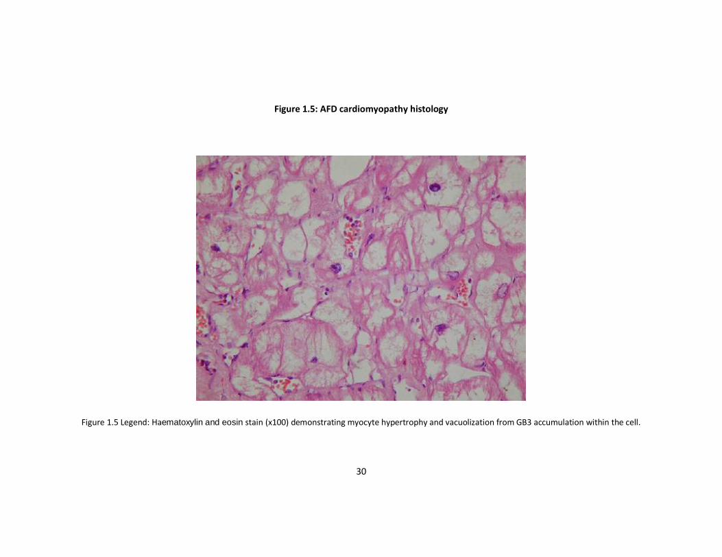
Figure 1.6: Fibrosis in Fabry disease cardiomyopathy
Figure 1.6 legend: Localised myocardial scar seen on cardiac MRI scan correlated to myocardial fibrosis on Picro-sirius red staining at post mortem examination of the
same heart, in the absence of significant coronary artery disease
32
The ‘cardiac variant’
Patients with residual enzyme activity (1-5%) can have a delayed presentation in middle
age with predominantly cardiovascular symptoms, LVH, cardiac rhythm abnormalities
and valve disease65-67. However, while there is an absence of other ‘classical features’,
rigorous clinical characterisation usually reveals organ involvement elsewhere. Hence
AFD should be considered in all patients with otherwise unexplained left ventricular
hypertrophy from the middle decade of life.
Heart disease in females
Clinical manifestations have generally been thought to be rare or mild in female
carriers. However, data from the Fabry Outcome Survey (FOS) and other sources show
that most patients have signs or symptoms of disease with a similar prevalence of
fatigue, neurological, gastrointestinal signs and symptoms in men and women10, 35, 44.
Despite a lower prevalence of left ventricular hypertrophy, females have a similar
prevalence of cardiac symptoms such as angina, dyspnoea and palpitations44. The
prevalence of AFD related symptoms and signs increase with age in both males and
females and it is suggested that females with AFD have a 15 year reduction in their life
span when compared to the general population35. A study by Kampmann et al which
examined 55 females with AFD confirmed on mutational analysis showed that the
severity of the cardiac involvement in this group of patients may be as great as in that
of males68. A twin study has suggested the mechanism of non-random X inactivation
(Lyonisation) may result in disease expression in females43. This hypothesis is further
strengthened by a recent study that demonstrates a higher Mainz Severity Score (a
33
validated severity score for disease severity) in females with non-random X inactivation
demonstrated in peripheral blood compared to those without69.
Enzyme replacement therapy
Exogenous proteins result in the possibility of sensitising the patient with a resulting
reduced efficacy or allergy to the protein, hence initial efforts at enzyme replacement
therapy (ERT) focused on a human source of A-Gal. Enzyme extracted from human
placental tissue was infused into 2 patients with AFD it resulted in a rapid reduction in
circulating Gb3, returning to pre-infusion levels in 48 hours27. Additional studies were
carried out with A-Gal extracted from spleen and plasma with promising results70;
however it became apparent that these sources would only provide limited quantities
of the enzyme. This resulted in 2 biotechnology corporations synthesising recombinant
A-Gal. Transkaryotic Therapies, Inc., Cambridge MA, subsequently known as Shire
Human Genetic Therapies, Inc. A-Gal was initially produced in a cultured human skin
fibroblast cell line using a proprietary gene-activation technique. Known as
Agalsidase–alfa (ReplagalTM) it is administered in a dose of 0.2 mg/kg of body weight
every second week in intravenous (iv) infusions of 40 min. Initial studies with this
infusion resulted in > 30% reduction in hepatic and urinary Gb371. A randomised
controlled trial by Schiffmann et al demonstrated a significant improvement in
neuropathic pain, renal function, cardiac conduction and weight gain72.
34
The Genzyme Corporation, Cambridge, MA, used transduced Chinese hamster ovary
cells to produce recombinant a-galactosidase A, Agalsidase–beta (FabrazymeTM), also
administered on a biweekly basis, but in a dose of 1 mg/kg in a 4–6 h iv infusion. The
initial trial with agalsidase beta revealed clearance of Gb3 from the kidney, skin and
heart and reduction of plasma and urinary Gb3 levels73. This study demonstrated that
88% of patients developed antibodies to the enzyme, but it was felt that this did not
affect the efficacy end points73.
Several studies using both enzyme products have demonstrated an improvement in
renal, neurological and quality of life improvements72, 73. There has been a phase three
trial that has demonstrated a reduction in QRS duration on ECG, cardiac structural and
functional improvement and clearance of GB3 from the vascular endothelium72, 74, 75.
However, post mortem studies have demonstrated continued GB3 storage within
myocytes despite several years of enzyme replacement therapy76. The true prognostic
effect of enzyme replacement therapy remains to be determined.
Aims of the thesis
Exercise limitation
Male and female patients with AFD have an equal prevalence of cardiac symptoms,
including effort intolerance, despite differences in the severity of cardiac disease at
presentation68, 77, 78. The mechanism and clinical significance of exercise limitation in
patients with AFD have not been studied in detail. I will determine the exercise capacity
35
in a large referral population of patients with AFD and its relation to markers of disease
severity.
Left ventricular function
I have described above that there are data to support progressive left ventricular
hypertrophy. Weidemann et al. demonstrate that enzyme replacement therapy
improves cardiac contractile function as measured by strain rate on echocardiography,
however there are no data to document the natural history of left ventricular systolic
function in AFD74. We have long term follow up data on a large cohort of AFD patients
allowing the description of the above and its relationship to symptoms.
Arrhythmia
The majority of adult patients with AFD will have abnormal resting ECGs, the most
common abnormalities being voltage criteria for left ventricular hypertrophy and
repolarisation abnormalities. Short PR intervals, usually in the absence of an accessory
pathway, AV conduction disease, sinus node disease and bundle branch block have also
been described68, 77, 78. Nevertheless, the importance of cardiac arrhythmia in the
natural history of AFD is unknown. I will describe the prevalence of arrhythmia in a
prospectively studied cohort of patients with AFD, and determine its impact on clinical
outcomes.

36
Pathogenesis
Myocardial fibrosis has been described as a prominent feature of AFD cardiomyopathy,
however there are little data regarding the pathogenesis of this and its relationship to
symptoms and overall disease severity63. I will investigate myocardial ischaemia and
collagen turn over and their relation to symptoms and disease severity.
37
Summary of my role in the research project
Together with Professor Perry Elliott, I developed the idea for the research project
presented in this thesis and obtained funding for it. I was responsible for recruiting
patients into the project, and carried out the clinical and echocardiographic evaluation
of patients recruited between 2002 and 2005. I also retrospectively entered clinical
and echocardiographic data onto the database for all patients recruited into the study,
including all patients evaluated before 2002, by manually searching through patients’
medical records, echocardiograms and other investigations. In addition, I modified the
database so that data relevant to the AFD population could be entered and analysed. I
carried out all the analyses presented in this thesis.
38
Chapter 2
Characterisation of cardiac
arrhythmia
39
Abstract
Background
Over 60% of patients with AFD have symptoms and signs of cardiac involvement. The
prevalence and clinical significance of arrhythmia in AFD are unknown.
Methods and results
The patient cohort comprised seventy-eight patients (43 male, 43.5 ± 15.0 years, 13.0-
83.0 years) with AFD, studied for a mean of 1.9 years (0.25-10 years). All patients
underwent clinical evaluation, 12-lead ECG and echocardiography. Sixty patients
(76.9%) underwent 24 hour ambulatory ECG (Holter) monitoring. Atrial fibrillation (AF)
was seen at baseline in 3/78 (3.9%) patients. On Holter monitoring 8 (13.3%) had
paroxysmal AF (PAF) and 5 (8.3%) had non-sustained ventricular tachycardia (NSVT).
Patients with NSVT were all male with a maximal left ventricular wall thickness > 20mm.
Univariate analysis identified age (p<0.001), left atrial diameter (p = 0.001), maximal
left ventricular wall thickness (p = 0.003), left ventricular mass index (p = 0.009) and
angina (p = 0.02) as predictors of AF/PAF. Stepwise logistic regression identified age as
the only independent predictor of AF/PAF (OR = 1.2, 95% CI = 1.1 to 1.3, p = 0.001).
Four patients had pacemakers implanted for bradyarrhythmia and 1 had a bi-
ventricular pacemaker and internal cardioverter defibrillator implanted for heart
failure and symptomatic NSVT.

40
Conclusion
Brady- and tachyarrhythmias are common in older patients with AFD. Patients with AFD
should undergo regular cardiological review, as the development of arrhythmia in these
patients may have an impact on the natural history of this disease.
41
Aims
The aims of this study were to determine the prevalence of cardiac arrhythmia in a
large consecutively referred population of patients with AFD, and to assess their clinical
significance.
Methods
Between January 1993 and December 2003, 78 patients (43 (55.1%) male; 35 (44.9%)
female; mean age ( SD) = 43.5 15.0 years; range = 13-83) with AFD were assessed at
The Heart Hospital, University College London, UK. Patients were diagnosed with AFD
on the basis of low plasma -Gal level and / or on mutational analysis. Seven patients
had been identified during the screening of patients with unexplained left ventricular
hypertrophy 79. The remaining patients were direct referrals for cardiac evaluation from
The Charles Dent Metabolic Unit, The National Hospital for Neurology and
Neurosurgery and The Lysosomal Storage Disorders Unit, The Royal Free Hospital,
London, UK. Forty-one patients (6 female, 35 male) were on enzyme replacement
therapy (ERT) at the base line visit.
Clinical Assessment
All patients underwent clinical history and examination. Cardiac symptoms were
defined as angina (central chest pain occurring during exertion, lasting for less than 15
minutes), dyspnoea (graded using New York Heart Association criteria; NYHA), syncope
and palpitation.
42
ECG Assessment
All patients had a standard supine electrocardiogram (ECG) performed using a Hewlett-
Packard Page Writer, Andover, MA, USA. ECGs were recorded at 25 mm/s with an
amplitude of 1 mV/10mm and 50 Hz filtering. The following features were recorded:
Rhythm, PR interval and QRS duration. LVH was assessed using the Romhilt-Estes (RE)
score 80. A normal P-R interval was defined as > 120ms and ≤ 200ms. A prolonged QRS
duration was defined as > 120ms.
Twenty-four hour ambulatory ECG (Holter) was performed in 60/78 (76.9%) patients
using Reynolds Medical tape Holter monitors, Hertford, UK. Non-sustained ventricular
tachycardia (NSVT) was defined as three or more consecutive ventricular ectopics at a
rate > 120 beats per minute lasting less than 30 seconds. Atrial fibrillation (AF) was
defined on the basis of the baseline ECG showing the replacement of consistent P
waves by rapid oscillations or fibrillatory waves that vary in size, shape, and timing 81.
Paroxysmal atrial fibrillation (PAF) was defined as episodes of AF lasting less than seven
days, diagnosed from documented arrhythmia on 24 hour ambulatory ECG 82.
Persistent AF was defined as AF lasting more than seven days, but potentially amenable
to cardioversion82. Permanent AF was defined as AF in which chemical/electrical
cardioversion failed or was not attempted82.
Echocardiography
M-mode, 2D, and Doppler echocardiography were performed using a GE System V
echocardiograph. The following left ventricular parameters were measured from M-

43
mode tracings according to recommendations of the American Society of
Echocardiography: end-diastolic left ventricular internal cavity diameter (LVed),
interventricular septum thickness (IVS), posterior wall thickness (PW) and end systolic
internal cavity diameter (LVes)83. The severity and distribution of left ventricular
hypertrophy were assessed in the parasternal short-axis plane at the mitral valve and
papillary muscle level. Maximum left ventricular wall thickness was defined as the greatest
thickness in any single segment measured in diastole. Relative wall thickness (RWT) was
calculated as ((IVS + PW)/LVed) assessed at the mitral valve level. Left Ventricular
remodeling or left ventricular hypertrophy was defined as a RWT > 0.4583. Left
ventricular mass was calculated by the Devereux modified cube formula 83 and
indexed by body surface area. Normal left ventricular mass index (LVMI) was defined
as <134 gm/m2 for men and <110 gm/m2 for women 84. Left ventricular geometry was
classified as normal (normal LVMI and normal RWT), concentric remodelling (normal
LVMI and increased RWT), eccentric left ventricular hypertrophy (increased LVMI and
normal RWT), and concentric left ventricular hypertrophy (increased LVMI and
increased RWT). Fractional shortening (FS) ((LVed-LVes/LVed)*100) was used to
measure systolic performance85.
Follow Up
Follow-up started with the date of the baseline cardiovascular assessment. Data were
collected at routine clinic visits, and where necessary by direct communication with
patients, their attending physicians and patients' general practitioners; all data were
verified by the author.
44
Statistical Analysis
Statistical analysis was performed using SPSS v19.0. The χ2 test was used to compare
non-continuous variables. Continuous variables were tested for normality, non-
normally distributed variables were tested using the Wilcoxon Rank sum and normally
distributed variables compared using the 2-tailed independent sample Student’s t-test.
All values are expressed as means standard deviation. Statistical significance was
defined as p < 0.05.
Results
Clinical Evaluation
Table 2.1 describes the prevalence of cardiac and AFD symptoms separated by gender.
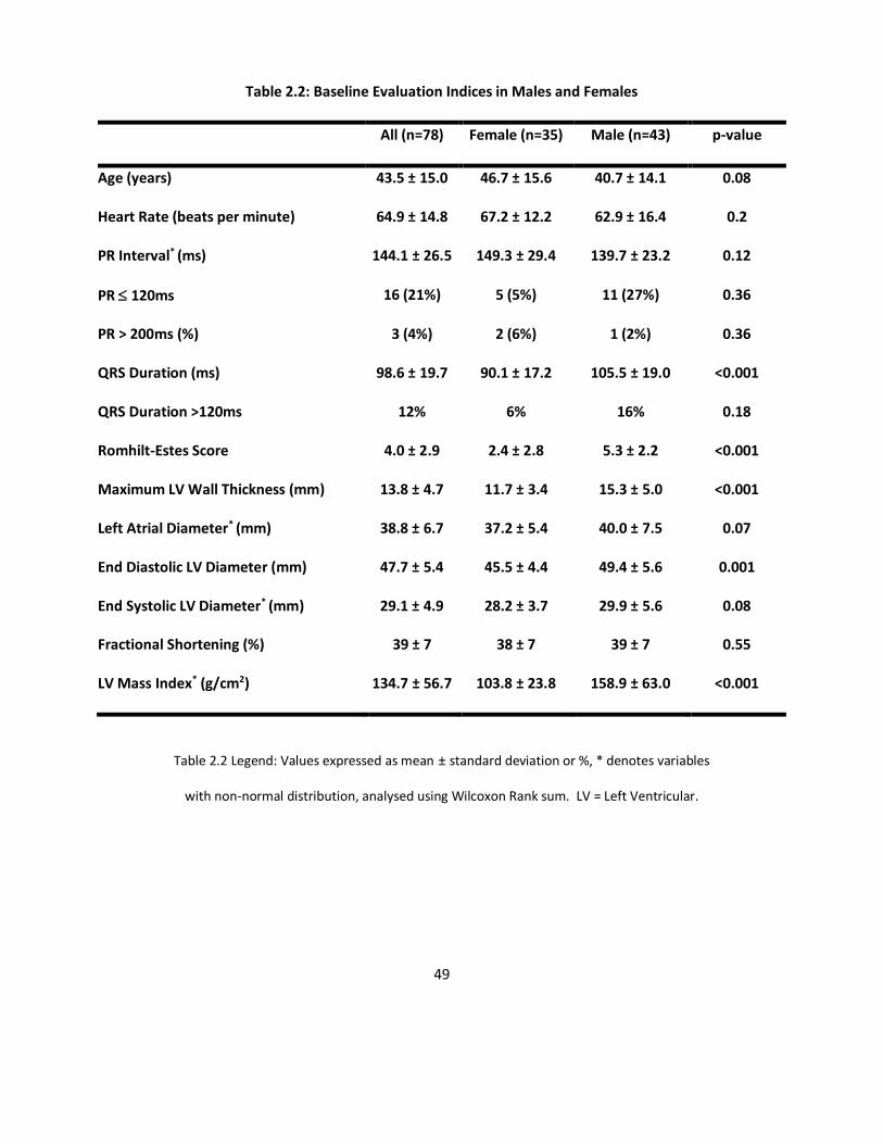
Table 2.2 displays the important findings in the baseline electrocardiographic and
echocardiographic findings for this cohort. There were no differences between males
and females in the prevalence of cardiac symptoms. Men were more likely to suffer
from AFD symptoms and signs compared to women.
ECG Changes at Baseline
There were no significant differences between men and women in the baseline heart
rate, QRS axis or P-R interval. The mean RE score was greater (mean difference = 2.8,
95% CI = 1.7 to 4.0, p = <0.001) and the mean QRS duration was greater (p < 0.001) in
men compared to women (see Table 2.2). Two males had evidence of pre-excitation
on their baseline ECG. One of these patients had an intracardiac Electrophysiological
study. No accessory pathway was identified; however there was accelerated AV
45
conduction with a fixed HV time of less than 10ms and normal decremental AV and VA
conduction. The second patient went into complete heart block and there was
evidence of a presumed accessory pathway on his Cardiomemo ECG as demonstrated
by a pre-excited QRS complex followed by a normally conducted QRS complex (Figure
2.1). Three of the 78 patients had AF (3.9%; 2 persistent and 1 permanent). There was
no difference in the prevalence of AF in patients on ERT compared to those not on ERT.
Holter assessment at baseline
Sixty patients underwent Holter assessment at the baseline visit. Eight of the 60
patients (13.3%) had PAF (lasting less than 24 hours). Five of the 60 patients (8.3%) had
NSVT. There was no difference in the prevalence of AF/PAF between males and
females. There seemed to be no difference in the prevalence of PAF/NSVT on Holter
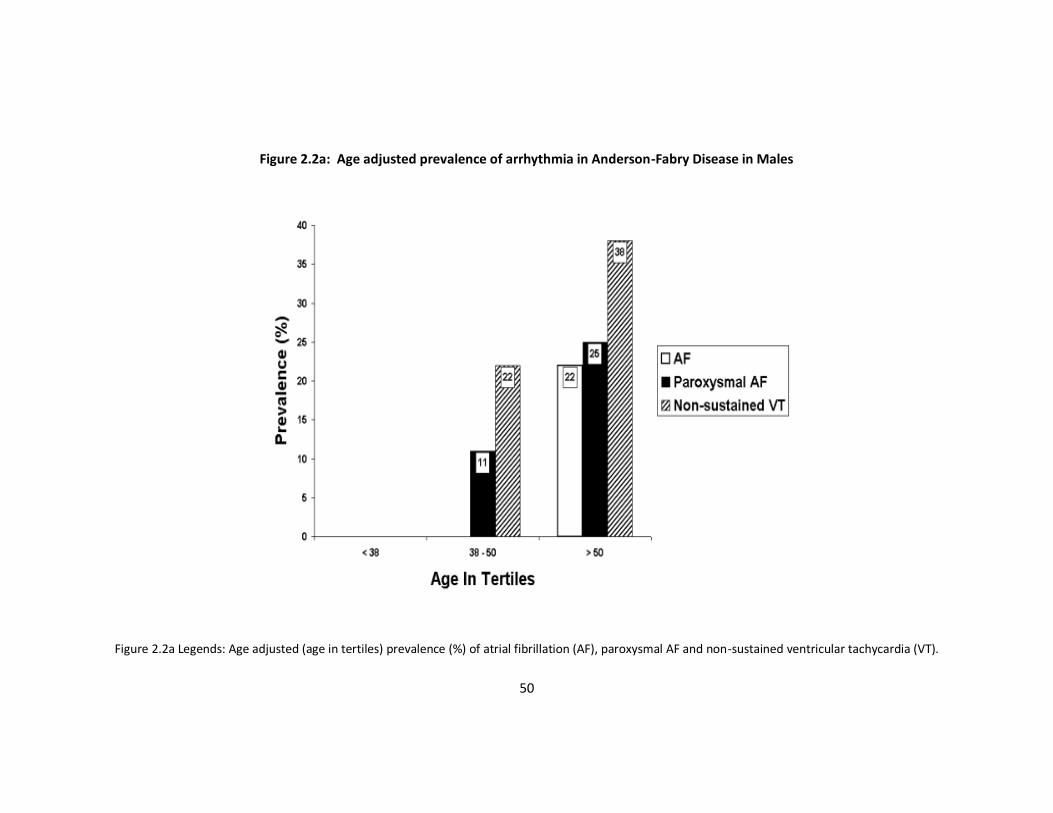
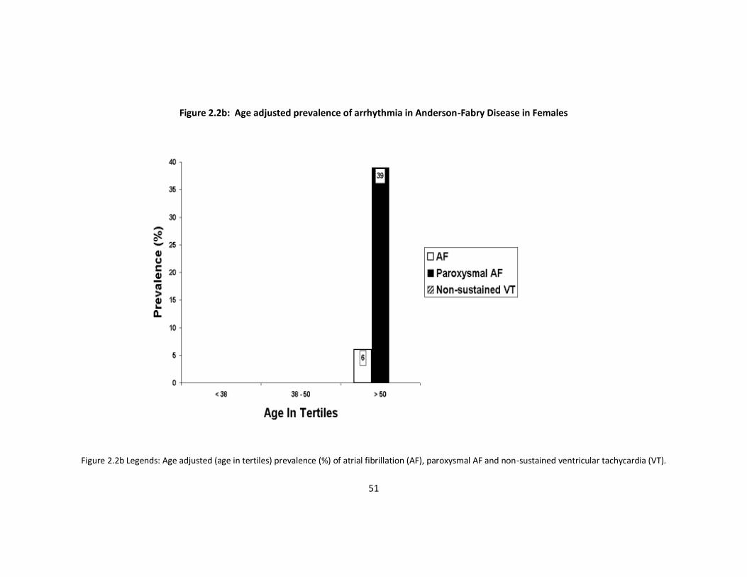
between patients on ERT and those not on ERT. There was a significant increase in the
prevalence of arrhythmia with age (Figure 2.2a and b).
Echocardiographic Parameters at Baseline
Compared to females, males had greater maximal left ventricular wall thickness
(mean difference = 3.6mm, 95% CI = 1.6, 5.6mm, p = <0.001), larger LVed (mean
difference =3.9 mm, 95% CI = 1.6, 6.2mm, p = 0.001) and higher LVMI (p < 0.001, see
Table 2.2). There was a trend to a larger LA size and LVes in men compared to
women (p = 0.07 and p = 0.08 respectively; Table 2.2).
46
Table 2.1: Symptoms at Baseline Evaluation in Males and Females
All (n=78) Female (n=35) Male (n=43) p-value
Chest Pain 15 (19%) 8 (23%) 7 (16%) 0.57
Dyspnoea* 22 (28%) 7 (20%) 15 (36%) 0.21
Palpitations 22 (28%) 10 (29%) 12 (28%) 1.0
Syncope 6 (8%) 2 (6%) 4 (9%) 0.7
Acroparaesthesiae 45 (58%) 14 (40%) 31 (72%) 0.005
Hypohydrosis 24 (31%) 5 (14%) 19 (44%) 0.006
Angiokeratoma 27 (35%) 4 (11%) 23 (54%) <0.001
Abdominal Pain 22 (28%) 6 (17%) 16 (37%) 0.048
Tinnitus 15 (19%) 1 (3%) 14 (33%) 0.001
Table 2.1 Legend: * = New York Heart Association class II and above.
47
Figure 2.1: Complete heart block
Figure 2.1 Legends: Cardio-memo strip showing complete heart block in a 55-year-old male patient requiring permanent pacemaker implantation. *Conducted beat
showing pre-excitation. **Conducted beat shows a normal PR interval.
* **
48
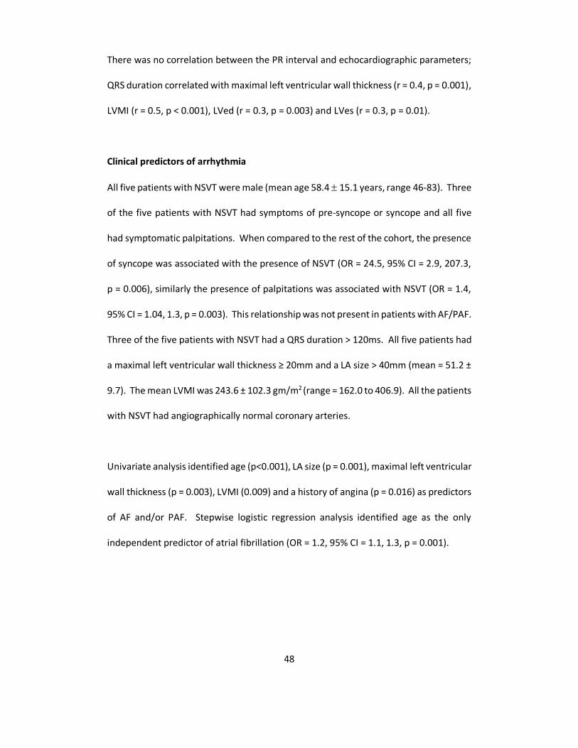
There was no correlation between the PR interval and echocardiographic parameters;
QRS duration correlated with maximal left ventricular wall thickness (r = 0.4, p = 0.001),
LVMI (r = 0.5, p < 0.001), LVed (r = 0.3, p = 0.003) and LVes (r = 0.3, p = 0.01).
Clinical predictors of arrhythmia
All five patients with NSVT were male (mean age 58.4 15.1 years, range 46-83). Three
of the five patients with NSVT had symptoms of pre-syncope or syncope and all five
had symptomatic palpitations. When compared to the rest of the cohort, the presence
of syncope was associated with the presence of NSVT (OR = 24.5, 95% CI = 2.9, 207.3,
p = 0.006), similarly the presence of palpitations was associated with NSVT (OR = 1.4,
95% CI = 1.04, 1.3, p = 0.003). This relationship was not present in patients with AF/PAF.
Three of the five patients with NSVT had a QRS duration > 120ms. All five patients had
a maximal left ventricular wall thickness ≥ 20mm and a LA size > 40mm (mean = 51.2 ±
9.7). The mean LVMI was 243.6 ± 102.3 gm/m2 (range = 162.0 to 406.9). All the patients
with NSVT had angiographically normal coronary arteries.
Univariate analysis identified age (p<0.001), LA size (p = 0.001), maximal left ventricular
wall thickness (p = 0.003), LVMI (0.009) and a history of angina (p = 0.016) as predictors
of AF and/or PAF. Stepwise logistic regression analysis identified age as the only
independent predictor of atrial fibrillation (OR = 1.2, 95% CI = 1.1, 1.3, p = 0.001).
49
Table 2.2: Baseline Evaluation Indices in Males and Females
All (n=78) Female (n=35) Male (n=43) p-value
Age (years) 43.5 ± 15.0 46.7 ± 15.6 40.7 ± 14.1 0.08
Heart Rate (beats per minute) 64.9 ± 14.8 67.2 ± 12.2 62.9 ± 16.4 0.2
PR Interval* (ms) 144.1 ± 26.5 149.3 ± 29.4 139.7 ± 23.2 0.12
PR 120ms 16 (21%) 5 (5%) 11 (27%) 0.36
PR > 200ms (%) 3 (4%) 2 (6%) 1 (2%) 0.36
QRS Duration (ms) 98.6 ± 19.7 90.1 ± 17.2 105.5 ± 19.0 <0.001
QRS Duration >120ms 12% 6% 16% 0.18
Romhilt-Estes Score 4.0 ± 2.9 2.4 ± 2.8 5.3 ± 2.2 <0.001
Maximum LV Wall Thickness (mm) 13.8 ± 4.7 11.7 ± 3.4 15.3 ± 5.0 <0.001
Left Atrial Diameter* (mm) 38.8 ± 6.7 37.2 ± 5.4 40.0 ± 7.5 0.07
End Diastolic LV Diameter (mm) 47.7 ± 5.4 45.5 ± 4.4 49.4 ± 5.6 0.001
End Systolic LV Diameter* (mm) 29.1 ± 4.9 28.2 ± 3.7 29.9 ± 5.6 0.08
Fractional Shortening (%) 39 ± 7 38 ± 7 39 ± 7 0.55
LV Mass Index* (g/cm2) 134.7 ± 56.7 103.8 ± 23.8 158.9 ± 63.0 <0.001
Table 2.2 Legend: Values expressed as mean ± standard deviation or %, * denotes variables
with non-normal distribution, analysed using Wilcoxon Rank sum. LV = Left Ventricular.
50
Figure 2.2a: Age adjusted prevalence of arrhythmia in Anderson-Fabry Disease in Males
Figure 2.2a Legends: Age adjusted (age in tertiles) prevalence (%) of atrial fibrillation (AF), paroxysmal AF and non-sustained ventricular tachycardia (VT).
51
Figure 2.2b: Age adjusted prevalence of arrhythmia in Anderson-Fabry Disease in Females
Figure 2.2b Legends: Age adjusted (age in tertiles) prevalence (%) of atrial fibrillation (AF), paroxysmal AF and non-sustained ventricular tachycardia (VT).

52
Follow-Up
Follow up data were available in 66/78 (84.6%) patients. The mean follow up time was
1.9 years (range = 3 months – 10 years). During follow up, 7/66 (10.6%) patients had
permanent pacemakers implanted; 1 for complete heart block; (patient with pre-
excitation, Figure 2.1); 3 for symptomatic bradycardia; 1 for symptomatic LV outflow
tract gradient reduction; 1 for complete heart block post alcohol septal ablation (the
latter two patients had been followed-up with a presumed diagnosis of hypertrophic
cardiomyopathy for several years prior to the diagnosis of AFD); 1 had a bi-ventricular
device and internal cardioverter defibrillator for heart failure and symptomatic NSVT
(Figure 2.3).
There were 2/66 (3.0%) new cases of persistent AF, one patient with persistent AF at
baseline evaluation had been successfully cardioverted to sinus rhythm and one
remained in permanent AF. There were 2 new cases of PAF documented.
Discussion
This study shows that arrhythmia is common in patients with AFD and that it is
associated with significant morbidity.
Population based studies have shown that the overall prevalence of atrial fibrillation
in the normal population is less than 1% 86; this prevalence is age dependent, with a
prevalence of less than 1% in people less than 55, rising to 9% or more in the over
eighties 86.
53
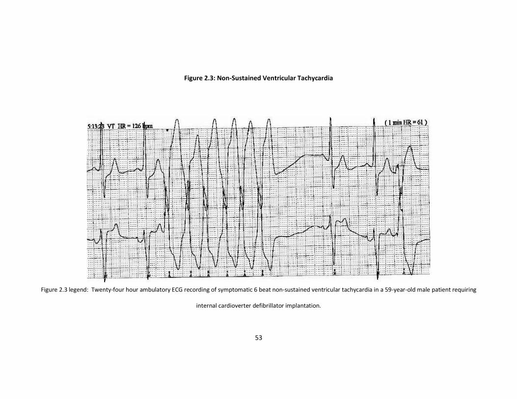
Figure 2.3: Non-Sustained Ventricular Tachycardia
Figure 2.3 legend: Twenty-four hour ambulatory ECG recording of symptomatic 6 beat non-sustained ventricular tachycardia in a 59-year-old male patient requiring
internal cardioverter defibrillator implantation.
54
In this study, the overall prevalence of AF was four times higher than in the general
population; in the over fifties it was present in 12%. This suggests that AFD is associated
with a substantial risk of AF in middle aged and elderly patients.
Gender related differences
Although AFD is an X-linked disorder, most females who carry a pathogenic mutation
in the -Gal gene develop signs and symptoms of the disease, albeit at an older age
than men 68. A striking observation in this study was that, in spite of a higher incidence
of “classical” signs and symptoms such as angiokeratomata, neuropathic pain and
hypohidrosis in men, the incidence of cardiovascular symptoms was similar in males
and females. Men tended to have a longer QRS duration and higher Romhilt-Estes
score than women, reflecting their greater left ventricular mass. Similarly, all the
patients with NSVT were men; there was, however, no gender related difference in the
frequency of AF, the only independent risk factor being age. It is likely, therefore, that
most patients with AFD become prone to cardiac disease and arrhythmia if they live
into middle age and beyond.
Clinical Implications
The impact of arrhythmia on mortality in patients with AFD cannot be determined from
this study, but the high prevalence of atrial fibrillation, the occurrence of complete
heart block and symptomatic ventricular tachycardia, suggests that it may contribute
to the shortened life expectancy of patients with this disease. This observation
suggests that all patients with AFD should undergo regular assessment with ECG and
Holter monitoring.
55
Patients with AFD have a higher incidence of stroke that is usually attributed to
microvascular dysfunction87-89. Additionally, an increased thrombotic potential,
dilative arteriopathy and changes in regional cerebral hyperperfusion may contribute
to cerebrovascular disease in patients with AFD49-51. Lenders et al suggest that up to
7% of patient with AFD also carry a mutation in the Factor V Leiden gene. These patient
have a 5 fold increase (95% CI HR = 2-13) in the risk of suffering a thromboembolic
event90. The high prevalence of atrial arrhythmia in this study, suggests that
thromboembolism may be an additional risk factor for stroke, and consideration of
anticoagulation in patients with permanent or frequent paroxysms of AF may be
needed. Unfortunately, the early age of onset of atrial arrhythmia compared to the
general population precludes the use thromboembolism risk scores such as the
CHA2DS2-VASc scoring system in this patient group91.
There has been no systematic study of sudden death in AFD. To date, there have been
four case reports published describing the unexpected death of patients with AFD.
Three were asymptomatic elderly females (diagnosed at post-mortem) 92, 93, one was a
26-year-old male known to have AFD who died whilst running 94; and one was a male
known to have AFD who had ventricular fibrillation resistant to defibrillation 95. Three
of the four patients had post-mortem evidence of significant cardiomyopathy with
hearts weighing >450gms. All the patients with NSVT in our study had evidence for
significant cardiac disease (all male, increased LVMI and maximal left ventricular wall
thickness > 20mm). Together these data suggest that sudden death in AFD is associated
with significant cardiac involvement and may be related to NSVT.

56
Conclusions
This study demonstrates that arrhythmias are common in older patients with AFD.
This, together with the high prevalence of permanent pacemaker implantation,
suggests that there is a need for regular cardiology follow-up in this patient group,
monitoring for rhythm abnormalities.

57
Chapter 3
The natural history of left
ventricular systolic performance
58
Abstract
Aim
Although AFD cardiomyopathy is usually associated with left ventricular hypertrophy,
conduction disease and valvular thickening, a recent study has demonstrated an
impairment of contractile function as well. The aim of this study was to determine the
importance of systolic function in the natural history of AFD.
Methods and results
Twelve patients 9 male, (aged; 54.5 ± 12.2) years; minimum = 42 and maximum = 82)
with AFD were studied. All patients underwent clinical, electrocardiographic and
echocardiographic evaluation. Two echocardiograms >1 year apart were performed on
all patients. Patients on enzyme replacement therapy (ERT) were excluded from the
study.
The mean follow up between echocardiograms was 3.3 ± 2.7 years (minimum = 1,
maximum = 9). There was no change in left ventricular mass. There was an increase
in the end systolic left ventricular diameter of 5.0mm (95% confidence interval (CI) =
0.2, 9.6, p = 0.038) and a decrease in fractional shortening of 6.1% (95% CI = 0.3, 11.9,
p=0.042). Three patients (25%) developed symptomatic congestive heart failure.
Conclusions
Systolic impairment in patients with AFD cardiomyopathy is common. The decline in
systolic function may represent a measure of disease severity and provide a clinically

59
important surrogate marker for response to therapy.
60
Aims
The aim of this study was to determine the importance of systolic performance in the
natural history of AFD and its potential value as marker of disease progression, we
performed a retrospective analysis of an untreated cohort of patients followed for at
least one year.
Methods
A total of 75 patients with AFD recruited from The Heart Hospital, London, The Royal Free
Hospital and The National Hospital for Neurology and Neurosurgery, were evaluated
between 1st January 1993 and 1st December 2003. Diagnosis was based on low
plasma α-Gal levels and on genetic mutational analysis. The mean (standard
deviation) follow up from diagnosis of AFD was 5.8 ± 4.8 years. Of the 75 patients 24
had been followed up for more than one year and 12 were receiving ERT at the time
of evaluation. The final study cohort comprised 12 untreated patients (9 male, 3
female patients mean age 54.5 ± 12.2 years; minimum age = 42, maximum = 82) with
at least 2 echocardiograms separated by one year or more. Six patients were detected
during screening of unexplained hypertrophic cardiomyopathy (HCM)79, the remaining
six were patients referred for cardiac assessment of patients with AFD.
Clinical assessment
All patients underwent clinical examination, supine 12-lead electrocardiography (ECG)
(Hewlett-Packard, USA) and 24 hour ambulatory ECG (Reynolds Medical, UK)
monitoring as described in chapter 2.
61
Plasma α-Gal analysis
Plasma α-Gal activity was measured with the fluorogenic substrate 4-
methylumbelliferyl-α-Dgalactopyranoside (Sigma), with N-acetyl-D-galactosamine
(Nacalai Tesque) used as an inhibitor of α-N-acetylgalactosaminidase as described
previously 96. On the basis of previously published data, a plasma α-Gal activity of 1.2
nmol·h–1·mL–1 was considered diagnostic of AFD 96. All patients with diagnostic plasma
α-Gal levels went on to have confirmation of their diagnosis with genetic mutational
analysis.
Assessment of left ventricular mass and function
M-mode, 2D, and Doppler echocardiographic assessment of all patients was as previously
described in chapter 2 using a GE System V echocardiograph.
Statistical analysis
Statistical analysis was performed using SPSS v19.0 (Chicago, USA). The χ2 test was
used to compare non-continuous variables, and the 2-tailed paired sample Student’s
t-test was used to compare continuous variables. All values are expressed as means ±
standard deviation. Statistical significance was defined as p 0.05.
Results
Table 3.1 describes the baseline symptoms and echocardiographic findings in this
cohort. The mean time between the analysed echocardiograms was 3.3 ± 2.7 years
(minimum = 1, maximum = 9).
Four patients (41%) had two symptoms or signs of AFD, and one in retrospect had 4
62
symptoms or signs of AFD. Ten patients (83%) presented with a cardiovascular
symptom. Six patients (50%) had two or more cardiac symptoms (see table 3).
During the follow up, 2 (17%) patients had documented non-sustained ventricular
tachycardia; one (8%) had an internal cardiac defibrillator implanted prophylactically for
this. Four (33.3%) patients required permanent pacemaker implantation: two for
symptomatic bradycardia; one for congestive heart failure (bi-ventricular device); and
one patient for complete heart block following alcohol septal ablation prior to the
diagnosis of AFD. Two other patients progressed to congestive heart failure.
Electrocardiography
The baseline mean P-R interval was 141.5 ± 22.3 ms with a baseline mean QRS duration
of 112.7 ± 25.0 ms. During follow up, no difference in the P-R interval was seen. There
was an increase in the QRS duration (mean = 14.2ms; 95% CI = 4.0, 24.4ms, p = 0.012).
Patients with pacemakers were excluded from this analysis.
Echocardiography
Three patients (25%) had normal left ventricular wall thickness, eight (66.7%) had
concentric LVH and one patient (8.3%) had distal LVH. Figures 3.1 & 3.2 plot the changes
in LVes, LVed and FS in individual patients. There was an increase in the LVes (mean 5
± 6.9 mm, 95% CI = 0.2, 9.6, p=0.038), accompanied by a reduction in the FS (mean 6.1
± 8.7 mm, 95% CI = 0.3, 11.9 mm, p=0.042) and a trend towards an increasing LVed
(mean 2.9 ± 5.4, 95% CI = -0.5, 6.35 mm, p=0.088). The rate of dilatation in the left
ventricular end systolic dimension was 1.9mm/year with an associated decline of 2.7
%/year in the fractional shortening. There was no correlation between age and
baseline LVes.
63
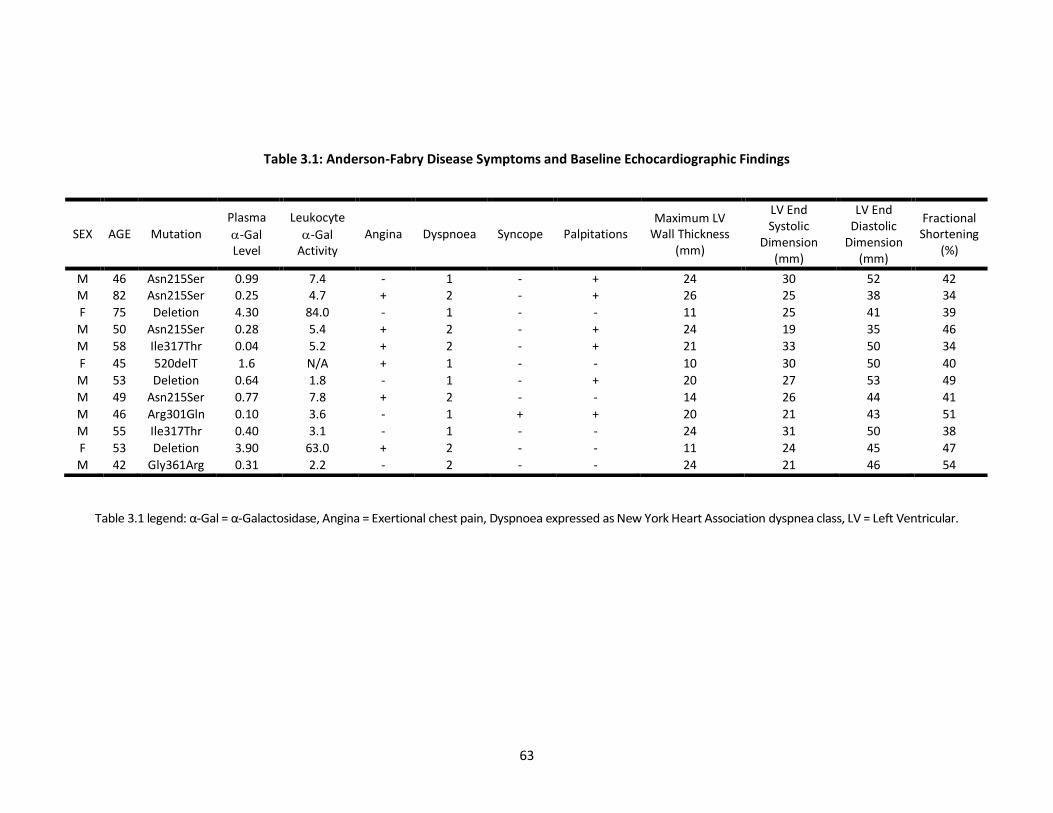
Table 3.1: Anderson-Fabry Disease Symptoms and Baseline Echocardiographic Findings
Table 3.1 legend: α-Gal = α-Galactosidase, Angina = Exertional chest pain, Dyspnoea expressed as New York Heart Association dyspnea class, LV = Left Ventricular.
SEX AGE Mutation Plasma
-Gal Level
Leukocyte
-Gal Activity
Angina Dyspnoea Syncope Palpitations Maximum LV
Wall Thickness (mm)
LV End Systolic
Dimension (mm)
LV End Diastolic
Dimension (mm)
Fractional Shortening
(%)
M 46 Asn215Ser 0.99 7.4 - 1 - + 24 30 52 42 M 82 Asn215Ser 0.25 4.7 + 2 - + 26 25 38 34
F 75 Deletion 4.30 84.0 - 1 - - 11 25 41 39
M 50 Asn215Ser 0.28 5.4 + 2 - + 24 19 35 46
M 58 Ile317Thr 0.04 5.2 + 2 - + 21 33 50 34
F 45 520delT 1.6 N/A + 1 - - 10 30 50 40
M 53 Deletion 0.64 1.8 - 1 - + 20 27 53 49
M 49 Asn215Ser 0.77 7.8 + 2 - - 14 26 44 41
M 46 Arg301Gln 0.10 3.6 - 1 + + 20 21 43 51
M 55 Ile317Thr 0.40 3.1 - 1 - - 24 31 50 38
F 53 Deletion 3.90 63.0 + 2 - - 11 24 45 47
M 42 Gly361Arg 0.31 2.2 - 2 - - 24 21 46 54
64
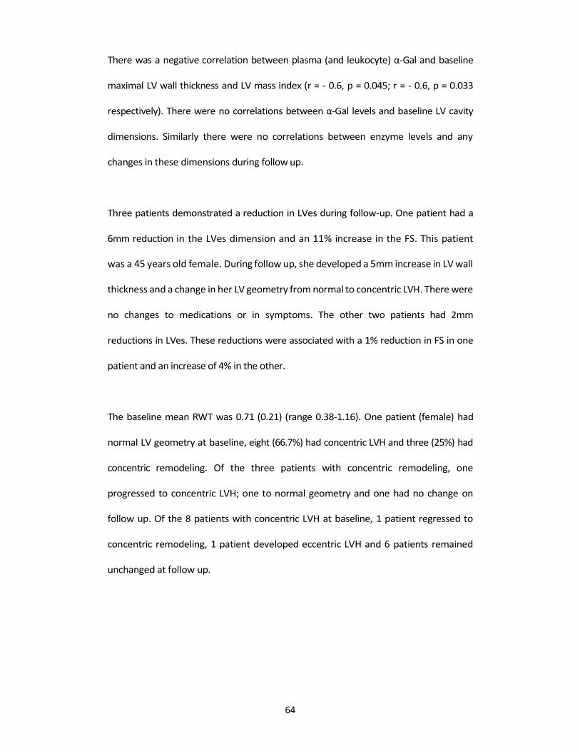
There was a negative correlation between plasma (and leukocyte) α-Gal and baseline
maximal LV wall thickness and LV mass index (r = - 0.6, p = 0.045; r = - 0.6, p = 0.033
respectively). There were no correlations between α-Gal levels and baseline LV cavity
dimensions. Similarly there were no correlations between enzyme levels and any
changes in these dimensions during follow up.
Three patients demonstrated a reduction in LVes during follow-up. One patient had a
6mm reduction in the LVes dimension and an 11% increase in the FS. This patient
was a 45 years old female. During follow up, she developed a 5mm increase in LV wall
thickness and a change in her LV geometry from normal to concentric LVH. There were
no changes to medications or in symptoms. The other two patients had 2mm
reductions in LVes. These reductions were associated with a 1% reduction in FS in one
patient and an increase of 4% in the other.
The baseline mean RWT was 0.71 (0.21) (range 0.38-1.16). One patient (female) had
normal LV geometry at baseline, eight (66.7%) had concentric LVH and three (25%) had
concentric remodeling. Of the three patients with concentric remodeling, one
progressed to concentric LVH; one to normal geometry and one had no change on
follow up. Of the 8 patients with concentric LVH at baseline, 1 patient regressed to
concentric remodeling, 1 patient developed eccentric LVH and 6 patients remained
unchanged at follow up.
65
Figure 3.1: The Change in Left Ventricular End Systolic Diameter, and Left Ventricular End Diastolic Diameter During Follow Up
Figure 3.1 legend: Error bars represent mean (SD) for baseline and follow-up respectively: Left Ventricular End Systolic Diameter (mm); 26.5(4.4), 31.5(7.0): Left
Ventricular End Diastolic Diameter (mm); 45.6(5.7), 48.5(5.7).
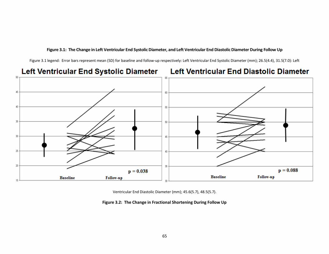
Figure 3.2: The Change in Fractional Shortening During Follow Up
66
Figure 3.2 legend: Error bars
represent mean (SD) for baseline and follow-up respectively: Fractional Shortening (%); 42.9(6.5), 36.1(8.6).
67
Discussion
This study demonstrates that left ventricular systolic function progressively
deteriorates in patients with AFD. Importantly, the change in systolic performance was not
accompanied by a change in left ventricular mass index. This suggests that systolic
function may be a sensitive surrogate marker of disease severity and prognosis in
patients with AFD cardiomyopathy.
Cardiac disease in AFD
The most commonly reported cardiac abnormalities in patients with AFD include
increased QRS voltage and repolarisation changes, short PR interval, QRS prolongation,
concentric left ventricular hypertrophy and valvular thickening 77, 97, 98. Previous isolated
case reports have reported congestive cardiac failure from restrictive cardiomyopathy
and LV dilatation, with one case requiring cardiac transplantation62, 99-103.
To the best of our knowledge, this is the first time that the deterioration in systolic
dysfunction and progression to congestive cardiac failure has been reported in a
prospectively studied cohort of patients with AFD.
Mechanism of systolic dysfunction
Although the genetic and biochemical basis of AFD is well described, the pathophysiology
of cardiac disease in AFD is inadequately understood. Histologically, AFD is
characterised by myocyte vacuolation and intralysosomal inclusions on electron
microscopy 97. However, the consequences of these abnormalities on myocyte
function are not known. Moreover, some data suggest that substrate accumulation
68
accounts for only 3% of the increased LV mass seen in AFD patients 62. Patients with
AFD have areas of interstitial expansion on gadolinium enhanced cardiac magnetic
resonance imaging 63. It is unclear whether this is a primary or secondary
phenomenon, but the presence of interstitial abnormalities suggest that progressive
fibrosis may also contribute to the reduction in systolic performance observed in this
study.
Clinical implications and limitations
Our study suggests that systolic function may be a surrogate marker of disease severity
in AFD. Weidemann et al have recently shown that enzyme replacement therapy using a
recombinant α-galactosidase A preparation improves longitudinal and radial strain rate
in patients with AFD 74. Strain rate imaging is a new technique that quantifies changes
in regional myocardial deformation, and is thought to be a sensitive measure of
myocardial function 104. Our findings and Weidemann et al study support the
hypothesis that systolic function may also be a useful surrogate end-point with which
to assess the response to treatment. This study is limited by the small size of the cohort
and the relatively simple assessment of systolic function.
Conclusions
This study suggests that cardiac systolic dysfunction is common in patients with AFD
and hence systolic performance should be monitored in all patients with AFD.

69
Chapter 4
Exercise capacity
70
Abstract
Background
Patients with AFD frequently complain of exercise limitation, but few studies have
quantified its severity and relation to disease severity.
Methods and Result
Cardiopulmonary exercise testing (CPET) was performed in 103 consecutive patients
(mean age 43.2±14.8 years, 53 (51.5% male) with AFD; the relation between gas
exchange parameters, cardiovascular manifestations and other markers of disease
severity was determined. Oxygen consumption at peak exercise (pVO2) was less than
80% of predicted maximum (%VO2max) in 51 patients (33 (64.7%) male, 18 (35.3%)
female; p=0.01). Males had lower % predicted VO2max (p=0.001), % predicted peak
work rate (p<0.001), and anaerobic threshold (p=0.04) when compared to females.
Stepwise linear regression identified Mainz severity score index (MSSI) score as the only
independent predictor of % predicted VO2 max (β=-1.02, p<0.001).
Conclusions
Males with AFD have more severe exercise limitation when compared to females. The
utility of pVO2 as a surrogate marker of disease severity warrants further investigation.
Aim
Many patients with AFD die prematurely in the fourth and fifth decade of life from
stroke, heart failure and renal failure, but throughout life, experience fatigue and
exercise limitation 105. The aim of this study was to determine exercise capacity using
71
symptom limited cardiopulmonary exercise testing (CPET) in a large referral population
of patients with AFD and its relation to markers of disease severity.
Methods
One hundred and eleven consecutively referred patients (mean age 43.0±15.6 years;
56 (50.5%) males) with AFD were assessed at the Heart Hospital, University College
Hospitals, London, between January 1993 and February 2005. The diagnosis of AFD was
based on plasma A-Gal levels and mutational analysis. Sixty-two patients (55.9%, 44.9
(12.4) years, 45 (81.8%) males) were receiving enzyme replacement therapy. All
patients underwent clinical history and physical examination. The following cardiac
symptoms were noted: angina (exertional central chest pain lasting ≤15 minutes);
dyspnoea (graded using New York Heart Association criteria, NYHA); syncope and
palpitation.
All patients underwent clinical examination, supine 12-lead electrocardiography (ECG)
(Hewlett-Packard, USA) and 24 hour ambulatory ECG (Reynolds Medical, UK)
monitoring as described in chapter 2.
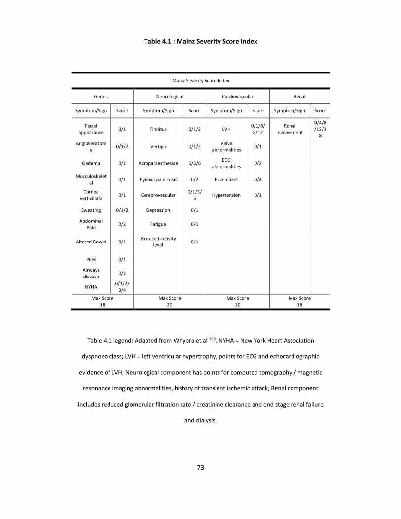
Mainz severity score index (MSSI)
In brief, the MSSI scoring system comprises four components: general, neurological,
cardiovascular, and renal106. Each component consists of a group of signs and
symptoms associated with AFD, weighted according to their contribution to morbidity.
For each sign and symptom, a score is assigned and summed to produce a total score
72
for that system component. These individual component scores are then totaled to give
the final MSSI score (Table 4.1).
Cardiopulmonary testing
Patients were exercised in the upright position using a bicycle ergometer (Ergometrics
800S, SensorMedics, Inc., Yorba Linda, California, USA) with continuous 12-lead
electrocardiographic monitoring (Max 1, Marquette Electronics Inc., Milwaukee,
Wisconsin).
Breath-by-breath respiratory gas sampling was performed using a VMax 229 Console,
(SensorMedics, Inc). Patients were exercised using a 10 to 25 W/min incremental ramp
protocol adjusted to patients’ subjective assessment of their exercise capacity. Patients
cycled at a constant rate of 60 to 65 rpm to the point of maximum effort. Respiratory
gases were sampled continuously from a mouthpiece and analyzed using a 1111D/000
paramagnetic transducer for oxygen and a 2900 MMC non-dispersive infrared sensor
for carbon dioxide.
73
Table 4.1 : Mainz Severity Score Index
Mainz Severity Score Index
General Neurological Cardiovascular Renal
Symptom/Sign Score Symptom/Sign Score Symptom/Sign Score Symptom/Sign Score
Facial appearance
0/1 Tinnitus 0/1/2 LVH 0/1/6/8/12
Renal involvement
0/4/8/12/1
8
Angiokeratoma
0/1/2 Vertigo 0/1/2 Valve
abnormalities 0/1
Oedema 0/1 Acroparaesthesiae 0/3/6 ECG
abnormalities 0/2
Musculoskeletal
0/1 Pyrexia pain crisis 0/2 Pacemaker 0/4
Cornea verticillata
0/1 Cerebrovascular 0/1/3/
5 Hypertension 0/1
Sweating 0/1/2 Depression 0/1
Abdominal Pain
0/2 Fatigue 0/1
Altered Bowel 0/1 Reduced activity
level 0/1
Piles 0/1
Airways disease
0/2
NYHA 0/1/2/
3/4
Max Score 18
Max Score 20
Max Score 20
Max Score 18
Table 4.1 legend: Adapted from Whybra et al 106. NYHA = New York Heart Association
dyspnoea class; LVH = left ventricular hypertrophy, points for ECG and echocardiographic
evidence of LVH; Neurological component has points for computed tomography / magnetic
resonance imaging abnormalities, history of transient ischemic attack; Renal component
includes reduced glomerular filtration rate / creatinine clearance and end stage renal failure
and dialysis.
74
The signals underwent analogue to digital conversion for the calculation of oxygen
consumption (VO2) and carbon dioxide output (VCO2) using an established technique107,
108. A printout of VO2 (l/min), VCO2 (l/min), heart rate (HR) (beats per min), work rate
(W) and respiratory quotient (RQ) was obtained and averaged at 10 s intervals to obtain
smooth graphical representation. Plots of VCO2 against VO2 were used to estimate
anaerobic threshold (AT). Blood pressure was recorded at 2-minute intervals during
exercise and for 10 minutes into recovery using a brachial cuff and
sphygmomanometer. Change in blood pressure (defined as peak exercise SBP – pre-
exercise SBP) was recorded. All tests were supervised by an experienced senior
physiologist.
The following calculations were made:
1. Peak oxygen consumption (pVO2) was defined as the highest oxygen consumption
achieved during exercise. This was the highest measured VO2 value over the last 10 s
of exercise. Data were presented as values normalized for body weight (ml/kg/min).
The predicted maximal oxygen consumption (VO2 max) was calculated using
established equations based on age and gender109, 110. The normalized value was then
expressed as a percentage of the predicted maximum value. Values <80% were
considered abnormal because they fall below previously established 95% confidence
limits 111.
2. Anaerobic threshold is the VO2 above which aerobic energy production is
supplemented by anaerobic mechanisms and is reflected by an increase in lactate and
lactate/pyruvate ratio in the muscle and arterial blood 111 21. This can be estimated
75
noninvasively by plotting VCO2 versus VO2 (“V slope method”; 112. The AT was expressed
as a percentage of the predicted VO2 max. Values<40% predicted VO2 max were
considered abnormal 112.
3. Oxygen pulse (O2P) is the ratio of VO2 to heart rate (HR) (equation 1). It is the product
of stroke volume (SV) and the systemic arteriovenous oxygen difference (A-V) O2
(equation 2).
O2P=VO2/HR (1)
VO2 max=Cardiac output×(A-V) O2
Cardiac output=SV×HR
O2P=SV×(A-V) O2. (2)
Values were expressed as a percentage of the maximum predicted O2P calculated from
equation 1 using established formulae for predicted VO2 max 109, 110 and predicted
maximum heart rate 113, 114. Values >80% predicted VO2 max were regarded as normal
if the individual had achieved a maximal heart rate of at least 80% to exclude
chronotropic incompetence, because this is associated with falsely high readings 113, 114.
Statistical analysis
Statistical analysis was performed using SPSS v19.0 (SPSS, Inc., Chicago, Illinois, USA).
Analyses were stratified for gender. The chi-square test was used to compare non-
continuous variables and the two-tailed, independent-samples student t test was used
to compare continuous variables. All values were expressed as mean ± SD. Stepwise
linear regression analysis was used to identify independent predictors of pVO2 using
76
those variables significantly associated with pVO2 in a univariate analysis. Statistical
significance was defined as p <0.05.
Results
CPET data were unavailable in 8 (7 %) of the 111 patients. The reasons were as follows:
3 patients were less than 16 years of age (all female); 1 was 80 years of age (female); 2
were wheelchair bound (of which one was male); 1 had fast atrial fibrillation at
presentation (male) and 1 refused testing (female).
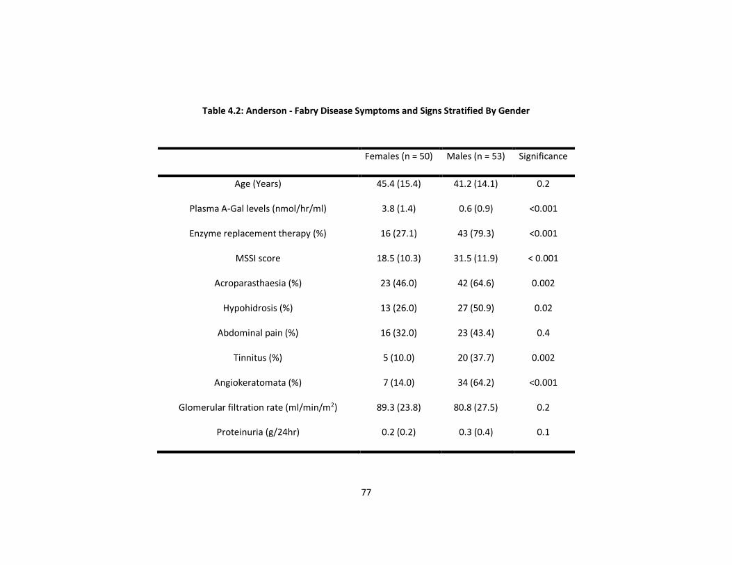
Baseline characteristics of the final study cohort (n=103) are shown in Table 4.2. Males
had a higher prevalence of AFD symptoms and higher MSSI scores compared to females
(mean difference = 13.0, 95% CI = 8.5, 17.4, p < 0.001). Males tended to have higher
levels of proteinuria and lower glomerular filtration rates when compared to females
(Table 4.2).
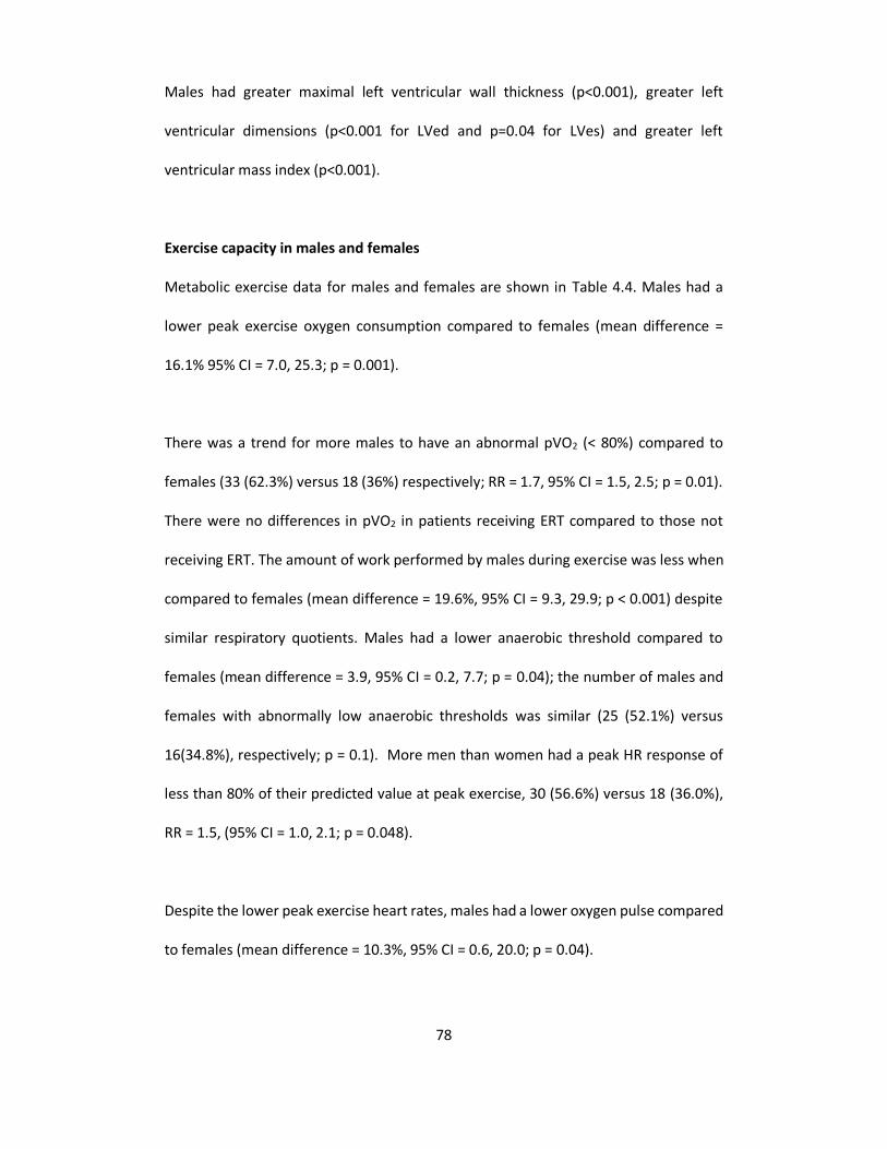
Cardiac disease
Cardiac findings in males and females are summarized in Table 4.3. There was no
difference in the prevalence of cardiovascular symptoms between males and females.
Males tended to have lower resting heart rates compared to females (mean difference
= 5.5 bpm, 95% CI = -0.1, 11.1; p = 0.05). The QRS duration was greater in males
compared to females (mean difference = 15.7 ms, 95% CI = 8.4, 23.0; p < 0.001). Males
had a higher RE score compared to females (mean difference = 2.6, 95% CI = 1.5, 3.7; p
< 0.001, Table 4.2).
77
Table 4.2: Anderson - Fabry Disease Symptoms and Signs Stratified By Gender
Females (n = 50) Males (n = 53) Significance
Age (Years) 45.4 (15.4) 41.2 (14.1) 0.2
Plasma A-Gal levels (nmol/hr/ml) 3.8 (1.4) 0.6 (0.9) <0.001
Enzyme replacement therapy (%) 16 (27.1) 43 (79.3) <0.001
MSSI score 18.5 (10.3) 31.5 (11.9) < 0.001
Acroparasthaesia (%) 23 (46.0) 42 (64.6) 0.002
Hypohidrosis (%) 13 (26.0) 27 (50.9) 0.02
Abdominal pain (%) 16 (32.0) 23 (43.4) 0.4
Tinnitus (%) 5 (10.0) 20 (37.7) 0.002
Angiokeratomata (%) 7 (14.0) 34 (64.2) <0.001
Glomerular filtration rate (ml/min/m2) 89.3 (23.8) 80.8 (27.5) 0.2
Proteinuria (g/24hr) 0.2 (0.2) 0.3 (0.4) 0.1
78
Males had greater maximal left ventricular wall thickness (p<0.001), greater left
ventricular dimensions (p<0.001 for LVed and p=0.04 for LVes) and greater left
ventricular mass index (p<0.001).
Exercise capacity in males and females
Metabolic exercise data for males and females are shown in Table 4.4. Males had a
lower peak exercise oxygen consumption compared to females (mean difference =
16.1% 95% CI = 7.0, 25.3; p = 0.001).
There was a trend for more males to have an abnormal pVO2 (< 80%) compared to
females (33 (62.3%) versus 18 (36%) respectively; RR = 1.7, 95% CI = 1.5, 2.5; p = 0.01).
There were no differences in pVO2 in patients receiving ERT compared to those not
receiving ERT. The amount of work performed by males during exercise was less when
compared to females (mean difference = 19.6%, 95% CI = 9.3, 29.9; p < 0.001) despite
similar respiratory quotients. Males had a lower anaerobic threshold compared to
females (mean difference = 3.9, 95% CI = 0.2, 7.7; p = 0.04); the number of males and
females with abnormally low anaerobic thresholds was similar (25 (52.1%) versus
16(34.8%), respectively; p = 0.1). More men than women had a peak HR response of
less than 80% of their predicted value at peak exercise, 30 (56.6%) versus 18 (36.0%),
RR = 1.5, (95% CI = 1.0, 2.1; p = 0.048).
Despite the lower peak exercise heart rates, males had a lower oxygen pulse compared
to females (mean difference = 10.3%, 95% CI = 0.6, 20.0; p = 0.04).
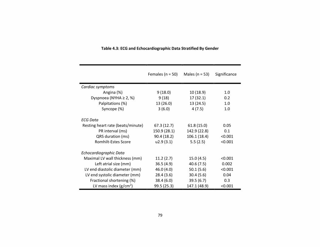
79
Table 4.3: ECG and Echocardiographic Data Stratified By Gender
Females (n = 50) Males (n = 53) Significance
Cardiac symptoms Angina (%) 9 (18.0) 10 (18.9) 1.0
Dyspnoea (NYHA ≥ 2, %) 9 (18) 17 (32.1) 0.2 Palpitations (%) 13 (26.0) 13 (24.5) 1.0
Syncope (%) 3 (6.0) 4 (7.5) 1.0 ECG Data Resting heart rate (beats/minute) 67.3 (12.7) 61.8 (15.0) 0.05
PR interval (ms) 150.9 (28.1) 142.9 (22.8) 0.1 QRS duration (ms) 90.4 (18.2) 106.1 (18.4) <0.001
Romhilt-Estes Score u2.9 (3.1) 5.5 (2.5) <0.001 Echocardiographic Data
Maximal LV wall thickness (mm) 11.2 (2.7) 15.0 (4.5) <0.001 Left atrial size (mm) 36.5 (4.9) 40.6 (7.5) 0.002
LV end diastolic diameter (mm) 46.0 (4.0) 50.1 (5.6) <0.001 LV end systolic diameter (mm) 28.4 (3.6) 30.4 (5.6) 0.04
Fractional shortening (%) 38.4 (6.0) 39.5 (6.7) 0.3 LV mass index (g/cm2) 99.5 (25.3) 147.1 (48.9) <0.001
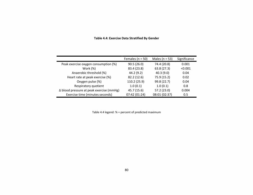
80
Table 4.4: Exercise Data Stratified By Gender
Females (n = 50) Males (n = 53) Significance
Peak exercise oxygen consumption (%) 90.5 (26.0) 74.4 (20.8) 0.001 Work (%) 83.4 (23.8) 63.8 (27.3) <0.001
Anaerobic threshold (%) 44.2 (9.2) 40.3 (9.0) 0.04 Heart rate at peak exercise (%) 82.2 (12.6) 75.9 (15.2) 0.02
Oxygen pulse (%) 110.2 (25.9) 99.8 (22.7) 0.04 Respiratory quotient 1.0 (0.1) 1.0 (0.1) 0.8
Δ blood pressure at peak exercise (mmHg) 45.7 (15.6) 57.2 (23.0) 0.004 Exercise time (minutes:seconds) 07:42 (01:24) 08:01 (02:37) 0.5
Table 4.4 legend: % = percent of predicted maximum

81
Figure 4.1: Correlation between percentage predicted VO2 max and MSSI score
82
Males had a larger increase in systolic blood pressure during exercise compared to
females (mean difference = 12.0mmHg, 95% CI = 4.2, 19.7; p = 0.003).
Clinical correlates of pVO2
There was no relation between pVO2 and a history of chest pain, palpitations or
syncope. Patients with dyspnoea (NYHA class ≥ II) had a lower pVO2 compared to those
without (mean difference = 16.4%, 95% CI = 5.6, 27.2; p = 0.003). There was a negative
correlation between percentage predicted VO2 max and MSSI score (Figure 4.1; r = -
0.5, p < 0.001). There was no correlation between percentage predicted VO2 max and
the degree of proteinuria (quantified through 24 hour urine collection) or GFR
(measured using EDTA nuclear medicine techniques).
There was a negative correlation between percentage predicted VO2 max and the QRS
duration on ECG and the RE score (r = - 0.3 and r = - 0.3; p = 0.006 and p = 0.001
respectively). There were significant correlations between percentage predicted VO2
max and maximal LV wall thickness, left atrial size and LV mass index (r = - 0.2, - 0.3, -
0.2; p = 0.04, 0.007, 0.02 respectively).
To identify the clinical predictors of percentage predicted pVO2, a stepwise regression
analysis model was used. Co-variates entered into the model were those that were
identified as being significantly associated with percentage predicted VO2 max; namely:
gender, NYHA class ≥ II, MSSI score, QRS duration, RE score, maximal LV wall thickness,
LA size and LV mass index. Using this model the only independent clinical predictor of
percentage predicted VO2 max was MSSI score (β = - 1.0, 95% CI = - 0.6, -1,4; p < 0.001).
83
Discussion
This study shows that the majority of men and a substantial minority of women with
AFD have objective evidence for exercise limitation. The degree of impairment
correlates with overall disease severity.
There are many potential mechanisms of exercise limitation in patients with AFD.
Glycosphingolipid accumulation in cardiomyocytes and myocardial fibrosis are
common in adults with AFD115, 116. By contributing to abnormal myocardial contractility
and diastolic dysfunction, these abnormalities would be expected to impair stroke
volume response to exercise and result in elevated left atrial pressure, thereby leading
to pulmonary congestion and exertional dyspnea. In this study, many patients had
diminished heart rate responses which contributed to a reduced cardiac output at peak
exercise. The mechanism of this chronotropic incompetence is unknown, but it
probably reflects the effect of Gb3 accumulation in the sinoatrial node and specialized
conduction system of the heart 117 as well as impaired autonomic function 118. Valvular
disease is also common in patients with AFD, and could theoretically contribute to
exercise limitation if severe 61.
In this study a number of exercise, ECG and echocardiographic parameters were
significantly worse in males compared to females. This would be consistent with more
severe disease manifestations in hemizygous males compared to heterozygous
females. There was however no significant difference in cardiac symptoms between
males and females which is consistent with previous data119.
84
Mild airflow obstruction is common in patients with AFD 120. It is similar to the fixed
obstruction that is seen in COPD with infiltration of inflammatory cells, smooth muscle
hyperplasia, uncoupling of smooth muscle from the adventitia, bronchial wall
hypertrophy and fibrosis being potential mechanisms 120. This was not quantified in this
study but may have impacted on pVO2.
The percentage of predicted VO2 max correlated significantly with MSSI score, ECG
parameters (RE score and QRS duration) and echocardiographic parameters (maximal
LV wall thickness, left atrial size and LVMI). However, the only independent predictor
of pVO2 was found to be MSSI score, This is perhaps not surprising given that both MSSI
and pVO2 take account of multiple organ systems affected by AFD, but suggests that
cardiopulmonary exercise testing may be a useful method for assessing disease severity
and perhaps the response to enzyme replacement therapy.
In conclusion, objective impairment of exercise capacity is common in patients with
AFD and may be a useful marker of disease severity which deserves further
investigation.

85
Chapter 5
Coronary microvascular
function, its relation to
symptoms and response to
enzyme replacement therapy

86
Abstract
Background
In chapter 3, I described how patients with AFD develop progressive systolic
dysfunction. AFD cardiomyopathy is also associated with myocardial fibrosis. These
phenomena could be secondary to coronary microvascular dysfunction. Aim of this
study was to measure coronary flow reserve (CFR), an index of microvascular function,
in AFD at baseline and after enzyme replacement therapy (ERT).
Methods and results
Myocardial blood flow (MBF) at rest and during hyperaemia (adenosine 140 μg/kg/min)
was measured in 10 male, non-smoking patients (53.8±10.9 years, cholesterol 5.5±1.3
mmol/l) and 24 age matched male, non-smoking controls (52.0±7.6 years, cholesterol
4.5±0.6 mmol/l) using positron emission tomography (PET). Resting and hyperaemic
MBF and CFR (hyperaemic/resting MBF) were reduced in patients compared to controls
(0.99±0.17 vs. 1.17±0.25 ml/g/min, p<0.05; 1.37±0.32 vs. 3.44±0.78 ml/g/min,
p<0.0001 and 1.410.39 vs. 3.030.85, p<0.0001, respectively). This coronary
microvascular dysfunction was independent of cholesterol levels. A repeat PET scan
was carried out in 5 patients after 10.1±2.3 months of ERT; resting and hyperaemic
MBF and CFR were unchanged after ERT (0.9930.16 vs. 0.9910.16 ml/g/min, p=ns;
1.560.29 vs. 1.710.3 ml/g/min, p=ns and 1.60.37 vs. 1.740.28, p=ns respectively).
Conclusions
The results of the present study demonstrate that patients with AFD have markedly
abnormal coronary microvascular function. Our preliminary data suggest that ERT has

87
no effect on coronary microvascular dysfunction. Further work is necessary to
determine whether treatment at an earlier stage in the course of the disease may
improve coronary microvascular function in AFD patients.
88
Aims
Patients with AFD complain of angina despite angiographically normal coronary
arteries. I have previously described a progressive deterioration in left ventricular
systolic function and a previous study from this group has demonstrated myocardial
scarring in patients with AFD cardiomyopathy63, 121. The aim of this study was to see if
these clinical abnormalities may be caused by coronary microvascular dysfunction.
The aims of this study were to measure coronary flow reserve (CFR), an index of
coronary microvascular function, in a consecutive cohort of patients with AFD using
positron emission tomography (PET), and to determine the effect of ERT on
microvascular abnormalities.
Methods
Patient and Controls
Ten non-smoking male patients with AFD (53.8 ± 10.9 years, range 43-82, cholesterol
5.5±1.3 mmol/l) referred to the Heart Hospital, London, UK were studied. The diagnosis
of AFD was based on the identification of an -Gal gene mutation and a low plasma -
Gal (mean 0.45 0.35 nmol/hr/ml, range 0.04-0.99 nmol/hr/ml). Nine patients were
identified through screening of patients presenting with unexplained left ventricular
hypertrophy (LVH). One patient was referred with an established diagnosis of AFD (#
6), and 1 was identified through family screening; (# 10). At the time of diagnosis, none
of these patients were receiving ERT.
89
All patients had ECG and 2-D/Doppler echocardiography performed using the
previously described methods in chapter 2. Nine patients had symptoms and/or signs
suggestive of myocardial ischaemia and underwent coronary angiography to exclude
coronary artery disease.
Twenty four healthy non-smoking age matched males (52.08 years, cholesterol
4.5±0.6 mmol/l) with normal electrocardiograms (ECG), and no evidence of cardiac
disease served as controls for the MBF and CFR data.
Enzyme Replacement Therapy
Five patients (Table 5.1) received FabrazymeTM (Genzyme Corporation, Cambridge MA,
USA) at a dose of 1 or 2 mg/kg every two weeks as part of a separate randomised study.
Follow up PET scans were obtained 17.11.9 months after the baseline scan while
patients were on treatment (mean 10.1±2.3 months on ERT). Plasma
globotriaosylceramide (neutral glycosphingolipid) levels were measured using tandem
mass spectrometry pre- and post ERT122.
The study was approved by the Local Research Ethics Committees and all participants
gave written informed consent. Radiation exposure was approved by the UK
Administration of Radioactive Substances Advisory Committee (ARSAC).
Positron Emission Tomography (PET)
All patients and controls underwent PET scanning to measure MBF at rest and during
adenosine-induced hyperaemia (140 μg/kg/min i.v.). Scanning was performed with an
90
ECAT 931-08/12 15-slice tomograph with a 10.5-cm axial field of view (CTI/Siemens,
Knoxville, TN). Resting and hyperemic MBF were measured using oxygen-15 labeled
water (H215O) as previously reported123. Arterial blood pressure was recorded by
automatic cuff sphygmomanometer at one-minute intervals and the ECG was
monitored continuously throughout the procedure.
PET Data Analysis
All emission and transmission data were reconstructed using a Hanning filter with a
cut-off frequency of 0.5 units of the reciprocal of the sampling interval of the projection
data resulting in an image resolution of 8.4x8.3x6.6 mm full width at half maximum at
the centre of the field of view. Myocardial and blood pool images were then generated
directly from the dynamic H215O study as previously reported124. Regions of interest
were drawn within the left atrium and ventricular myocardium on consecutive image
planes. These were projected onto the dynamic H215O images to generate blood and
tissue time activity curves. Arterial and tissue activity curves were fitted to a single tis-
sue compartment tracer kinetic model to give values of MBF (ml/g/min)124. Coronary
resistance (mmHg/ml/min/g) was calculated as the ratio of mean arterial pressure to
MBF and CFR as the ratio of hyperaemic MBF to resting MBF.
Statistical Analysis
Comparisons between continuous variables were performed using the paired Student
t-test (SPSS v19.0; SPSS, Inc., Chicago, Illinois, USA). Linear regression was performed
to assess the relationship between cholesterol levels, LVM index, MBF and CFR. Data
are reported as mean ± SD values. A p<0.05 was considered significant.
91
Results
Patient Characteristics
The clinical characteristics of the patients are shown in Table 5.1. Six (60%) patients
complained of exertional chest pain. None of the nine patients had angiographically
significant coronary artery disease. Three patients had rate responsive dual chamber
permanent pacemakers: one for left ventricular outflow tract gradient reduction and
two for heart block. None of the patients had a PR interval less than 120ms. Two
patients had non-sustained ventricular tachycardia on Holter (# 2 and 3), and 2 were in
atrial fibrillation (# 5 and 11). Seven years prior to the diagnosis of AFD, patient #1 had
undergone an assessment of coronary sinus pH during dipyridamole infusion125. The
peak change in coronary sinus pH during hyperaemia was -0.086 units, indicating
severe myocardial ischaemia.
Serum cholesterol was higher in the patients compared to controls (mean difference
0.97 mmol/l, 95% CI = 0.3-1.6, p = 0.006). Patients also had higher HDL cholesterol
levels compared to controls (1.7±0.5 mmol/l vs. 1.1±0.4 mmol/l, respectively, p =
0.002). There were no differences in LDL cholesterol (3.1±0.8 mmol/l vs. 3.7±1.3
mmol/l, patients vs. controls respectively, p = 0.2) and triglyceride levels (1.4±0.7
mmol/l vs. 1.8±1.6 mmol/l, patients vs. controls respectively, p = 0.5).
Two patients had acroparaesthesiae, 1 had hypohidrosis and 1 had renal dysfunction
(creatinine clearance 52 ml/min). None of the patients were hypertensive and one was
a type 2 diabetic. No angiokeratomata were noted in any of the patients. All patients

92
had a maximal left ventricular wall thickness 13mm (17 4.0 mm, range 14-26 mm).
Nine patients had concentric left ventricular hypertrophy (LVH) and 1 had asymmetric
septal hypertrophy. At the time of study no patient had left ventricular outflow tract
obstruction; the mean left ventricular end-systolic, end-diastolic and left atrial
diameters were 330.9, 5208 and 47 11 mm respectively. The mean LVM index was
23481.3g/m2 (range 141- 407 g/m2).
Myocardial Blood Flow and Coronary Flow Reserve
Heart rate and mean arterial blood pressure were similar in controls and AFD patients
at rest and during adenosine infusion. The rate pressure product (RPP= systolic blood
pressure x heart rate) was similar in the controls and patients during all study
conditions.
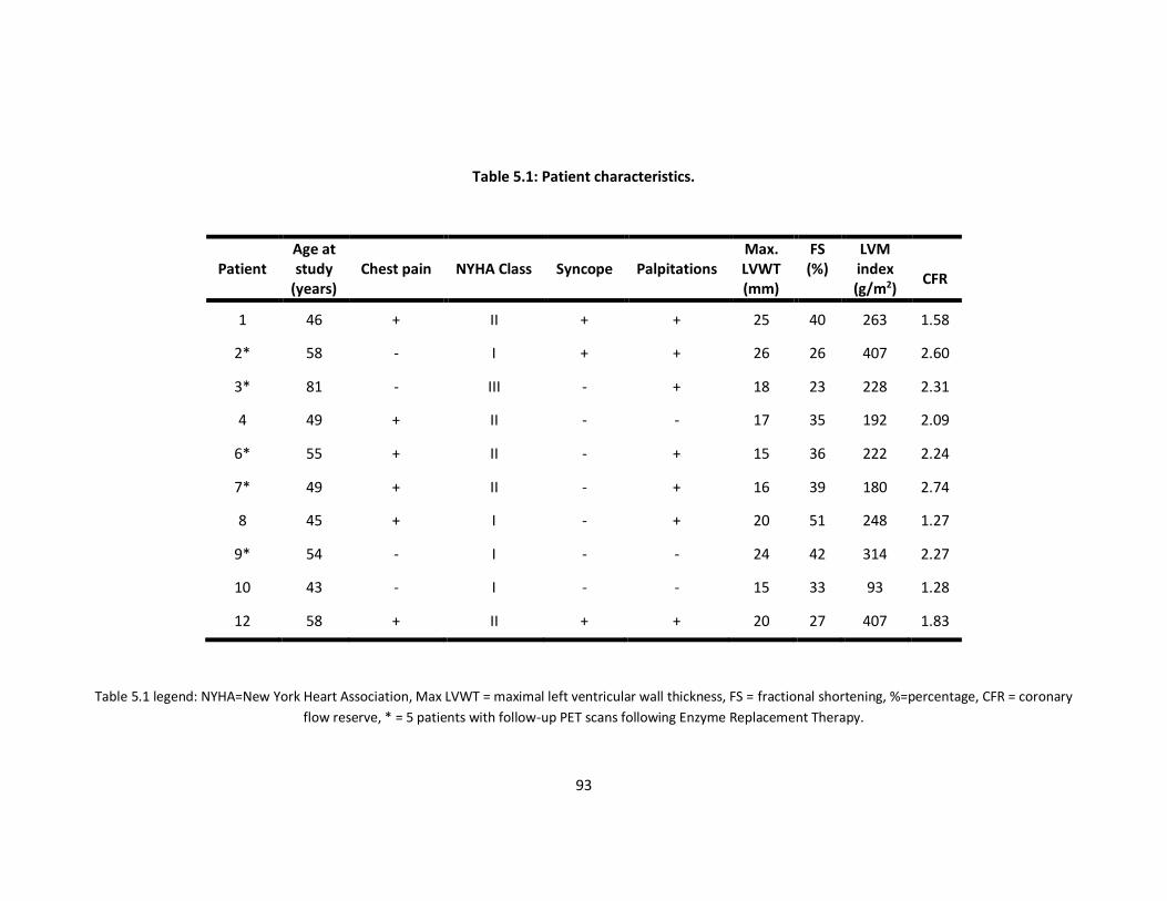
93
Table 5.1: Patient characteristics.
Patient Age at study
(years) Chest pain NYHA Class Syncope Palpitations
Max. LVWT (mm)
FS (%)
LVM index (g/m2)
CFR
1 46 + II + + 25 40 263 1.58
2* 58 - I + + 26 26 407 2.60
3* 81 - III - + 18 23 228 2.31
4 49 + II - - 17 35 192 2.09
6* 55 + II - + 15 36 222 2.24
7* 49 + II - + 16 39 180 2.74
8 45 + I - + 20 51 248 1.27
9* 54 - I - - 24 42 314 2.27
10 43 - I - - 15 33 93 1.28
12 58 + II + + 20 27 407 1.83
Table 5.1 legend: NYHA=New York Heart Association, Max LVWT = maximal left ventricular wall thickness, FS = fractional shortening, %=percentage, CFR = coronary
flow reserve, * = 5 patients with follow-up PET scans following Enzyme Replacement Therapy.
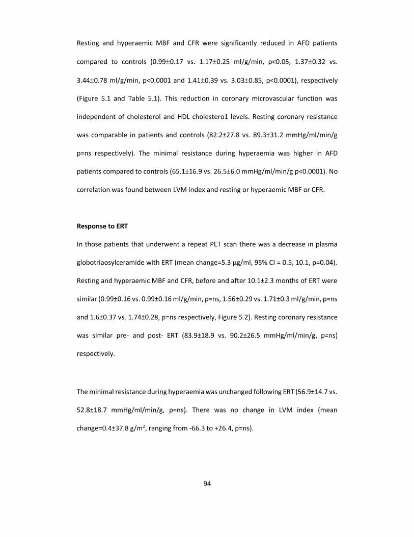
94
Resting and hyperaemic MBF and CFR were significantly reduced in AFD patients
compared to controls (0.990.17 vs. 1.170.25 ml/g/min, p<0.05, 1.370.32 vs.
3.440.78 ml/g/min, p<0.0001 and 1.410.39 vs. 3.030.85, p<0.0001), respectively
(Figure 5.1 and Table 5.1). This reduction in coronary microvascular function was
independent of cholesterol and HDL cholestero1 levels. Resting coronary resistance
was comparable in patients and controls (82.2±27.8 vs. 89.3±31.2 mmHg/ml/min/g
p=ns respectively). The minimal resistance during hyperaemia was higher in AFD
patients compared to controls (65.1±16.9 vs. 26.5±6.0 mmHg/ml/min/g p<0.0001). No
correlation was found between LVM index and resting or hyperaemic MBF or CFR.
Response to ERT
In those patients that underwent a repeat PET scan there was a decrease in plasma
globotriaosylceramide with ERT (mean change=5.3 μg/ml, 95% CI = 0.5, 10.1, p=0.04).
Resting and hyperaemic MBF and CFR, before and after 10.1±2.3 months of ERT were
similar (0.99±0.16 vs. 0.99±0.16 ml/g/min, p=ns, 1.56±0.29 vs. 1.71±0.3 ml/g/min, p=ns
and 1.6±0.37 vs. 1.74±0.28, p=ns respectively, Figure 5.2). Resting coronary resistance
was similar pre- and post- ERT (83.9±18.9 vs. 90.2±26.5 mmHg/ml/min/g, p=ns)
respectively.
The minimal resistance during hyperaemia was unchanged following ERT (56.9±14.7 vs.
52.8±18.7 mmHg/ml/min/g, p=ns). There was no change in LVM index (mean
change=0.4±37.8 g/m2, ranging from -66.3 to +26.4, p=ns).
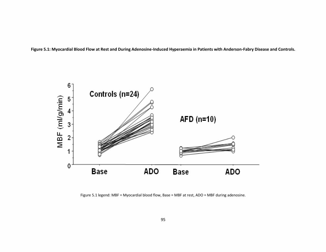
95
Figure 5.1: Myocardial Blood Flow at Rest and During Adenosine-Induced Hyperaemia in Patients with Anderson-Fabry Disease and Controls.
Figure 5.1 legend: MBF = Myocardial blood flow, Base = MBF at rest, ADO = MBF during adenosine.
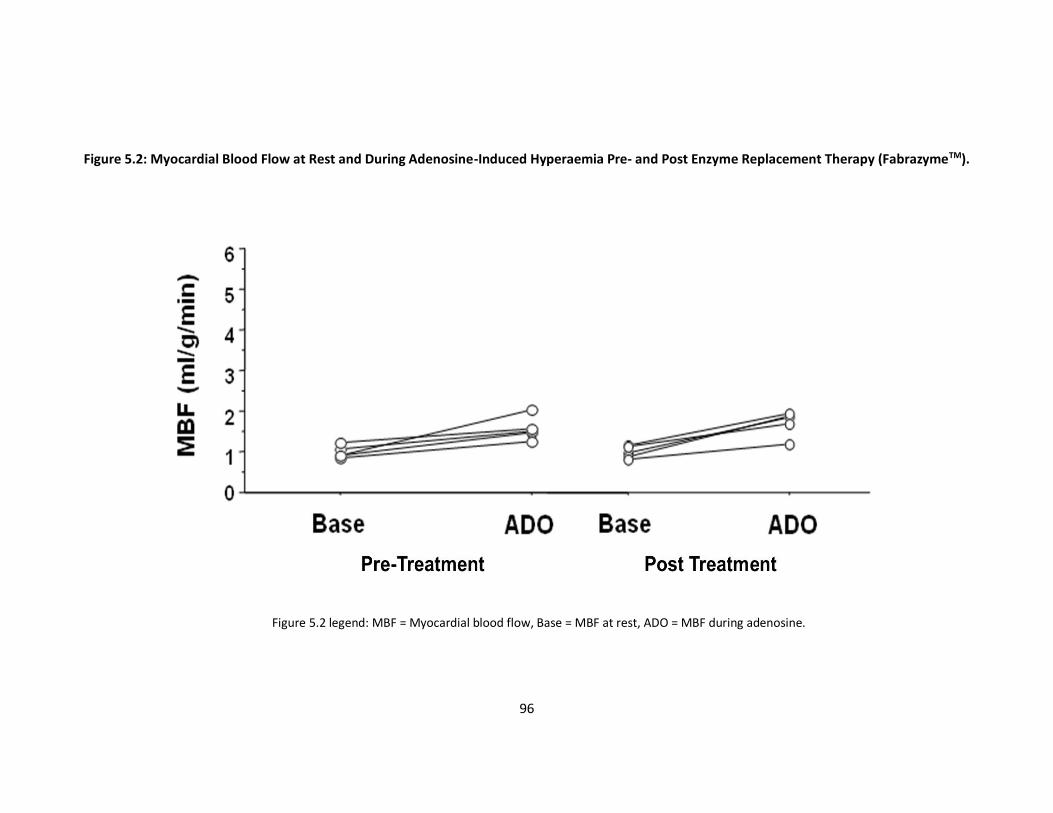
96
Figure 5.2: Myocardial Blood Flow at Rest and During Adenosine-Induced Hyperaemia Pre- and Post Enzyme Replacement Therapy (FabrazymeTM).
Figure 5.2 legend: MBF = Myocardial blood flow, Base = MBF at rest, ADO = MBF during adenosine.
97
There were no changes in the left ventricular cavity dimensions (mean change in end
systolic cavity dimension=0.1±0.3mm, p=ns; mean change in end diastolic cavity
dimension=0.1±0.2mm, p=ns).
There was a trend to a reduction in fractional shortening (mean fractional shortening
pre-ERT=34.8±6.7%, mean post-ERT=31.4±6.3%, p=0.08). There was no change in the
maximal left ventricular wall thickness (mean change=0.02±0.3mm, p=ns).
Discussion
This study shows that patients with AFD have a substantial reduction in hyperaemic
MBF and CFR compared to normal controls. The significance of these findings for
individual patients remains to be determined, but the severity of the microvascular
dysfunction suggests that these abnormalities may have a substantial influence on the
natural history of AFD.
Mechanisms of Microvascular Dysfunction
Symptoms and signs suggestive of myocardial ischaemia in the absence of coronary
disease are frequent in patients with cardiomyopathies and in individuals with left
ventricular hypertrophy secondary to pressure overload. Reductions in CFR similar to
those seen in this study have been demonstrated in all these conditions, although
different mechanisms are involved. In patients with hypertrophic cardiomyopathy,
remodelling of the intramural arterioles is important126,127, whereas in aortic stenosis,
increased perivascular fibrosis and intramyocardial pressure and reduced diastolic
filling are the dominant mechanisms for microvascular dysfunction128, 129.
98
In AFD, a number of mechanisms may contribute to microvascular dysfunction. AFD
cardiomyopathy is characterised by GB3 deposition in myocytes, conduction tissue,
vascular endothelium and valvular tissue. This is accompanied by secondary changes
such as myocyte hypertrophy and fibrosis130 which cause raised coronary vascular
resistance and increased myocardial oxygen demand. Although endothelial GB3
deposits may lead to microvascular dysfunction, a recent publication has shown that
patients with AFD have enhanced nitric oxide independent endothelial function
measured using forearm venous plethysmography131. It remains to be seen whether
similar abnormalities are seen in the coronary microvasculature.
Response to ERT
In this study there was a significant reduction in the plasma levels of
globotriaosylceramide with ERT, but no improvements in coronary microvascular
function or LVM were seen. However, interpretation of this observation is limited by
the small size of the cohort and by the fact that the cohort was considerably older, and
had more severe cardiac disease compared to patients included in previous trials of
ERT73, 75, 132. Along with an improvement in AFD symptoms these trials have
demonstrated a reduction in LVM and an improvement in systolic function with ERT. In
order to understand the impact of ERT on coronary microvascular function a larger
study conducted over a longer period of time is required.

99
Conclusion
This study shows that microvascular dysfunction is important in the pathophysiology of
AFD cardiomyopathy. Further studies are required to assess the impact of enzyme
replacement therapy on microvascular function.

100
Chapter 6
Extracellular matrix turnover in
Anderson-Fabry disease
101
Abstract
Background
Patients with AFD develop progressive left ventricular (LV) remodelling and heart
failure. We hypothesized that altered extracellular matrix turnover (ECM) contributes
to the pathophysiology of cardiac disease in AFD.
Methods and results
Twenty-nine (44.1±11.7 years, 15 male) consecutive patients with AFD and 21 normal
controls (39.7±11.3 years, 10 male) had serum analysed for matrix metalloproteinase-
9 (MMP-9), and tissue inhibitor of matrix metalloproteinase 1 and 2 (TIMP-1, TIMP-2).
All patients underwent clinical assessment, echocardiography and Mainz Severity Score
Index measurement (MSSI); a validated severity score in AFD.
MMP-9 levels were significantly higher in patients than controls (1003.8±337.8ng/ml
vs. 576.7±276.3ng/ml respectively, p < 0.001). There were no differences in TIMP levels
between patients and controls. There was a positive correlation between MMP-9 levels
and MSSI (r = 0.5, p=0.01). There was a negative correlation between MMP-9 and
fractional shortening (FS, r = -0.5, p=0.01). There was no correlation between LV mass
or maximal LV wall thickness and MMP-9 levels. These relations were independent of
age and gender using stepwise linear regression analysis.
Conclusions
Patients with AFD have abnormal ECM turnover compared to normal controls. The
correlation between MMP-9 levels and FS suggests that altered ECM turnover is

102
important in cardiac remodelling. The association between MMP-9 and overall disease
severity suggests that circulating levels of matrix metalloproteinases may provide a
useful marker for assessing the response of patients with AFD to enzyme replacement
treatment.
103
Aim
Previous chapters have described progressive left ventricular dysfunction, significant
arrhythmia and significant exercise limitation.
Matrix metalloproteinases (MMPs) and their endogenous inhibitors, tissue inhibitors
of metalloproteinases (TIMPs) are the key mediators of extracellular matrix (ECM)
turnover, the balance between constitutive enzyme activity and inhibitor activity
ultimately determining ECM composition for a variety of elements. While tissue
composition is the key a variety of studies have suggested that the measurement of
circulating MMPs and TIMPs are associated and provide a non-invasive assessment of
ECM turnover133-135. For example circulating levels of TIMP-1 appear to correlate with
the severity of structural left ventricular hypertrophy and related diastolic filling
abnormalities in hypertensive heart disease136, 137.
The aim of this study was to assess if patients with AFD have abnormal collagen
turnover as compared to age matched normal healthy cohort and to assess its
relationship to overall disease severity.
Methods
The study cohort comprised 29 consecutive patients (mean age 44.1 ± 11.7 years, 15
(60.0%) male and 14 (40.4%) female) with a genetically confirmed diagnosis of AFD and
21 age and gender matched healthy normal controls (mean age 39.7 ± 11.3 years, 10
(47.6%) male and 11 (52.4%) female). The latter were asymptomatic and had no clinical
evidence of vascular, neoplastic, metabolic or inflammatory disease (assessed by
104
history, examination and routine laboratory analysis). All controls were normotensive
(supine blood pressure <140/90 mmHg), and had a normal resting ECG. All were free
of drug therapy.
The study was approved by the local research ethics committee and informed, written
consent was obtained from all patients and controls.
Clinical Assessment
All patients underwent a complete clinical assessment with clinical history,
examination, a standard 12 – lead electrocardiogram (ECG) and echocardiography as
described previously in chapter 2.
Mainz Severity Score Index (MSSI)
In brief, the MSSI scoring system comprises four components: general, neurological,
cardiovascular and renal (chapter 4, Table 4.1)106. Each component consists of a group
of signs and symptoms associated with AFD, weighted according to their contribution
to morbidity. For each sign and symptom, a score is assigned and summed to produce
a total score for that component (chapter 4, Table 4.1). These individual component
scores are then totalled to give the final MSSI score.
MMP-9, TIMP-1 and TIMP-2 Evaluation
Circulating concentrations of serum MMP-9, TIMP-1 and TIMP-2 were measured by an
in house enzyme linked immunosorbent assay as described previously135. In brief,
serum was stored in aliquots at -20ºC and batch analysed for MMP and TIMP.
105
Monoclonal primary and biotinylated secondary antihuman antibody for total MMP-9,
TIMP-1 and TIMP-2 were tested against respective recombinant human MMP and TIMP
(MAB911, BAF911, 911-MP; MAB970, BAF970, 970-TM; MAB9711, BAF971, 971-TM;
R&D Systems Europe Ltd, Abingdon, UK) and against pooled human plasma to
determine the optimal concentration of primary (2 μg/mL, 4 μg/mL and 2 μg/mL
respectively) and secondary antibody (0.1 μg/mL, 0.1 μg/mL and 0.2 μg/mL
respectively) sample dilution (1/100) and standard. The mean intra-assay coefficient of
variation (CV) and mean inter-assay CV for the assays were <5% and <10% respectively.
Statistics
Statistical analysis was performed using SPSS v19.0. The χ2 test was used to compare
non-continuous variables. Normally distributed variables were compared using the 2-
tailed independent sample Student t-test and non-normally distributed variables were
compared using the Man Whitney U test. Correlations for normally distributed
variables were expressed as a Pearson’s correlation coefficient and non-normally
distributed variables as a Spearman’s rho coefficient. All values were expressed as
means (standard deviation) for normally distributed variables and median
(interquartile range) for non-normally distributed variables.
Stepwise linear regression was used to assess the relationship between circulating
MMP and TIMP levels and markers of disease severity were adjusted for covariates. As
females develop signs and symptoms of the disease at older ages, gender was entered
into the regression model; age was entered into the regression model as there is a
progressive accumulation of glycosphingolipid with advancing age.
106
Table 6.1: Baseline Characteristics in Males and Females
Total Female (14) Male (15) p-
value
*TIMP-2 (ng/ml) 210 (190-250) 198.5 (188.8-265) 210 (190-240) 0.9
TIMP-1 (ng/ml) 1029.7 (299.3) 996.8 (276.2) 1060.3 (326.0) 0.6
MMP-9 (ng/ml) 1003.8 (337.8) 887.1 (307.9) 1112.7 (337.5) 0.07
Age (years) 44.1 (11.7) 45.7 (13.5) 42.5 (10.0) 0.5
MSSITOTAL 26.9 (14.7) 17.1 (10.4) 36.0 (12.1) <0.001
MSSIGENERAL 6.5 (4.2) 3.6 (2.1) 9.0 (4.1) <0.001
MSSINEUROLOGICAL 5.6 (4.6) 4.2 (3.4) 6.9 (5.2) 0.1
MSSICARDIAC 8.7 (5.8) 5.2 (5.5) 11.9 (4.0) 0.001
MSSIRENAL 5.5 (5.2) 3.1 (3.6) 7.7 (5.5) 0.01
PR interval (ms) 147.3 (30.1) 156.5 (35.4) 138.7 (22.0) 0.1
QRS Duration (ms) 93.2 (14.3) 82.9 (7.9) 102.9 (12.0) <0.001
Max LV Wall Thickness (mm) 13.4 (4.3) 10.9 (1.8) 15.8 (4.5) 0.001
*RWT 0.5 (0.4-0.6) 0.4 (0.4-0.5) 0.6 (0.5-0.6) 0.01
LVed (mm) 47.4 (4.0) 45.9 (2.2) 48.7 (4.8) 0.06
LVes (mm) 29.0 (3.9) 27.4 (2.7) 30.4 (4.3) 0.04
FS (%) 38.7 (7.5) 40.1 (5.3) 37.3 (9.1) 0.3
*LV Mass Index (g/cm2) 96.8 (91-145) 92.0 (82.6-95.8) 132.4 (101-183) <0.001
Table 6.1 legend: TIMP = tissue inhibitor of matrix metalloproteinases; MMP = matrix
metalloproteinase; MSSI = Mainz Severity Score Index; Max LV = maximal left ventricular; RWT
= relative wall thickness; LVed = left ventricular end diastolic cavity dimension; LVes = left
ventricular end systolic cavity dimension; FS = fractional shortening; * = non-normally
distributed variables comparison made using Man Whitney U test, expressed as median
(interquartile range), normally distributed variables expressed as mean (standard deviation).
107
Other covariates were entered into the model if there was a relationship between the
covariate and MMP or TIMP with a p-value of < 0.05 on univariate analysis. Statistical
significance was defined as p < 0.05.
Results
The clinical characteristics of the patients are shown in Table 6.1. MMP-9 levels were
elevated in patients with AFD by a mean of 427.1 ng/ml (95% CI = 252.1, 602.2 ng/ml,
p < 0.001, Figure 6.1). There were no differences between patients and controls in
TIMP-1 and TIMP-2 levels.
Disease Severity and MMP-9
Table 6.1 describes each of the MSSI component scores stratified by gender. The mean
MSSITOTAL score was higher in men compared to women (19.1, 95% CI = 10.7 to 27.5,
Table 6.1).
Similarly, the mean MSSIGENERAL, MSSICARDIAC and MSSIRENAL but not
MSSINEUROLOGICAL scores were higher in men compared to women (5.4, 95% CI = 2.7
to 7.6; 6.7, 95% CI = 2.9 to 10.4; 4.6, 95% CI = 1.0 – 8.2; 2.7, 95% CI = -0.6 to 6.1
respectively, table 6.1). There was a positive correlation between MMP-9 levels and
MSSITOTAL, MSSIGENERAL and MSSINEUROLOGICAL, scores (r = 0.5, p = 0.01, Figure
6.2; r = 0.4, p = 0.03; r = 0.4, p = 0.047 respectively), but not with MSSICARDIAC or
MSSIRENAL scores.
108
Figure 6.1: Difference in Matrix Metalloproteinase – 9 between Patients and Controls
Figure 6.1 legend: Box and whiskers plot for controls and patients. Boxes represent median ± interquartile range (580ng/ml ± 920 ng/ml for controls and 1100 ng/ml ±
1220 ng/ml for patients, p< 0.001).
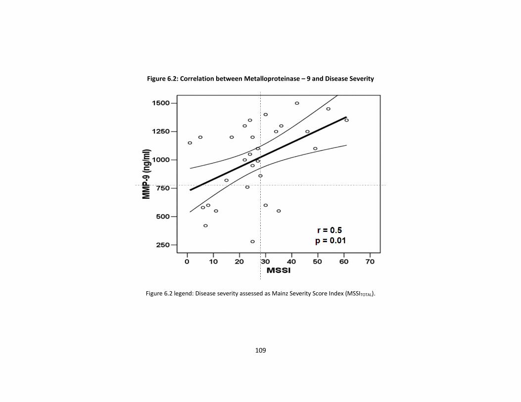
109
Figure 6.2: Correlation between Metalloproteinase – 9 and Disease Severity
Figure 6.2 legend: Disease severity assessed as Mainz Severity Score Index (MSSITOTAL).
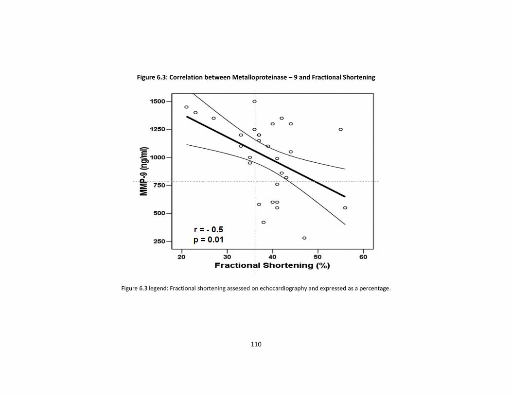
110
Figure 6.3: Correlation between Metalloproteinase – 9 and Fractional Shortening
Figure 6.3 legend: Fractional shortening assessed on echocardiography and expressed as a percentage.
111
Cardiovascular Evaluation
Four of the 29 (13.8%) patients complained of angina, 5/29 (17.2%) were in NYHA class
2 or greater, 8/29 (27.6%) complained of palpitations and none had experienced pre-
syncope/syncope. There were no gender differences in the frequency of cardiac
symptoms.
All patients were in sinus rhythm. The mean PR interval was 147.3 ± 30.1 ms (100 – 223
ms) with a mean QRS duration of 93.2 ± 14.3 ms (72 – 132 ms). Men had a higher QRS
duration when compared to women (mean difference = 19.9 ms, 95% CI = 12.1ms to
27.8ms, p < 0.001, Table 6.1).
Seventeen (58.6%, 12 males and 5 females) patients had evidence of cardiac
remodelling. Eight (27.6%, seven males) had evidence of concentric hypertrophy, 9
(31.0%, 5 males) had concentric remodelling and 12 (41.4%, 3 males) had normal LV
geometry.
The mean maximum LV wall thickness was 13.4 ± 4.3 mm (8 - 26mm), with a mean RWT
of 0.5 ± 0.1 (0.3 - 1.0) and a mean LVMI of 121.3 ± 48.4 gm/m2. Men had higher maximal
LV wall thickness (mean of 4.9mm, 95% CI = 2.3mm to 7.6mm); higher RWT (mean of
0.1, 95% CI = 0.03 to 0.2); and higher LVMI (mean 56.1 gm/m2, 95% CI = 25.8 to 86.4
gm/m2) when compared to women (Table 6.1). Men had a higher LVes, a trend to
higher LVed and no difference in the FS when compared to women (Table 6.1).
There was a negative correlation between MMP-9 levels and FS (r = -0.4, p = 0.02, Figure
6.3). There was a trend to a positive correlation between MMP-9 levels and QRS
112
duration (r = 0.4, p = 0.047) and RWT (r = 0.4, p = 0.06). MMP-9 levels did not correlate
with LVMI or left ventricular cavity dimensions.
Stepwise linear regression demonstrated a relation between MMP-9 levels and MSSI
score and FS that was independent of age and gender (β = 11.0, 95% CI = 3.9 to 18.7, p
= 0.004; β = -20.2, 95% CI = -6.4 to -33.9, p = 0.006).
Discussion
This study demonstrates that patients with AFD have elevated levels of circulating
MMP-9 when compared to normal controls. MMP-9 levels correlated with clinical
markers of disease severity, suggesting that abnormal ECM turnover plays an important
role in the pathogenesis of AFD.
MMP in Heart Disease
The ECM is composed of a wide range of elements including collagen, elastin,
specialised proteins (fibrillin and fibronectin) and proteoglycans. It has an important
role in the maintenance of the structural integrity and function of organs and influences
both intracellular signalling and cell-to-cell interactions. Although the ECM is
ubiquitous, its composition can vary from organ to organ138.
There are more than twenty different MMP’s, each acting on a number of ECM
substrates138, 139. MMP-9, also known as gelatinase, is a 92-kD protein secreted by
myocytes, fibroblasts, smooth muscle cells and neutrophils; its function is to degrade a
number of interstitial proteins including basement membrane components such as
laminin and fibronectin138.
113
Increased MMP expression occurs in several disease states including tumour
angiogenesis and metastasis, rheumatoid arthritis and vascular disease140, 141. It has
been shown recently that circulating levels of MMP-9 predict disease severity and
prognosis in stable and unstable coronary artery disease, peripheral arterial disease
and hypertension134, 142, 143. Myocardial and blood levels of MMP-9 are elevated in
patients with ischaemic and non-ischaemic left ventricular dilatation144, 145. In the low
risk population studied in The Framingham Heart Study blood levels of MMP-9 were
associated with increased left ventricular cavity dimensions and mass index146. TIMP-1
activity is increased in hypertension and diabetes and is associated with myocardial
fibrosis and diastolic dysfunction 137; TIMP-2 has been linked with angiogenesis147.
Significance of ECM Turnover in AFD
Although the genetic and biochemical basis of AFD is well described, the
pathophysiology of cardiac disease in AFD is inadequately understood. Histologically,
AFD cardiomyopathy is characterised by myocyte hypertrophy and vacuolation97.
However, substrate accumulation accounts for only 1% of the increased LV mass seen
in AFD cardiomyopathy62.
The findings in this study support recent data from cardiac magnetic resonance imaging
suggesting that myocardial fibrosis may be a key determinant of cardiac function in
AFD63. The main impact of elevated MMP-9 levels is to increase collagen type I / III
ratio139. Collagen type I is not a major substrate for MMP-9, and hence raised levels of
MMP-9 may reflect the breakdown of components other than collagen type I139.
114
Although TIMP-1 directly inhibits the degradation of collagen type I in the ECM, there
were no differences in circulating levels between patients and controls. It is possible
that elevated MMP-9 levels may directly increase collagen gene expression in the
absence of elevated TIMP-1 levels148. Moreover, it is likely that the balance between
TIMP and MMP activities is more important than abnormalities in either factor in
isolation.
Monitoring of Disease Activity
The recent introduction of enzyme replacement therapy for the treatment of AFD
represents a major advance in the treatment of lysosomal diseases75. A new challenge
for clinicians treating such diseases is the monitoring of response to therapy. In some
diseases there are highly sensitive circulating markers (e.g. chitotriosidase in Gaucher
disease) that can be used; in AFD plasma and urine levels of Gb3 have been used, but
recent evidence suggests that this may not be the most appropriate marker for
monitoring response to treatment122. The association between MMP-9 and overall
disease severity suggest that ECM turnover may be a useful alternative surrogate for
the response to enzyme replacement treatment
Conclusion
Patients with AFD have abnormal ECM turnover; the relation between MMP-9 levels
and disease severity suggests that altered ECM remodelling is central to the
pathogenesis of AFD related complications.

115
Chapter 7
Summary of findings and current
literature
116
Summary of findings and current literature
The aim of this thesis was to perform the first detailed characterisation of cardiac
manifestations of AFD and to explore their relation to outcomes. Many of the findings
were completely novel and have been confirmed in subsequent studies from our own
group and other investigators.
Arrhythmia
In chapter two, I demonstrated that there is an increase in prevalence of both
supraventricular and ventricular arrhythmia with age and that approximately 5% of
patients required a permanent pacemaker at follow up. These findings suggested that
cardiac arrhythmia may be a significant contributor to long-term morbidity and
mortality in AFD. The high contribution of arrhythmia to symptoms has been confirmed
in subsequent studies reporting palpitations or documented arrhythmia in 19.3% of the
population in the international Fabry outcome survey not on ERT and 34.5% of the
population on ERT119. Older patients with AFD are more likely to develop bundle branch
block and atrioventricular conduction/sinus node disease149 and in the FOS registry 3%
of these patients required antibradycardia pacing119. Additionally most males have a
resting bradycardia and chronotropic incompetence (also described in chapter 4)150 and
male children have reduced heart rate variability151.
Work from our own cohort has shown that PR interval and QRS duration increase with
age and are independent predictors of future anti-bradycardia pacing (hazard ratio HR
1.03, 95% CI 1.004–1.060, p = 0.02; HR 1.05, 95% CI 1.02–1.09, p < 0.001 respectively)152.
In this study the annual implant rate for any cardiac device (anti-bradycardia or high

117
energy defibrillator device) was 2.1% (95% CI = 1.2-3.1) with a 5 year cumulative
incidence of 12% (95% CI = 6.1-17.9%)152.
Although the high prevalence of tachyarrhythmia has been documented in chapter 2
and other studies, its impact on the natural history of the disease is still uncertain. In
rare instances it may be the first presentation of AFD cardiomyopathy153. Sudden
cardiac death is rare in AFD, however it is likely from published literature that
bradycardia as well as ventricular tachycardia would contribute95, 154. Weidemann et al
followed 40 patients with advanced AFD on ERT and demonstrated a sudden cardiac
death risk of 15% (6/40, total of 7 deaths in cohort)155. All of these patients had
documented non-sustained ventricular tachycardia and myocardial fibrosis on cardiac
MRI scanning155. Further research is needed to identify those at greatest risk and
institute appropriate preventative therapies for the prevention of sudden cardiac
death156.
Histological examination of the conduction tissue demonstrates that the greater the
involvement of the conduction tissue with GB3 deposition the greater the accelerated
conduction with prolonged refractoriness and the greater the electrical instability76, 157.
With the high prevalence of stroke in patients with AFD it is likely that AF contributes
to this, but there are no data regarding thromboprophylaxis for patients with AFD
cardiomyopathy and documented AF44. The pathophysiology of cerebrovascular
disease is not completely understood, but is likely to be multifactorial including
microvascular disease, increased thrombotic potential, arteriopathy and
118
thromboembolic49-51. Although risk stratification algorithms such as CHA2DS2-VASc
score have been validated in the general population, it is unlikely that this would be
applicable to the AFD population as the age of onset of atrial arrhythmia is much
younger than in the general population91. It would seem intuitive that they should be
considered for formal anticoagulation, particularly if there are high risk factors such as
a mutation in the Factor V Leiden gene90.
Left ventricular systolic function
Chapter 3 described the progressive deterioration in LV systolic function in a small
cohort of patients with severe AFD cardiomyopathy. This change occurred without
change in LV mass index and suggested that systolic function may be a useful marker
for the assessment of disease progression and response to treatment. The relationship
with FS and disease progression is likely to be related to inferior posterior myocardial
scar that is seen on histology and CMR and is a limitation of the study76, 158.
The most common finding in AFD cardiomyopathy is that of LV hypertrophy and is
associated with cardiac symptoms119. The prevalence of LV hypertrophy increases with
age, occurring earlier in males than in females and inversely associated with renal
function119. Early stages of AFD cardiomyopathy are characterised by concentric LV
remodelling which progresses to LV hypertrophy with time, with asymmetric septal
hypertrophy seen in 5% of cases119. Additionally, right ventricular hypertrophy is also
seen, usually in association with LV hypertrophy159. Right ventricular hypertrophy is
seen in equal frequencies in males and females159. It becomes increasingly recognised
with advancing age. The impact of right ventricular hypertrophy on right ventricular

119
function is contradictory in the literature159, 160 and its impact on prognosis remains to
be determined.
While echocardiography remains the mainstay for diagnosis of LV hypertrophy, there
have been surprising few studies that have examining the change in systolic function in
prospective cohorts. Tissue Doppler imaging appears to be a sensitive marker for early
AFD cardiomyopathy with longitudinal velocities being depressed in the lateral and
septal aspects of the mitral valve annulus prior to overt LV hypertrophy116. These tissue
Doppler indices (in-vivo) correlate with resting and active tension in cardiomyocytes
(in-vitro) but not with cardiomyocyte area, % storage of GB3 or % fibrosis161. These
findings suggest that myofilament degradation and dysfunction contribute to systolic
impairment161. Importantly, tissue Doppler indices appear to improve with enzyme
replacement therapy, unless there is advanced AFD cardiomyopathy present74, 162. A
recent study has suggested that a binary appearance of the left ventricular endocardial
border on 2-dimensional echocardiography is a highly sensitive and specific
discriminator of AFD from hypertrophic cardiomyopathy, however our group have not
been able to reproduce this finding163, 164.
Exercise limitation
In chapter 4 I demonstrated that exercise limitation is common in males and in a
substantial minority of females. The degree of impairment was not only related to
severity of cardiac involvement, but also to overall disease severity as expressed by the
MSSI score. This relationship was not influenced by the degree of proteinuria or GFR.
120
Additionally, males tended to have a degree of chronotropic incompetence which may
have contributed to their exercise limitation.
Exercise limitation assessed with cardiopulmonary exercise testing has subsequently
been demonstrated in several studies. Bierer et al. demonstrate a higher proportion
of patients with abnormal peak exercise oxygen consumption as compared to
controls165. The main finding from this study was a significant drop in diastolic blood
pressure in a significant proportion of patients with exercise compared to controls165.
This was not apparent in my study. In a follow up study they demonstrated an
improvement in exercise capacity with ERT166. The drop in diastolic blood pressure
seen in their original study was not replicated in the follow up study nor was there clear
evidence for change in diastolic blood pressure response to exercise with ERT,
particularly in patients included in both studies166. Lobo et al. demonstrate similar
exercise limitation with cardiopulmonary exercise testing, but their follow up cohort
failed to show an improvement in exercise capacity with 1 year of ERT150. These
conflicting data question the utility of peak exercise oxygen consumption as a way of
monitoring response to treatment.
An interesting study by Spinelli et al. where patients with AFD underwent radionuclide
angiography during rest and exercise along with resting echocardiography and cardiac
MRI scanning and compared to controls167. The study demonstrates that the majority
of patients compared to controls had impaired exercise capacity. This impaired exercise
capacity was associated with a failure to augment stroke volume with exercise and
significant proportion having a reduction in stroke volume with exercise. This study
121
suggests that the predominant mechanism for exercise intolerance is cardiac
dysfunction as opposed to pulmonary, musculoskeletal or hypohidrosis.
Coronary microvascular dysfunction
Chapter 5 demonstrates that patients with AFD cardiomyopathy have coronary
microvascular dysfunction when compared to normal controls and that this does not
improve with enzyme replacement therapy, at least in the short-medium term. These
findings have been confirmed by other studies168, 169. Up to 20% of patients experience
anginal symptoms119 despite obstructive coronary artery disease being present in a
minority of patients10. Volumetric intravascular ultrasound analysis of the coronary
tree in patients with AFD and chest pain showed that patients have diffuse involvement
of their coronary arteries with plaques that are less echogenic170. These findings are
probably caused by Gb3 accumulation and disease-specific trophic influences rather
than accelerated atherosclerosis171. Histologically, there is a diffuse storage
arteriopathy affecting all parts of the arterial wall resulting in intimal thickening and
vacuolation as well as medial hypertrophy76. Additionally, there is significant
calcification associated with the arteriopathy, as in classical atherosclerotic disease;
however seems to be present in the media as opposed to the intima170. Chimenti et al.
have suggested that luminal narrowing of intramural arteries (due to GB3 deposition
and hypertrophy in smooth muscle cells and endothelium) correlates with coronary
flow and histological myocardial fibrosis172; suggesting that small vessel disease
contributes to angina symptoms, myocardial fibrosis and potentially progressive heart
failure in AFD cardiomyopathy172.
122
Extracellular matrix turnover
Chapter 6 demonstrates that patients with AFD have abnormal extracellular matrix
(ECM) turnover; the relation between MMP-9 levels and disease severity, as measured
with the Mainz severity score index (MSSI), suggests that altered ECM remodelling is
central to the pathogenesis of AFD related complications. My study demonstrates that
circulating MMP-9 is elevated in patients with AFD. In patients with hypertrophic
cardiomyopathy (HCM) caused by sarcomeric gene mutations, circulating MMP-2 levels
and not MMP-9 levels are positively associated with left ventricular end systolic
diameter, left atrial dimension and left ventricular systolic function173. The same study
also suggests that MMP-2 in HCM was associated with worse symptoms, higher
circulating B type natriuretic peptide (BNP), and worse prognosis173.
A recent study of N-terminal proBNP (NT-proBNP) in patients with AFD demonstrated
that concentrations are increased in patients with AFD and correlate with non-invasive
markers of diastolic dysfunction174. Increased NT-proBNP levels were present in
patients without echocardiographic evidence of LV hypertrophy174. These findings
suggest that measurement of NT-proBNP levels might assist in decisions on the timing
of ERT174.
Cardiac magnetic resonance (CMR) imaging studies suggest that 50% of males and
females with AFD have late gadolinium enhancement in posterior LV wall segments63,
175. This late enhancement seen in AFD cardiomyopathy correlate with myocardial
collagen deposition when comparing histologically with pre-mortem CMR scans (Figure
1.5)158. Importantly this macroscopic change in the extracellular matrix is associated
123
with a lack of regression of left ventricular hypertrophy or improvement in segmental
myocardial function with ERT162, 176.
Although a deficiency in A-Gal activity results in GB3 accumulation in numerous cell
types, it is clear that this is only the start of a cascade of cellular signalling activation
that lead to hypertrophy, apoptosis, necrosis and fibrosis. The mechanisms underlying
this cascade remain largely unknown. Recently it has been suggested that a circulating
growth promoting factor perhaps lyso-Gb3, may have a causative role in the
development of LVH in the hearts of patients with AFD171, 177. Another mechanism
suggested for these cardiac changes was reported by Shen et al. They reported that
excess intracellular GB3 induces oxidative stress and up regulation and expression of
cellular adhesion molecules in vascular endothelial cells of patients, resulting in
dysfunction of the coronary microvascular bed178. It has also been reported that GB3
accumulation alters mitochondrial energy metabolism. Alterations in mitochondrial
metabolism in the LV wall of patients with sarcomeric HCM can be replicated in skin
fibroblasts from patients with AFD disease, suggesting that GB3 deposits may cause
disturbances in respiratory-chain activity, leading to reduced levels of creatinine
phosphate, ADP, and ATP 179-181.
Screening for AFD in defined populations
I discussed new born screening for AFD in the introduction. These specific studies have
identified a higher than expected incidence of AFD. These reported incidences are
likely to still be an underestimation of the true incidence due to a failure to detect
females and misdiagnosed cases. New born screening for several other conditions
124
already exists and the addition of AFD to this would not pose a technical challenge.
However, children lost to follow up, false positive rates, missed female cases and the
lack of prospective longitudinal data to demonstrate the efficacy of this strategy28-30, 32-
34 result in an ethical dilemma of whether there should be wide spread use of this
strategy.
An alternative strategy is to target high risk populations and establish the prevalence
of AFD in these populations (case finding studies) and then to screen the family of the
identified individuals. These populations include patients on dialysis, with unexplained
LVH and patients with cryptogenic stroke. A systematic review of the literature by
Linthorst et al in 2014 identified 20 studies (10 of which looked at males and females)
that looked at screening of high risk populations. The screening method was the
identification of individual cases using A-Gal activity and then confirmatory genetic
testing. The overall prevalence for AFD in men on dialysis was 0.33% (95% CI 0.20% to
0.47%) and 0.10% (95% CI 0% to 0.19%) in females. The prevalence of AFD in males
with renal transplants was 0.33% (95% CI 0.07% to 0.69%) and 0% in females.
Methodological differences in the selection of the study population and screening
techniques in the studies looking at patients with unexplained LVH hampered the
calculation of the overall prevalence which ranged from 0.9% to 3.9% in men and 1.1%
to 11.8% in women. In premature strokes (n=2 studies), overall AFD prevalence was
4.2% (95% CI 2.4% to 6.0%) in men and 2.1% (95% CI 0.5% to 3.7%) in women182.
Interestingly, most of the studies looking at screening females for AFD used A-Gal
activity, even though it is known that this method fails to diagnose up to a third of
patients and hence the prevalence in females is likely to be an under estimate182.
125
Disease targeted therapies in AFD
Since I completed the work in thesis, the impact of ERT has been evaluated in several
observational studies. Several have demonstrated progression of cardiac disease
(especially in those with established cardiac involvement) despite ERT76, 155, 183. In the
latest review from the Cochrane database of ERT, it concluded that ‘there is no robust
evidence for ERT in AFD’184. This may be in part to potential problems with the poor
uptake of enzyme in cardiac and renal cells but is almost certainly also affected by the
relatively late stage of disease at which ERT is commenced185, 186. However, while
robust evidence showing that ERT prevents hard endpoints is still lacking, treatment is
not futile as symptoms such as neuropathic pain, hypohidrosis/anhidrosis improve and
there is a reduction in the rate of progression of renal disease all resulting in an
improved quality of life72, 73, 75, 155, 187-190.
The two available ERT agents are produced in different ways as described in the
introduction and infused at different doses. Agalsidase alfa is infused at 0.2 mg/kg
biweekly and agalsidase beta is infused at 1.0 mg/kg biweekly. There are limited data
available for head to head efficacy comparison. Vedder et al showed no effect when
both products were dosed at 0.2 mg/kg biweekly191, however Beer et al, Imbriaco et al
and Weidemann et al reported that left ventricular mass and measures of myocardial
function were improved with agalsidase beta (1.0mg/kg biweekly) treatment74, 162, 176,
192. Hughes et al and Baehner et al reported improved left ventricular mass in patients
treated with agalsidase alfa193, 194.

126
A recent systematic review (including 31 studies) of the efficacy of ERT based on the
stage of disease progression demonstrates that patients with a GFR>60 ml/min/1.73
m2, the decline of renal function was similar for treated and untreated patients
(historical controls), however males with a GFR<60 ml/min/1.73 m2 had a slower rate
of decline in renal function, possibly attributable to anti- proteinuric therapy.
Regardless of baseline LVH, LV mass remained stable or increased, albeit at a slower
rate compared to historical controls when treated with ERT. In females with LVH, LV
mass decreased and in those without LVH, LV mass remained stable on ERT compared
to historical controls. There was no evidence to support the use of ERT to treat white
matter lesions in AFD in this systematic review195. This study highlights the difficulties
in assessing the effectiveness of ERT in this chronic disease, there appears to be a
differential effect of ERT in different organ systems as well as differing effects at
different disease stages. The true long term efficacy of ERT in this chronic disease
remains uncertain.
Small molecule ‘chaperone therapy’ either alone or in combination with ERT may also
offer hope to patients with this disease196. These molecules are inhibitors of the
enzyme and can increase the catalytic activity of mutated forms of the enzyme,
however, approximately 45% of patients have nonsense mutations in the A-Gal gene
resulting in very low levels of the enzyme. Future techniques to identify patients
suitable for this therapy will assist in deciding who will benefit from this form of
therapy197.
127
Several other therapies are under investigation. Substrate reduction therapy is
accomplished by blocking specific enzymatic steps in the biosynthesis of the
accumulating substance. This form of therapy has been shown to help some patients
with Gaucher Disease and has seen interest in the AFD community198. The effect of an
inhibitor of glucocerebroside biosynthesis was examined in Fabry knockout mice.
There was a 50% reduction in Gb3 in kidneys, liver and heart following 8 weeks of intra-
peritoneal therapy with the inhibitor199. The precise mechanism of Gb3 clearance is
unclear, but deserves further investigation.
The availability of the A-Gal knockout mice has made it possible to carry out critical
experiments in regards to eventual gene therapy in patients with AFD200. A single
injection with a recombinant adeno-associated viral vector containing modified chicken
α-actin promoter resulted in normalisation of Gb3 levels in the liver and spleen, with
an 85% reduction in the heart and 66% reduction in the lungs at 6 months. The kidney
demonstrated an 82% reduction at 2 months, but had returned to 60% of the pre-
treatment levels at 6 months201, 202. These are promising results that need further
investigation, however gene therapy in this and many other metabolic conditions have
been delayed due to the occurrence of insertional mutagenesis causing leukaemia in
human recipients203. There is hope that these oncogenic hazards will be eliminated by
using self-inactivating lentival vectors in stem cell derived erythroid cells204.
Limitations of this thesis
Work subsequent to mine has demonstrated that there are techniques to identify AFD
related cardiac disease early using advanced echocardiographic techniques such as
128
tissue Doppler and strain rate imaging74, 155, 162. These were not in wide usage at the
time of my studies and although FS was seen to decline in patients with advanced AFD,
this is likely a reflection of progressive replacement fibrosis that occurs with disease
progression76. Additional imaging modalities such as cardiac MRI scanning, which has
now been established as a method of differentiating underlying aetiologies of
unexplained LVH would have added to this body of work158. Additionally, subsequent
work has suggested that other biomarkers such as brain naturetic peptide may be
useful in identifying patients with early cardiac involvement with AFD cardiomyopathy
and exploring this and other biomarkers would have added to this body of work174.
Conclusions
Together the chapters in my thesis demonstrate that progression of AFD
cardiomyopathy is associated with cardiac systolic dysfunction and progressive
arrhythmic disturbance. These changes are associated with exercise limitation. The
underlying mechanisms are not clear, but it seems likely that coronary microvascular
dysfunction and changes in the extracellular matrix play a role in the pathogenesis.
There is a suggestion that early initiation of ERT, prior to cardiac fibrosis, may prevent
progression of AFD cardiomyopathy, this however remains to be proven.

129
References
130
1. Desnick RJ, Ioannou YA, Eng CM. A-galactosidase a deficiency: Fabry disease.
In: Scriver CR, Beaudet AL, Sly WS, Valle D, eds. The metabolic and molecular
bases of inherited disease. New York: McGraw-Hill; 2001:3733-3774.
2. Vellodi A. Lysosomal storage disorders. Br J Haematol. 2005;128:413-431
3. Meikle PJ, Hopwood JJ, Clague AE, Carey WF. Prevalence of lysosomal storage
disorders. Jama. 1999;281:249-254
4. Poorthuis BJ, Wevers RA, Kleijer WJ, Groener JE, de Jong JG, van Weely S,
Niezen-Koning KE, van Diggelen OP. The frequency of lysosomal storage
diseases in the netherlands. Human Genetics. 1999;105:151-156
5. de Duve C. Exploring cells with a centrifuge. Science. 1975;189:186-194
6. de Duve C. From cytases to lysosomes. Fed Proc. 1964;23:1045-1049
7. de Duve C. Lysosomes revisited. Eur J Biochem. 1983;137:391-397
8. Hers HG. Alpha-glucosidase deficiency in generalized glycogenstorage disease
(pompe's disease). Biochem J. 1963;86:11-16
9. Baudhuin P, Hers HG, Loeb H. An electron microscopic and biochemical study
of type ii glycogenosis. Lab Invest. 1964;13:1139-1152
10. MacDermot KD, Holmes A, Miners AH. Anderson-fabry disease: Clinical
manifestations and impact of disease in a cohort of 98 hemizygous males.
Journal of Medical Genetics. 2001;38:750-760
11. Anderson W. A case of angiokeratoma. Br J Dermat. 1898;1:113-117
12. Fabry J. Ein beitrag zur kenntnis der purpura haemorrhagic a nodularis (purpura
papulosa haemorrhagic a hebrae). Arch Dermatol Syph. 1898;43:187-200
13. Linhart A, Elliott PM. The heart in anderson-fabry disease and other lysosomal
storage disorders. Heart. 2007;93:528-535
131
14. Steiner L, Voerner H. Angiokeratoma miliare, eineideio-pathische
gefaesserkrankung. Deutsch Arch Klin Med. 1909;96:105
15. Weicksel J. Angiomatosis bezw, angiokeratoma universalis. Deutsch Med
Woschr. 1925;51:898 - 900
16. Ruiter M, Pompen AWM. Angiokeratoma corporis diffusum (universale) mit
kardiovasorenale m symptomenkomplex bei 3 brudern. Arch Derm Syph.
1939;179:169
17. Pompen AW, Ruiter M, Wyers HJ. Angiokeratoma corporis diffusum
(universale) fabry, as a sign of an unknown internal disease; two autopsy
reports. Acta Med Scand. 1947;128:234-255
18. Scriba K. Zur pathogenes. Des angiokeratom a corporis diffusum fabry mit
cardio-vasorenale m symptomenkomplex verhandlg. Deutsch Path Gesellsch.
1950;34:221
19. Colley JR, Miller DL, Hutt MS, Wallace HJ, De Wardener HE. The renal lesion in
angiokeratoma corporis diffusum. Br Med J. 1958;1:1266-1268
20. Ruiter M. [angiokeratoma corporis diffusum syndrome and its cutaneous
manifestations; review and personal experience in the last ten years]. Hautarzt.
1958;9:15-19
21. De Groot WP. Genetic aspects of the thesaurismosis lipoidica hereditaria
ruiter-pompen-wyers (angiokeratoma corporis diffusum fabry).
Dermatologica. 1964;129:281-282
22. Hashimoto K, Gross BG, Lever WF. Angiokeratoma corporis diffusum (fabry).
Histochemical and electron microscopic studies of the skin. J Invest Dermatol.
1965;44:119-128
132
23. Dempsey H, Hartley MW, Carroll J, Balint J, Miller RE, Frommeyer WB, Jr.
Fabry's disease (angiokeratoma corporis diffusum): Case report on a rare
disease. Ann Intern Med. 1965;63:1059-1068
24. Opitz JM, Stiles FC, Wise D, Race RR, Sanger R, Von Gemmingen GR, Kierland
RR, Cross EG, De Groot WP. The genetics of angiokeratoma corporis diffusum
(fabry's disease) and its linkage relations with the xg locus. Am J Hum Genet.
1965;17:325-342
25. Brady RO, Gal AE, Bradley RM, Martensson E, Warshaw AL, Laster L. Enzymatic
defect in fabry's disease. Ceramidetrihexosidase deficiency. N Engl J Med.
1967;276:1163-1167
26. Desnick RJ, Simmons RL, Allen KY, Woods JE, Anderson CF, Najarian JS, Krivit
W. Correction of enzymatic deficiencies by renal transplantation: Fabry's
disease. Surgery. 1972;72:203-211
27. Brady RO, Tallman JF, Johnson WG, Gal AE, Leahy WR, Quirk JM, Dekaban AS.
Replacement therapy for inherited enzyme deficiency. Use of purified
ceramidetrihexosidase in fabry's disease. N Engl J Med. 1973;289:9-14
28. Inoue T, Hattori K, Ihara K, Ishii A, Nakamura K, Hirose S. Newborn screening
for fabry disease in japan: Prevalence and genotypes of fabry disease in a pilot
study. J Hum Genet. 2013;58:548-552
29. Hopkins PV, Campbell C, Klug T, Rogers S, Raburn-Miller J, Kiesling J. Lysosomal
storage disorder screening implementation: Findings from the first six months
of full population pilot testing in missouri. J Pediatr. 2015;166:172-177
30. Ozkara HA, Topcu M. Sphingolipidoses in turkey. Brain Dev. 2004;26:363-366
133
31. Pinto R, Caseiro C, Lemos M, Lopes L, Fontes A, Ribeiro H, Pinto E, Silva E, Rocha
S, Marcao A, Ribeiro I, Lacerda L, Ribeiro G, Amaral O, Sa Miranda MC.
Prevalence of lysosomal storage diseases in portugal. Eur J Hum Genet.
2004;12:87-92
32. Spada M, Pagliardini S, Yasuda M, Tukel T, Thiagarajan G, Sakuraba H, Ponzone
A, Desnick RJ. High incidence of later-onset fabry disease revealed by newborn
screening. Am J Hum Genet. 2006;79:31-40
33. Hwu WL, Chien YH, Lee NC, Chiang SC, Dobrovolny R, Huang AC, Yeh HY, Chao
MC, Lin SJ, Kitagawa T, Desnick RJ, Hsu LW. Newborn screening for fabry
disease in taiwan reveals a high incidence of the later-onset gla mutation
c.936+919g>a (ivs4+919g>a). Hum Mutat. 2009;30:1397-1405
34. Matern D, Gavrilov D, Oglesbee D, Raymond K, Rinaldo P, Tortorelli S. Newborn
screening for lysosomal storage disorders. Semin Perinatol. 2015;39:206-216
35. MacDermot KD, Holmes A, Miners AH. Anderson-fabry disease: Clinical
manifestations and impact of disease in a cohort of 60 obligate carrier females.
J Med Genet. 2001;38:769-775
36. Germain DP. Fabry disease. Orphanet J Rare Dis. 2010;5:30
37. Altarescu GM, Goldfarb LG, Park KY, Kaneski C, Jeffries N, Litvak S, Nagle JW,
Schiffmann R. Identification of fifteen novel mutations and genotype-
phenotype relationship in fabry disease. Clinical Genetics. 2001;60:46-51
38. Blaydon D, Hill J, Winchester B. Fabry disease: 20 novel gla mutations in 35
families. Human Mutation. 2001;18
134
39. Ashley GA, Shabbeer J, Yasuda M, Eng CM, Desnick RJ. Fabry disease: Twenty
novel alpha-galactosidase a mutations causing the classical phenotype. J
Human Genet. 2001;46:192-196
40. Eng CM, Resnick-Silverman LA, Niehaus DJ, Astrin KH. Nature and frequency of
mutations in the alpha-galactosidase a gene that cause fabry disease. Am J
Human Genet. 1993;53:1186-1197
41. Shabbeer J, Yasuda M, Luca E, Desnick RJ. Fabry disease: 45 novel mutations in
the alpha-galactosidase a gene causing the classical phenotype. Mol Genet &
Metab. 2002;76:23-30
42. Pastores GM, Lien YH. Biochemical and molecular genetic basis of fabry
disease. J Am Soc Nephrol. 2002;13 Suppl 2:S130-133
43. Redonnet-Vernhet I, Ploos van Amstel JK, Jansen RPM, Wevers RA, Salvayre R,
Levade T. Uneven x inactivation in a female monozygotic twin pair with fabry
disease and discordant expression of a novel mutation in the alpha-
galactosidase a gene. J Med Genet. 1996;33:682-688
44. Mehta A, Ricci R, Widmer U, Dehout F, Garcia de Lorenzo A, Kampmann C,
Linhart A, Sunder-Plassmann G, Ries M, Beck M. Fabry disease defined:
Baseline clinical manifestations of 366 patients in the fabry outcome survey.
Eur J Clin Invest. 2004;34:236-242
45. Sheth KJ, Werlin SL, Freeman ME, Hodach AE. Gastrointestinal structure and
function in fabry's disease. Am J Gastroenterol. 1981;76:246-251
46. Shelley ED, Shelley WB, Kurczynski TW. Painful fingers, heat intolerance, and
telangiectases of the ear: Easily ignored childhood signs of fabry disease.
Pediatr Dermatol. 1995;12:215-219
135
47. Hilz MJ, Stemper B, Kolodny EH. Lower limb cold exposure induces pain and
prolonged small fiber dysfunction in fabry patients. Pain. 2000;84:361-365
48. Mitsias P, Levine SR. Cerebrovascular complications of fabry's disease. Ann
Neurol. 1996;40:8-17
49. Mendez MF, Stanley TM, Medel NM, Li Z, Tedesco DT. The vascular dementia
of fabry's disease. Dement Geriatr Cogn Disord. 1997;8:252-257
50. DeGraba T, Azhar S, Dignat-George F, Brown E, Boutiere B, Altarescu G,
McCarron R, Schiffmann R. Profile of endothelial and leukocyte activation in
fabry patients. Annals of Neurology. 2000;47:229-233
51. Moore DF, Scott LT, Gladwin MT, Altarescu G, Kaneski C, Suzuki K, Pease-Fye
M, Ferri R, Brady RO, Herscovitch P, Schiffmann R. Regional cerebral
hyperperfusion and nitric oxide pathway dysregulation in fabry disease:
Reversal by enzyme replacement therapy. Circulation. 2001;104:1506-1512
52. Moore DF, Altarescu G, Ling GS, Jeffries N, Frei KP, Weibel T, Charria-Ortiz G,
Ferri R, Arai AE, Brady RO, Schiffmann R. Elevated cerebral blood flow velocities
in fabry disease with reversal after enzyme replacement. Stroke. 2002;33:525-
531
53. Sims K, Politei J, Banikazemi M, Lee P. Stroke in fabry disease frequently occurs
before diagnosis and in the absence of other clinical events: Natural history
data from the fabry registry. Stroke. 2009;40:788-794
54. O'Brien BD, Shnitka TK, McDougall R, Walker K, Costopoulos L, Lentle B, Anholt
L, Freeman H, Thomson AB. Pathophysiologic and ultrastructural basis for
intestinal symptoms in fabry's disease. Gastroenterology. 1982;82:957-962
136
55. Larralde M, Boggio P, Amartino H, Chamoles N. Fabry disease: A study of 6
hemizygous men and 5 heterozygous women with emphasis on dermatologic
manifestations. Arch Dermatol. 2004;140:1440-1446
56. Mohrenschlager M, Henkel V, Ring J. Fabry disease: More than
angiokeratomas. Arch Dermatol. 2004;140:1526-1528
57. Kanda A, Nakao S, Tsuyama S, Murata F, Kanzaki T. Fabry disease:
Ultrastructural lectin histochemical analyses of lysosomal deposits. Virchows
Arch. 2000;436:36-42
58. Orssaud C, Dufier J, Germain D. Ocular manifestations in fabry disease: A survey
of 32 hemizygous male patients. Ophthalmic Genet. 2003;24:129-139
59. Alroy J, Sabnis S, Kopp JB. Renal pathology in fabry disease. J Am Soc Nephrol.
2002;13 Suppl 2:S134-138
60. Branton M, Schiffmann R, Kopp JB. Natural history and treatment of renal
involvement in fabry disease. J Am Soc Nephrol. 2002;13 Suppl 2:S139-143
61. Desnick RJ, Blieden LC, Sharp HL, Hofschire PJ, Moller JH. Cardiac valvular
anomalies in fabry disease. Clinical, morphologic, and biochemical studies.
Circulation. 1976;54:818-825
62. Elleder M, Bradova V, Smid F, Budesinsky M, Harzer K, Kustermann-Kuhn B,
Ledvinova J, Belohlavek, Kral V, Dorazilova V. Cardiocyte storage and
hypertrophy as a sole manifestation of fabry's disease. Report on a case
simulating hypertrophic non-obstructive cardiomyopathy. Virchows Archiv A,
Pathol Anat & Histopath. 1990;417:449-455
63. Moon JC, Sachdev B, Elkington AG, McKenna WJ, Mehta A, Pennell DJ, Lee PJ,
Elliott PM. Gadolinium enhanced cardiovascular magnetic resonance in
137
anderson-fabry disease. Evidence for a disease specific abnormality of the
myocardial interstitium. Eur Heart J. 2003;24:2151-2155
64. Linhart A, Palecek T, Bultas J, Ferguson JJ, Hrudova J, Karetova D, Zeman J,
Ledvinova J, Poupetova H, Elleder M, Aschermann M. New insights in cardiac
structural changes in patients with fabry's disease. Am Heart J. 2000;139:1101-
1108
65. von Scheidt W, Eng CM, Fitzmaurice TF, Erdmann E, Hubner G, Olsen EG,
Christomanou H, Kandolf R, Bishop DF, Desnick RJ. An atypical variant of fabry's
disease with manifestations confined to the myocardium. New Engl J Med.
1991;324:395-399
66. Nagao Y, Nakashima H, Fukuhara Y, Shimmoto M, Oshima A, Ikari Y, Mori Y,
Sakuraba H, Suzuki Y. Hypertrophic cardiomyopathy in late-onset variant of
fabry disease with high residual activity of alpha-galactosidase a. Clin Genet.
1991;39:233-237
67. Ishii S, Kase R, Sakuraba H, Suzuki Y. Characterization of a mutant alpha-
galactosidase gene product for the late-onset cardiac form of fabry disease.
Bioch Biophysic R Comm. 1993;197:1585-1589
68. Kampmann C, Baehner F, Whybra C, Martin C, Wiethoff CM, Ries M, Gal A, Beck
M. Cardiac manifestations of anderson-fabry disease in heterozygous females.
J Am Coll Cardiol. 2002;40:1668-1674
69. Dobrovolny R, Dvorakova L, Ledvinova J, Magage S, Bultas J, Lubanda JC, Elleder
M, Karetova D, Pavlikova M, Hrebicek M. Relationship between x-inactivation
and clinical involvement in fabry heterozygotes. Eleven novel mutations in the
138
alpha-galactosidase a gene in the czech and slovak population. J Mol Med
(Berl). 2005;83:647-654
70. Desnick RJ, Dean KJ, Grabowski G, Bishop DF, Sweeley CC. Enzyme therapy in
fabry disease: Differential in vivo plasma clearance and metabolic effectiveness
of plasma and splenic alpha-galactosidase a isozymes. Proc Natl Acad Sci U S A.
1979;76:5326-5330
71. Schiffmann R, Murray GJ, Treco D, Daniel P, Sellos-Moura M, Myers M, Quirk
JM, Zirzow GC, Borowski M, Loveday K, Anderson T, Gillespie F, Oliver KL,
Jeffries NO, Doo E, Liang TJ, Kreps C, Gunter K, Frei K, Crutchfield K, Selden RF,
Brady RO. Infusion of alpha-galactosidase a reduces tissue
globotriaosylceramide storage in patients with fabry disease. Proc Natl Acad
Sci U S A. 2000;97:365-370
72. Schiffmann R, Kopp JB, Austin HA, 3rd, Sabnis S, Moore DF, Weibel T, Balow JE,
Brady RO. Enzyme replacement therapy in fabry disease: A randomized
controlled trial.[comment]. Jama. 2001;285:2743-2749
73. Eng CM, Guffon N, Wilcox WR, Germain DP, Lee P, Waldek S, Caplan L, Linthorst
GE, Desnick RJ, International Collaborative Fabry Disease Study G. Safety and
efficacy of recombinant human alpha-galactosidase a--replacement therapy in
fabry's disease. New Engl J Med. 2001;345:9-16
74. Weidemann F, Breunig F, Beer M, Sandstede J, Turschner O, Voelker W, Ertl G,
Knoll A, Wanner C, Strotmann JM. Improvement of cardiac function during
enzyme replacement therapy in patients with fabry disease: A prospective
strain rate imaging study. Circulation. 2003;108:1299-1301
139
75. Beck M, Ricci R, Widmer U, Dehout F, de Lorenzo AG, Kampmann C, Linhart A,
Sunder-Plassmann G, Houge G, Ramaswami U, Gal A, Mehta A. Fabry disease:
Overall effects of agalsidase alfa treatment. Eur J Clin Invest. 2004;34:838-844
76. Sheppard MN, Cane P, Florio R, Kavantzas N, Close L, Shah J, Lee P, Elliott P. A
detailed pathologic examination of heart tissue from three older patients with
anderson-fabry disease on enzyme replacement therapy. Cardiovasc Pathol.
2010;19:293-301
77. Kampmann C, Wiethoff CM, Martin C, Wenzel A, Kampmann R, Whybra C,
Miebach E, Beck M. Electrocardiographic signs of hypertrophy in fabry disease-
associated hypertrophic cardiomyopathy. Acta Paediatrica Supplement.
2002;91:21-27
78. Linhart A, Magage S, Palecek T, Bultas J. Cardiac involvement in fabry disease.
Acta Paediatrica Supplement. 2002;91:15-20
79. Sachdev B, Takenaka T, Teraguchi H, Tei C, Lee P, McKenna WJ, Elliott PM.
Prevalence of anderson-fabry disease in male patients with late onset
hypertrophic cardiomyopathy. Circulation. 2002;105:1407-1411
80. Romhilt DW, Estes EH, Jr. A point-score system for the ecg diagnosis of left
ventricular hypertrophy. Am Heart J. 1968;75:752-758
81. Bellet S. Clinical disorders of the heart beat. Philadelphia: Lea & Febiger; 1971.
82. Fuster V, Ryden LE, Asinger RW, Cannom DS, Crijns HJ, Frye RL, Halperin JL, Kay
GN, Klein WW, Levy S, McNamara RL, Prystowsky EN, Wann LS, Wyse DG,
Gibbons RJ, Antman EM, Alpert JS, Faxon DP, Gregoratos G, Hiratzka LF, Jacobs
AK, Russell RO, Smith SC, Alonso-Garcia A, Blomstrom-Lundqvi C, de Backer G,
Flather M, Hradec J, Oto A, Parkhomenko A, Silber S, Torbicki A. Acc/aha/esc
140
guidelines for the management of patients with atrial fibrillation: Executive
summary a report of the american college of cardiology/american heart
association task force on practice guidelines and the european society of
cardiology committee for practice guidelines and policy conferences
(committee to develop guidelines for the management of patients with atrial
fibrillation) developed in collaboration with the north american society of
pacing and electrophysiology. Circulation. 2001;104:2118-2150
83. Devereux RB, Alonso DR, Lutas EM, Gottlieb GJ, Campo E, Sachs I, Reichek N.
Echocardiographic assessment of left ventricular hypertrophy: Comparison to
necropsy findings. Am J Cardiol. 1986;57:450-458
84. Devereux RB, Reichek N. Echocardiographic determination of left ventricular
mass in man. Anatomic validation of the method. Circulation. 1977;55:613-618
85. Devereux RB, Liebson PR, Horan MJ. Recommendations concerning use of
echocardiography in hypertension and general population research.
Hypertension. 1987;9:II97-104
86. Go AS, Hylek EM, Phillips KA, Chang Y, Henault LE, Selby JV, Singer DE.
Prevalence of diagnosed atrial fibrillation in adults: National implications for
rhythm management and stroke prevention: The anticoagulation and risk
factors in atrial fibrillation (atria) study. JAMA. 2001;285:2370-2375
87. Utsumi K, Yamamoto N, Kase R, Takata T, Okumiya T, Saito H, Suzuki T, Uyama
E, Sakuraba H. High incidence of thrombosis in fabry's disease. Intern Med.
1997;36:327-329
88. Grewal RP. Stroke in fabry's disease. J Neurol. 1994;241:153-156
141
89. Ritter M, Dittrich R, Droste DW. Microembolus detection in four patients with
fabry's disease: Further support for a primarily microangiopathic origin of early
cerebrovascular symptoms. Eur Neurol. 2003;50:141-145
90. Lenders M, Karabul N, Duning T, Schmitz B, Schelleckes M, Mesters R, Hense
HW, Beck M, Brand SM, Brand E. Thromboembolic events in fabry disease and
the impact of factor v leiden. Neurology. 2015;84:1009-1016
91. Camm AJ, Lip GY, De Caterina R, Savelieva I, Atar D, Hohnloser SH, Hindricks G,
Kirchhof P. 2012 focused update of the esc guidelines for the management of
atrial fibrillation: An update of the 2010 esc guidelines for the management of
atrial fibrillation. Developed with the special contribution of the european
heart rhythm association. Eur Heart J. 2012;33:2719-2747
92. Sivaloganathan S. Fabry's disease--a rare cause of sudden death. Med Sci Law.
1992;32:263-266
93. Carter N, Milroy CM, Shepherd RT. Sudden death in elderly women with fabry's
disease. Am J Forensic Med Pathol. 1995;16:21-26
94. Efthimiou J, McLelland J, Betteridge D. Short pr intervals and tachyarrhythmias
in fabry's disease. Postgrad Med J. 1986;62:285-287
95. Eckart RE, Kinney KG, Belnap CM, Le TD. Ventricular fibrillation refractory to
automatic internal cardiac defibrillator in fabry's disease. Review of
cardiovascular manifestations. Cardiology. 2000;94:208-212
96. Nakao S, Takenaka T, Maeda M, Kodama C, Tanaka A, Tahara M, Yoshida A,
Kuriyama M, Hayashibe H, Sakuraba H, et al. An atypical variant of fabry's
disease in men with left ventricular hypertrophy. New Engl J Med.
1995;333:288-293
142
97. Ferrans VJ, Hibbs RG, Burda CD. The heart in fabry's disease. A histochemical
and electron microscopic study. Am J Cardiol. 1969;24:95-110
98. Goldman ME, Cantor R, Schwartz MF, Baker M, Desnick RJ. Echocardiographic
abnormalities and disease severity in fabry's disease. J Am Coll Cardiol.
1986;7:1157-1161
99. Bannwart F. [fabry's disease. Light and electron microscopic cardiac findings 12
years after successful kidney transplantation]. Schweiz Med Wochenschr.
1982;112:1742-1747
100. Broadbent JC, Edwards WD, Gordon H, Hartzler GO, Krawisz JE. Fabry
cardiomyopathy in the female confirmed by endomyocardial biopsy. Mayo Clin
Proc. 1981;56:623-628
101. Cantor WJ, Daly P, Iwanochko M, Clarke JT, Cusimano RJ, Butany J. Cardiac
transplantation for fabry's disease. Can J Cardiol. 1998;14:81-84
102. Kramer W, Thormann J, Mueller K, Frenzel H. Progressive cardiac involvement
by fabry's disease despite successful renal allotransplantation. Int J Cardiol.
1985;7:72-75
103. Maben P, Evans R, Lin J, Vaughan D. Endomyocardial biopsy. Diagnosis of
fabry's disease in congestive cardiomyopathy. J Kans Med Soc. 1983;84:556-
557
104. Heimdal A, Stoylen A, Torp H, Skjaerpe T. Real-time strain rate imaging of the
left ventricle by ultrasound. J Am Soc Echocardiogr. 1998;11:1013-1019
105. Mehta A, Widmer U. Natural history of fabry disease. In: Mehta A, Beck M,
Sunder-Plassman G, eds. Fabry disease perspectives from 5 years of fos. Oxford:
PharmaGenesis; 2006:183-201.
143
106. Whybra C, Kampmann C, Krummenauer F, Ries M, Mengel E, Miebach E,
Baehner F, Kim K, Bajbouj M, Schwarting A, Gal A, Beck M. The mainz severity
score index: A new instrument for quantifying the anderson-fabry disease
phenotype, and the response of patients to enzyme replacement therapy. Clin
Genet. 2004;65:299-307
107. Beaver WL, Wasserman K, Whipp BJ. On-line computer analysis and breath-by-
breath graphical display of exercise function tests. J Appl Physiol. 1973;34:128-
132
108. Jones NL. Evaluation of a microprocessor-controlled exercise testing system. J
Appl Physiol. 1984;57:1312-1318
109. Bruce RA, Kusumi F, Hosmer D. Maximal oxygen intake and nomographic
assessment of functional aerobic impairment in cardiovascular disease. Am
Heart J. 1973;85:546-562
110. Cooper DM, Weiler-Ravell D, Whipp BJ, Wasserman K. Aerobic parameters of
exercise as a function of body size during growth in children. J Appl Physiol.
1984;56:628-634
111. Wasserman KH, Hansen JE, Sue DY, Whipp BJ, Casaburi R. Principles of exercise
testing and interpretation: Including pathophysiology and clinical applications.
London: Lippincott Williams & Wilkins; 1999.
112. Beaver WL, Wasserman K, Whipp BJ. A new method for detecting anaerobic
threshold by gas exchange. J Appl Physiol. 1986;60:2020-2027
113. Hansen JE, Sue DY, Wasserman K. Predicted values for clinical exercise testing.
Am Rev Respir Dis. 1984;129:S49-55
114. Jones NL, Campbell EJM. Clinical exercise testing. Philadelphia: Saunders; 1988.
144
115. Cantor WJ, Butany J, Iwanochko M, Liu P. Restrictive cardiomyopathy
secondary to fabry's disease. Circulation. 1998;98:1457-1459
116. Pieroni M, Chimenti C, Ricci R, Sale P, Russo MA, Frustaci A. Early detection of
fabry cardiomyopathy by tissue doppler imaging. Circulation. 2003;107:1978-
1984
117. Ikari Y, Kuwako K, Yamaguchi T. Fabry's disease with complete atrioventricular
block: Histological evidence of involvement of the conduction system. Br Heart
J. 1992;68:323-325
118. Galanos J, Nicholls K, Grigg L, Kiers L, Crawford A, Becker G. Clinical features of
fabry's disease in australian patients. Intern Med J. 2002;32:575-584
119. Linhart A, Kampmann C, Zamorano JL, Sunder-Plassmann G, Beck M, Mehta A,
Elliott PM. Cardiac manifestations of anderson-fabry disease: Results from the
international fabry outcome survey. Eur Heart J. 2007;28:1228-1235
120. Aubert JD, Barbey F. Pulmonary involvement in fabry disease. In: Mehta A, Beck
M, Sunder-Plassman G, eds. Fabry disease perspectives from 5 years of fos.
Oxford: PharmaGenesis; 2006:263-270.
121. Shah JS, Lee P, Hughes D, Thaman R, Sachdev B, Pellerin D, Mehta A, Elliott PM.
The natural history of left ventricular systolic function in anderson-fabry
disease. Heart. 2005;91:533-534
122. Mills K, Vellodi A, Morris P, Cooper D, Morris M, Young E, Winchester B.
Monitoring the clinical and biochemical response to enzyme replacement
therapy in three children with fabry disease. Eur J Pediatr. 2004;163:595-603
123. Hermansen F, Rosen SD, Fath-Ordoubadi F, Kooner JS, Clark JC, Camici PG,
Lammertsma AA. Measurement of myocardial blood flow with oxygen-15
145
labelled water: Comparison of different administration protocols. Eur J Nucl
Med. 1998;25:751-759
124. Hermansen F, Ashburner J, Spinks TJ, Kooner JS, Camici PG, Lammertsma AA.
Generation of myocardial factor images directly from the dynamic oxygen-15-
water scan without use of an oxygen-15-carbon monoxide blood-pool scan. J
Nucl Med. 1998;39:1696-1702
125. Elliott PM, Rosano GM, Gill JS, Poole-Wilson PA, Kaski JC, McKenna WJ. Changes
in coronary sinus ph during dipyridamole stress in patients with hypertrophic
cardiomyopathy. Heart. 1996;75:179-183
126. Camici P, Chiriatti G, Lorenzoni R, Bellina RC, Gistri R, Italiani G, Parodi O,
Salvadori PA, Nista N, Papi L, et al. Coronary vasodilation is impaired in both
hypertrophied and nonhypertrophied myocardium of patients with
hypertrophic cardiomyopathy: A study with nitrogen-13 ammonia and positron
emission tomography. J Am Coll Cardiol. 1991;17:879-886
127. Maron BJ, Wolfson JK, Epstein SE, Roberts WC. Intramural ("small vessel")
coronary artery disease in hypertrophic cardiomyopathy. J Am Coll Cardiol.
1986;8:545-557
128. Rajappan K, Rimoldi OE, Dutka DP, Ariff B, Pennell DJ, Sheridan DJ, Camici PG.
Mechanisms of coronary microcirculatory dysfunction in patients with aortic
stenosis and angiographically normal coronary arteries. Circulation.
2002;105:470-476
129. Rajappan K, Rimoldi OE, Camici PG, Bellenger NG, Pennell DJ, Sheridan DJ.
Functional changes in coronary microcirculation after valve replacement in
patients with aortic stenosis. Circulation. 2003;107:3170-3175
146
130. von Scheidt W, Eng CM, Fitzmaurice TF, Erdmann E, Hubner G, Olsen EG,
Christomanou H, Kandolf R, Bishop DF, Desnick RJ. An atypical variant of fabry's
disease with manifestations confined to the myocardium. N Engl J Med.
1991;324:395-399
131. Altarescu G, Moore DF, Pursley R, Campia U, Goldstein S, Bryant M, Panza JA,
Schiffmann R. Enhanced endothelium-dependent vasodilation in fabry disease.
Stroke. 2001;32:1559-1562
132. Weidemann F, Breunig F, Beer M, Sandstede J, Turschner O, Voelker W, Ertl G,
Knoll A, Wanner C, Strotmann JM. Improvement of cardiac function during
enzyme replacement therapy in patients with fabry disease: A prospective
strain rate imaging study. Circulation. 2003;108:1299-1301
133. Tayebjee MH, Lip GY, MacFadyen RJ. Matrix metalloproteinases in coronary
artery disease:Clinical and therapeutic implications and pathological
significance. . Cur Medic Chem. 2005;12:763-771
134. Tayebjee MH, Nadar S, Blann AD, Gareth Beevers D, MacFadyen RJ, Lip GY.
Matrix metalloproteinase-9 and tissue inhibitor of metalloproteinase-1 in
hypertension and their relationship to cardiovascular risk and treatment: A
substudy of the anglo-scandinavian cardiac outcomes trial (ascot). Am J
Hypertens. 2004;17:764-769
135. Tayebjee MH, Tan KT, MacFadyen RJ, Lip GYH. Abnormal circulating levels of
metalloprotease 9 and its tissue inhibitor 1 in angiographically proven
peripheral arterial disease: Relationship to disease severity. J Intern Med.
2005;257:110-116
147
136. Tayebjee MH, Nadar SK, MacFadyen RJ, Lip GY. Tissue inhibitor of
metalloproteinase-1 and matrix metalloproteinase-9 levels in patients with
hypertension relationship to tissue doppler indices of diastolic relaxation. Am
J Hypertens. 2004;17:770-774
137. Lindsay MM, Maxwell P, Dunn FG. Timp-1: A marker of left ventricular diastolic
dysfunction and fibrosis in hypertension. Hypertension. 2002;40:136-141
138. Nagase H, Woessner JF, Jr. Matrix metalloproteinases. J Biol Chem.
1999;274:21491-21494
139. McCawley LJ, Matrisian LM. Matrix metalloproteinases: They're not just for
matrix anymore! Curr Opin Cell Biol. 2001;13:534-540
140. McDonnell S, Morgan M, Lynch C. Role of matrix metalloproteinases in normal
and disease processes. Biochem Soc Trans. 1999;27:734-740
141. Nelson AR, Fingleton B, Rothenberg ML, Matrisian LM. Matrix
metalloproteinases: Biologic activity and clinical implications. J Clin Oncol.
2000;18:1135-1149
142. Inokubo Y, Hanada H, Ishizaka H, Fukushi T, Kamada T, Okumura K. Plasma
levels of matrix metalloproteinase-9 and tissue inhibitor of metalloproteinase-
1 are increased in the coronary circulation in patients with acute coronary
syndrome. Am Heart J. 2001;141:211-217
143. Kaden JJ, Dempfle CE, Sueselbeck T, Brueckmann M, Poerner TC, Haghi D,
Haase KK, Borggrefe M. Time-dependent changes in the plasma concentration
of matrix metalloproteinase 9 after acute myocardial infarction. Cardiology.
2003;99:140-144
148
144. Altieri P, Brunelli C, Garibaldi S, Nicolino A, Ubaldi S, Spallarossa P, Olivotti L,
Rossettin P, Barsotti A, Ghigliotti G. Metalloproteinases 2 and 9 are increased
in plasma of patients with heart failure. Eur J Clin Invest. 2003;33:648-656
145. Reinhardt D, Sigusch HH, Hensse J, Tyagi SC, Korfer R, Figulla HR. Cardiac
remodelling in end stage heart failure: Upregulation of matrix
metalloproteinase (mmp) irrespective of the underlying disease, and evidence
for a direct inhibitory effect of ace inhibitors on mmp. Heart. 2002;88:525-530
146. Sundstrom J, Evans JC, Benjamin EJ, Levy D, Larson MG, Sawyer DB, Siwik DA,
Colucci WS, Sutherland P, Wilson PWF, Vasan RS. Relations of plasma matrix
metalloproteinase-9 to clinical cardiovascular risk factors and
echocardiographic left ventricular measures: The framingham heart study.
Circulation. 2004;109:2850-2856
147. Stetler-Stevenson WG, Seo DW. Timp-2: An endogenous inhibitor of
angiogenesis. Trends Mol Med. 2005;11:97-103
148. Dolgilevich SM, Siri FM, Atlas SA, Eng C. Changes in collagenase and collagen
gene expression after induction of aortocaval fistula in rats. Am J Physiol Heart
Circ Physiol. 2001;281:H207-214
149. Linhart A, Lubanda JC, Palecek T, Bultas J, Karetova D, Ledvinova J, Elleder M,
Aschermann M. Cardiac manifestations in fabry disease. J Inherit Metab Dis.
2001;24 Suppl 2:75-83; discussion 65
150. Lobo T, Morgan J, Bjorksten A, Nicholls K, Grigg L, Centra E, Becker G.
Cardiovascular testing in fabry disease: Exercise capacity reduction,
chronotropic incompetence and improved anaerobic threshold after enzyme
replacement. Intern Med J. 2008;38:407-414
149
151. Kampmann C, Wiethoff CM, Whybra C, Baehner FA, Mengel E, Beck M. Cardiac
manifestations of anderson-fabry disease in children and adolescents. Acta
Paediatr. 2008;97:463-469
152. O'Mahony C, Coats C, Cardona M, Garcia A, Calcagnino M, Murphy E, Lachmann
R, Mehta A, Hughes D, Elliott PM. Incidence and predictors of anti-bradycardia
pacing in patients with anderson-fabry disease. Europace. 2011;13:1781-1788
153. Frustaci A, Chimenti C. Images in cardiovascular medicine. Cryptogenic
ventricular arrhythmias and sudden death by fabry disease: Prominent
infiltration of cardiac conduction tissue. Circulation. 2007;116:e350-351
154. Shah JS, Hughes DA, Sachdev B, Tome M, Ward D, Lee P, Mehta AB, Elliott PM.
Prevalence and clinical significance of cardiac arrhythmia in anderson-fabry
disease. Am J Cardiol. 2005;96:842-846
155. Weidemann F, Niemann M, Stork S, Breunig F, Beer M, Sommer C, Herrmann
S, Ertl G, Wanner C. Long-term outcome of enzyme-replacement therapy in
advanced fabry disease: Evidence for disease progression towards serious
complications. J Intern Med. 2013;274:331-341
156. Mehta AB. Fabry disease: Is there a role for enzyme replacement therapy? J
Intern Med. 2013;274:329-330
157. Frustaci A, Morgante E, Russo MA, Scopelliti F, Grande C, Verardo R, Franciosa
P, Chimenti C. Pathology and function of conduction tissue in fabry disease
cardiomyopathy. Circ Arrhythm Electrophysiol. 2015
158. Moon JC, Sheppard M, Reed E, Lee P, Elliott PM, Pennell DJ. The histological
basis of late gadolinium enhancement cardiovascular magnetic resonance in a
150
patient with anderson-fabry disease. J Cardiovasc Magn Reson. 2006;8:479-
482
159. Palecek T, Dostalova G, Kuchynka P, Karetova D, Bultas J, Elleder M, Linhart A.
Right ventricular involvement in fabry disease. J Am Soc Echocardiogr.
2008;21:1265-1268
160. Kampmann C, Baehner FA, Whybra C, Bajbouj M, Baron K, Knuf M, Wiethoff
CM, Trubel H, Beck M. The right ventricle in fabry disease. Acta Paediatr Suppl.
2005;94:15-18; discussion 19-10
161. Chimenti C, Hamdani N, Boontje NM, DeCobelli F, Esposito A, Bronzwaer JG,
Stienen GJ, Russo MA, Paulus WJ, Frustaci A, van der Velden J. Myofilament
degradation and dysfunction of human cardiomyocytes in fabry disease. Am J
Pathol. 2008;172:1482-1490
162. Weidemann F, Niemann M, Breunig F, Herrmann S, Beer M, Stork S, Voelker W,
Ertl G, Wanner C, Strotmann J. Long-term effects of enzyme replacement
therapy on fabry cardiomyopathy: Evidence for a better outcome with early
treatment. Circulation. 2009;119:524-529
163. Pieroni M, Chimenti C, De Cobelli F, Morgante E, Del Maschio A, Gaudio C,
Russo MA, Frustaci A. Fabry's disease cardiomyopathy: Echocardiographic
detection of endomyocardial glycosphingolipid compartmentalization. J Am
Coll Cardiol. 2006;47:1663-1671
164. Kounas S, Demetrescu C, Pantazis AA, Keren A, Lee PJ, Hughes D, Mehta A,
Elliott PM. The binary endocardial appearance is a poor discriminator of
anderson-fabry disease from familial hypertrophic cardiomyopathy. J Am Coll
Cardiol. 2008;51:2058-2061
151
165. Bierer G, Kamangar N, Balfe D, Wilcox WR, Mosenifar Z. Cardiopulmonary
exercise testing in fabry disease. Respiration. 2005;72:504-511
166. Bierer G, Balfe D, Wilcox WR, Mosenifar Z. Improvement in serial
cardiopulmonary exercise testing following enzyme replacement therapy in
fabry disease. J Inherit Metab Dis. 2006;29:572-579
167. Spinelli L, Nicolai E, Acampa W, Imbriaco M, Pisani A, Rao MA, Scopacasa F,
Cianciaruso B, De Luca N, Cuocolo A. Cardiac performance during exercise in
patients with fabry's disease. Eur J Clin Invest. 2008;38:910-917
168. Kalliokoski RJ, Kantola I, Kalliokoski KK, Engblom E, Sundell J, Hannukainen JC,
Janatuinen T, Raitakari OT, Knuuti J, Penttinen M, Viikari J, Nuutila P. The effect
of 12-month enzyme replacement therapy on myocardial perfusion in patients
with fabry disease. J Inherit Metab Dis. 2006;29:112-118
169. Kalliokoski RJ, Kalliokoski KK, Sundell J, Engblom E, Penttinen M, Kantola I,
Raitakari OT, Knuuti J, Nuutila P. Impaired myocardial perfusion reserve but
preserved peripheral endothelial function in patients with fabry disease. J
Inherit Metab Dis. 2005;28:563-573
170. Kovarnik T, Mintz GS, Karetova D, Horak J, Bultas J, Skulec R, Skalicka H,
Aschermann M, Elleder M, Linhart A. Intravascular ultrasound assessment of
coronary artery involvement in fabry disease. J Inherit Metab Dis. 2008;31:753-
760
171. Barbey F, Brakch N, Linhart A, Rosenblatt-Velin N, Jeanrenaud X, Qanadli S,
Steinmann B, Burnier M, Palecek T, Bultas J, Hayoz D. Cardiac and vascular
hypertrophy in fabry disease: Evidence for a new mechanism independent of
152
blood pressure and glycosphingolipid deposition. Arterioscler Thromb Vasc
Biol. 2006;26:839-844
172. Chimenti C, Morgante E, Tanzilli G, Mangieri E, Critelli G, Gaudio C, Russo MA,
Frustaci A. Angina in fabry disease reflects coronary small vessel disease. Circ
Heart Fail. 2008;1:161-169
173. Kitaoka H, Kubo T, Okawa M, Hayato K, Yamasaki N, Matsumura Y, Doi YL.
Impact of metalloproteinases on left ventricular remodeling and heart failure
events in patients with hypertrophic cardiomyopathy. Circ J. 2010;74:1191-
1196
174. Coats CJ, Parisi V, Ramos M, Janagarajan K, O'Mahony C, Dawnay A, Lachmann
RH, Murphy E, Mehta A, Hughes D, Elliott PM. Role of serum n-terminal pro-
brain natriuretic peptide measurement in diagnosis of cardiac involvement in
patients with anderson-fabry disease. Am J Cardiol. 2013;111:111-117
175. De Cobelli F, Esposito A, Belloni E, Pieroni M, Perseghin G, Chimenti C, Frustaci
A, Del Maschio A. Delayed-enhanced cardiac mri for differentiation of fabry's
disease from symmetric hypertrophic cardiomyopathy. AJR Am J Roentgenol.
2009;192:W97-102
176. Beer M, Weidemann F, Breunig F, Knoll A, Koeppe S, Machann W, Hahn D,
Wanner C, Strotmann J, Sandstede J. Impact of enzyme replacement therapy
on cardiac morphology and function and late enhancement in fabry's
cardiomyopathy. Am J Cardiol. 2006;97:1515-1518
177. Aerts JM, Groener JE, Kuiper S, Donker-Koopman WE, Strijland A, Ottenhoff R,
van Roomen C, Mirzaian M, Wijburg FA, Linthorst GE, Vedder AC, Rombach SM,
Cox-Brinkman J, Somerharju P, Boot RG, Hollak CE, Brady RO, Poorthuis BJ.
153
Elevated globotriaosylsphingosine is a hallmark of fabry disease. Proc Natl Acad
Sci U S A. 2008;105:2812-2817
178. Shen JS, Meng XL, Moore DF, Quirk JM, Shayman JA, Schiffmann R, Kaneski CR.
Globotriaosylceramide induces oxidative stress and up-regulates cell adhesion
molecule expression in fabry disease endothelial cells. Mol Genet Metab.
2008;95:163-168
179. Ashrafian H, Redwood C, Blair E, Watkins H. Hypertrophic cardiomyopathy:A
paradigm for myocardial energy depletion. Trends Genet. 2003;19:263-268
180. Jung WI, Sieverding L, Breuer J, Hoess T, Widmaier S, Schmidt O, Bunse M, van
Erckelens F, Apitz J, Lutz O, Dietze GJ. 31p nmr spectroscopy detects metabolic
abnormalities in asymptomatic patients with hypertrophic cardiomyopathy.
Circulation. 1998;97:2536-2542
181. Lucke T, Hoppner W, Schmidt E, Illsinger S, Das AM. Fabry disease: Reduced
activities of respiratory chain enzymes with decreased levels of energy-rich
phosphates in fibroblasts. Mol Genet Metab. 2004;82:93-97
182. Linthorst GE, Bouwman MG, Wijburg FA, Aerts JM, Poorthuis BJ, Hollak CE.
Screening for fabry disease in high-risk populations: A systematic review. J Med
Genet. 2010;47:217-222
183. Rombach SM, Smid BE, Bouwman MG, Linthorst GE, Dijkgraaf MG, Hollak CE.
Long term enzyme replacement therapy for fabry disease: Effectiveness on
kidney, heart and brain. Orphanet J Rare Dis. 2013;8:47
184. El Dib RP, Nascimento P, Pastores GM. Enzyme replacement therapy for
anderson-fabry disease. Cochrane Database Syst Rev. 2013;2:CD006663
154
185. Keslova-Veselikova J, Hulkova H, Dobrovolny R, Asfaw B, Poupetova H, Berna L,
Sikora J, Golan L, Ledvinova J, Elleder M. Replacement of alpha-galactosidase a
in fabry disease: Effect on fibroblast cultures compared with biopsied tissues
of treated patients. Virchows Arch. 2008;452:651-665
186. Murray GJ, Anver MR, Kennedy MA, Quirk JM, Schiffmann R. Cellular and tissue
distribution of intravenously administered agalsidase alfa. Mol Genet Metab.
2007;90:307-312
187. Frustaci A, Chimenti C, Ricci R, Natale L, Russo MA, Pieroni M, Eng CM, Desnick
RJ. Improvement in cardiac function in the cardiac variant of fabry's disease
with galactose-infusion therapy. New Engl J Med. 2001;345:25-32
188. Ries M, Ramaswami U, Parini R, Lindblad B, Whybra C, Willers I, Gal A, Beck M.
The early clinical phenotype of fabry disease: A study on 35 european children
and adolescents. Eur J Pediatr. 2003;162:767-772
189. Ramaswami U, Parini R, Pintos-Morell G. In: Mehta a, beck m, sunder-
plassmann g, editors. Fabry disease: Perspectives from 5 years of fos. Oxford:
Oxford pharmagenesis. Natural history and effects of enzyme replacement
therapy in children and adolescents with fabry disease. Chapter 31. 2006.
190. Ramaswami U. Fabry disease during childhood: Clinical manifestations and
treatment with agalsidase alfa. Acta Paediatr Suppl. 2008;97:38-40
191. Vedder AC, Linthorst GE, Houge G, Groener JE, Ormel EE, Bouma BJ, Aerts JM,
Hirth A, Hollak CE. Treatment of fabry disease: Outcome of a comparative trial
with agalsidase alfa or beta at a dose of 0.2 mg/kg. PLoS One. 2007;2:e598
192. Imbriaco M, Pisani A, Spinelli L, Cuocolo A, Messalli G, Capuano E, Marmo M,
Liuzzi R, Visciano B, Cianciaruso B, Salvatore M. Effects of enzyme-replacement
155
therapy in patients with anderson-fabry disease: A prospective long-term
cardiac magnetic resonance imaging study. Heart. 2009;95:1103-1107
193. Hughes DA, Elliott PM, Shah J, Zuckerman J, Coghlan G, Brookes J, Mehta AB.
Effects of enzyme replacement therapy on the cardiomyopathy of anderson-
fabry disease: A randomised, double-blind, placebo-controlled clinical trial of
agalsidase alfa. Heart. 2008;94:153-158
194. Baehner F, Kampmann C, Whybra C, Miebach E, Wiethoff CM, Beck M. Enzyme
replacement therapy in heterozygous females with fabry disease: Results of a
phase iiib study. J Inherit Metab Dis. 2003;26:617-627
195. Rombach SM, Smid BE, Linthorst GE, Dijkgraaf MG, Hollak CE. Natural course
of fabry disease and the effectiveness of enzyme replacement therapy: A
systematic review and meta-analysis: Effectiveness of ert in different disease
stages. J Inherit Metab Dis. 2014;37:341-352
196. Germain DP, Giugliani R, Hughes DA, Mehta A, Nicholls K, Barisoni L, Jennette
CJ, Bragat A, Castelli J, Sitaraman S, Lockhart DJ, Boudes PF. Safety and
pharmacodynamic effects of a pharmacological chaperone on alpha-
galactosidase a activity and globotriaosylceramide clearance in fabry disease:
Report from two phase 2 clinical studies. Orphanet J Rare Dis. 2012;7:91
197. Shin SH, Kluepfel-Stahl S, Cooney AM, Kaneski CR, Quirk JM, Schiffmann R,
Brady RO, Murray GJ. Prediction of response of mutated alpha-galactosidase a
to a pharmacological chaperone. Pharmacogenet Genomics. 2008;18:773-780
198. Cox T, Lachmann R, Hollak C, Aerts J, van Weely S, Hrebicek M, Platt F, Butters
T, Dwek R, Moyses C, Gow I, Elstein D, Zimran A. Novel oral treatment of
156
gaucher's disease with n-butyldeoxynojirimycin (ogt 918) to decrease substrate
biosynthesis. Lancet. 2000;355:1481-1485
199. Abe A, Gregory S, Lee L, Killen PD, Brady RO, Kulkarni A, Shayman JA. Reduction
of globotriaosylceramide in fabry disease mice by substrate deprivation. J Clin
Invest. 2000;105:1563-1571
200. Ohshima T, Murray GJ, Swaim WD, Longenecker G, Quirk JM, Cardarelli CO,
Sugimoto Y, Pastan I, Gottesman MM, Brady RO, Kulkarni AB. Alpha-
galactosidase a deficient mice: A model of fabry disease. Proc Natl Acad Sci U S
A. 1997;94:2540-2544
201. Jung SC, Han IP, Limaye A, Xu R, Gelderman MP, Zerfas P, Tirumalai K, Murray
GJ, During MJ, Brady RO, Qasba P. Adeno-associated viral vector-mediated
gene transfer results in long-term enzymatic and functional correction in
multiple organs of fabry mice. Proc Natl Acad Sci U S A. 2001;98:2676-2681
202. Park J, Murray GJ, Limaye A, Quirk JM, Gelderman MP, Brady RO, Qasba P.
Long-term correction of globotriaosylceramide storage in fabry mice by
recombinant adeno-associated virus-mediated gene transfer. Proc Natl Acad
Sci U S A. 2003;100:3450-3454
203. Howe SJ, Mansour MR, Schwarzwaelder K, Bartholomae C, Hubank M, Kempski
H, Brugman MH, Pike-Overzet K, Chatters SJ, de Ridder D, Gilmour KC, Adams
S, Thornhill SI, Parsley KL, Staal FJ, Gale RE, Linch DC, Bayford J, Brown L, Quaye
M, Kinnon C, Ancliff P, Webb DK, Schmidt M, von Kalle C, Gaspar HB, Thrasher
AJ. Insertional mutagenesis combined with acquired somatic mutations causes
leukemogenesis following gene therapy of scid-x1 patients. J Clin Invest.
2008;118:3143-3150

157
204. Enquist IB, Nilsson E, Ooka A, Mansson JE, Olsson K, Ehinger M, Brady RO,
Richter J, Karlsson S. Effective cell and gene therapy in a murine model of
gaucher disease. Proc Natl Acad Sci U S A. 2006;103:13819-13824

158
Publications arising from
dissertation

159
Shah JS, Hughes DA, Sachdev B, Tome M, Ward D, Lee P, Mehta AB, Elliott PM.
Prevalence and clinical significance of cardiac arrhythmia in Anderson-Fabry disease.
Am J Cardiol. 2005; 96(6):842-846.
Shah JS, Lee P, Hughes D, Thaman R, Sachdev B, Pellerin D, Mehta A, Elliott PM. The
natural history of left ventricular systolic function in Anderson-Fabry disease. Heart.
2005; 91(4):533-534.
Shah JS, Elliott PM. Fabry disease and the heart: an overview of the natural history and
the effect of enzyme replacement therapy. Acta Paediatr Suppl. Mar 2005; 94(447):11-
14; discussion 19-10.
Elliott PM, Kindler H, Shah JS, Sachdev B, Rimoldi O, Thaman R, Tome MT, McKenna WJ,
Lee P, Camici PG. Coronary microvascular dysfunction in male patients with Anderson-
Fabry disease and the effect of treatment with alpha galactosidase A. Heart.
2006;92(3):357-60
Shah JS, Hughes DA, Tayebjee MH, MacFadyen RJ, Mehta AB, Elliott PM. Extracellular
matrix turnover and disease severity in Anderson-Fabry disease. J Inherit Metab Dis.
2007; 30(1):88-95.
Sheppard MN, Cane P, Florio R, Kavantzas N, Close L, Shah J, Lee P, Elliott P. A detailed
pathologic examination of heart tissue from three older patients with anderson-fabry
disease on enzyme replacement therapy. Cardiovasc Pathol. 2010;19:293-301