Clinical Cell attachment to adhesive dressings: … attachment to adhesive dressings: qualitative...
8
Clinical RESEARCH/AUDIT 35 Wounds UK, 2008, Vol 4, No 3 Cell attachment to adhesive dressings: qualitative and quantitative analysis Mike Waring, Mark Rippon, Stephan Bielfeldt, Marianne Brandt Background: Adhesive skin contact layers can cause trauma and pain upon removal. Aims: To compare two adhesive foam dressings,Allevyn ® Adhesive and Mepilex ® Border, in terms of the peel force required to remove them and any trauma and pain caused on removal. Methods: Strips of the dressings were applied to 22 healthy volunteers and were removed 48 and 72 hours after application and the peel force required to remove them was measured. Dressings were also applied to the forearms of the volunteers and removed 24 hours after application when they were asked to rate the pain severity of the removal. The sites were also inspected for erythema. Complete dressings were also applied to the wounds of nine patients. Following removal, the wound contact layers were analysed by scanning electron microscopy and assayed for protein. Results: The peel force measurements showed no difference between the dressings. Mepilex Border exhibited significantly lower pain on removal and less skin erythema than Allevyn Adhesive. Electron micrographs showed large number of skin cells attached to the surface of Allevyn Adhesive dressings and few cells attached to Mepilex Border dressings. Protein analysis demonstrated significantly more protein on the Allevyn Adhesive dressings. Conclusions: This study showed that Mepilex Border was atraumatic and did not remove significant amounts of skin cells. Conflict of interest: This study was supported by an educational grant from Mölnlycke Health Care. Abstract Mike Waring is Director, Devicemark Limited, Wirral, United Kingdom; Mark Rippon is Medical Marketing Manager, Mölnlycke Health Care, Gothenburg, Sweden, and Stephan Bielfeldt is Director of Cosmetic Re- search and Marianne Brandt is Project Manager, both at ProDERM Ltd, Hamburg, Germany KEY WORDS Adhesive dressings Trauma Pain Cell attachment Soft silicone Safetac T echnological advances in materials have provided new opportunities in a number of healthcare applications. In wound care, new materials have allowed dressings to be developed that are able to interact with the wound environment (Bishop et al, 2003). The introduction of dressings with adhesive skin contact layers, that enable the dressings to be secured in place without the need for secondary support or fixation, has been a significant development and is associated with additional benefits such as lowering the overall treatment costs and reducing the time required for dressing changes (Williams, 1999; Meaume et al, 2003a; Meaume et al, 2003b). Unfortunately, some adhesive dressings may adhere too aggressively to the wound or to the fragile peri- wound skin, resulting in trauma and pain upon removal (Dykes, 2007). Exogenous damage to the skin caused by the repeated application and removal of adhesives that are used in dressings results in stripping of the skin. Variable levels of damage may be inflicted, usually involving the stratum corneum. Skin stripping may result in pain, inflammatory damage, oedema, and soreness, all of which can have an adverse effect on skin barrier function (Dykes et al, 2001; Meaume et al, 2003b; Tokumura et al, 2005). Dressing removal, which has the potential to cause trauma to delicate healing tissue in wounds and the surrounding skin is considered to be one of the most painful wound care interventions (Hollinworth and Collier, 2000). Pain, particularly at dressing change has been identified as a major problem in wound management (Meaume et al, 2004). Chronic wound pain is psychologically distressing, resulting in physiological stresses on the body, which can compromise wound healing and ultimately affect the patient’s quality of life (Soon and Acton, 2006). The bonds that form between adhesives and the skin play a major role in skin stripping, especially at the edge of dressings, and contribute to the cutaneous trauma and pain caused at dressing changes. Damage to the stratum corneum and irritation resulting from skin stripping are known to be subject to many variables including adhesive strength, anatomical site and seasonal variation (Fluhr et al, 2002; Tokumura et al, 2005). The repeated peeling of adhesives from the same skin site increases the potential for cutaneous damage and results in significant adverse effects on skin topography. The quantity and depth of stripped corneocytes that are removed is directly related to the degree of skin irritancy and adhesive applied. The repeated application of adhesives enhances these detrimental effects (Karwoski and Plaut, 2004; Tokumura et al, 2005). Studies conducted on both
Transcript of Clinical Cell attachment to adhesive dressings: … attachment to adhesive dressings: qualitative...

Clinical RESEARCH/AUDIT
35Wounds UK, 2008, Vol 4, No 3
Cell attachment to adhesive dressings: qualitative and quantitative analysis
Mike Waring, Mark Rippon, Stephan Bielfeldt, Marianne Brandt
Background: Adhesive skin contact layers can cause trauma and pain upon removal. Aims: To compare two adhesive foam dressings, Allevyn® Adhesive and Mepilex® Border, in terms of the peel force required to remove them and any trauma and pain caused on removal. Methods: Strips of the dressings were applied to 22 healthy volunteers and were removed 48 and 72 hours after application and the peel force required to remove them was measured. Dressings were also applied to the forearms of the volunteers and removed 24 hours after application when they were asked to rate the pain severity of the removal. The sites were also inspected for erythema. Complete dressings were also applied to the wounds of nine patients. Following removal, the wound contact layers were analysed by scanning electron microscopy and assayed for protein. Results: The peel force measurements showed no difference between the dressings. Mepilex Border exhibited significantly lower pain on removal and less skin erythema than Allevyn Adhesive. Electron micrographs showed large number of skin cells attached to the surface of Allevyn Adhesive dressings and few cells attached to Mepilex Border dressings. Protein analysis demonstrated significantly more protein on the Allevyn Adhesive dressings. Conclusions: This study showed that Mepilex Border was atraumatic and did not remove significant amounts of skin cells. Conflict of interest: This study was supported by an educational grant from Mölnlycke Health Care.
Abstract
Mike Waring is Director, Devicemark Limited, Wirral, United Kingdom; Mark Rippon is Medical Marketing Manager, Mölnlycke Health Care, Gothenburg, Sweden, and Stephan Bielfeldt is Director of Cosmetic Re-search and Marianne Brandt is Project Manager, both at ProDERM Ltd, Hamburg, Germany
KEY WORDSAdhesive dressingsTraumaPainCell attachmentSoft siliconeSafetac
Technological advances in materials have provided new opportunities in a number of
healthcare applications. In wound care, new materials have allowed dressings to be developed that are able to interact with the wound environment (Bishop et al, 2003). The introduction of dressings with adhesive skin contact layers, that enable the dressings to be secured in place without the need
for secondary support or fixation, has been a significant development and is associated with additional benefits such as lowering the overall treatment costs and reducing the time required for dressing changes (Williams, 1999; Meaume et al, 2003a; Meaume et al, 2003b).
Unfortunately, some adhesive dressings may adhere too aggressively to the wound or to the fragile peri-wound skin, resulting in trauma and pain upon removal (Dykes, 2007). Exogenous damage to the skin caused by the repeated application and removal of adhesives that are used in dressings results in stripping of the skin. Variable levels of damage may be inflicted, usually involving the stratum corneum. Skin stripping may result in pain, inflammatory damage, oedema, and soreness, all of which can have an adverse effect on skin barrier function (Dykes et al, 2001; Meaume et al, 2003b; Tokumura et al, 2005). Dressing removal, which has the potential to cause trauma to delicate healing tissue in wounds and the surrounding skin is considered to be one of the most painful wound care interventions (Hollinworth and Collier, 2000). Pain, particularly at dressing change has
been identified as a major problem in wound management (Meaume et al, 2004). Chronic wound pain is psychologically distressing, resulting in physiological stresses on the body, which can compromise wound healing and ultimately affect the patient’s quality of life (Soon and Acton, 2006).
The bonds that form between adhesives and the skin play a major role in skin stripping, especially at the edge of dressings, and contribute to the cutaneous trauma and pain caused at dressing changes. Damage to the stratum corneum and irritation resulting from skin stripping are known to be subject to many variables including adhesive strength, anatomical site and seasonal variation (Fluhr et al, 2002; Tokumura et al, 2005). The repeated peeling of adhesives from the same skin site increases the potential for cutaneous damage and results in significant adverse effects on skin topography. The quantity and depth of stripped corneocytes that are removed is directly related to the degree of skin irritancy and adhesive applied. The repeated application of adhesives enhances these detrimental effects (Karwoski and Plaut, 2004; Tokumura et al, 2005). Studies conducted on both
p35-47Adhesion4(3).indd 1 3/9/08 02:48:54

was undertaken by ProDERM Ltd (Hamburg, Germany), following the international standard of Good Clinical Practice. Dressings were also applied to nine patients (Allevyn Adhesive, n=5; Mepilex Border, n=4) with a variety of wound aetiologies in a typical clinic setting in line with standard nursing practice.
Assessment of peel adhesion force Strips of dressings (15 x 80 mm) were applied to test sites on the backs of healthy volunteers in order to measure the peel adhesion force on removal. Replicates of each dressing strip were applied randomly to positions either side of the centre of the back. A 5mm long tab was made at the end of the dressing strip to enable the sample to be gripped in the jaws of the universal testing machine (UTM) (Zwicki 1120, Zwick GmbH, Ulm). To ensure good contact between the skin and the dressing strip, the dressing strip was pressed against the test site using a 1kg metal roller (rolled back and forth five times). This was done to ensure
Clinical RESEARCH/AUDIT
36 Wounds UK, 2008, Vol 4, No 3
the normal skin of healthy volunteers and the peri-wound skin of patients with chronic wounds have addressed aspects of skin surface-adhesive interactions and demonstrated clear differences in the level of skin stripping related to various types of dressing adhesives and the associated forces (known as peel forces) required to remove them (Dykes et al, 2001; Karlsmark, 2006; Zillmer et al, 2006; Dykes, 2007).
The development of Safetac® adhesive technology by Mölnlycke Health Care (Gothenberg, Sweden) has resulted in the availability of wound dressings that are designed to overcome the problem of dressing adherence to the wound and damage to the peri-wound skin. These dressings rely upon an adhesive technology involving the use of soft silicone, a material that adheres readily to intact dry skin but not to the surface of a moist wound or to the surrounding skin. The consequence of this is that such dressings can be applied and reapplied without causing damage to newly forming tissue in the wound or skin stripping in the peri-wound region (White, 2005). Dressings that use Safetac soft silicone adhesive technology have been described as ‘atraumatic dressings’ (World Union of Wound Healing Societies, 2004).
This ar ticle reports on a study that was undertaken to examine and compare two commercially available wound dressings that use different adhesive systems (one incorporating an acrylic adhesive; the other using Safetac soft silicone adhesive) in terms of their propensity to cause skin stripping (the removal of stratum corneum cells) when they are removed.
AimsThe study was undertaken to compare two absorbent foam dressings in terms of their potential to cause skin trauma on removal. Microscopy and other analytical techniques were used in order to compare the dressings in relation to the following parameters:
8 Peel adhesion force required to remove the dressings from the skin of volunteers
8 Pain severity and skin erythema on removal of dressings from the skin of volunteers
8 Level of skin cell removal after application to both the skin of volunteers and to patients with wounds.
Methods Two currently marketed wound dressings were used in the study; Allevyn Adhesive (Smith & Nephew, Hull) which uses acrylic adhesive and Mepilex Border (Mölnlycke Health Care, Gothenburg, Sweden) which use Safetac soft silicone adhesive.
Study populationA total of 22 volunteers, all female with an average age of 44.4 years ±11.8 (SD), who satisfied the inclusion and exclusion criteria presented in Table 1, participated in the study. The application of dressing strips and dressings to the volunteers
Table 1Inclusion and exclusion criteria for volunteers
Inclusion criteria 8 Male or female8 Between 18 and 65 years of age8 Signed informed consent form to participate
in the study8 Willingness to actively participate in the
study and to come to the scheduled visits8 Willingness to discontinue the use of
detergents and/or cosmetic products such as creams and moisturisers, in the treatment areas throughout the course of the study
8 Willingness not to bring the test area into contact with water (e.g. showering, bathing) during the application period
Exclusion criteria8 Pregnancy or lactation8 Drug addiction or alcoholism8 AIDS or infectious hepatitis if known
to the panellist8 Serious illness that might require systemic
medication, such as insulin-dependent diabetes or cancer, or conditions which exclude participation or might influence the test reaction/evaluation
8 Active skin disease at test area8 Documented allergies to patch systems8 Moles, tattoos, scars, irritated skin or hairs
at the test area that could influence the investigation
8 Application of cosmetic products 24 hours before the start of the study
8 Participation in similar cosmetic and/or pharmaceutical studies or being in the waiting period after participation in similar studies
All inclusion and exclusion criteria were checked by a questionnaire before the start of the study
p35-47Adhesion4(3).indd 2 3/9/08 02:48:54

Clinical RESEARCH/AUDIT
room temperature before performing a bicinchoninic acid (BCA) protein assay. A series of dilutions of known concentration were prepared from bovine serum albumin protein standard and assayed alongside the supplied samples and the concentration of each unknown determined based on the standard curve.
ResultsPeel adhesion forcesComparable values of peel adhesion force were observed for both Allevyn Adhesive and Mepilex Border after an application period of 48 hours: mean values of 0.520 Newtons (N) (Allevyn Adhesive) and 0.504 N (Mepilex Border) were recorded. After an application period of 72 hours, the peel adhesion force values decreased very slightly although comparable values for both dressings were again recorded (Table 3). In a repeated measures analysis of variance (ANOVA) with the factors dressing and time (two levels each), no significant main effects for product (p=0.521) and time (p=0.225) were found. The results also showed that for both dressings there was wide variation in the levels of adhesion to the skin of different volunteers. As it is known that the level of skin adhesion of products can vary quite significantly from person to person, this observation was not unexpected. Interestingly, a subjective observation suggested that Mepilex Border adhered more effectively to dry skin
that the same level of application force was applied to both dressings so that comparisons could be made more confidently.
About 48 and 72 hours after the dressing strip had been applied, the volunteers were acclimatised in a controlled environment (21±1°C and 50±5% relative humidity) for at least 10 minutes before the dressing strips were removed at a rate of 300mm/min using the UTM. The dressing strips were removed from the test sites at an angle of approximately 135°. After removal, the samples were retained and placed into sample dishes, awaiting fur ther analysis. Dressing strips that partially peeled away were fixed with an adhesive strip at the end of the strip that was folded to obtain a star ting point for the UTM. These additional adhesive strips were carefully removed before the actual peel force measurements were taken.
Assessment of pain severity on dressing removalComplete dressings were randomly applied to the volar forearms of the volunteers and removed simultaneously 24 hours after application. The volunteers were asked to rate pain severity directly after dressing removal using a visual analogue scale (VAS). The assessed parameter was the distance between left border and a mark made by the volunteer on the analogue scale in mm (total length of scale: 100mm). The limits of the analogue scale ranged from zero ‘no pain at all’ to 100 ‘worst pain ever’.
Assessment of skin erythemaAfter removal of the dressing strips
from the backs of volunteers and the complete dressings from their volar forearms, the test sites were inspected by an attending technician for the presence of erythema and the observations recorded using the scale shown in Table 2.
Analysis of dressings after removalFollowing the removal of the dressings from volunteers and patients, the sides that had been in contact with the skin/wounds were analysed by scanning electron microscopy and assayed for protein by Intertek NWTC (Bebington, United Kingdom). Protein analysis was undertaken to indicate the amount of skin attached to the dressings on removal.
Electron microscopySamples were mounted and coated with gold and examined using a Cambridge S360 Scanning Electron Microscope. Electron micrographs were taken of the dressing samples and control samples of unused dressings for qualitative evaluation.
Protein assayThe adhesive layer (including any adhered skin) was detached from the film backing of each dressing sample using diethyl ether and placed in an appropriate vessel. Residual diethyl ether associated with this procedure was allowed to evaporate in air. A known volume of sodium dodecyl sulphate in sodium hydroxide was added to the vessel and the solutions were allowed to stand overnight at
38 Wounds UK, 2008, Vol 4, No 3
Table 3Comparison of Allevyn Adhesive and Mepilex Border in terms of peel adhesion force on removal
Product Mean adhesive force (N) Results of repeated measures ANOVA p-values of F-test
After 48 hours After 72 hours
n Mean n Mean Product Time Interaction product/time
Allevyn Adhesive
22 0.520 22 0.505 0.521 n.s. 0.225 n.s. 0.517 n.s.
Mepilex Border
21 0.504 21 0.474
n.s. = not significant
Table 2Erythema scoring system
Response ScoreNo erythema 0Very slight erythema 0.5Slight erythema 1Moderate erythema 2Strong erythema 3
p35-47Adhesion4(3).indd 4 3/9/08 02:48:55

Clinical RESEARCH/AUDITClinical RESEARCH/AUDITClinical RESEARCH/AUDIT
Table 3Demographic data
than Allevyn Adhesive, whereas the latter appeared to adhere better to oily skin.
Pain severity on dressing removalA comparison of the two dressings in terms of the mean pain severity scores recorded at dressing removal revealed a significant difference between them (Table 4). When the peel adhesion force values are considered alongside the pain severity scores, it suggests that Allevyn Adhesive may adhere to skin in a different manner to Mepilex Border. While the peel adhesion force for Mepilex Border was similar to that for Allevyn Adhesive, Mepilex Border was associated with significantly less pain severity on removal. The results of the scanning electron microscopy described below, which show less skin cells attached to Mepilex Border than to Allevyn Adhesive after removal (Figures 1 and 2), add further weight to the suggestion that the mechanisms of adhesion of the two dressings are quite different.
Skin erythemaAfter an application period of 48 hours, very slight to slight skin erythema was observed on the backs of seven volunteers where strips of Allevyn Adhesive had been applied. Moderate to strong reactions were documented for three volunteers on whom strips of Allevyn Adhesive had been applied. Slight-to-moderate skin erythema was observed on the back of only one of the volunteers on whom strips of Mepilex Border had been applied for a period of 48 hours.
Table 4Comparison of Allevyn Adhesive and Mepilex Border in terms of pain severity scores at dressing removal
Product Pain severity score (visual analogue scale) after 24 hour application
Comparison of products p-value of t-test
n Mean
Allevyn Adhesive 22 44.18 <0.001 (significant)
Mepilex Border 21 17.19
Figure 1. Scanning electron micrographs of Allevyn Adhesive (left side) and Mepilex Border (right side) removed from volunteer 1 after 48 (above) and 72 hours (below).
Figure 2. Scanning electron micrographs of Allevyn Adhesive (left side) and Mepilex Border (right side) removed from volunteer 7 after 48 (above) and 72 hours (below).
40 Wounds UK, 2008, Vol 4, No 3
Table 5Comparison of Allevyn Adhesive and Mepilex Border in terms of levels of protein attached after 48 and 72 hours of application
Dressing Protein level (µg/ml)
After 48 hours
After 72 hours
n mean n mean
Allevyn Adhesive 17 795 14 1075
Mepilex Border 17 446 16 572
p35-47Adhesion4(3).indd 6 3/9/08 02:48:58
Clinical RESEARCH/AUDITClinical RESEARCH/AUDIT Clinical RESEARCH/AUDITClinical RESEARCH/AUDIT
42 Wounds UK, 2008, Vol 4, No 3
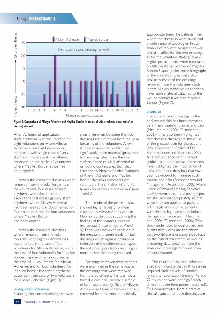
After 72 hours of application, slight erythema was documented for eight volunteers on whom Allevyn Adhesive strips had been applied, compared with single cases of very slight and moderate skin erythema observed on the backs of volunteers where Mepilex Border strips had been applied.
When the complete dressings were removed from the volar forearms of the volunteers, four cases of slight erythema were documented for each of the test dressings. Very slight erythema where Allevyn Adhesive had been applied was documented for four volunteers and for four volunteers where Mepilex Border had been applied.
When the complete dressings where removed from the volar forearms, very slight erythema was documented in the case of four volunteers for Allevyn Adhesive, and in the case of four volunteers for Mepilex Border. Slight erythema occurred in the case of 11 volunteers for Allevyn Adhesive, and for four volunteers for Mepilex Border. Moderate erythema occurred in the case of two volunteers for Allevyn Adhesive (Figure 3).
Dressing analysis after removalScanning electron microscopy showed
appropriate time. The patients from whom the dressings were taken had a wide range of aetiologies. Protein analysis of replicate samples showed similar profiles for the two dressings as for the volunteer study (Figure 6). Higher protein levels were measured on Allevyn Adhesive than on Mepilex Border. Scanning electron micrographs of the clinical samples were also similar to those of the dressings removed from the volunteer study in that Allevyn Adhesive was seen to have more material attached to the wound contact layer than Mepilex Border (Figure 7).
DiscussionThe adherence of dressings to the peri-wound skin has been shown to be a major cause of trauma and pain (Meaume et al, 2004; Zillmer et al, 2006). It has also been highlighted that dressing changes are the cause of the greatest pain for the patient (Hollinworth and Collier, 2000; Kammerlander and Eberlein, 2002). As a consequence of this, recent guidelines and consensus documents have highlighted the importance of using atraumatic dressings that have been developed to minimise such trauma and pain (European Wound Management Association, 2002; World Union of Wound Healing Societies 2004). However, some dressing types are still used inappropriately so that when they are applied to patients with fragile skin, such as patients with chronic leg ulcers, they induce damage and hence pain (Meaume et al, 2004; Zillmer et al, 2006). This study undertook to qualitatively and quantitatively evaluate the effects that two different dressing types have on the skin of volunteers, as well as presenting data obtained from the analysis of dressings removed from patients’ wounds.
The results of the peel adhesion evaluation indicate that both dressings required similar levels of removal force after application times of 48 and 72 hours and were not significantly different at the time points measured). This demonstrates, from a practical clinical aspect, that both dressings are
Figure 3. Comparison of Allevyn Adhesive and Mepilex Border in terms of skin erythema observed after dressing removal.
clear differences between the two dressings after removal from the volar forearms of the volunteers. Allevyn Adhesive was observed to have significantly more material (presumed to have originated from the skin surface, hence cellular) attached to its wound contact area than that attached to Mepilex Border. Examples of Allevyn Adhesive and Mepilex Border dressings removed from volunteers 1 and 7 after 48 and 72 hours application are shown in Figures 1 and 2.
The results of the protein assay showed higher levels of protein attached to Allevyn Adhesive than Mepilex Border, thus supporting the findings of the scanning electron microscopy (Table 5; Figures 4 and 5). There was, however, variation in the measured protein levels for both dressings which again is probably a reflection of the different skin types in the volunteer population resulting in more or less skin being removed.
Dressings removed from patients were examined in the same way as the dressings that were removed from the volunteers. This was not a formal clinical trial, merely a sample of both test dressings (five of Allevyn Adhesive and four of Mepilex Border) removed from patients at a clinically
���
���
���
���
���
���
1 2 3 4 5 6 7 8 9 10 11 12 13 14 15 16 17 18 19 20 21 22
Skin response post dressing removal
Allevyn Adhesive Mepilex Border
Numbered study participants
3.0
No
eryt
hem
aSt
rong
ery
them
a
p35-47Adhesion4(3).indd 8 3/9/08 02:49:00

Clinical RESEARCH/AUDITClinical RESEARCH/AUDIT
44 Wounds UK, 2008, Vol 4, No 3
liable to stay in place on the patient when subjected to similar forces and environments, therefore confirming that soft silicone dressings are capable of adhering to the patient just as well as other advanced wound dressings. The results do show that there is a wide variation in peel force data for both dressings and this is believed to reflect the different skin types of the volunteers. Whether or not the measurement of peel removal force is a good indicator of dressing adhesion
is debatable, as that may only be truly measured by the length of time the dressing is secured in place rather than its removal.
The evaluation of pain severity on the healthy volunteers using the VAS demonstrated that, after 24 hours application, Mepilex Border was less painful to remove than Allevyn Adhesive and also caused less erythema. This is consistent with data already in the literature that highlights
the reduced pain associated with soft silicone dressings versus dressings using acrylic adhesives (Woo et al, 2007; White, 2008).
Qualitatively, a scanning electron micrograph technique was used to visualise the surface of the dressings after they had been in contact with skin of volunteers. The results of the electron microscopy work showed that less cellular material had attached to Mepilex Border than to Allevyn Adhesive. This suppor ts the data relating to the volunteers’ responses to the measurement of pain severity on removal, in that traumatic removal of cells from the patients’ skin will result in pain. As a quantitative measure of this trauma, protein analysis of the material attached to the dressings was under taken. The results of this analysis indicate that Allevyn Adhesive is more aggressive than Mepilex Border upon removal from the skin. Protein measurement showed significantly higher values for Allevyn Adhesive than Mepilex Border.
For the most part the results from this study are as expected in that higher levels of pain severity are associated with more cells being visibly adherent to Allevyn Adhesive than Mepilex Border with greater residual protein. However, the protein residue on Mepilex Border was higher than would be expected when comparing the data with the scanning electron micrographs, which show vir tually no cell attachment. It is thought that this anomaly may be explained by the presence of hairs on some of the micrographs for Mepilex Border which could have influenced the protein results.
ConclusionTrauma and pain associated with dressing removal is a very real concern for both patients and carers. The results of this study support the view that dressings utilising Safetac soft silicone adhesive are associated with less trauma and pain at dressing removal than an advanced dressing with an acrylic adhesive. This should be
Figure 4. Levels of protein attached to Allevyn Adhesive and Mepilex Border after 48 hours of application.
Figure 5. Levels of protein attached to Allevyn Adhesive and Mepilex Border after 72 hours of application.
�
���
���
���
���
����
����
����
����
����Allevyn Adhesive Mepilex Border
2 3 4 5 6 8 9 11 12 13 14 15 16 17 18 20 22Numbered study participants
µg/m
l
�
���
���
���
���
����
����
����
����
����
����Allevyn Adhesive Mepilex Border
2 3 4 5 6 8 9 11 12 13 14 15 16 17 18 20 22Numbered study participants
µg/m
l
p35-47Adhesion4(3).indd 10 3/9/08 02:49:03

Clinical RESEARCH/AUDITClinical RESEARCH/AUDIT
46 Wounds UK, 2008, Vol 4, No 3
taken into consideration by clinicians when choosing dressings for patients, particularly for those that have friable skin that is easily damaged.
ReferencesBishop SM, Walker M, Rogers AA, Chen WY (2003) Importance of moisture balance at the wound-dressing interface. J Wound Care 12(4): 125–8
Dykes PJ (2007) The effect of adhesive
�
����
����
����
����
����
����
����
Prot
ein
ug/m
l
Allevyn Adhesive
5961 239 5962 100 5405
Mepilex Border
2817 989 100 100
Protein analysis - Clinical samples
Figure 6. Levels of protein attached to Allevyn Adhesive and Mepilex Border after 48 hours of application to patients with wounds
Figure 7. Scanning electron micrographs of Allevyn Adhesive (left side) and Mepilex Border (right side) removed from patients with wounds.
Key Points
8 Some adhesive dressings may adhere too aggressively to wounds or peri-wound skin, resulting in trauma and pain on removal.
8 Two foam dressings, Allevyn Adhesive (which uses acrylic adhesive) and Mepilex Border (which uses Safetac soft silicone adhesive technology) were compared in terms of peel force, pain and skin erythema on removal from the skin of volunteers; and the amount of cell removal from the skin of volunteers and patients with wounds.
8 No difference was observed between the dressings in terms of peel force, but Mepilex Border was associated with significantly lower pain on removal and less skin erythema.
8 Electron microscopy identified large numbers of skin cells attached to the surface of Allevyn Adhesive, whereas Mepilex Border did not remove significant numbers of cells.
8 Assays demonstrated significantly more protein on Allevyn Adhesive after removal.
8These findings provide further evidence of the atraumatic properties of dressings with Safetac.
WUK
Clinical RESEARCH/AUDITClinical RESEARCH/AUDIT
p35-47Adhesion4(3).indd 12 3/9/08 02:49:06

Clinical RESEARCH/AUDITClinical RESEARCH/AUDIT
dressing edges on cutaneous irritancy and skin barrier function. J Wound Care 16(3): 97–100
Dykes PJ, Heggie R, Hill SA (2001) Effects of adhesive dressings on the stratum corneum of skin. J Wound Care 10(2): 7–10
European Wound Management Association (2002) Position Document: Pain at Wound Dressing Changes. Medical Education Partnership, London http://www.tendra.com/Files/Tendra/safetac/ENGLISH.pdf (last accessed 19 March 2008
Fluhr JW, Dickel H, Kuss O, Weyher I, Diepgen TL, Berardesca E (2002) Impact of anatomical location on barrier recovery, surface pH and stratum corneum hydration after acute barrier disruption. Br J Dermatol 146(5): 770–6
Hollinworth H, Collier M (2000) Nurses’ views about pain and trauma at dressing changes: results of a national survey. J Wound Care 9(8): 369–73
Kammerlander G, Eberlein T (2002) Nurses’ views about pain and trauma at dressing changes: a central European perspective. J Wound Care 11(2): 76–9
Karlsmark T (2006) Dressing adherence to skin: a clinical evaluation in leg ulcer
patients. World Wide Wounds www.worldwidewounds.com/News/News.html (last accessed 19 March 2008)
Karwoski AC, Plaut RH (2004) Experiments on peeling adhesive tapes from human forearms. Skin Res Technol 10(4): 271–7
Meaume S, Moffat CJ, Franks PJ (2003a) ‘Atraumatic dressings’, a new terminology in wound care. Soins 678: 40
Meaume S, Van de Looverbosch D, Heyman H et al (2003b) A study to compare a new soft silicone dressing with a self-adherent polymer dressing in stage II pressure ulcers. Ostomy Wound Manage 49(9): 44–51
Meaume S, Teot L, Lazareth I, Martini J, Bohbot S (2004) The importance of pain reduction through dressing selection in routine wound management: the MAPP study. J Wound Care 13(10): 409–13
Soon K, Acton C (2006) Pain-induced psychological stress: a barrier to wound healing. Wounds UK 2(4): 92–101
Tokumura F, Umekage K, Sado M et al (2005) Skin irritation due to repetitive application of adhesive tape: the influence of adhesive strength and seasonal
variability. Skin Res Technol 11(2): 102–6
White R (2005) Evidence for atraumatic soft silicone dressing use. Wounds UK 1(3): 104–9
White R (2008) A multinational survey of the assessment of pain when removing dressings. Wounds UK 4(1): 14–22
Williams C (1999) The benefits and application of the Lyofoam product range. Br J Nurs 8(11): 745, 748–9
Woo K, Price P, Harding KG, Sibbald RG (2007) Pain experience during dressing change comparing two foam dressings. Poster presentation. Symposium on
Advances in Wound Care. Tampa, USA
World Union of Wound Healing Societies (2004) Principles of Best Practice: Minimising Pain at Wound Dressing-Related Procedures. A Consensus Document. Medical Education Partnership, London. http://www.wuwhs.org/datas/2_1/2/A_consensus_docment_-_Minimising_pain_at-wound_dressing_related_procedures.pdf (Last accessed 19 March 2008)
Zillmer R, Agren MS, Gottrup F, Karlsmark T (2006) Biophysical effects of repetitive removal of adhesive dressings on peri-ulcer skin. J Wound Care 15(5): 187–91
Clinical RESEARCH/AUDITClinical RESEARCH/AUDIT
47Wounds UK, 2008, Vol 4, No 3
p35-47Adhesion4(3).indd 13 3/9/08 02:49:06