CLINICAL CASES IN SPORTS MEDICINE (TENNIS ELBOW), IN ... · promoting physiological wound healing...
36
UNIVERSITA’ DEGLI STUDI DI NAPOLI FEDERICO II DIPARTIMENTO DI INGEGNERIA DEI MATERIALI E DELLA PRODUZIONE P.le Vincenzo Tecchio, 80 - 80125 NAPOLI - ITALIA Pagina 1 CLINICAL CASES IN SPORTS MEDICINE (TENNIS ELBOW), IN ORTHOPEDIC SURGERY (ACHILLES TENDON), MAXILLO-FACIAL SURGERY (ELEVATION OF THE MAXILLARY SINUS), IN PLASTIC SURGERY (TREATMENT OF WRINKLES) SOFT TISSUE SPORTS INJURY MANAGEMENT Introduction Platelet-rich plasma (PRP) therapy is an emerging technology that aims to improve the process of tissue repair via the delivery of bioactive agents, which will provide chemotactic, proliferative, and anabolic cellular responses and enhance recovery of tissue function. 1 Platelet-rich plasma products are easily prepared from the patients’own blood and typically involve the local injection of a set volume of PRP or the application of PRP gel form during surgery directly at the site of injury. The clinical use of PRP for promoting physiological wound healing was introduced in the 1980s for the treatment of cutaneous ulcers. 2 Early studies on PRP examined the healing effects of purified and isolated recombinant growth factors, such as platelet- derived growth factor BB (PDGF-BB), as therapeutic molecules for wound healing. However, knowledge of healing mechanisms has led to the conclusion that isolated growth factors cannot mediate all biological aspects required for tissue repair. Thus, a more rational strategy would be the administration of a balanced combination of mediators that would act in synergy to mimic the physiological needs of the injured tissue. 1 In the 1990s, advances in oral implantology were fueled by the potential regenerative effects of PRPs in bone tissue, and observing the healing properties in soft tissues. Since then, the use of PRP has spread to many other clinical areas, including ophthalmology, orthopedics, sports medicine, cardiology, dermatology, plastic surgery, and neurology. 3 The first reported application of PRP in sports injuries was in the arthroscopic management of an avulsion of articular cartilage in a young soccer player. 4 Further developments in PRP therapies have introduced new opportunities for tissue repair in sports medicine, such as novel therapies for the management of chronic pathologies (eg, tendinopathy and osteoarthritis). 5-7 Platelet-rich plasma has the potential to accelerate the process of healing and tissue regeneration in clinical settings. In sports medicine, this may accelerate return to play, particularly in elite and professional athletes. Platelet-Rich Plasma Therapies and Healing Mechanisms In the past decades, an increased understanding of the physiological role of platelets in wound healing has led to the concept of using platelets as therapeutic tools. Platelets are produced in large numbers from megakaryocytes in the bone marrow. The normal platelet concentration is 150 000 to 350 000/μL. Anucleate platelets circulate for 7 to 10 days and mediate primary hemostasis. On activation, platelets secrete multiple signaling proteins involved in the healing of musculoskeletal tissues. Relevant growth factors present in PRP include transforming growth factor-β1 (TGF-β1), platelet- derived growth factor (PDGF)-AB and PDGF-BB, vascular endothelial growth factor A (VEGF-A), epithelial growth factor
Transcript of CLINICAL CASES IN SPORTS MEDICINE (TENNIS ELBOW), IN ... · promoting physiological wound healing...

UNIVERSITA’ DEGLI STUDI DI NAPOLI FEDERICO IIDIPARTIMENTO DI INGEGNERIA DEI MATERIALI E DELLA PRODUZIONE
P.le Vincenzo Tecchio, 80 - 80125 NAPOLI - ITALIA
Pagina 1
CLINICAL CASES IN SPORTS MEDICINE (TENNIS ELBOW), IN ORTHOPEDIC SURGERY (ACHILLES TENDON), MAXILLO-FACIAL SURGERY (ELEVATION OF THE MAXILLARY SINUS), IN PLASTIC SURGERY (TREATMENT OF WRINKLES) SOFT TISSUE SPORTS INJURY MANAGEMENT Introduction Platelet-rich plasma (PRP) therapy is an emerging technology that aims to improve the process of tissue repair via the delivery of bioactive agents, which will provide chemotactic, proliferative, and anabolic cellular responses and enhance recovery of tissue function.1
Platelet-rich plasma products are easily prepared from the patients’own blood and typically involve the local injection of a set volume of PRP or the application of PRP gel form during surgery directly at the site of injury. The clinical use of PRP for promoting physiological wound healing was introduced in the 1980s for the treatment of cutaneous ulcers.2
Early studies on PRP examined the healing effects of purified and isolated recombinant growth factors, such as platelet-derived growth factor BB (PDGF-BB), as therapeutic molecules for wound healing. However, knowledge of healing mechanisms has led to the conclusion that isolated growth factors cannot mediate all biological aspects required for tissue repair. Thus, a more rational strategy would be the administration of a balanced combination of mediators that would act in synergy to mimic the physiological needs of the injured tissue.1
In the 1990s, advances in oral implantology were fueled by the potential regenerative effects of PRPs in bone tissue, and observing the healing properties in soft tissues. Since then, the use of PRP has spread to many other clinical areas, including ophthalmology, orthopedics, sports medicine, cardiology, dermatology, plastic surgery, and neurology.3
The first reported application of PRP in sports injuries was in the arthroscopic management of an avulsion of articular cartilage in a young soccer player.4
Further developments in PRP therapies have introduced new opportunities for tissue repair in sports medicine, such as novel therapies for the management of chronic pathologies (eg, tendinopathy and osteoarthritis).5-7
Platelet-rich plasma has the potential to accelerate the process of healing and tissue regeneration in clinical settings. In sports medicine, this may accelerate return to play, particularly in elite and professional athletes. Platelet-Rich Plasma Therapies and Healing Mechanisms In the past decades, an increased understanding of the physiological role of platelets in wound healing has led to the concept of using platelets as therapeutic tools. Platelets are produced in large numbers from megakaryocytes in the bone marrow. The normal platelet concentration is 150 000 to 350 000/µL. Anucleate platelets circulate for 7 to 10 days and mediate primary hemostasis. On activation, platelets secrete multiple signaling proteins involved in the healing of musculoskeletal tissues. Relevant growth factors present in PRP include transforming growth factor-β1 (TGF-β1), platelet-derived growth factor (PDGF)-AB and PDGF-BB, vascular endothelial growth factor A (VEGF-A), epithelial growth factor

UNIVERSITA’ DEGLI STUDI DI NAPOLI FEDERICO IIDIPARTIMENTO DI INGEGNERIA DEI MATERIALI E DELLA PRODUZIONE
P.le Vincenzo Tecchio, 80 - 80125 NAPOLI - ITALIA
Pagina 2
(EGF), hepatocyte growth factor (HGF) and insulin-like growth factor (IGF)-I and IGF-II, among others.8
These signaling proteins control cell activities by interacting with receptors located on the membrane of the target cells. This binding activates various intracellular signaling pathways that induce the synthesis of proteins needed for regenerative processes, such as angiogenesis or extracellular matrix formation. In addition to providing initial signals for local cell activation and homing of precursor cells to the injury and differentiation, PRPs contain potent adhesive substrates for cells, such as fibrin, fibronectin, vitronectin, thrombospondin, osteocalcin, and osteonectin.8
Considering these properties, PRPs are crucial in the process of repair of tendons, muscles, ligaments, cartilage, and bone injuries.
In the physiological process of wound healing, platelets embedded in the blood clot serve as a primary source of biologically active factors. Typically, after muscle strain or contusion, the hematoma that originates as a consequence of vessel disruption contains about 94% red blood cells, a small amount of platelets (4%), and < 1% leukocytes (Figure 1).
The rationale for the use of PRP involves replacing the blood clot with PRP, thus minimizing the presence of red blood cells and increasing platelet concentration at the injury site.
These unique properties of PRPs have led to the commercial development of multiple systems that offer an easy, cost-effective strategy to obtain high concentrations of factors for tissue healing and regeneration in the clinical setting.
Preparation of PRP and Products
In the past few years, several semiautomatic machines have been developed for centrifugal separation of PRP for therapeutic use. The process of PRP preparation is relatively straightforward and can be performed in the clinic or operating room. It can usually be completed within minutes. The cost to both medical practitioners and patients varies widely depending on the method used to produce the PRP concentrate.
The cost of a commercial kit “DENSITY PLATELET GEL” is about $300 to $600.
For PRP preparation, peripheral blood is drawn from the patient under sterile conditions, with or without anticoagulants, and the plasma is prepared by centrifugation or filtration. The volume can be adapted to the extent of the size of the injury and phase of injury, ranging from 10 to 100 mL. Essentially, the methods of producing PRPs determine the composition and concentration in terms of leukocytes, erythrocytes, and platelets in a given plasma volume.
There are 3 methods used to make these determinations: 1) double-spinning methods using automated machines along with commercial kits, 2) single-spinning methods using conventional laboratory centrifuges followed by manual PRP separation, or 3) selective blood filtration using commercially available technology. When using single spinning, the platelet yield is 1- to 3-fold baseline levels, while 5- to 8-fold baseline levels are achieved by double spinning. Double spinning also concentrates leukocytes.
Platelet concentrates have been categorized in pure PRP (P-PRP), in which leukocytes are purposely eliminated from the PRP, and leukocyte and platelet-rich plasma (L-PRP), which contains a high concentration of leukocytes.9
Whether leukocytes have detrimental effects in particular orthopedic sports medicine applications is controversial, but basic

UNIVERSITA’ DEGLI STUDI DI NAPOLI FEDERICO IIDIPARTIMENTO DI INGEGNERIA DEI MATERIALI E DELLA PRODUZIONE
P.le Vincenzo Tecchio, 80 - 80125 NAPOLI - ITALIA
Pagina 3
evidence points toward a deleterious effect of neutrophils, particularly in joints and muscle injuries.10
The improved homogeneity of P-PRP and its reduced donor-to- donor variability would support the view that some PRP production techniques are more reproducible and predictable than others.
There is little consensus regarding the dose of platelets and growth factors needed to obtain efficient clinical results. The clinical variability of different studies suggests that some techniques might not produce a sufficient number of functional platelets to produce the expected outcome. Similarly, there is no consistency between the methods of applying the therapy, the timing of treatment, the number of injections per series, or the volume of injection. This has precluded the establishment of the standards necessary to combine the results of independent studies and provide an estimate of the treatment effects. For example, the methods for PRP preparation vary widely between practitioners and the volume of plasma used. Double-spinning techniques yield a PRP concentration of around 10% of the blood volume drawn (ie, 20 mL of whole blood would result in 2 mL of PRP), in contrast to 40% to 50% of the blood volume obtained after single spinning. Also, each method leads to a different product, with varying biological properties and potential uses.11 It is unclear whether these differences have any clinical relevance.10
Some authors have suggested that PRP preparations containing only moderately elevated platelet concentrations induce optimal biological benefit, while other authors suggest lower platelet concentrations produce suboptimal effects, and higher ones produce inhibitory effects.12,13
According to other authors, the therapeutic dose of PRP is ≥ 4 to 6 times higher than the normal platelet count.14
To add to the discussions, the actual growth factor content does not correlate with the platelet count in whole blood or in PRP when leukocytes are present in the preparation, and there is no evidence that gender or age affects platelet count or growth factor concentrations.15
However, age may influence the number of receptors of local cells interacting with the plasma signals.16
Once the PRP is separated from the whole blood, it is stable for about 8 hours. However, because these procedures are considered an autograft by regulatory organizations, the plasma should be prepared and used immediately at the point of care, and should not be stored. Prior to application, platelets can be slowly activated by initiating the coagulation cascade with the addition of calcium chloride, a necessary cofactor for prothrombin conversion into thrombin. Alternatively, coagulation and platelets can be activated by instantly adding standard solution of 1000 U/mL of bovine or human thrombin along with 10% calcium chloride to the PRP. After plasma activation, the fibrin scaffold can be formed in vivo or ex vivo; the latter is suitable for implantation in surgery or in ulcer care, and provides a gradual release of growth factors in the area where it has been applied. Product Safety There are safety concerns about the routine use of PRP. Any concerns regarding transmission of diseases such as human immunodeficiency virus, hepatitis, or Creutzfeldt-Jakob disease, or of immunogenic reactions (a concern with allografts or xenografts), are by definition not applicable, given the autologous nature of PRP.17
However, some systems use purified bovine thrombin to activate the platelets. This may produce coagulopathies, and most

UNIVERSITA’ DEGLI STUDI DI NAPOLI FEDERICO IIDIPARTIMENTO DI INGEGNERIA DEI MATERIALI E DELLA PRODUZIONE
P.le Vincenzo Tecchio, 80 - 80125 NAPOLI - ITALIA
Pagina 4
authorities now use human recombinant thrombin.
Some authors have raised the issue of genetic instability, and hypothesized that the use of PRP may lead to the development of neoplasms. Growth factors act on receptors located on the cell membranes rather than on the cell nucleus, and activate normal gene expression via intracellular signaling proteins, which promote normal, not abnormal, gene expression.18
Growth factors are not directly mutagenic, and their activities in normal wound healing are highly regulated by various feedback control mechanisms. Furthermore, until now, no systemic effect on circulating growth factors has been shown after PRP application.19
Some antimicrobial activity of PRP (platelet-leukocyte gel) against Staphylococcus aureus has been shown in vitro20 and in vivo, although this antimicrobial activity is not comparable with systemic antibiotic treatment.21 Therapeutic Application of PRP in Sports-Related Injuries The therapeutic administration of PRP extends to the treatment of multiple musculoskeletal injuries in orthopedics and sports medicine. Its widespread clinical use has been popularized following its use in high-profile athletes. Orthopedists, primary care sports medicine physicians, and rheumatologists are among the practitioners using PRP in the management of tendon, ligament, muscle, nerve, bone, and joint injuries. However, PRP use should be considered experimental in all applications of sports medicine. Studies of varying levels of evidence have demonstrated the safety and beneficial effects of PRP in some of these applications, but adequate level I randomized trials needed to perform meta-analyses are lacking. Moreover, advances in the field of clinical research on the use of PRP are hindered by the lack of standardization of the various formulations and administration regimens and modalities. In this section, we focus on the use of PRPs in soft tissue injuries.
Tendon Injuries
One interesting application of PRP therapies is the management of tendon injuries. Tendon injuries are a major cause of musculoskeletal morbidity affecting professional and recreational athletes in various anatomical locations.
Tendinopathy is associated with a failed healing response. The tendon cell-mediated process involves increased turnover and remodeling, and gradual transformation in the quality and quantity of extracellular matrix that habitually precedes tendon rupture. Tendon cells (tenoblasts and tenocytes) have a central role in the repair and maintenance of extracellular matrix, synthesizing new proteins and producing the enzymes that degrade them.
The activities of tendon cells are likely to be influenced by external growth factors and cytokines released from PRPs.22
This is the core hypothesis of PRP application that is supported by recent advances in basic laboratory research providing a more detailed understanding of the biological mechanisms influenced by PRPs.
In an animal study, Kajikawa et al23 showed the chemotactic action of PRPs in tendon injury.
In addition, PRPs stimulated cell proliferation and the synthesis of angiogenic factors, such as VEGF and HGF, which act in

UNIVERSITA’ DEGLI STUDI DI NAPOLI FEDERICO IIDIPARTIMENTO DI INGEGNERIA DEI MATERIALI E DELLA PRODUZIONE
P.le Vincenzo Tecchio, 80 - 80125 NAPOLI - ITALIA
Pagina 5
a paracrine manner on endothelial cells, promoting angiogenesis.24-26
Moreover, PRPs induced the synthesis of molecules of the extracellular matrix, such as collagens or hyaluronan.27
However, PRPs should be combined with an appropriate loading regimen to enhance extracellular matrix organization in the short term. Indeed, injections of PRP 1 week postoperatively increased tendon regenerate strength after 4 weeks if combined with early therapy.28
A placebo-controlled experimental trial in 6 horses reported less inflammation and increased metabolic activity and maturation, higher strength at failure, and elastic modulus in tendons treated with PRP.29
In a case series study including 12 athletes who were undergoing surgical repair of Achilles tendon tears, Sánchez et al30 applied basic research findings to a clinical investigation. In their study, they applied P-PRP with a moderate concentration of platelets (2–3 times the concentration of platelets compared with whole blood) clotted ex vivo and injected it in liquid form, activated with calcium chloride. Controls were treated with an identical surgical procedure performed by the same surgeon, but they did not receive PRP during surgery. The authors reported an enhancement in the range of motion and a faster return to sporting activities in the group that received PRP during surgery. The cross-sectional area of these Achilles tendons had fewer differences compared with the contralateral tendon after 18 months, indicating a more physiological repair of the PRP-treated tendon.
These authors also reported the use of P-PRP in 2 configurations (clotted ex vivo and calcium chloride-activated liquid) to treat complications derived from the surgical repair of the Achilles tendon.31
In a recent randomized clinical trial, de Vos et al32 treated patients with Achilles tendinopathy with eccentric exercises and 1 injection of buffered L-PRP (6–8 times the concentration of platelets compared with whole blood) in the experimental group (although leukocytes play a significant role in the biology of these products, their concentration was not reported), while the control group was managed with eccentric exercises and 1 injection of saline. The patients were followed-up at 6, 12, and 24 weeks. Buffered L-PRP injection did not improve pain or activity for patients who were all treated with a concurrent eccentric exercise regimen. However, if testing a combination therapy, the optimal study design to address the buffered L-PRP hypothesis has to consider various options for control groups. In fact, a third arm including patients who do not receive any active therapy (ie, needling trauma) could have produced more reliable results. To note, this trial used only 1 injection of L-PRP: most practitioners report benefits in Achilles tendinopathy, and tendinopathy as whole, after 2 to 3 injections.33
It is possible that there are clinical differences in the effects of PRP injections between anatomical locations. Preliminary studies in wrist extensor and flexor tendinopathy have been favorable to PRP treatment. Mishra and Pavelko34 reported the effects of buffered L-platelet concentrate injections in a small group of patients and found a significant improvement in pain after 8 weeks. More recently, in a randomized clinical trial (level I), Peerbooms et al35 also reported their findings after administering buffered L-PRP in the experimental group and corticoids in the control group of patients with chronic tennis elbow. The group treated wtih corticosteroids appeared to recover initially, but improvement eventually declined, whereas the L-PRP group progressively improved. An observational case series study (level III) reported significant functional improvement on arthroscopic rotator cuff repair in 14 patients followed up at 12 and 24 months postoperatively.36 Another prospective observational study37 (level IV) reported decreased pain and enhanced functional recovery after PRP application in 20 athletes with chronic patellar tendinopathy (jumper’s knee). In a prospective case-control study (level III),

UNIVERSITA’ DEGLI STUDI DI NAPOLI FEDERICO IIDIPARTIMENTO DI INGEGNERIA DEI MATERIALI E DELLA PRODUZIONE
P.le Vincenzo Tecchio, 80 - 80125 NAPOLI - ITALIA
Pagina 6
Filardo et al38 administered 3 injections of activated L-PRP (6 times the concentration of platelets compared with whole blood, leukocyte concentration was not evaluated) in the patellar tendon, with a significant functional improvement after 6 months.
The effects of PRP on rotator cuff pathology have been mixed. Everts et al39 reported better functional recovery and less pain in a prospective cohort study using L-PRP open subacromial decompression. However, no group differences were found at 2 years. It can be suggested that PRP could bring forth an early biological and clinical response that becomes less pronounced in the long term. In a recent randomized clinical trial, Weber and Kauffman40 reported that platelet-rich fibrin matrix (DENSITY PLATELET GEL®; Industrie biomediche e Farmaceutiche Srl, Scafati, Italy) applied during rotator cuff surgery had the same outcomes as controls.
Ligament Injuries
Bone-tendon-bone patellar grafts and hamstring grafts are both used in anterior cruciate ligament (ACL) reconstruction. The goal of this surgery is to obtain rapid tendon ligamentization (tendon transformation into a ligament-like structure) and rapid bone-bone or tendon-bone healing. In an observational case series study (level III), Sánchez et al5 applied P-PRP to both types of grafts, and evidenced enhanced functional recovery. Recently, the same authors41 reported enhanced ligamentization at histological examination of tendon grafts treated with P-PRP injections. Confirming these findings, in a case-control study (level III), Radice et al42 reported enhanced ligamentization after evaluating 100 ACL grafts by magnetic resonance imaging (MRI) using ex vivo clotted L-PRP. Orrego et al43 found better graft maturation evaluated by MRI signal intensity, without any significant effect in the osteoligamentous interface or tunnel widening evolution (level II). In a recent prospective randomized clinical study (level I), Vogrin et al44 reported better anteroposterior knee stability at 6 months postsurgery in the ex vivo clotted L-PRP (platelet gel) group. However, Nin et al,45 in a randomized, case-controlled trial, and Silva and Sampaino46 and Figueroa et al,47 in case-controlled studies (level I and III, respectively), could not find any improvement after applying ex vivo clotted L-PRP. Differences in the results could be attributed to both the PRP product and the procedures of application. Some researchers in our group (OMD, GM) have just completed a randomized controlled trial in professional athletes with anterior tibiofibular ligament tears (high ankle sprain). Injection of P-PRP under ultrasound guidance resulted in quicker return to play when compared with untreated controls (unpublished data). Mei-Dan et al48 reported on an Olympic medalist judoka who won the gold medal world championship < 6 months after sustaining a complete tear of the elbow medial ligamentous complex, which was then injected twice with P-PRP. Osteoarthritis and Cartilage Damage to the knee ACL, cartilaginous tissue, or meniscus in an early stage of life can lead to osteoarthritis (OA) later. Posttraumatic or secondary OA is a relatively common condition for athletes with a history of joint injury. In a laboratory study, PRP application can improve the quality of synovial fluid by inducing the endogenous secretion of hyaluronic acid by synovial cells.49 In a retrospective cohort study (level III), Sánchez et al50 reported decreased pain and enhanced function, as assessed using the WOMAC scale, after intra-articular injection of activated P-PRP in knee OA compared with intra-articular hyaluronic acid. The same group is using activated P-PRP injections in hip OA with promising preliminary results. Recently, in a case series (level IV) involving 115 knees of young patients with a low degree of articular degeneration, Kon et al51 reported reduced pain and improved function after L-PRP treatment.

UNIVERSITA’ DEGLI STUDI DI NAPOLI FEDERICO IIDIPARTIMENTO DI INGEGNERIA DEI MATERIALI E DELLA PRODUZIONE
P.le Vincenzo Tecchio, 80 - 80125 NAPOLI - ITALIA
Pagina 7
Muscle Injuries Athletes often experience muscle strains and contusions, which temporarily disable them from training and competition. Applying the rest, ice, compression, and elevation protocol shortly after injury relieves pain and minimizes swelling. Thereafter, the combined injection of Traumeel®/Actovegin® (the former is a homeopathic formulation and the latter an amino acid mixture) for the management of acute muscle strains is popular in many countries. The use of autologous plasma preparations might be a safe alternative to this treatment. In a nonrandomized and nonblinded pilot study (level III), Wright-Carpenter et al52 compared both treatments, assessing the time needed to resume full sports activities after moderate strains in 18 patients receiving 5 mL of autologous conditioned serum (ACS) every second day versus 11 patients receiving the same volume of Traumeel®/Actovegin®. Autologous conditioned serum incubates whole blood with glass beads; it contains signalling proteins including interleukin-1β (IL-1β), TNF-α, IL-7, fibroblast growth factor-2 (FGF-2), IL-1Ra, HGF, PDGF-AB, TGFβ1, and IGF-1. The mean time needed to resume full competition was shorter for the ACS group. Moreover, regression of edema/bleeding was faster in the ACS group as monitored by MRI.
In the Second World Congress of Regenerative Medicine, Sánchez et al53 reported activated P-PRP injections in 21 muscle injuries of varying severity and anatomical locations. These athletes, who played in division 1 teams of the Spanish Soccer League, resumed normal training activities after half the time needed by matched historical controls. Using the same leukocyte-free PRP preparation, Loo et al54 reported good outcome after application to adductor longus strain in a single case report. More recently, in a laboratory-controlled study, Hammond et al55 injected either L-PRP or PRP in 2 models of muscle strains in the tibialis anterioris of rats (8 animals per group). The authors found enhanced myogenesis and improved contractile function with PRP. Doping Concerns Because PRP contains growth factors, its use may go against anti-doping rules. In 2008, the World Anti-Doping Association (WADA) and the International Olympic Committee (IOC) organized an international meeting to discuss possible conflicts with the WADA Code. The resulting position paper, the Aspetar Consensus, discussed the use of PRP in muscle injury in relation to evidence-based medicine and doping. The recommendations from the Aspetar Consensus were equivocal and left the decision to further research. However, therapeutic use exemption when wishing to use growth factor technologies in elite athletes was recommended. Currently, in Section (S.2.6) of the current 2010 Prohibited List, intramuscular injections of PRP are prohibited, while other applications require a declaration of use. In the same Section (S.2.5), the growth factors explicitly mentioned in connection with PRP are IGF-1, PDGF, FGFs, VEGF, and HGF. The concentration of all these factors, while present in PRP, is in the physiological range. Moreover, in these preparations, HGF and IGF-1 are not readily available because they are bound to proteins that regulate their physiological actions.
Finally, after more discussion in 2010, guidance has emerged from the WADA clarifying that PRP formulations (as they exist currently) do not increase muscle growth beyond return to a normal physiological state. Hence, because the use of PRP injections for therapeutic purposes does not violate the spirit of sports, the prohibition for intramuscular injections of PRPs has been removed from the 2011 prohibited list.
UNIVERSITA’ DEGLI STUDI DI NAPOLI FEDERICO IIDIPARTIMENTO DI INGEGNERIA DEI MATERIALI E DELLA PRODUZIONE
P.le Vincenzo Tecchio, 80 - 80125 NAPOLI - ITALIA
Pagina 8
TREATMENT OF ACHILLES TENDONPATHY WITH PRP Introduction Musculoskeletal injuries are the most common cause of severe long-term pain and physical disability (Woolf and Pfleyer, 2003). The Achilles tendon is an area commonly injured as a result of overuse. Initially, conservative measures such as ice, rest, orthotics, and physical therapy are used, with a failure rate of 25% eventually leading to surgery (Mishra, 2009; Chang, 2010). However, patients who undergo surgery have a long recovery period, with increased incidence of complications (Mishra, 2009; Chang, 2010). Cortisone injections directly into the tendon are frequently performed, but are not recommended because of increased incidence of tendon rupture (Sampson et al, 2008). The last decade has brought about significant advances in musculoskeletal injury repair, including concentrated autologous platelet-rich plasma (PRP) therapy (Everts, 2006; Sampson, 2008). PRP’s healing properties result from its ability to release growth factors and chemoattractants, congregating macrophages and fibroblasts to the site of injury to facilitate tendon repair (Everts et al, 2006) The growth factors found in platelets have been shown to be key in the reparative process of tendons (Sanchez et al, 2007). PRP is typically administered into chronic or subacute tendon tears or tendinosis when conservative measures fail, or at the time of surgery to improve post-operative recovery. This case report demonstrates the use of PRP to potentially accelerate healing in a more acute setting and without having exhausted conservative measures. At the 2010 Annual American Academy of Orthopaedic Surgeons meeting, preliminary data was presented that demonstrated that 28 out of the 30 patients with Achilles tendinosis who failed 6 months of conservative treatment were able to return to their preinjury activity levels after a single injection, within several months following PRP therapy, without requiring additional therapy (Leahy, 2010). Moreover, Aspenberg and Virchenko (2004) where able to show that PRP was able to facilitate the repair of a transected Achilles tendon in rats, when administered six hours after the initial tear. This is in contrast to published human studies, in which PRP is typically injected at a point of subacute or chronic status of Achilles tendinopathy. The rat tendons demonstrated increased callus strength, stiffness and material characteristics. Contrastingly, a recent study by De Vos et al (2010) showed that ultrasound guided PRP, versus normal saline injections followed by eccentric exercises, made no significant difference in the function and pain levels of individuals who had chronic Achilles tendinopathy. However, there were several limitations to their study, including the lack of follow-up after a 6-month period, and the absence of follow-up imaging. Another limitation was that they relied on the tendon collagen, rather than calcium chloride and thrombin, to activate the platelets, which may have caused the spread of the platelets to other regions, decreasing the effectiveness of PRP. Both study groups demonstrated symptomatic improvement, which were attributed to eccentric exercises, although this variable was not isolated as a control. Also, Sanchez et al (2007) showed that patients who received Achilles tendon surgery with PRP had a quicker recovery time then those who only received surgery. The individuals with PRP were able to return back to their sport at 14 weeks, while the ones who only had surgery returned back to their sport at 22 weeks. There is also evidence that suggests that dry needling with autologous blood may stimulate repair in patellar tendons. (James et al, 2007) There is conflicting data that is emerging regarding Achilles tendonapathies being treated with PRP.
UNIVERSITA’ DEGLI STUDI DI NAPOLI FEDERICO IIDIPARTIMENTO DI INGEGNERIA DEI MATERIALI E DELLA PRODUZIONE
P.le Vincenzo Tecchio, 80 - 80125 NAPOLI - ITALIA
Pagina 9
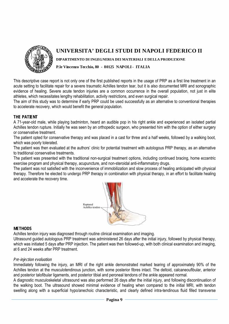
This descriptive case report is not only one of the first published reports in the usage of PRP as a first line treatment in an acute setting to facilitate repair for a severe traumatic Achilles tendon tear, but it is also documented MRI and sonographic evidence of healing. Severe acute tendon injuries are a common occurrence in the overall population, not just in elite athletes, which necessitates lengthy rehabilitation, activity restrictions, and even surgical repair. The aim of this study was to determine if early PRP could be used successfully as an alternative to conventional therapies to accelerate recovery, which would benefit the general population. THE PATIENT A 71-year-old male, while playing badminton, heard an audible pop in his right ankle and experienced an isolated partial Achilles tendon rupture. Initially he was seen by an orthopedic surgeon, who presented him with the option of either surgery or conservative treatment. The patient opted for conservative therapy and was placed in a cast for three and a half weeks, followed by a walking boot, which was poorly tolerated. The patient was then evaluated at the authors’ clinic for potential treatment with autologous PRP therapy, as an alternative to traditional conservative treatments. The patient was presented with the traditional non-surgical treatment options, including continued bracing, home eccentric exercise program and physical therapy, acupuncture, and non-steroidal anti-inflammatory drugs. The patient was not satisfied with the inconvenience of immobilization and slow process of healing anticipated with physical therapy. Therefore he elected to undergo PRP therapy in combination with physical therapy, in an effort to facilitate healing and accelerate the recovery time.
METHODS Achilles tendon injury was diagnosed through routine clinical examination and imaging. Ultrasound guided autologous PRP treatment was administered 26 days after the initial injury, followed by physical therapy, which was initiated 5 days after PRP injection. The patient was then followed-up, with both clinical examination and imaging, at 6 and 24 weeks after PRP treatment. Pre-injection evaluation Immediately following the injury, an MRI of the right ankle demonstrated marked tearing of approximately 90% of the Achilles tendon at the musculotendinous junction, with some posterior fibres intact. The deltoid, calcaneoufibular, anterior and posterior talofibular ligaments, and posterior tibial and peroneal tendons of the ankle appeared normal. A diagnostic musculoskeletal ultrasound was also performed 26 days after the initial injury, and following discontinuation of the walking boot. The ultrasound showed minimal evidence of healing when compared to the initial MRI, with tendon swelling along with a superficial hypo/anechoic characteristic, and clearly defined intra-tendinous fluid filled transverse

UNIVERSITA’ DEGLI STUDI DI NAPOLI FEDERICO IIDIPARTIMENTO DI INGEGNERIA DEI MATERIALI E DELLA PRODUZIONE
P.le Vincenzo Tecchio, 80 - 80125 NAPOLI - ITALIA
Pagina
10
tearing. Additionally, there was a focal hyperechoic area at the deep margin representing poorly organized fusiform, blunt contracted fibres in the distal segment. Clinically, the patient had severe compromise in his ambulatory potential and activities of daily living, due to pain and weakness of the affected ankle. A physical exam revealed a mildly swollen Achilles tendon, with maximum point tenderness located 2 cm from its calcaneal insertion. The patient was observed to have an antalgic gait. Diagnostic imaging sequence Magnetic Resonance Imaging (MRI) images were acquired on a Hitachi Airis II High Performance open MRI Scanner (Figure1a–c) at the time of initial injury through the orthopedic clinic. The patient had > 90% complete tear of the Achilles tendon, as the radiologist’s MRI report. The first diagnostic musculoskeletal ultrasound was done at 26 days post injury, and after cast removal, to ensure no significant changes had occurred since the MRI (Figure 2) and initial trauma. Ultrasound imaging was performed with a 7.6–13.0 MHz high frequency linear transducer Sonosite Micromaxx, B-mode ultrasound technique. Colour power Doppler application was used for detection of hyperemia. The Patient was also re-imaged by means of ultrasound at 6 weeks (Figure 2) and MRI at 24 weeks post PRP Injection (Figure 1a–c).
Figure 1a. Sagittal T1 images of the Achilles tendon pre and post-platelet rich plasma (PRP) injection. The image on the left shows a tear of the Achilles tendon pre-PRP at the
musculotendinous junction (arrow). The image on the right shows the intact Achilles tendon without evidence of tear at the musculotendinous junction (arrow).

UNIVERSITA’ DEGLI STUDI DI NAPOLI FEDERICO IIDIPARTIMENTO DI INGEGNERIA DEI MATERIALI E DELLA PRODUZIONE
P.le Vincenzo Tecchio, 80 - 80125 NAPOLI - ITALIA
Pagina
11
PRP preparation and delivery PRP was prepared using DENSITY PLATELET GEL® (Industrie biomediche e Farmaceutiche Srl, Scafati, Italy). A total of 9 cc of autologous blood was drawn into a 10 cc syringe (BD), containing 1 cc of ACD-A, and processed to yield a total of 5 cc of PRP (~8.5 X Baseline; manufacturer’s internal validation). The patient was anesthetized locally with 5 cc of 1% lidocaine into the corresponding subcutaneous tissue. Thereafter, the PRP sample was combined with 0.9 cc of CaCl and administered using a 22G 3.5 in needle into the Achilles tendon, under musculoskeletal ultrasound guidance. Needling of the affected tendon fibres was performed under live ultrasound in colour Doppler mode to ensure proper flow of PRP into the tendon. The patient tolerated the procedure well and without any intra or post-procedure adverse reactions.
Figure 1b. STIR images of the Achilles tendon. The image on the left, which is pre-platelet-rich plasma (PRP) injection, shows a large tear with few intact posterior fibres (arrow). The image on the right is post-PRP injections, which shows a thickened Achilles tendon indicative of tendonitis without evidence of rupture (arrow).
Figure 1c. T2 Axial images of the Achilles Tendon pre- and post-platelet-rich plasma (PRP) injection. The image on the left is indicative of the Achilles tendon tear (arrow), while the image on the right shows repaired Achilles tendon, which is now thickened and demonstrates fluid compatible with
Achilles tendinitis (arrow).

UNIVERSITA’ DEGLI STUDI DI NAPOLI FEDERICO IIDIPARTIMENTO DI INGEGNERIA DEI MATERIALI E DELLA PRODUZIONE
P.le Vincenzo Tecchio, 80 - 80125 NAPOLI - ITALIA
Pagina
12
Integrating physical therapy into the treatment plan Physical therapy was started five days after the injection, and carried out twice weekly.
Prior to starting therapy the patient was instructed to weight bear as tolerated, along with discontinuing the walking boot, which was promoting biomechanical dysfunctions. Soft tissue mobilization and edema massage, along with modalities including laser, electrical stimulation and ice were performed to reduce swelling. Active treatment consisted of range of motion, stationary cycling, and lower extremity strengthening, and eccentric exercises. Eccentric exercises are thought to promote collagen fibre cross-linkage in the tendon facilitating tendon repair (Woodley et al, 2007). Studies have shown that eccentric exercise improves pain, strength and function (Andes and Murrell, 2008).
Figure 2a,b. The image on the left is pre-platelet-rich-plasma (PRP) injection treatment. The ultrasound image demonstrates a swollen tendon characterized by a superficial hypo/anechoic characcteristic, as well as clearly
defined intra-tendinous fluid filled transverse tearing. Focal hyperechoic area at the deep margin represents poorly organized fusiform, blunt contracted fibres in the distal segment. The image on the right is post-PRP treatment. The
ultrasound image demonstrates decreased fluid filled tear lines in the superficial segment. Additionally, the fusiform non-organized hyperechoic segment is decreased in size. The overall tendon appearance is more
organized, with a smaller degree of blunt contraction.

UNIVERSITA’ DEGLI STUDI DI NAPOLI FEDERICO IIDIPARTIMENTO DI INGEGNERIA DEI MATERIALI E DELLA PRODUZIONE
P.le Vincenzo Tecchio, 80 - 80125 NAPOLI - ITALIA
Pagina
13
Follow-up evaluation (6 weeks) After PRP injection, the patient had chosen to discontinue use of his walking boot. He reported increased swelling in the affected foot since his initial evaluation and PRP treatment, which subsided significantly with physical therapy treatments. Follow-up examination at 6-weeks (following PRP injection) demonstrated mild pitting edema of the right ankle, with no tenderness to palpation. Additionally, tenderness to palpation at the Achilles tendon was absent in spite of palpable thickening. The patient displayed normal ankle range of motion and manual muscle testing with intact sensation of the right ankle. The patient continued to ambulate with an antalgic gait with persistent reduced ambulatory potential, as compared to his pre-injury status. A diagnostic musculoskeletal ultrasound of the right Achilles tendon showed an enlarged cross-section of the Achilles tendon that was indicative of early regeneration (Figure 2b). Focal fibrosis was present at the mid tendon region and only a small focal tear remained. Follow-up evaluation (24 weeks) A follow-up MRI of the Achilles tendon revealed an intact, thickened tendon without evidence of tears at the musculo-tendonous junction (Figure 1a). MRI revealed T2 hyper-intensity and T1 hypo-intensity demonstrating fluid build-up within the thickened Achilles tendon, indicative of tendonitis. The fluid in the central portion of the Achilles tendon had a cranio-caudal height of 2.5 cm and an antero-posterior extent of 8 mm. Nonetheless, there was no evidence of the previously described severe Achilles tear. At this time, the patient reported full functional return of his pre-injury activity levels, including stair climbing and playing badminton. CONCLUSION Post-operative tendon healing is accomplished by several stages including inflammation, formative, and remodeling stages (Aspenberg, 2007). This process often takes a while to occur and the resulting tendon is usually weaker. One of the factors that contributes to limited tendon healing is poor vascular supply. In fact, most tears are located at the avascular calcaneal insertion (Filardo et al, 2010). Recently several studies have shown the significance of growth factors in the reparative process of tendons. PRP contains multiple growth factors that contribute to the healing process. Vascular endothelial growth factor (VEGF) promotes angiogenesis.

UNIVERSITA’ DEGLI STUDI DI NAPOLI FEDERICO IIDIPARTIMENTO DI INGEGNERIA DEI MATERIALI E DELLA PRODUZIONE
P.le Vincenzo Tecchio, 80 - 80125 NAPOLI - ITALIA
Pagina
14
Platelet derived growth factor (PDGF), insulin like growth factor (IGF-1) and fibroblast growth factor (FGF), together facilitate healing and proliferation. Lastly, transforming growth factor beta (tgf-b) increases mechanical strength, as well as increasing expression of pro-collagen 1 and 3 (Molloy, 2003; Kashiwagi, 2004). A recent study demonstrated that local injection of PRP into a partially torn Achilles tendon six days after initial injury facilitated a rapid repair of the tendon, with pain relief and return to previous activity level within two to three months of the injection (Filardo et al, 2010). In this current case report, non-surgical, ultrasound guided injection of PRP into the Achilles tendon within one month of injury, significantly improved a 90% Achilles tendon rupture in 6 weeks, leaving only a small focal tear. At 24 weeks, the tear was completely resolved, and the patient returned to full functional activity without limitations, and without using a brace that restricted activities of daily living. This preliminary case report supports the potential utility of PRP as a first-line treatment to repair acute Achilles tendon tears, without having to resort to surgery or undergo a prolonged rehabilitation process with activity restrictions secondary to bracing. Unfortunately, interpretation of the data in this single case report is limited. Further studies should incorporate baseline and follow-up measures, including the foot and ankle outcomes questionnaires and visual analog pain scale. Ultimately, a large randomized multi-centered study with long term follow-up supported with serial MRI and sonographic imaging is needed before the adoption of PRP injection as a standard treatment option for acute Achilles tendon tears. While there remains optimism regarding the promise of biological based therapies, larger controlled trials are needed to determine if this particular therapy is safe and effective for the treatment of various tendon injuries for acute and chronic settings. Due to the increasing number of individuals participating in physical activities, more and more musculoskeletal injuries are occurring, and tendon injuries, especially of the Achilles tendon, are one of the major concerns in sports medicine. Tissue repair in musculoskeletal lesions is often a slow, and sometimes incomplete, process. In particular, significant partial tendon ruptures seem to respond poorly to conservative measures, and do not improve with time, so surgery is most often considered the preferable treatment option for this kind of lesion. However, as correctly highlighted by the authors of this article, patients who undergo surgery have a long recovery period, with increased incidence of complications. The search for a minimally-invasive solution to improve tendon healing is therefore highly desirable, especially in sports patients where fast recovery of full efficiency and return to competition are of primary importance. An optimal treatment should aim to restore patients to their pre-injury status in a safe, cost-effective way, and as quickly as possible. Several studies have revealed a complex regulation of growth factors (GFs) for the normal tissue structure and the reaction to tissue damage. Therefore, their use is thought to be useful in clinical practice. Platelets contain a reservoir of GFs. In this view, the positive effects of platelet concentrate injections on tissue healing might be attributed to the higher content and secretion of GFs, which can be placed directly into the lesion site in physiological proportions. In fact, with respect to purified individual GFs or experimental associations, platelet-rich plasma (PRP) has the theoretical advantage of containing numerous bioactive molecules with a natural balance of anabolic and catabolic functions, thus potentially optimizing the tissue environment and favouring the healing process (Kon et al, 2010). The attractive possibility to use the patients’own GFs to enhance reparative process in tissues with low healing potential, the promising preclinical studies and preliminary clinical findings other than the safety of this treatment approach, explain its worldwide clinical application. REFERENCES
UNIVERSITA’ DEGLI STUDI DI NAPOLI FEDERICO IIDIPARTIMENTO DI INGEGNERIA DEI MATERIALI E DELLA PRODUZIONE
P.le Vincenzo Tecchio, 80 - 80125 NAPOLI - ITALIA
Pagina
15
Andes BM, Mur rel l GAC (2008) Treatment of tendinopathy: what works, what does not, and what is on the horizon. Clin Orthop Relat Res 466(7): 1539–54 Aspenberg P (2007) Stimulation of tendon repair: mechanical loading, GDFs and platelets. A minireview. Int Orthop 31(6): 783–9 Aspenberg P, Virchenko O (2004) Platelet concentrate injection improves Achilles tendon repair in rats. Acta Orthop Scand 75(1): 93–9 Chang HJ, Burke AE, Glass RM (2010) JAMA patient page. Achilles tendinopathy. JAMA 303(2): 188 De Vos RJ, Weir A, Van Schie HTM et al (2010) Platelet-rich plasma injection for chronic Achilles tendinopathy: a randomized controlled trial. JAMA 303(2): 144–9 Everts P, Knape J, Weirich G et al (2006) Platelet-rich plasma and platelet gel: a review. J Extra Corpor Technol 38(2): 174–87 Filardo, G, Lo Presti M, Kon E, Marcacci M (2010) Nonoperative biological treatment approach for partial Achilles tendon lesion. Orthopedics 33(2): 120–3 James SL, Ali K, Pocock C, Robertson C, Walter J, Bell J, Connell D (2007) Ultrasound guided dry needling and autologous blood injection for patellar tendinosis. Br J Sports Med 41(8): 518–21 Kashiwagi K, Mochizuki Y, Yasunaga Y, Ishida O, Deie M, Ochi M (2004) Effects of t ransforming growth fa c tor -be t a 1 on the e a r l y stages of healing of the Achilles tendon in a rat model. Scand J Plast Reconstr Surg Hand Surg 38(4): 193–7 Leahy M (2010) PRP Effective in Treating Chronic Achi l l e s Tendinos i s . Ame r i can Academy of Orthopaedic Surgeons Now 4(3). Online. http://tinyurl.com/4oobooh (accessed 18 January 2011) Metzl JA, Ahmad CS, Levine WN (2008) The ruptured Achilles tendon: operative and non-operative treatment options. Curr Rev Musculoskelet Med 1(2): 161–4 Mishra A,Woodall Jr. J,Vieira (2009) Treatment of tendon and muscle using platelet-rich plasma. Clin Sports Med 28(1): 113–25 Molloy T, Wang Y, Murrell G (2003) The roles of growth factors in tendon and ligament healing. Sports Med 33(5): 381–94 Sánchez M, Anitua E, Azofra J, Andía I, Padilla S, Mujika (2007) Comparison of surgically repaired Achilles tendon tears using platelet-rich fibrin matrices. Am J Sports Med 35(2): 245–51 Sampson SJ, Gerhardt M, Mandelbaum M (2008) Platelet rich plasma injection grafts for musculoskeletal injuries: a review. Curr Rev Musculoskelet Med 1(3-4): 165–74 Woolf AD, Pfleyer B (2003) Burden of major musculoskeletal conditions. Bull Word Health Organ 81(9): 646–56 Kon E, Filardo G, Di Martino A, Marcacci M (2010) Plateletrich plasma (PRP) to treat sports injuries: evidence to support its use. Knee Surg Sports Traumatol Arthrosc [Epubahead of print] De Vos RJ, Weir A, van Schie HT et al (2010) Platelet-rich plasma injection for chronic Achilles tendinopathy: a randomized controlled trial. JAMA 303(2): 144–9 Peerbooms JC, Sluimer J, Bruijn DJ, Gosens T (2010) Positive effect of an autologous platelet concentrate in lateral epicondylitis in a double-blind randomized controlled trial: platelet-rich plasma versus corticosteroid injection with a 1-year follow-up. Am J Sports Med 38(2): 255–62 Filardo G, Presti ML, Kon E, Marcacci M (2010) Nonoperative biological treatment approach for partial Achilles tendon lesion. Orthopedics 33(2): 120–3 Aspenberg P, Virchenko O (2004) Platelet concentrate injection improves Achilles tendon repair in rats. Acta Orthop Scand 75(1): 93–9.

UNIVERSITA’ DEGLI STUDI DI NAPOLI FEDERICO IIDIPARTIMENTO DI INGEGNERIA DEI MATERIALI E DELLA PRODUZIONE
P.le Vincenzo Tecchio, 80 - 80125 NAPOLI - ITALIA
Pagina
16
HISTOLOGIC EVALUATIONS OF PRF (PLATELET-RICH FIBRIN) EFFECTS ON BONE ALLOGRAFT MATURATION IN SINUS LIFT Introduction Elevation of the sinus floor to increase the alveolar bone needed to place implants is considered to be a highly predictable and effective treatment option.1-3 Many techniques have been described to achieve vertical augmentation of the maxillary sinus mucosa. When considering a lateral approach to the sinus, the major differences between the various surgeries consist of the type of grafting material used and the decision of immediate or delayed implant placement.4 In case of severe atrophy of the maxillary alveolar process, sinus floor elevation and implant insertion are usually performed in 2 stages.5 When autogenous bone graft is used, it takes approximately 6 months following augmentation for the transplanted bone to be integrated and substituted by osteoconduction (creeping substitution). Alternatively, autogenous bone transplants can be replaced by bone substitutes, eg, Beta-Tricalcium Phosphate (Beta TCP), to avoid donor site morbidity.6 Maturation of these materials may take up to 8 months if used for sinus augmentation. It would be beneficial for the patient to reduce this time interval by accelerating the process of the transplanted bone or the bone substitute. Use of platelet-rich plasma was a promising option that remains controversial.7-25 Use of fibrin glue to improve bone regeneration is well documented.26-32 (Platelet-rich fibrin (PRF) is an autologous fibrin matrix used to enhance bone). The aim of this histologic study is to evaluate the potential of PRF in combination with Beta TCP to enhance bone regeneration in sinus floor elevation. MATERIALS AND METHODS Patient selection This study is a case of 9 sinus elevations performed between January 2009 and June 2011 with bone synthetic filler based on Beta-Tricalcium Phosphate (SINT-OSS; IBF, Italy) with or without PRF (Table I).
The patients were informed about the aim and design of the study and written consent was obtained. Patients with immunologic diseases, unstable diabetes mellitus, ongoing chemo- or radiotherapy, or a history of drug abuse were excluded. The inclusion criteria were a blood concentration of thrombocytes within the normal range and an absence of a history of maxillary sinus inflammations. Clinical examination and preoperative radiographs showed a severe atrophy of the maxilla. Surgical procedure

UNIVERSITA’ DEGLI STUDI DI NAPOLI FEDERICO IIDIPARTIMENTO DI INGEGNERIA DEI MATERIALI E DELLA PRODUZIONE
P.le Vincenzo Tecchio, 80 - 80125 NAPOLI - ITALIA
Pagina
17
Surgery was performed with local anesthesia. Access to the lateral maxillary wall was achieved via a mucosal crestal incision, and anterior and posterior releasing vestibular incisions. A bony window of approximately 15-20 mm2 was outlined by a round bur with constant saline irrigation. It was then moved medially and left in that position, still attached to the sinus membrane. After careful elevation of the schneiderian membrane without perforation, 1-2 g of SINT-OSS containing beta-TCP granules of 250 to 500 µm diameter were instilled for augmentation of the sinus floor. In 3 cases, the sinus was filled with Neta-TCP only (control group). In the 6 other cases, PRF was added to the bone graft particles (test group).
PRF preparation The PRF was produced using the technique previously described.33 The patient’s blood samples were taken during the surgery in the operating room, prior to the sinus elevation. Immediately after the blood draw, the dried monovettes (without anticoagulant) were centrifugated at 5,200 rpm for 15 minutes in a laboratory centrifuge. The PRF clots were recovered and used in 2 ways: — Some were placed in sterile cups and cut in few millimeter fragments. Then they were mixed with Beta-TCP particles. The mixture obtained constituted an easy-to-use homogeneous graft material. — Others were packed tightly in 2 sterile compresses in order to obtain resistant fibrin membranes transferable to the schneiderian membrane (to prevent or treat perforation) and on the grafting material before wound closure. They can also

UNIVERSITA’ DEGLI STUDI DI NAPOLI FEDERICO IIDIPARTIMENTO DI INGEGNERIA DEI MATERIALI E DELLA PRODUZIONE
P.le Vincenzo Tecchio, 80 - 80125 NAPOLI - ITALIA
Pagina
18
be placed under the incision line to improve mucosal healing. Harvesting of the bone specimen Implant insertion was performed 4 months following sinus floor augmentation for the test group and 8 months for the control group. During this procedure, a bone biopsy from the augmented site was harvested using a trephine bur of 3 mm diameter. To guarantee that the augmented region of interest was examined, a drill was used before the trephine bur to eliminate the superficial and non-regenerated bone. The healing time and number of collected bone specimens are summarized in Table I. In the control group, bone sampling after a healing time of 4 months was not possible. Histologic examination Bone fragments were removed, fixed in formaldehyde solution, dehydrated in alcohol, and embedded in methylmethacrylate resin. Undecalcified sections were made and stained according to 2 protocols: toluidine blue/PAS and Masson trichrome staining. The images were assessed at magnification 1003 to 6303 for qualitative analysis and digitized for quantitative analysis. From digital images of these sections, different histologic structures were separated and measured (in pixels) using image analyzer software. In staining by Masson trichrome, mineralized trabecular bone is identified in green, osteoid borders in red, and medullary spaces in pink (Fig. 1, A and B). In staining by toluidine blue/PAS, mineralized matrix appears blue, osteoı¨d borders are red, and medullary spaces are orange-pink (Fig. 1, C and D). Measures of each histologic structure are expressed in total section area percentage (Figs. 2 and 3).
It is difficult to differentiate new bone formation and Beta-TCP particles, because both collagenous matrixes are very similar. Therefore, a meticulous histologic observation of bone vitality is necessary to quantify the new bone areas: When osteocytic lacunas are filled with a well distinguished osteocyt, it is new vital bone. On the other hand, when the lacunae are empty, it is inert graft bone. Thus, histomorphometric evaluation is dependent on the operator. That is why in this study, histomorphometric evaluations were performed by 3 different laboratories.

UNIVERSITA’ DEGLI STUDI DI NAPOLI FEDERICO IIDIPARTIMENTO DI INGEGNERIA DEI MATERIALI E DELLA PRODUZIONE
P.le Vincenzo Tecchio, 80 - 80125 NAPOLI - ITALIA
Pagina
19
RESULTS Preliminary analyses highlight mineralized trabecular bone rich in osteocytes with important oteoid borders in contact with dense cellular osteoblast fronts (Fig. 1). Nevertheless, trabecular bone areas are less massive, more spaced and surrounded by adipose tissue. These observations especially concern apical parts of the samples. This phenomenon is explainable by the difficulty of correctly packing the bone graft particles in the entire sinus cavity during surgery. Even if such areas are less dense, they represent strong matrix turnover activity. In one case, a perforation of the sinus membrane was treated using the PRF membrane. After this fibrin membrane placement, the sinus filling was able to be completed. Four months later, histologic evaluation showed normal bone density. The rate of vital bone/inert bone in the bone trabecular areas makes it possible to evaluate the importance of turnover. One can observe one-third inert bone graft and two-thirds new vital bone (Figs. 2-3, Table II) for both groups (Beta TCP and Beta-TCP/PRF). The importance of osteoid tissue in both types of sample gives evidence of substantial turnover. Finally, the histomorphometric results of control group (Beta-TCP without PRF) after 8 months appear equivalent to those of the test group (Beta TCP with PRF) after 4 months. This fact constitutes the essential strength of these histologic observations. It is the first evaluation of the quality of newbone formation within the bone graft when PRF is added to Beta-TCP in case of sinus lift augmentation after 4 months healing time.
DISCUSSION The aim of this histologic study was to evaluate the potential of PRF in combination with freeze-dried bone allograft to enhance bone regeneration in sinus floor elevation. Histomorphometric analysis shows that bone structures between control group (Beta-TCP alone) and test group (Beta-TCP-PRF) seem to be similar. But the healing period of the 2 groups was not identical (8 months and 4 months, respectively). Therefore, use of PRF with Beta-TCP to perform sinus floor augmentation seems to accelerate bone regeneration and allow implant placement after only 4 months of healing. Thus, healing time between sinus graft and implant placement could be considerably reduced by using PRF. These histologic analyses highlight other advantages of using PRF. PRF adjunction to Beta-TCP makes it possible to enhance the graft volume without injuring the maturation quality in new bone. That is why, in the case of autogenous graft, addition of PRF has to be tested to show if it can lead to a reduction of the volume of bone harvesting. From a fundamental point of view, it is still difficult to know if the addition of a fibrin clot really permits enhancement of new

UNIVERSITA’ DEGLI STUDI DI NAPOLI FEDERICO IIDIPARTIMENTO DI INGEGNERIA DEI MATERIALI E DELLA PRODUZIONE
P.le Vincenzo Tecchio, 80 - 80125 NAPOLI - ITALIA
Pagina
20
bone deposit. Nevertheless these histologic results concur with other studies focusing on the rule of fibrin network on tissue regeneration.35-37 This fibrin matrix will guide the healing processes. PRF contains platelet growth factors as well, but these cytokines seem to have a secondary rule in the bioactivity of PRF. This hypothesis can be reinforced by the histologic evaluation of the osteocyt number in both control and test group samples, which is identical. Therefore, PRF does not appear to enhance cellular proliferation in the long term, but may play an important role in the revascularization of the graft by supporting angiogenesis. CONCLUSIONS In this study, the histologic similarities observed between these 2 groups (Beta-TCP alone and Beta-TCP/PRF) make it possible to consider sinus floor augmentation surgery with a shorter healing period before implant placement (4 months instead of 8 months). Furthermore, the quantity of bone material used to fill the sinus cavity can be safely reduced without injuring the final bone density. Finally, the PRF membranes appear to be able to treat sinus membrane perforation and permit the surgery to be completed. The use of PRF, in addition to a bone graft material, to perform sinus floor augmentation is attractive from a histologic point of view. Nevertheless, other major prospective clinical studies must be conducted to validate the healing period of 4 months between sinus floor procedures and implant placement. REFERENCES 1. Jensen OT, Shulman LB, Block MS, Iacono VJ. Report of the Sinus Consensus Conference of 1996. Int J Oral Maxillofac Implants 1998;13(Suppl):11-45. 2. Shulman LB, Jensen OT. Sinus Graft Consensus Conference. Introduction. Int J Oral Maxillofac Implants 1998;13(Suppl):5-6. 3. Geurs NC, Wang IC, Shulman LB, Jeffcoat MK. Retrospective radiographic analysis of sinus graft and implant placement procedures from the Academy of Osseointegration Consensus Conference on Sinus Grafts. Int J Periodont Restor Dent 2001; 21:517-23. 4. Cordaro L. Bilateral simultaneous augmentation of the maxillary sinus floor with particulated mandible. Report of a technique and preliminary results. Clin Oral Implants Res 2003;14:201-6. 5. Boyne PJ, James RA. Grafting of the maxillary sinus floor with autogenous marrow and bone. J Oral Surg 1980;38:613-6. 6. Kassolis JD, Rosen PS, Reynolds MA. Alveolar ridge and sinus augmentation utilizing platelet-rich plasma in combination with freeze-dried bone allograft: case series. J Periodontol 2000;71:1654-61. 7. Sanchez AR, Sheridan PJ, Kupp LI. Is platelet-rich plasma the perfect enhancement factor? A current review. Int J Oral Maxillofac Implants 2003;18:93-103. 8. Froum SJ, Wallace SS, Tarnow DP, Cho SC. Effect of plateletrich plasma on bone growth and osseointegration in human maxillary sinus grafts: three bilateral case reports. Int J Periodontics Restorative Dent 2002;22:45-53. Marx RE, Carlson ER, Eichstaedt RM, Schimmele SR, Strauss JE, Georgeff KR. Platelet-rich plasma: growth factor enhancement for bone grafts. Oral Surg Oral Med Oral Pathol Oral Radiol Endod 1998;85:638-46. 10. ZechnerW, Tangl S, Tepper G, Furst G, Bernhart T, Haas R, et al. Influence of platelet-rich plasma on osseous healing of dental implants: a histologic and histomorphometric study in minipigs. Int J Oral Maxillofac Implants 2003;18:15-22.

UNIVERSITA’ DEGLI STUDI DI NAPOLI FEDERICO IIDIPARTIMENTO DI INGEGNERIA DEI MATERIALI E DELLA PRODUZIONE
P.le Vincenzo Tecchio, 80 - 80125 NAPOLI - ITALIA
Pagina
21
11. Wiltfang J, Schlegel KA, Schultze-Mosgau S, Nkenke E, Zimmermann R, Kessler P. Sinus floor augmentation with beta-tricalciumphosphate (beta-TCP): does platelet-rich plasma promote its osseous integration and degradation? Clin Oral Implants Res 2003;14:213-8. 12. Soffer E, Ouhayoun JP, Anagnostou F. Fibrin sealants and platelet preparations in bone and periodontal healing. Oral Surg Oral Med Oral Pathol Oral Radiol Endod 2003;95:521-8. 13. Dohan D, Donsimoni J-M, Navarro G, Gaultier F. [Platelet concentrates. Part 1: Technologies.] Implantodontie 2003;12:5-16. French. 14. Dohan D, Donsimoni J-M, Navarro G, Gaultier F. [Platelet concentrates. Part 2: Associated biology.] Implantodontie 2003;12: 17-25. French. 15. Gaultier F, Navarro G, Donsimoni J-M, Dohan D. [Platelet concentrates. Part 3: Clinical applications.] Implantodontie 2004;13: 3-11. French. 16. Aghaloo TL, Moy PK, Freymiller EG. Investigation of plateletrich plasma in rabbit cranial defects: a pilot study. J Oral Maxillofac Surg 2002;60:1176-81. 17. Aghaloo TL, Moy PK, Freymiller EG. Evaluation of platelet-rich plasma in combination with anorganic bovine bone in the rabbit cranium: a pilot study. Int J Oral Maxillofac Implants 2004;19: 59-65. 18. Choi BH, Im CJ, Huh JY, Suh JJ, Lee SH. Effect of platelet-rich plasma on bone regeneration in autogenous bone graft. Int J Oral Maxillofac Surg 2004;33:56-9. 19. Grageda E. Platelet-rich plasma and bone graft materials: a review and a standardized research protocol. Implant Dent 2004; 13:301-9. 20. Jakse N, Tangl S, Gilli R, Berghold A, Lorenzoni M, Eskici A, et al. Influence of PRP on autogenous sinus grafts. An experimental study on sheep. Clin Oral Implants Res 2003;14:578-83. 21. Jensen TB, Rahbek O, Overgaard S, Soballe K. Platelet rich plasma and fresh frozen bone allograft as enhancement of implantfixation. An experimental study in dogs. J Orthop Res 2004;22:653-8. 22. Jensen TB, Rahbek O, Overgaard S, Soballe K. No effect of platelet-rich plasma with frozen or processed bone allograft around noncemented implants. Int Orthop, 2005. 23. Mazor Z, Peleg M, Garg AK, Luboshitz J. Platelet-rich plasma for bone graft enhancement in sinus floor augmentation with simultaneous implant placement: patient series study. Implant Dent 2004;13:65-72. 24. Oyama T, Nishimoto S, Tsugawa T, Shimizu F. Efficacy of platelet-rich plasma in alveolar bone grafting. J Oral Maxillofac Surg 2004;62:555-8. 25. Wiltfang J, Kloss FR, Kessler P, Nkenke E, Schultze-Mosgau S, Zimmermann R, Schlegel KA. Effects of platelet-rich plasma on bone healing in combination with autogenous bone and bone substitutes in critical-size defects. An animal experiment. Clin Oral Implants Res 2004;15:187-93. 26. Bonucci E, Marini E, Valdinucci F, Fortunato G. Osteogenic response to hydroxyapatite-fibrin implants in maxillofacial bone defects. Eur J Oral Sci 1997;105:557-61. 27. Gibble JW, Ness PM. Fibrin glue: the perfect operative sealant? Transfusion 1990;30:741-7. 28. Yamada Y, Boo JS, Ozawa R, Nagasaka T, Okazaki Y, Hata K, Ueda M. Bone regeneration following injection of mesenchymal stem cells and fibrin glue with a biodegradable scaffold. J Craniomaxillofac Surg 2003;31:27-33. 29. Tayapongsak P, O’Brien DA, Monteiro CB, Arceo-Diaz LY. Autologous fibrin adhesive in mandibular reconstruction with particulate cancellous bone and marrow. J Oral Maxillofac Surg 1994;52:161-5, discussion 166. 30. Matras H. Fibrin sealant in maxillofacial surgery. Development and indications. A review of the past 12 years. Facial Plast Surg 1985;2:297-313.

UNIVERSITA’ DEGLI STUDI DI NAPOLI FEDERICO IIDIPARTIMENTO DI INGEGNERIA DEI MATERIALI E DELLA PRODUZIONE
P.le Vincenzo Tecchio, 80 - 80125 NAPOLI - ITALIA
Pagina
22
31. Matras H. Fibrin seal: the state of the art. J Oral Maxillofac Surg 1985;43:605-11. 32. Gurevich O, Vexler A, Marx G, Prigozhina T, Levdansky L, Slavin S, et al. Fibrin microbeads for isolating and growing bone marrow-derived progenitor cells capable of forming bone tissue. Tissue Eng 2002;8:661-72. 33. Choukroun J, Adda F, Schoeffler C,Vervelle A. Une opportunite´ en paro-implantologie: le PRF. Implantodontie 2001;42:55-62. French. 34. Simonpieri A, Choukroun J, Girard MO, Ouaknine T, Dohan D. [Immediate post-extraction implantation: interest of the PRF.] Implantodontie 2004;13:177-89. French. 35. Clark RA. Fibrin and wound healing. Ann N Y Acad Sci 2001; 936:355-67. 36. van Hinsbergh VW, Collen A, Koolwijk P. Role of fibrin matrix in angiogenesis. Ann N Y Acad Sci 2001;936:426-37. 37. Vinazzer H. Fibrin sealing: physiologic and biochemical background. Facial Plast Surg 1985;2:291-5. Reprint requests: David M. Dohan, DDS, MS Faculty of Dental Surgery Biophysics Laboratory 1 Rue Maurice Arnoux 92120 Montrouge France Volume 101, Number 3 Choukroun et al. 303.
PLATELET-RICH PLASMA: APPLICATIONS IN PLASTIC SURGERY Introduction
UNIVERSITA’ DEGLI STUDI DI NAPOLI FEDERICO IIDIPARTIMENTO DI INGEGNERIA DEI MATERIALI E DELLA PRODUZIONE
P.le Vincenzo Tecchio, 80 - 80125 NAPOLI - ITALIA
Pagina
23
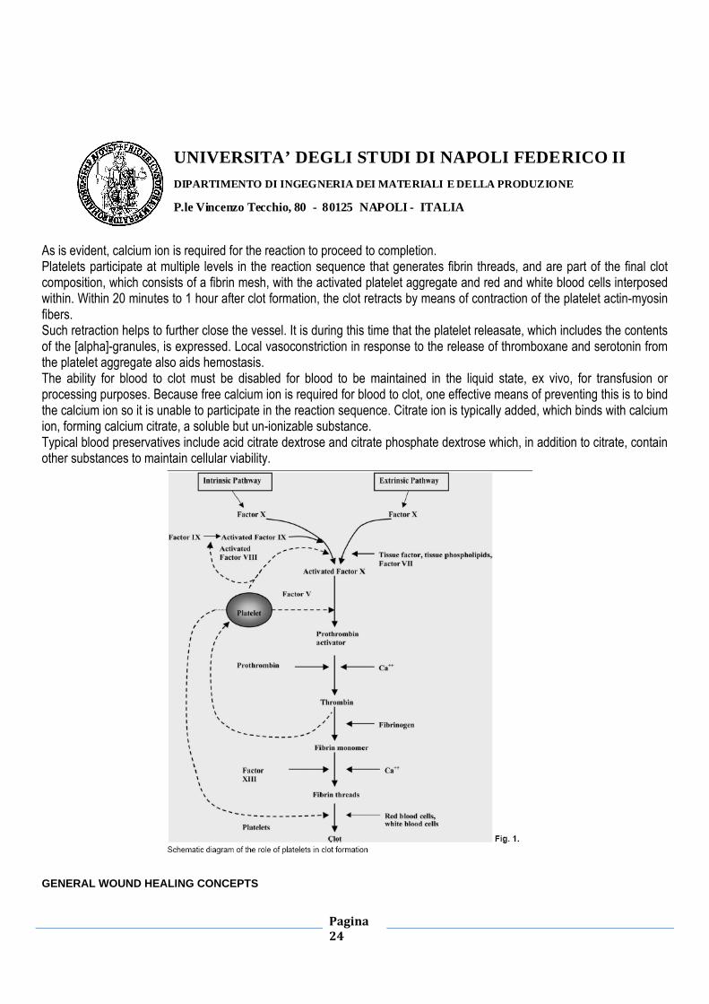
Platelets are cytoplasmic fragments of megakaryocytes (a type of white blood cell), are formed in the marrow, and are round or oval in shape, approximately 2 µm in diameter. They have a trilaminar cell membrane with a glycoprotein receptor surface overlying and partially interspersed with and penetrating a bilayer of phospholipids and cholesterol. Platelets lack nuclei but contain organelles and structures such as mitochondria, microtubules, and granules ([alpha], [delta], and [lambda]). There are approximately 50 to 80 [alpha]-granules per platelet, each bound by a unit membrane and formed during megakaryocyte maturation. The [alpha]-granules are approximately 200 to 500 nm in diameter and contain over 30 bioactive proteins, many of which have a fundamental role in hemostasis and/or tissue healing. The platelet cytoplasm contains an open, canalicular system that increases the effective surface area for intake of stimulatory agonists and the discharge of effector secretions. The submembrane region contains microfilaments of actin and myosin that mediate morphologic alterations. These cells possess a tricarboxylic acid cycle and use glucose by means of the glycolytic and hexose monophosphate shunt pathways. Their function is closely linked to their metabolic activity. Platelets reside intravascularly and are concentrated in the spleen. The normal concentration of platelets in blood is approximately 140,000 to 400,000 platelets/mm3. These remain in the circulation for an average of approximately 10 days before removal by macrophages of the reticuloendothelial system. PLATELET FUNCTION IN HEMOSTASIS AND WOUND HEALING Functionally, platelets are involved with both hemostasis and the initiation of wound healing. This, however, is a somewhat arbitrary division because hemostasis can be considered to be the first stage of healing. PLATELET ROLE IN HEMOSTASIS After tissue injury, platelets become exposed to damaged blood vessels, which places them in direct contact with collagen, the basement membranes of capillaries, and subendothelial microfibrils. This interaction causes the platelets to aggregate at the site and change from a rounded shape to one that includes large, sticky protuberances, or pseudopodia. This process is called activation. During activation, the [alpha]-granules fuse with the platelet plasma membrane and release their protein contents to the surroundings, a topic that is described in more detail below as related to the role of platelets in tissue healing. Other factors that mediate activation include adenosine diphosphate, which is released by activated platelets, and thrombin and adrenalin. For small vascular defects, this platelet plug may be sufficient to stop blood loss; however, if the defect is large, a blood clot may be required. Blood clotting is initiated by one of two pathways, namely, the intrinsic and extrinsic pathways. The intrinsic pathway is initiated by damage or alteration to the blood, itself, whereas the extrinsic pathway is initiated by contact of blood with factors that are extraneous to the blood (e.g., damaged tissue). Both pathways involve a cascaded reaction sequence whereby inactive factors become activated which, in turn, catalyze the formation of other products from precursors that go on to catalyze subsequent reactions, leading to the formation of a formal clot. Although both pathways begin differently, they converge and share many of the latter steps in the reaction series, as shown in Fig. 1.
UNIVERSITA’ DEGLI STUDI DI NAPOLI FEDERICO IIDIPARTIMENTO DI INGEGNERIA DEI MATERIALI E DELLA PRODUZIONE
P.le Vincenzo Tecchio, 80 - 80125 NAPOLI - ITALIA
Pagina
24
As is evident, calcium ion is required for the reaction to proceed to completion. Platelets participate at multiple levels in the reaction sequence that generates fibrin threads, and are part of the final clot composition, which consists of a fibrin mesh, with the activated platelet aggregate and red and white blood cells interposed within. Within 20 minutes to 1 hour after clot formation, the clot retracts by means of contraction of the platelet actin-myosin fibers. Such retraction helps to further close the vessel. It is during this time that the platelet releasate, which includes the contents of the [alpha]-granules, is expressed. Local vasoconstriction in response to the release of thromboxane and serotonin from the platelet aggregate also aids hemostasis. The ability for blood to clot must be disabled for blood to be maintained in the liquid state, ex vivo, for transfusion or processing purposes. Because free calcium ion is required for blood to clot, one effective means of preventing this is to bind the calcium ion so it is unable to participate in the reaction sequence. Citrate ion is typically added, which binds with calcium ion, forming calcium citrate, a soluble but un-ionizable substance. Typical blood preservatives include acid citrate dextrose and citrate phosphate dextrose which, in addition to citrate, contain other substances to maintain cellular viability.
GENERAL WOUND HEALING CONCEPTS

UNIVERSITA’ DEGLI STUDI DI NAPOLI FEDERICO IIDIPARTIMENTO DI INGEGNERIA DEI MATERIALI E DELLA PRODUZIONE
P.le Vincenzo Tecchio, 80 - 80125 NAPOLI - ITALIA
Pagina
25
There are three overlapping stages to wound healing (1) inflammatory, (2) proliferative, and (3) remodeling. Inflammation is the initial response to tissue injury, whereby the goal is to provide rapid hemostasis and begin the sequence of events that leads to regeneration tissue. As blood escapes from the damaged vessels, a hematoma forms, filling the tissue space, with platelets playing a key role, as described above. Growth factors and cytokines are released by activated platelets and other cells, resulting in cell migration, proliferation, differentiation, and matrix synthesis. The fibrin mesh of the hematoma functions as a provisional matrix to maintain the regenerative space and provide a scaffold for cell migration and proliferation. The first inflammatory cells to invade the wound site are neutrophils, which provide rapid protection against infection and removal of tissue debris, having lifetimes measured in hours and days. Next, there is an influx of monocytes and T-lymphocytes. The monocytes differentiate to macrophages and become the predominant cell type. Macrophages have lifetimes measured in days to months and assist the neutrophils in their function and in secreting factors that direct succeeding events. The role of the T lymphocytes in successful wound repair is presently unclear. Mesenchymal stem cells migrate into the region, providing the uncommitted cell line that will be responsible for formation of bone, cartilage, fibrous tissue, blood vessels, and other tissues. Fibroblasts migrate into the region and begin to proliferate, producing extracellular matrix. Blood vessel endothelial cells near the injury proliferate and form new capillaries that extend into the injured site. This begins the process of angiogenesis. Near the end of the inflammatory phase, granulation tissue, with a pink, soft, granular appearance, forms. This is a transient, well-vascularized tissue devoid of nerves but rich in fibroblasts, capillaries, and chronic inflammatory cells that provides a metabolically rich environment to aid repair. During the second, or proliferative, phase of wound healing, the damaged, necrotic tissue is removed and replaced by living tissue that is specific to the local tissue environment (e.g., bone, cartilage, fibrous tissue). The mesenchymal stem cells differentiate into osteoblasts, fibroblasts, chondrocytes, and other cell types as required to generate the appropriate type of tissue. Local factors, including the growth factor and cytokine profile, hormones, nutrients, pH, oxygen tension, and the electrical and mechanical environment, mediate the appropriate differentiation. The third and final phase of wound healing is remodeling. During this phase, the newly generated tissue reshapes and reorganizes to more closely resemble the original tissue. Changes that occur include a reduction in cell density and vascularity, removal of excess repair matrix, and orientation of the collagen fibers of the repair matrix along lines of stress to maximize strength. Bone remodeling is generally described by Wolff's law. This final stage of healing can require years for completion. Scar tissue differs from normal tissue in that it is regenerated tissue that consists primarily of fibroblasts and matrix and may restore integrity but not form and function. Soft tissue and skin heals by scar formation. The healed tissue, however, may consist of some components of the original tissue that have reformed within the scar. Bone is unique in that it typically heals without scar (i.e., the healed tissue cannot be distinguished from uninjured bone). Tissue, patient, and treatment variables affect the rate and quality of the healing response. PLATELET ROLE IN WOUND HEALING

UNIVERSITA’ DEGLI STUDI DI NAPOLI FEDERICO IIDIPARTIMENTO DI INGEGNERIA DEI MATERIALI E DELLA PRODUZIONE
P.le Vincenzo Tecchio, 80 - 80125 NAPOLI - ITALIA
Pagina
26
Numerous proteins are contained within the [alpha]-granules of platelets that strongly influence wound healing, including platelet-derived growth factor (PDGF) (including [alpha][alpha], [beta][beta], and [alpha][beta] isomers), transforming growth factor (TGF)-[beta] (including [beta]1 and [beta]2 isomers), platelet factor 4 (PF4), interleukin (IL)-1, platelet-derived angiogenesis factor (PDAF), vascular endothelial growth factor (VEGF), epidermal growth factor (EGF), platelet-derived endothelial growth factor (PDEGF) epithelial cell growth factor (ECGF), insulin-like growth factor (IGF), osteocalcin, osteonectin, fibrinogen, vitronectin, fibronectin, and thrombospondin (TSP). Collectively, these proteins are members of the families of growth factors, cytokines, and chemokines which, for the purpose of this review, are broadly referred to as secretory proteins. Activation, also known as degranulation, causes the [alpha]-granules to fuse to the platelet cell membrane, where at least some of the secretory proteins (e.g., PDGF and TGF-[beta]) are transformed to a bioactive state by the addition of histones and carbohydrate side chains. The active proteins are then secreted, allowing them to bind to the trans-membrane receptors of target cells (e.g., mesenchymal stem cells, osteoblasts, fibroblasts, endothelial cells, and epidermal cells). Once bound to the transmembrane receptors, intracellular signal proteins are activated, which results in the expression of a gene sequence that directs cellular proliferation, matrix formation, osteoid production, collagen synthesis, and so forth. Platelets begin to actively secrete these proteins within 10 minutes after clotting, with more than 95 percent of the presynthesized growth factors secreted within 1 hour. After this initial burst of protein release, the platelets synthesize and secrete additional proteins for the balance of their life (5 to 10 days). As the direct platelet influence begins to subside, macrophages, which arrive by means of vascular ingrowth stimulated by the platelets, assume responsibility for wound-healing regulation by secreting their own factors. Thus, the platelets at the repair site ultimately set the pace for wound repair. The many proteins secreted by the activated platelets influence many aspects of wound healing, and Anitua et al. have provided a recent, detailed review. For example, PDGF is chemotactic for macrophages whereas, collectively, PDGF, TGF-[beta], and IGF assist in chemotaxis and mitogenesis of stem cells and osteoblasts, angiogenesis for capillary ingrowth, bone matrix formation, and collagen synthesis. TGF-[beta] and PDGF also assist in bone mineralization. As a group, the adhesive proteins fibrinogen, fibronectin, vitronectin, and TSP-1 participate in thrombus formation, and some appear to have some mitogenic characteristics as well. Some of the secretory proteins released from platelets are absent in chronic, nonhealing wounds, providing further evidence of their role in wound healing. Although it is generally believed that platelets do not contain bone morphogenetic proteins (BMPs), Sipe et al. recently identified both BMP-2 and BMP-4 within platelet lysate, suggesting the possibility that this might contribute to the platelet role in bone formation and repair. A schematic diagram of the role of platelets in wound healing is shown in Figure 2. Although platelets are central to normal wound healing, healing can occur in their absence. For instance, animals rendered thrombocytopenic, although displaying altered wound-healing characteristics that may be site-dependent, may go on to heal, perhaps through the involvement of compensatory mechanisms.

UNIVERSITA’ DEGLI STUDI DI NAPOLI FEDERICO IIDIPARTIMENTO DI INGEGNERIA DEI MATERIALI E DELLA PRODUZIONE
P.le Vincenzo Tecchio, 80 - 80125 NAPOLI - ITALIA
Pagina
27
PLATELET-RICH PLASMA Platelet-rich plasma is defined as a portion of the plasma fraction of autologous blood having a platelet concentration above baseline. As such, platelet-rich plasma contains not only a high level of platelets but also the full complement of clotting factors, the latter of which typically remain at their normal, physiologic levels. Other terms in the literature that are sometimes used to describe platelet preparations include platelet concentrate, platelet gel, and platelet releasate. Ideally, there should be universal agreement regarding definitions and terminology; however, at the very least, the nature of the platelet derivative studied should be precisely and unambiguously described. The processing required to concentrate platelets in a portion of the plasma will leave the balance essentially devoid of platelets. Such platelet-poor plasma can function clinically as a fibrin sealant for hemostasis. Five important issues relating to platelets are discussed in the following order: platelet concentration ratio, processing technique, quantification of secretory protein concentration, handling, and application, and clinical use. Platelet Concentration Ratio To a first approximation, the amount of hematoma that forms in response to trauma is proportional to the degree of tissue injury. In this way, delivery of platelet-rich plasma can be thought of as responding with hematoma in excess of that which would have been physiologically produced. It is likely that the effect of platelet-rich plasma on wound healing is a function of many variables, including the platelet concentration, platelet-rich plasma volume delivered, the extent and type of injury, and the overall medical condition of the patient. The large number of variables and their potential for interaction is probably the

UNIVERSITA’ DEGLI STUDI DI NAPOLI FEDERICO IIDIPARTIMENTO DI INGEGNERIA DEI MATERIALI E DELLA PRODUZIONE
P.le Vincenzo Tecchio, 80 - 80125 NAPOLI - ITALIA
Pagina
28
reason that there is no single recommendation for the degree of increase of platelets in platelet-rich plasma over baseline. Some investigators have suggested that platelet-rich plasma should achieve a 3 to 5-fold increase in platelet concentration over baseline, although the dependence of clinical benefit on platelet concentration versus total number of platelets delivered may need to await further investigation. Platelet concentration ratios of less than 2-fold to 8.5-fold have been reported. Weibrich et al. suggest that different individuals may require different platelet concentration ratios to achieve comparable biological effect. Processing Technique During surgery, platelets will collect at the surgical site to initiate clotting and healing, somewhat reducing the whole blood platelet count. As such, blood should be drawn before surgery begins because the surgery itself will lead to platelet activation that may interfere with preparation. When anticoagulated blood is centrifuged, three layers form as a function of density: the bottom layer consisting of red blood cells (specific gravity, 1.09), the middle layer consisting of platelets and white blood cells (buffy coat; specific gravity, 1.06), and the top plasma layer (specific gravity, 1.03). Centrifugation forms the basis of current methods for producing platelet-rich plasma, with the yield approximately 10 percent, by volume, of whole blood drawn. Platelet fragmentation during processing should be avoided. Because it is the process of activation that results in the completion of the tertiary structure of some of the secretory proteins, such fragmentation during processing could result in the release of high levels of proteins, with compromised bioactivity. The integrity of the platelet membrane can be preserved by use of acid citrate dextrose type A anticoagulant and low gravity forces during centrifugation. In addition, platelet activation occurring during processing should be kept to a minimum. Although bioactive secretory proteins would be produced, they might be lost and not transferred to the surgical bed when the clot is implanted, although the magnitude of this may be a function of the mode of delivery. Pselectin is a protein contained in the inner face of the [alpha]-granule membrane. On platelet activation, the [alpha]-granule membrane fuses with the platelet membrane and P-selectin becomes expressed on the platelet surface, where it can be measured and the amount of platelet activation determined. Thus, measurement of P-selectin provides valuable information regarding the platelet-rich plasma preparation. Although it is possible to use a standard laboratory centrifuge to produce platelet-rich plasma, the process is labor intensive, generally requiring two spins and multiple transfers; consequently, sterility may be difficult to maintain. Furthermore, such techniques may not reliably maximize the platelet concentration or the levels of key secretory proteins. Standard cell separators and salvage devices can be used to produce platelet-rich plasma. These devices operate on a unit of blood and typically use continuous-flow centrifuge bowl or continuous-flow disk separation technology and both a hard (fast) and a soft (slow) spin, yielding platelet concentrations from two to four times baseline. Many surgical procedures require use of relatively small volumes of platelet-rich plasma. Some of these procedures may be performed in an office setting, making draw of a full unit of blood undesirable and legally precluding the reintroduction of the unused portion of the blood to the patient. Consequently, small, compact office systems have been developed that produce approximately 6 ml of platelet-rich plasma from 45 to 60 ml of blood, obviating the need for reinfusion. There are many such systems, including the GPS (Biomet, Warsaw, Ind.), the PCCS (Implant Innovations, Inc., Palm Beach

UNIVERSITA’ DEGLI STUDI DI NAPOLI FEDERICO IIDIPARTIMENTO DI INGEGNERIA DEI MATERIALI E DELLA PRODUZIONE
P.le Vincenzo Tecchio, 80 - 80125 NAPOLI - ITALIA
Pagina
29
Gardens, Fla.), the Symphony II (DePuy, Warsaw, Ind.), the SmartPReP (Harvest Technologies Corp., Norwell, Mass.), and the DENSITY PLATELET GEL® (Industrie Biomediche e Farmaceutiche Srl, Scafati, Italy). Although all operate on a small volume of drawn blood (45 to 60 ml) and on the principle of centrifugation, these systems differ widely in their ability to collect and concentrate platelets, with approximately 30 to 85 percent of the available platelets collected and from a less than 2-fold to an approximately 8-fold increase in the platelet concentration over baseline. In general, most systems, whether large or small volume, do not concentrate the plasma proteins of the coagulation cascade. Quantification of Secretory Protein Concentration The regenerative potential of platelet-rich plasma depends, to large extent, on the levels of secretory proteins that are released on platelet activation. These protein levels will depend on several factors, including (1) the concentrations of these proteins contained in the platelets (a patient variable); (2) the processing technique, which will influence platelet concentration and whether platelets are activated or fragmented during preparation; and (3) the completeness of platelet activation before measurement. The secretory proteins must first be released from the platelets before they can be measured. Release can be accomplished through platelet activation or through physical disruption of the platelet/[alpha]-granule structure. The most common method of platelet activation is to add calcium chloride and thrombin to the platelet-rich plasma. The thrombin directly activates platelets, and the calcium ion replenishes that which was bound by the acid citrate dextrose type A anticoagulant. Although this method is often used to activate platelet-rich plasma clinically, the activation that occurs during clot formation does not necessarily lead to complete release. Another activation method uses adenosine diphosphate, which acts directly on the platelets. Secretory protein levels are commonly expressed in concentration units (e.g., measured amount per milliliter of releasate or per 100,000 platelets). Weibrich et al., using a freeze/thaw cycle to release proteins, measured the levels of PDGF-[alpha][beta], PDGF-[beta][beta], TGF-1, TGF-[beta], and IGF-1 in specimens of platelet-rich plasma derived from 115 patients. Minimum and maximum values for each typically spanned one to two orders of magnitude, with means ± SD of 117.5 ± 63.4 ng/ml, 9.9 ± 7.5 ng/ml, 169.4 ± 84.5 ng/ml, 0.4 ± 0.3 ng/ml, and 84.2 ± 23.6 ng/ml, respectively. They found statistical correlations between the concentrations of the following pairs of growth factors: PDGF-[alpha][beta]/PDGF-[beta][beta], PDGF-[alpha][beta]/TGF-[beta], and PDGF-[beta][beta]/TGF-[beta]. There was little or no correlation between the levels of these individual proteins and donor age and gender attributes. Eppley et al. used thrombin/calcium chloride to activate platelets and release proteins from platelet-rich plasma derived from 10 healthy volunteers. Secretory protein levels measured were 17 ± 8 ng/ml (PDGF-[beta][beta]), 120 ± 42 ng/ml (TGF-[beta]1), 955 ± 1030 ng/ml (VEGF), 129 ±61 ng/ml (EGF), and 72 ± 25 ng/ml (IGF-1). Zimmermann et al. used various methods to initiate platelet release and measured levels of PDGF-[alpha][beta], PDGF-[beta][beta], and TGF-[beta]1 in platelet-rich plasma preparations both rich and deficient in white blood cells, expressing levels both on a per-milliliter and a per-100,000 platelet basis. For a given protein, there was typically a 3- to 4-fold range in measured level versus release method, and the authors concluded that the release of each growth factor by a given sample preparation method must be investigated and interpreted separately. All else being equal, one would expect that the concentration of released secretory proteins would be linearly proportional to the platelet concentration ratio. Although such a relationship between some secretory proteins (i.e., PDGF-[alpha][beta], TGF-[beta], VEGF, and EGF) and platelet count has been reported, an additional study by the same principal author confirmed this relationship only for PDGF, TGF-[beta], and EGF, but not for VEGF and IGF.60 Although a general trend of

UNIVERSITA’ DEGLI STUDI DI NAPOLI FEDERICO IIDIPARTIMENTO DI INGEGNERIA DEI MATERIALI E DELLA PRODUZIONE
P.le Vincenzo Tecchio, 80 - 80125 NAPOLI - ITALIA
Pagina
30
increasing protein content and platelet count for a variety of secretory proteins (PDGF-[alpha][beta], PDGF-[beta][beta], TGF-1, TGF-[beta], VEGF, EGF, and IGF-1) was demonstrated, Eppley et al. and Weibrich et al.found little value in using platelet concentration ratio to predict resultant platelet-rich plasma secretory protein levels. Eppley et al., using thrombin/calcium chloride to activate the platelets, found variable concentration ratios for several secretory proteins, all lower than the platelet concentration ratio. Incomplete platelet activation and variable binding of the expressed proteins to the clot, which would not have been measured in the platelet-rich plasma supernatant, could be a partial explanation. Handling and Application of Platelet-Rich Plasma After preparation, platelet-rich plasma is stable, in the anticoagulated state, for 8 hours, or longer, permitting the blood to be drawn before surgery and used, as needed, during lengthy operations. The platelet-rich plasma must be activated for the platelets to release their [alpha]-granule contents, with the clot that forms providing a vehicle to contain the secreted proteins and maintain their presence at the wound site. This is most commonly performed by adding a solution of 1000 units of topical bovine thrombin per milliliter of 10% calcium chloride to the platelet-rich plasma. Marx et al. described a technique in which 6 ml of platelet-rich plasma, 1 ml of the calcium chloride/thrombin mix, and 1 ml of air (to act as a mixing bubble) is introduced into a 10-ml syringe. The syringe is agitated for 6 to 10 seconds to initiate clotting, and then the clot delivered. Alternatively, Man et al. described another technique for delivering the activated platelet-rich plasma. The platelet-rich plasma and calcium Chloride/thrombin solution are mixed in a 10:1 (volume/volume) ratio by use of a dual-syringe mixing system. The platelet-rich plasma is drawn into a 10-ml syringe and the activating solution is drawn into a 1-ml syringe. Both syringe plungers are connected to move together with both output ports connected to a dual-spray applicator tip that allows both solutions to be mixed as they are applied to the wound. Plateletpoor plasma can be delivered similarly to function as a fibrin glue or hemostatic agent. Because the [alpha]-granules quickly release their contents on activation, Marx states that the clotted platelet-rich plasma should be used within 10 minutes of clot initiation. This is not an issue with the dual-syringe spray delivery, as the platelet-rich plasma is delivered to the wound site immediately after activation. In the case of other mixing techniques, it is important to transfer the clot to the surgical site before clot retraction; otherwise, the transferred clot may be deficient in the secretary proteins that were expressed. In the early to mid 1990s, there were a few reports of the development of antibovine antibodies (antibovine factor V) that cross-reacted with human clotting factors in response to use of the bovine product to provide hemostasis to open, bleeding vessels. Although bovine thrombin is often used (along with calcium chloride) for clinical platelet activation, there is little evidence that such cross-reaction occurs for this application. This may be because current processing methods remove much more bovine factor V contamination and its use in platelet-rich plasma gel precludes its exposure to the systemic circulation, possibly explaining why platelet-rich plasma has not produced postsurgical bleeding or shown elevation in postoperative prothrombin time or the development of detectable antibovine antibodies.

UNIVERSITA’ DEGLI STUDI DI NAPOLI FEDERICO IIDIPARTIMENTO DI INGEGNERIA DEI MATERIALI E DELLA PRODUZIONE
P.le Vincenzo Tecchio, 80 - 80125 NAPOLI - ITALIA
Pagina
31
Clinical Use Proponents of platelet-rich plasma technology suggest that benefits include an increase in hard- and softtissue wound healing and a decrease in postoperative infection, pain, and blood loss. There have been numerous publications on the use of platelet-rich plasma for several clinical applications, including periodontal and oral surgery, maxillofacial surgery, aesthetic plastic surgery, spinal fusion, heart bypass surgery, and treatment of chronic skin and softtissue ulcers. The details of the quantity of platelet-rich plasma used and the methods of application are procedure-specific. Although the vast majority of these studies have yielded excellent outcomes, most are only limited case studies or series. As such, wound-healing enhancement by platelet-rich plasma remains largely anecdotal. There exists, however, a small collection of clinical studies with prospective or retrospective controls that have demonstrated a significant enhancement of hard- and soft-tissue healing with the use of platelet-rich plasma. In plastic surgery, the use of autologous blood–derived products has been largely limited to fibrin glues, primarily used to obtain hemostasis and adherence of skin flaps. A study of 20 patients by Man et al. demonstrated that the use of autologous fibrin glue and platelet gel in cosmetic surgical procedures involving the creation of flaps, such as in face and neck lifts and breast reductions and augmentations, resulted in numerous advantages. These included the elimination of the need for drains, a reduction in postoperative pain and swelling, and improved wound healing. In another skin flap study, a clinical series of eight patients were unilaterally treated with autologous platelet–rich plasma mixed with thrombin and calcium chloride to form an autologous platelet gel to determine the effects on postoperative recovery from deep-plane rhytidectomy. Staged postoperative facial photographs were graded in a blinded fashion for postoperative ecchymosis and edema. Although no statistically significant differences were identified in the data, trends suggested that treatment with autologous platelet gel was better at preventing or improving ecchymosis rather than edema, and was chiefly demonstrable in the early phases of recovery. In a larger face-lift case series, a single postoperative hematoma among a cohort of 100 consecutive patients undergoing face lifting treated with bilateral autologous platelet gel occurred, an outcome reported as a significant reduction in postoperative complication incidence by the authors. No drains or postoperative dressings were used in the fibrin glue–treated group, which patients found most favorable. Similarly, Oliver et al. in 2001 reported on a prospective, randomized, double-blind trial of the use of fibrin sealant for face lifts in 20 patients. A significant difference in drain output occurred between control and treated sides (average, 30 ml for the control side and 10 ml for the fibrin glue side). It was suggested that postoperative drains may not be needed with this technique, and that pain and bruising may likewise be reduced. Favorable results were also reported by Fezza in 2002. In 24 consecutive patients, fibrin glue was used in face lifts without drains. These patients had less bruising and swelling, no incidence of hematoma, and shorter operative times compared with another group of 24 face-lift patients where glue was not used. The most recent study of the efficacy of a commercial (non-autologously derived) fibrin glue on face lifts was reported by Marchac in 2005, nearly 10 years after his initial report in 1994. In a prospective study of 30 patients, there were minimal differences between glued and unglued sides in terms of drain output, bruising, swelling, or the incidence of hematomas. After 17 years of clinical use, this experienced author now concludes that the theoretical benefit of fibrin in face lifts is not as great as previously hoped. In a companion experience, Jones et al. in 2004 did not find any statistical difference in hematoma rates with the use of fibrin glue compared with other prevention techniques such as dressings, drains, or the use of tumescence. Results with fibrin glue fixation in forehead endoscopy were reported in 1998. In 206 endoscopic forehead procedures in 196 patients in which fibrin glue was used, good results were obtained in 86 percent of the cases, with minimal

UNIVERSITA’ DEGLI STUDI DI NAPOLI FEDERICO IIDIPARTIMENTO DI INGEGNERIA DEI MATERIALI E DELLA PRODUZIONE
P.le Vincenzo Tecchio, 80 - 80125 NAPOLI - ITALIA
Pagina
32
complications. Fibrin glue provided stable fixation, with the possibility of displacement in several directions, including transversal expansion. Numerous clinical trials have reported favorable results evaluating wounds treated with platelet releasate. Knighton et al. observed that 17 of 21 chronic lower extremity ulcers reepithelialized during an 8.6-week (average) course of twice-daily wound treatment with platelet releasate suspended on a collagen base (platelet-derived wound-healing factor) compared with two of 13 similar wounds treated with placebo. After crossover of the placebo group, all 11 nonhealed wounds achieved 100 percent epithelialization in an average of 7.1 weeks. An earlier study by the same group, also using plateletderived wound-healing factor, demonstrated a 93 percent reepithelialization rate among 71 chronic wounds in 41 patients after daily treatments with autologous platelet concentrate. A similar clinical protocol was reported by Ganio et al. in a case series of 171 patients with 355 chronic (average, 75 weeks before presentation) lower extremity wounds. Results included a 78 percent rate of limb salvage in patients for whom amputation was initially recommended after daily 12-hour treatments with plateletderived wound-healing factor for an average of 10 weeks. Marx et al.22 reported enhanced bone formation in mandibular bone grafts treated with platelet-rich plasma, an effect lasting up to 6 months postoperatively that was documented by biopsy-proven enhanced bone deposition among platelet-rich plasma-treated grafts. CONCLUSIONS Platelets immediately appear at the site of tissue injury in large numbers. As such, it makes evolutionary sense that platelets direct wound healing because, by design, they will be present exactly where and when needed, to create a local environment conducive to tissue regeneration. By releasing secretory proteins from their [alpha]-granules on activation, platelets set the pace of wound healing, with their effects remaining long after the clot has been cleared. The enhancement of healing by the placement of a supraphysiologic concentration of autologous platelets at the site of tissue injury is supported by basic science studies. There exist a small number of controlled, clinical studies that provide evidence that the use of autologous platelet-rich plasma does, indeed, accelerate soft- and hard-tissue healing in at least some applications. The future role of platelet-rich plasma in plastic surgery largely remains both theoretical and anecdotal at this time. Many more controlled clinical studies will be required to establish the conditions under which the application of platelet-rich plasma has merit. Currently, platelet-rich plasma use seems to offer some efficacy in certain types of acute and chronic wounds, although the timing of application is not clear. In aesthetic plastic surgery, although theoretically appealing and not harmful, the potential benefits of platelet-rich plasma may be difficult to measure in these patients with otherwise normal wound healing. REFERENCES 1. Beasley, L. S., and Einhorn, T. A. Role of growth factors in fracture healing. In E. Canalis (Ed.), Skeletal Growth Factors. New York: Lippincott Williams & Wilkins, 2000. Pp. 311–322 2. Bhanot, S., and Alex, J. C. Current applications of platelet gels in facial plastic surgery. Facial Plast. Surg. 18: 27, 2002 3. Anitua, E., Andia, I., Ardanza, B., et al. Autologous platelets as a source of proteins for healing and tissue regeneration. Thromb. Haemost. 91: 4, 2004. 4. Buckwalter, J. A., Einhorn, T. A., Bolander, M. E., et al. Healing of musculoskeletal tissues. In C. A. Rockwood, Jr., R. W. Bucholz, and D. P. Green (Eds.), Fractures in Adults. Philadelphia: Lippincott-Raven, 1996. Pp. 261–304. 5. Anderson, J. M. The cellular cascades of wound healing. In J. E. Davies (Ed.), Bone Engineering. Toronto: em squared

UNIVERSITA’ DEGLI STUDI DI NAPOLI FEDERICO IIDIPARTIMENTO DI INGEGNERIA DEI MATERIALI E DELLA PRODUZIONE
P.le Vincenzo Tecchio, 80 - 80125 NAPOLI - ITALIA
Pagina
33
inc., 2000. Pp. 81–93 6. Marx, R. E. Platelet-rich plasma: Evidence to support its use. J. Oral Maxillofac. Surg. 62: 489, 2004. 7. Haynesworth, S. E., Kadiyala, S., Liang, L., et al. Mitogenic stimulation of human mesenchymal stem cells by platelet releasate suggest a mechanism for enhancement of bone repair by platelet concentrates (Poster no. 462). Transactions of the 48th Annual Meeting of the Orthopaedic Research Society, Dallas, Texas, February 10–13, 2002 8. Liu, Y., Kalen, A., Risto, O., et al. Fibroblast proliferation due to exposure to a platelet concentrate in vitro is pH dependent. Wound Repair Regen. 10: 336, 2002. 9. Carter, C. A., Jolly, D. G., Worden, C. E., Sr., et al. Platelet-rich plasma gel promotes differentiation and regeneration during equine wound healing. Exp. Mol. Pathol. 74: 244, 2003. 10. Fennis, J. P. M., Stoelinga, P. J. W., and Jansen, J. A. Mandibular reconstruction: A clinical and radiographic animal study on the use of autogenous scaffolds and platelet-rich plasma. J. Oral Maxillofac. Surg. 31: 281, 2002. 11. Marx, R. E. Platelet concentrate: A strategy for accelerating and improving bone regeneration. In J. E. Davies (Ed.), Bone Engineering. Toronto: University of Toronto, 2000. Pp. 447–453. 12. Anitua, E. Plasma rich in growth factors: Preliminary results of use in the preparation of future sites for implants. J. Oral Implantol. 14: 529, 1999. 13. Bose, B., and Balzarini, M. A. Bone graft gel: Autologous growth factors used with autograft bone for lumbar spine fusions. Adv. Ther. 19: 170, 2002. 14. Della Valle, A., Sammartino, G., Marenzi, G., et al. Prevention of postoperative bleeding in anticoagulated patients undergoing oral surgery: Use of platelet-rich plasma gel. J. Oral Maxillofac. Surg. 61: 1275, 2003. 15. DelRossi, A. J., Cernaianu, A. C., Vertrees, R. A., et al. Platelet-rich plasma reduces postoperative blood loss after cardiopulmonary bypass. J. Thorac. Cardiovasc. Surg. 100: 281, 1990 16. Froum, S. J., Wallace, S. S., Tarnow, D. P., et al. Effect of platelet-rich plasma on bone growth and osseointegration in human maxillary sinus grafts: Three bilateral case reports. Int. J. Periodontics Restorative Dent. 22: 45, 2002. 17. Hee, H. T., Majd, M. E., Holt, R. T., et al. Do autologous growth factors enhance transforaminal lumbar interbody fusion? Eur. Spine J. 12: 400, 2003. 18. Kassolis, J. D., Rosen, P. S., and Reynolds, M. A. Alveolar ridge and sinus augmentation utilizing platelet-rich plasma in combination with freeze-dried bone allograft: Case series. J. Periodontol. 71:1654, 2000 19. Lowery, G. L., Kulkarni, S., and Pennisi, A. E. Use of autologous growth factors in lumbar spinal fusion. Bone 25 (2 Suppl.): 47S, 1999 20. Man, D., Plosker, H., and Winland-Brown, J. E. The use of autologous platelet-rich plasma (platelet gel) and autologous platelet-poor plasma (fibrin glue) in cosmetic surgery. Plast. Reconstr. Surg. 107: 229, 2001. 21. Margolis, D. J., Kantor, J., Santanna, J., et al. Effectiveness of platelet releasate for the treatment of diabetic neuropathic foot ulcers. Diabetes Care 24: 483, 2001. 22. Marx, R. E., Carlson, E. R., Eichstaedt, R. M., et al. Platelet-rich plasma: Growth factor enhancement for bone grafts. Oral Surg. Oral Med. Oral Pathol. Oral Radiol. Endod. 85: 638, 1998. 23. Petrungaro, P. S. Using platelet-rich plasma to accelerate soft tissue maturation in esthetic periodontal surgery. Compend. Contin. Educ. Dent. 22: 729, 2001 24. Robiony, M., Polini, F., Costa, F., et al. Osteogenesis distraction and platelet-rich plasma for bone restoration of the severely atrophic mandible: Preliminary results. J. Oral Maxillofac. Surg. 60: 630, 2002 25. Soffer, E., Ouhayoun, J. P., and Anagnostou, F. Fibrin sealants and platelet preparations in bone and periodontal healing. Oral Surg. Oral Med. Oral Pathol. Oral Radiol. Endod. 95: 521, 2003.
UNIVERSITA’ DEGLI STUDI DI NAPOLI FEDERICO IIDIPARTIMENTO DI INGEGNERIA DEI MATERIALI E DELLA PRODUZIONE
P.le Vincenzo Tecchio, 80 - 80125 NAPOLI - ITALIA
Pagina
34
26. Welsh, W. J. Autologous platelet gel: Clinical function and usage in plastic surgery. Cosmetic Derm. 11: 13, 2000 27. Whitman, D. H., Berry, R. L., and Green, D. M. Platelet gel: An autologous alternative to fibrin glue with applications in oral and maxillofacial surgery. J. Oral Maxillofac. Surg. 55: 1294, 1997. 28. Shanaman, R., Filstein, M. R., and Danesh-Meyer, M. J. Localized ridge augmentation using GBR and platelet-rich plasma: Case reports. Int. J. Periodontics Restorative Dent. 21: 345, 2001 29. Eppley, B. L., Woodell, J. E., and Higgins, J. Platelet quantification and growth factor analysis from platelet-rich plasma: Implications for wound healing. Plast. Reconstr. Surg. 114: 1502, 2004. 30. Kevy, S. V., and Jacobson, M. S. Comparison of methods for point of care preparation of autologous platelet gel. J. Extra Corpor. Technol. 36: 28, 2004 31. Marx, R. E. Platelet-rich plasma (PRP): What is PRP and what is not PRP? Implant Dent. 10: 225, 2001. 32. Waters, J. H., and Roberts, K. C. Database review of possible factors influencing point-of-care platelet gel manufacture. J. Extra Corpor. Technol. 36: 250, 2004. 33. Zimmermann, R., Arnold, D., Strasser, E., et al. Sample preparation technique and white cell content influence the detectable levels of growth factors in platelet concentrates. Vox Sang. 85: 283, 2003 34. Guyton, A. C. Physiology of the Human Body. Philadelphia: Saunders College Publishing, 1979. 35. Tischler, M. Platelet rich plasma: The use of autologous growth factors to enhance bone and soft tissue grafts. N. Y. State Dent. J. 68: 22, 2002 36. Conley, C. L. Hemostasis. In V. B. Mountcastle (Ed.), Medical Physiology. St. Louis: Mosby, 2004. Pp. 1137–1146 37. Caro, C. D., Pedley, T. J., Schroter, R. C., et al. The Mechanics of the Circulation. Oxford: Oxford University Press, 1978. 38. Harrison, P., and Cramer, E. M. Platelet alpha-granules. Blood Rev. 7: 52, 1993 39. Szpaderska, A. M., Egozi, E. I., Gamelli, R. L., et al. The effect of thrombocytopenia on dermal wound healing. J. Invest. Dermatol. 120: 1130, 2003 40. Lowe, H. C., Rafty, L. A., Collins, T., et al. Biology of platelet-derived growth factor. In E. Canalis (Ed.), Skeletal Growth Factors. Philadelphia: Lippincott Williams & Wilkins, 2000. Pp. 129–151. 41. Woodward, S. C., and Salthouse, T. N. The tissue response to implants and its evaluation by light microscopy. In A. F. von Recum (Ed.), Handbook of Biomaterials Evaluation. New York: Macmillan, 1986. Pp. 364–378 42. Frost, H. M. A 2003 update of bone physiology and Wolff's law for clinicians. Angle Orthod. 74: 3, 2004 43. Roseborough, I. E., Grevious, M. A., and Lee, R. C. Prevention and treatment of excessive dermal scarring. J. Natl. Med. Assoc. 96: 108, 2004 44. Weibrich, G., Kleis, W. K., Hafner, G., et al. Growth factor levels in platelet-rich plasma and correlations with donor age, sex, and platelet count. J. Craniomaxillofac. Surg. 30: 97, 2002. 45. Lariviere, B., Rouleau, M., Picard, S., et al. Human plasma fibronectin potentiates the mitogenic activity of platelet-derived growth factor and complements its wound healing effects. Wound Repair Regen. 11: 79, 2003 46. Zhou, Y. Q., Levesque, J. P., Hatzfeld, A., et al. Fibrinogen potentiates the effect of interleukin-3 on early human hematopoietic progenitors. Blood 82: 800, 1993. 47. Sipe, J. B.Waits, C. A.Skikne, B. et al. The presence of bone morphogenetic proteins (BMPs) in megakaryocytes and platelets. Presented at the 24th Annual Meeting of the American Society for Bone and Mineral Research, San Antonio, Texas, September 20–24, 2002. 48. Gonshor, A. Technique for producing platelet-rich plasma and platelet concentrate: Background and process. Int. J. Periodontics Restorative Dent. 22: 547, 2002.

UNIVERSITA’ DEGLI STUDI DI NAPOLI FEDERICO IIDIPARTIMENTO DI INGEGNERIA DEI MATERIALI E DELLA PRODUZIONE
P.le Vincenzo Tecchio, 80 - 80125 NAPOLI - ITALIA
Pagina
35
49. Mann, K. G. Thrombin formation. Chest 124 (3 Suppl.): 4S, 2003. 50. Weibrich, G., Kleis, W. K., Kunz-Kostomanolakis, M., et al. Correlation of platelet concentration in platelet-rich plasma to the extraction method, age, sex, and platelet count of the donor. Int. J. Oral Maxillofac. Implant. 16: 693, 2001. 51. Slater, M., Patava, J., Kingham, K., et al. Involvement of platelets in stimulating osteogenic activity. J. Orthop. Res. 13: 655, 1995 52. Arm, D. M. Autologous platelet-based therapies for orthopaedic tissue regeneration. Orthopedics 25: 169, 2002 53. Hannon, T. J., Polston, G., Pekarske, W. J., et al. Determination of Platelet Yields from Platelet Rich Plasma for Five Autotransfusion Devices. Cardiothoracic Research and Education Foundation, 1999. 54. Marlovits, S., Mousavi, M., Gabler, C., et al. A new simplified technique for producing platelet-rich plasma: A short technical note. Eur. Spine J. 13 (Suppl. 1): S102, 2004 55. Lozada, J. L., Caplanis, N., Proussaefs, P., et al. Platelet-rich plasma application in sinus graft surgery: Part I. Background and processing techniques. J. Oral Implantol. 27: 38, 2001 56. Hood, A. G., and Arm, D. M. Topical application of autogenous tissue growth factors for augmentation of structural bone graft fusion. Presented at the American Society of Extra-Corporeal Technology 11th Annual Symposium on New Advances in Blood Management, Las Vegas, Nev. April 20–23, 2004. 57. Arm, D. M. Characterization of an autologous platelet gel containing multiple growth factors. Transactions of the 45th Annual Meeting of the Orthopaedic Research Society, Anaheim, Calif., February 1–4, 1999 58. Weibrich, G., Kleis, W. K., and Hafner, G. Growth factor levels in the platelet-rich plasma produced by 2 different methods: Curasan-type PRP kit versus PCCS PRP system. Int. J. Oral Maxillofac. Implants 17: 184, 2002. 59. Kevy, S. V., Jacobson, M. S., Blasetti, L., et al. Preparation of growth factor enriched autologous platelet gel (Paper no. 262). Transactions of the Society for Biomaterials 27th Annual Meeting, St. Paul, Minn., April 24–29, 2001. 60. Kevy, S. V., Jacobson, M. S., and Kadiyala, S. Characterization of growth factor levels in platelet concentrates. Presented at the 5th Annual Hilton Head Workshop on Engineering Tissues, Hilton Head, S.C., February 21–25, 2001 61. Anderson, N. A., Pamphilon, D. H., Tandy, N. J., et al. Comparison of platelet-rich plasma collection using the Haemonetics PCS and Baxter Autopheresis C. Vox Sang. 60: 155, 1991. 62. Christie, R. J., Carrington, L., and Alving, B. Postoperative bleeding induced by topical bovine thrombin: Report of two cases. Surgery 121: 708, 1997. 63. Rapaport, S. I., Zivelin, A., Minow, R. A., et al. Clinical significance of antibodies to bovine and human thrombin and factor V after surgical use of bovine thrombin. Am. J. Clin. Pathol. 97: 84, 1992. 64. Zehnder, J. L., and Leung, L. L. Development of antibodies to thrombin and factor V with recurrent bleeding in a patient exposed to topical bovine thrombin. Blood 76: 2011, 1990. 65. Knighton, D. R., Ciresi, K., Fiegel, V. D., et al. Stimulation of repair in chronic, nonhealing, cutaneous ulcers using platelet-derived wound healing formula. Surg. Gynecol. Obstet. 170: 56, 1990. 66. Ellis, D. A., and Shaikh, A. The ideal tissue adhesive in facial plastic and reconstructive surgery. J. Otolaryngol. 19: 68, 1990 67. Mandel, M. A. Minimal suture blepharoplasty: Closure of incisions with autologous fibrin glue. Aesthetic Plast. Surg. 16: 269, 1992 68. Powell, D. M., Chang, E., and Farrior, E. H. Recovery from deep-plane rhytidectomy following unilateral wound treatment with autologous platelet gel. Arch. Facial Plast. Surg. 3: 245, 2001. 69. Marchac, D., and Sandor, G. Facelifts and sprayed fibrin glue: An outcome analysis of 200 patients. Br. J. Plast. Surg. 47: 306, 1994

UNIVERSITA’ DEGLI STUDI DI NAPOLI FEDERICO IIDIPARTIMENTO DI INGEGNERIA DEI MATERIALI E DELLA PRODUZIONE
P.le Vincenzo Tecchio, 80 - 80125 NAPOLI - ITALIA
Pagina
36
70. Oliver, D. W., Hamilton, S. A., Figle, A. A., Wood, S. H., and Lamberty, B. G. A prospective, randomized, double-blind trial of the use of fibrin sealant for facelifts. Plast. Reconstr. Surg. 108: 2102, 2001. 71. Fezza, J. P., Cartwrwight, M., Mack, W., and Flaharty, P. The sue of aerosolized fibrin glue in face-lift surgery. Plast. Reconstr. Surg. 110: 658, 2002 72. Marchac, D., and Greensmith, A. L. Early postoperative efficacy of fibrin glue in facelifts: A prospective randomized trial. Plast. Reconstr. Surg. 115: 911, 2005. 73. Jones, B. M., and Grover, R. Avoiding hematoma in cervicofacial rhytidectomy: A personal 8-year quest. Reviewing 910 patients. Plast. Reconstr. Surg. 113: 381, 2004. 74. Marchac, D., Ascherman, J., and Arnaud, E. Fibrin glue fixation in forehead endoscopy: Evaluation of our experience in 206 cases. Plast. Reconstr. Surg. 100: 704, 1997. 75. Knighton, D. R., Fiegal, V. D., Doucette, M., et al. The use of topically applied platelet growth factors in chronic nonhealing wounds: A review. Wounds 1: 71, 1989. 76. Ganio, C., Tenewitz, F. E., Wilson, R., et al. The treatment of chronic nonhealing wounds using autologous platelet-derived growth factors. J. Foot Ankle Surg. 32: 263, 1993