
Clinical Case series: Fetal* MR Imaging of the Brain at 3T MRI/Fetal MR 3T cases... · ·...
4
100 MAGNETOM Flash · 2/2011 · www.siemens.com/magnetom-world Clinical Fetal Imaging Background Fetal MR imaging (MRI) is a recognized complementary method to fetal ultra- sound to identify fetal central nervous system (CNS) pathology. It can provide additional and diagnostically-relevant information, add certainty to ultrasound diagnosis and help parental counselling. In most imaging centers MRI is performed at 1 or 1.5 Tesla field strength, with no proven deleterious effect on the fetus to date. According to national and inter- national guidelines, fetal MRI is recom- mended from the second trimester of gestation, and specific absorption rates (SAR) for each pulse sequences are limited by the manufacturers to ensure that increase in body temperature is less than 0.5°C. Experimental physical evi- dence suggests that SAR deposition to the fetus in utero is only minimally higher at 3 Tesla, and still within the guidelines recommendation, but care still needs to be taken. This case series intends to provide an insight into the enhanced visualization of fetal CNS in selected brain patholo- gies at 3 Tesla. Materials and methods All images in this case series were acquired on a 48-channel whole-body 3T scanner (MAGNETOM Skyra) with a combination of the Body Matrix coil anteriorly and integrated spine coil pos- teriorly. Fetal brain protocol typically consists of T2-weighted (w) HASTE single- shot in all three planes, T1w FLASH in the axial plane, Diffusion-tensor imaging in the axial plane. Selected sequence parameters were as follows and adapted to the patient: Case series: Fetal* MR Imaging of the Brain at 3T Jacques F. Schneider University Children’s Hospital, Basel, Switzerland Sequence parameters Axial / Coronal / Sagittal T2w HASTE (free-breathing) TR/TE = 1100 / 96 ms, SL = 3 mm, FOV = (280*280) mm 2 , matrix = (256*230) px 2 Axial T1w FLASH (breathhold) TR/TE = 122 / 2.46 ms, SL= 3 mm, FOV = (280*280) mm 2 , matrix = (224*168) px 2 Axial epi2D mddw20 (free-breathing) TR/TE = 4300 / 85 ms, SL 3 mm, FOV = (220*220) mm 2 , matrix = (142*142) px 2 , b-value = 800 Sagittal T2w trufi2d (breathhold) TR/TE = 3.88 / 1.7 ms, FOV = (376*376) mm 2 , matrix = (614*768) px 2 1A 1D *MR scanning has not been established as safe for imaging fetuses and infants under two years of age. The responsible physician must evaluate the benefits of the MRI examination compared to other imaging procedures. Conclusion This short case series shows that fetal MRI at 3T is feasible and delivers excellent visualization of fetal brain and spine structures. With growing experience, the use of 3T systems for fetal MRI could add much-needed certainty in the diagnostic workup of fetal brain pathologies.
Transcript of Clinical Case series: Fetal* MR Imaging of the Brain at 3T MRI/Fetal MR 3T cases... · ·...

100 MAGNETOM Flash · 2/2011 · www.siemens.com/magnetom-world
Clinical Fetal Imaging
Background
Fetal MR imaging (MRI) is a recognized complementary method to fetal ultra-sound to identify fetal central nervous system (CNS) pathology. It can provide additional and diagnostically-relevant information, add certainty to ultrasound diagnosis and help parental counselling. In most imaging centers MRI is performed at 1 or 1.5 Tesla field strength, with no proven deleterious effect on the fetus to date. According to national and inter-national guidelines, fetal MRI is recom-mended from the second trimester of gestation, and specific absorption rates (SAR) for each pulse sequences are limited by the manufacturers to ensure that increase in body temperature is less than 0.5°C. Experimental physical evi-dence suggests that SAR deposition to the fetus in utero is only minimally higher at 3 Tesla, and still within the guidelines recommendation, but care still needs to be taken.This case series intends to provide an insight into the enhanced visualization of fetal CNS in selected brain patholo-gies at 3 Tesla.
Materials and methodsAll images in this case series were acquired on a 48-channel whole-body 3T scanner (MAGNETOM Skyra) with a combination of the Body Matrix coil anteriorly and integrated spine coil pos-teriorly. Fetal brain protocol typically consists of T2-weighted (w) HASTE single-shot in all three planes, T1w FLASH in the axial plane, Diffusion-tensor imaging in the axial plane. Selected sequence parameters were as follows and adapted to the patient:
Case series: Fetal* MR Imaging of the Brain at 3TJacques F. Schneider
University Children’s Hospital, Basel, Switzerland
Sequence parametersAxial / Coronal / Sagittal T2w HASTE (free-breathing)TR/TE = 1100 / 96 ms, SL = 3 mm, FOV = (280*280) mm2, matrix = (256*230) px2
Axial T1w FLASH (breathhold)TR/TE = 122 / 2.46 ms, SL= 3 mm, FOV = (280*280) mm2, matrix = (224*168) px2
Axial epi2D mddw20 (free-breathing)TR/TE = 4300 / 85 ms, SL 3 mm, FOV = (220*220) mm2, matrix = (142*142) px2, b-value = 800Sagittal T2w trufi2d (breathhold)TR/TE = 3.88 / 1.7 ms, FOV = (376*376) mm2, matrix = (614*768) px2
1A
1D
* MR scanning has not been established as safe for imaging fetuses and infants under two years of age. The responsible physician must evaluate the benefits of the MRI examination compared to other imaging procedures.
ConclusionThis short case series shows that fetal MRI at 3T is feasible and delivers excellent visualization of fetal brain and spine structures. With growing experience, the use of 3T systems for fetal MRI could add much-needed certainty in the diagnostic workup of fetal brain pathologies.
MAGNETOM Flash · 2/2011 · www.siemens.com/magnetom-world 101
Fetal Imaging Clinical
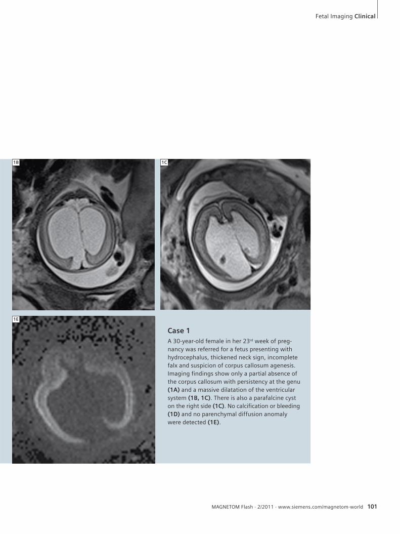
1B 1C
1E
Case 1A 30-year-old female in her 23rd week of preg-nancy was referred for a fetus presenting with hydrocephalus, thickened neck sign, incomplete falx and suspicion of corpus callosum agenesis. Imaging findings show only a partial absence of the corpus callosum with persistency at the genu (1A) and a massive dilatation of the ventricular system (1B, 1C). There is also a parafalcine cyst on the right side (1C). No calcification or bleeding (1D) and no parenchymal diffusion anomaly were detected (1E).

102 MAGNETOM Flash · 2/2011 · www.siemens.com/magnetom-world
Clinical Fetal Imaging
2B
Case 2A 29-year-old female in her 26th week of pregnancy was referred for a fetus with progressive hydrocephalus and inconsistent visualization of the midline structures. MRI shows intact corpus callosum and midline structures, a moderate dilatation of both side ventricles but clear depiction of cysts at the caudothalamic groove (2A), as well as intraventricular septations in both occipital horns (2B), raising the suspicion of an intrauterine infection. Diffusion trace images did not show parenchymal ischemia or infarctions (2C) nor were calcifications detected on T1w imaging (2D). Recent cytomega-lovirus infection was subsequently confirmed in the mother, and in the fetus after delivery.
Contact Jacques Schneider, MDHead of Pediatric RadiologyUniversity Children’s Hospital UKBB
CH-4005 Basel Switzerland Phone +41 61 704 [email protected]
308 %209 %
2A
2D2C
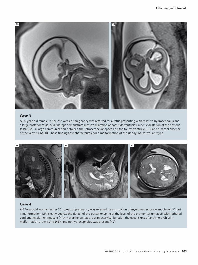
3A 3B
Case 3A 30-year-old female in her 26th week of pregnancy was referred for a fetus presenting with massive hydrocephalus and a large posterior fossa. MRI findings demonstrate massive dilatation of both side ventricles, a cystic dilatation of the posterior fossa (3A), a large communication between the retrocerebellar space and the fourth ventricle (3B) and a partial absence of the vermis (3A–B). These findings are characteristic for a malformation of the Dandy-Walker variant type.
MAGNETOM Flash · 2/2011 · www.siemens.com/magnetom-world 103
Fetal Imaging Clinical
Case 4A 35-year-old woman in her 36th week of pregnancy was referred for a suspicion of myelomeningocele and Arnold Chiari II malformation. MRI clearly depicts the defect of the posterior spine at the level of the promontorium at L5 with tethered cord and myelomeningocele (4A). Never theless, at the craniocervical junction the usual signs of an Arnold-Chiari II malformation are missing (4B), and no hydrocephalus was present (4C).
4A 4B 4C


![3T]caP[[h ]Tgc [TeT[ 3T[TQaPcT - Novotel Sydney Central · 3t[tqapct 3t]cap[[h 5if(spwf$pdlubjm1bdlbhf qfsqfstpo ipvsdbobqft ipvstpgcfwfsbhft $pdlubjm1bdlbhf qfsqfstpo ipvstpgefmjdjpvt{dbobqft{](https://static.fdocuments.net/doc/165x107/5f6aa72c2199805f6a1a97e5/3tcaph-tgc-tet-3ttqapct-novotel-sydney-central-3ttqapct-3tcaph-5ifspwfpdlubjm1bdlbhf.jpg)


