Clinical Case Challenges In Neuro-Optometry III Thomas J. Landgraf, O.D., F.A.A.O.
68
Clinical Case Challenges In Neuro- Optometry III Thomas J. Landgraf, O.D., F.A.A.O.
-
Upload
rudolph-mervyn-hall -
Category
Documents
-
view
217 -
download
1
Transcript of Clinical Case Challenges In Neuro-Optometry III Thomas J. Landgraf, O.D., F.A.A.O.
Clinical Case Challenges In Neuro-Optometry III
Thomas J. Landgraf, O.D., F.A.A.O.
Before We Get Started….. On our LAST hour
together Foundations & Support
Groups Myasthenia Gravis Multiple Sclerosis

Case #6: Don’t Assume Anything
Or “The Case Of The Chronic Sixth Nerve Palsy”70 yo maleDiplopia with isolated abduction deficit OS
Other eye movements intact
No other significant neuro-eye findings

Case #6: Don’t Assume Anything Or “The Case Of The
Chronic Sixth Nerve Palsy” Most likely a case of
ischemic sixth nerve palsy Patient of vasculopathic age Sixth nerve palsy is isolated
May follow the patient without neuro-imaging
Expectation of improvement or resolution in 8-12 weeks

Case #6: Don’t Assume Anything Or “The Case Of The Chronic Sixth
Nerve Palsy” No expected recovery in 6-8 weeks upon re-
exam Refer to neurologist MRI with gadolinium
DDx includes: a mass lesion Pons, along the clivus, in the nasopharynx, at the
base of the brain, in the cavernous sinus, in the orbit

Case #6: Don’t Assume Anything
Or “The Case Of The Chronic Sixth Nerve Palsy”MRI with gadolinium
Large pontine mass consistent with glioma A death sentence in a young person Elderly, grows very slowly

Case #6: Don’t Assume Anything
Or “The Case Of The Chronic Sixth Nerve Palsy”Neurosurgical consult
Monitor without intervention
Prismatic correction

CN VI Palsy
BackgroundCommon cause of horizontal diplopiaMost commonly affected of the ocular motor nerves

CN VI Palsy
BackgroundMost emergent
Age 40 and underIf non-isolated
Think microvascular in elderly If isolated, most patients recover fully and may not
require referral

CN VI Palsy
Background: AnatomyLongest subarachnoid courseNucleus in the pons Innervates the ipsilateral lateral rectus
T2-weighted MRI in a patient with a chronic CN VI palsy shows a left pontine glioma

CN VI Palsy
Diagnosis: Who?Any ageVariety of causes
Ischemia most common in adults Elderly with DM and HTN

CN VI Palsy Diagnosis: Who?
Variety of causesNeoplasm
Mass lesion of CNS most common in children and young adults MRI of large posterior fossa tumor associated with hydrocephalus
and CN VI palsy in a 6 yoInflammation
Post-viral and ear infections in kidsAneurysmTrauma

CN VI Palsy
Diagnosis: SymptomsDiplopia
Binocular or monocular?Horizontal or vertical?
Pain, especially if growing lesion in the cavernous sinus
Additional neurologic signsDepends on the etiology

CN VI Palsy
Diagnosis: SignsAbduction deficit
EsotropiaMaximum on gaze to the side of the
palsy
Head turn

CN VI Palsy
Diagnosis: additional signs dependent on etiologyPapilledema with nausea, vomiting, tinnitus, HA’sProptosis Ptosis Increased ESR with HA and jaw claudicationRetraction of globe and narrowing of lid fissure on
attempted abduction
CN VI Palsy
Differential Diagnosis Ischemia InflammationNeoplasmAneurysmTrauma
CN VI Palsy
Ancillary Tests: Optometric In-OfficeVisual fieldsForced duction?

CN VI Palsy
Ancillary Tests: Referral Indications for Neuro-Imaging
Emergent if age < 30 yearsHead traumaPainNon-isolated Other etiologies besides microvascular, myasthenia gravis,
thyroid, Giant Cell, congenital

CN VI Palsy Ancillary Tests: Referral
Indications for Neuro-Imaging Consult with neuro-eye doc or neurologist reassures: “that level of comfort thing again”
Workup if microvascular: DM, HTN

CN VI Palsy
ManagementMicrovascular, trauma, idiopathic
Resolve spontaneously within 6 monthsComfort: patch, blur, block, Botox for temporary treatmentIf need long-term: prism, surgery

CN VI Palsy
ManagementFollow-up
CN VI every 6 weeks over 6 monthsIf you expect improvement?Neuro consult if no improvement

CN VI Palsy
My Clinical ExperienceAll isolated (majority) have been:
ElderlyMicrovascularIf it all makes sense, I hold off on the “Neuro-massage”
All non-isolated: YoungerPoor prognosis

Case #7: A Quickie
On-call ResidentPtosis ODSeveral months prior Ptosis OS
Seen by another resident
Don’t assume you are smarter than another residentDocumentation was correct
What is really going on?

Myasthenia Gravis
BackgroundAutoimmune Disease
Autoantibodies against acetylcholine receptors
Abnormal fatigueability of muscles under voluntary control
Usually orbital and facial muscles
Myastenia Gravis
BackgroundPrevalence: 1:20,000
But we see it!Ocular involvement: 90%Account for initial complaint in 75%

Myasthenia Gravis
Diagnosis: Who?Females under 50 / 7:3Males peak in late 50’sAssociated conditions: thymoma, thyroid disease,
diabetes, lupus, rheumatoid

Myasthenia Gravis
Diagnosis: SymptomsMajority present with ocular
symptomsPtosis: asymmetricDiplopia: any motility defectAnd spreadVariability of ocular fatigue
Worse at the end of the day Hx, Hx, Hx!

Myasthenia Gravis
Diagnosis: SymptomsNon-Ocular
Within two years of ocularLimb fatigueFacial muscle weakness
Difficulty breathing, chewing, talking, swallowing

Myasthenia Gravis
Diagnosis: SignsPtosis & EOM involvementCogan’s lid twitchExposure keratitisOphthalmoplegia
Orbicularis oculi weakness

Myasthenia Gravis
Differential DiagnosisPupils are never affectedNo eye painThyroid ophthalmopathy, INO (Internuclear Ophthalmoplegia), orbital
pseudotumor, botulism, myotonic dystrophy, Chronic Progressive External Ophthalmoplegia

Myasthenia Gravis
Ancillary Tests: Optometric In-OfficeMeasure palpebral apertures
Pupil center to upper lid margin
Sustained up gazeSqueezing of eyelids closed Initial VF
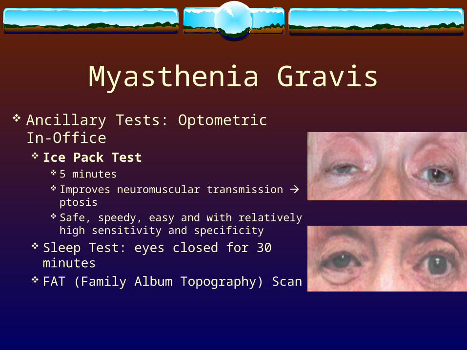
Myasthenia Gravis Ancillary Tests: Optometric In-
Office Ice Pack Test
5 minutes Improves neuromuscular transmission
ptosis Safe, speedy, easy and with relatively high
sensitivity and specificity
Sleep Test: eyes closed for 30 minutes FAT (Family Album Topography) Scan

Myasthenia Gravis
Ancillary Tests: ReferralTensilon (Endrophonium HCL) Test
IV 10 mg of TensilonWhy refer in Tennessee?
Rate of complications low but life-threatening Hypotension, bradycardia, cardiac arrest, respiratory arrest, seizures,
vomiting
Improves eyelid / motility defectAnticholinesterase

Myasthenia Gravis
Ancillary Tests: ReferralEMG (Electomyography)Acetylcholine antibody receptor test

Myasthenia Gravis
ManagementReferral
Neurologist, neuro-eye docInternist or PCP
Lab testing for associated conditionsCT scan of chest / mediastinum for thymoma

Myasthenia Gravis
Management: MedicalAnticholinesterases, steroids, immunosuppressantsThymectomyPlasmapharesis IV gammaglobulin
Myasthenia Gravis
Management: OptometricLid crutches, tapeOcclusionRarely prism, ptosis or strabismus surgeryFollow-up as needed post-diagnosis
Monitor for steroid side effects
Myasthenia Gravis
My Clinical ExperienceAm I missing this?Teaching about it may help…..

Case #8: OK To Not Refer?
History2006 68 yo Caucasian female
My patient since 1997“my glaucoma drops are too expensive”
Alphagan-P bid OU
H/O thinner that normal pachymetry OUH/O highest tonometry 20 mm Hg OU

Case #8: OK To Not Refer?
History: Of interest to us today…..1997: first visit
Referred for pupil and glaucoma work-up Anisicoria noted over 10 years ago

Case #8: OK To Not Refer?
HistoryHTN, hypercholesterolemiaH/O bypassHyzarr, Metiprolol, Lipitor, Aspirin, Lyrica, vitamins
No wonder the glc drop is too expensive
POAG, Horner’s Syndrome, ERM
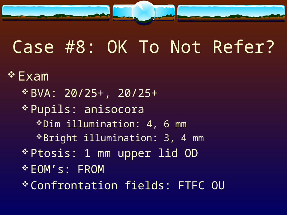
Case #8: OK To Not Refer?
ExamBVA: 20/25+, 20/25+Pupils: anisocora
Dim illumination: 4, 6 mmBright illumination: 3, 4 mm
Ptosis: 1 mm upper lid ODEOM’s: FROMConfrontation fields: FTFC OU
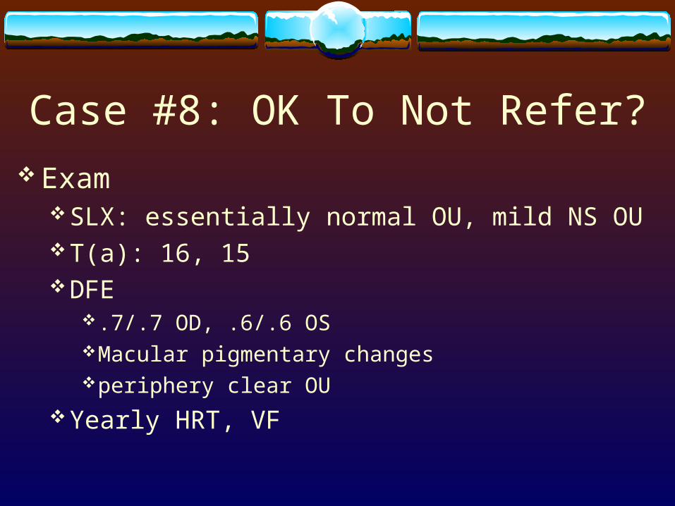
Case #8: OK To Not Refer?
ExamSLX: essentially normal OU, mild NS OUT(a): 16, 15DFE
.7/.7 OD, .6/.6 OSMacular pigmentary changes periphery clear OU
Yearly HRT, VF

Case #8: OK To Not Refer
Latest VF OD
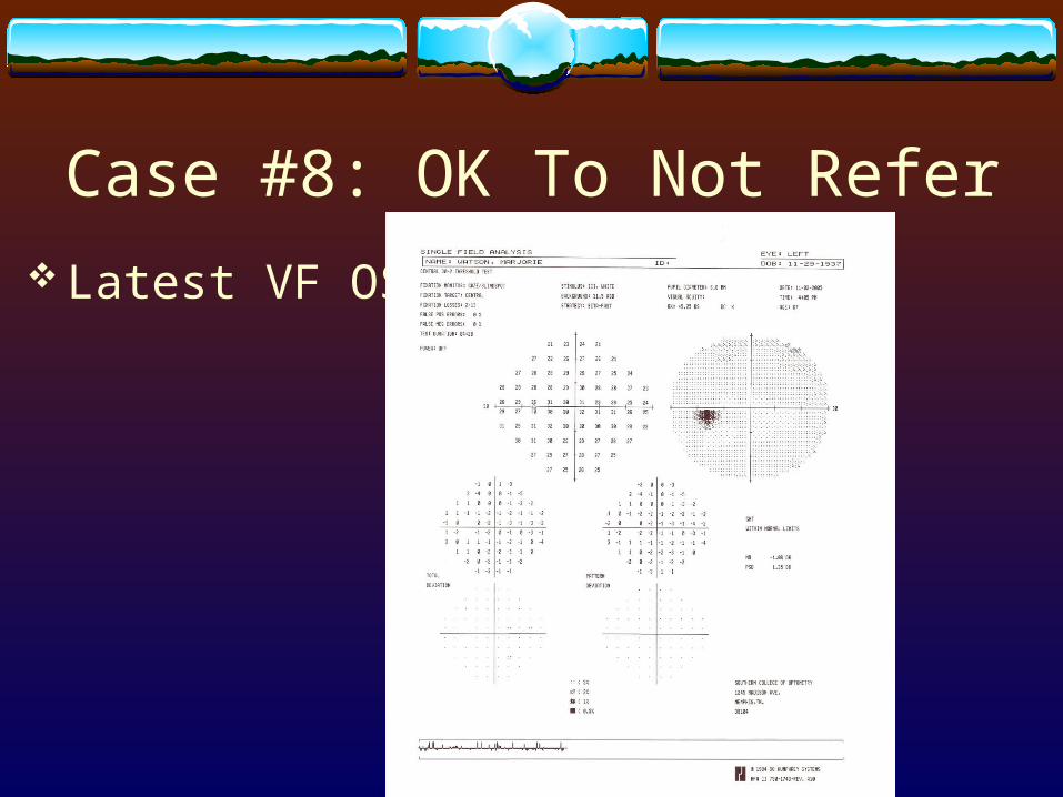
Case #8: OK To Not Refer
Latest VF OS

Case #8: OK To Not Refer
Latest HRT OD

Case #8: OK To Not Refer
Latest HRT OS

Case #8: OK To Not Refer?
Assessment1. POAG OU
Stable HRT, VF, ONH appearance, IOPAlphagan-P too expensive
2. H/O Horner’s Syndrome ODBenign and stable
3. Macular pigmentary changes
Case #8: OK To Not Refer?
Plan:1. Switch Alphagan-P to Brimonidine, RTC 1 month
IOP check, Education potential side effects of Brimonidine
2,3. To monitor

Case #8: OK To Not Refer?
After preparing this lectureLooked way back in the record again10/5/83: “Anisocoria noted 1-2 years ago; neuro-
opthalmology work-up with no known causes” I never got the cocaine in ‘97Good idea eventually:
IopidineParedrine or Pholedrine
Horner’s Syndrome
Background1852: Claude Bernard first noted experimentally1869: Swiss ophthalmologist Johann Friedrich Horner
notedShould be called?
Bernard’s SyndromeBernard-Horner’s Syndrome

Horner’s Syndrome
Background: Sympathetic Anatomy of Eye & FaceFirst order sympathetic neuron
Begins in ipsilateral hypothalamusDescends through midbrain, pons, and medullaEnds at ciliospinal center of Budge at levels of C8-T1 in the
spinal cord
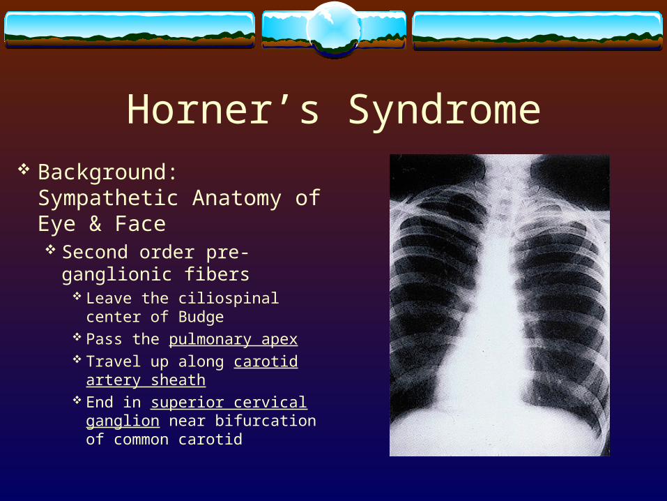
Horner’s Syndrome Background: Sympathetic
Anatomy of Eye & Face Second order pre-ganglionic
fibers Leave the ciliospinal center of
Budge Pass the pulmonary apex Travel up along carotid artery
sheath End in superior cervical
ganglion near bifurcation of common carotid

Horner’s Syndrome Background: Sympathetic Anatomy
of Eye & Face Third order post-ganglionic neuron
Travels along internal carotid artery to the cavernous sinus
Leave the internal carotid, travel with the abducens and join the ophthalmic division of the trigeminal
Enter the orbit with the naso-ciliary branch

Horner’s Syndrome Background: Sympathetic Anatomy of Eye
& Face In the orbit, the sympathetic fibers pass through
the ciliary ganglion Join the two long ciliary branches of the
nasociliary nerve and innervate the iris dilator muscle
Other sympathetic branches travel with branches of the ophthalmic artery to innervate the lacrimal gland, Muller’s muscles, and orbital vessels
Sympathetic fibers controlling facial sweating travel with the external carotid artery
Horner’s Syndrome
Why?Dysfunction of the sympathetic innervation to eye and
parts of face Interruption of oculosympathetic nerve supply
somewhere between hypothalamus and the eye

Horner’s Syndrome
Diagnosis: Who?No predilection
Age, race, gender, geographics
CongenitalPresents by age two with heterochromia

Horner’s Syndrome
Diagnosis: SignsPtosis Mullers muscle
1-2 mm upper eyelid
Reverse ptosisSlight elevation lower eyelid
Miosis iris dilator muscle

Horner’s Syndrome
Diagnosis: SignsAnisicoria > darkness
Dilator iris muscle normally more active
Dilation lag: prolonged redilation of the pupil after dimming the light
Hypochromia Iridis: typical if congenitalAnhidrosis
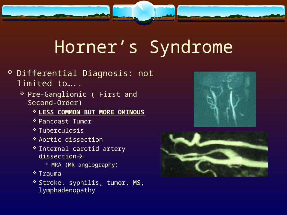
Horner’s Syndrome Differential Diagnosis: not limited
to….. Pre-Ganglionic ( First and Second-Order)
LESS COMMON BUT MORE OMINOUS
Pancoast Tumor Tuberculosis Aortic dissection Internal carotid artery dissection
MRA (MR angiography) Trauma Stroke, syphilis, tumor, MS,
lymphadenopathy

Horner’s Syndrome Differential Diagnosis
Post-Ganglionic (Third-Order) Aneurysm Atherosclerosis Herpes Zoster Trauma Sinusitis
Painful Horner’s Carotid dissection until proven othewise

Horner’s Syndrome
Differential DiagnosisVs Third Nerve Palsy
PtosisAnisicoria exaggerated in dim illumination Horner’s

Horner’s Syndrome
Ancillary Tests: Optometric In-Office?Pupil Testing 1: Horner’s?
Cocaine blocks reuptake of norepinephrine at the sympathetic nerve endings
Dilates normal eye after an hour No dilation in Horner’s due to lack of norepinephrine at nerve
endings
Availability of cocaine 10% solution?

Horner’s Syndrome Ancillary Tests: Optometric In-Office
Pupil Testing 1: Horner’s? Alternative: Iopidine (Apraclonidine)
Weak, direct action on alpha-1 receptors Normal: no dilation Horner’s: dennervation supersensitivity to
norepinephrine increase in alpha-1 receptors in iris stroma dilation

Horner’s Syndrome Ancillary Tests: Optometric
In-Office Pupil Testing 2: pre- or post-
ganglionic? Paredrine 1%
(Hydroxyamphetamine) releases norepinephrine from stores in nerve endings
Mydriasis in a normal pupil No mydriasis in a post-
ganglionic Horner’s due to destroyed nerve endings
“Fail-safe”? Postive Paredrine Test OD
did not dilate

Horner’s Syndrome
Ancillary Tests: Optometric In-OfficePupil Testing 2: pre- or post-ganglionic?Alternative: Pholedrine 5%
With third neuron damageHorner’s pupil will not dilatePost-ganglionic lesionIf dilates

Horner’s Syndrome
Management: Referral unless congenitalNeurologist, Neuro-Eye DocPCP, InternistCardiologist, Oncologist, Vascular SurgeonNo treatment to improve or reverse the condition

Horner’s Syndrome My Clinical Experience
Some pharmacist in Memphis is upset with me!
Glad we have Iopidine now