Clinical Article Spontaneous Resolution of Chronic ...
9
628 However, its outcomes may not be satisfactory because of re- currence and physical infirmity associated with aging 26) . The re- ported rate of recurrence after surgery for cSDH is approxi- mately 5–30% 11) . When brain CT scan reveals slight compression of the brain parenchyma caused by hematoma and patients have no neuro- logical symptoms or have mild symptoms, it is occasionally dif- ficult to select the treatment strategy between surgical and con- servative treatments and we cannot be sure about the surgical decision. Although many neurosurgeons are aware of sponta- neously resolving subdural hematoma (SDH), the clinical and radiological characteristics have not yet been adequately de- scribed in the literature 9) . We reported 16 cases of close observation of cSDH. We com- pared and evaluated the clinical and radiological characteristics between the spontaneously cured cases and the cases requiring operation. INTRODUCTION Chronic subdural hematoma (cSDH) is defined as a condi- tion consisting of a slowly progressive accumulation of liquefied blood within the subdural space 9) . This accumulation of blood in the subdural space may produce brain parenchymal com- pression, and at that time, patients have neurological symptoms such as headache, hypesthesia and hemiparesis. The incidence of cSDH increases significantly with age. The other risk factors of this disease have been established in many studies like taking blood thinners, long-term alcohol abuse, and history of head trauma 23) . cSDH is common and more prevalent in the aged popula- tion 9) . Surgical treatment might generally be indicated in condi- tions of large hematoma width (>10 mm) or shift of the midline (>5 mm) on brain computed tomography (CT) scan and surgi- cal intervention has been accepted as the treatment of choice 8) . Spontaneous Resolution of Chronic Subdural Hematoma : Close Observation as a Treatment Strategy Hyung Chan Kim, M.D., 1 Jung Ho Ko, M.D., Ph.D., 1 Dong Soo Yoo, M.D., Ph.D., 2 Sang-Koo Lee, M.D., Ph.D. 1 Departments of Neurological Surgery, 1 Radiology, 2 College of Medicine, Dankook University, Cheonan, Korea Objective : Chronic subdural hematoma (cSDH) is common condition in neurosurgical field. It is difficult to select the treatment modality between the surgical method and the conservative method when patients have no or mild symptoms. The purpose of this study is to provide a suggestion that the patients could be cured with conservative treatment modality. Methods : We enrolled 16 patients who had received conservative treatment for cSDH without special medications which could affect hematoma resolution such as mannitol, steroids, tranexamic acid and angiotensin converting enzyme inhibitors. The patients were classified according to the Markwalder’s Grading Scale. Results : Among these 16 patients, 13 (81.3%) patients showed spontaneously resolved cSDH and 3 (18.7%) patients received surgery due to symptom aggravation and growing hematoma. They were categorized into two groups based on whether they were cured with conservative treat- ment or not. The first group was the spontaneous resolution group. The second group was the progression-surgery group. The mean hematoma volume in the spontaneous resolution group was 43.1 mL. The mean degree of midline shift in the spontaneous resolution group was 5.3 mm. The mean hematoma volume in the progression-surgery group was 62.0 mL. The mean degree of midline shift in the second group was 6 mm. Conclusion : We suggest that the treatment modality should be determined according to the patient’s symptoms and clinical condition and close observation could be performed in patients who do not have any symptoms or in patients who have mild to moderate headache without neurologi- cal deterioration. Key Words : Close observation · Chronic subdural hematoma · Treatment. Clinical Article • Received : March 29, 2016 • Revised : July 25, 2016 • Accepted : August 12, 2016 • Address for reprints : Jung Ho Ko, M.D., Ph.D. Department of Neurological Surgery, College of Medicine, Dankook University, 201 Manghyang-ro, Dongnam-gu, Cheonan 31116, Korea Tel : +82-41-550-6280, Fax : +82-41-550-6034, E-mail : [email protected] • This is an Open Access article distributed under the terms of the Creative Commons Attribution Non-Commercial License (http://creativecommons.org/licenses/by-nc/3.0) which permits unrestricted non-commercial use, distribution, and reproduction in any medium, provided the original work is properly cited. J Korean Neurosurg Soc 59 (6) : 628-636, 2016 http://dx.doi.org/10.3340/jkns.2016.59.6.628 Copyright © 2016 The Korean Neurosurgical Society Print ISSN 2005-3711 On-line ISSN 1598-7876 www.jkns.or.kr
Transcript of Clinical Article Spontaneous Resolution of Chronic ...

628
However, its outcomes may not be satisfactory because of re-currence and physical infirmity associated with aging26). The re-ported rate of recurrence after surgery for cSDH is approxi-mately 5–30%11).
When brain CT scan reveals slight compression of the brain parenchyma caused by hematoma and patients have no neuro-logical symptoms or have mild symptoms, it is occasionally dif-ficult to select the treatment strategy between surgical and con-servative treatments and we cannot be sure about the surgical decision. Although many neurosurgeons are aware of sponta-neously resolving subdural hematoma (SDH), the clinical and radiological characteristics have not yet been adequately de-scribed in the literature9).
We reported 16 cases of close observation of cSDH. We com-pared and evaluated the clinical and radiological characteristics between the spontaneously cured cases and the cases requiring operation.
INTRODUCTION
Chronic subdural hematoma (cSDH) is defined as a condi-tion consisting of a slowly progressive accumulation of liquefied blood within the subdural space9). This accumulation of blood in the subdural space may produce brain parenchymal com-pression, and at that time, patients have neurological symptoms such as headache, hypesthesia and hemiparesis. The incidence of cSDH increases significantly with age. The other risk factors of this disease have been established in many studies like taking blood thinners, long-term alcohol abuse, and history of head trauma23).
cSDH is common and more prevalent in the aged popula-tion9). Surgical treatment might generally be indicated in condi-tions of large hematoma width (>10 mm) or shift of the midline (>5 mm) on brain computed tomography (CT) scan and surgi-cal intervention has been accepted as the treatment of choice8).
Spontaneous Resolution of Chronic Subdural Hematoma : Close Observation as a Treatment Strategy
Hyung Chan Kim, M.D.,1 Jung Ho Ko, M.D., Ph.D.,1 Dong Soo Yoo, M.D., Ph.D.,2 Sang-Koo Lee, M.D., Ph.D.1
Departments of Neurological Surgery,1 Radiology,2 College of Medicine, Dankook University, Cheonan, Korea
Objective : Chronic subdural hematoma (cSDH) is common condition in neurosurgical field. It is difficult to select the treatment modality between the surgical method and the conservative method when patients have no or mild symptoms. The purpose of this study is to provide a suggestion that the patients could be cured with conservative treatment modality.Methods : We enrolled 16 patients who had received conservative treatment for cSDH without special medications which could affect hematoma resolution such as mannitol, steroids, tranexamic acid and angiotensin converting enzyme inhibitors. The patients were classified according to the Markwalder’s Grading Scale. Results : Among these 16 patients, 13 (81.3%) patients showed spontaneously resolved cSDH and 3 (18.7%) patients received surgery due to symptom aggravation and growing hematoma. They were categorized into two groups based on whether they were cured with conservative treat-ment or not. The first group was the spontaneous resolution group. The second group was the progression-surgery group. The mean hematoma volume in the spontaneous resolution group was 43.1 mL. The mean degree of midline shift in the spontaneous resolution group was 5.3 mm. The mean hematoma volume in the progression-surgery group was 62.0 mL. The mean degree of midline shift in the second group was 6 mm.Conclusion : We suggest that the treatment modality should be determined according to the patient’s symptoms and clinical condition and close observation could be performed in patients who do not have any symptoms or in patients who have mild to moderate headache without neurologi-cal deterioration.
Key Words : Close observation · Chronic subdural hematoma · Treatment.
Clinical Article
• Received : March 29, 2016 • Revised : July 25, 2016 • Accepted : August 12, 2016• Address for reprints : Jung Ho Ko, M.D., Ph.D. Department of Neurological Surgery, College of Medicine, Dankook University, 201 Manghyang-ro, Dongnam-gu, Cheonan 31116, Korea Tel : +82-41-550-6280, Fax : +82-41-550-6034, E-mail : [email protected]• This is an Open Access article distributed under the terms of the Creative Commons Attribution Non-Commercial License (http://creativecommons.org/licenses/by-nc/3.0) which permits unrestricted non-commercial use, distribution, and reproduction in any medium, provided the original work is properly cited.
J Korean Neurosurg Soc 59 (6) : 628-636, 2016
http://dx.doi.org/10.3340/jkns.2016.59.6.628
Copyright © 2016 The Korean Neurosurgical Society
Print ISSN 2005-3711 On-line ISSN 1598-7876www.jkns.or.kr
629
Close Observation for Chronic Subdural Hematoma | HC Kim, et al.
MATERIALS AND METHODS
Clinical and radiological records of 241 patients who had pre-sented with a low density to iso-dense SDH with parenchymal compression on brain CT scan between July 2005 and Decem-ber 2014 were retrospectively reviewed. Patients were excluded if they had initially received surgery for cSDH or if they had presented with a low Glasgow coma scale (GCS) score below 8 due to difficulty in assessing the neurological status. From among these patients, we enrolled 16 patients who had received conservative treatment for cSDH without special medications which could affect hematoma resolution such as mannitol, ste-roids, tranexamic acid and angiotensin converting enzyme (ACE) inhibitors. Their brain CT scans showed the hematoma exerted a mass effect on the brain which may require surgical treatment such as burr-hole trephination with or without drain-age. They were treated conservatively due to mild symptoms, their medical comorbidities, or their denial for operation. All patients had cSDH which was identified on the initial brain CT or some patients had a chronic stage of traumatic SDH which had progressed from the initially identified acute SDH.
We decided the treatment strategy for cSDH such as surgery or observation according to the clinical and radiological findings. If patients had neurological symptoms such as decreased men-tality, motor weakness, sensory change or progressive severe headache, we preferred to perform surgical treatment. Depend-ing on the patient’s condition, we closely observed the patients and assessed the neurological status daily in case of admitted pa-tients or performed the follow-up assessment once a week in outpatients. We preferred close observation in cases with high possibility of recurrence after the initial operation such as those with antiplatelet use and hematological disorder and in cases belonging to the high risk group of general anesthesia. We want-ed to reduce the number of unnecessary operations due to op-erative or anesthetic complications. Therefore, we recommend-ed nonsurgical treatment for the enrolled patients. We routinely performed follow-up brain CT once a week for 3 weeks and then once or twice a month until detection of nearly total resolution. Within the follow-up period, if neurologic symptoms had newly developed or progressed, brain CT was performed to consider surgery as soon as possible. Surgical methods were burr-hole trephination with drainage and saline irrigation. Generally, when it is decided to treat patients with cSDH surgically, the opera-tion is carried out under local anesthesia. However, at our insti-tution, considering the safety of patients and the possibility of lack of patients’ cooperation during the operation, all surgeries were carried out under general anesthesia. Thus, prior to the treat-ment, it was inevitable to collect data regarding patient’s comor-bidities. During the observation period, we considered continu-ation of observation at every follow-up visit by comparing the change in hematoma amount and symptoms. We considered that the major criterion for surgical decision was the change in symp-toms. Complete cure was defined when the SDH had nearly dis-
appeared on follow-up brain CT. The demographic data were collected after obtaining informa-
tion on clinical factors; age, sex, history of trauma, past medical history such as diabetic mellitus, hypertension and medication especially antithrombotic agents, GCS, clinical symptoms and symptom duration, hematoma thickness and volume, degree of midline shift, accompanying intracranial lesion, Glasgow out-come scale (GOS) score. The patients received a CT scan of the brain at admission. We measured the volume of hematoma from the CT scans; all of these measurements were performed using the PACS imaging display software (Viewrex, TechHeim, Seoul, Korea). The volume measurement was hand-traced by the radi-ologist (D.S. Yoo) on each axial image, and then these areas were added together and multiplied by the slice thickness. The midline shift was identified as deviation of the septum pellu-cidum from the central position. The patients were classified ac-cording to the Markwalder’s Grading Scale (MGS)18). The grad-ing score was used for patient classification and it ranged from 0 to IV. Grade 0 indicates neurologically intact patients. Grade I indicates alert and oriented patients who often complains of a headache. Grade II indicates drowsy or disoriented patients. Sometimes, the patients who are classified as having grade II have hemiparesis. Grade III indicates patients who are stupor-ous but respond appropriately to noxious stimuli and have hemi-plegic symptom. The last grade, grade IV indicates patients who are comatose with absent motor responses to painful stimuli. They might have decerebrate or decorticate posturing.
The measured data were analyzed by using the R ver 3.2.4 software for Windows. We compared data such as the measured hematoma volume and the degree of midline shift. This compari-son was performed by using the Wilcoxon rank sum test. The results were considered statistically significant if p was <0.05.
RESULTS
Among these 16 patients, 13 (81.3%) patients showed sponta-neously resolved cSDH and 3 (18.7%) patients received surgery due to symptom aggravation and growing hematoma. They were categorized into two groups based on whether they were cured with conservative treatment or not. The first group was the spontaneous resolution group (Table 1). There were 9 men and 4 women with a mean age of 62 years (range, 25 to 81 years). There were 8 patients over 65 years of age (61.5%) and there were 5 below 65 years of age (38.5%). Eight patients (61.5%) had a medical history of hypertension, diabetes mellitus, hema-tologic disorder due to anaplastic anemia and alcohol abuse, anticoagulant use because of previous cerebral infarction or mi-tral valve replacement surgery and hemodialysis due to end-stage renal disease. Ten patients (76.9%) had a trauma history before admission. The most common presenting symptom was mild headache without neurological deficit. Case 2 patient only had hemiparesis. Their MGS ranged from grade I to grade II. The number of MGS grade I patients was 10 (76.9%) and the

630
J Korean Neurosurg Soc 59 | November 2016
number of MGS grade II patients was 3 (23.1%). The fronto-pa-rietal area was the most common area of hematoma involve-ment. The mean hematoma volume was 43.1 mL (range, 11.9 to 68.3 mL). The mean hematoma thickness on brain CT was 13.3 mm (range, 9.3 to 17.3 mm). The mean degree of midline shift was 5.3 mm (range, 0 to 10.4 mm). All patients had good outcomes (GOS 5) and their clinical symptoms improved. There were no recurrences of cSDH. Mean duration from iden-tification to complete resolution of cSDH was 17 weeks (range, 4 to 96 weeks). The second group was the progression-surgery patients. Table 2 summarizes their demographic data. There were 2 men and 1 woman with a mean age of 72 years (range, 67 to 81 years). One patient had a history of antiplatelet medica-tion. All patients had a trauma history before admission. All pa-tients presented with mild headache without neurological defi-cit. The mean hematoma volume was 62.0 mL (range, 41.0 to 75.0 mL). The mean hematoma thickness was 17.5 mm (range, 14.3 to 21.5 mm). The mean degree of midline shift was 6.0 mm (range, 0 to 15.0 mm). Mean duration from the decision of surgery for hematoma was 5.5 weeks (range, 2 to 8 weeks). All patients had good outcomes (GOS 5) and their clinical symp-toms improved. There were no recurrences of cSDH.
The results of univariate analysis with the Wilcoxon rank sum test are described in Table 3. The hematoma volumes showed no significant difference between the two groups. However, the point estimates of the median in the spontaneous resolution group and the progression-surgery group were 44.6 and 70, re-spectively. The p-value for this analysis was 0.146. This study is limited in its significance due to the small sample size. There-fore, further analysis with a larger sample size for strengthening statistical power is needed to strengthen our results15).
Case illustration
A 75-year-old maleThe patient was admitted due to minor head trauma. He com-
plained of mild headache and had an alert mentality. The initial brain CT revealed a scanty acute SDH in the right hemisphere (Fig. 1A). He was treated conservatively and his clinical course was uneventful. Follow-up brain CT performed on the 20th hospital day (HD) showed a liquefied SDH that compressed the brain parenchyma with a mild midline shift (Fig. 1B). The mea-sured volume of the hematoma was 51.5 mL and the length of midline shift was 4.4 mm. Because his symptom had been im-proving slowly and he did not have any other neurological signs, we made a decision of performing close observation. Follow-up brain CT scan performed on the 46th HD showed an improved mass effect and a remarkably decreased amount of hematoma (Fig. 1C). He was discharged at that time and brain CT scan was acquired in the outpatient department 2 weeks later. The final brain CT scan revealed nearly complete resolution of the hema-toma (Fig. 1D). He has not experienced any recurrence of symp-tom and hematoma.Ta
ble
1. C
linic
al a
nd ra
diol
ogic
cha
ract
eris
tics
of th
e sp
onta
neou
s re
solu
tion
grou
p
No.
Sex/
Age
Trau
ma H
x.Sy
mpt
omM
arkw
alder
Loc.
Thic.
(mm
)Vo
l. (m
L)M
id. (
mm
)O
utco
me (
wks
)C
omor
bidi
ties
Low
den
sity l
ine
1M
/34
Yes
Dro
wsin
ess
Gra
de II
F-T-
P10
.229
.313
GO
S5 (4
)N
oN
o
2M
/25
No
Hem
ipar
esis
Gra
de II
F-T-
P15
.362
10.4
GO
S5 (1
4)Ap
lastic
anem
iaYe
s
3M
/68
Yes
Hea
dach
eG
rade
IF-
T12
33.1
09.6
GO
S5 (1
0)A
lcoho
lic ab
use
No
4M
/67
No
Hea
dach
eG
rade
IF-
P13
.244
.602
GO
S5 (7
)Pr
evio
us ce
rebr
al in
farc
tion
Yes
5M
/77
Yes
Hea
dach
eG
rade
IBi
lat F
-P16
52.2
00G
OS5
(24)
No
Yes
6F/
79Ye
sD
row
sines
sG
rade
IIF-
P17
.341
.904
.8G
OS5
(12)
Hyp
erte
nsio
nYe
s
7M
/75
Yes
Hea
dach
eG
rade
IF-
P11
.851
.504
.4G
OS5
(4)
Hyp
erte
nsio
nN
o
8F/
54Ye
sH
eada
che
Gra
de I
Bilat
F-P
14.7
/12.
611
.900
GO
S5 (4
)D
iabe
tes m
ellitu
sYe
s
9M
/81
Yes
Hea
dach
eG
rade
IF-
P09
.336
.600
GO
S5 (8
)N
oYe
s
10F/
81Ye
sH
eada
che
Gra
de I
F-P
12.1
61.7
03.3
GO
S5 (1
8)N
oYe
s
11M
/66
No
Hea
dach
eG
rade
IF-
P16
.246
.905
.8G
OS5
(10)
No
No
12M
/41
Yes
Hea
dach
eG
rade
IF-
P15
.668
.308
.2G
OS5
(96)
End
stage
rena
l dise
ase
No
13F/
54Ye
sH
eada
che
Gra
de I
F-T-
P10
.721
.507
.2G
OS5
(12)
Prev
ious
mitr
al va
lve o
pera
tion
histo
ryN
oF-
T-P
: fro
ntot
empo
ropa
rieta
l, F-
T : f
ront
otem
pora
l, F-
P : f
ront
opar
ieta
l, G
OS
: gla
sgow
out
com
e sc
ale

631
Close Observation for Chronic Subdural Hematoma | HC Kim, et al.
A 25-year-old maleThe patient had aplastic anemia which was diagnosed 10
years ago and he was frequently treated by blood transfusion due to a hematological disorder. He presented with left side hemiparesis and headache. The initial brain CT scan showed a mixed subacute and cSDH that compressed the brain paren-chyma and caused a midline shift (Fig. 2A). The measured vol-ume of the hematoma was 62.0 mL and the length of midline shift was 10.4 mm. He had severe thrombocytopenia (measured platelet count was 31000/uL) which might have led to sponta-neous bleeding and his medical condition was very poor. Be-
cause we were concerned about hemostasis and poor general condition, we recommended conservative treatment and in-formed consent was obtained from the patient and his relatives. Close observation and conservative treatment including trans-fusion were performed in the intensive care unit. His neurologi-cal status was not aggravated and the clinical course was un-eventful for 2 weeks. The hemiparesis recovered slowly from the 8th HD onwards. Follow-up brain CT performed on the 14th HD revealed decreased amount of hematoma and mass effect (Fig. 2B). His neurologic symptom disappeared on the 30th HD, and brain CT scan revealed a remarkable reduction in he-
Table 2. Clinical and radiologic characteristics of the progression-surgery group
No. Sex/Age Trauma Hx. Symptom Markwalder Loc. Thic. (mm) Vol. (mL) Mid. (mm) Outcome (wks) Comorbid Low
density line1 M/81 Yes Headache Grade I Bilat F-T-P 21.5/18.2 44/70 00 OP Hypertension Yes2 F/67 Yes Headache Grade I F-T-P 14.3 41 15 OP Hypertension No3 M/69 Yes Dizziness Grade I F-P 16.2 75 03 OP No No
OP : operation, F-T-P : frontotemporoparietal, F-P : frontoparietal
Fig. 1. A 75-year-old male complained of mild headache and he had alert mentality after minor head trauma [Table 1 (Case 7)]. A : Initial brain CT re-veals scanty acute SDH in the right hemisphere. B : Follow-up brain CT scan on the 20th hospital day (HD) shows liquefied subdural hematoma that compressed the brain parenchyma with a mild midline shift. C : Follow-up brain CT scan on the 46th HD shows an improved mass effect and a re-markably decreased amount of hematoma. D : Final brain CT scan reveals nearly complete resolution of the hematoma. SDH : subdural hematoma.
DCBA
Fig. 2. A 25-year-old male presented with left side hemiparesis and headache [Table 1 (Case 2)]. A : Initial brain CT scan shows that a mixed sub-acute to chronic subdural hematoma compresses the brain parenchyma and causes a midline shift. B : Follow-up brain CT scan on the 14th hospital day (HD) reveals decreased amount of hematoma and mass effect. C : Follow-up brain CT scan on the 30th HD identifies remarkable reduction in he-matoma volume. D : Final brain CT scan shows complete disappearance of the hematoma.
DCBA
Table 3. The univariate analysis of the hematoma volume between two groups
Spontaneously resolution group (n=13) Progression-surgery group (n=3)p-value*
Mean SD Median Mean SD MedianHematoma volume (mL) 43.2 16.5 44.6 62 18.4 70 0.146*p-value was calculated by using Wilcoxon rank sum test. SD : standard deviation

632
J Korean Neurosurg Soc 59 | November 2016
matoma volume (Fig. 2C). After discharge, brain CT scan was acquired in the outpatient department 1 month later. The final brain CT scan showed that the hematoma had completely dis-appeared (Fig. 2D).
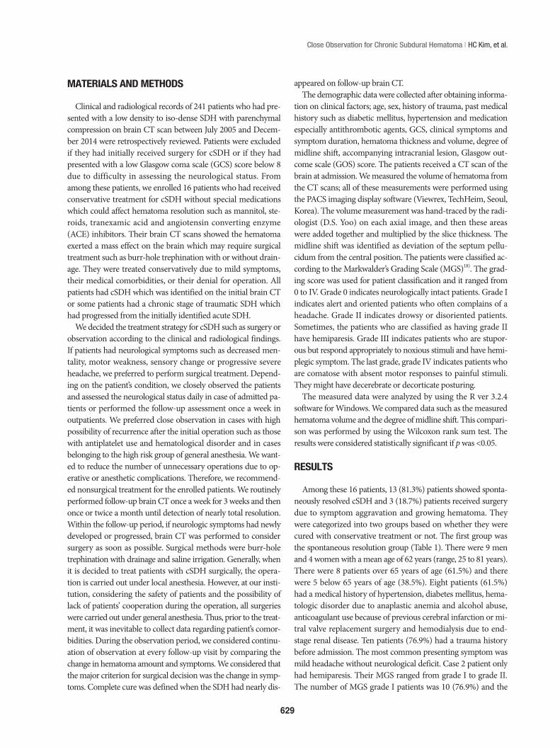
An 81-year-old maleHe was brought to our institution via the neurosurgical outpa-
tient department. The brain CT scan revealed an iso-dense le-sion in the subdural space in the bilateral convexity (Fig. 3A). He presented with unspecific myalgia without neurological symp-toms and signs. We explained to his relative that this lesion had a chance of growing into a hematoma. Close observation was performed. During the admission period, he did not have aggra-vation of symptoms. After 2 weeks, follow-up CT scan showed no change compared with the previous CT scan. We decided to discharge him and perform outpatient follow-up. At the first follow-up after discharge, his relative informed that the patient did not prefer to talk with other people as found in people with mutistic nature. Brain CT scan showed an increased amount of hematoma (Fig. 3B). Surgical evacuation of the hematoma was performed and the patient fully recovered (Fig. 3C).
DISCUSSION
The pathophysiologic mechanism of hematoma growthThe cSDH tends to gradually increase in volume. Several hy-
potheses about the mechanism of increase in the hematoma volume have been proposed since a long time. One hypothesis is the osmotic pressure theory after formation of internal and external capsules. As the hematoma volume increases, tension in the hematoma capsule is increased followed by micro-tear of its capillary vessels. Then hemorrhage occurs and it becomes a factor involved in increasing the osmotic pressure in the hema-toma25). This osmotic theory recently been doubted due to the following factors : 1) there was no significant increase in the volume in this theory when hematoma membranes were used;
2) fresh erythrocytes were always introduced in the hematoma fluid on repeated tapping; 3) it was not possible to prove that the arachnoid acts as a membrane permeable to cerebrospinal fluid; and 4) it has been shown that albumin, the most osmoti-cally active protein, cannot be found in destroyed red blood cells but is derived from the plasma17). This previously accepted hypothesis has been proven wrong4,28). Rather, hematoma ex-pansion is thought to result from repeated micro-hemorrhage from the fragile neo-membranes4,13). The phenomenon of re-current bleeding from the hematoma capsule as an etiological factor for the development of chronic SDH was assumed by Putnam and Cushing in 1932, and later by Dandy. This theory was corroborated by the experimental studies performed by Markwalder.17). Currently, various clinical and experimental studies indicate that fibrinolysis and liquefaction of the initial clot occur rapidly and subsequently inhibit the blood coagula-tion system in the hematoma. Additionally, the byproducts of blood breakdown and the fibrinolytic process also cause thick-ening of the inner dural layer with ingrowing neocapillaries which play an important role in the leakage of blood, causing enlargement of the subdural collection due to microhemor-rhage and further increase in the fibrinolytic activity. The corre-lation between this cycle of re-bleeding and fibrinolysis, and re-absorption of the subdural collection will determine whether the cSDH will resolve, persist or enlarge24). Some authors stated that the hyperfibrinolytic activity has been proved to be critical for liquefaction of the hematoma and progression of cSDH. Several studies have indicated the hyperfibrinolytic and coagu-lative activity in cSDH and some studies have shown that in-creased permeability of the capillaries in the hematoma outer membrane can influence the enlargement of a cSDH5). Based on this rationale, they applied an antifibrinolytic agent for pre-vention of enlargment and recurrence of cSDH11).
Close observation studyPrevious reports on spontaneous resolution of cSDH without
Fig. 3. An 81-year-old male presented with an incidental subdural hematoma [Table 2 (Case 1)]. A : Initial brain CT scan reveals an isodense lesion in the subdural space in bilateral convexity. B : When the symptom changed, follow-up brain CT shows an increased amount of hematoma. C : Post-operative brain CT scan shows reduction of hematoma through drainage catheters.
A B C

633
Close Observation for Chronic Subdural Hematoma | HC Kim, et al.
special medication and surgery have been published1,7,9,10,12,16,20,22). Most of them were case studies which were composed of small cases series with 4 or 5 cases in one report. To the best of our knowledge, our study has the highest number of enrolled cases compared to the studies reported so far, and unlike other inves-tigations, this study has a unique aspect as it considered the rate of spontaneous resolution of cSDH. Table 4 presents the list and characteristics of previous studies. The total number of all col-lected cases was eighteen. The mean age of this group was 66.5 years (range, 21 to 87 years). There were 11 men (61.1%) and 7 women (38.9%). Most of them, except for 3 patients had a trau-ma history such as traffic accident, fall, and blunt trauma. All patients had symptoms such as headache, dizziness, drowsiness and unstable gait. Two patients had a mild hemiparesis and one patient had diplopia. The most commons site was the frontal area. Mean hematoma thickness was 16.7 mm (range, 9 to 34 mm) and there was no data for thickness in 7 cases. The aver-age degree of midline shift was 2.8 mm (range, 5 to 12 mm). All of the patients had a good clinical course. Based on this table, their results were similar to our results.
Naganuma et al.20) reported 4 cases of spontaneous resolution of cSDHs and suggested the three following characteristic CT findings associated with spontaneous resolution of chronic SDH : low density hematoma or iso-dense hematoma, small size, and ventricular dilatation. They considered that the dilated ventricle could cause elevated counterpressure against cSDH. This theory might have some role in the resolution of SDH. We reviewed the radiological data of the spontaneous resolution group. Except for one case, there were no other cases of ventric-
ular dilatation in this group and the mean Evan’s ratio ranged from 0.19 to 0.32.
Horikoshi et al.9) reported that the clinical and radiological find-ings of four cases (five hematomas) were compared to those of 19 surgical cases. They considered that spontaneously resolving cSDH were asymptomatic or only caused mild transient head-ache, and disappeared within 4 to 9 months after head injury. All spontaneously resolving SDHs were located in the frontal region, and maximum thickness and midline displacement were less 5 mm. Our results were similar to their results. Table 1 shows that the most common location of cSDH was the frontal region. The mean value of midline displacement in our study was 5.2 mm and thickness was 13.3 mm. We suggest that close observa-tion could be chosen when hematoma volume or thickness is below 43 mL and 13 mm, respectively, and midline shift is below 5 mm on brain CT scans. According to our results, there were no statistically significant differences between the two groups. However, the spontaneous resolution group had a relatively less amount of hematoma. This shows that the patients who have a definite large amount of cSDH (>43 mL) should be informed about the possibility of surgery during the close observation pe-riod. Especially, Horikoshi et al.11) suggested that brain CT scan demonstrated a low density line between the hematoma and the cerebral cortex, indicative of remaining cerebrospinal fluid space in four of five hematomas9). In our study, in 7 patients (53.8%) of the spontaneously resolution group, the low density line on the initial brain CT scans was visible. While in only one patient (33.3%) of the progression-surgery group, the low density line was visible.
Table 4. Previous reports about spontaneous resolution of chronic subdural hematoma without special medication or surgery
No. Author/Year Sex/Age Trauma Hx. Symptom Markwalder Loc. Thic.
(mm)Vol.
(mL)Mid. shift
(mm)Outcome
(wks)01 Naganuma et al.20)/1986 79/M Yes No Grade 0 F 20 - - Improved (33)02 Naganuma et al. 75/M Yes GCS14 Grade I F 16 - - Improved (46)03 Naganuma et al. 41/M Yes Somnolence Grade II F-P/O 34/64 - - Improved (35)04 Naganuma et al. 21/M Yes No Grade 0 F 9 - - Improved (11)05 Horikoshi et al.9)/1998 F/75 Yes Double vision Grade I F 12 - 05 Improved (20)06 Horikoshi et al. M/69 Yes Dizziness Grade I Bil. F 13/18 - 00 Improved (11)07 Horikoshi et al. M/68 - Headache Grade I F 10 - 05 Improved (-)08 Horikoshi et al. F/75 - Headache Grade I F 15 - 00 Improved (-)09 Parlato et al.22)/2000 M/79 Yes Unstable gait Grade II F-P - - 00 Improved (7)10 Parlato et al. M/68 Yes Headache Grade I F-T-P - - 00 Improved (6)11 Parlato et al. F/76 Yes Headache Grade I F-P - - 00 Improved (6)12 Parlato et al. M/72 Yes Headache Grade I F-P - - 00 Improved (6)13 Parlato et al. F/71 Yes Headache Grade I F-P - - 00 Improved (6)14 Göksu et al.7)/2009 M/35 Yes Speech disturbance Grade II F-P - 76.5 00 Improved (19)15 Marcikić et al.16)/2010 F/76 Yes Unstable gait Grade II F-P - - - Improved (1)16 Juković et al.10)/2012 F/65 Yes Disorientation Grade II F-P 17 - 07 Improved (28)17 Baldawa et al.1)/2015 M/65 - Hemiparesis Grade II F-P 15 - 08 Improved (96)18 Lee et al.12)/2015 F/87 Yes Hemiparesis Grade II F-T-P 22 - 12 Improved (20)
F : frontal, O : occipital, F-T-P : frontotemporoparietal, F-P : frontoparietal, GCS : glasgow coma scale

634
J Korean Neurosurg Soc 59 | November 2016
Some authors suggested that age over 70 years, worsening mental function, the presence of brain atrophy, and absence of clinical and radiological symptoms due to increased intracranial pressure are clinical and radiological findings that allow one to choose conservative therapy. In our study, the number of pa-tients who were aged over 70 years was 5 (38.5%) in the sponta-neously resolution group. However, in this aging society, age cannot be an independent factor that affects the decision mak-ing process. Many physicians should be cautious about patients’ symptoms. If the symptoms have not begun to resolve after 7 or 10 days of clinical observation, surgery should be performed immediately in patients with progressive neurological deficits and increase in size of the SDH regardless of the age22). We sug-gest that the treatment modality could be determined according to the patient’s symptoms, and the clinical indication for close observation is patients who do not have any symptoms or pa-tients who have moderate headache without neurological dete-rioration (MGS 0 or I).
Nakamura et al.21) stated that the decreased fibrinolytic activ-ity of the hematoma capsule and the fluid might have caused a spontaneous resolution. They also noted that the resolving he-matoma appeared as a low density area or an area of decreasing density, from mixed to low, in successive CT scans.
Spontaneous resolution rate of cSDHMiranda et al.19) reported about the elderly patients who suf-
fered from cSDH. Of the 209 cases, primary surgical interven-tion was performed in 137 (65.5%) patients. The remaining 72 (34.5%) patients were simply observed. In this report, the natu-ral course or results obtained in the simply observed patients were not described in detail. Lee et al.14) reported about the forty patients who were admitted to their institution due to cSDH and finally only two (5%) patients had a spontaneously absorbed cSDH. These results were comparable to our results. Finally, the spontaneous resolution rate was 5.3% (13/241) in our study.
The kind of conservative treatment with special medication and its rationale
Some authors believed that only reduction in the internal pressure of a hematoma will cure cSDH, and therefore, the prob-lem may be solved nonsurgically if the internal pressure within a SDH can be reduced by any method, not necessarily cranioto-my. Hence, nonsurgical treatment consisting of osmotherapy with 20% mannitol was performed in a consecutive series. Dis-appearance or marked reduction of the hematoma content and complete clinical recovery were obtained in 22 out of 23 pa-tients25). Sun et al.24) reported regarding corticosteroid treatment for 26 cSDH patients who had GCS score of 15 with no neuro-logical deficits, but they had symptoms such as headache or un-steady gait (MGS I). Among them, only one case (4%) required surgical drainage. They supported the rationale for dexametha-sone treatment suggested by experimental studies that the de-velopment of cSDH is due to an inflammatory process induced
by the presence of erythrocyte breakdown products in the sub-dural space24), and they showed that corticosteroids inhibited the formation of protein permeable membranes and decreased the size of chronic hematomas in rats6). Also, Delagado-LÓpez et al.3) reported that although 21.8% of the patients on dexametha-sone ultimately required surgical drain, a favorable outcome was obtained in 96% of the patients who had MGS I and II. The rationale for the use of dexamethasone in cSDH lies in its anti-angiogenic properties over the subdural clot membrane, as it is derived from experimental studies and very few clinical obser-vations have been published. Surgical evacuation of cSDH is known to achieve excellent results, but there are no well-de-signed trials comparing medical versus surgical therapies3). In the study by Delgado-LÓpez et al.3), they suggested that the only indication for surgery as an initial emergency treatment may be the case of a patient with depressed level of consciousness and severe neurological deterioration occurring acutely and it seems reasonable to propose the steroid trial as the first therapeutic choice. Even though they conservatively treated MGS II pa-tients using corticosteroids, their outcome was almost favorable. We think it may be a noteworthy result, and when patient has a moderate neurologic deficit or decreased mentality and if the patient’s condition is very poor to undergo surgery, corticoste-roid treatment could be chosen. In a recent report, tranexamic acid has been shown to prevent the early stages of cSDH that can occur after head trauma and the recurrence of cSDH after sur-gery. Tranexamic acid is an antifibrinolytic agent that has fewer side effects than other agents and is widely used for hemostasis. In several studies assessing the role of hyperfibrinolytic activi-ties in the liquefaction and enlargement of cSDH5), they hy-pothesized that tranexamic acid would inhibit the hyperfibri-nolytic activity within the cSDH11). Aberrant angiogenesis and localized inflammation contribute to the formation of cSDH. Atorvastatin is active in promoting angiogenesis and modulat-ing inflammation26). There is increasing evidence that impaired angiogenesis in the neomembrane and localized inflammation play a key role in the formation of a cSDH. Impaired angiogen-esis results in blood leakage from immature vessels of the neo-membrane and localized inflammation hampers angiogenesis and prevents leaked blood from being absorbed. 3-Hydroxy-3-methylglutaryl-Coenzyme A reductase inhibitors, which are the first-line treatment in patients with high cholesterol and cor-onary heart disease, have been demonstrated to improve angio-genesis and reduce inflammation26). The researchers reported that 22 of the 23 patients experienced improvements in symp-toms, and reduction in hematoma volume within the first month of the treatment. Hematoma was completely resolved in 17 pa-tients (77.3%) and shrank in 5 patients (22.7%) within 3 months after the treatment was initiated26). Because the cSDH is charac-terized by pathological vascularization of the parietal mem-brane, plasma leakage from immature vessels may be involved in hematoma enlargement and recurrence. The researchers test-ed the hypothesis that the antiangiogenic mechanism of ACE-

635
Close Observation for Chronic Subdural Hematoma | HC Kim, et al.
inhibitor treatment for the control of arterial hypertension re-duces the risk of recurrence of cSDH. Their data suggest that ACE-inhibitor treatment for the control of arterial hypertension lowers the risk of recurrence in patients undergoing operation for cSDH and possibly even the development of cSDH. This ef-fect might be the result of an antiangiogenic mechanism of ACE-inhibitors27).
Drawbacks of nonsurgical treatmentIn comparison with surgical treatment, the nonsurgical treat-
ment has several disadvantages. First, the prolonged presence of a hematoma on the cerebral surface even with low pressure may raise fears of an ill effect on cerebral function. The usual surgi-cal treatment eliminates the hematoma as soon as it is discovered, while nonsurgical treatment cannot eliminate a mass lesion by one effort25). The second drawback is the side effect of each medi-cation. The third drawback is that longer follow-up periods may be required. Because cSDH frequently occurs in the elderly group, special attention should be paid to this side effect. The mannitol treatment is not completely free from problems. It should be performed with combined sufficient hydration and with careful attention being paid to the electrolyte balance. The side effect of long-term use of corticosteroids had been widely accepted. Gastric mucosal hemorrhage, edema, and increased risk of infection are associated with use of steroids2). Also, tranexamic acid had several side effects including gastrointesti-nal symptoms and ischemic events. ACE inhibitors, which com-prise a class of anti-hypertensive medications, may have unsolic-ited influence on blood pressure, and their effect on cSDH is yet to be proven27).
LimitationsThe present study has some limitations. First, the present study
is performed by using a retrospective chart review. This method may increase the risk of selection bias. Second, the number of patients enrolled in this study is too small to reach a statistical significance. Further study with a large number of patients is needed to establish the criteria for conservative management. Despite these limitations, when physicians encounter a similar situation, our study might provide guidance in selecting the treat-ment modality.
CONCLUSION
We think that spontaneously resolving SDH might be more frequent than formerly expected. We suggest that the treatment modality should be determined according to the patient’s symp-toms and clinical condition and close observation could be per-formed in patients who do not have any symptoms or in patients who have mild to moderate headache without neurological de-terioration (MGS grade 0 or I). When a patient has a moderate neurologic deficit or decreased mentality and if the patient’s con-dition is too poor to undergo surgery, corticosteroid treatment
could be chosen. Also, we suggest that close observation could be chosen when hematoma volume or thickness is below 43 mL or 13 mm, respectively, and midline shift is below 5 mm on brain CT scan.
References 1. Baldawa SS, Nayak N : Spontaneous resolution of bilateral chronic sub-
dural hematoma. Turk Neurosurg 25 : 835-836, 2015 2. Berghauser Pont LM, Dirven CM, Dippel DW, Verweij BH, Dammers
R : The role of corticosteroids in the management of chronic subdural hematoma : a systematic review. Eur J Neurol 19 : 1397-1403, 2012
3. Delgado-López PD, Martín-Velasco V, Castilla-Díez JM, Rodríguez-Salazar A, Galacho-Harriero AM, Fernández-Arconada O : Dexameth-asone treatment in chronic subdural haematoma. Neurocirugia (Astur) 20 : 346-359, 2009
4. Ducruet AF, Grobelny BT, Zacharia BE, Hickman ZL, DeRosa PL, An-dersen KN, et al. : The surgical management of chronic subdural hema-toma. Neurosurg Rev 35 : 155-169; discussion 169, 2012
5. Fujisawa H, Ito H, Kashiwagi S, Nomura S, Toyosawa M : Kallikrein-ki-nin system in chronic subdural haematomas : its roles in vascular perme-ability and regulation of fibrinolysis and coagulation. J Neurol Neurosurg Psychiatry 59 : 388-394, 1995
6. Glover D, Labadie EL : Physiopathogenesis of subdural hematomas. Part 2 : inhibition of growth of experimental hematomas with dexameth-asone. J Neurosurg 45 : 393-397, 1976
7. Göksu E, Akyüz M, Uçar T, Kazan S : Spontaneous resolution of a large chronic subdural hematoma : a case report and review of the literature. Ulus Travma Acil Cerrahi Derg 15 : 95-98, 2009
8. Greenberg MS : Handbook of Neurosurgery, ed 7. New York : Thieme, 2010, pp899-902
9. Horikoshi T, Naganuma H, Fukasawa I, Uchida M, Nukui H : Comput-ed tomography characteristics suggestive of spontaneous resolution of chronic subdural hematoma. Neurol Med Chir (Tokyo) 38 : 527-532; dis-cussion 532-533, 1998
10. Juković M, Kojadinović Z, Popovska B, Till V : Complete spontaneous resolution of compressive chronic subdural hematoma in a patient with liver failure. Med Glas (Zenica) 9 : 417-420, 2012
11. Kageyama H, Toyooka T, Tsuzuki N, Oka K : Nonsurgical treatment of chronic subdural hematoma with tranexamic acid. J Neurosurg 119 : 332-337, 2013
12. Lee GS, Park YS, Min KS, Lee MS : Spontaneous resolution of a large chronic subdural hematoma which required surgical decompression. J Korean Neurosurg Soc 58 : 301-303, 2015
13. Lee JY, Ebel H, Ernestus RI, Klug N : Various surgical treatments of chronic subdural hematoma and outcome in 172 patients : is membra-nectomy necessary? Surg Neurol 61 : 523-527; discussion 527-528, 2004
14. Lee TS, Yang DD, Sung KW, Kim JO, Lee WH, Rhee HY : Two cases of spontaneously absorbed chronic subdural hematoma. J Korean Neuro-surg Soc 15 : 861-866, 1986
15. Lieber RL : Statistical significance and statistical power in hypothesis testing. J Orthop Res 8 : 304-309, 1990
16. Marcikić M, Hreckovski B, Samardzić J, Martinović M, Rotim K : Spon-taneous resolution of post-traumatic chronic subdural hematoma : case report. Acta Clin Croat 49 : 331-334, 2010
17. Markwalder TM : Chronic subdural hematomas : a review. J Neurosurg 54 : 637-645, 1981
18. Markwalder TM, Steinsiepe KF, Rohner M, Reichenbach W, Mark-walder H : The course of chronic subdural hematomas after burr-hole craniostomy and closed-system drainage. J Neurosurg 55 : 390-396, 1981
19. Miranda LB, Braxton E, Hobbs J, Quigley MR : Chronic subdural hema-toma in the elderly : not a benign disease. J Neurosurg 114 : 72-76, 2011

636
J Korean Neurosurg Soc 59 | November 2016
20. Naganuma H, Fukamachi A, Kawakami M, Misumi S, Nakajima H, Wakao T : Spontaneous resolution of chronic subdural hematomas. Neu-rosurgery 19 : 794-798, 1986
21. Nakamura N, Ogawa T, Hashimoto T, Yuki K, Kobayashi S : [Reevalua-tion on resolving subdural hematoma (author’s transl)]. Neurol Med Chir (Tokyo) 21 : 491-500, 1981
22. Parlato C, Guarracino A, Moraci A : Spontaneous resolution of chronic subdural hematoma. Surg Neurol 53 : 312-315; discussion 315-317, 2000
23. Sim YW, Min KS, Lee MS, Kim YG, Kim DH : Recent changes in risk factors of chronic subdural hematoma. J Korean Neurosurg Soc 52 : 234-239, 2012
24. Sun TF, Boet R, Poon WS : Non-surgical primary treatment of chronic subdural haematoma : preliminary results of using dexamethasone. Br J
Neurosurg 19 : 327-333, 200525. Suzuki J, Takaku A : Nonsurgical treatment of chronic subdural hema-
toma. J Neurosurg 33 : 548-553, 197026. Wang D, Li T, Tian Y, Wang S, Jin C, Wei H, et al. : Effects of atorvastatin
on chronic subdural hematoma : a preliminary report from three medi-cal centers. J Neurol Sci 336 : 237-242, 2014
27. Weigel R, Hohenstein A, Schlickum L, Weiss C, Schilling L : Angioten-sin converting enzyme inhibition for arterial hypertension reduces the risk of recurrence in patients with chronic subdural hematoma possibly by an antiangiogenic mechanism. Neurosurgery 61 : 788-792; discussion 792-793, 2007
28. Weir B : The osmolality of subdural hematoma fluid. J Neurosurg 34 : 528-533, 1971