Clinical and molecular characterization of KCNT1-related...
14
ARTICLE OPEN ACCESS Clinical and molecular characterization of KCNT1-related severe early-onset epilepsy Amy McTague, MBChB,* Umesh Nair, BScHons,* Sony Malhotra, PhD, Esther Meyer, PhD, Natalie Trump, PhD, Elena V. Gazina, PhD, Apostolos Papandreou, MBBS, Adeline Ngoh, MBBS, Sally Ackermann, MBChB, Gautam Ambegaonkar, MBBS, MRCPCH, Richard Appleton, MA(oxon), Archana Desurkar, MBBS, Christin Eltze, MBBS, MPhil, Rachel Kneen, BMedSci, BMBS, FRCPCH, Ajith V. Kumar, MD, MRCP, Karine Lascelles, MBBS, MRCPCH, Tara Montgomery, BMBS, BMedSci, FRCP, Venkateswaran Ramesh, MBBS, Rajib Samanta, MBBS, FRCPCH, Richard H. Scott, PhD, Jeen Tan, MBChB, MRCPCH, William Whitehouse, MBBS, MRCP, Annapurna Poduri, MD, MPH, Ingrid E. Scheffer, MBBS, PhD, W.K. “Kling” Chong, FRCR, J. Helen Cross, MBChB, PhD, Maya Topf, PhD, Steven Petrou, PhD and Manju A. Kurian, MRCPCH, PhD Neurology® 2018;90:e55-66. doi:10.1212/WNL.0000000000004762 Correspondence Dr. McTague [email protected] or Dr. Kurian [email protected] Abstract Objective To characterize the phenotypic spectrum, molecular genetic findings, and functional con- sequences of pathogenic variants in early-onset KCNT1 epilepsy. Methods We identified a cohort of 31 patients with epilepsy of infancy with migrating focal seizures (EIMFS) and screened for variants in KCNT1 using direct Sanger sequencing, a multiple-gene next-generation sequencing panel, and whole-exome sequencing. Additional patients with non- EIMFS early-onset epilepsy in whom we identified KCNT1 variants on local diagnostic multiple gene panel testing were also included. When possible, we performed homology modeling to predict the putative effects of variants on protein structure and function. We undertook elec- trophysiologic assessment of mutant KCNT1 channels in a xenopus oocyte model system. Results We identified pathogenic variants in KCNT1 in 12 patients, 4 of which are novel. Most variants occurred de novo. Ten patients had a clinical diagnosis of EIMFS, and the other 2 presented with early-onset severe nocturnal frontal lobe seizures. Three patients had a trial of quinidine with good clinical response in 1 patient. Computational modeling analysis implicates abnormal pore function (F346L) and impaired tetramer formation (F502V) as putative disease mecha- nisms. All evaluated KCNT1 variants resulted in marked gain of function with significantly increased channel amplitude and variable blockade by quinidine. Conclusions Gain-of-function KCNT1 pathogenic variants cause a spectrum of severe focal epilepsies with onset in early infancy. Currently, genotype-phenotype correlations are unclear, although clinical *These authors contributed equally to this work. From Molecular Neurosciences (A.M., E.M., A., A.N., M.A.K.), Developmental Neurosciences, UCL Great Ormond Street Institute of Child Health; Department of Neurology (A.M., A., A. N., C.E., J.H.C., M.A.K.) and Neuroradiology (W.K.C.), Great Ormond Street Hospital for Children, London, UK; Florey Institute of Neuroscience and Mental Health (U.N., E.V.G., I.E.S., S. P.), Melbourne, Australia; Department of Biological Sciences (S.M., M.T.), Institute of Structural and Molecular Biology, Birkbeck College, University of London; Regional Molecular Genetics Laboratory (N.T., R.H.S.), North East Thames Regional Genetics Service, and Department of Clinical Genetics (A.V.K., R.H.S.), Great Ormond Street Hospital, London, UK; Department of Paediatric Neurology (S.A.), Red Cross War Memorial Children’s Hospital, Cape Town, South Africa; Department of Paediatric Neurology (G.A.), Addenbrooke’s Hospital, Cambridge; Roald Dahl EEG Unit (R.A.), Department of Neurology, and Department of Neurology (R.K.), Alder Hey Children’s Hospital, Liverpool; Department of Paediatric Neurology (A.D.), Sheffield Children’s Hospital; Clinical Neurosciences (C.E., J.H.C.), Developmental Neurosciences, UCL Great Ormond Street Institute of Child Health, London; Institute of Infection and Global Health (R.K.), University of Liverpool; Department of Paediatric Neurology (K.L.), Evelina Children’s Hospital, Guys and St. Thomas’ NHS Foundation Trust, London; Department of Clinical Genetics (T.M.), Northern Genetics Service; Department of Pediatric Neurology (V.R.), Great North Children’s Hospital, Newcastle Upon Tyne; Department of Paediatric Neurology (R.S.), University Hospital Leicester Children’s Hospital; Department of Paediatric Neurology (J.T.), Royal Manchester Children’s Hospital; Department of Paediatric Neurology (W.W.), Nottingham University Hospitals NHS Trust, UK; Epilepsy Genetics Program (A. Poduri), Department of Neurology, Boston Children’s Hospital; De- partment of Neurology (A. Poduri), Harvard Medical School, Boston, MA; University of Melbourne (I.E.S.), Austin Health and Royal Children’s Hospital, Australia; and Department of Medicine (S.P.), Royal Melbourne Hospital, University of Melbourne, Australia. Dr. Malhotra is currently at the Department of Biochemistry, University of Cambridge, UK. Go to Neurology.org/N for full disclosures. Funding information and disclosures deemed relevant by the authors, if any, are provided at the end of the article. The Article Processing Charge was funded by The Wellcome Trust, Medical Research Council. This is an open access article distributed under the terms of the Creative Commons Attribution License 4.0 (CC BY), which permits unrestricted use, distribution, and reproduction in any medium, provided the original work is properly cited. Copyright © 2017 The Author(s). Published by Wolters Kluwer Health, Inc. on behalf of the American Academy of Neurology. e55
-
Upload
nguyendang -
Category
Documents
-
view
215 -
download
0
Transcript of Clinical and molecular characterization of KCNT1-related...

ARTICLE OPEN ACCESS
Clinical and molecular characterization ofKCNT1-related severe early-onset epilepsyAmyMcTague,MBChB,*UmeshNair, BScHons,* SonyMalhotra, PhD, EstherMeyer, PhD,Natalie Trump, PhD,
Elena V. Gazina, PhD, Apostolos Papandreou, MBBS, Adeline Ngoh, MBBS, Sally Ackermann, MBChB,
Gautam Ambegaonkar, MBBS, MRCPCH, Richard Appleton, MA(oxon), Archana Desurkar, MBBS,
Christin Eltze, MBBS, MPhil, Rachel Kneen, BMedSci, BMBS, FRCPCH, Ajith V. Kumar, MD, MRCP,
Karine Lascelles, MBBS, MRCPCH, Tara Montgomery, BMBS, BMedSci, FRCP, Venkateswaran Ramesh, MBBS,
Rajib Samanta, MBBS, FRCPCH, Richard H. Scott, PhD, Jeen Tan, MBChB, MRCPCH,
William Whitehouse, MBBS, MRCP, Annapurna Poduri, MD, MPH, Ingrid E. Scheffer, MBBS, PhD,
W.K. “Kling” Chong, FRCR, J. Helen Cross, MBChB, PhD, Maya Topf, PhD, Steven Petrou, PhD
and Manju A. Kurian, MRCPCH, PhD
Neurology® 2018;90:e55-66. doi:10.1212/WNL.0000000000004762
Correspondence
Dr. McTague
or Dr. Kurian
AbstractObjectiveTo characterize the phenotypic spectrum, molecular genetic findings, and functional con-sequences of pathogenic variants in early-onset KCNT1 epilepsy.
MethodsWe identified a cohort of 31 patients with epilepsy of infancy with migrating focal seizures(EIMFS) and screened for variants in KCNT1 using direct Sanger sequencing, a multiple-genenext-generation sequencing panel, and whole-exome sequencing. Additional patients with non-EIMFS early-onset epilepsy in whomwe identifiedKCNT1 variants on local diagnostic multiplegene panel testing were also included. When possible, we performed homology modeling topredict the putative effects of variants on protein structure and function. We undertook elec-trophysiologic assessment of mutant KCNT1 channels in a xenopus oocyte model system.
ResultsWe identified pathogenic variants in KCNT1 in 12 patients, 4 of which are novel. Most variantsoccurred de novo. Ten patients had a clinical diagnosis of EIMFS, and the other 2 presentedwith early-onset severe nocturnal frontal lobe seizures. Three patients had a trial of quinidinewith good clinical response in 1 patient. Computational modeling analysis implicates abnormalpore function (F346L) and impaired tetramer formation (F502V) as putative disease mecha-nisms. All evaluated KCNT1 variants resulted in marked gain of function with significantlyincreased channel amplitude and variable blockade by quinidine.
ConclusionsGain-of-function KCNT1 pathogenic variants cause a spectrum of severe focal epilepsies withonset in early infancy. Currently, genotype-phenotype correlations are unclear, although clinical
*These authors contributed equally to this work.
From Molecular Neurosciences (A.M., E.M., A., A.N., M.A.K.), Developmental Neurosciences, UCL Great Ormond Street Institute of Child Health; Department of Neurology (A.M., A., A.N., C.E., J.H.C., M.A.K.) and Neuroradiology (W.K.C.), Great Ormond Street Hospital for Children, London, UK; Florey Institute of Neuroscience and Mental Health (U.N., E.V.G., I.E.S., S.P.), Melbourne, Australia; Department of Biological Sciences (S.M., M.T.), Institute of Structural and Molecular Biology, Birkbeck College, University of London; Regional MolecularGenetics Laboratory (N.T., R.H.S.), North East Thames Regional Genetics Service, and Department of Clinical Genetics (A.V.K., R.H.S.), Great Ormond Street Hospital, London, UK;Department of Paediatric Neurology (S.A.), Red Cross War Memorial Children’sHospital, Cape Town, South Africa; Department of Paediatric Neurology (G.A.), Addenbrooke’sHospital,Cambridge; Roald Dahl EEG Unit (R.A.), Department of Neurology, and Department of Neurology (R.K.), Alder Hey Children’s Hospital, Liverpool; Department of Paediatric Neurology(A.D.), Sheffield Children’s Hospital; Clinical Neurosciences (C.E., J.H.C.), Developmental Neurosciences, UCL Great Ormond Street Institute of Child Health, London; Institute ofInfection and Global Health (R.K.), University of Liverpool; Department of Paediatric Neurology (K.L.), Evelina Children’s Hospital, Guys and St. Thomas’NHS Foundation Trust, London;Department of Clinical Genetics (T.M.), Northern Genetics Service; Department of Pediatric Neurology (V.R.), Great North Children’s Hospital, Newcastle Upon Tyne; Department ofPaediatric Neurology (R.S.), University Hospital Leicester Children’s Hospital; Department of Paediatric Neurology (J.T.), Royal Manchester Children’s Hospital; Department ofPaediatric Neurology (W.W.), Nottingham University Hospitals NHS Trust, UK; Epilepsy Genetics Program (A. Poduri), Department of Neurology, Boston Children’s Hospital; De-partment of Neurology (A. Poduri), Harvard Medical School, Boston, MA; University of Melbourne (I.E.S.), Austin Health and Royal Children’s Hospital, Australia; and Department ofMedicine (S.P.), Royal Melbourne Hospital, University of Melbourne, Australia. Dr. Malhotra is currently at the Department of Biochemistry, University of Cambridge, UK.
Go to Neurology.org/N for full disclosures. Funding information and disclosures deemed relevant by the authors, if any, are provided at the end of the article.
The Article Processing Charge was funded by The Wellcome Trust, Medical Research Council.
This is an open access article distributed under the terms of the Creative Commons Attribution License 4.0 (CC BY), which permits unrestricted use, distribution, and reproduction in anymedium, provided the original work is properly cited.
Copyright © 2017 The Author(s). Published by Wolters Kluwer Health, Inc. on behalf of the American Academy of Neurology. e55

outcome is poor for the majority of cases. Further elucidation of disease mechanisms may facilitatethe development of targeted treatments, much needed for this pharmacoresistant genetic epilepsy.
GlossaryADNFLE = autosomal dominant nocturnal frontal lobe epilepsy; EIMFS = epilepsy of infancy with migrating focal seizures;EOEE = early-onset epileptic encephalopathy; NFLE = nocturnal frontal lobe epilepsy; RCK = regulator of potassiumconductance; SLACK = sequence like a calcium-dependent potassium channel; WT = wild-type.
Autosomal dominant pathogenic variants in KCNT1, encod-ing the sodium-activated potassium channel, are identified ina wide spectrum of epileptic disorders with variable age atonset and cognitive outcome. These include severeearly-onset epileptic encephalopathies such as Ohtaharaand West syndromes1,2 and epilepsy of infancy withmigrating focal seizures (EIMFS),3–14 as well as auto-somal dominant and sporadic severe nocturnal frontallobe epilepsies (ADNFLE and NFLE),10,15,16 but thegenotype-phenotype relationship appears to be unclear.We undertook detailed clinical, molecular genetic, andfunctional characterization of a cohort of patients withKCNT1-related epilepsy.
MethodsPatient recruitmentWe recruited patients with EIMFS (n = 31) to a research studyinvestigating the genetic basis of early-onset epileptic en-cephalopathy (EOEE) between 2011 and 2016, followingan earlier national surveillance study.4 Inclusion criteria wereepilepsy with onset at <2 years and unknown etiology.Diagnostic criteria for EIMFS were as described in the pre-vious study.4 Patients were recruited at Great Ormond StreetHospital, London, UK, and by referral from other centers inthe United Kingdom and internationally. Two patientswho had routine local diagnostic multiple gene panel testingrevealing KCNT1 variants were also included.
Standard protocol approvals, registrations,and patient consentsWe obtained written informed consent from families inwhom research genetic investigations were undertaken. Thestudy was approved by the National Research Ethics Service(London-Bloomsbury, Research Ethics Committee reference13/LO/0168, Integrated Research Application System pro-ject identifier 95005). We collected anonymized data frompatients tested on the diagnostic next-generation sequencingpanel (n = 3) as part of an approved case note review project(Great Ormond Street Hospital Research and DevelopmentDepartment, 16NM11).
Genetic testingWe used a variety of different methods (table e-1, http://links.lww.com/WNL/A6), including direct Sanger sequencing,
multiple gene panel testing with the TruSeq Custom Ampli-con panel and SureSelect panel, exome sequencing (e-Methods, http://links.lww.com/WNL/A8; tables e-1 and e-2,http://links.lww.com/WNL/A6), and diagnostic chromo-somal microarray.
Homology modelingHMMscan17 against Pfam (database of sequence-based do-main families)18 identified 2 domains in the sequenceof human KCNT1 (isoform 1): ion channel (PF07885, atposition 278–346) and calcium-activated BK potassiumchannel alpha-subunit family (PF03493, at position495–598) (e-Methods).
Electrophysiologic assessment of mutantKCNT1 in xenopus oocyte modelWe introduced variants into a wild-type (WT) humanKCNT1 expression construct19 using QuikChange Light-ning Site-Directed Mutagenesis Kit (Agilent Technologies,Santa Clara, CA). cDNAs were transcribed in vitro(mMessage mMachine; Ambion, Austin, TX). Oocyteswere prepared, and 2-electrode voltage clamp recordingwas performed after 14 to 24 hours of expression. We alsorecorded currents before and after the application ofQuinidine (e-Methods).
ResultsClinical and molecular genetic features ofKCNT1 mutation–positive patientsClinical presentationWe identified pathogenic variants in KCNT1 in 12 patients, 5through direct Sanger sequencing, 2 from whole-exome se-quencing, and 5 from the Great Ormond Street Hospital di-agnostic panel (5 of 800 tested patients with EOEE/developmental delay).
Clinical features are summarized in table 1. Median age atseizure onset was 3.5 weeks (range 1 day–6 months). Mostpatients developed seizures consistent with EIMFS. Twopatients (patients 3 and 11) presented with severe, early-onsetNFLE, characterized by asymmetric tonic posturing and laterfencing posture. We noted similar frontal seizure semiology inpatients with EIMFS (e.g., patient 12). All patients developedaxial hypotonia, and upper motor neuron signs emerged in 3
e56 Neurology | Volume 90, Number 1 | January 2, 2018 Neurology.org/N

Table 1 Clinical and genetic features for 12 patients with KCNT1 mutations
Patient
CDS/proteinchange Inheritance
Dxmethods ECS
Ageatonset Initial seizure type
Subsequentseizure types
Autonomicfeatures
MD (age atonset)
Additionalfeatures
Bestdevelopmentalstage attained
Best responseto treatment
Previous functionalvalidation?
1a c.811G>T,V271F
Unknown(parentalDNA notavailable)
WES, SS EIMFS 2 wk HV, eye jerking, oralautomatisms, FM upperlimbs
Asymmetric tonicposturing, My,GTCS
Facialflushing
— — No developmentalmilestonesachieved
None Gain of function4,28
2 c.820C>A,L274I
De novo SS, NGSP EIMFS 1 d HV, ED and jerking, TSupper limbs
FM seizures offace and arm
— — — No developmentalmilestonesachieved
None(includingquinidine)
No
3 c.862G>A,G288S
De novo NGSP NFLE 6 mo Predominantly nocturnal,asymmetric tonicposturing
ED, chokingnoises, fencingposture, briefgeneralized TSand GTCS
Facialflushing
— Right-sidedneglect,increased toneon right side,peripheralhyperreflexia inlower limbs
Grasps objects andstanding briefly at2.5 y, vocalizing andbabbling
None resultedin seizurefreedom
Gain offunction5,8–10,13,26,28,33
4 c.1038C>G,F346L
De novo SS EIMFS 7 wk Exaggerated startle, reflexwarm water clonic/My,evolved to HV and ED,tonic posturing upperlimbs, FM all limbs
Rapid alternatingED, facialgrimacing leadingto airwayobstruction
Drooling,salivation,apneas
HK MDaffectingupper limbs(18 mo)
Coarse facialfeatures, gumhypertrophy
Normaldevelopment until10 wk, thenregression with lossof social smile andhead control
None No
5 c.1504T>G,F502V
Maternalinheritance,likelysomaticmosaicism
SS EIMFS 3 mo Behavioural arrest,staring, upward eyerolling, HV and ED to eitherside, asymmetric tonicposturing and elevation oflimbs
Flexor spasmsinvolving thetrunk, clonicseizures of limbs,eyelid twitching,gelastic seizures
HK MDdisorderinvolvinghead and alllimbs (18mo)
Cleft of hardpalate
Early social smileand visualinteraction, lostafter onset ofepilepsy
KD withvigabatrin,effect later lostQuinidine-markedreduction inseizures
No
6 c.2687T>A,M896K
De novo NGSP EIMFS 2 wk Brief FM all limbs(twitching)
HV, dystonicposturing upperlimbs, ED andjerking
Facialflushing,noisybreathing
HKmovementperioralmuscles,tongue,hand, andwrists (2 y)
Systemicproliferativevasculopathy ofpulmonary andmediastinalvessels
No developmentalmilestonesachieved
None(includingquinidine)
No
7 c.2849G>A,R950Q
Paternallyinherited
SS EIMFS 5 mo HV, TS TS, gelasticseizures
— HK MD (14mo)
— Normal untilseizure onset,(smile and headcontrol), regressionat 5 mo with nofurtherdevelopment
None No9
Continued
Neurolo
gy.org/N
Neurology
|Volum
e90,N
umber
1|
January2,2018
e57

Table 1 Clinical and genetic features for 12 patients with KCNT1 mutations (continued)
Patient
CDS/proteinchange Inheritance
Dxmethods ECS
Ageatonset Initial seizure type
Subsequentseizure types
Autonomicfeatures
MD (age atonset)
Additionalfeatures
Bestdevelopmentalstage attained
Best responseto treatment
Previous functionalvalidation?
8 c.2800G>A,A934T
De novo SS EIMFS 4 wk HV, ED, fisting of hands Asymmetric tonicposturing, ED, oral9 automatisms
Facialflushing
Limbdystoniaand severescoliosis (18mo)
Peripheralhypertonia
Babbling, somedegree of headcontrol
Steroids andKD incombinationat 7–12 mo
Gain offunction3,4,8,9,20
9a c.2800G>A,A934T
De novo WES EIMFS 2 wk HV, ED, vocalization T10S withadversivecomponent
Facialflushing,pupillarydilation
— Gastrointestinaldysmotility
Partial head control,smiling
Nitrazepam at5 mo
10 c.2800G>A,A934T
De novo NGSP EIMFS 3 wk HV, ED with pupil jerking,FM arm and face, oralautomatisms
HV, ED, drooling,TS with adversivecomponent, FMupper limbs
Facialflushing
— — Smiling, visualawareness, somehead control, rolling
Stiripentol,levetiracetam,andclonazepam incombination
11 c.2800G>A,A934T
De novo NGSP NFLE 8 wk Focal motor seizureshands, ED and jerking, TSupper limb and FMcontralateral lower limbSeizures only in sleepStopped at 4–5 mo
From 11 mo: TSwith fist clenching,ED, asymmetrictonic posturing,mainly from sleep
— — Right-sidedweakness withperipheralhyperreflexia
Walking beforeregression at 11mo,best subsequentstage sittingindependently
No sustainedresponse
12 c.2800G>A,A934T
De novo NGSP EIMFS 3 wk ED with eye flickering, HV,TS upper limbs
FM upper andlower limbs withlip smacking,hand fisting, HVand ED, fencingposture of arm
Facialflushing
— — Social smile andreaching for objectsuntil regression andloss of these skills at5 mo
KD andlacosamide incombination
Abbreviations: CDS = coding sequence; Dx = diagnostic; ECS = electroclinical syndrome; ED = eye deviation; EIMFS = epilepsy of infancy with migrating focal seizures; FM = focal motor; GTCS = generalized tonic-clonic seizures;HK = hyperkinetic; HV = head version; KD = ketogenic diet; MD =movement disorder; My =myoclonic seizures; NFLE = nocturnal frontal lobe epilepsy; NGSP = next-generation sequencing panel; SS = Sanger sequencing; TS =tonic seizures.a Previously described by McTague et al.4
e58
Neurology
|Vo
lume90,N
umber
1|
January
2,2018Neurology.org/N

patients. Four patients had a choreiform movement disorder(onset 14–24 months); 1 patient developed generalized dys-tonia at 18 months. Onset of hyperkinesia was not related tomedication (including vigabatrin) nor triggered by in-tercurrent illness. Initial age at presentation, disease course,response to medication, brain MRI, and EEG findings weresimilar for both KCNT1 pathogenic variant–positive and –negative patients from the cohort. However, 5 of 12 patho-genic variant–positive patients with EIFMS presented witha severe movement disorder compared to 2 of 19 KCNT1-negative cases. Most had extensive uninformative laboratorymetabolic and genetic investigations. Abnormal muscle re-spiratory chain enzyme activity for complex I and/or II wasdetected in patients 4 and 8 of uncertain significance (table e-3, http://links.lww.com/WNL/A6). For patient 4, the musclebiopsy was taken during an intercurrent illness and repeatedafter clinical recovery, revealing a more borderline result. Inpatient 8, borderline abnormalities in complex I and II ratioswere found. Neither patient had other systemic, biochemical,or radiologic features of mitochondrial disease or concurrentsodium valproate treatment.
In general, neurodevelopmental outcome was markedlyimpaired in all patients with EIMFS. All patients had a trial ofat least 5 different medications. Response to treatment was,in general, poor (table 1). Three of 8 patients who receivedthe ketogenic diet in combination with other antiepilepticdrugs responded with ≈75% seizure reduction. Threepatients were treated with quinidine. Patient 2 received 40mg/kg/d without adverse events but with no effect on sei-zure burden. Patient 5 was treated with quinidine at 40 mg/kg/d, leading to a marked reduction in seizure frequency.Patient 6 showed some initial transient reduction in seizurefrequency at 30 mg/kg/d. For this patient, the unexpecteddevelopment of a severe proliferative pulmonary and medi-astinal vasculopathy resulted in life-threatening pulmonaryhemorrhage. Investigations failed to identify an underlyingvasculitis, and quinidine was subsequently withdrawn. Thepatient later died despite initial successful pulmonaryembolization.
EEG featuresAll patients with an EIMFS phenotype had a “migrating” ictalfocus with discrete ictal involvement of differing cortical areaswithin the same EEG (table e-4, http://links.lww.com/WNL/A6). Although not always evident at initial presentation, itdeveloped by 7 months of age in most patients. Periods ofEEG suppression or burst suppression were noted in 8 of 12patients; 6 of these patients had seizure onset in the first 4weeks of life. Further atypical EEG features included a gener-alized electrodecremental response in 5 patients and hypsar-rhythmia in 1 patient.
Radiologic featuresNeuroimaging was available for review in 11 of 12 patients. Themajority developed predominantly frontal cerebral atrophy by 3years of age (figures e-1A and e-1B, http://links.lww.com/
WNL/A7; table e-5, http://links.lww.com/WNL/A6). Cere-bellar atrophy was also evident in 4 patients (figure e-1C).We noted an open operculum in the first 6 months of life inpatient 4 (figure e-1B). Delayed myelination was evident in9 of 11 patients who had imaging after 3 months of age. Insome patients, early brain imaging was normal. Magneticresonance spectroscopy was abnormal with a relativelyreduced N-acetylcholine peak in 3 of 4 patients.
Molecular genetic findingsWe identified 12 patients with pathogenic variants in KCNT1(table 1 and table e-6, http://links.lww.com/WNL/A6); 8have been previously reported and 4 are unpublished.4,5,9,10,16
Eight of 12 patients had C-terminus variants, of whom 5 hadthe commonly reported variant A934T. We have identified 4(including 2 unpublished) pathogenic variants causingEIMFS, namely V271F, L274I, G288S, and F346L located inor between transmembranes 5 and 6. All are missense variantsthat are predicted to be pathogenic (table e-6), affecting highlyconserved amino acid residues (figure e-2, http://links.lww.com/WNL/A7), and are not reported in 1000 Genomes, theExAC database, or the Exome Variant Server.20–24 For 9 of 12cases, variants occurred de novo. Parental DNA was notavailable for patient 1. In patient 5, we found the sameKCNT1variant in an asymptomatic mother and her affected child. Wenoted a lower heterozygous peak on Sanger sequencing ofboth salivary and blood-derived maternal genomic DNA(figure e-3), which may reflect somatic mosaicism. In patient7, the variant was inherited from the unaffected father with nodifference in peak size on Sanger sequencing (figure e-4). Therecurrent A934T variant was identified in 5 patients, 4 with anEIMFSpresentation and 1 (patient 11)with anNFLEphenotype.
Protein homology modeling of mutant KCNT1Homology modeling was performed for 2 novel mutations:F346L, located in the ion channel domain (residues270–353), and F502V, located in the gating region (residues373–1174, although residues 1,045–1,174 could not bemodeled). F346 is located on the inner helix of the trans-membrane pore (figure 1, A and B). It is part of the hydro-phobic cavity, which mediates interactions between the inner-membrane helices of 2 adjacent subunits (figure 1C) and is thusresponsible formaintaining the stability of the open conformation.In the modeled closed-state conformation, the helix containingF346 and the inner helix from the other protomer undergoconformational changes (figure e-5, http://links.lww.com/WNL/A7). Therefore, mutation to leucine (F346L) is likely to de-stabilize the open state by perturbing the hydrophobic inter-actions because the side chain of leucine is smaller (figure 1D),affecting the equilibrium between the closed and open states. Inaddition, the packing arrangement in the K+ channels involvingthe pore and the inner helix is known to be critical for the stabilityof the tetrameric assembly, ion conduction function, and cationselectivity. Thus, F346Lmight be detrimental to these functions.25
Within each protomer of the KCNT1 gating region, there are2 tandem RCK domains (RCK1 and RCK2) that serve as
Neurology.org/N Neurology | Volume 90, Number 1 | January 2, 2018 e59

Figure 1 Modeling the ion channel and gating apparatus of KCNT1
(A) Side view of the homologymodel of the KCNT1 ion channel (residues 278–346) as a tetramer. F346 is present on the edge of the inner helix (in gold) and interactswith the inner helix of the adjacent subunit in the tetrameric arrangement. Membrane position is shown in spheres. (B) Top view of the tetramer arrangement of theion channel and location of F346 on the inner helix. (C) F346 is part of the hydrophobic cavity (shown as surface), which mediates interactions between the innermembranehelices of the 2 subunits. F346 is shown in green; the surrounding hydrophobic residues are shown in red. (D)Onmutation to leucine (F346L, in green), thehydrophobic interactions between the2 subunits are likely tobe reduced (black circle) because the side chain of leucine ismuch shorter thanphenylalanine. (E)Modelof a dimer of the gating ring (residues 373–1,044; residues 1,045–1,174 could not be modeled), which is a tetramer (dimer of the modeled dimer). Each subunitpossesses 2 RCK domains: RCK1 (in blue) and RCK2 (in gold). F502 (in green) is present in the RCK1 domain, near the intersubunit interface (assembly interface). TheRCK1-RCK2 intrasubunit interface ispurple (residues fromRCK1)andorange (residues fromRCK2). Thedimer interfaces formedbybothRCK-1andRCK-2are indicatedby anarrow. (F) F502 (green) and its neighboring hydrophobic residues (red), includingW476,withwhich it could potentially formapi-pi interaction. Distance betweenthe centroid (spheres) of the 2 rings (F502andW476) is 4.7 A, and theanglebetween the ringplanes is 27.3°. (G) F502V could abolish the formationof thepotential pi-piinteraction with W476 and is likely to reduce the hydrophobic interactions (black circle) because the side chain of valine is smaller than that of phenylalanine.
e60 Neurology | Volume 90, Number 1 | January 2, 2018 Neurology.org/N
regulators of potassium conductance (figure 1E). These formflexible intrasubunit and intersubunit (figure 1E) interfacesthat facilitate functional tetramer formation.26 F502 is locatedin RCK1 and predicted to form a pi-pi interaction with W476from αD (figure 1F). F502 is also surrounded by a number ofhydrophobic residues (I472, L473, A475, V500, and A503),which may play a role in stabilizing the gating ring (figure 1F).The amino acid substitution F502V is predicted to result indestabilization of these hydrophobic interactions, given thesmaller valine side chain (figure 1G), and abolition of po-tential pi-stacking with resultant disruption of the stable as-sembly interface.
Electrophysiologic assessment ofmutant KCNT1We evaluated the 4 previously unpublished variants andV271F, which we previously described4 and was recentlystudied in a xenopus oocyte system.27 All mutations resulted inan increased current magnitude compared toWT (figure 2A).We noted that for variants V271F and F346L, the rate ofactivation was slowed at higher voltages compared toWT, andin others (M896K, F502V and L274I), the activation rateswere generally faster thanWT (figure 2A). Investigation of thecurrent-voltage relationship showed that mutant channelswere very weakly voltage dependent, and in some cases,voltage dependence of steady-state activation was essentiallyabsent (figure 2, B and C) with only a residual Goldman-Hodgkin-Katz rectification. Assessment of average peak cur-rents at 10 mV revealed a significant difference between bothindividual mutant channels and summated data compared toWT (figure 2, D and E).
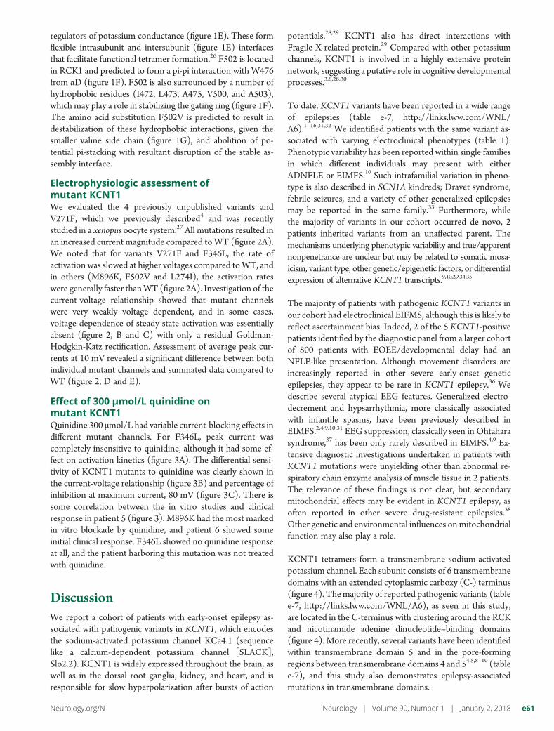
Effect of 300 μmol/L quinidine onmutant KCNT1Quinidine 300 μmol/L had variable current-blocking effects indifferent mutant channels. For F346L, peak current wascompletely insensitive to quinidine, although it had some ef-fect on activation kinetics (figure 3A). The differential sensi-tivity of KCNT1 mutants to quinidine was clearly shown inthe current-voltage relationship (figure 3B) and percentage ofinhibition at maximum current, 80 mV (figure 3C). There issome correlation between the in vitro studies and clinicalresponse in patient 5 (figure 3). M896K had the most markedin vitro blockade by quinidine, and patient 6 showed someinitial clinical response. F346L showed no quinidine responseat all, and the patient harboring this mutation was not treatedwith quinidine.
DiscussionWe report a cohort of patients with early-onset epilepsy as-sociated with pathogenic variants in KCNT1, which encodesthe sodium-activated potassium channel KCa4.1 (sequencelike a calcium-dependent potassium channel [SLACK],Slo2.2). KCNT1 is widely expressed throughout the brain, aswell as in the dorsal root ganglia, kidney, and heart, and isresponsible for slow hyperpolarization after bursts of action
potentials.28,29 KCNT1 also has direct interactions withFragile X-related protein.29 Compared with other potassiumchannels, KCNT1 is involved in a highly extensive proteinnetwork, suggesting a putative role in cognitive developmentalprocesses.3,8,28,30
To date, KCNT1 variants have been reported in a wide rangeof epilepsies (table e-7, http://links.lww.com/WNL/A6).1–16,31,32 We identified patients with the same variant as-sociated with varying electroclinical phenotypes (table 1).Phenotypic variability has been reported within single familiesin which different individuals may present with eitherADNFLE or EIMFS.10 Such intrafamilial variation in pheno-type is also described in SCN1A kindreds; Dravet syndrome,febrile seizures, and a variety of other generalized epilepsiesmay be reported in the same family.33 Furthermore, whilethe majority of variants in our cohort occurred de novo, 2patients inherited variants from an unaffected parent. Themechanisms underlying phenotypic variability and true/apparentnonpenetrance are unclear but may be related to somatic mosa-icism, variant type, other genetic/epigenetic factors, or differentialexpression of alternative KCNT1 transcripts.9,10,29,34,35
The majority of patients with pathogenic KCNT1 variants inour cohort had electroclinical EIFMS, although this is likely toreflect ascertainment bias. Indeed, 2 of the 5 KCNT1-positivepatients identified by the diagnostic panel from a larger cohortof 800 patients with EOEE/developmental delay had anNFLE-like presentation. Although movement disorders areincreasingly reported in other severe early-onset geneticepilepsies, they appear to be rare in KCNT1 epilepsy.36 Wedescribe several atypical EEG features. Generalized electro-decrement and hypsarrhythmia, more classically associatedwith infantile spasms, have been previously described inEIMFS.2,4,9,10,31 EEG suppression, classically seen in Ohtaharasyndrome,37 has been only rarely described in EIMFS.4,9 Ex-tensive diagnostic investigations undertaken in patients withKCNT1 mutations were unyielding other than abnormal re-spiratory chain enzyme analysis of muscle tissue in 2 patients.The relevance of these findings is not clear, but secondarymitochondrial effects may be evident in KCNT1 epilepsy, asoften reported in other severe drug-resistant epilepsies.38
Other genetic and environmental influences onmitochondrialfunction may also play a role.
KCNT1 tetramers form a transmembrane sodium-activatedpotassium channel. Each subunit consists of 6 transmembranedomains with an extended cytoplasmic carboxy (C-) terminus(figure 4). Themajority of reported pathogenic variants (tablee-7, http://links.lww.com/WNL/A6), as seen in this study,are located in the C-terminus with clustering around the RCKand nicotinamide adenine dinucleotide–binding domains(figure 4). More recently, several variants have been identifiedwithin transmembrane domain 5 and in the pore-formingregions between transmembrane domains 4 and 54,5,8–10 (tablee-7), and this study also demonstrates epilepsy-associatedmutations in transmembrane domains.
Neurology.org/N Neurology | Volume 90, Number 1 | January 2, 2018 e61

To date, different model systems have been used to determinethe functional effects ofKCNT1 variants.1,5,7,10,13,19,27 Our proteinhomology structural modeling data predict abnormal gating orprotein instability within the pore-forming region as a putativedisease mechanism. In silico modeling of G288S has predictedsimilar detrimental effects,5 while Y775H is predicted to affectsodium sensitivity of the channel.27 KCNT1 variants maytherefore alter structural properties of the protein, contributingto altered channel function. Consistent with previousreports1,3,13,19,27 (table e-7, http://links.lww.com/WNL/A6), ourxenopus oocyte model demonstrated that KCNT1 pathogenicvariants display a gain-of-function effect with increasedcurrent amplitude (figure 2). Previous studies have sought tocorrelate disease severity with the degree of gain of
function.1,19 However, in keeping with more recent studies,8
such correlation was not evident in our study. KCNT1 variantsresult in an increased Po (probability of the channel beingopen), which may be due to increased mutant channel coop-erativity or altered sodium sensitivity.8,27 In a recent study, so-dium removal from the pipette solution had a less negativeeffect on G288S channel amplitude than WT, suggestingreduced sodium sensitivity in the mutant.13 Heterotetramerformation may be of importance in vivo. In 1 study, mutantKCNT1 homomers revealed a more marked gain of functionthan mutant WT heteromers.13 A significant remainingquestion is howKCNT1 gain-of-function variants with predictedeffects on neuronal hyperpolarization result in epilepsy.28
Altered voltage sensitivity may result in KCNT1 channels
Figure 2 Functional investigation of KCNT1 mutations in a xenopus oocyte model
(A) Representative current traces obtained from oocytes expressing WT and EIMFS mutants (M896K, F502V, V271F, F346L, and L274I). Oocytes were held at−90 mV and stepped from −80 to 80 mV for 600 milliseconds every 5 seconds. Scale bars apply to all traces. (B) Current-voltage relationships for WT (n = 32),M896K (n = 15), F502V (n = 13), V271F (n = 9), F346L (n = 11), and L274I (n = 12). Currents were averaged and then normalized to the value at a test potential of80 mV (Imax). (C) Comparison of current-voltage relationships between WT (solid circles, n = 32) and EIMFS mutations (M896K [squares, n = 15], F502V[triangles, n = 13], V271F [hexagons, n = 9], F346L [diamonds, n = 11], and L274I [inverted triangles, n = 12]). Currents were averaged and then normalized tothe value at a test potential of 80mV (Imax). (D) Average peak currents at 10 mV for WT (n = 44), M896K (n = 19), F502V (n = 16), V271F (n = 10), F346L (n = 11),and L274I (n = 12) channels. Peak currents for eachmutant channel at 10mVwere compared to the peak currents for theWT channel at 10mV. ***p < 0.001,****p < 0.0001. (E) Comparison of pooled WT (n = 44) and EIMFS (n = 68) currents at 10 mV. ****p < 0.0001. EIMFS = epilepsy of infancy with migrating focalseizures; WT = wild-type.
e62 Neurology | Volume 90, Number 1 | January 2, 2018 Neurology.org/N

opening at more depolarized potentials, allowing a persistenthyperpolarizing current, with resultant interneuronal dis-inhibition as reported in SCN1A-related epilepsy.39 Con-versely, increased repolarization permitting more frequentand rapid action potentials may also play a role.27,34
Recently, quinidine has been identified as a novel therapyfor patients with KCNT1-related epilepsy. In in vitromodels, quinidine has been shown to reduce the abnormalincrease in mutant KCNT1 channel amplitude.19 For 1patient with EIMFS with the KCNT1 variant R428Q , invitro testing showed quinidine sensitivity, and treatmentresulted in a dramatic improvement in seizure control withneurodevelopmental gains.3,7 However, in more recentstudies, patient response has been variable and not alwaysas predicted by in vitro studies.11 Indeed, another patient
with the same variant (R428Q) but different epilepsyphenotype (unclassified EOEE) failed to respond to quin-idine, albeit at a later stage in the disease course.14 Mostrecently, a patient withWest syndrome had a good responsebut only with a higher dose of 60 mg/kg/d.2 Clinical re-sponse may possibly be determined by the specific variant,other genetic factors, epilepsy phenotype, and drug timingwithin a therapeutic window. In our series, we treated 3patients with quinidine, and 1 patient showed a clinical response.One patient developed a severe pulmonary vasculopathy, afterwhich quinidine was discontinued. Systemic vasculitis has beenreported with quinidine treatment.40 While investigations in ourpatient did not reveal overt evidence of vasculitis, the observedpulmonary dysfunction may represent an adverse drug-relatedevent. The precisemechanism of KCNT1 blockade by quinidineis unclear, and it is possible that the disease mechanism for
Figure 3 Effect of quinidine on xenopus oocytes expressing hKCNT1 channels
(A) Representative current traces obtained from oocytes expressing WT and EIMFS mutants (M896K and F346L) with application of vehicle (ND96) and 300μmol/L quinidine. Oocyteswere held at−90mVand stepped from −80 to 80mV for 600milliseconds every 5 seconds. Scale bars apply to all traces. (B) Current-voltage relationships forWT (n = 32), M896K (n = 15), F502V (n = 13), V271F (n = 9), F346L (n = 11), and L274I (n = 12) hKCNT1 channels in the presence of vehicle(ND96) and 300μmol/L quinidine. Currentswere averagedand then normalized to the value at a test potential of 80mV (Imax). (C) Average percent inhibition at80 mV of WT (n = 31) and EIMFS (M896K, n = 15; F502V, n = 13; V271F, n = 9; F346L, n = 11; and; L274I, n = 12) hKCNT1 channels by quinidine (300 μmol/L)depicting the variable degree of block by 300μmol/L quinidine (1-way analysis of variance followed by Bonferroni post hoc analysis). *p < 0.1. EIMFS = epilepsyof infancy with migrating focal seizures; WT = wild-type.
Neurology.org/N Neurology | Volume 90, Number 1 | January 2, 2018 e63

F346L, perhaps involving abnormal channel-opening dynamicsas suggested by themodeling data, is notmodifiable by quinidine.Our data suggest that quinidine should be considered as a ther-apeutic option for patients with KCNT1 variants, but used withcaution. Larger studies will provide further guidance about clin-ical utility, patient selection, optimum age at administration, anddose. Other KCNT1 modulators, including bepridil and clofili-lum, have been identified as possible alternative therapies.28 Likequinidine, bepridil has been shown in vitro to reversibly blockmutant KCNT1 channels at a lower concentration than WTchannels.13 However, similar to quinidine, potential cardiaceffects and lack of specificity may limit use in patients.
Pathogenic variants in KCNT1 cause a wide spectrum of se-vere epilepsies typically associated with impaired neurologic
development and significant disease burden. As demonstrated,in vitromodel systemsmay be useful to validate putative variantsand to confirm pathogenicity, although genotype-phenotypecorrelations remain unclear. Evaluation of new therapies, in-cluding KCNT1-specific blockers, remains a research priorityfor this devastating pharmacoresistant group of epilepsies.
Note added in proofRecently, 3 patients with de novo KCNT1 mutations andmassive systemic to pulmonary collateral artery formation,presenting with pulmonary hemorrhage requiring emboliza-tion, were described. These patients had not been treated withquinidine. However, the mechanism remains unclear andfurther investigation of the expression and role of KCNT1 inthe cardiovascular system is required.41
Figure 4 Schematic diagram of the location of mutations in KCNT1 in this and previously published studies
KCNT1 encodes sequence like a calcium-dependent potassium channel (SLACK), which forms tetramers (top left) or heteromers with KCNT2 or sequence like anintermediate conductance K channel (SLICK). The structure comprises 6 transmembrane domains with a pore-forming region, regulator of potassium conductance(RCK), and nicotinamide adenine dinucleotide–binding (NAD-B) domains. EIMFS phenotypes are shaded in purple, ADNFLE or NFLE in pink, others (Ohtaharasyndrome, leukoencephalopathy, focal epilepsy, EOEE,West syndrome,unaffected) inorange.Mutationsgiving rise to>1phenotypeareshadedwitha combinationofthe corresponding colors. Novel mutations identified in this study are outlined in green, those identified in previous studies in turquoise. ADNFLE = autosomaldominant nocturnal frontal lobe epilepsy; EIMFS = epilepsy of infancy with migrating focal seizures; EOEE = early-onset epileptic encephalopathy; NFLE = nocturnalfrontal lobe epilepsy.
e64 Neurology | Volume 90, Number 1 | January 2, 2018 Neurology.org/N

Author contributionsAmy McTague, Umesh Nair, Sony Malhotra, and Esther Meyerhave contributed to drafting/revising themanuscript for content,including medical writing for content, study concept or design,acquisition of data, analysis or interpretation of data. NatalieTrump has contributed to drafting/revising the manuscript forcontent, including medical writing for content, acquisition ofdata, analysis or interpretationof data. ElenaV.Gazina, ApostolosPapandreou, and Adeline Ngoh have contributed to drafting/revising the manuscript for content, acquisition of data, analysisor interpretation of data. Sally Ackermann and Gautam Ambe-gaonkar have contributed to drafting/revising the manuscript forcontent, acquisition of data. Richard Appleton has contributed todrafting/revising the manuscript for content, including medicalwriting for content, acquisition of data, analysis or interpretationof data. Archana Desurkar has contributed to drafting/revisingthemanuscript for content, acquisition of data. Christin Eltze andRachel Kneen have contributed to drafting/revising the manu-script for content, including medical writing for content, acqui-sition of data, analysis or interpretation of data. Ajith V. Kumarand Karine Lascelles have contributed to drafting/revising themanuscript for content, acquisition of data. Tara Montgomeryhas contributed to drafting/revising the manuscript for content,acquisition of data, analysis or interpretation of data. Ven-kateswaran Ramesh and Rajib Samanta have contributed todrafting/revising the manuscript for content, acquisition of data.Richard H. Scott has contributed to drafting/revising the man-uscript for content, acquisition of data, analysis or interpretationof data. Jeen Tan has contributed to drafting/revising the man-uscript for content, acquisition of data. William Whitehouse andAnnapurna Poduri have contributed to drafting/revising themanuscript for content, including medical writing for content,acquisition of data. Ingrid E. Scheffer,W.K. “Kling”Chong, and J.Helen Cross have contributed to drafting/revising the manu-script for content, including medical writing for content, acqui-sition of data, analysis or interpretation of data. Maya Topf,Steven Petrou, and Manju A. Kurian have contributed todrafting/revising the manuscript for content, including medicalwriting for content, study concept or design, acquisition of data,analysis or interpretation of data.
AcknowledgmentThe authors thank the patients and their families for theirparticipation in this study. They thank all clinicians referringpatients with EIMFS for inclusion in the research study (Dr.Mike Pike, Dr. Stefan Spinty, Dr. Ailsa McLellan, Dr. MaryKing, Dr. Andrew Curran, Dr. Hans Randby, Dr. Linda deMeirleir, Dr. Jost Richter, Dr. Elaine King, Dr.Martin Piepkorn,Dr. Mary O’Regan, Dr. Ariane Biebl, Dr. Gary McCullagh, andDr. Siobhan West). They thank Erin Heinzen, PharmD, PhD,for whole-exome sequencing of 5 patients at the Duke Centerfor Genomic Medicine, Durham, NC.
Study fundingThis project was supported by the National Institute forHealth Research Biomedical Research Centre at Great
Ormond Street Hospital for Children NHS Foundation Trustand University College London.
DisclosureA. McTague was funded by a Medical Research CouncilClinical Research Training Fellowship (MR/L001497/1), theMedical Research Foundation for travel and conference fees(MRF-007-0003-STD-MCTAG), a Grass Foundation TravelBursary from the American Epilepsy Society, and a JuniorInvestigator travel scholarship from the University of SouthCalifornia. U. Nair and S. Malhotra report no disclosuresrelevant to the manuscript. E. Meyer is funded by SPARKS.N. Trump reports no disclosures relevant to the manuscript.E.V. Gazina is funded by the Australian Research Council(APP1106027). A. Papandreou is funded by a joint ActionMedical Research and British Paediatric Neurology Associa-tion research training fellowship. A. Ngoh is funded byGuarantors of Brain, Friends of Landau Kleffner Syndromeand Action Medical Research. S. Ackermann and G. Ambe-gaonkar report no disclosures relevant to the manuscript. R.Appleton served on the scientific committees for and receiveda single honorarium fromNovartis and GWPharma and is theChief Investigator for the EcLiPSE Study funded by an Na-tional Institute for Health Research Health Technology As-sessment grant (12/127/134). A. Desurkar, C. Eltze, R.Kneen, A.V. Kumar, K. Lascelles, T. Montgomery, V. Ramesh,R. Samanta, and R.H. Scott report no disclosures relevant tothe manuscript. J. Tan received an industry-funded bursary forconference travel (£300) from UCB Pharma. W. Whitehouseserves on the editorial board of NeuroEducation. A. Poduri ison the Scientific Advisory Board of the Dravet SyndromeFoundation, serves as an associate editor for Epilepsia andcontributing editor for Epilepsy Currents, and is on the editorialboard for Annals of Neurology. She receives research supportfrom the National Institute of Neurological Disorders andStroke (NINDS), Citizens United for Research in Epilepsy,and the Boston Children’s Hospital Translational ResearchProgram. I.E. Scheffer serves on the editorial boards of Neu-rology and Epileptic Disorders; may accrue future revenue ona pending patent WO61/010176: Therapeutic Compoundthat relates to discovery of PCDH19 gene; is one of theinventors listed on a patent held by Bionomics Inc on di-agnostic testing of using the SCN1A gene, WO2006/133508;has received speaker honoraria or scientific board consultingfees from Athena Diagnostics, UCB, GSK, Eisai, and Trans-genomics; has received funding for travel from Athena Diag-nostics, UCB, Eisai, and GSK; and has received researchsupport from the National Health and Medical ResearchCouncil of Australia, NINDS, Health Research Council ofNew Zealand, US Department of Defense, and March ofDimes. W.K. Chong reports no disclosures relevant to themanuscript. J.H. Cross is on the advisory boards of Shire,GSK, Eisai, Vitaflo, UCB, and Takeda for which remunerationhas been made to her department; has a patent in applicationwith Vitaflo; and is the chief investigator for clinical trials withVitaflo, GWPharma, and Zogenix for which remuneration hasbeen made to the department. M. Topf reports no disclosures
Neurology.org/N Neurology | Volume 90, Number 1 | January 2, 2018 e65

relevant to the manuscript. S. Petrou is scientific founder ofPraxis Precision Medicine, Boston, and a Scientific AdvisoryBoard Member of Pairnomix, Minneapolis, and has receivedshares for both. Dr. Petrou’s research is supported by the LuluFoundation, Vitaflo/Nestle, Citizens United for Research inEpilepsy,Mallinckrodt Pharmaceuticals, Clarus Ventures, DHBFoundation, Australian Research Council, National Health andMedical Research Council, and SCN2A Research Foundation.M.A. Kurian receives research/grant support from a WellcomeTrust Intermediate Clinical Fellowship (WT098524MA). Goto Neurology.org/N for full disclosures.
Received November 7, 2016. Accepted in final form September 26,2017.
References1. Martin HC, Kim GE, Pagnamenta AT, et al. Clinical whole-genome sequencing in
severe early-onset epilepsy reveals new genes and improves molecular diagnosis. HumMol Genet 2014;23:3200–3211.
2. Fukuoka M, Kuki I, Kawawaki H, et al. Quinidine therapy for West syndrome withKCNT1 mutation: a case report. Brain Dev 2017;39:80–83.
3. Barcia G, Fleming MR, Deligniere A, et al. De novo gain-of-function KCNT1 channelmutations cause malignant migrating partial seizures of infancy. Nat Genet 2012;44:1255–1259.
4. McTague A, Appleton R, Avula S, et al. Migrating partial seizures of infancy: ex-pansion of the electroclinical, radiological and pathological disease spectrum. Brain2013;136:1578–1591.
5. Ishii A, Shioda M, Okumura A, et al. A recurrent KCNT1 mutation in two sporadiccases with malignant migrating partial seizures in infancy. Gene 2013;531:467–471.
6. Vanderver A, Simons C, Schmidt JL, et al. Identification of a novel de novo p.Phe932Ile KCNT1 mutation in a patient with leukoencephalopathy and severe epi-lepsy. Pediatr Neurol 2014;50:112–114.
7. BeardenD, StrongA, Ehnot J, DiGiovineM,DlugosD,Goldberg EM.Targeted treatmentof migrating partial seizures of infancy with quinidine. Ann Neurol 2014;76:457–461.
8. KimGE, Kronengold J, Barcia G, et al. Human slack potassium channel mutations increasepositive cooperativity between individual channels. Cell Rep 2014;9:1661–1672.
9. Ohba C, Kato M, Takahashi N, et al. De novo KCNT1 mutations in early-onsetepileptic encephalopathy. Epilepsia 2015;56:e121–e128.
10. Møller RS, Heron SE, Larsen LHG, et al. Mutations in KCNT1 cause a spectrum offocal epilepsies. Epilepsia 2015;56:e114–e120.
11. Mikati MA, Jiang Y-H, Carboni M, et al. Quinidine in the treatment of KCNT1positive epilepsies. Ann Neurol 2015;78:995–999.
12. Allen NM, Conroy J, Shahwan A, et al. Unexplained early onset epileptic encepha-lopathy: exome screening and phenotype expansion. Epilepsia 2015;57:e12–e17.
13. Rizzo F, Ambrosino P, Guacci A, et al. Characterization of two de novo KCNT1mutations in children with malignant migrating partial seizures in infancy. Mol CellNeurosci 2016;72:54–63.
14 Chong PF, Nakamura R, Saitsu H, Matsumoto N, Kira R. Ineffective quinidine therapyin early-onset epileptic encephalopathy with KCNT1 mutation. Ann Neurol 2016;79:502–503.
15. Heron SE, Smith KR, Bahlo M, et al. Missense mutations in the sodium-gated po-tassium channel gene KCNT1 cause severe autosomal dominant nocturnal frontallobe epilepsy. Nat Genet 2012;44:1188–1190.
16 Hildebrand MS, Myers CT, Carvill GL, et al. A targeted resequencing gene panel forfocal epilepsy. Neurology 2016;86:1605–1612.
17. Eddy SR. Accelerated profile HMM searches. PLoS Comput Biol 2011;7:e1002195.18 Finn RD, Mistry J, Tate J, et al. The Pfam protein families database. Nucleic Acids Res
2009;38:D211–D222.19. Milligan CJ, Li M, Gazina EV, et al. KCNT1 gain of function in 2 epilepsy phenotypes
is reversed by quinidine. Ann Neurol 2014;75:581–590.20. 1000 Genomes. Available at: http://browser.1000genomes.org/index.html. Accessed
June 14, 2016.21. ExAC Browser (beta)|Exome Aggregation Consortium. Available at: http://exac.
broadinstitute.org/. Accessed June 14, 2016.22. NHLBI Exome Sequencing Project (ESP). Available at: http://evs.gs.washington.
edu/EVS/. Accessed June 14, 2016.23. PolyPhen-2. Available at: http://genetics.bwh.harvard.edu/pph2/. Accessed June 14,
2016.24. Available at: http://provean.jcvi.org/genome_submit_2.php. Accessed June 14,
2016.25. Doyle DA, Morais Cabral J, Pfuetzner RA, et al. The structure of the potassium
channel: molecular basis of K+ conduction and selectivity. Science 1998;280:69–77.
26. Yuan P, Leonetti MD, Pico AR, Hsiung Y, MacKinnon R. Structure of the humanBK channel Ca2+-activation apparatus at 3.0 A resolution. Science 2010;329:182–186.
27. Tang Q-Y, Zhang F-F, Xu J, et al. Epilepsy-related slack channel mutants lead tochannel over-activity by two different mechanisms. Cell Rep 2016;14:129–139.
28. Kaczmarek LK. Slack, slick, and sodium-activated potassium channels. ISRNNeurosci2013;2013:354262.
29. Brown MR, Kronengold J, Gazula V-R, et al. Amino-termini isoforms of the slack K+channel, regulated by alternative promoters, differentially modulate rhythmic firingand adaptation. J Physiol 2008;586:5161–5179.
30. Kim GE, Kaczmarek LK. Emerging role of the KCNT1 slack channel in intellectualdisability. Front Cell Neurosci 2014;8:209.
31. Allen AS, Berkovic SF, Cossette P, et al. De novo mutations in epileptic encephalo-pathies. Nature 2013;501, 217–221.
32. Arai-Ichinoi N, Uematsu M, Sato R, et al. Genetic heterogeneity in 26 infants witha hypomyelinating leukodystrophy. Hum Genet 2015;135:89–98.
33. Scheffer IE, Zhang Y-HH, Jansen FE, Dibbens L. Dravet syndrome or genetic(generalized) epilepsy with febrile seizures plus? Brain Dev 2009;31:394–400.
34. Lim CX, Ricos MG, Dibbens LM, Heron SE. KCNT1 mutations in seizuredisorders: the phenotypic spectrum and functional effects. J Med Genet 2016;53:217–225.
35. Chen H, Kronengold J, Yan Y, et al. The N-terminal domain of slack determines theformation and trafficking of slick/slack heteromeric sodium-activated potassiumchannels. J Neurosci 2009;29:5654–5665.
36. Howell KB, McMahon JM, Carvill GL, et al. SCN2A encephalopathy: a major cause ofepilepsy of infancy with migrating focal seizures. Neurology 2015;85:958–966.
37. Ohtahara S. A study on the age dependent epileptic encephalopathy. No To Hattatsu1977;9:2–21.
38. Folbergrova J, Kunz WS. Mitochondrial dysfunction in epilepsy. Mitochondrion2012;12:35–40.
39. Yu FH, Mantegazza M, Westenbroek RE, et al. Reduced sodium current inGABAergic interneurons in a mouse model of severe myoclonic epilepsy in infancy.Nat Neurosci 2006;9:1142–1149.
40. Lipsker D,Walther S, Schulz R, Nave S, Cribier B. Life-threatening vasculitis related toquinidine occurring in a healthy volunteer during a clinical trial. Eur J Clin Pharmacol1998;54:815.
41. Kawasaki Y, Kuki I, Ehara E, et al. Three cases of KCNT1 mutations: malignantmigrating partial seizures in infancy with massive systemic to pulmonary collateralarteries. J Pediatr Epub 2017 October 5.
e66 Neurology | Volume 90, Number 1 | January 2, 2018 Neurology.org/N

SOURCE ARTICLE NPub.org/22ubfy
Clinical and molecular characterization ofKCNT1-related severe early-onset epilepsyAmy McTague, MBChB, Umesh Nair, BSc Hons, Sony Malhotra, PhD, Esther Meyer, PhD, Natalie Trump, PhD,
Elena V. Gazina, PhD, Apostolos Papandreou, MBBS, Adeline Ngoh, MBBS, Sally Ackermann, MBChB,
Gautam Ambegaonkar, MBBS, MRCPCH, Richard Appleton, MA(Oxon), Archana Desurkar, MBBS,
Christin Eltze, MBBS, MPhil, Rachel Kneen, BMedSci, BMBS, FRCPCH, Ajith V. Kumar, MD, MRCP,
Karine Lascelles, MBBS, MRCPCH, TaraMontgomery, BM, BS, BMedSci, FRCP, Venkateswaran Ramesh,MBBS,
Rajib Samanta,MBBS, FRCPCH, RichardH. Scott, PhD, Jeen Tan,MBChB,MRCPCH,WilliamWhitehouse,MBBS,
MRCP, Annapurna Poduri, MD, MPH, Ingrid E. Scheffer, MBBS, PhD, W. K. “Kling” Chong, FRCR,
J. Helen Cross, MBChB, PhD, Maya Topf, PhD, Steven Petrou, PhD, and Manju A. Kurian, MRCPCH, PhD
Neurology® 2018;90:24. doi:10.1212/WNL.0000000000004762
Correspondence
Dr. McTague
Dr. Kurian
Study questionWhat are the phenotypic, molecular genetic, and functional char-acteristics of KCNT1 variants that cause early-onset epilepsy?
Summary answerGain-of-function KCNT1 mutations can cause diverse severefocal epilepsies with onset in early infancy, but genotype-phenotype relationships remain unclear.
What is known and what this paper addsAutosomal dominant pathogenic variants of the sodium-activatedpotassium channel gene KCNT1 are associated with a broadspectrum of epileptic disorders. The study explores the clinical,molecular genetic, and functional properties of a cohort ofpatients with KCNT1-related epilepsy.
Participants and settingThirty-one patients with epilepsy of infancy with migrating focalseizures (EIMFS) were recruited from 2011 to 2016. Twopatientswhohad undergone routine genetic testing that includedKCNT1 analysis were also included. The patients wererecruited at London’s Great Ormond Street Hospital and byreferrals from UK and international centers.
Design, size, and durationThe patients’ KCNT1 variants were identified via multiplemethods, and homologymodeling was performed. ThemutantKCNT1 variants were electrophysiologically assessed ina Xenopus oocyte model.
Main results and the role of chancePathogenic KCNT1 mutations were detected in 12 patients.These patients exhibited diverse symptoms. The ages at onsetranged from 1 day to 6 months, and all had neurodevelopmentalimpairments. Treatment outcomes were generally poor. ThepathogenicKCNT1 variants included 8 previously reported and4 unreported variants. Homology modeling for 2 unreportedvariants indicated that they would induce abnormal gating orprotein instability. Electrophysiologic assessments of 5
variants, including the 4 unreported ones, revealed abnormalchannel functions, including increased current magnitudesrelative to the wild-type (WT) channel and variable blockadeby quinidine, in keeping with the clinical response.
Bias, confounding, and other reasonsfor cautionFocusing on patients with EIMFS may have introduced as-certainment bias.
Generalizability to other populationsPathogenic KCNT1 mutations have been detected in patientswith epileptic conditions other than EIMFS, including noc-turnal frontal lobe epilepsy. Such mutations may have differenteffects in other conditions.
Study funding/potential competing interestsThis study was funded by the UK National Health Service andUniversity College London. Some authors report receiving fund-ing, personal compensation, and advisory committee appoint-ments from various companies, journals, scholarly societies, andgovernment agencies. Go to Neurology.org/N for fulldisclosures.
A draft of the short-form article was written byM. Dalefield, a writer with Editage, a division of Cactus Communications. The authors of the full-length article and the journal editors edited and approved the final version.
24 Copyright © 2017 American Academy of Neurology
SHORT-FORM ARTICLE

DOI 10.1212/WNL.00000000000047622018;90;e55-e66 Published Online before print December 1, 2017Neurology
Amy McTague, Umesh Nair, Sony Malhotra, et al. -related severe early-onset epilepsyKCNT1Clinical and molecular characterization of
This information is current as of December 1, 2017
ServicesUpdated Information &
http://n.neurology.org/content/90/1/e55.fullincluding high resolution figures, can be found at:
References http://n.neurology.org/content/90/1/e55.full#ref-list-1
This article cites 35 articles, 6 of which you can access for free at:
Subspecialty Collections
http://n.neurology.org/cgi/collection/neonatal_seizuresNeonatal seizures
http://n.neurology.org/cgi/collection/ion_channel_gene_defectsIon channel gene defectsfollowing collection(s): This article, along with others on similar topics, appears in the
Permissions & Licensing
http://www.neurology.org/about/about_the_journal#permissionsits entirety can be found online at:Information about reproducing this article in parts (figures,tables) or in
Reprints
http://n.neurology.org/subscribers/advertiseInformation about ordering reprints can be found online:
ISSN: 0028-3878. Online ISSN: 1526-632X.Wolters Kluwer Health, Inc. on behalf of the American Academy of Neurology.. All rights reserved. Print1951, it is now a weekly with 48 issues per year. Copyright Copyright © 2017 The Author(s). Published by
® is the official journal of the American Academy of Neurology. Published continuously sinceNeurology