Clinical anatomy as a modern concept for 21st century...
5
99 Review Article Kitasato Med J 2013; 43: 99-103 Clinical anatomy as a modern concept for 21st century teaching, postgraduate education, and research Bernhard Hirt, Thomas Shiozawa Institute of Anatomy, Department of Clinical Anatomy, Eberhard Karls University, Tuebingen, Germany Anatomy faces new challenges in the 21st century. Exponential growth of knowledge, paradigm shifts in medical education, and the scientific trend to subcellular and molecular research make an impact on the institutions of today. We described our concept of clinical anatomy as a contemporary and innovative model for student teaching, postgraduate training, and applied clinical research. Anatomy can also foster interdisciplinary teaching along with the clinical, especially surgical, professions to give students a direct link to the application of anatomical knowledge. Furthermore, clinical students can be relegated to the conceptual framework of basic sciences during their clinical rotations. Clinical anatomy can provide a unique platform for postgraduate training, because cadaveric human body specimens can be used as realistic and safe models for practicing even the most complex surgical procedures. Lastly, the study of clinical anatomy can facilitate applied clinical research, as new operation techniques and new devices and instruments can be tested and evaluated in a realistic setting. Therefore, this new concept of the study of clinical anatomy provides a modern, up-to-date model for teaching anatomy in this day and age. Key words: clinical anatomy, teaching, dissection, postgraduate surgical training Background natomy is one of the oldest subjects in medicine. Although having evolved over time, the common purpose of the scientific field, over decades, and even centuries, is the explanation of the structure and morphology of the human body. Traditionally, most professional anatomists were also clinically trained; and, therefore, their research was oriented toward the human structure. However, the vast expanding knowledge in the second half of the 20th century more and more turned to sub- and ultrastructure; therefore, the applied and clinical aspects of anatomy began to fade. This resulted in the reduction of anatomical content in the curricula and the decline of clinically qualified anatomists. However, in the '70s and '80s of the last century the growing demand in different medical branches for detailed knowledge of specialized anatomy, e.g., the development of new technology, was recognized. 1 This rejuvenation was attended by the foundation of the British and American Association of Clinical Anatomists, in 1977 and 1984, respectively, and also the launch of the journal. Clinical Anatomy In Germany, the discussion about anatomy, and how anatomy related to clinical medicine, peaked at that time in a well-remembered article by the anatomist Herbert Lippert about the "Dehumanisation of anatomy and medicine." 2 He called for a more clinical and living, teaching approach for undergraduate anatomy, resulting in a broad controversial discussion in the whole medical community. This emphasis on clinical anatomy, against the background of the development of many different medical technologies, also brought the need for profound anatomical knowledge back on screen for science and research and, for the first time, for postgraduate, surgical, training. The first Center for Clinical Anatomy in Germany was founded by Bernhard Tillmann, in 1994, in Kiel, followed by similar institutions in Cologne, Hanover, Mainz, Muenster, Halle, and Tuebingen. To date, many anatomical institutes have hosted postgraduate training courses; however, not many devote themselves to clinical and macroscopic anatomy as a field of research. The concept of clinical anatomy in Tuebingen was A Received 18 February 2013, accepted 2 April 2013 Correspondence to: Bernhard Hirt, Department of Clinical Anatomy, Eberhard Karls University Elfriede-Aulhorn-Straße 8, 72076 Tuebingen, Germany E-mail: [email protected]
Transcript of Clinical anatomy as a modern concept for 21st century...

99
Review Article Kitasato Med J 2013; 43: 99-103
Clinical anatomy as a modern concept for 21st century teaching,postgraduate education, and research
Bernhard Hirt, Thomas Shiozawa
Institute of Anatomy, Department of Clinical Anatomy, Eberhard Karls University, Tuebingen, Germany
Anatomy faces new challenges in the 21st century. Exponential growth of knowledge, paradigm shiftsin medical education, and the scientific trend to subcellular and molecular research make an impact onthe institutions of today. We described our concept of clinical anatomy as a contemporary andinnovative model for student teaching, postgraduate training, and applied clinical research.Anatomy can also foster interdisciplinary teaching along with the clinical, especially surgical, professionsto give students a direct link to the application of anatomical knowledge. Furthermore, clinicalstudents can be relegated to the conceptual framework of basic sciences during their clinical rotations.Clinical anatomy can provide a unique platform for postgraduate training, because cadaveric humanbody specimens can be used as realistic and safe models for practicing even the most complex surgicalprocedures. Lastly, the study of clinical anatomy can facilitate applied clinical research, as newoperation techniques and new devices and instruments can be tested and evaluated in a realistic setting.Therefore, this new concept of the study of clinical anatomy provides a modern, up-to-date model forteaching anatomy in this day and age.
Key words: clinical anatomy, teaching, dissection, postgraduate surgical training
Background
natomy is one of the oldest subjects in medicine.Although having evolved over time, the common
purpose of the scientific field, over decades, and evencenturies, is the explanation of the structure andmorphology of the human body. Traditionally, mostprofessional anatomists were also clinically trained; and,therefore, their research was oriented toward the humanstructure. However, the vast expanding knowledge inthe second half of the 20th century more and more turnedto sub- and ultrastructure; therefore, the applied andclinical aspects of anatomy began to fade. This resultedin the reduction of anatomical content in the curriculaand the decline of clinically qualified anatomists.However, in the '70s and '80s of the last century thegrowing demand in different medical branches fordetailed knowledge of specialized anatomy, e.g., thedevelopment of new technology, was recognized.1 Thisrejuvenation was attended by the foundation of the Britishand American Association of Clinical Anatomists, in 1977and 1984, respectively, and also the launch of the journal.
Clinical Anatomy
In Germany, the discussion about anatomy, and howanatomy related to clinical medicine, peaked at that timein a well-remembered article by the anatomist HerbertLippert about the "Dehumanisation of anatomy andmedicine."2 He called for a more clinical and living,teaching approach for undergraduate anatomy, resultingin a broad controversial discussion in the whole medicalcommunity.
This emphasis on clinical anatomy, against thebackground of the development of many different medicaltechnologies, also brought the need for profoundanatomical knowledge back on screen for science andresearch and, for the first time, for postgraduate, surgical,training. The first Center for Clinical Anatomy inGermany was founded by Bernhard Tillmann, in 1994,in Kiel, followed by similar institutions in Cologne,Hanover, Mainz, Muenster, Halle, and Tuebingen. Todate, many anatomical institutes have hosted postgraduatetraining courses; however, not many devote themselvesto clinical and macroscopic anatomy as a field of research.
The concept of clinical anatomy in Tuebingen was
A
Received 18 February 2013, accepted 2 April 2013Correspondence to: Bernhard Hirt, Department of Clinical Anatomy, Eberhard Karls UniversityElfriede-Aulhorn-Straße 8, 72076 Tuebingen, GermanyE-mail: [email protected]
100
Hirt, et al.
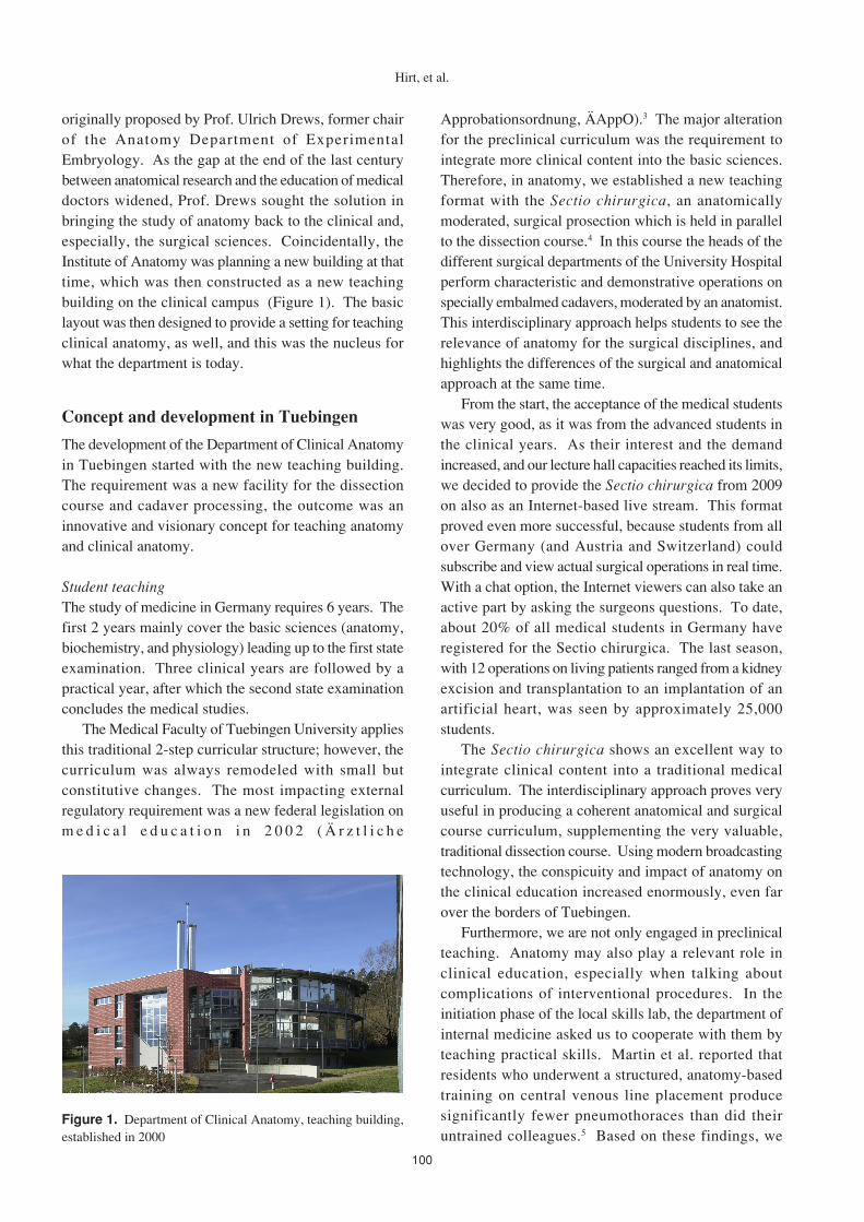
originally proposed by Prof. Ulrich Drews, former chairof the Anatomy Department of ExperimentalEmbryology. As the gap at the end of the last centurybetween anatomical research and the education of medicaldoctors widened, Prof. Drews sought the solution inbringing the study of anatomy back to the clinical and,especially, the surgical sciences. Coincidentally, theInstitute of Anatomy was planning a new building at thattime, which was then constructed as a new teachingbuilding on the clinical campus (Figure 1). The basiclayout was then designed to provide a setting for teachingclinical anatomy, as well, and this was the nucleus forwhat the department is today.
Concept and development in Tuebingen
The development of the Department of Clinical Anatomyin Tuebingen started with the new teaching building.The requirement was a new facility for the dissectioncourse and cadaver processing, the outcome was aninnovative and visionary concept for teaching anatomyand clinical anatomy.
Student teachingThe study of medicine in Germany requires 6 years. Thefirst 2 years mainly cover the basic sciences (anatomy,biochemistry, and physiology) leading up to the first stateexamination. Three clinical years are followed by apractical year, after which the second state examinationconcludes the medical studies.
The Medical Faculty of Tuebingen University appliesthis traditional 2-step curricular structure; however, thecurriculum was always remodeled with small butconstitutive changes. The most impacting externalregulatory requirement was a new federal legislation onm e d i c a l e d u c a t i o n i n 2 0 0 2 ( Ä r z t l i c h e
Approbationsordnung, ÄAppO).3 The major alterationfor the preclinical curriculum was the requirement tointegrate more clinical content into the basic sciences.Therefore, in anatomy, we established a new teachingformat with the Sectio chirurgica, an anatomicallymoderated, surgical prosection which is held in parallelto the dissection course.4 In this course the heads of thedifferent surgical departments of the University Hospitalperform characteristic and demonstrative operations onspecially embalmed cadavers, moderated by an anatomist.This interdisciplinary approach helps students to see therelevance of anatomy for the surgical disciplines, andhighlights the differences of the surgical and anatomicalapproach at the same time.
From the start, the acceptance of the medical studentswas very good, as it was from the advanced students inthe clinical years. As their interest and the demandincreased, and our lecture hall capacities reached its limits,we decided to provide the Sectio chirurgica from 2009on also as an Internet-based live stream. This formatproved even more successful, because students from allover Germany (and Austria and Switzerland) couldsubscribe and view actual surgical operations in real time.With a chat option, the Internet viewers can also take anactive part by asking the surgeons questions. To date,about 20% of all medical students in Germany haveregistered for the Sectio chirurgica. The last season,with 12 operations on living patients ranged from a kidneyexcision and transplantation to an implantation of anartificial heart, was seen by approximately 25,000students.
The Sectio chirurgica shows an excellent way tointegrate clinical content into a traditional medicalcurriculum. The interdisciplinary approach proves veryuseful in producing a coherent anatomical and surgicalcourse curriculum, supplementing the very valuable,traditional dissection course. Using modern broadcastingtechnology, the conspicuity and impact of anatomy onthe clinical education increased enormously, even farover the borders of Tuebingen.
Furthermore, we are not only engaged in preclinicalteaching. Anatomy may also play a relevant role inclinical education, especially when talking aboutcomplications of interventional procedures. In theinitiation phase of the local skills lab, the department ofinternal medicine asked us to cooperate with them byteaching practical skills. Martin et al. reported thatresidents who underwent a structured, anatomy-basedtraining on central venous line placement producesignificantly fewer pneumothoraces than did theiruntrained colleagues.5 Based on these findings, we
Figure 1. Department of Clinical Anatomy, teaching building,established in 2000

101
Clinical anatomy as a modern concept
developed an anatomical training session with emphasison the neck, regarding the catheter placement techniqueand possible complications.6 This became a requiredmodule in the internal medicine skills training, whichactually takes place in the anatomical dissection hall. Asa second means of educating medical students in a realisticsurgical environment, we initiated a course teachingendoscopic anatomy. Students were trained to analyzeanatomy via endoscopic views on cadavers. They learnedto use endoscopes dynamically to search for specificstructures. We recognized that this educational conceptis able to longitudinally connect basic anatomicalknowledge with clinical relevance.
Postgraduate trainingHaving started with loose cooperation with several clinicalpartners in the university hospital, the contribution of theDepartment of Clinical Anatomy to postgraduateeducation has also changed tremendously over the lastyears. The building and basic hardware proved a perfectsetting for surgical training courses to many disciplines.Starting with anesthesiology and orthopaedic surgery,soon training courses in neurosurgery, ear, nose, andthroat (ENT) surgery, maxillofacial surgery, gynecology,and trauma surgery followed. The present infrastructureallows us to host training courses of varying size (Figure2). In 2012, we hosted 60 national and internationaltraining course events, training about 1,000 surgeons from12 different disciplines.
InfrastructureThe floor plan is centered around the semicircular shapeddissection hall on the ground floor, the first floorcomprises seminar rooms and a histology lecture hall.The basic hardware: operating room (OR) equipment
with an OR table, operation microscope, ultrasonic device,and laparoscopy tower, was already included. Over theyears, we acquired more equipment. Now we offer 11workplaces with OR tables (Trumpf Merkur, TrumpfGmbH + Co. KG, Ditzingen, Germany), High definition(HD) laparoscopic/arthroscopic tower (Karl Storz GmbH& Co. KG, Tuttlingen, Germany), basic surgicalinstruments (Aesculap AG, Tuttlingen, Germany; KLSMartin Group, Tuttlingen, Germany; Karl Storz), andLED (light-emitting diodes) surgical operating lamps(KLS Martin Group). After beginning with live (realtime) surgery transmissions in 2008, we established oneKarl Storz OR1 master workplace (Karl Storz) fordemonstrations (Figure 3). Several HD camera systemsprovide high quality overview and insights for telemedialpurposes. In 2011, a livestream broadcast studio wasadded. We established technical architecture to combinethe existing OR1 master workplace including the KarlStorz AIDA (Karl Storz) documentation system with aTricasterTM 855 live production system (NewTek Inc.,San Antonio, TX, USA). Sound processing is facilitatedwith a Sennheiser Integrated System (SennheiserElectronic GmbH & Co. KG, Wedemark, Germany).
Cadaver processingFor the dissection course, formalin (4%) fixation is used.For the training of surgical procedures, an alcohol-glycerol mixture, as described previously,7 proved veryvaluable. Advantages are the authentic haptics of thesoft tissue, which allow full mobility of joints forarthroscopy and CO2 insufflation of the abdomen forlaparoscopy. Limitations to these fixation methods arethe preservation of the brain and the relatively fastevaporation time of the fixation fluids. The latter requiresa more intensive maintenance of the specimen if stored
Figure 2. Example setting of a postgraduate training course inthe Department of Clinical Anatomy
Figure 3. Karl Storz OR1 system integrated in the dissectionhall of the Department of Clinical Anatomy

102
and used over a longer period of time.
Applied anatomy researchThe cooperation with various clinical disciplines andmedical devices companies naturally suggests scientificcooperation. In a clinical anatomy department, thepossibilities and limitations of new surgical techniquescan be tested under safe conditions on the most realisticmodel−the human cadaveric body.
Special ties exist to the departments of neurosurgery,ENT, obstetrics andgynecology, and hand and plasticsurgery. In neurosurgery, several new approaches weredescribed, with a focus on endoscopy versus microscopy(Figure 4). The endoscopic extension of the retrosigmoidsuprameatal approach optimizes the visualization of thesellar and parasellar regions.8 First usability testing ofdifferent new prototype endoscope systems wereperformed in an anatomical setting to check usability inneurosurgical standard approaches like the anterolateralor retrosigmoidal,9 as well as for a ventriculoscopicapproach,10 and visualization for aneurysm surgery.11
A special challenge in gynecology is pelvic floorsurgery, because the numbers of patients increase rapidlyand many new, mainly minimal invasive operationtechniques arise. As the pelvis presents an anatomicallyhighly complex and three-dimensionally challengingregion, we evaluated several new pelvic floor repairsystems. The anatomical conditions for different meshapplicators like the Gynecare TVT-O,12 GynecarePROLIFT13 and Gynecare PROSIMA14 were firstimplanted in anatomical cadavers to evaluate the requisitesurgical procedures. This was clearly foresighted becausenowadays many of these products are viewed criticallybecause of the increasing occurrences of complications.
Surgical cadavers were also used to develop newsurgical measures for free flap transplantations in thefield of plastic and reconstructive surgery.15-17 Moreover,
with the study of clinical anatomy, we provide a platformfor companies to test their newly developed implantsand for surgeons to test and/or practice new surgicalapproaches. For example, the first retinal microchipallowing blind patients to see again18 was first tested forthe surgical procedure and fitting of the implant on acadaveric specimen in our facility.
Discussion
The 21st century is a great challenge for anatomy becausethe settings change with new educational paradigms,technological advances, and the demands of the variousinstitutions and societies. Against this background, theneed for a sound anatomical education is now moreimportant than ever.19 The surgical disciplines involvedhave already expressed their concerns about the qualityof anatomical education.20,21 In 2009, the German Societyof Surgery even made "Anatomia−quo vadis?"("Anatomy−where to go?") a main topic of their annualcongress. There is also amounting data supporting adecline of anatomical knowledge in the recent years.22,23
Our concept of teaching clinical anatomy tries tocounteract this development, taking effect on severallevels:・The undergraduate teaching concept connects basic
sciences and clinical application. With the Sectiochirurgica, we have succeeded in creating aninnovative, transdisciplinary, nationwide, combinedanatomical and surgical lecture course. It is clearlynecessary to discuss how this approach may betransferable to other institutions. And we also clearlysee a need to teach anatomy clinically applied in thepreclinical phase, and then to review anatomy duringclinical studies so that students remember it and wecan point out its relevance, e.g., to help avoidcomplications.
・Anatomy can take a major part in postgraduateeducation, because we can exclusively provide themost realistic training model there is−the human body,with all its anatomical variants. With modified fixationprocedures and sufficient OR equipment, we can offeran up-to-date surgical training environment, includingaudio and video technology for telemedialtransmissions via the Internet accessible fromanywhere in the world.
・To further support the development of medicaltechnology and surgical science, the study of clinicalanatomy can play a key role for interdisciplinarycooperation and scientific exchange, providing aplatform for cutting-edge applied research. The
Figure 4. Clinical anatomical research setting: evaluation ofdifferent surgical visualization techniques in skull base surgery
Hirt, et al.

103
accelerating technological advances are also realitiesin medicine; and novel surgical devices, innovativeprocedures, and ground-breaking, new approachesprovide a great opportunity for clinical anatomy to bein the vanguard of this great technological evolution.We believe this concept to be a beacon for a 21st
century study of clinical anatomy and would encourageother institutions to follow on this path. It may lead thismagnificent academic field back to clinical recognition,to expedite the applied sciences, and to a better quality ofmedical and health care for the next generation.
References
1. Ger R, Scothorne RJ. What is clinical anatomy?Does it need, or deserve, a new journal? Clin Anat1988; 1: 1-2.
2. Lippert H. Die Inhumanität der Medizin und dieAnatomie. Deutsches Ärzteblatt 1984; 81: 2540-2(in German).
3. Ärzt l iche Apporbat ionsordnung (ÄAppO,Bundesgesetzblatt). Kraft Getreten 2002; 44 (inGerman).
4. Hirt B, Shiozawa T, Herlan S, et al. Surgicalprosection in a traditional anatomical curriculum-Tubingens' Sectio chirurgica. Ann Anat 2010; 192:349-54.
5. Martin M, Scalabrini B, Rioux A, et al. Trainingfourth-year medical students in critical invasive skillsimproves subsequent patient safety. Am Surg 2003;69: 437-40.
6. Shiozawa T, Drews U, Schrauth M, et al.Praxisbezogene anatomische Demonstrationunterstützt den praktischen Unterricht im Skills LabInnere Medizin. J Gesellschaft MedizinischeAusbildung (Cologne) 2006; 11: 11 (in German).
7. Shiozawa T, Huebner M, Hirt B, et al. Nerve-preserving sacrocolpopexy: anatomical study andsurgical approach. Eur J Obstet Gynecol ReprodBiol 2010; 152: 103-7.
8. Ebner FH, Koerbel A, Roser F, et al. Microsurgicaland endoscopic anatomy of the retrosigmoidintradural suprameatal approach to lesions extendingfrom the posterior fossa to the central skull base.Skull Base 2009; 19: 319-23.
9. Ebner FH, Marquardt JS, Hirt B, et al. Developmentsin neuroendoscopy: trial of a miniature rigidendoscope with a multidirectional steerable tipcamera in the anatomical lab. Neurosurg Rev 2012;35: 45-50.
10. Ebner FH, Hirt B, Marquardt JS, et al. Actual stateof EndActive ventricular endoscopy. Childs NervSyst 2012; 28: 87-91.
11. Ebner FH, Marquardt JS, Hirt B, et al. Broadeninghorizons of neuroendoscopy with a variable-viewrigid endoscope: an anatomical study. Eur J SurgOncol 2010; 36: 195-200.
12. Reisenauer C, Kirschniak A, Drews U, et al.Transobturator vaginal tape inside-out. A minimallyinvasive treatment of stress urinary incontinence:surgical procedure and anatomical conditions. Eur JObstet Gynecol Reprod Biol 2006; 127: 123-9.
13. Reisenauer C, Kirschniak A, Drews U, et al.Anatomical conditions for pelvic floor reconstructionwith polypropylene implant and its application forthe treatment of vaginal prolapse. Eur J ObstetGynecol Reprod Biol 2007; 131: 214-25.
14. Reisenauer C, Shiozawa T, Huebner M, et al.Anatomic study of prolapse surgery withnonanchored mesh and a vaginal support device. AmJ Obstet Gynecol 2010; 203: 590.e1-7.
15. Rahmanian-Schwarz A, Spetzler V, Amr A, et al. Acomposite osteomusculocutaneous free flap from themedial femoral condyle for reconstruction of complexdefects. J Reconstr Microsurg 2011; 27: 251-60.
16. Rahmanian-Schwarz A, Rothenberger J, Hirt B, etal. A combined anatomical and clinical study forquantitative analysis of the microcirculation in theclassic perfusion zones of the deep inferior epigastricartery perforator flap. Plast Reconstr Surg 2011;127: 505-13.
17. Rahmanian-Schwarz A, Schiefer J, Amr A, et al.Vascularized tendon incorporated in reversehomodigital and heterodigital island flaps for thereconstruction of dorsal digital defects. Microsurgery2012; 32: 178-82.
18. Zrenner E, Bartz-Schmidt KU, Benav H, et al.Subretinal electronic chips allow blind patients toread letters and combine them to words. Proc BiolSci 2011; 278: 1489-97.
19. Older J. Anatomy: a must for teaching the nextgeneration. Surg J R Coll Surg Edinb Irel 2004; 2:79-90.
20. Monkhouse WS. Anatomy and the medical schoolcurriculum. Lancet 1992; 340: 834-5.
21. Waterston SW, Stewart IJ. Survey of clinicians'attitudes to the anatomical teaching and knowledgeof medical students. Clin Anat 2005; 18: 380-4.
22. McKeown PP, Heylings DJ, Stevenson M, et al. Theimpact of curricular change on medical students'knowledge of anatomy. Med Educ 2003; 37: 954-61.
23. Prince KJ, Scherpbier AJ, van Mameren H, et al. Dostudents have sufficient knowledge of clinicalanatomy? Med Educ 2005; 39: 326-32.
Clinical anatomy as a modern concept