(CLIM/LDB/NLI) Maintains Basal Mammary Epithelial Stem Cells ...
16
The Co-factor of LIM Domains (CLIM/LDB/NLI) Maintains Basal Mammary Epithelial Stem Cells and Promotes Breast Tumorigenesis Michael L. Salmans 1,2 , Zhengquan Yu 3 , Kazuhide Watanabe 1 , Eric Cam 1 , Peng Sun 1 , Padhraic Smyth 2,4 , Xing Dai 1 , Bogi Andersen 1,2,5 * 1 Department of Biological Chemistry, University of California, Irvine, Irvine, California, United States of America, 2 Institute for Genomics and Bioinformatics, University of California, Irvine, Irvine, California, United States of America, 3 State Key Laboratories for AgroBiotechnology, College of Biological Sciences, China Agricultural University, Beijing, PR China, 4 Department of Computer Science, University of California, Irvine, Irvine, California, United States of America, 5 Department of Medicine, University of California, Irvine, Irvine, California, United States of America Abstract Mammary gland branching morphogenesis and ductal homeostasis relies on mammary stem cell function for the maintenance of basal and luminal cell compartments. The mechanisms of transcriptional regulation of the basal cell compartment are currently unknown. We explored these mechanisms in the basal cell compartment and identified the Co- factor of LIM domains (CLIM/LDB/NLI) as a transcriptional regulator that maintains these cells. Clims act within the basal cell compartment to promote branching morphogenesis by maintaining the number and proliferative potential of basal mammary epithelial stem cells. Clim2, in a complex with LMO4, supports mammary stem cells by directly targeting the Fgfr2 promoter in basal cells to increase its expression. Strikingly, Clims also coordinate basal-specific transcriptional programs to preserve luminal cell identity. These basal-derived cues inhibit epidermis-like differentiation of the luminal cell compartment and enhance the expression of luminal cell-specific oncogenes ErbB2 and ErbB3. Consistently, basal-expressed Clims promote the initiation and progression of breast cancer in the MMTV-PyMT tumor model, and the Clim-regulated branching morphogenesis gene network is a prognostic indicator of poor breast cancer outcome in humans. Citation: Salmans ML, Yu Z, Watanabe K, Cam E, Sun P, et al. (2014) The Co-factor of LIM Domains (CLIM/LDB/NLI) Maintains Basal Mammary Epithelial Stem Cells and Promotes Breast Tumorigenesis. PLoS Genet 10(7): e1004520. doi:10.1371/journal.pgen.1004520 Editor: Kent W. Hunter, National Cancer Institute, United States of America Received March 12, 2014; Accepted June 3, 2014; Published July 31, 2014 Copyright: ß 2014 Salmans et al. This is an open-access article distributed under the terms of the Creative Commons Attribution License, which permits unrestricted use, distribution, and reproduction in any medium, provided the original author and source are credited. Data Availability: The authors confirm that all data underlying the findings are fully available without restriction. All relevant data are within the paper and its Supporting Information files. All gene expression microarray data areavailable from GEO (Accession number GSE57724). Funding: This work was supported by National Institutes of Health Grant AR44882 (to BA), National Institutes of Health - National Library of Medicine Biomedical Informatics Training Grant T15LM0744 (to MLS), and California Institute for Regenerative Medicine (CIRM) Training Grant TG2-01152 (to MLS). The funders had no role in study design, data collection and analysis, decision to publish, or preparation of the manuscript. Competing Interests: The authors have declared that no competing interests exist. * Email: [email protected] Introduction Mouse mammary gland morphogenesis begins during mid- gestation with the development of two bilateral epithelial ridges along the ventral epidermis that form invasive, multipotent stem cell-enriched placodes migrating into the underlying mesenchyme, later branching to form a rudimentary ductal tree by birth. The structure remains relatively quiescent until hormonal stimuli at puberty initiate the formation of stem cell-enriched terminal end buds (TEBs) that rapidly proliferate and invade into the mammary fat pad, frequently bifurcating to generate a ductal network. The resulting ducts consist of luminal epithelial cells surrounded by a basal myoepithelial cell layer. Basal and luminal cells communi- cate signals to each other through paracrine and direct cell-cell interactions to regulate proper morphogenesis [1,2]; the nature of these complex cell-cell interactions remains to be fully defined. Mammary stem cells (MaSCs) coordinate ductal morphogenesis and homeostasis of the luminal and basal cell compartments in the adult mammary gland. Two models have been proposed for the function of MaSCs in the mammary gland: either committed unipotent luminal and basal epithelial stem cells maintain their respective compartment [3], or bipotent MaSCs in the basal cell compartment give rise to both lineages [4]. CD49f Hi CD29- Hi CD24 + basal epithelial cells maintain a small population of basal stem cells (BSCs) with the potential to regenerate a functional mammary gland [5,6], while luminal stem cells (LSCs), enriched in the CD49f L uCD29 L uCD24 + luminal epithelial cell population, maintain unipotent potential to preserve the luminal cell population [3,4] and cannot regenerate the mammary gland. Transcription factors that control the maintenance of stem cells and lineage specification along the mammary epithelial cell (MEC) hierarchy are best characterized in the luminal cell compartment [7]. However, the current knowledge of transcriptional regulation of BSCs and their differentiation is limited. The LIM domain, a tandem zinc finger motif that serves as an interface for protein-protein interactions, is found in a variety protein families, including the LIM-homeodomain (Lhx) and LIM-only (LMO) transcription factors [8]. Originally discovered as a co-activator of Lhx and LMO [9–12], the co-factor of LIM domains (CLIM/LDB/NLI) coordinates the organization of PLOS Genetics | www.plosgenetics.org 1 July 2014 | Volume 10 | Issue 7 | e1004520
Transcript of (CLIM/LDB/NLI) Maintains Basal Mammary Epithelial Stem Cells ...

The Co-factor of LIM Domains (CLIM/LDB/NLI) MaintainsBasal Mammary Epithelial Stem Cells and PromotesBreast TumorigenesisMichael L. Salmans1,2, Zhengquan Yu3, Kazuhide Watanabe1, Eric Cam1, Peng Sun1, Padhraic Smyth2,4,
Xing Dai1, Bogi Andersen1,2,5*
1 Department of Biological Chemistry, University of California, Irvine, Irvine, California, United States of America, 2 Institute for Genomics and Bioinformatics, University of
California, Irvine, Irvine, California, United States of America, 3 State Key Laboratories for AgroBiotechnology, College of Biological Sciences, China Agricultural University,
Beijing, PR China, 4 Department of Computer Science, University of California, Irvine, Irvine, California, United States of America, 5 Department of Medicine, University of
California, Irvine, Irvine, California, United States of America
Abstract
Mammary gland branching morphogenesis and ductal homeostasis relies on mammary stem cell function for themaintenance of basal and luminal cell compartments. The mechanisms of transcriptional regulation of the basal cellcompartment are currently unknown. We explored these mechanisms in the basal cell compartment and identified the Co-factor of LIM domains (CLIM/LDB/NLI) as a transcriptional regulator that maintains these cells. Clims act within the basal cellcompartment to promote branching morphogenesis by maintaining the number and proliferative potential of basalmammary epithelial stem cells. Clim2, in a complex with LMO4, supports mammary stem cells by directly targeting the Fgfr2promoter in basal cells to increase its expression. Strikingly, Clims also coordinate basal-specific transcriptional programs topreserve luminal cell identity. These basal-derived cues inhibit epidermis-like differentiation of the luminal cell compartmentand enhance the expression of luminal cell-specific oncogenes ErbB2 and ErbB3. Consistently, basal-expressed Climspromote the initiation and progression of breast cancer in the MMTV-PyMT tumor model, and the Clim-regulated branchingmorphogenesis gene network is a prognostic indicator of poor breast cancer outcome in humans.
Citation: Salmans ML, Yu Z, Watanabe K, Cam E, Sun P, et al. (2014) The Co-factor of LIM Domains (CLIM/LDB/NLI) Maintains Basal Mammary Epithelial Stem Cellsand Promotes Breast Tumorigenesis. PLoS Genet 10(7): e1004520. doi:10.1371/journal.pgen.1004520
Editor: Kent W. Hunter, National Cancer Institute, United States of America
Received March 12, 2014; Accepted June 3, 2014; Published July 31, 2014
Copyright: � 2014 Salmans et al. This is an open-access article distributed under the terms of the Creative Commons Attribution License, which permitsunrestricted use, distribution, and reproduction in any medium, provided the original author and source are credited.
Data Availability: The authors confirm that all data underlying the findings are fully available without restriction. All relevant data are within the paper and itsSupporting Information files. All gene expression microarray data areavailable from GEO (Accession number GSE57724).
Funding: This work was supported by National Institutes of Health Grant AR44882 (to BA), National Institutes of Health - National Library of Medicine BiomedicalInformatics Training Grant T15LM0744 (to MLS), and California Institute for Regenerative Medicine (CIRM) Training Grant TG2-01152 (to MLS). The funders had norole in study design, data collection and analysis, decision to publish, or preparation of the manuscript.
Competing Interests: The authors have declared that no competing interests exist.
* Email: [email protected]
Introduction
Mouse mammary gland morphogenesis begins during mid-
gestation with the development of two bilateral epithelial ridges
along the ventral epidermis that form invasive, multipotent stem
cell-enriched placodes migrating into the underlying mesenchyme,
later branching to form a rudimentary ductal tree by birth. The
structure remains relatively quiescent until hormonal stimuli at
puberty initiate the formation of stem cell-enriched terminal end
buds (TEBs) that rapidly proliferate and invade into the mammary
fat pad, frequently bifurcating to generate a ductal network. The
resulting ducts consist of luminal epithelial cells surrounded by a
basal myoepithelial cell layer. Basal and luminal cells communi-
cate signals to each other through paracrine and direct cell-cell
interactions to regulate proper morphogenesis [1,2]; the nature of
these complex cell-cell interactions remains to be fully defined.
Mammary stem cells (MaSCs) coordinate ductal morphogenesis
and homeostasis of the luminal and basal cell compartments in the
adult mammary gland. Two models have been proposed for the
function of MaSCs in the mammary gland: either committed
unipotent luminal and basal epithelial stem cells maintain their
respective compartment [3], or bipotent MaSCs in the basal cell
compartment give rise to both lineages [4]. CD49fHiCD29-HiCD24+ basal epithelial cells maintain a small population of basal
stem cells (BSCs) with the potential to regenerate a functional
mammary gland [5,6], while luminal stem cells (LSCs), enriched in
the CD49fLuCD29LuCD24+ luminal epithelial cell population,
maintain unipotent potential to preserve the luminal cell
population [3,4] and cannot regenerate the mammary gland.
Transcription factors that control the maintenance of stem cells
and lineage specification along the mammary epithelial cell (MEC)
hierarchy are best characterized in the luminal cell compartment
[7]. However, the current knowledge of transcriptional regulation
of BSCs and their differentiation is limited.
The LIM domain, a tandem zinc finger motif that serves as an
interface for protein-protein interactions, is found in a variety
protein families, including the LIM-homeodomain (Lhx) and
LIM-only (LMO) transcription factors [8]. Originally discovered
as a co-activator of Lhx and LMO [9–12], the co-factor of LIM
domains (CLIM/LDB/NLI) coordinates the organization of
PLOS Genetics | www.plosgenetics.org 1 July 2014 | Volume 10 | Issue 7 | e1004520
transcriptional complexes through two key regulatory domains:
the amino-terminal homodimerization domain and the carboxy-
terminal LIM-interacting domain (LID) [9,13,14]. Additionally,
Clims can associate with other DNA-binding proteins, including
GATA, bHLH, and Otx family members [15,16]. It is through
these higher order transcriptional complexes that Clims mediate
enhancer-promoter interactions to influence gene regulation [17–
19]. The Clim family consists of two highly conserved members:
the ubiquitously expressed Clim2 (Ldb1/Nli), and the more
regionally expressed Clim1 (Ldb2) [10]. Germline deletion of
Clim2 in the mouse causes embryonic lethality by day 9.5 [20],
while Clim1 knockout mice display no developmental defects (as
indicated by the Mouse Genome Informatics website) possibly due
to compensatory effects by Clim2. The function of Clims in
mammary gland development has not been investigated. Howev-
er, in breast cancer, Clim expression correlates with estrogen
receptor (ER) positivity where Clims are involved in coordinating
transcriptional networks through ERa [21].
Because of the lethality of germline Clim2 deletion and the
possible overlap in function between Clim1 and Clim2, we
investigated the role of Clims in the mammary gland using the
Keratin 14 (K14) promoter to express a dominant-negative Clim
(DN-Clim), consisting of the highly conserved LID fused to a
nuclear localization signal and Myc-tag [22], directing its
expression to basal MECs. The DN-Clim molecule, which targets
all Clim family members, interacts with LIM domain proteins
through the LID, but lacks the dimerization domain essential for
coordinating enhancer-promoter interactions [18], thereby inhib-
iting Clim-mediated transcriptional regulation. Using the K14-
DN-Clim mouse model we discovered novel transcriptional
regulatory programs coordinated by Clims in the basal cells of
the mammary gland that promote branching morphogenesis
through the maintenance of BSCs, in addition to controlling basal
cell-derived transcriptional programs that preserve luminal cell
identity. Transcriptional networks within the stem cell-enriched
TEB and differentiated duct correlate with human breast cancer
gene expression and patient outcome in an opposing fashion; a
significant proportion of these networks are regulated by Clims.
Furthermore, Clim-mediated transcriptional machinery in the
basal cell population permits tumor initiation and progression in
the MMTV-Polyoma Middle T (PyMT) breast cancer model.
Altogether, these data describe novel mechanisms of transcrip-
tional regulation in BSCs and their role in breast cancer.
Results
Clims promote mammary gland branchingmorphogenesis
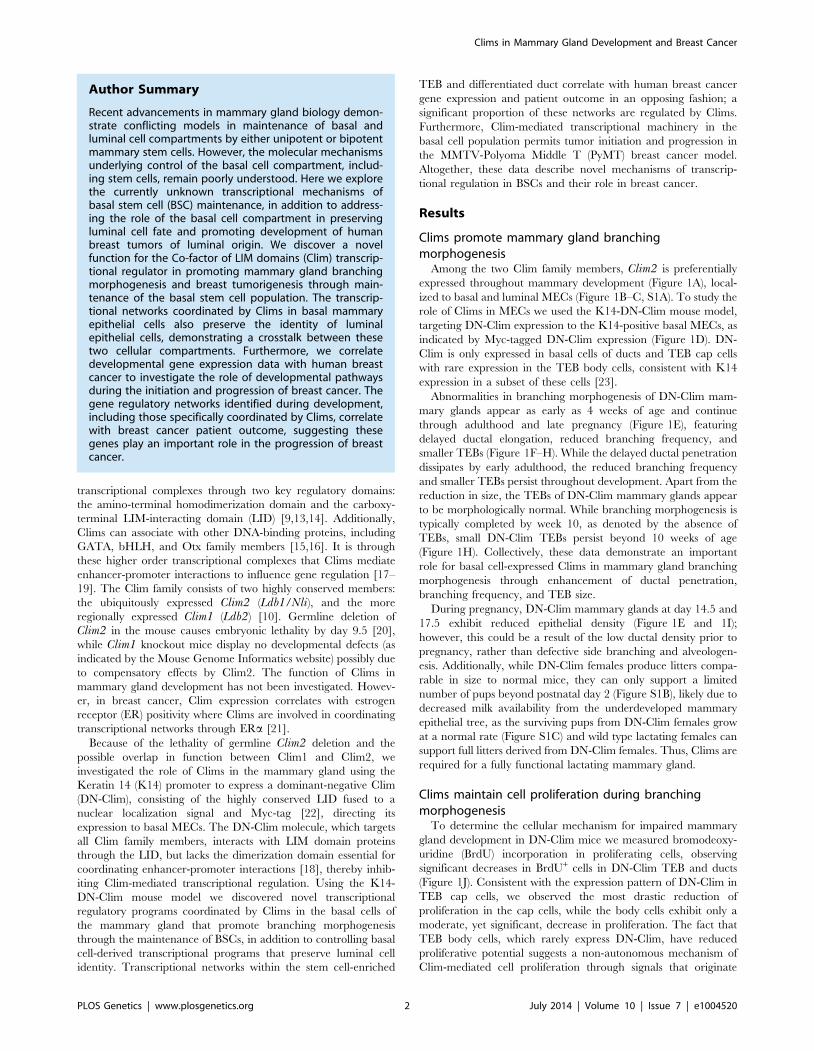
Among the two Clim family members, Clim2 is preferentially
expressed throughout mammary development (Figure 1A), local-
ized to basal and luminal MECs (Figure 1B–C, S1A). To study the
role of Clims in MECs we used the K14-DN-Clim mouse model,
targeting DN-Clim expression to the K14-positive basal MECs, as
indicated by Myc-tagged DN-Clim expression (Figure 1D). DN-
Clim is only expressed in basal cells of ducts and TEB cap cells
with rare expression in the TEB body cells, consistent with K14
expression in a subset of these cells [23].
Abnormalities in branching morphogenesis of DN-Clim mam-
mary glands appear as early as 4 weeks of age and continue
through adulthood and late pregnancy (Figure 1E), featuring
delayed ductal elongation, reduced branching frequency, and
smaller TEBs (Figure 1F–H). While the delayed ductal penetration
dissipates by early adulthood, the reduced branching frequency
and smaller TEBs persist throughout development. Apart from the
reduction in size, the TEBs of DN-Clim mammary glands appear
to be morphologically normal. While branching morphogenesis is
typically completed by week 10, as denoted by the absence of
TEBs, small DN-Clim TEBs persist beyond 10 weeks of age
(Figure 1H). Collectively, these data demonstrate an important
role for basal cell-expressed Clims in mammary gland branching
morphogenesis through enhancement of ductal penetration,
branching frequency, and TEB size.
During pregnancy, DN-Clim mammary glands at day 14.5 and
17.5 exhibit reduced epithelial density (Figure 1E and 1I);
however, this could be a result of the low ductal density prior to
pregnancy, rather than defective side branching and alveologen-
esis. Additionally, while DN-Clim females produce litters compa-
rable in size to normal mice, they can only support a limited
number of pups beyond postnatal day 2 (Figure S1B), likely due to
decreased milk availability from the underdeveloped mammary
epithelial tree, as the surviving pups from DN-Clim females grow
at a normal rate (Figure S1C) and wild type lactating females can
support full litters derived from DN-Clim females. Thus, Clims are
required for a fully functional lactating mammary gland.
Clims maintain cell proliferation during branchingmorphogenesis
To determine the cellular mechanism for impaired mammary
gland development in DN-Clim mice we measured bromodeoxy-
uridine (BrdU) incorporation in proliferating cells, observing
significant decreases in BrdU+ cells in DN-Clim TEB and ducts
(Figure 1J). Consistent with the expression pattern of DN-Clim in
TEB cap cells, we observed the most drastic reduction of
proliferation in the cap cells, while the body cells exhibit only a
moderate, yet significant, decrease in proliferation. The fact that
TEB body cells, which rarely express DN-Clim, have reduced
proliferative potential suggests a non-autonomous mechanism of
Clim-mediated cell proliferation through signals that originate
Author Summary
Recent advancements in mammary gland biology demon-strate conflicting models in maintenance of basal andluminal cell compartments by either unipotent or bipotentmammary stem cells. However, the molecular mechanismsunderlying control of the basal cell compartment, includ-ing stem cells, remain poorly understood. Here we explorethe currently unknown transcriptional mechanisms ofbasal stem cell (BSC) maintenance, in addition to address-ing the role of the basal cell compartment in preservingluminal cell fate and promoting development of humanbreast tumors of luminal origin. We discover a novelfunction for the Co-factor of LIM domains (Clim) transcrip-tional regulator in promoting mammary gland branchingmorphogenesis and breast tumorigenesis through main-tenance of the basal stem cell population. The transcrip-tional networks coordinated by Clims in basal mammaryepithelial cells also preserve the identity of luminalepithelial cells, demonstrating a crosstalk between thesetwo cellular compartments. Furthermore, we correlatedevelopmental gene expression data with human breastcancer to investigate the role of developmental pathwaysduring the initiation and progression of breast cancer. Thegene regulatory networks identified during development,including those specifically coordinated by Clims, correlatewith breast cancer patient outcome, suggesting thesegenes play an important role in the progression of breastcancer.
Clims in Mammary Gland Development and Breast Cancer
PLOS Genetics | www.plosgenetics.org 2 July 2014 | Volume 10 | Issue 7 | e1004520
Clims in Mammary Gland Development and Breast Cancer
PLOS Genetics | www.plosgenetics.org 3 July 2014 | Volume 10 | Issue 7 | e1004520

from the cap cells. Thus, Clims promote proliferation in both the
TEB and duct during branching morphogenesis.
Gene sets differentially expressed in the TEB and ductrepresent distinct developmental functions duringbranching morphogenesis and are differentiallyexpressed in breast cancer subtypes
The current knowledge of the molecular mechanisms that
regulate branching morphogenesis is limited, especially regarding
the transcriptional programs within the stem cell-enriched TEB
and differentiated duct structures. To elucidate these transcrip-
tional programs, and specifically those controlled by Clims, we
profiled gene expression in TEB and duct cells from wild type
(WT) and DN-Clim mice, using laser capture microdissection to
isolate these structures from four, six, eight, and ten week old mice.
Consistent with its preferential expression during branching
morphogenesis, Clim2 was expressed higher than Clim1 in both
the TEB and duct cells (Figure S2A).
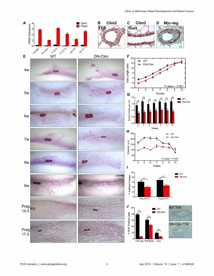
To define developmentally regulated genes over the time course
we applied the Bayesian Estimation of Temporal Regulation
(BETR) algorithm [24] identifying 8,030 and 6,488 probe sets
(7,318 and 5,870 genes) temporally regulated in TEB and duct
cells, respectively, with 1,313 TEB and 815 duct genes changing
1.5-fold or greater in two or more time points (Figure 2A–C,
Dataset S1 and S2). TEB genes are associated with proliferation,
while the duct genes are associated with differentiation (Fig-
ure 2D–E). Because the TEB represents a stem cell-enriched
population [25], and because of our direct comparison of TEB and
duct cells, these gene signatures are representative of mammary
stem cell and differentiated MECs, respectively.
The hyperproliferative and invasive nature of the TEB bears
similarity to molecular properties observed in aggressive breast
and other adenocarcinomas. In addition, poorly differentiated
basal-like tumors are more aggressive and possess stem cell-like
transcriptional characteristics compared to the differentiated
luminal subtypes [26]. This prompted us to determine whether
the less differentiated TEB and fully differentiated duct gene
signatures can be used as predictors of prognosis in human breast
cancer. Utilizing three data sets that profile the transcriptome and
classify the intrinsic subtypes of primary human breast tumors
[27–29] we found the TEB signature is highly expressed in the
poorly differentiated claudin-low and basal-like breast cancer
subtypes, while the duct signature is more highly expressed in the
differentiated luminal subtypes (Figure 2F). High TEB signature
expression predicted poor overall survival in breast cancer patients
(Figure 2G). An opposite trend was observed with the duct
signature (Figure 2H), suggesting the expression of differentiation
genes suppresses tumorigenesis. These trends are also observed
within the different subtypes of breast cancer (Figure 2I). Even
after removal of proliferation genes, which significantly contribute
to prognosis prediction [30], the TEB signature retains prognostic
relevance, albeit with reduced power (Figure 2G). The duct
signature, which is poorly enriched for proliferation genes, was
relatively unaffected by removal of these genes (Figure 2H). Thus,
these developmental signatures hold prognostic value in breast
cancer and may be useful to identify novel genes of interest for
understanding the biology of breast cancer in relationship to
normal development.
Clims regulate developmentally associated TEB and ductgenes
To determine genes regulated by Clims during branching
morphogenesis we used the CyberT algorithm [31] to define
differentially expressed genes (DEGs) in the DN-Clim TEB and
duct: 66 TEB and 137 duct genes were significantly differentially
expressed in at least two time points (Figure 3A–B, Dataset S3 and
S4), a sizable proportion of which are differentially expressed in
both TEB and duct cells (Figure S2B). Each DEG set is
significantly enriched with genes from their respective develop-
mental signature (Figure S2C–D), indicating an important role for
Clims in regulating branching morphogenesis. Using the Molec-
ular Signature Database [32] to characterize the biological
properties of the Clim-regulated TEB and duct gene sets revealed
that both have functions in mammary stem cells, pubertal
mammary gland development, several breast cancer subtypes,
and Wnt-signaling in the mammary gland (Figure 3C–D).
Additionally, TEB DEGs participate in metallopeptidase activity
and possess serum response factor (SRF) and androgen receptor
(AR) transcription factor binding motifs, while duct DEGs are
enriched with smooth muscle and mammary basal epithelial cell
genes, participate in FGF signaling, and possess TCF3 and SOX9
transcription factor binding motifs. Furthermore, high expression
of Clim-regulated genes in breast cancer is an indicator of poor
prognosis (Figure 3E). Collectively, these data support the
involvement of Clims in gene expression networks important for
both stem cell function and breast cancer.
To determine genes differentially expressed in basal and luminal
cell compartments we sorted MECs into Lin2CD29HiCD24+ and
Lin2CD29LuCD24+ populations, respectively (Figure S3A–D).
Basal and luminal cell purity from WT and DN-Clim MECs was
confirmed by qPCR for K8, K14, and the K14-DN-Climtransgene expression (Figure S3E–G); as expected, the K14-DN-Clim transgene is only expressed in basal cells, and not luminal
cells. Basal markers (K5, K14, and smooth muscle actin) and
luminal markers (K8, K18, Gata3, estrogen and progesterone
receptors) are significantly overexpressed (CyberT, p-value,
0.001) in their respective cell types, indicating effective basal and
luminal cell separation (Dataset S5). DN-Clim basal and luminal
Figure 1. Clims promote branching morphogenesis and proliferation. (A) Clim2 mRNA is predominantly expressed over Clim1 throughoutmammary gland development. Gene expression is normalized to 18s mRNA. (B–C) IHC showing expression of Clim2 in the (B) TEB and (C) duct inmammary glands from 6 week old mice. Clim2 expression is observed in basal and luminal cells, with slightly higher expression in basal cells. (D) IHCdetecting the Myc-tag of DN-Clim demonstrates expression in the K14-positive basal layer of mammary glands from 6 week old mice. (E) Wholemount analysis of mammary glands throughout branching morphogenesis and pregnancy demonstrates defective branching morphogenesis andTEB maintenance in DN-Clim mammary glands. Images are representative of at least three mice per time point. (F) Mean ductal penetration of thethree longest ducts throughout branching morphogenesis indicates a delay in DN-Clim ductal penetration. Data are normalized using the center ofthe lymph node as the central reference point. P-value derived from repeated measures ANOVA. (G) Mean number of branch points for the threelongest ducts normalized to the length of the duct (mm) indicates reduced branching frequency in DN-Clim mammary glands. ** P-value,0.01determined by Student’s t-test. (H) TEB diameter (mm) is reduced in DN-Clim mammary glands throughout branching morphogenesis. P-value derivedfrom repeated measures ANOVA. (I) Proportion of pregnancy stage mammary fat pad filled with epithelial tissue demonstrates a reduced epithelialdensity in DN-Clim mammary glands. ** p-value,0.01 determined by Student’s t-test. (J) Proportion of BrdU positive cells in TEB (cap and body cells)and duct cells is reduced in DN-Clim mammary glands. Representative images of the TEB are shown. *** p-value,0.01 determined by Student’s t-test.Quantification for A, F-J represent mean 6 SEM for at least three mice.doi:10.1371/journal.pgen.1004520.g001
Clims in Mammary Gland Development and Breast Cancer
PLOS Genetics | www.plosgenetics.org 4 July 2014 | Volume 10 | Issue 7 | e1004520
cells differentially express 422 (221 up and 201 down) and 227
(139 up and 88 down) genes with at least 1.5-fold change,
respectively (Dataset S6 and S7), indicating both cell and non-cell
autonomous mechanisms of gene regulation coordinated by Clims;
a significant proportion of basal- and luminal-specific DEGs
overlap with the DEGs identified in the TEB and duct populations
(Figure S3H–I). Basal-specific DEGs participate in mammary
gland morphogenesis processes, including cell proliferation,
adhesion, and stem cell properties (Figure 3F and S3J). Interest-
ingly, DEGs in the luminal cell population are enriched with
keratinization and squamous epithelial, specifically esophageal and
epidermal, differentiation genes (Figure 3G and S3K), suggesting a
transepidermal differentiation of the luminal cells in the DN-Clim
mammary gland. Thus, Clims coordinate gene expression in the
basal cell compartment that ultimately help maintain the identity
of luminal cells.
Several genes and pathways related to mammary gland stem
cell biology are differentially expressed in the DN-Clim mammary
gland throughout the developmental time course and within the
basal and luminal cell compartments (Figure 3C–D, and S3J–K).
Ontology analyses revealed involvement of Clim-regulated genes
in mammary stem cells and the Wnt signaling pathway. Lgr5, a
marker of stem cells in CD29HiCD24+ basal cells [33], is
consistently downregulated in basal cells and both the TEB and
duct (Dataset S3, S4, and S6). Additional Clim-regulated genes
that have known roles in maintenance of the stem cell enriched
TEB and branching morphogenesis are the Fgfr2 gene [34,35]
and the luminal cell-specific ErbB2 and ErbB3 genes (Figure 3G
Figure 2. Developmental TEB and duct gene signatures derived from time course expression profiling predict breast cancerprognosis. (A) Identification of developmentally regulated genes with the BETR algorithm in TEB and duct cells over the time course of branchingmorphogenesis (4, 6, 8, and 10 weeks). TEB and duct signatures were further refined to by selecting genes with at least 1.5-fold expression change inat least two time points for each cell type. (B–C) Heat map of resulting (B) 1,313 TEB and (C) 815 duct gene signatures expressed at least 1.5-fold orgreater in their respective cell type in at least two time points. (D–E) Biological processes associated with (D) TEB and (E) duct gene signatures. TheTEB and duct signatures represent a proliferation and differentiation signature, respectively. Vertical line represents p-value at 0.05. (F) TEB and Ductsignatures are differentially expressed in the molecular breast cancer subtypes. The TEB and duct signatures are more highly expressed in the poorly-and well-differentiated breast cancer subtypes, respectively. (G–H) Analysis of overall survival (OS) in NKI 295 cohort of patients classified into (G) TEBand (H) duct high and low expression groups based on median expression the respective gene signature, including the effects of removingproliferation genes (-Prol). High expression of the proliferative TEB signature confers poor prognosis, while high expression of the differentiated ductsignature confers improved prognosis. P-value derived from Log-rank test. (I) Hazard ratios (HR) derived from overall survival analysis for TEB and ductsignatures in the molecular subtypes of breast cancer in three separate data sets demonstrate similar trends observed in G–H. Red samples representTEB signature and blue samples represent duct. Error bars represent 95% confidence interval; filled circles represent p-value,0.1, squares representtrends with p-value,0.25, and triangles represent non-significant changes with p-value.0.25.doi:10.1371/journal.pgen.1004520.g002
Clims in Mammary Gland Development and Breast Cancer
PLOS Genetics | www.plosgenetics.org 5 July 2014 | Volume 10 | Issue 7 | e1004520

and S4) [36,37]. Given this data, we hypothesized that Clims
promote branching morphogenesis through gene regulation within
stem cells of the basal MEC compartment.
Clims maintain the basal mammary epithelial stem cellpopulation
The reduced size of the stem cell-enriched TEB in DN-Clim
mice and the enrichment of mammary gland stem cell genes in our
DEG sets, along with previous evidence that Clim2 affects stem
cells in the hair follicle [22], prompted us to examine the stem cell
populations in the DN-Clim mammary gland. Quantification of
the basal and luminal MEC populations revealed a reduction of
the CD29HiCD24+ BSC-enriched population in the DN-Clim
mammary gland (Figure 4A), while the proportion of the
CD29LuCD24+ LSC-enriched population remains unchanged.
No differences were observed in the luminal progenitor cell
population when using CD61 as a marker for these cells (Figure
S5A) [38]. Limiting dilution transplant analysis with
CD29HiCD24+ cells revealed a nearly complete ablation of DN-
Clim mammary repopulating units (MRU) (Table 1, Figure 4B);
only two DN-Clim transplants resulted in mammary outgrowths,
both of which developed poorly structured mammary trees (Figure
S5B). Thus, Clims are of paramount importance in the
maintenance of BSCs.
Mammary colony forming cell (MaCFC) and mammo-
sphere functional assays confirmed the depleted basal stem/
progenitor cell population. Unsorted Lin2 bulk and sorted
CD29HiCD24+Lin2 DN-Clim primary epithelial cells have fewer
MaCFCs and form smaller colonies, while a slight and insignif-
icant decrease in MaCFCs is observed in the sorted CD29LuC-
D24+Lin2 population (Figure 4C–D). Consistently, Lin2 bulk and
CD29HiCD24+Lin2 sorted DN-Clim primary mouse MECs
grown in suspension produced significantly fewer and smaller
mammospheres (Figure 4E–F), while CD29LuCD24+Lin2 cells
exhibit comparable sphere forming efficiency to WT cells. Serial
passaging of the mammospheres every seven days resulted in
enhanced depletion rates of Lin2 bulk and CD29HiCD24+Lin2
DN-Clim stem/progenitor cells. Collectively, these results suggest
Clims maintain the number and proliferative potential of the basal
stem/progenitor cell population.
To further confirm the requirement of Clims in promoting
stem-like features, Clim1 and Clim2 were knocked down by
siRNA, both independently and together, in the MCF10A and
MCF7 mammary epithelial cell lines (Figure S5C–D). Clim2
knockdown resulted in significantly reduced mammosphere
forming efficiency in both cell lines, while Clim1 knockdown
resulted in significant decreases in MCF10A cells, but not MCF7
cells (Figure 4G). Consistently, we observed a synergistic decrease
in mammosphere formation efficiency in the combined siClim1/2
in MCF10A cells, but not MCF7 cells. These cell type-specific
differences in promoting stem-like properties may be attributed to
higher levels of Clim1 in MCF10A cells than MCF7 cells.
Altogether these data further support the role of Clims in
promoting stem-like features of MECs.
Figure 3. Clims coordinate the expression of developmental gene signatures. (A–B) Heat map of (A) TEB (66 genes) and (B) duct (137genes) DEGs in the DN-Clim mammary gland with CyberT p-value,0.01 in at least two time points. (C–D) Categories of enriched gene sets from theMolecular Signatures Database that significantly overlap with the (C) TEB and (D) duct DEG sets. Both gene sets are enriched with mammary stem celland breast cancer genes. Vertical line represents p-value at 0.05. (E) Analysis of overall survival (OS) in UNC 337 cohort of patients classified into highand low expression groups based on median expression of the combined list of DEGs in the DN-Clim TEB and ducts demonstrates that highexpression of Clim-regulated genes confers poor prognosis. HR: Hazard Ratio with 95% confidence interval. P-value derived from Log-rank test. (F)Fold change of Wnt-signaling factors in DN-Clim sorted basal cells suggests altered Wnt signaling. Fgfr2 is also downregulated. (G) Increasedexpression of epidermal differentiation genes in DN-Clim luminal cells suggests a role for basal cells in maintaining luminal cell fate. Luminal cellsignaling factors ErbB2/3 and Fgfr2 are also downregulated.doi:10.1371/journal.pgen.1004520.g003
Clims in Mammary Gland Development and Breast Cancer
PLOS Genetics | www.plosgenetics.org 6 July 2014 | Volume 10 | Issue 7 | e1004520

Clim2 is known to interact with LMO4 in MECs; LMO4
promotes mammary gland morphogenesis and breast cancer [39–
42]. To determine whether Clim2 may be acting through LMO4
to maintain the mammary stem/progenitor cell population we
transiently knocked down LMO4 gene expression individually and
in conjunction with Clim2 in MCF10A and MCF7 cells (Figure
S5E). We found similar reduction in mammosphere forming
efficiency with individual and combined knockdowns (Figure 4G),
suggesting these two transcription factors may target similar
mechanisms to promote stem cell features, consistent with these
Figure 4. Clims maintain basal mammary epithelial stem cells. (A) Flow cytometry analysis of Lin2 MECs with CD29 and CD24 markers revealsdecreased CD29HiCD24+ basal epithelial cell population. (B) DN-Clim mammary glands are absent of MRUs as demonstrated in representative wholemounts from transplants of CD29HiCD24+ BSC-enriched population. See Table 1 for results from limiting dilution analysis. (C) DN-Clim basal cells, andnot luminal cells, maintain fewer colony forming units determined by colony-forming cell assays with Lin2 bulk and sorted primary mammaryepithelial cells. (D) Reduced growth rates in colonies that form from DN-Clim basal cells determined by quantification of colony size for thecorresponding MaCFC assays described in (C). (E) DN-Clim basal cells, and not luminal cells, maintain fewer mammosphere forming units determinedby mammosphere assays with Lin2 bulk and sorted primary mammary epithelial cells. (F) Reduced growth rates in spheres that form from DN-Climbasal cells determined by quantification of mammosphere diameter for the first passage of the mammosphere assays described in (E). (G) Clim1,Clim2 and the Clim interaction factor LMO4 are essential for maintaining stem-like features of MCF10A cells, as determined by mammosphere assayswith transient siRNA knockdown of Clim1, Clim2, and LMO4. In MCF7 cells, only Clim2 and LMO4 maintain stem-like features. Data represent mean 6SEM from at least three mice (C–F) or at least three experiments (G). ** p-value,0.01; *** p-value,0.001, ns: not significant.doi:10.1371/journal.pgen.1004520.g004
Clims in Mammary Gland Development and Breast Cancer
PLOS Genetics | www.plosgenetics.org 7 July 2014 | Volume 10 | Issue 7 | e1004520

two factors forming a tight complex [43–45]. Thus, Clim2 may
regulate gene expression in these cells through the organization of
transcriptional complexes involving LMO4.
Clim2 regulates Fgfr2 to maintain BSCsFgfr2 is significantly downregulated in the DN-Clim mammary
gland throughout the developmental time course (Figure 5A) as
validated by qPCR in the TEB and duct cells from 6 week old
mice (Figure 5B). Fgfr2 expression was also decreased in the
sorted basal and luminal cells from DN-Clim mice (Figure 5C).
While the decrease in basal cells is likely due to cell autonomous
effects of DN-Clim, the decreased Fgfr2 expression in luminal
cells is likely non-cell autonomous as the DN-Clim molecule is not
expressed in these cells. Since Fgfr2 is essential for maintaining the
BSC population [46], and DN-Clim is localized to the K14-
positive basal MECs, we hypothesized that Clims directly regulate
the expression of Fgfr2 to maintain BSCs. Indeed, the FGFR2
protein is decreased in DN-Clim basal cells (Figure 5D–E).
Transient knockdown of Fgfr2 in the Fgfr2-high MCF7 cell line
impairs mammosphere forming efficiency (Figure 5F), while stable
overexpression of Fgfr2 in the Fgfr2-low MCF10A cell line with
transient knockdown of Clims partially rescues mammosphere
forming efficiency (Figure 5G). These results suggest Clims
regulate Fgfr2 expression to maintain stem-like features.
To determine if Clims directly bind the Fgfr2 promoter, we
performed ChIP assays for Clim2, DN-Clim (Myc-tag), H3K4me3
(a marker for actively transcribed genes), and the Clim-interacting
partner LMO4, followed by qPCR targeting multiple sites in the
2 kb region surrounding the Fgfr2 transcriptional start site (TSS).
Clim2 binds to a region 538 bp to 708 bp upstream of the TSS in
the WT mammary gland, with enriched H3K4me3 surrounding
the TSS (Figure 6A), while DN-Clim binds the same region
upstream of the TSS in DN-Clim mammary glands, with a drastic
reduction in H3K4me3 enrichment (Figure 6B). Additionally,
LMO4 is enriched at the same region in WT mammary glands,
but is absent in the DN-Clim mammary gland, suggesting a
necessity for Clim2 to recruit LMO4 to the promoter. To
determine if Clim-binding activates transcription, we cloned the
promoter-binding region (433 to 1010 bp upstream of the Fgfr2TSS) into the pGL3-promoter vector. In Clim2-positive MCF10A
cells, this region of DNA enhances transcription of the luciferase
reporter, and DN-Clim inhibits its transcription (Figure 6C).
Furthermore, Fgfr2 expression is downregulated in MCF10A
and MCF7cells upon knockdown of Clim1/2 and LMO4
(Figure 6D). Altogether, these data suggest that Clim2 promotes
the maintenance of the BSC population by directly binding the
Fgfr2 promoter to drive transcription of the gene.
Clim2 promotes breast tumorigenesisThe CLIM protein is expressed in ER-positive breast tumors
[21], and we show here that the Clim-regulated gene network is
associated with poor prognosis in breast cancer (Figure 3E).
Furthermore, its interacting partner LMO4 is upregulated in
breast cancer and promotes breast tumorigenesis [40,42,47,48].
We examined the expression of Clims in the molecular subtypes of
breast cancer and observed highest expression of Clim2 in the less
differentiated claudin-low and basal-like breast cancer subtypes,
while Clim1 is most highly expressed in the differentiated luminal
cell types (Figure 7A–B). Accordingly, Clim2 expression alone
predicts poor prognosis in breast cancer, while Clim1 expression
displays an opposite trend (Figure S6). The combined expression
level of both Clim genes is a strong predictor of poor outcome
(Figure 7C), suggesting Clim2 is a more powerful predictor of
breast cancer outcome than Clim1.
To determine if Clims play a role in breast tumorigenesis, we
bred the K14-DN-Clim gene into the MMTV-PyMT mouse
model. PyMT mice develop palpable tumors in nearly every
mammary gland, whereas DN-Clim/PyMT mice only develop
tumors in no more than six mammary glands (Figure 7D).
Additionally, DN-Clim/PyMT mice exhibit a significant delay in
the development of palpable tumors (Figure 7E) and the growth
rate of these tumors is drastically impaired (Figure 7F). While
tumor development is rapid and frequent in PyMT mice, with
consecutive palpable tumors developing within days of each other,
there was a significant delay in the time required for the second
palpable DN-Clim/PyMT tumor to develop and the growth rate
of the second tumor was significantly slower than the first
(Figure 7E–F). Taken together, these data demonstrate that Clim
promotes the initiation and progression of breast cancer in the
PyMT model.
To determine the stage at which Clim may be promoting breast
cancer, we collected whole mounts of mammary glands from as
early as 3 weeks to as late as 3 months. Hyperplastic lesions appear
in the mammary glands of 3-week-old PyMT and DN-Clim/
PyMT mice (Figure 7G). However, by 7 weeks entire arms of the
mammary tree display hyperplastic lesions in PyMT mice, while
the DN-Clim/PyMT mice only exhibit sparse lesions (Figure 7G).
Consistently, after removal of the primary tumor from mammary
glands of 3 month old mice we observed a decrease in the area of
the epithelial tree covered in tumor tissue. The drastic difference in
Table 1. Clims maintain the basal mammary epithelial stem cell population.
CD29HiCD24+ Basal Cells MaSC Frequency
Wild Type DN-Clim
5000 2/2 1/2
1000 2/2 0/2
500 3/3 0/3
100 2/3 1/3
50 3/5 0/5
MRU Frequency (95% CI) 1/69 (1/28 to 1/172) 1/5566 (1/1288 to 1/24,055)
p-value (WT vs DN-Clim) p = 3.58e-09
p-value (single-hit Poisson model) p = 0.748 p = 0.222
doi:10.1371/journal.pgen.1004520.t001
Clims in Mammary Gland Development and Breast Cancer
PLOS Genetics | www.plosgenetics.org 8 July 2014 | Volume 10 | Issue 7 | e1004520

the number of hyperplastic nodules developing from the mam-
mary tree suggest that Clims act during the early stages of
tumorigenesis. IHC staining of 6 week old mammary glands
demonstrated that these tumors primarily express K8, and not
K14 (Figure 7H), confirming they are luminal tumors [49]. PyMT
tumors exhibit a rare population of K14-expressing cells at the
leading, basal edge of the tumor, which also express DN-Clim in
the DN-Clim/PyMT tumors (Figure 7H). These data demonstrate
Figure 5. Fgfr2 downregulation in the mammary gland leads to loss of stem/progenitor cell activity. (A) Fgfr2 is downregulated in TEBand duct cells throughout branching morphogenesis. The data are derived from the time course gene expression microarray analysis. (B) qPCRvalidation of decreased Fgfr2 expression in DN-Clim TEB and duct cells. Gene expression is normalized to 18s mRNA. (C) qPCR validation of decreasedFgfr2 expression in sorted basal and luminal cells. Gene expression is normalized to 18s mRNA. (D) Immunofluorescence analysis of FGFR2 proteinexpression reveals decreased protein in the basal layer of DN-Clim mammary glands. (E) Semi-quantitative analysis of FGFR2 protein expression in TEBand luminal structures suggests reduced protein levels in basal cells of the DN-Clim mammary gland. WT mammary glands exhibit more protein inbasal cells when compared to luminal cells, but DN-Clim mammary glands exhibit equal FGFR2 protein levels in basal and luminal cells. Thepercentage of TEB and duct structures that exhibit higher FGFR2 protein in basal cells was quantified by counting at least 10 structures from at least10 frozen sections from three WT and three DN-Clim mammary glands. The data represent the mean 6 SEM from three biological replicates. (F)Transient siRNA knockdown of Fgfr2 in MCF7 reduces sphere forming efficiency in mammosphere assays. (G) Fgfr2 overexpression partially rescuesreduced mammosphere forming efficiency upon transient siRNA knockdown of Clims in the MCF10A cell line. Data represent mean 6 SEM from atleast two littermate mice (A–C, E), or three replicate experiments (F–G).doi:10.1371/journal.pgen.1004520.g005
Clims in Mammary Gland Development and Breast Cancer
PLOS Genetics | www.plosgenetics.org 9 July 2014 | Volume 10 | Issue 7 | e1004520

the necessity of Clim-mediated, basal cell-specific gene expression
for robust PyMT-mediated tumorigenesis, and suggest a functional
role for Clim-dependent mechanisms in basal MECs during the
initiation and progression of luminal breast tumors.
Discussion
Transcriptional networks regulating the development and
maintenance of the mammary gland luminal cell compartment
are well characterized, with GATA3, Notch1, Elf5, and STAT5a
being key factors [38,50–52]. However, transcriptional regulation
within the basal cell compartment remains poorly understood.
Here, we describe a Clim-driven developmental transcriptional
network in the basal cell compartment that maintains the number
and proliferative potential of BSCs. Interfering with this
transcriptional network leads to defective branching morphogen-
esis and impaired PyMT-mediated tumorigenesis. In addition,
Clim-dependent basal cell-derived transcriptional programs sup-
port the expression of key luminal growth factor receptors and
maintain the identity of luminal cells (Figure 8).
The widely expressed Clims regulate unique transcriptional
programs in diverse cell types. In part, this likely depends on
Clim’s tissue specific interactions with LIM domains of distinct
LHX and LMO family members, such as LMO2 and LHX2 in
erythroid cells [12,53], LHX3 in the neural tube [54], LHX2 in
hair follicles [22,55,56], and LMO4 in the mammary gland [40].
Despite this diversity in interactions, Clims seem to play a general
role in promoting stem cell maintenance as previously shown in
intestinal and hair follicle epithelia [22,57], blood [58], and ES
cells [59]. How Clims maintain stem cell features is largely
unexplained. We address this question in the mouse mammary
gland with the K14-DN-Clim mouse model that targets both Clim
family members in the basal cell population. DN-Clim, effectively
disrupts Clim/LIM domain-mediated transcriptional complexes invivo, as has been demonstrated in Drosophila, Zebrafish, and
mouse [22,60]. Additionally, knockout of Lhx2, the Clim-
interacting partner in hair follicle stem cells, gives a similar
phenotype as expression of DN-Clim [22,55]. Lacking the
dimerization domain, DN-Clim binds LIM domain proteins to
inhibit their interactions with endogenous Clims, thereby
Figure 6. Clims and LMO4 directly target and the Fgfr2 promoter to induce gene expression. (A–B) ChIP-qPCR assays of Myc-tagged DN-Clim, Clim2, LMO4, and H3K4me3 enrichment at the indicated locations of the 62 kb surrounding the Fgfr2 promoter (top panel) in (A) WT and (B)DN-Clim MECs. MECs were collected from three littermate mice then pooled for ChIP-qPCR experiments. The data suggest Clim2 binds approximately0.5–1.0 kb upstream of the Fgfr2 TSS. When DN-Clim is bound to this region, LMO4 binding is lost and H3K4me3 levels are decreased. (C) Luciferaseassays from MCF10A cells transiently expressing the Clim-binding promoter region of Fgfr2 in the pGL3-promoter luciferase reporter vector indicateClim acts as an inducer of Fgfr2 expression. The DN-Clim expression vector was transiently titrated to demonstrate reduction of luciferase activity withincreasing DN-Clim expression. (D) Expression of Fgfr2 in the MCF10A and MCF7 cells is reduced upon transient siRNA knockdown of Clim1, Clim2,and LMO4. Expression is normalized to GAPDH. Data represent mean 6 SEM from three replicate experiments (C–D).doi:10.1371/journal.pgen.1004520.g006
Clims in Mammary Gland Development and Breast Cancer
PLOS Genetics | www.plosgenetics.org 10 July 2014 | Volume 10 | Issue 7 | e1004520

preventing Clim-mediated looping involved with higher order
transcriptional complexes. We still observed DN-Clim at the
promoter of the Fgfr2 gene, indicating that these interactions still
allow the binding of LIM domain transcription factors to their
target promoters. The N-terminal dimerization domain may have
other functions not inhibited by DN-Clim, including possible
interactions with other non-LIM domain transcriptional regulators
[15,16] and interactions with the RLIM ubiquitin ligase that leads
to degradation of associated LIM domain proteins [61]. DN-Clim
has been demonstrated to stabilize nuclear LIM domain proteins
Figure 7. Clims promote the initiation and progression of tumors in the MMTV-PyMT breast cancer mouse model. (A–B) Expression of(A) Clim2 and (B) Clim1 in the molecular subtypes of breast cancer. Clim2 is more highly expressed in less differentiated Claudin-low (CLow) andbasal-like (BasL) tumor subtypes and Clim1 is more highly expressed in the differentiated Luminal A and B (LumA and LumB) subtypes. (C) Survivalanalysis within the UNC 337 cohort of breast cancer patients grouped by median expression of Clim1 and Clim2 demonstrates worse prognosis inpatients with highest expression of Clim1/2. (D) DN-Clim prevents tumorigenesis in the PyMT tumor model as determined by quantification of thenumber of mammary glands with palpable tumors. Data represent mean 6 SEM from ten mice of each genotype. (E) DN-Clim expression in the PyMTtumor model significantly delays development of palpable tumors. In PyMT mice, the second palpable tumor is detected within a few days of the firsttumor, but when DN-Clim is expressed there is a significant increase in the time to detect the second palpable tumor. P-values derived from Log-ranktest. (F) Decreased rate of tumor progression in DN-Clim/PyMT mice. P-values derived from repeated measures ANOVA. (G) Representative wholemounts of PyMT tumors with or without DN-Clim suggest a function for Clim in the tumor initiation stages. The primary tumor was excised fromthree month old mice before whole mount of the mammary gland. (H) K8, K14, and Myc-tag (DN-Clim) expression in mammary glands from 6 weekold PyMT mice with or without DN-Clim. High K8 and low K14 expression demonstrate the luminal subtype features of PyMT tumors. DN-Climexpression is observed in basal cells of the tumor.doi:10.1371/journal.pgen.1004520.g007
Clims in Mammary Gland Development and Breast Cancer
PLOS Genetics | www.plosgenetics.org 11 July 2014 | Volume 10 | Issue 7 | e1004520

[62], which may result in increased levels of these proteins in the
basal cells of the mammary gland.
In the mammary gland, several lines of evidence argue that
Clims maintain basal stem cells by stimulating expression of Fgfr2.
Expression of a DN-Clim molecule in the mouse mammary gland
and knockdown of Clims in human MECs lead to downregulation
of Fgfr2. Also, Fgfr2 overexpression can rescue the impaired
mammosphere formation after Clim knockdown in MECs.
Furthermore, we demonstrate that Clims associate with and
regulate the Fgfr2 promoter in MECs. This model is consistent
with work showing that FGF signaling is essential to branching
morphogenesis in several epithelial organs, including the drosoph-
ila trachea and the mammalian mammary gland, lung, kidney,
and salivary gland [63]. FGF signaling promotes mammary
embryonic placode formation [64], and Fgfr2 is necessary to
maintain the TEB structure and promote primary branching
[34,35]. In addition, Fgfr2 is required for maintenance of the
CD29HiCD24+ basal cell population and accordingly the main-
tenance of regenerative BSCs within this population [46]. The
defects in branching morphogenesis, basal epithelial cell frequen-
cy, and MRU capacity observed in these Fgfr2 transgenic mouse
models closely resemble the DN-Clim mammary gland phenotype.
Thus, Clim acts immediately upstream of Fgfr2 signaling in the
basal cell population through direct regulation of the Fgfr2 gene
to maintain BSCs and promote branching morphogenesis.
Our data suggest that Clim associates with its known
mammary gland binding partner LMO4 to regulate the Fgfr2gene. We find that LMO4 binds to the same region of the
Fgfr2 promoter, and knockdown of LMO4 also leads to
decreased Fgfr2 gene expression. Furthermore, we find that
LMO4 knockdown leads to decreased mammosphere forma-
tion in MECs. That these proteins function as a complex is
supported by experiments showing that the combined Clim/
LMO4 knockdown does not lead to further decrease in
mammosphere formation over individual knockdowns. Fur-
thermore, functional studies of LMO4 in the mammary gland
demonstrate its role in promoting branching morphogenesis
and lobuloalveoar development by sustaining cell proliferation
[39,41]. Altogether, these observations suggest Clims act
through LMO4 in the mammary gland to promote BSC
maintenance and development.
In addition to regulating the number and replicative potential of
BSCs, we find evidence that Clims regulate a basal cell program
that maintains luminal cell fate. Since one of the models of stem
cell hierarchy in the mammary gland proposes that BSCs give rise
to the luminal cells [4], this program may be established cell
autonomously in the basal progenitors and persists in the luminal
cells. Alternatively, the Clim-controlled program may act through
basal-to-luminal cell signaling; there is previous evidence for
crosstalk between the basal and luminal compartments through
paracrine and cell adhesion-mediated mechanisms [1,2]. Irrespec-
tive of the underlying mechanisms, since DN-Clim is selectively
expressed in the basal cell compartment, our gene expression data
in basal and luminal cell compartments clearly reveals an effect of
basal-specific Clim function on gene expression in luminal cells. In
particular, DN-Clim mice exhibit downregulation of key growth
factor receptors ErbB2 and ErbB3, and upregulation of several
epidermal differentiation genes. These observations demonstrate
an essential role for Clims in the basal cell compartment to
coordinate gene expression programs in the luminal cell compart-
ment, and for the first time demonstrate the importance of basal
cell factors on regulating the expression of key luminal regulators
in the EGFR family. These mechanisms may contribute to the
abnormal mammary development and decreased breast tumori-
genesis in the DN-Clim/PyMT mice.
The role of Clims is not limited to mammary gland
development as our work also suggests they act within basal cells
during the early stages of tumorigenesis. Expression of DN-Clim
within the basal cell compartment significantly impairs the ability
of PyMT to initiate neoplastic lesions. In the murine MMTV-
PyMT tumor model Fgfr2 is overexpressed in the CD29HiCD24+
tumor initiating cell population and is necessary to initiate
tumorigenesis in these mice [65]. As Clims are direct regulators
of Fgfr2 in the mammary gland, the reduced tumorigenicity
observed in DN-Clim/PyMT mice could in part be explained by
Clim-mediated regulation of Fgfr2 in cancer stem-like cells. The
MMTV promoter directs PyMT expression to both luminal and
basal cells [66], and while the histology and gene expression
patterns group PyMT breast tumors with the luminal subtype of
breast cancer, their cell of origin remains unknown [67]. Clims
could be promoting early tumorigenesis through one of two
scenarios: 1) Clims specifically maintain a basal cell-derived tumor
Figure 8. Regulatory roles for Clims in normal development and cancer. Clims promote branching morphogenesis and carcinogenesis inthe mammary gland (left). Direct regulation of Fgfr2 expression by Clims and LMO4 in the basal cell compartment facilitate basal stem cell self-renewal (right). Clims mediate a transcriptional program within the basal cell compartment to promote expression of Fgfr2, ErbB2, and ErbB3 in theluminal cell compartment, in addition to maintaining luminal epithelial cell fate by restricting the expression of epidermal differentiation genes.doi:10.1371/journal.pgen.1004520.g008
Clims in Mammary Gland Development and Breast Cancer
PLOS Genetics | www.plosgenetics.org 12 July 2014 | Volume 10 | Issue 7 | e1004520
initiating cell, or 2) Clims coordinate the communication of basal-
derived tumorigenic signals to the luminal cell compartment, such
as promoting the expression of Her2 in luminal cells. These signals
would have to occur during the early stages of tumorigenesis when
K14-positive basal cells are still present in premalignant lesions
[68]. While basal cells have previously been assumed to act as
inhibitors of breast cancer progression [69], the restricted
tumorigenicity observed in the DN-Clim/PyMT mammary gland
suggests basal cells may permit tumorigenesis.
Corresponding to these functional mouse experiments, we
observed differential expression of Clims in the molecular subtypes
of breast cancer, with high expression leading to poor outcome. It
is well known that Clims are expressed in luminal cells and
coordinate transcriptional programs through ERa in human
breast cancer [21]. However, our K14-DN-Clim mouse model
suggests an additional function for Clims during breast tumori-
genesis in the ER-negative basal cell population, either through
direct regulation of the basal cell or through indirect effects on the
luminal cell. Furthermore, we find that a Clim-regulated gene
module correlates with poor breast cancer prognosis arguing, for
relevance of the mouse experiments for human breast cancer. It is
possible that Clims may in part promote breast cancer through
activation of Fgfr2 expression. Deregulation of FGF signaling is
observed in breast cancer [70], with FGFR2 amplification and
overexpression observed in 5–10% of human breast cancers,
correlating with poor prognosis [71,72]. In addition, single point
mutations in the FGFR2 gene are associated with increased risk in
human breast cancer [73].
In conclusion, we have demonstrated that Clims are a
necessary factor in mammary gland branching morphogenesis,
maintaining the BSC compartment. Clim2 coordinates with
LMO4 to govern these processes through direct regulation of the
Fgfr2 gene. In a breast cancer mouse model Clims act within the
basal cells to permit tumorigenesis, possibly through the
maintenance of cancer stem-like cells. As Clims are expressed
in human breast cancer and correlate with poor differentiation of
ER-positive tumors, elucidating Clim targets at a global scale
may give insight into the transcriptional mechanisms that
maintain primitive cancer cells. The developmental transcrip-
tome of the TEB and duct populations reported will provide a
valuable resource for delineating the molecular relationships
between development and tumorigenesis, in addition to identi-
fying novel prognostic and therapeutic targets.
Materials and Methods
Mouse strainsGeneration and maintenance of K14-DN-Clim mice were as
previously described [22]. MMTV-PyMT mice (Jackson Labs)
were maintained on the K14-DN-Clim mixed background. Fox
Chase SCID Beige mice (Charles River) were used as recipients for
transplant experiments. All experiments conform to the regulatory
guidelines approved by the International Animal Care and Use
Committee of the University of California, Irvine.
Tissue and cell preparationMammary glands 4 and 9 were dissected and either whole
mounted, paraffin embedded, or frozen in O.C.T. For prolifer-
ation assays, mice were injected with BrdU (50 mg/g, Sigma-
Aldrich) 4 hours prior to sacrificing. Adult (8 to 12 week)
mammary glands for single-cell suspensions were generated
according to Stem Cell Technologies protocol. When collecting
RNA, the collagenase/hyaluronidase digestion was reduced to
1.5 hours.
Flow cytometry, sorting, and in vitro cultureSingle-cell suspensions were incubated with Propidium
Iodide (2 mg/mL, Sigma-Aldrich), CD31-APC, CD45-APC,
TER119-APC, CD24-PE (BD Biosciences) and analyzed by flow
cytometry on FACSCalibur (BD Biosciences) or sorted on
FACSAriaII (BD Biosciences). Bulk primary MECs for in vitro
culture were incubated with biotinylated CD31/CD45/TER119
cocktail (Stem Cell Technologies) and magnetically separated to
remove lineage cells.
Cleared fat pad transplantationFACS-sorted CD29HiCD24+ cells were transplanted into
cleared fat pads of 3 week old immunocompromised SCID Beige
recipients. Mammary glands were evaluated 8 weeks after
transplantation. Limiting dilution analysis was performed on the
ELDA Web-based tool (http://bioinf.wehi.edu.au/software/
elda/).
Microarray and bioinformatic analysisRNA was collected from laser capture microdissected (Leica LS-
AMD) or FACS sorted MECs with the RNeasy Mini Kit (Qiagen).
Gene expression was measured with Affymetrix Mouse Gene
1.0ST array. Data were analyzed with PLIER algorithm.
Differential expression was determined with the CyberT algorithm
[31] and time course developmental gene expression analysis was
performed with the BETR algorithm [24]. Detailed methods of
data analysis are described in the supplemental methods accom-
panying this manuscript.
Accession numbersThe gene expression microarray data has been submitted to
GEO (http://www.ncbi.nlm.nih.gov/geo/) under the accession
number GSE57724.
Additional details for the above methods are presented in Text
S1, as well as methods for IHC, immunofluorescence, whole
mount, ChIP, RT-qPCR, mammosphere and colony forming
assays, siRNA transfection, lentiviral expression, luciferase assays
and microarray analysis.
Supporting Information
Dataset S1 List of TEB signature genes. BETR p-values and fold
change at each time point is presented for each TEB signature gene.
Signature genes have BETR p-value,0.001 and 61.5 fold change.
(XLSX)
Dataset S2 List of Duct signature genes. BETR p-values and
fold change at each time point is presented for each Duct signature
gene. Signature genes have BETR p-value,0.001 and 61.5 fold
change.
(XLSX)
Dataset S3 List of DEGs in the DN-Clim TEB. CyberT p-
values and fold change at each time point for the TEB are
presented. DEGs have p-value,0.01 and 61.5 fold change in at
least two time points. NA: gene did not meet expression criteria in
the specified time point.
(XLSX)
Dataset S4 List of DEGs in the DN-Clim Duct. CyberT p-
values and fold change at each time point for the Duct are
presented. DEGs have p-value,0.01 and 61.5 fold change in at
least two time points. NA: gene did not meet expression criteria in
the specified time point.
(XLSX)
Clims in Mammary Gland Development and Breast Cancer
PLOS Genetics | www.plosgenetics.org 13 July 2014 | Volume 10 | Issue 7 | e1004520

Dataset S5 List of DEGs in the WT Basal vs Luminal cell
comparison. CyberT p-values and fold change are presented.
DEGs have p-value,0.001 and fold change 61.5.
(XLSX)
Dataset S6 List of DEGs in the DN-Clim basal cell population.
CyberT p-values and fold change are presented. DEGs have p-
value,0.001 and fold change 61.5.
(XLSX)
Dataset S7 List of DEGs in the DN-Clim luminal cell
population. CyberT p-values and fold change are presented.
DEGs have p-value,0.001 and fold change 61.5.
(XLSX)
Figure S1 Specificity of Clim2 antibody and DN-Clim females
fail to support full litters. A) The Clim2 antibody specifically
targets the Clim2 protein with no reactivity to Clim1, as
determined by western blot on protein lysates from HEK293 cells
overexpressing the Clim1 and Clim2 proteins. The Clim1/2
antibody detects Clim1 and Clim2 only in their respective
overexpression lysates. Vector Ctrl Lysate = Vector transfected
lysate control. (B) Average number of pups per litter from WT and
DN-Clim females. DN-Clim mice are unable to support the full
litter after postnatal day 2. (C) Growth rate of pups from WT and
DN-Clim females. Surviving pups from DN-Clim females grow at
a normal rate compared to pups from the WT mother.
(PDF)
Figure S2 Time course analysis of Clim expression and
comparison of Clim-regulated genes to TEB and duct genes. (A)
Expression of Clim1 and Clim2 from time course analysis of TEB
and duct cells. (B) Significant overlap of differentially expressed
genes from the DN-Clim TEB and duct. (C–D) DEGs in the DN-
Clim (C) TEB and (D) duct are significantly enriched in their
respective developmental gene set.
(PDF)
Figure S3 Gene expression profiling in sorted basal and luminal
mammary epithelial cells. (A) Selection of live (PI-negative), Lin2
(TER119-, CD45-, and CD31-negative) single cells. (B) Gating for
basal (Lin2CD29HiCD24+) and luminal (Lin2CD29LuCD24+)
MECs. (C) Post-sort analysis of basal MECs. (D) Post-sort analysis
of luminal MECs. APC: Lin markers, PE: CD24, FITC: CD29. (E–
F) qPCR validation of (E) Krt14 and (F) Krt8 in sorted cells indicates
pure basal and luminal cell populations. (G) qPCR validation of
DN-Clim transgene expression confirms expression of DN-Clim in
basal cells. (H) DN-Clim basal and (I) DN-Clim luminal DEGs are
significantly enriched in the combined list of DN-Clim TEB and
Duct DEGs. Ontology analysis of (J) DN-Clim basal DEGs and (K)
DN-Clim luminal DEGs. The categories represent top hits from
DAVID and the Molecular Signatures Database.
(PDF)
Figure S4 Reduced expression of ErbB2 and ErbB3 receptor
tyrosine kinases in the DN-Clim mammary gland. Expression of
the (A) ErbB2 and (B) ErbB3 in the time course microarray (left
panel), as determined by qPCR in 6 week old laser capture
microdissected TEB and duct cells (middle panel), or in 8 week old
sorted basal (Bas) and luminal (Lum) cells (right panel). Each are
significantly downregulated in the TEB and duct cells. Their
expression is restricted to the luminal cell compartment, and their
downregulation in DN-Clim luminal cells suggests non-autono-
mous regulation of these genes by Clims through the basal cell
population. Data represent mean 6 SEM from at least two
littermate mice. * p-value,0.05, ** p-value,0.01, *** p-value,
0.001, ns: not significant.
(PDF)
Figure S5 Luminal progenitor cell analysis, representative whole
mounts from DN-Clim transplants and validation of gene
knockdown by siRNA. (A) CD61 was used as a marker for
luminal progenitor cells in the Lin-CD29luCD24+ population. No
differences were observed in the quantity of these cells in the DN-
Clim mammary gland. (B) Whole mounts of the two successful
mammary transplants of DN-Clim CD29HiCD24+ cells. Both
mammary glands exhibit defects in ductal penetration and
branching morphogenesis. Inset from the fat pad transplanted
with 100 DN-Clim cells shows the epithelial outgrowth indicated
by the arrow. (C–E) Expression of Clim1 (C), Clim2 (D), and
LMO4 (E) validates specific transient knockdown of mRNA for
each respective gene.
(PDF)
Figure S6 Contribution of Clim expression to prognosis
prediction. Survival analysis based on expression of (A) Clim1 or
(B) Clim2. Patients were divided into high and low expressing
groups based on median expression of each gene. P-values derived
from the Log-rank test.
(PDF)
Text S1 Supplemental materials and methods.
(DOCX)
Acknowledgments
We thank the UCI Genomics High Throughput Facility (GHTF) and the
UCI Stem Cell Core Facility for expert service.
Author Contributions
Conceived and designed the experiments: MLS BA. Performed the
experiments: MLS ZY KW EC PSu. Analyzed the data: MLS ZY KW.
Contributed reagents/materials/analysis tools: MLS BA XD PSm.
Contributed to the writing of the manuscript: MLS BA.
References
1. Moumen M, Chiche A, Cagnet S, Petit V, Raymond K, et al. (2011) Themammary myoepithelial cell. Int J Dev Biol 55: 763–771.
2. Forster N, Saladi SV, van Bragt M, Sfondouris ME, Jones FE, et al. (2014) BasalCell Signaling by p63 Controls Luminal Progenitor Function and Lactation via
NRG1. Dev Cell 28: 147–160.
3. Van Keymeulen A, Rocha AS, Ousset M, Beck B, Bouvencourt G, et al. (2011)Distinct stem cells contribute to mammary gland development and maintenance.
Nature 479: 189–193.
4. Rios AC, Fu NY, Lindeman GJ, Visvader JE (2014) In situ identification ofbipotent stem cells in the mammary gland. Nature 506: 322–327.
5. Shackleton M, Vaillant F, Simpson KJ, Stingl J, Smyth GK, et al. (2006)
Generation of a functional mammary gland from a single stem cell. Nature 439:84–88.
6. Stingl J, Eirew P, Ricketson I, Shackleton M, Vaillant F, et al. (2006) Purification
and unique properties of mammary epithelial stem cells. Nature 439: 993–997.
7. Siegel PM, Muller WJ (2010) Transcription factor regulatory networks in
mammary epithelial development and tumorigenesis. Oncogene 29: 2753–2759.
8. Zheng Q, Zhao Y (2007) The diverse biofunctions of LIM domain proteins:determined by subcellular localization and protein-protein interaction. Biol Cell
99: 489–502.
9. Agulnick AD, Taira M, Breen JJ, Tanaka T, Dawid IB, et al. (1996) Interactionsof the LIM-domain-binding factor Ldb1 with LIM homeodomain proteins.
Nature 384: 270–272.
10. Bach I, Carriere C, Ostendorff HP, Andersen B, Rosenfeld MG (1997) A familyof LIM domain-associated cofactors confer transcriptional synergism between
LIM and Otx homeodomain proteins. Genes Dev 11: 1370–1380.
11. Jurata LW, Kenny DA, Gill GN (1996) Nuclear LIM interactor, a rhombotin
and LIM homeodomain interacting protein, is expressed early in neuronal
development. Proc Natl Acad Sci U S A 93: 11693–11698.
Clims in Mammary Gland Development and Breast Cancer
PLOS Genetics | www.plosgenetics.org 14 July 2014 | Volume 10 | Issue 7 | e1004520

12. Visvader JE, Mao X, Fujiwara Y, Hahm K, Orkin SH (1997) The LIM-domain
binding protein Ldb1 and its partner LMO2 act as negative regulators of
erythroid differentiation. Proc Natl Acad Sci U S A 94: 13707–13712.
13. Jurata LW, Gill GN (1997) Functional analysis of the nuclear LIM domain
interactor NLI. Mol Cell Biol 17: 5688–5698.
14. Matthews JM, Visvader JE (2003) LIM-domain-binding protein 1: a multifunc-
tional cofactor that interacts with diverse proteins. EMBO Rep 4: 1132–1137.
15. Dawid IB, Breen JJ, Toyama R (1998) LIM domains: multiple roles as adapters
and functional modifiers in protein interactions. Trends Genet 14: 156–162.
16. Bach I (2000) The LIM domain: regulation by association. Mech Dev 91: 5–17.
17. Soler E, Andrieu-Soler C, de Boer E, Bryne JC, Thongjuea S, et al. (2010) The
genome-wide dynamics of the binding of Ldb1 complexes during erythroid
differentiation. Genes Dev 24: 277–289.
18. Deng W, Lee J, Wang H, Miller J, Reik A, et al. (2012) Controlling long-range
genomic interactions at a native locus by targeted tethering of a looping factor.
Cell 149: 1233–1244.
19. Song SH, Kim A, Ragoczy T, Bender MA, Groudine M, et al. (2010) Multiple
functions of Ldb1 required for beta-globin activation during erythroid
differentiation. Blood 116: 2356–2364.
20. Mukhopadhyay M, Teufel A, Yamashita T, Agulnick AD, Chen L, et al. (2003)
Functional ablation of the mouse Ldb1 gene results in severe patterning defects
during gastrulation. Development 130: 495–505.
21. Johnsen SA, Gungor C, Prenzel T, Riethdorf S, Riethdorf L, et al. (2009)
Regulation of estrogen-dependent transcription by the LIM cofactors CLIM and
RLIM in breast cancer. Cancer Res 69: 128–136.
22. Xu X, Mannik J, Kudryavtseva E, Lin KK, Flanagan LA, et al. (2007) Co-
factors of LIM domains (Clims/Ldb/Nli) regulate corneal homeostasis and
maintenance of hair follicle stem cells. Dev Biol 312: 484–500.
23. Sun P, Yuan Y, Li A, Li B, Dai X (2010) Cytokeratin expression during mouse
embryonic and early postnatal mammary gland development. Histochem Cell
Biol 133: 213–221.
24. Aryee MJ, Gutierrez-Pabello JA, Kramnik I, Maiti T, Quackenbush J (2009) An
improved empirical bayes approach to estimating differential gene expression in
microarray time-course data: BETR (Bayesian Estimation of Temporal
Regulation). BMC Bioinformatics 10: 409.
25. Bai L, Rohrschneider LR (2010) s-SHIP promoter expression marks activated
stem cells in developing mouse mammary tissue. Genes Dev 24: 1882–1892.
26. Prat A, Perou CM (2011) Deconstructing the molecular portraits of breast
cancer. Mol Oncol 5: 5–23.
27. Parker JS, Mullins M, Cheang MC, Leung S, Voduc D, et al. (2009) Supervisedrisk predictor of breast cancer based on intrinsic subtypes. J Clin Oncol 27:
1160–1167.
28. van de Vijver MJ, He YD, van’t Veer LJ, Dai H, Hart AA, et al. (2002) A gene-
expression signature as a predictor of survival in breast cancer. N Engl J Med
347: 1999–2009.
29. Guedj M, Marisa L, de Reynies A, Orsetti B, Schiappa R, et al. (2012) A refined
molecular taxonomy of breast cancer. Oncogene 31: 1196–1206.
30. Mosley JD, Keri RA (2008) Cell cycle correlated genes dictate the prognostic
power of breast cancer gene lists. BMC Med Genomics 1: 11.
31. Baldi P, Long AD (2001) A Bayesian framework for the analysis of microarray
expression data: regularized t -test and statistical inferences of gene changes.
Bioinformatics 17: 509–519.
32. Subramanian A, Tamayo P, Mootha VK, Mukherjee S, Ebert BL, et al. (2005)
Gene set enrichment analysis: a knowledge-based approach for interpreting
genome-wide expression profiles. Proc Natl Acad Sci U S A 102: 15545–15550.
33. Plaks V, Brenot A, Linnemann JR, Wong KC, Werb Z (2013) Lgr5-Expressing
Cells Are Sufficient and Necessary for Postnatal Mammary Gland Organogen-
esis. Cell Reports 3: 70–78.
34. Lu P, Ewald AJ, Martin GR, Werb Z (2008) Genetic mosaic analysis reveals
FGF receptor 2 function in terminal end buds during mammary gland
branching morphogenesis. Dev Biol 321: 77–87.
35. Parsa S, Ramasamy SK, De Langhe S, Gupte VV, Haigh JJ, et al. (2008)
Terminal end bud maintenance in mammary gland is dependent upon FGFR2b
signaling. Dev Biol 317: 121–131.
36. Jackson-Fisher AJ, Bellinger G, Breindel JL, Tavassoli FA, Booth CJ, et al.
(2008) ErbB3 is required for ductal morphogenesis in the mouse mammary
gland. Breast Cancer Res 10: R96.
37. Jackson-Fisher AJ, Bellinger G, Ramabhadran R, Morris JK, Lee KF, et al.
(2004) ErbB2 is required for ductal morphogenesis of the mammary gland. Proc
Natl Acad Sci U S A 101: 17138–17143.
38. Asselin-Labat ML, Sutherland KD, Barker H, Thomas R, Shackleton M, et al.
(2007) Gata-3 is an essential regulator of mammary-gland morphogenesis and
luminal-cell differentiation. Nat Cell Biol 9: 201–209.
39. Wang N, Kudryavtseva E, Ch’en IL, McCormick J, Sugihara TM, et al. (2004)
Expression of an engrailed-LMO4 fusion protein in mammary epithelial cells
inhibits mammary gland development in mice. Oncogene 23: 1507–1513.
40. Wang N, Lin KK, Lu Z, Lam KS, Newton R, et al. (2007) The LIM-only factor
LMO4 regulates expression of the BMP7 gene through an HDAC2-dependent
mechanism, and controls cell proliferation and apoptosis of mammary epithelial
cells. Oncogene 26: 6431–6441.
41. Sum EY, Shackleton M, Hahm K, Thomas RM, O’Reilly LA, et al. (2005) Loss
of the LIM domain protein Lmo4 in the mammary gland during pregnancy
impedes lobuloalveolar development. Oncogene 24: 4820–4828.
42. Sum EY, Segara D, Duscio B, Bath ML, Field AS, et al. (2005) Overexpressionof LMO4 induces mammary hyperplasia, promotes cell invasion, and is a
predictor of poor outcome in breast cancer. Proc Natl Acad Sci U S A 102:
7659–7664.
43. Sugihara TM, Bach I, Kioussi C, Rosenfeld MG, Andersen B (1998) Mousedeformed epidermal autoregulatory factor 1 recruits a LIM domain factor,
LMO-4, and CLIM coregulators. Proc Natl Acad Sci U S A 95: 15418–15423.
44. Deane JE, Ryan DP, Sunde M, Maher MJ, Guss JM, et al. (2004) Tandem LIMdomains provide synergistic binding in the LMO4:Ldb1 complex. Embo J 23:
3589–3598.
45. Kenny DA, Jurata LW, Saga Y, Gill GN (1998) Identification andcharacterization of LMO4, an LMO gene with a novel pattern of expression
during embryogenesis. Proc Natl Acad Sci U S A 95: 11257–11262.
46. Pond AC, Bin X, Batts T, Roarty K, Hilsenbeck S, et al. (2013) Fibroblastgrowth factor receptor signaling is essential for normal mammary gland
development and stem cell function. Stem Cells 31: 178–189.
47. Montanez-Wiscovich ME, Seachrist DD, Landis MD, Visvader J, Andersen B,et al. (2009) LMO4 is an essential mediator of ErbB2/HER2/Neu-induced
breast cancer cell cycle progression. Oncogene 28: 3608–3618.
48. Visvader JE, Venter D, Hahm K, Santamaria M, Sum EY, et al. (2001) TheLIM domain gene LMO4 inhibits differentiation of mammary epithelial cells in
vitro and is overexpressed in breast cancer. Proc Natl Acad Sci U S A 98:
14452–14457.
49. Smith BA, Shelton DN, Kieffer C, Milash B, Usary J, et al. (2012) Targeting thePyMT Oncogene to Diverse Mammary Cell Populations Enhances Tumor
Heterogeneity and Generates Rare Breast Cancer Subtypes. Genes Cancer 3:550–563.
50. Bouras T, Pal B, Vaillant F, Harburg G, Asselin-Labat ML, et al. (2008) Notch
signaling regulates mammary stem cell function and luminal cell-fatecommitment. Cell Stem Cell 3: 429–441.
51. Oakes SR, Naylor MJ, Asselin-Labat ML, Blazek KD, Gardiner-Garden M, et
al. (2008) The Ets transcription factor Elf5 specifies mammary alveolar cell fate.Genes Dev 22: 581–586.
52. Yamaji D, Na R, Feuermann Y, Pechhold S, Chen W, et al. (2009) Development
of mammary luminal progenitor cells is controlled by the transcription factorSTAT5A. Genes Dev 23: 2382–2387.
53. Kitajima K, Kawaguchi M, Iacovino M, Kyba M, Hara T (2013) Molecular
functions of the LIM-homeobox transcription factor Lhx2 in hematopoietic
progenitor cells derived from mouse embryonic stem cells. Stem Cells 31: 2680–2689.
54. Ostendorff HP, Tursun B, Cornils K, Schluter A, Drung A, et al. (2006)
Dynamic expression of LIM cofactors in the developing mouse neural tube. DevDyn 235: 786–791.
55. Folgueras AR, Guo X, Pasolli HA, Stokes N, Polak L, et al. (2013) Architectural
niche organization by LHX2 is linked to hair follicle stem cell function. CellStem Cell 13: 314–327.
56. Mardaryev AN, Meier N, Poterlowicz K, Sharov AA, Sharova TY, et al. (2011)
Lhx2 differentially regulates Sox9, Tcf4 and Lgr5 in hair follicle stem cells topromote epidermal regeneration after injury. Development 138: 4843–4852.
57. Dey-Guha I, Mukhopadhyay M, Phillips M, Westphal H (2009) Role of ldb1 in
adult intestinal homeostasis. Int J Biol Sci 5: 686–694.
58. Li L, Jothi R, Cui K, Lee JY, Cohen T, et al. (2011) Nuclear adaptor Ldb1regulates a transcriptional program essential for the maintenance of hemato-
poietic stem cells. Nat Immunol 12: 129–136.
59. Hwang M, Gorivodsky M, Kim M, Westphal H, Geum D (2008) The neuronaldifferentiation potential of Ldb1-null mutant embryonic stem cells is dependent
on extrinsic influences. Stem Cells 26: 1490–1495.
60. Bach I, Rodriguez-Esteban C, Carriere C, Bhushan A, Krones A, et al. (1999)RLIM inhibits functional activity of LIM homeodomain transcription factors via
recruitment of the histone deacetylase complex. Nat Genet 22: 394–399.
61. Ostendorff HP, Peirano RI, Peters MA, Schluter A, Bossenz M, et al. (2002)Ubiquitination-dependent cofactor exchange on LIM homeodomain transcrip-
tion factors. Nature 416: 99–103.
62. Becker T, Ostendorff HP, Bossenz M, Schluter A, Becker CG, et al. (2002)
Multiple functions of LIM domain-binding CLIM/NLI/Ldb cofactors duringzebrafish development. Mech Dev 117: 75–85.
63. Lu P, Sternlicht MD, Werb Z (2006) Comparative mechanisms of branching
morphogenesis in diverse systems. J Mammary Gland Biol Neoplasia 11: 213–228.
64. Mailleux AA, Spencer-Dene B, Dillon C, Ndiaye D, Savona-Baron C, et al.
(2002) Role of FGF10/FGFR2b signaling during mammary gland developmentin the mouse embryo. Development 129: 53–60.
65. Kim S, Dubrovska A, Salamone RJ, Walker JR, Grandinetti KB, et al. (2013)
FGFR2 promotes breast tumorigenicity through maintenance of breast tumor-initiating cells. PLoS One 8: e51671.
66. Wagner KU, McAllister K, Ward T, Davis B, Wiseman R, et al. (2001) Spatial
and temporal expression of the Cre gene under the control of the MMTV-LTRin different lines of transgenic mice. Transgenic Res 10: 545–553.
67. Herschkowitz JI, Simin K, Weigman VJ, Mikaelian I, Usary J, et al. (2007)
Identification of conserved gene expression features between murine mammarycarcinoma models and human breast tumors. Genome Biol 8: R76.
68. Maglione JE, Moghanaki D, Young LJ, Manner CK, Ellies LG, et al. (2001)
Transgenic Polyoma middle-T mice model premalignant mammary disease.
Cancer Res 61: 8298–8305.
Clims in Mammary Gland Development and Breast Cancer
PLOS Genetics | www.plosgenetics.org 15 July 2014 | Volume 10 | Issue 7 | e1004520

69. Pandey PR, Saidou J, Watabe K (2010) Role of myoepithelial cells in breast
tumor progression. Front Biosci (Landmark Ed) 15: 226–236.70. Dickson C, Spencer-Dene B, Dillon C, Fantl V (2000) Tyrosine kinase signalling
in breast cancer: fibroblast growth factors and their receptors. Breast Cancer Res
2: 191–196.71. Adnane J, Gaudray P, Dionne CA, Crumley G, Jaye M, et al. (1991) BEK and
FLG, two receptors to members of the FGF family, are amplified in subsets ofhuman breast cancers. Oncogene 6: 659–663.
72. Sun S, Jiang Y, Zhang G, Song H, Zhang X, et al. (2012) Increased expression
of fibroblastic growth factor receptor 2 is correlated with poor prognosis in
patients with breast cancer. J Surg Oncol 105: 773–779.
73. Hunter DJ, Kraft P, Jacobs KB, Cox DG, Yeager M, et al. (2007) A genome-
wide association study identifies alleles in FGFR2 associated with risk of sporadic
postmenopausal breast cancer. Nat Genet 39: 870–874.
Clims in Mammary Gland Development and Breast Cancer
PLOS Genetics | www.plosgenetics.org 16 July 2014 | Volume 10 | Issue 7 | e1004520