Classification and etiology of caries review
58
Classification and Classification and Etiology of Caries Etiology of Caries
Transcript of Classification and etiology of caries review

Classification and Classification and Etiology of CariesEtiology of Caries

What causes caries?What causes caries?
HostResistanceResistance

What causes caries?What causes caries?
BacterialPlaque

What causes caries?What causes caries?
DietDiet(sugars)

What causes caries?What causes caries?
Time

Host
Bacterialplaque
Hostresistance
Time
Diet

Bacteria implicated in Bacteria implicated in caries infectioncaries infection
� Mutans streptococcus�Primary initiator�Primary initiator
� Lactobacillus�Responsible for progression
What Mutans streptococcus What Mutans streptococcus looks like…looks like…
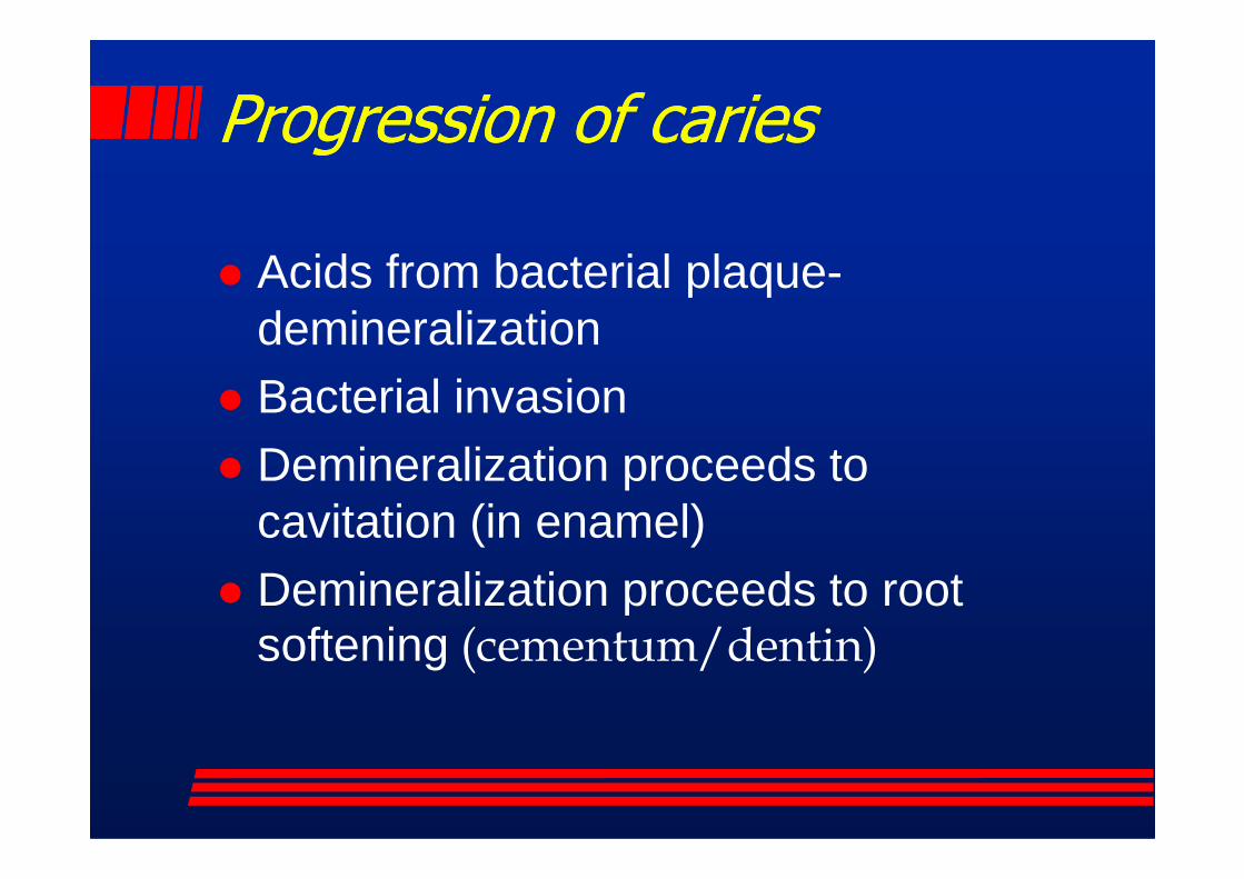
Progression of cariesProgression of caries
� Acids from bacterial plaque-demineralization
� Bacterial invasionDemineralization proceeds to � Demineralization proceeds to cavitation (in enamel)
� Demineralization proceeds to root softening (cementum/dentin)

Classification of carious lesions and Classification of carious lesions and tooth preparationstooth preparations
� Systemic groupings first described by G.V. Black, the father of operative dentistry �Classification according to locations in �Classification according to locations in
permanent teeth
� Classifies according to number of surfaces prepared
� Classifies by location tooth preparation surfaces

Black’s classificationBlack’s classification
� Class 1 (I)� Class 2 (II)� Class 3 (III)� Class 4 (IV)� Class 5 (V)According to Schlutz� Class 6 (VI)

Number of surfaces preparedNumber of surfaces prepared
� Simple- involving one surface of a tooth
� Compound- involving two surfaces of a toothof a tooth
� Complex- involving three of more surfaces of a tooth

Location of tooth preparationLocation of tooth preparation
EXAMPLES� O- occlusal� MO- mesio-occlusal� DO- disto-occlusal� DO- disto-occlusal� MOD- mesio-occlusodistal� F- facial� DL- disto-lingual� MID- mesioincisodistal� DF- distofacial

Class I: pit and fissure caries to Class I: pit and fissure caries to include posterior occlusal surfaces, include posterior occlusal surfaces, facial and lingual surfaces; buccal facial and lingual surfaces; buccal pits of molars; lingual pits of anterior pits of molars; lingual pits of anterior teethteeth

Class I: pit and fissure caries to Class I: pit and fissure caries to include posterior occlusal surfaces, include posterior occlusal surfaces, facial and lingual surfaces; buccal facial and lingual surfaces; buccal pits of molars; lingual pits of anterior pits of molars; lingual pits of anterior teethteeth
Pit or fissure
DEJ
Pit or fissureProgression ofcaries are twocones base tobase

CLASS ICLASS I--OCCLUSAL PIT AND FISSUREOCCLUSAL PIT AND FISSURE

CLASS ICLASS I--OCCLUSAL PIT AND FISSUREOCCLUSAL PIT AND FISSURE

Caries Lesion ContinuumCaries Lesion Continuum
Sound
< ½ thicknessRadiographic evidence
Definite dentinal caries
(radiographically);Enamel surface intact
SoundTooth
No caries
Histologicalevidence of caries
Definite enamel caries(radiographically);Dentin affected histologically;
Enamel surface intact
Slight enamelSurface cavitation
RESTORATIONINDICATED
remineralization demineralization
Adapted from Suddick et al, Caries activity estimates and implications:insights into risk vs activity. J Dent Educ 61:876-884, 1997)

Caries Management ParadigmCaries Management Paradigmclassifying lesions by appropriate careclassifying lesions by appropriate care
Lesions into pulp
Clinically detectableLesions into dentin(open and closed)
Preventive andOperative care
Progressive/cavitatedStable/non-cavitated
Clinically detectable“cavities” limited to enamel
Clinically detectable enamel lesionsWith intact surfaces
Lesions detectable with additional Diagnostic aids (transillumination and radiographs)
Sub-clinical initial lesions in a dynamicState of progression/regression
Preventivecare advised
No activecaries

Caries extension = preparationCaries extension = preparation
Caries diagnosed extension into
pits and fissures = preparation
composite resin or amalgam
Occlusal caries Extension to avoid unsupported enamel and caries removal
Caries removal

Caries extension = preparationCaries extension = preparation
Diagnosed expected caries extension
bitewing x-ray + pits and fissures = preparation
Radiographicdiagnosis
Clinical appearanceAnd diagnosis

Caries extension = preparationCaries extension = preparation
Diagnosed expected caries extension
bitewing x-ray + pits and fissures = preparation
Radiographicdiagnosis
Clinical appearanceAnd diagnosis

Class IClass Ilingual or buccal pitlingual or buccal pit

Class II: smooth surface caries on Class II: smooth surface caries on the proximal surfaces of posterior the proximal surfaces of posterior teethteeth
Progression ofcaries are twocones tip tobase
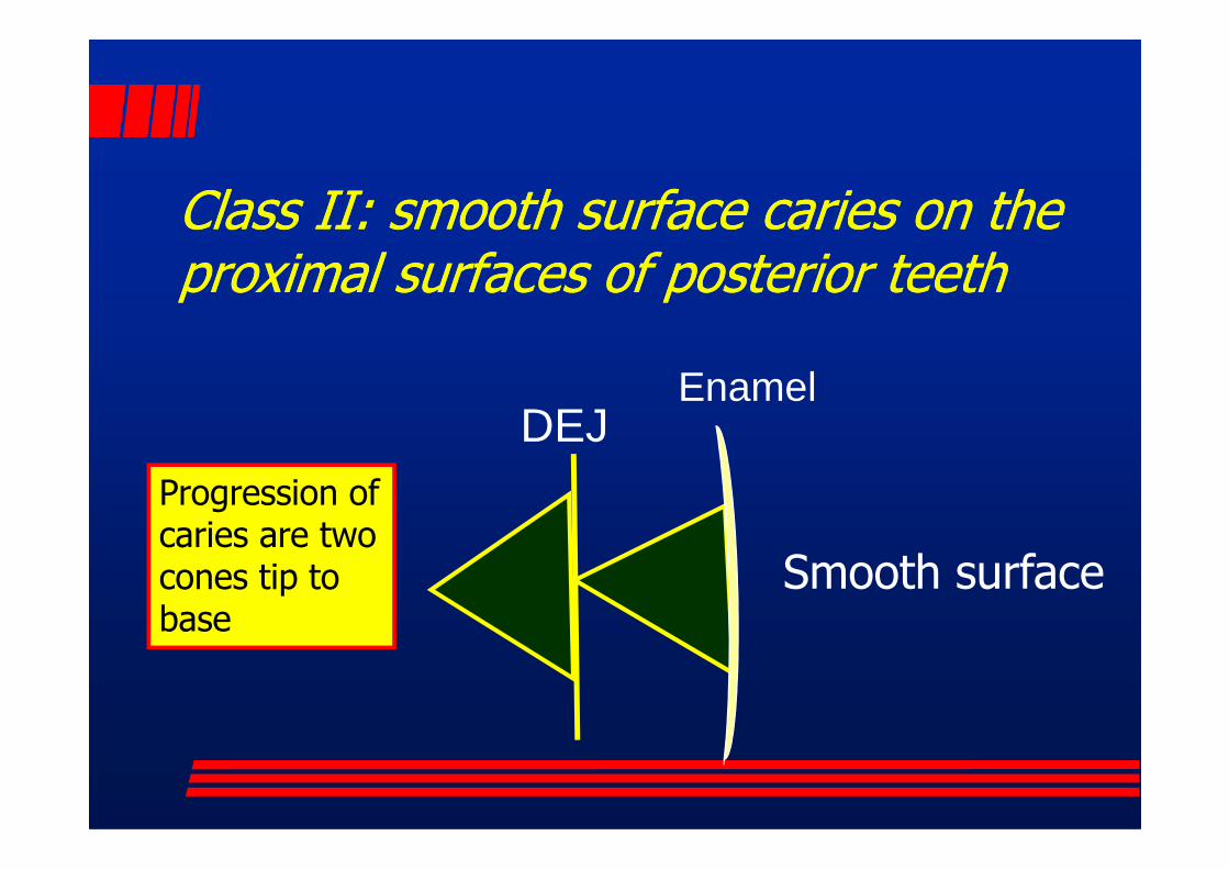
Class II: smooth surface caries on the Class II: smooth surface caries on the proximal surfaces of posterior teethproximal surfaces of posterior teeth
DEJEnamel
DEJ
Smooth surface
Progression ofcaries are twocones tip tobase

CLASS IICLASS II--interproximal cariesinterproximal caries

CLASS IICLASS II--interproximal caries; occlusal no interproximal caries; occlusal no caries not caries susceptiblecaries not caries susceptible
Class IIamalgam

Radiographic appearance of Class II caries

Radiographic appearance of Class II amalgam restorations and caries
Class IIcaries
Well contoured and carved Class II amalgam gingival margin
caries

Radiographic appearance of Class II amalgam restorations
Do you see any problems?

Radiographic appearance of Class II amalgam restorations
Did you See theClass II
Amalgam restorations with overhangs.
Class IIcaries

Radiographic appearance of Class II caries-note the Caries on the primary molars
Erupting Permanent1st premolar

Class III: smooth surface caries on the Class III: smooth surface caries on the proximal surfaces of anterior teethproximal surfaces of anterior teeth
Progression ofcaries are twocones tip tobase

Class III: smooth surface caries on the Class III: smooth surface caries on the proximal surfaces of anterior teethproximal surfaces of anterior teeth
DEJ Enamel
Smooth surface
Progression ofcaries are twocones tip tobase

Radiographic appearance of lesion
� Visible, invasive to <1/2 thickness of enamel
� Definite enamel caries (radiographically); dentin affected histologically;
CLASS IIICLASS III-- Proximal cariesProximal caries
affected histologically; enamel surface intact
� Definite dentinal caries; enamel surface intact
� Dentinal caries with enamel cavitation
radiographdemonstratesClass III lesion
on mesial surface of Maxillary lateral incisor.

Clinical appearance� Discolored with no cavitation;
depth within the enamel as seen through light transillumination
� Non-cavitated but visible with
CLASS IIICLASS III-- Proximal cariesProximal caries
� Non-cavitated but visible with transillumination as being through enamel into the dentin
� Clinically cavitated verified with explorer
TransilluminationdemonstratesClass III lesion
on mesial surface of Mandibular incisor.

Class IIIClass IIIcaries or defective restorationcaries or defective restoration

Class IV: lesions on the Class IV: lesions on the proximal surface of anterior proximal surface of anterior teeth that include the incisal teeth that include the incisal edgeedge
Usually dueto traumaticfracture
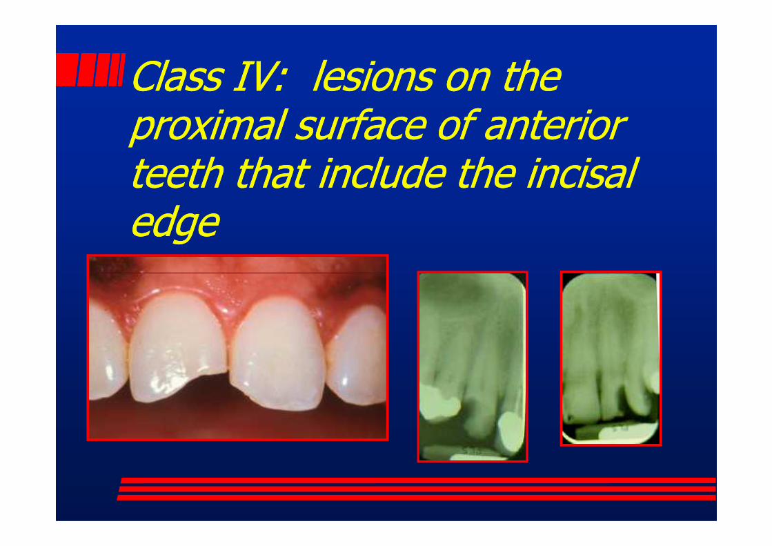
Class IV: lesions on the Class IV: lesions on the proximal surface of anterior proximal surface of anterior teeth that include the incisal teeth that include the incisal edgeedge

Class IV Class IV incisal edge with proximal surfaceincisal edge with proximal surface

Class IVClass IVcaries or defective restorationscaries or defective restorations

Traumatic fracture with Traumatic fracture with pulpal exposurepulpal exposure

Whenever restoring Class IV fractures, occlusal considerations will provide important information regarding tooth preparation before restoration.
Class IV restoration and Class IV restoration and its relationship to occlusionits relationship to occlusion
This restoration has fractured two previous times over a period of 4 months. Note the edge to edge occlusion that must be changed to expect a more
durable long lasting restoration.

Class V caries: smooth surface caries on Class V caries: smooth surface caries on the facial or lingual surfaces of both the facial or lingual surfaces of both anterior and posterior teeth at the anterior and posterior teeth at the gingival third; may involve cementum or gingival third; may involve cementum or dentin as well as enamel surfacesdentin as well as enamel surfaces
Progression ofcaries are twocones tip tobase

Class V caries: smooth surface caries on Class V caries: smooth surface caries on the facial or lingual surfaces of both the facial or lingual surfaces of both anterior and posterior teeth at the anterior and posterior teeth at the gingival third; may involve cementum or gingival third; may involve cementum or dentin as well as enamel surfacesdentin as well as enamel surfaces
DEJSmooth surface
EnamelDEJSmooth surface
Facial or lingual
Progression ofcaries are twocones tip tobase

Root Surface Caries MorphologyRoot Surface Caries Morphology
Carious lesion is a softening of the root surface that progresses following the
paths of the dentinal tubules

Root surface demineralization Root surface demineralization with softeningwith softening

Class V also describes nonClass V also describes non--carious smooth surface loss of carious smooth surface loss of tooth substance lesions on tooth substance lesions on facial and lingual surfacesfacial and lingual surfaces

� Erosion
NonNon--carious Class V lesionscarious Class V lesions
� Abrasion� Abfraction
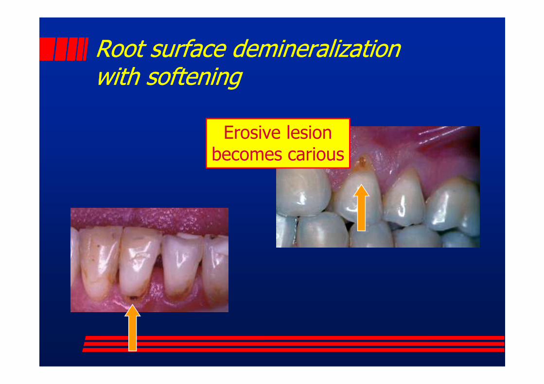
Root surface demineralization Root surface demineralization with softeningwith softening
Erosive lesionbecomes carious
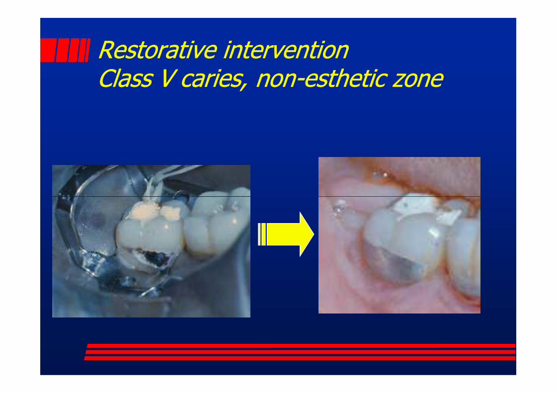
Restorative interventionRestorative interventionClass V caries, nonClass V caries, non--esthetic zoneesthetic zone

Class V cariesClass V caries

DiagnosisDiagnosis
Calculus, teeth must be cleaned for definitive diagnosis

Class VI: lesions are pit or wear Class VI: lesions are pit or wear defects on the incisal edges of defects on the incisal edges of anterior teeth or cusp tips of anterior teeth or cusp tips of posterior teeth; posterior teeth; lesions can be caries but don’t lesions can be caries but don’t have to behave to behave to behave to be

Class VI: lesions are pit or wear Class VI: lesions are pit or wear defects on the incisal edges of defects on the incisal edges of anterior teeth or cusp tips of anterior teeth or cusp tips of posterior teeth; posterior teeth; lesions can be caries but don’t lesions can be caries but don’t lesions can be caries but don’t lesions can be caries but don’t have to behave to be

Class VI: WearClass VI: Wear

Class VI: CariesClass VI: Caries

ReviewReview
� Class 1� Class 2� Class 3� Class 3� Class 4� Class 5� Class 6