Class #3. Pelvis Supports the trunk and organs in the lower abdomen (pelvic cavity) Absorbs stress...
38
Class #3
-
Upload
ellen-terry -
Category
Documents
-
view
215 -
download
0
Transcript of Class #3. Pelvis Supports the trunk and organs in the lower abdomen (pelvic cavity) Absorbs stress...
Class #3
Pelvis Supports the trunk and organs in the lower
abdomen (pelvic cavity) Absorbs stress from lower limbs when
moving (walking/jumping) Female pelvis is adapted for pregnancy and
childbirth and is wider and lighter than male pelvis
Bones of the pelvis Ilium-forms superior flared portion,
impt.for muscle attachment Ischium- inferior portion and strongest
bone of the pelvis Pubis- anterior portion of pelvis
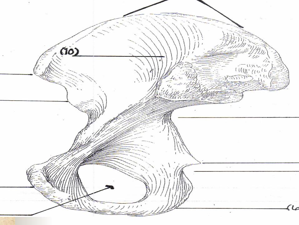
Landmarks of the Pelvis: Iliac crest Iliac fossa Ant.Superior Iliac Spine (ASIS) Ant. Inf. Iliac Spine (AIIS) Post. Sup. Iliac spine (PSIS) Post. Inf. Iliac spine (PIIS) Greater Sciatic Notch Gluteal Lines
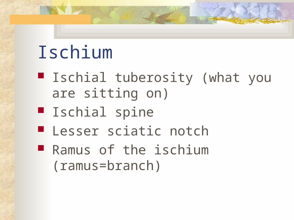
Ischium Ischial tuberosity (what you are sitting on) Ischial spine Lesser sciatic notch Ramus of the ischium (ramus=branch)
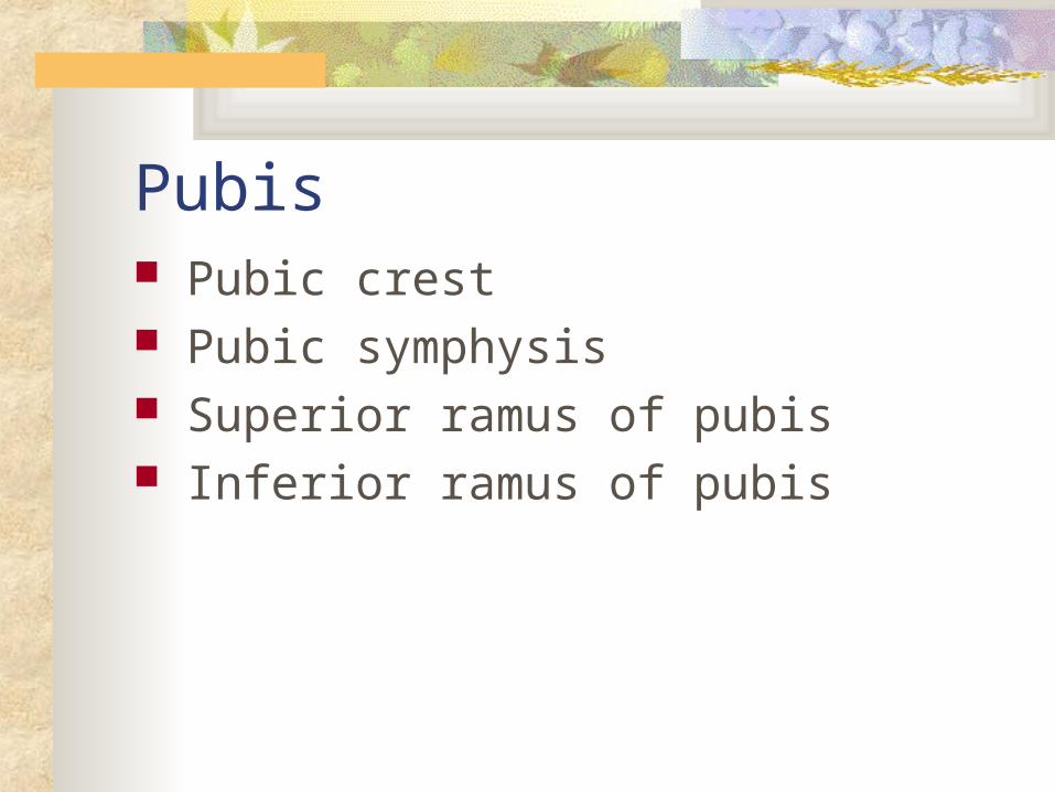
Pubis Pubic crest Pubic symphysis Superior ramus of pubis Inferior ramus of pubis
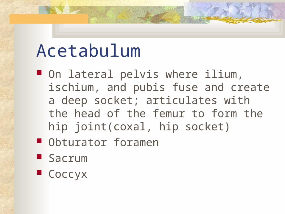
Acetabulum On lateral pelvis where ilium, ischium, and
pubis fuse and create a deep socket; articulates with the head of the femur to form the hip joint(coxal, hip socket)
Obturator foramen Sacrum Coccyx
Quadriceps GroupMain action is to extend the leg at the knee
joint (kicking a ball) also to move the thigh into extension at the knee; standing up from seated position, coming up into straight leg position from squat
Quad=four; cep=headed
Quadriceps muscle Rectus Femoris Vastus Lateralis Vastus Medialis Vastus Intermedius
About Muscles O: (Origin)- Where the muscle begins I: (Insertion)- Where the muscle ends A: (Action)- This is what the muscle does
when it contracts or shortens.
Insertions point always moves close to the origin.
Extension of the leg at the Knee Rectus femoris Vastus lateralis Vastus medialis
Vastus intermedius
Quadriceps Rectus FemorisRectus=straight or upright;Femoris=related to thighOrigin: AIISInsertion: Tibial Tuberosity, via the patella and patellar
ligamentAction: Ext. of the leg at the knee joint
Flexion of the thigh at the hip jointCombined actions seen as leg is brought forward in walking.
Quadriceps Vastus Lateralis
Vastus=vast or large; lateralis=related to the side
O: Linea aspera, ant. Aspect of greater trochanter
I: Tibial tuberosity, via patella & patellar lig.
A: Ext. of the leg at knee joint(also restrains medial pull on patella by Vastus Medialis)
Quadriceps Vastus Medialis (Medialis=related to the
middle
O: linea aspera
I: Tibial tuberosity via the patella & patellar lig.
A: Ext. of the leg at knee joint
Quadriceps Vastus Intermedius (intermedius=among the
middle, intermedius lies deep to the other quadriceps muscles.
O: linea aspera, anterior and lateral femoral shaft.
I: Tibial tuberosity via the patella & patellar lig.
A: Ext. of the leg at the knee joint
All 4 muscles are innervated by the Femoral Nerve
All 4 have common insertion on the tibial tuberosity
Osgood-Schlatter Dz Irritation and inflammation of the tibial
tuberosity; most often in boys between 10-15. The tuberosity becomes inflammed and/or separates from tibia, because of irritation caused when patellar tendon pulls on tuberosity during periods of rapid growth or overuse of quadriceps.
Medial Thigh MusclesAdduction of the Hip
Adductor magnus Adductor longus Adductor brevis
Gracilis Pectineus
Psoas major Iliacus
Gluteus maximus (lower fibers)
Adductor group Muscles of medial thigh Main action is hip adduction Also do medial rotation of hip, and all but
Gracilis assist with hip flexion
Adductor Muscles Pectineus Adductor Longus Adductor Brevis Adductor Magnus Gracilis Sartorius
Adductor Muscles Pectineus (means related to the pubic bone)
O: Ring around the obturator foramen (ant. Pubis)
I: linea aspera
A: adduction of femur at hip joint
Flexion of femur at hip joint
Adductor Muscles Adductor Longus
O: Ring around the obturator foramen (ant. Pubis)
I: linea aspera
A: adduction of femur at hip joint
assists with flexion of femur at hip
Adductor Muscles Adductor Brevis
Brevis is deep to longus
O: ring around the obturator foramen (ant. Pubis)
I: linea aspera
A: adduction of femur at hip joint
assist with flexion of femur at hip

Adductor Muscles Adductor Magnus (magnus=great)
Largest and deepest
O: ring around obturator foramen (inf. Ramus of pubis and ramus of ischium, ischial tuberosity)
I: linea aspera; (gluteal tuberosity, adductor tubercle of femur)
A: adduction of femur at hip
assists with ext. of femur at hip
Adductor Muscles Gracilis = slenderO: ring around the obturator foramen (inf ramus of
ant. Pubis)I: Proximal anteromedial tibia at the pes anserinus
tendon.A: adduction of femur at hip joint
assists with medial rotation of hipassists with flexion of leg at kneeassists with medial rotation of leg at knee
Adduction musclesCommon origin: ring around obturator
foramen
Common insertion: linea aspera
Nerve to adductor muscles is the Obturator
Adductor Muscles Sartorius “tailor’s muscles”Longest muscle in body, most superficial thigh muscleO: ASISI: proximal anteromedial tibia at the pes anserinusA: hip: assists with flexion
abductionLateral rotaton
Knee: assists with flexion, and medial rotation of leg at knee
Posterior Thigh MusclesFlexion of the leg at the knee
Biceps femoris Semitendinosus
Semimembranosus Gracilis
Sartorius Gastroncnemius
Popliteus Plantaris
Hamstring Group Named so because butchers used to hang
the carcass of a pig by the hamstring tendons.
Cross two joints: hip and knee, so involved with flexing leg at knee joint and extending femur at hip joint.

Hamstring Muscles Biceps Femoris Semitendinosus Semimembranosus
Hamstrings Muscles Biceps Femoris:
Biceps=two headed; femoris=related to thigh
O: Long head-ischial tuberosity
Short head-linea aspera
I: head of the fibula (lateral aspect)
A: Long head: ext. of femur at hip
Long and Short heads: flexion of leg at knee
lat. Rotation of leg at knee
Hamstring Muscles Semitendinosus:
Means half tendon
O: ischial tuberosity
I: pes anserinus (proximal anteromedial tibia)
A: flexion of leg at knee joint
med. Rot. Of leg at knee joint (knee must be semiflexed for medial rot. To occur)
ext. of femur at hip joint
Hamstring Muscles Semimembranosus
Means half membrane
O: ischial tuberosity
I: posteromedial tibial condyle
A: flexion of leg at knee joint
med. Rot. Of leg at knee joint(knee must be semiflexed for med. Rot. To occur)
extension of femur at hip joint
Hamstring MusclesCommon origin of all hamstring muscles is
the ischial tuberosity(your sits bone).
Pes Anserinus (the proximal anteriomedial tibia)
The common insertion for three thigh muscles
Anterior- Sartorious
Medial- Gracilis
Posterior- Semitendinosus
Flexion of the thigh at the hip Rectus Femoris
Gluteus medius (ant. fibers) Gluteus minimus
Adductor magnus (assists) Adductor longus (assists) Adductor brevis (assists)
Pectineus (assists) TFL
Sartorius Psoas major
Iliacus
Extension of the thigh at the hip Biceps femoris
Semitendinosus Semimembranosus Gluteus maximus
Gluteus medius (post. fibers) Adductor magnus (post. Fibers)
End of Class 3