Citrus Tristeza Virus: Survival at the Edge of the Movement
12
University of Nebraska - Lincoln DigitalCommons@University of Nebraska - Lincoln Papers in Plant Pathology Plant Pathology Department 7-1-2008 Citrus Tristeza Virus: Survival at the Edge of the Movement Continuum Svetlana Y. Folimonova University of Florida Alexey S. Folimonov University of Florida Satyanarayana Tatineni University of Nebraska-Lincoln, [email protected] William O. Dawson University of Florida, wodtmv@ufl.edu Follow this and additional works at: hp://digitalcommons.unl.edu/plantpathpapers Part of the Plant Pathology Commons is Article is brought to you for free and open access by the Plant Pathology Department at DigitalCommons@University of Nebraska - Lincoln. It has been accepted for inclusion in Papers in Plant Pathology by an authorized administrator of DigitalCommons@University of Nebraska - Lincoln. Folimonova, Svetlana Y.; Folimonov, Alexey S.; Tatineni, Satyanarayana; and Dawson, William O., "Citrus Tristeza Virus: Survival at the Edge of the Movement Continuum" (2008). Papers in Plant Pathology. 106. hp://digitalcommons.unl.edu/plantpathpapers/106
Transcript of Citrus Tristeza Virus: Survival at the Edge of the Movement

University of Nebraska - LincolnDigitalCommons@University of Nebraska - Lincoln
Papers in Plant Pathology Plant Pathology Department
7-1-2008
Citrus Tristeza Virus: Survival at the Edge of theMovement ContinuumSvetlana Y. FolimonovaUniversity of Florida
Alexey S. FolimonovUniversity of Florida
Satyanarayana TatineniUniversity of Nebraska-Lincoln, [email protected]
William O. DawsonUniversity of Florida, [email protected]
Follow this and additional works at: http://digitalcommons.unl.edu/plantpathpapers
Part of the Plant Pathology Commons
This Article is brought to you for free and open access by the Plant Pathology Department at DigitalCommons@University of Nebraska - Lincoln. Ithas been accepted for inclusion in Papers in Plant Pathology by an authorized administrator of DigitalCommons@University of Nebraska - Lincoln.
Folimonova, Svetlana Y.; Folimonov, Alexey S.; Tatineni, Satyanarayana; and Dawson, William O., "Citrus Tristeza Virus: Survival at theEdge of the Movement Continuum" (2008). Papers in Plant Pathology. 106.http://digitalcommons.unl.edu/plantpathpapers/106

JOURNAL OF VIROLOGY, July 2008, p. 6546–6556 Vol. 82, No. 130022-538X/08/$08.00�0 doi:10.1128/JVI.00515-08Copyright © 2008, American Society for Microbiology. All Rights Reserved.
Citrus Tristeza Virus: Survival at the Edge of the MovementContinuum�
Svetlana Y. Folimonova, Alexey S. Folimonov,† Satyanarayana Tatineni,‡ and William O. Dawson*Citrus Research and Education Center, University of Florida, 700 Experiment Station Road, Lake Alfred, Florida 33850
Received 7 March 2008/Accepted 14 April 2008
Systemic invasion of plants by viruses is thought to involve two processes: cell-to-cell movement betweenadjacent cells and long-distance movement that allows the virus to rapidly move through sieve elements andunload at the growing parts of the plant. There is a continuum of proportions of these processes thatdetermines the degrees of systemic infection of different plants by different viruses. We examined the systemicdistribution of Citrus tristeza virus (CTV) in citrus species with a range of susceptibilities. By using a “pure”culture of CTV from a cDNA clone and green fluorescent protein-labeled virus we show that both cell-to-celland long-distance movement are unusually limited, and the degree of limitation varies depending on the citrushost. In the more-susceptible hosts CTV infected only a small portion of phloem-associated cells, and more-over, the number of infection sites in less-susceptible citrus species was substantially decreased further,indicating that long-distance movement was reduced in those hosts. Analysis of infection foci in the two mostdifferential citrus species, Citrus macrophylla and sour orange, revealed that in the more-susceptible host theinfection foci were composed of a cluster of multiple cells, while in the less-susceptible host infection foci wereusually single cells, suggesting that essentially no cell-to-cell movement occurred in the latter host. Thus, CTVin sour orange represents a pattern of systemic infection in which the virus appears to function with only thelong-distance movement mechanism, yet is able to survive in nature.
Viruses that infect higher plants share a number of commonprinciples with animal viruses. Among those are virion mor-phology and strategies for replication and expression of theirgenomes. However, to establish a productive infection in a hostthe plant virus needs to be able to move throughout a plantfrom an initially infected cell. Success depends upon compat-ible interactions between viral and host factors. Generally,systemic movement is thought to involve two distinct pro-cesses: cell-to-cell movement, which according to our defini-tion is a process that allows the virus to transverse the cell wallbetween adjacent cells, and long-distance movement, which isa process that allows the virus to enter the sieve element froman adjacent nucleated cell and rapidly move through the con-nected sieve elements, followed by its exit into another adja-cent phloem-associated cell at a distal region of the plant. Amajor obstacle for the spreading virus is to cross the bound-aries represented by the cell wall. For this purpose most virusesutilize specific virus-encoded movement proteins as well assome host proteins that facilitate their translocation throughplasmodesmata channels. The viral proteins and their interac-tions with the host during cell-to-cell movement are fairly wellknown (reviewed in references 26, 43, and 45). However, themechanisms of long-distance transport and factors that aid
virus entrance into phloem tissue, further vascular movement,and unloading from phloem are much less understood.
Different viruses utilize different ratios of cell-to-cell andlong-distance movement, which results in significant differ-ences in the extent and patterns of systemic invasion of theirhosts. Often the virus-host interaction results in no diseasewhen the virus is able to replicate in initially infected cells butis not able to move throughout the plant, which is considereda nonhost of the virus. One ultimate example of spread isexemplified by the well-studied system of Tobacco mosaic virus(TMV) in tobacco, where the virus moves efficiently, infectingmost of the cells throughout the entire plant. This sequencetypically results from cell-to-cell movement from an initiallyinfected cell through plasmodesmata to neighboring cells untilthe virus reaches phloem cells. Then, long-distance movementallows the virus to rapidly move through sieve elements andunload at the growing parts of the plant, where further cell-to-cell movement from phloem-associated cells allows the virusto invade most of the cells of these distal plant organs. Oncethe virus approaches the growing point via long distance move-ment, continued cell-to-cell movement parallels plant growthto maintain the systemic infection. Other patterns of move-ment allow a range of more limited systemic infection ofplants. Some viruses spread systemically throughout plants butare confined mainly to cells associated with phloem. Therefore,in nature the virus is usually introduced directly into phloem-associated tissues by a vector, often an insect. The virus then isable to move normally by long-distance movement but is lim-ited in cell-to-cell movement to nearby phloem-associatedcells.
Not only must the virus have the capacity to replicate andmove in a particular plant host, but also it must have the abilityto escape from the host’s surveillance system. Along with
* Corresponding author. Mailing address: Citrus Research and Ed-ucation Center, University of Florida, 700 Experiment Station Road,Lake Alfred, FL 33850. Phone: (863) 956-1151. Fax: (863) 956-4631.E-mail: [email protected].
† Present address: Center Bioengineering, Russian Academy of Sci-ences, Moscow 117312, Russia.
‡ Present address: U.S. Department of Agriculture Agricultural Re-search Service and Department of Plant Pathology, University of Ne-braska, Lincoln, NE 68583.
� Published ahead of print on 23 April 2008.
6546

movement functions, viruses also encode another group offactors termed silencing suppressors that counteract the RNAinterference plant defense system to allow a systemic infectionto be established and maintained (33, 35, 44). Mutations ofviral suppressor genes often result in reduction or preventionof systemic infection (9, 21, 32). In fact, the tissue limitationscan be due to the plant defense system. Experiments withcoinoculation of phloem-limited viruses, the polerovirus Potatoleafroll virus, or bipartite geminiviruses Bean golden mosaicvirus and Abutilon mosaic virus with certain other viruses re-sulted in alleviation of their phloem limitation, which demon-strated that perhaps confinement to phloem could be ex-plained by lack of necessary factors that enable these viruses tounload into mesophyll tissue and move further cell to celland/or by lack of the mechanisms to overcome the plant de-fense system outside the phloem (6, 7, 27, 36, 46).
Citrus tristeza virus (CTV) is limited to phloem-associatedcells in citrus trees. It is the largest and most complex memberof the Closteroviridae family, which contains viruses withmono-, bi-, and tripartite genomes (1, 5, 11, 12, 20). Members
of this family are transmitted by different types of insects:aphids, whiteflies, and mealybugs. CTV has long flexuous viri-ons (2,000 nm by 10 to 12 nm) encapsidated by two coatproteins and a single-stranded RNA genome of approximately19.3 kb. The major coat protein (CP) encapsidates about 97%of the genomic RNA, while the minor coat protein (CPm)covers the rest of the genome at its 5� end (13, 41). The RNAgenome of CTV encodes 12 open reading frames (ORFs) (18,28) (Fig. 1). ORFs 1a and 1b are expressed from the genomicRNA and encode polyproteins required for virus replication.Ten 3�-end ORFs are expressed by 3�-coterminal subgenomicRNAs (sgRNAs) (17, 19) and encode the following proteins:CP, CPm, p65 (HSP70 homolog), and p61, which are involvedin assembly of virions (38); a hydrophobic p6 protein with aproposed role in virus movement (12, 42); p20 and p23, whichalong with CP are suppressors of RNA silencing (25); p33, p13,and p18, whose functions remain unknown.
The host range of CTV generally is limited to citrus speciesand relatives, and the different species exhibit differential de-grees of susceptibility to CTV infection (8; S. M. Garnsey,
FIG. 1. (A) Schematic diagram of the genome organization of wild-type CTV (CTV9R) and its derivative encoding GFP. The open boxesrepresent ORFs and their translation products. PRO, papain-like protease domain; MT, methyltransferase; HEL, helicase; RdRp, an RNA-dependent RNA polymerase; HSP70h, HSP70 homolog. Bent arrows indicate positions of BYV (BCP) or CTV CP (CCP) sgRNA controllerelements. Inserted elements are shown in gray. (B) Replication of CTV9R (lane 1) or CTV-BC5/GFP (lane 2) in N. benthamiana mesophyllprotoplasts inoculated with virions extracted from infected C. macrophylla seedlings. Total RNA was isolated from protoplasts 4 days postinocu-lation. Northern blot hybridizations were carried out using CTV 3� positive RNA strand-specific riboprobe. Positions of sgRNAs are shown.
VOL. 82, 2008 LIMITED MOVEMENT OF CITRUS TRISTEZA VIRUS 6547

personal communication). On the other hand, a number ofcitrus relatives, including Poncirus trifoliata, Swinglea glutinosa,and Severinia buxifolia, demonstrate immunity to infection bymost CTV isolates (15, 47, 48, 49). Since CTV can replicate inprotoplasts isolated from these plants, the resistance at awhole-plant level possibly results from a lack of virus move-ment (2). Experiments with the resistant genotype used as aninterstock grafted between two susceptible genotypes showedthat the virus moves though the resistant interstock unimpededbut does not multiply detectably in the resistant plant (S. M.Garnsey, personal communication). Therefore, it appearslikely that the virus is unable to egress from the sieve elementsinto adjacent phloem cells of the resistant genotype. It is pos-sible that the gradient of susceptibility of different citrus spe-cies to CTV is related to the ability of the virus to move intoand infect cells surrounding sieve elements.
The objective of this work was to examine the systemicdistribution of CTV in citrus species with a range of suscepti-bility and to attempt to interpolate movement mechanismsresponsible for the distribution. We show that CTV in citrustrees generally follows similar patterns, but the degrees of bothcell-to-cell and long-distance movement are more limited thanmost systems that have been described, and the limitationvaries depending on the citrus host. In all hosts, long-distancemovement appears to be limited to relatively few initial infec-tion sites. In the more-susceptible citrus species, CTV also haslimited cell-to-cell movement that produces small clusters ofinfected cells. However, in less-susceptible citrus species, itappears that no cell-to-cell movement occurs. The virus is ableto exit sieve elements but cannot spread to adjacent cells,resulting in infection of isolated single cells. Thus, here wedemonstrate a new pattern of systemic infection in which thevirus appears to function with only the long-distance move-ment mechanism, yet is able to survive in nature. Elucidationof this process aids in understanding how the virus surviveslong term (up to about 100 years) in infected trees and how itspreads to other trees.
MATERIALS AND METHODS
Virus constructs, amplification of virions in Nicotiana benthamiana proto-plasts, and inoculation of citrus trees. Full-length cDNA constructs were used inthis study: pCTV9R, a cDNA clone of T36 strain of CTV (37, 40), and constructscontaining insertion of green fluorescent protein (GFP) ORF in the regionbetween the CPm and CP genes in the CTV genome, pCTV-BC5/GFP (14) andCTV9-GFPC3 (42). SP6 RNA polymerase-derived transcripts of CTV cDNAslinearized with NotI restriction endonuclease were used for transfection of N.benthamiana mesophyll protoplasts according to the procedure described bySatyanarayana et al. (37). Protoplasts were harvested at 4 days postinoculationand stored at �70°C for subsequent protoplast passage of virions. Passaging ofvirions up to 11 successive cycles in protoplasts for amplification of the virus wasdone as described previously by Satyanarayana et al. (38). Accumulation of thevirus was monitored by Northern blot hybridization of the total RNA isolatedfrom protoplasts with a 3� positive-stranded CTV RNA-specific riboprobe (37).
Amplified progeny virions from the final passages in protoplasts were ex-tracted and concentrated by sucrose cushion centrifugation, and the concen-trated virions were used for mechanical “bark flap” inoculation of small trees ofCitrus macrophylla Wester as described by Robertson et al. (34). Infected treeswere later used as a source of virus inocula for subsequent graft inoculations ofyoung C. macrophylla trees as well as other citrus species: Mexican lime [Citrusaurantifolia (Christm. Swing)], Madam vinous sweet orange [Citrus sinensis (L.)Osbeck], sour orange (Citrus aurantium L.), and Duncan grapefruit (Citrus para-disi Macf.).
Serological assays. A double antibody sandwich indirect enzyme-linked im-munosorbent assay (ELISA) was performed as described previously using anti-
bodies specific to CTV virions (16) to confirm infection in inoculated plants andcompare titers of virus accumulation in different citrus species. Purified immu-noglobulin G from rabbit polyclonal antiserum CTV-908 (1 �g/ml) was used asa coating antibody. ECTV172, a broadly reactive CTV monoclonal antibody, wasused as a detecting antibody.
Examination of fluorescence in citrus plants infected with GFP-tagged CTV.Samples of bark tissue from CTV-BC5/GFP-infected trees were examined forGFP fluorescence at different time points beginning at 6 weeks after inoculationusing a Zeiss Stemi SV 11 UV fluorescence dissecting microscope (Carl ZeissJena, GmbH, Jena, Germany) with an attached Olympus Q-color 5 camera(Olympus America, Inc., Center Valley, PA). More detailed observation of theinfection foci in infected C. macrophylla and sour orange trees was performedusing a confocal scanning microscope (Leica TCS SL; Leica Microsystems, Inc.,Exton, PA).
Light and transmission electron microscopy and immunogold labeling. Peti-oles of young leaves of C. macrophylla and Mexican lime trees and bark tissuesamples of C. macrophylla and sour orange trees were harvested for electronmicroscopy and light microscopy studies, respectively, 6 to 8 weeks after inocu-lation with CTV9R. Tissue samples were fixed in phosphate buffer containing1.8% paraformaldehyde and 0.25% glutaraldehyde and embedded in LR Whiteresin (London Resin Company, Hampshire, United Kingdom). Procedures forthe specimen fixation and embedding, as well as immunogold labeling, werecarried out as described by Zhou et al. (50). The immunogold labeling of sections(semithin 2-�m sections for light microscopy or ultrathin 80- to 90-nm sectionsfor electron microscopy) of LR White-embedded samples was performed usingpurified immunoglobulin G from rabbit polyclonal antiserum CTV-908 (1 �g/ml,1:1,000 dilution) as primary antibody and goat anti-rabbit–10-nm gold conjugate(1:25 dilution; Sigma). For light microscopy studies gold labeling was enhancedby incubating with silver enhancing solution (BioCell Research Laboratories,Cardiff, United Kingdom) as described by Ding et al. (10). Specimens wereobserved using a Morgagni 268 transmission electron microscope (FEI, TheNetherlands) or Leitz LaborLux S light microscope (Leica, Germany).
RESULTS
Different citrus hosts support different titers of CTV. Thedogma is that CTV titers vary considerably in different citrusspecies (8; S. M. Garnsey, personal communication). (It shouldbe noted that citrus taxonomy is complex. For example, sweetorange [C. sinensis] and grapefruit [C. paradisi] are classified asspecies but now are known to be hybrids instead.) Titers alsovary with different isolates of virus, which generally are popu-lations composed of a mixture of viral genotypes whose se-quences fit into different sequence groups that we now con-sider strains. Some populations accumulate to higher levelsthan others in the same type of host. Also, measurements ofviral titers have been done in different countries, with differentenvironments, and in different citrus varieties. So, although ithas been an accepted phenomenon that some citrus varietiestend to support higher titers of CTV while other varietiessupport lower titers, the variation among different isolates anddifferent measurements has greatly confounded this under-standing.
The development of an infectious cDNA clone of the T36strain of CTV (37) allowed us to obtain a “pure” culture of thevirus in citrus plants (39) using an inoculum that originatedfrom RNA transcripts of the cDNA clone. The recombinantCTV (CTV9R) culture has remained a uniform populationduring the first 6 years of infection of citrus trees, which wasconfirmed by resequencing of the entire genome (Z. Xiong etal., unpublished data). We examined the susceptibility of dif-ferent citrus species to this strain of CTV in five Citrus spp.: C.macrophylla, Mexican lime, Madam vinous sweet orange, sourorange, and Duncan grapefruit. Three plants of each weregraft inoculated with CTV9R. CTV is usually assayed in thefirst flush of new leaves that develops after graft inoculation of
6548 FOLIMONOVA ET AL. J. VIROL.

the lower trunk. At 6 weeks after inoculation, the upper leaveswere harvested, extracted, and assayed by ELISA with CTV-specific antibodies. Higher titers of the virus were detected inC. macrophylla and Mexican lime trees, while in sour orangeand grapefruit titers of the virus were significantly lower. Sweetorange showed intermediate levels of CTV accumulation (Ta-ble 1). These data indicated that these species represent a goodselection of citrus hosts with different degrees of virus infec-tion.
CTV infects only a proportion of phloem-associated cells.Previous studies on cytopathology and ultrastructure of CTVin citrus plants showed that the virus accumulates preferen-tially in the phloem parenchyma and to a lesser degree incompanion cells (4, 22, 23, 50). CTV virions were also foundoccasionally in sieve elements. Although symptoms and cellu-lar reactions caused by the virus are different depending oncitrus species and isolates of virus, so far there is little infor-mation about virus movement and distribution in those hosts.A recent cytopathological study conducted using transmissionelectron microscopy also did not reveal any differences in virusdistribution between different hosts (50). We chose to furtherexamine the quality and quantity of cells in different hosts toattempt to better understand possible reasons for differenttiters.
We first examined the distribution of CTV in the moresusceptible hosts, C. macrophylla and Mexican lime, by trans-mission electron microscopy of thin sections of leaf petioles oftrees infected with CTV9R. Usually CTV induces several typesof inclusions in infected phloem parenchyma and companioncells. Those are viral arrays, fibrous inclusions, and cytoplasmicvesicles and have been described in detail previously (50).These characteristic ultrastructural changes make it relativelyeasy to differentiate the infected cells from uninfected onesusing the electron microscope. To further improve the detec-tion of infected cells, we also used immunogold labeling withT36 CTV-specific antibodies, which react with virions, makingthem readily distinguishable from P-proteins and other thread-like structures in infected phloem cells. Figures 2 and 3 showtransmission electron micrographs of phloem cells in petiolesof infected C. macrophylla and Mexican lime plants, respec-tively. Figures 2A and 3A show groups of cells at lower mag-nification, while Fig. 2B, C, and D and Fig. 3B and C showareas of infected cells at higher magnification to visualize im-munogold labeling. Both figures demonstrate that even in thecases of these two species, which usually have the highest virusconcentrations, the virus infects only a small proportion ofphloem-associated cells. In general, we found approximately 2to 3 infected cells per 10 to 20 cells in a viewed area. Consid-
ering that some species of citrus support much lower titers ofthe virus, including sour orange, which has less than 1/10 thevirus concentration of C. macrophylla or Mexican lime (Table1), we would expect to find an even lower number of infectedcells. This low proportion of infected cells, in addition to thedifficulty of obtaining statistically relevant sample sizes usingelectron microscopy, diminished the usefulness of this ap-proach to reveal the differences of CTV interactions with thevarious citrus hosts.
Distribution of CTV varies in different citrus hosts. Becauseof the limitations of the above approach, as an alternativemethod to examine the distribution of CTV in different citrusspecies, we used GFP-tagged CTV (14), which allowed obser-vations to be made using a fluorescence dissecting microscopeof many more samples from different plants and allowed scan-ning of larger areas of infected tissues. The CTV-based vector,CTV-BC5/GFP (Fig. 1), contained the GFP ORF inserted intothe virus genome as an extra gene (14). The initial work ofcharacterizing the vector was in C. macrophylla. At 5 weeksafter inoculation, the GPF-tagged virus was found to havereplicated and moved systemically into young leaves of thetree. The vector has been unusually stable, continuing produc-tion of GFP in citrus trees for 5 years so far. The biologicalcharacteristics of the CTV-GFP vector in citrus trees wereessentially identical to those of wild-type CTV, with both vi-ruses exhibiting similar time intervals for establishing systemicinfections, similar symptoms produced in infected plants, andappearing to be equally competitive when inoculated simulta-neously into the same tree (14). These features make the GFP-expressing CTV-based construct highly suitable for studying virusdistribution in citrus trees.
Small trees of C. macrophylla, Mexican lime, sweet orange,sour orange, and grapefruit were graft inoculated with CTV-BC5/GFP. Six weeks after inoculation, the new flush of leavesand stems of the infected trees was assayed for GFP fluores-cence.
In rapidly growing citrus trees, the bark slips, which meansthat the bark freely pulls away from the wood, separating at thecambial layer, leaving the xylem cells with the woody trunk andthe phloem-associated cells on the inside of the excised bark.This allows observation of the phloem-associated cells on theinside of the bark piece by using a dissecting fluorescencemicroscope. Examination of C. macrophylla bark tissue fromseveral young flushes revealed an intermittent pattern of dis-tribution of GFP fluorescence in the phloem-associated cells(Fig. 4). Stretches of cells exhibited fluorescence throughoutthe bark, but most cells on the phloem surface failed to exhibitfluorescence, demonstrating that only a portion of the phloem-
TABLE 1. Analysis of CTV accumulation in five citrus species
Exptl group
CTV titera in:
Citrus macrophylla Mexican lime Sweet orangeMadam Vinous Duncan grapefruit Sour orange
CTV9R infected 3.08 � 0.015 2.95 � 0.021 0.92 � 0.065 0.54 � 0.030 0.32 � 0.015Healthy 0.10 � 0.004 0.09 � 0.003 0.08 � 0.008 0.09 � 0.009 0.07 � 0.008
a CTV9R-infected trees were assayed at 6 weeks postinoculation by double antibody sandwich indirect ELISA using CTV-specific 908 IgG as trapping antibody ata 1-�g/ml concentration and ECTV 172 monoclonal antibody as detecting antibody at a 1:50,000 dilution. ELISA values (A405) are averages for three plants � standarddeviations.
VOL. 82, 2008 LIMITED MOVEMENT OF CITRUS TRISTEZA VIRUS 6549

associated cells became infected. Examination of bark fromolder flushes and leaf midribs and veins showed similar pat-terns of fluorescence. It is difficult to calculate the ratio of theinfected phloem cells to the uninfected ones, but based on theobservations of multiple bark pieces from infected C. macro-phylla trees we estimate that less than 10 to 20% of cellsfluoresced.
The distribution of GFP fluorescence in Mexican lime treesinfected with CTV-BC5/GFP was similar to that in C. macro-phylla trees (Fig. 4), which coincides with the similar ELISAvalues of CTV in these two hosts (Table 1). However, thepatterns of fluorescence in sour orange and grapefruit treeswere very different. Many fewer and smaller fluorescent spotswere seen in the bark pieces from these trees. The reducedGFP fluorescence paralleled the reduced viral titers in thesehosts. However, the intensity of fluorescence within individualinfected cells in sour orange and grapefruit appeared to besimilar to that in infected cells of C. macrophylla and Mexicanlime trees, suggesting that the reduced titers of virus revealed
by ELISA were correlated with fewer cells infected. Observa-tions of bark tissue from several infected sweet orange treesshowed that the frequency of fluorescent foci in these plantswas less than that in C. macrophylla and Mexican lime butmuch higher than that in sour orange or grapefruit. In all fivespecies, the pattern of distribution of CTV-induced fluores-cence was correlated with the ELISA values of virus titer inthose hosts (Fig. 4; Table 1).
Analysis of infection foci in duplex plants and grafted barkpatches. The numbers of fluorescent sites were much lower ingrapefruit and especially in sour orange than in C. macrophyllaand Mexican lime. This may have resulted from a decreasedability of the virus to move from sieve elements into adjacentcells or a lower susceptibility of the adjacent cells. However, italso could result from lower inocula being generated withininfected cells of these trees and less virus in the sieve elementsto move into adjacent cells. We examined this possibility bytwo different approaches. The first was to examine “duplex”plants, in which side grafts from healthy sour orange plants
FIG. 2. Transmission electron micrographs showing phloem cells in a petiole of C. macrophylla infected with CTV9R. (A) Groups of phloemcells at lower magnification. Infected cells are indicated with letters B, C, and D, corresponding to images in subsequent panels. (B to D) Areasfrom the cells shown in panel at higher magnification. Viral arrays were labeled with polyclonal CTV-specific antibodies used as primary antibodiesand secondary antibodies conjugated with 10-nm gold particles. No labeling was detected in noninfected cells.
6550 FOLIMONOVA ET AL. J. VIROL.

were put onto C. macrophylla seedlings infected with CTV-BCN5/GFP and new shoots of the sour orange and C. macro-phylla were trained to grow in parallel from the same infectedC. macrophylla rootstock. Thus, the inoculum source for bothnew shoots was identical. The second approach was to excisesmall squares of bark from the stems of C. macrophylla trees,replace them with bark pieces of an equal size from a sourorange tree, and allow the substituted bark patches to becomegrafted in place. Reciprocal grafts were made by replacing barkpatches from C. macrophylla into sour orange trees. Two and 6months after inoculation with GFP-expressing CTV, tissuefrom duplex plants and plants with bark patches was analyzedfor GFP expression. In both cases very few fluorescent infec-tion foci were found in sour orange tissue (Fig. 5). The C.macrophylla portion of the same trees appeared to be heavilyinfected and could have served as a continuous virus supply.Nevertheless, this did not affect the level of infection in sour
orange tissues, and the distribution of infection foci remainedsimilar to that observed earlier in individual sour orange plantsinoculated with CTV-BC5/GFP, demonstrating that the pat-tern of virus distribution in these hosts was not related to theamount of inoculum.
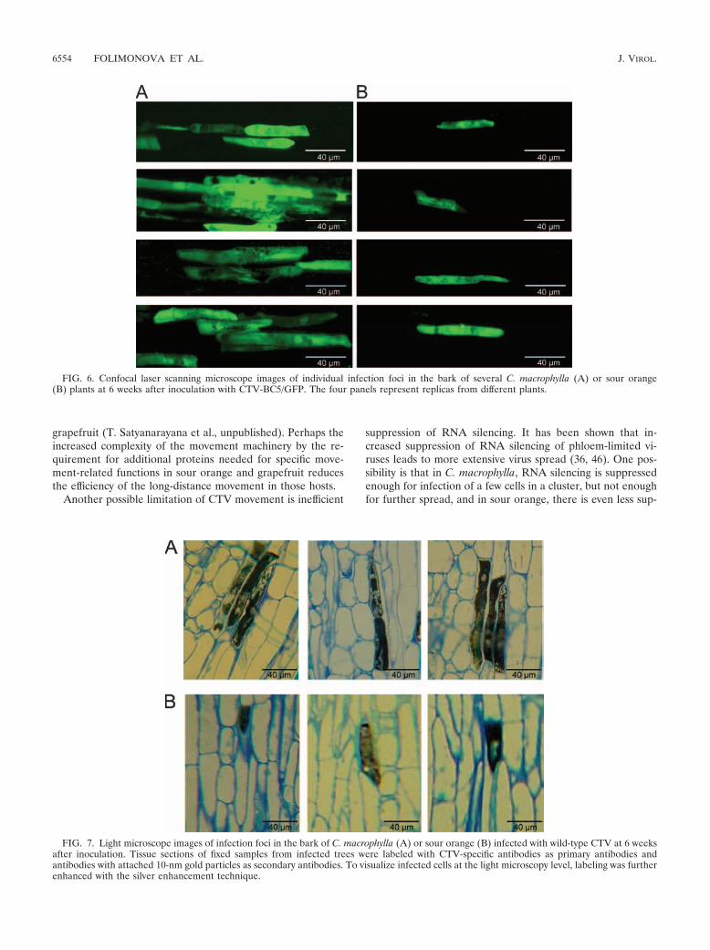
Confocal microscopy of infection foci produced in C. mac-rophylla and sour orange. We next examined the fluorescentfoci in the two most differential citrus species, C. macrophyllaand sour orange, by confocal microscopy to estimate the rela-tive amounts of fluorescence per cell as an estimate of the levelof virus replication per cell and determine how many cellsoccur in individual foci. In C. macrophylla, each infection focuswas composed of multiple cells, ranging from 3 to more than 12(Fig. 6). Strikingly, observations of sour orange tissues re-vealed an absence of cell clusters in sites of infection. Infectionfoci consisted of single cells (Fig. 6). These observations weremade for many individual tissue samples from several indepen-
FIG. 3. Transmission electron micrographs showing phloem cells in a petiole of Mexican lime infected with CTV9R. (A) Groups of phloemcells at lower magnification. Infected cells are indicated with letters B and C, corresponding to images in those panels. (B and C) Areas from theinfected cells shown in panel A at higher magnification. Masses of virions were labeled with polyclonal CTV-specific antibodies used as primaryantibodies and secondary antibodies conjugated with 10-nm gold particles.
VOL. 82, 2008 LIMITED MOVEMENT OF CITRUS TRISTEZA VIRUS 6551

dent infected trees, and all of them showed similar results.However, the intensity of fluorescence in infected cells ap-peared to be similar in C. macrophylla and sour orange, sug-gesting that the reduced titer of virus in sour orange resultedmainly from a reduction in number of cells infected.
To corroborate the confocal microscopy of the GFP-labeledCTV, we utilized a light microscopy technique to examinetissues of these two citrus hosts infected with wild-type CTV.Tissue sections of fixed samples from infected trees were la-beled with CTV-specific antibodies as primary antibodies andantibodies with attached gold particles as secondary antibod-ies. To visualize infected cells at the light microscopy level,labeling was further enhanced according to the silver enhance-ment technique. Figure 7 shows that groups of infected cellswere found in C. macrophylla samples, while only singlecells were present in sour orange tissues. Thus, patterns of cellsinfected with wild-type virus observed with the immunogold-silver enhancement technique in these two citrus species con-firmed our findings with confocal microscopy of the GFP-labeled virus.
DISCUSSION
Although the dogma has been that CTV accumulates todifferent amounts in different hosts, this has been confounded
by the use of different populations of virus, different hosts, anddifferent environments. Use of a pure culture of CTV from acDNA clone and the ability to label CTV with GFP allowedvisualization of virus replication, accumulation, and distribu-tion in different citrus species, clearly showing that some spe-cies were much more susceptible to the virus than others. Inthe more-susceptible species, C. macrophylla and Mexicanlime, many more cells became infected. Infection sites con-sisted of clusters of 3 to 12 cells. In the less-susceptible species,sour orange and grapefruit, there were fewer infection sitesand they usually were single cells. Sweet orange tended to beintermediate between these two extremes. Our interpretationis that systemic invasion of CTV begins when the virus enterssieve elements of the phloem, which transport the virus fromsome distal position in the direction of sugar movement(source to sink), after which at some point the virus exits intoan adjacent cell, usually in stems and leaf veins of a new flush.We assume that the adjacent cell is a companion or phloemparenchyma cell, but this differentiation in citrus phloem is notreadily apparent, especially when using confocal microscopy ofGFP-labeled virus. We refer to this process as long-distancemovement. We consider the movement of virus to fill thecluster of multiple cells as cell-to-cell movement.
The fewer infection sites in sour orange than in C. macro-
FIG. 4. Detection of GFP fluorescence in phloem-associated cells on the internal surface of bark of C. macrophylla (CM), Mexican lime (ML),sweet orange Madam Vinous (MV), Duncan grapefruit (DG), and sour orange (SO) trees infected with CTV-BC5/GFP at 6 weeks afterinoculation (micrographs in the top row). Indicated areas of fluorescing cells in bark of C. macrophylla and sour orange are shown at highermagnification (bottom row) to demonstrate dramatic differences in virus distribution in these two species.
6552 FOLIMONOVA ET AL. J. VIROL.

phylla suggest that the long-distance movement mechanism isreduced in sour orange. However, even in the more susceptiblehost the long-distance movement mechanism of CTV appearsto be inefficient, since the vast majority of phloem-associatedcells were not infected. Additionally, there appears to be afixed limit of cell-to-cell movement in each host. Cell-to-cellmovement was limited to 12 cells or less in C. macrophylla andMexican lime and essentially did not occur in sour orange andgrapefruit. However, even in C. macrophylla, there was varia-tion in how far the virus could move, resulting in clusters ofdifferent sizes, demonstrating that the limitation was not due toa particular cell type.
The observed movement and distribution of CTV corre-spond with observations of aphid transmissibility from and tospecific citrus species. It has been observed that grapefruit andsour orange are poor donor hosts for aphids to acquire thevirus. The reduced infection sites and the single cells would beexpected to be smaller targets for aphid stylets or to providereduced amounts of virus in sieve elements for aphid feeding.Similarly, these species are poorer receptor hosts for aphidtransmission experiments. Apparently, the more limited infec-tion sites correspond with less efficient infection when inocu-lated by an aphid.
The ability of a virus to systemically invade a plant is due tomultiple processes: the ability of the virus to efficiently repli-cate in a host; the facility of interaction of virus encoded
movement protein(s) with complementary host factors; theability of the virus to suppress the host RNA silencing surveil-lance mechanism. Based on the amount of GFP fluorescence,the levels of accumulation of CTV per infected cell in thedifferent hosts did not appear to differ substantially. Thus, themajor cause of the differences of virus titers in different hostsappears to be differences in numbers of cells infected, whichreflect differences in the effectiveness of movement. It is pos-sible that the movement proteins of CTV interact more effi-ciently with components of some hosts than others. We havenot been able to identify which CTV proteins are involved inmovement, because of technical problems associated with onlybeing able to inoculate citrus trees with CTV using intactvirions (39). However, based on work with another closterovi-rus, Beet yellow virus (BYV), several proteins were found to beassociated with movement. Those include both minor and ma-jor coat proteins, p6, HSP70 homolog, and p64 (the ortholog ofCTV p61), which are required for cell-to-cell movement (3,30), and the leader proteinase and p20 protein (that is uniqueto BYV), which play roles in long-distance transport (29, 31).We assume that similar CTV proteins are involved in move-ment. Additionally, we also have found that some CTV-specificproteins are required for movement in certain hosts. The p33,p18, and p13 genes of CTV can be deleted with normal infec-tion and movement in C. macrophylla (42). However, thesegenes are needed for virus movement in sour orange and
FIG. 5. (A) Detection of GFP fluorescence in phloem-associated cells on the internal surface of bark of C. macrophylla and sour orange shootsfrom a “duplex” plant created by grafting of sour orange onto a C. macrophylla tree infected with CTV-BC5/GFP. The image was taken at 6 monthsafter the graft was done. (B) Scheme of the “bark patch” experiment. A small piece of bark was excised from the stem of a C. macrophylla treeand replaced with a bark piece of an equal size from a sour orange tree. The substituted bark patch was then allowed to become grafted in place.Six months after inoculation with GFP-expressing CTV, fluorescence was observed in the bark tissue excised from C. macrophylla at a regioncontaining grafted bark patch of sour orange. CM, C. macrophylla; SO, sour orange.
VOL. 82, 2008 LIMITED MOVEMENT OF CITRUS TRISTEZA VIRUS 6553
grapefruit (T. Satyanarayana et al., unpublished). Perhaps theincreased complexity of the movement machinery by the re-quirement for additional proteins needed for specific move-ment-related functions in sour orange and grapefruit reducesthe efficiency of the long-distance movement in those hosts.
Another possible limitation of CTV movement is inefficient
suppression of RNA silencing. It has been shown that in-creased suppression of RNA silencing of phloem-limited vi-ruses leads to more extensive virus spread (36, 46). One pos-sibility is that in C. macrophylla, RNA silencing is suppressedenough for infection of a few cells in a cluster, but not enoughfor further spread, and in sour orange, there is even less sup-
FIG. 6. Confocal laser scanning microscope images of individual infection foci in the bark of several C. macrophylla (A) or sour orange(B) plants at 6 weeks after inoculation with CTV-BC5/GFP. The four panels represent replicas from different plants.
FIG. 7. Light microscope images of infection foci in the bark of C. macrophylla (A) or sour orange (B) infected with wild-type CTV at 6 weeksafter inoculation. Tissue sections of fixed samples from infected trees were labeled with CTV-specific antibodies as primary antibodies andantibodies with attached 10-nm gold particles as secondary antibodies. To visualize infected cells at the light microscopy level, labeling was furtherenhanced with the silver enhancement technique.
6554 FOLIMONOVA ET AL. J. VIROL.

pression. Though CTV has three different genes that havebeen identified as RNA silencing suppressors (25), none ofthem is able to alleviate phloem limitation of the virus, andthey could possibly have different functional modes in variouscitrus species.
An important question relative to the survival of a virus in aplant is what is the source of inoculum for infection of newgrowth of the plant. Since CTV often is found in single cells orsmall clusters of cells, a first question is what is the time periodthat an infected cell serves as a source of inoculum for newlydeveloping cells? For how long is the virus exported from a cellafter it becomes infected? In well-examined systems like TMVin tobacco, the movement machinery is thought to coincidewith replication. Thus, in tobacco infected with TMV, the cellwould be expected to be a source of inoculum for other cellsfor only 3 to 4 days. It is difficult to determine whether theinfected cell could serve as a movement-driven inoculumsource at later times, because the surrounding destinationshave already been used. However, with CTV in citrus trees it isnot known whether the infected cells serve as a source ofinoculum only during the replication process or whether virusis exported from the cell for the movement and spread of theinfection long after replication has stopped in that cell. In C.macrophylla the virus is able to move cell to cell, resulting inclusters. Even though the cell-to-cell spread was limited to 3 to12 cells in the newly infected flushes, there was some evidencethat some cell-to-cell spread continued as the tree grew. As thetree trunk grows, the phloem grows horizontally. When weexamined bark pieces from trunks of older trees that have beeninfected for 1 to 3 years, we still found strong GFP fluores-cence. From our experience with TMV-GFP in tobacco, GFPfluorescence fades in a couple of weeks, suggesting that brightfluorescence is an indication of recent replication of CTV. Thisresult suggests that as the more susceptible tree grows, CTV isable to continue cell-to-cell spread horizontally into newly de-veloping phloem-associated cells. This continued replicationcould serve as a nearby inoculum source for new flushes at theends of the tree limbs. The question about inoculum sourceand virus survival in a plant appears to be particularly inter-esting for sour orange, where the virus is localized to individualcells and apparently is not able to move cell to cell. Furtherelucidation of this process is necessary to understand how CTVestablishes and maintains systemic infections in this and otherless-susceptible citrus hosts.
Overall, results of this work emphasize that there is a con-tinuum of different degrees of systemic invasion of plants byviruses based on the ratios of cell-to-cell and long-distancemovement. In high-titer viruses that invade most of the cells ofthe plant, like TMV in tobacco, both long-distance and cell-to-cell movement are efficient. As we move along the contin-uum to phloem-limited viruses, the process of long-distancemovement may continue but cell-to-cell movement becomesmore limited, confining the virus to cells surrounding sieveelements. Movement of CTV in citrus is even more limited.CTV in the susceptible hosts C. macrophylla and Mexican limeexhibited both limited long-distance and limited cell-to-cellmovement. Infection in sour orange appeared to approach theextreme, with even more limited long-distance movement andno cell-to-cell movement. So, to generalize, at one extreme ofthe continuum of different degrees of systemic invasion by
viruses based on ratios of cell-to-cell and long-distance move-ment is sour orange, with limited long-distance movement andessentially no cell-to-cell movement. An example of the otherextreme of the continuum is the Citrus leprosis virus, which hasno long-distance movement and moves cell to cell only a fewmillimeters within localized chlorotic spots (24). This viruscauses a serious disease because it is transmitted efficiently byso many mites that essentially the whole tree becomes infected.Yet, the complete lack of one movement mechanism in thecases of these viruses does not result in resistance. These vi-ruses at both extremes of the continuum survive in these hostsin nature.
ACKNOWLEDGMENTS
We thank John Cook, Cecile Robertson, and Diann Achor for ex-cellent technical assistance. We also thank Bryce Falk, Valerian Dolja,and Moshe Bar-Joseph for critical reading of the manuscript.
This research was supported by the Florida Agricultural ExperimentStation, an endowment from the J. R. and Addie Graves family, andgrants from the Florida Citrus Production Research Advisory Board,the U.S.-Israel BARD, a USDA/ARS Cooperative Agreement, and aNational Research Initiative of the USDA Cooperative State Re-search, Education and Extension Service (grant no. 2005-35319-15291).
REFERENCES
1. Agranovsky, A. A. 1996. Principles of molecular organization, expression,and evolution of closteroviruses: over the barriers. Adv. Virus Res. 47:119–158.
2. Albiach-Martı, M. R., J. W. Grosser, S. Gowda, M. Mawassi, T. Satyanaray-ana, S. M. Garnsey, and W. O. Dawson. 2004. Citrus tristeza virus replicatesand forms infectious virions in protoplasts of resistant citrus relatives. Mol.Breed. 14:117–128.
3. Alzhanova, D. V., Y. Hagiwara, V. V. Peremyslov, and V. V. Dolja. 2000.Genetic analysis of the cell-to-cell movement of beet yellows closterovirus.Virology 268:192–200.
4. Bar-Joseph, M., G. Loebenstein, and J. Cohen. 1976. Comparison of particlecharacteristics and cytopathology of CTV with other morphologically similarviruses, p. 39–46. Proc. 7th Conf. IOCV. IOCV, Riverside, CA.
5. Bar-Joseph, M., S. M. Garnsey, and D. Gonsalves. 1979. The closteroviruses:a distinct group of elongated plant viruses. Adv. Virus Res. 25:93–168.
6. Barker, H. 1987. Invasion of non-phloem tissue in Nicotiana clevelandii byleafroll luteovirus is enhanced in plants also infected with potato virus Y.J. Gen. Virol. 68:1223–1227.
7. Barker, H. 1989. Specificity of the effect of sap-transmissible viruses inincreasing the accumulation of luteoviruses in co-infected plants. Ann. Appl.Biol. 115:71–78.
8. Brlansky, R. H., and R. F. Lee. 1990. Numbers of inclusion bodies producedby mild and severe strains of Citrus tristeza virus in seven citrus hosts. PlantDis. 74:297–299.
9. Chu, M., B. Desvoyes, M. Turina, R. Noad, and H. B. Scholthof. 2000.Genetic dissection of tomato bushy stunt virus p19-protein-mediated host-dependent symptom induction and systemic invasion. Virology 266:79–87.
10. Ding, X. S., M. H. Shintaku, S. A. Arnold, and R. S. Nelson. 1995. Accumu-lation of mild and severe strains of tobacco mosaic virus in minor veins oftobacco. Mol. Plant-Microbe Interact. 8:32–40.
11. Dolja, V. V., A. V. Karasev, and E. V. Koonin. 1994. Molecular biology andevolution of closteroviruses: sophisticated build-up of large RNA genomes.Annu. Rev. Phytopathol. 32:261–285.
12. Dolja, V. V., J. F. Kreuze, and J. P. T. Valkonen. 2006. Comparative andfunctional genomics of closteroviruses. Virus Res. 117:38–51.
13. Febres, V. J., L. Ashoulin, M. Mawassi, A. Frank, M. Bar-Joseph, K. L.Manjunath, R. F. Lee, and C. L. Niblett. 1996. The p27 protein is present atone end of citrus tristeza particles. Phytopathology 86:1331–1335.
14. Folimonov, A. S., S. Y. Folimonova, M. Bar-Joseph, and W. O. Dawson. 2007.A stable RNA virus-based vector for citrus trees. Virology 368:205–216.
15. Garnsey, S. M., H. C. Barrett, and D. J. Hutchison. 1987. Identification ofcitrus tristeza virus resistance in citrus relatives and its potential applications.Phytophylactica 19:187–191.
16. Garnsey, S. M., and M. Cambra. 1991. Enzyme-linked immunosorbent assay(ELISA) for citrus pathogens, p. 193–216. In C. N. Roistacher (ed.), Graft-transmissible diseases of citrus. Handbook for detection and diagnosis. FAO,Rome, Italy.
17. Hilf, M. E., A. V. Karasev, H. R. Pappu, D. J. Gumpf, C. L. Niblett, and S. M.
VOL. 82, 2008 LIMITED MOVEMENT OF CITRUS TRISTEZA VIRUS 6555

Garnsey. 1995. Characterization of citrus tristeza virus subgenomic RNAs ininfected tissue. Virology 208:576–582.
18. Karasev, A. V., V. P. Boyko, S. Gowda, O. V. Nikolaeva, M. E. Hilf, E. V.Koonin, C. L. Nibblet, K. Cline, D. J. Gumpf, R. F. Lee, S. M. Garnsey, andW. O. Dawson. 1995. Complete sequence of the citrus tristeza virus RNAgenome. Virology 208:511–520.
19. Karasev, A. V., M. E. Hilf, S. M. Garnsey, and W. O. Dawson. 1997. Tran-scriptional strategy of closteroviruses: mapping the 5� termini of the Citrustristeza virus subgenomic RNAs. J. Virol. 71:6233–6236.
20. Karasev, A. V. 2000. Genetic diversity and evolution of closteroviruses.Annu. Rev. Phytopathol. 38:293–324.
21. Kasschau, K. D., and J. C. Carrington. 2001. Long-distance movement andreplication maintenance functions correlate with silencing suppression activ-ity of potyviral HC-Pro. Virology 285:71–81.
22. Kitajima, E. W., A. R. Silva, G. Oliveira, W. Muller, and A. S. Costa. 1964.Thread-like particles associated with tristeza disease of citrus. Nature (Lon-don) 201:1011–1012.
23. Kitajima, E. W., G. W. Muller, and A. S. Costa. 1974. Electron microscopyof tristeza-infected Passiflora gracilis Jacq, p. 79–82. Proc. 6th Conf. IOCV.IOCV, Riverside, CA.
24. Kitajima, E. W., C. M. Chagas, and J. C. Rodrigues. 2003. Brevipalpus-transmitted plant virus and virus-like diseases: cytopathology and some re-cent cases. Exp. Appl. Acarol. 30:135–160.
25. Lu, R., A. Folimonov, M. Shintaku, W. X. Li, B. W. Falk, W. O. Dawson, andS. W. Ding. 2004. Three distinct suppressors of RNA silencing encoded by a20-kb viral RNA genome. Proc. Natl. Acad. Sci. USA 101:15742–15747.
26. Lucas, W. J. 2006. Plant viral movement proteins: agents for cell-to-celltrafficking of viral genomes. Virology 344:169–184.
27. Morra, M. R., and I. T. Petty. 2000. Tissue specificity of geminivirus infectionis genetically determined. Plant Cell 12:2259–2270.
28. Pappu, H. R., A. V. Karasev, E. J. Anderson, S. S. Pappu, M. E. Hilf, V. J.Febres, R. M. G. Eckloff, M. McCaffery, V. Boyko, S. Gowda, V. V. Dolja,E. V. Koonin, D. J. Gumpf, K. C. Cline, S. M. Garnsey, W. O. Dawson, R. F.Lee, and C. L. Niblett. 1994. Nucleotide sequence and organization of eightopen reading frames of the citrus tristeza closterovirus genome. Virology199:35–46.
29. Peng, C. W., A. J. Napuli, and V. V. Dolja. 2003. Leader proteinase of thebeet yellows virus functions in long-distance transport. J. Virol. 77:2843–2849.
30. Peremyslov, V. V., Y. Hagiwara, and V. V. Dolja. 1999. HSP70 homologfunctions in cell-to-cell movement of a plant virus. Proc. Natl. Acad. Sci.USA 96:14771–14776.
31. Prokhnevsky, A. I., V. V. Peremyslov, A. J. Napuli, and V. V. Dolja. 2002.Interaction between long-distance transport factor and Hsp 70-related move-ment protein of beet yellows virus. J. Virol. 76:11003–11011.
32. Qu, F., and T. J. Morris. 2002. Efficient infection of Nicotiana benthamianaby Tomato bushy stunt virus is facilitated by the coat protein and maintainedby p19 through suppression of gene silencing. Mol. Plant-Microbe Interact.15:193–202.
33. Qu, F., and J. Morris. 2005. Suppressors of RNA silencing encoded by plantviruses and their role in viral infections. FEBS Lett. 579:5958–5964.
34. Robertson, C. J., S. M. Garnsey, T. Satyanarayana, S. Folimonova, andW. O. Dawson. 2005. Efficient infection of citrus plants with different clonedconstructs of Citrus tristeza virus amplified in Nicotiana benthamiana pro-toplasts, p.187–195. Proc. 16th Conf. IOCV. IOCV, Riverside, CA.
35. Roth, B. M., G. J. Pruss, and V. B. Vance. 2004. Plant viral suppressors ofRNA silencing. Virus Res. 102:97–108.
36. Ryabov, E. V., G. Fraser, M. A. Mayo, H. Baker, and M. Taliansky. 2001.Umbravirus gene expression helps potato leafroll virus to invade mesophylltissues and to be transmitted mechanically between plants. Virology 286:363–372.
37. Satyanarayana, T., S. Gowda, V. P. Boyko, M. R. Albiach-Martı, M. Ma-wassi, J. Navas-Castillo, A. V. Karasev, V. Dolja, M. E. Hilf, D. J. Lewan-dowski, P. Moreno, M. Bar-Joseph, S. M. Garnsey, and W. O. Dawson. 1999.An engineered closterovirus RNA replicon and analysis of heterologousterminal sequences for replication. Proc. Natl. Acad. Sci. USA 96:7433–7438.
38. Satyanarayana, T., S. Gowda, M. Mawassi, M. R. Albiach-Martı, M. A.Ayllon, C. Robertson, S. M. Garnsey, and W. O. Dawson. 2000. Closterovirusencoded HSP70 homolog and p61 in addition to both coat proteins functionin efficient virion assembly. Virology 278:253–265.
39. Satyanarayana, T., M. Bar-Joseph, M. Mawassi, M. R. Albiach-Martı, M. A.Ayllon, S. Gowda, M. E. Hilf, P. Moreno, S. M. Garnsey, and W. O. Dawson.2001. Amplification of Citrus tristeza virus from a cDNA clone and infectionof citrus trees. Virology 280:87–96.
40. Satyanarayana, T., S. Gowda, M. A. Ayllon, and W. O. Dawson. 2003.Frameshift mutations in infectious cDNA clones of Citrus tristeza virus: astrategy to minimize the toxicity of viral sequences to Escherichia coli. Vi-rology 313:481–491.
41. Satyanarayana, T., S. Gowda, M. A. Ayllon, and W. O. Dawson. 2004.Closterovirus bipolar virion: evidence for initiation of assembly by minorcoat protein and its restriction for the genomic RNA 5� region. Proc. Natl.Acad. Sci. USA 101:799–804.
42. Satyanarayana, T., C. J. Robertson, S. M. Garnsey, M. Bar-Joseph, S.Gowda, and W. O. Dawson. Three genes of Citrus tristeza virus are dispens-able for infection and movement throughout some varieties of citrus trees.Virology, in press.
43. Scholthof, H. B. 2005. Plant virus transport: motions of functional equiva-lence. Trends Plant Sci. 10:376–382.
44. Voinnet, O., Y. M. Pinto, and D. C. Baulcombe. 1999. Suppression of genesilencing: a general strategy used by diverse DNA and RNA viruses of plants.Proc. Natl. Acad. Sci. USA 96:14147–14152.
45. Waigmann, E., S. Ueki, K. Trutnyeva, and V. Citovsky. 2004. The ins andouts of nondestructive cell-to-cell and systemic movement of plant viruses.Crit. Rev. Plant Sci. 23:195–250.
46. Wege, C., and D. Siegmund. 2007. Synergism of a DNA and an RNA virus:enhanced tissue infiltration of the begomovirus Abutilon mosaic virus(AbMV) mediated by Cucumber mosaic virus (CMV). Virology 357:10–28.
47. Yoshida, T. 1985. Inheritance of susceptibility to citrus tristeza virus intrifoliate orange. Bull. Fruit tree Res. Stn. B (Okitsu) 12:17–25. (In Japanesewith English summary.)
48. Yoshida, T. 1993. Inheritance of immunity to citrus tristeza virus of trifoliateorange. Bull. Fruit tree Res. Stn. B (Okitsu) 25:33–43. (In Japanese withEnglish summary.)
49. Yoshida, T. 1996. Graft compatibility of Citrus with plants in the Auran-tiodeae and their susceptibility to citrus tristeza virus. Plant Dis. 80:414.
50. Zhou, C. L. E., E.-D. Ammar, H. Sheta, S. Kelley, M. Polek, and D. E.Ullman. 2002. Citrus tristeza virus ultrastructure and associated cytopathol-ogy in Citrus sinensis and Citrus aurantifolia. Can. J. Bot. 80:512–525.
6556 FOLIMONOVA ET AL. J. VIROL.