cis-Cinnamic Acid Is a Novel, Natural Auxin Ef · PDF filecis-Cinnamic Acid Is a Novel,...
14
cis-Cinnamic Acid Is a Novel, Natural Auxin Ef flux Inhibitor That Promotes Lateral Root Formation 1[OPEN] Ward Steenackers, Petr Klíma, Mussa Quareshy, Igor Cesarino, Robert P. Kumpf, Sander Corneillie, Pedro Araújo, Tom Viaene, Geert Goeminne, Moritz K. Nowack, Karin Ljung, Ji rí Friml, Joshua J. Blakeslee, Ond rej Novák, Eva Za zímalová, Richard Napier, Wout Boerjan 2 *, and Bartel Vanholme 2 * Department of Plant Systems Biology, VIB, B-9052 Gent, Belgium (W.S., I.C., R.P.K., S.C., P.A., T.V., G.G., M.K.N., W.B., B.V.); Department of Plant Biotechnology and Bioinformatics, Ghent University, B-9052 Gent, Belgium (W.S., I.C., R.P.K., S.C., P.A., T.V., G.G., M.K.N., W.B., B.V.); Institute of Experimental Botany, Czech Academy of Sciences, CZ-16502 Prague, Czech Republic (P.K., E.Z.); School of Life Sciences, University of Warwick, Coventry CV4 7AL, United Kingdom (M.Q., R.N.); Department of Botany, Institute of Biosciences, University of São Paulo, Butantã, São Paulo 03178-200, Brazil (I.C.); Umeå Plant Science Centre, Department of Forest Genetics and Plant Physiology, Swedish University of Agricultural Sciences, SE-901 83 Umeå, Sweden (K.L., O.N.); Institute of Science and Technology, Austria, 3400 Klosterneuburg, Austria (J.F.); Department of Horticulture and Crop Science, The Ohio State University/Ohio Agricultural Research and Development Center, Wooster, Ohio 44691 (J.J.B.); and Laboratory of Growth Regulators, Centre of the Region Haná for Biotechnological and Agricultural Research, Institute of Experimental Botany CAS and Faculty of Science of Palacký University, CZ-78371 Olomouc, Czech Republic (O.N.) ORCID IDs: 0000-0001-5678-2919 (P.K.); 0000-0001-8115-9803 (M.Q.); 0000-0002-6789-2432 (I.C.); 0000-0002-4046-6935 (R.P.K.); 0000-0002-0337-2999 (G.G.); 0000-0003-2901-189X (K.L.); 0000-0002-8302-7596 (J.F.); 0000-0002-0605-518X (R.N.); 0000-0003-1495-510X (W.B.); 0000-0002-7214-7170 (B.V.). Auxin steers numerous physiological processes in plants, making the tight control of its endogenous levels and spatiotemporal distribution a necessity. This regulation is achieved by different mechanisms, including auxin biosynthesis, metabolic conversions, degradation, and transport. Here, we introduce cis-cinnamic acid (c-CA) as a novel and unique addition to a small group of endogenous molecules affecting in planta auxin concentrations. c-CA is the photo-isomerization product of the phenylpropanoid pathway intermediate trans-CA (t-CA). When grown on c-CA-containing medium, an evolutionary diverse set of plant species were shown to exhibit phenotypes characteristic for high auxin levels, including inhibition of primary root growth, induction of root hairs, and promotion of adventitious and lateral rooting. By molecular docking and receptor binding assays, we showed that c-CA itself is neither an auxin nor an anti-auxin, and auxin profiling data revealed that c-CA does not significantly interfere with auxin biosynthesis. Single cell-based auxin accumulation assays showed that c-CA, and not t-CA, is a potent inhibitor of auxin efflux. Auxin signaling reporters detected changes in spatiotemporal distribution of the auxin response along the root of c-CA-treated plants, and long-distance auxin transport assays showed no inhibition of rootward auxin transport. Overall, these results suggest that the phenotypes of c-CA-treated plants are the consequence of a local change in auxin accumulation, induced by the inhibition of auxin efflux. This work reveals a novel mechanism how plants may regulate auxin levels and adds a novel, naturally occurring molecule to the chemical toolbox for the studies of auxin homeostasis. Plant growth and development are tightly regulated by a plethora of signaling compounds, which are pre- sent within the plant at extremely low concentrations. Although the molecular working mechanism for sev- eral of these compounds has been described in detail (phytohormones, such as auxin and cytokinin, being among the best studied), for others the underlying mode of action is still unknown. Cinnamic acid (CA) is one of them, and whereas the first report on its bio- logical activity dates back to 1935 (Haagen-Smit and Went, 1935; Hitchcock, 1935), little additional research has been performed on this compound. CA is found in planta, both as trans ( t ) - and cis ( c)-isomers, though not in equal concentrations (Yang et al., 1999; Yin et al., 2003). t-CA is synthesized through the deamination of Phe by PHE AMMONIA-LYASE, after which it is hydroxylated to p-coumaric acid by CINNAMIC ACID-4-HYDROXYLASE (C4H; Boerjan et al., 2003). These are the first steps of the general phenylpropanoid pathway that lead toward a plethora of secondary metabolites such as flavonoids, stilbenes, tannins, and monolignols (Vogt, 2010; Supplemental Fig. S1). Besides being a crucial intermediate of an important pathway, t-CA itself has also been described as a bioac- tive compound, though its exact activity has remained a matter of debate. Depending on the experiment, t-CA has been described as inactive, anta-, or agonistic to auxin or an inhibitor of polar auxin transport (Van Overbeek et al., 1951; Åberg, 1961; Letham, 1978; Liu et al., 1993). c-CA is a photoisomerization product of 552 Plant Physiology Ò , January 2017, Vol. 173, pp. 552–565, www.plantphysiol.org Ó 2017 American Society of Plant Biologists. All Rights Reserved. www.plantphysiol.org on May 19, 2018 - Published by Downloaded from Copyright © 2017 American Society of Plant Biologists. All rights reserved.
-
Upload
truongduong -
Category
Documents
-
view
222 -
download
4
Transcript of cis-Cinnamic Acid Is a Novel, Natural Auxin Ef · PDF filecis-Cinnamic Acid Is a Novel,...
cis-Cinnamic Acid Is a Novel, Natural Auxin EffluxInhibitor That Promotes Lateral Root Formation1[OPEN]
Ward Steenackers, Petr Klíma, Mussa Quareshy, Igor Cesarino, Robert P. Kumpf, Sander Corneillie,Pedro Araújo, Tom Viaene, Geert Goeminne, Moritz K. Nowack, Karin Ljung, Ji�rí Friml,Joshua J. Blakeslee, Ond�rej Novák, Eva Za�zímalová, Richard Napier, Wout Boerjan2*, andBartel Vanholme2*
Department of Plant Systems Biology, VIB, B-9052 Gent, Belgium (W.S., I.C., R.P.K., S.C., P.A., T.V., G.G.,M.K.N., W.B., B.V.); Department of Plant Biotechnology and Bioinformatics, Ghent University, B-9052 Gent,Belgium (W.S., I.C., R.P.K., S.C., P.A., T.V., G.G., M.K.N., W.B., B.V.); Institute of Experimental Botany, CzechAcademy of Sciences, CZ-16502 Prague, Czech Republic (P.K., E.Z.); School of Life Sciences, University ofWarwick, Coventry CV4 7AL, United Kingdom (M.Q., R.N.); Department of Botany, Institute of Biosciences,University of São Paulo, Butantã, São Paulo 03178-200, Brazil (I.C.); Umeå Plant Science Centre, Department ofForest Genetics and Plant Physiology, Swedish University of Agricultural Sciences, SE-901 83 Umeå, Sweden(K.L., O.N.); Institute of Science and Technology, Austria, 3400 Klosterneuburg, Austria (J.F.); Department ofHorticulture and Crop Science, The Ohio State University/Ohio Agricultural Research and DevelopmentCenter, Wooster, Ohio 44691 (J.J.B.); and Laboratory of Growth Regulators, Centre of the Region Haná forBiotechnological and Agricultural Research, Institute of Experimental Botany CAS and Faculty of Science ofPalacký University, CZ-78371 Olomouc, Czech Republic (O.N.)
ORCID IDs: 0000-0001-5678-2919 (P.K.); 0000-0001-8115-9803 (M.Q.); 0000-0002-6789-2432 (I.C.); 0000-0002-4046-6935 (R.P.K.);0000-0002-0337-2999 (G.G.); 0000-0003-2901-189X (K.L.); 0000-0002-8302-7596 (J.F.); 0000-0002-0605-518X (R.N.);0000-0003-1495-510X (W.B.); 0000-0002-7214-7170 (B.V.).
Auxin steers numerous physiological processes in plants, making the tight control of its endogenous levels and spatiotemporaldistribution a necessity. This regulation is achieved by different mechanisms, including auxin biosynthesis, metabolicconversions, degradation, and transport. Here, we introduce cis-cinnamic acid (c-CA) as a novel and unique addition to asmall group of endogenous molecules affecting in planta auxin concentrations. c-CA is the photo-isomerization product of thephenylpropanoid pathway intermediate trans-CA (t-CA). When grown on c-CA-containing medium, an evolutionary diverse setof plant species were shown to exhibit phenotypes characteristic for high auxin levels, including inhibition of primary rootgrowth, induction of root hairs, and promotion of adventitious and lateral rooting. By molecular docking and receptor bindingassays, we showed that c-CA itself is neither an auxin nor an anti-auxin, and auxin profiling data revealed that c-CA does notsignificantly interfere with auxin biosynthesis. Single cell-based auxin accumulation assays showed that c-CA, and not t-CA, is apotent inhibitor of auxin efflux. Auxin signaling reporters detected changes in spatiotemporal distribution of the auxin responsealong the root of c-CA-treated plants, and long-distance auxin transport assays showed no inhibition of rootward auxintransport. Overall, these results suggest that the phenotypes of c-CA-treated plants are the consequence of a local change inauxin accumulation, induced by the inhibition of auxin efflux. This work reveals a novel mechanism how plants may regulateauxin levels and adds a novel, naturally occurring molecule to the chemical toolbox for the studies of auxin homeostasis.
Plant growth and development are tightly regulatedby a plethora of signaling compounds, which are pre-sent within the plant at extremely low concentrations.Although the molecular working mechanism for sev-eral of these compounds has been described in detail(phytohormones, such as auxin and cytokinin, beingamong the best studied), for others the underlyingmode of action is still unknown. Cinnamic acid (CA) isone of them, and whereas the first report on its bio-logical activity dates back to 1935 (Haagen-Smit andWent, 1935; Hitchcock, 1935), little additional researchhas been performed on this compound.
CA is found in planta, both as trans (t)- and cis(c)-isomers, though not in equal concentrations (Yanget al., 1999; Yin et al., 2003). t-CA is synthesized through
the deamination of Phe by PHE AMMONIA-LYASE,after which it is hydroxylated to p-coumaric acid byCINNAMIC ACID-4-HYDROXYLASE (C4H; Boerjanet al., 2003). These are the first steps of the generalphenylpropanoid pathway that lead toward a plethoraof secondary metabolites such as flavonoids, stilbenes,tannins, andmonolignols (Vogt, 2010; Supplemental Fig.S1). Besides being a crucial intermediate of an importantpathway, t-CA itself has also been described as a bioac-tive compound, though its exact activity has remained amatter of debate. Depending on the experiment, t-CAhas been described as inactive, anta-, or agonistic toauxin or an inhibitor of polar auxin transport (VanOverbeek et al., 1951; Åberg, 1961; Letham, 1978; Liuet al., 1993). c-CA is a photoisomerization product of
552 Plant Physiology�, January 2017, Vol. 173, pp. 552–565, www.plantphysiol.org � 2017 American Society of Plant Biologists. All Rights Reserved. www.plantphysiol.orgon May 19, 2018 - Published by Downloaded from
Copyright © 2017 American Society of Plant Biologists. All rights reserved.

t-CA and, in contrast to the latter, is detected only intrace amounts in plants (Yin et al., 2003; Wong et al.,2005). However, it has been suggested to have higherbiological activity compared to t-CA (Haagen-Smit andWent, 1935). c-CA inhibits the gravitropic response ofetiolated tomato (Lycopersicon esculentum) seedlings andyoung tomato plants (Yang et al., 1999) and promotescell-elongation in Pisum sativum (Haagen-Smit andWent, 1935; Koepfli et al., 1938; Went, 1939) and epi-nastic curvature of tomato plants (Yang et al., 1999).Although these effects resemble, to some extent, thephysiological effects caused by perturbed auxin or
ethylene homeostasis, further studies claimed that themode of action of c-CA might be different from that ofauxin and independent of ethylene-signaling (Yanget al., 1999; Wong et al., 2005).
In addition to this inconsistent view on the physio-logical role of CA in plants, an adequate explanationconcerning the molecular mechanism by which bothisomers independently affect plant growth and devel-opment is lacking. We evaluated the working mecha-nism of CA and demonstrate that t-CA is inactive as amolecular signal, consistent with its role as a primaryintermediate in the general phenylpropanoid pathway.In contrast, its c-isomer is biologically active and acts asa natural inhibitor of cellular auxin efflux, promotinglateral root formation.
RESULTS
CA Affects Plant Development
An evolutionary diverse set of plant species wasgrown on tissue culture medium supplemented withcommercially available CA and analyzed for aberrantgrowth phenotypes. In the higher land plants tested,CA inhibited primary root growth and inducedthe proliferation of adventitious and lateral roots in adose-dependentmanner (Fig. 1A; Supplemental Fig. S2,A–D). In the Pteridophyte Selaginella moellendorffii, CAaffected root apical meristem bifurcation, thickening ofthe root and root hair proliferation, resulting in a moredense root architecture (Supplemental Fig. S2E). InPhyscomitrella patens, representing the Bryophytes, noclear effect on rhizoid growth was observed, but CAdid stimulate cell and leaf elongation in the gameto-phores (Supplemental Fig. S2, F and G). These resultsindicate that the addition of CA to the growth mediumaffects plant growth and development throughout theplant lineage.
To study the underlying molecular working mecha-nism of this compound, we focused on Arabidopsis(Arabidopsis thaliana). In this model plant, the IC50-rootvalue (i.e. the CA concentration needed to reduce theprimary root length by 50%) was determined to be9.2mM under the conditions tested (Fig. 1B). Lateral rootformation and adventitious rooting were stimulated,and the overall increase in number of emerged lateralroots combined with the reduction in primary rootlength resulted in a considerable increase in lateral rootdensity (LRD). A 1.4- and 2.5-fold increase in LRD wasobtained at applied CA concentrations of 2.5 and 5 mM,respectively (Fig. 1C). Concentrations above 10 mM
resulted in the outgrowth of fasciated lateral rootsalong the primary root and a significant increase in thenumber of adventitious roots (Fig. 1, D and E). Besides,an increase in root hair number and length was ob-served not only on the primary root (Fig. 1F), but alsoon the lateral roots (Fig. 1G). Finally, a root-wavingphenotype was observed in CA-treated plants (Fig.1A), indicating gravitropism defect. This was confirmed
1 This work has been supported by grants from the Hercules Foun-dation for the Synapt Q-Tof (grant no. AUGE/014), by the Multidis-ciplinary Research Partnership “Biotechnology for a SustainableEconomy” (01MRB510W) of Ghent University, and by the StanfordUniversity Global Climate and Energy Project (“Lignin management:optimizing yield and composition in lignin-modified plants”); S.C. issupported by the Research Foundation Flanders for a predoctoral(3G032912) fellowship; W.S. is supported by the Agency for Innova-tion by Science and Technology in Flanders for a predoctoral fellow-ship; J.F. is supported by the European Research Council (projectERC-2011-StG-20101109-PSDP); R.P.K. acknowledges the OMICS@VIB postdoctoral program of the VIB; P.K. and E.Z. were supportedby the Czech Science Foundation (project no. 16-10948S) and EU Op-erational Programme Prague - Competitiveness (project no. CZ.2.16/3.1.00/21519), and O.N. acknowledges the Ministry of Education,Youth and Sport of the Czech Republic (the National Program forSustainability I, grant no. LO1204); K.L. acknowledges the SwedishGovernmental Agency for Innovation Systems and the Swedish Re-search Council; R.N. acknowledges the support of the Biotechnologyand Biological Sciences Research Council (BB/L009366); I.C. ac-knowledges the Foundation for Research of the State of São Paulofor the Young Investigators Awards research fellowship (grant 2015/02527-1); P.A. acknowledges Conselho Nacional de DesenvolvimentoCientífico e Tecnológico fellowship (201998/2011-4).
2 These authors contributed equally to the article.* Address correspondence to [email protected]
and [email protected] author responsible for distribution of materials integral to the
findings presented in this article in accordance with the policy de-scribed in the Instructions for Authors (www.plantphysiol.org) is:Bartel Vanholme ([email protected]).
W.S. designed the experiments, performed most of the experi-ments, analyzed the data, and wrote the article; P.K. performed theauxin accumulation assays; M.Q. performed auxin-binding and anti-auxin experiments using surface plasmon resonance and did dockinganalysis; I.C. assisted in designing the experiments, provided techni-cal assistance, and assisted in writing; S.C. and T.V. provided tech-nical assistance with all experiments performed with Physcomitrellapatens; R.P.K. and P.A. provided technical assistance with confocalimaging and diverse phenotyping experiments, respectively; G.G.provided technical assistance on ultraperformance liquidchromatography-mass spectrometry and performed data analysis;O.N. performed the auxin metabolite profiling; J.J.B. performed therootward auxin transport assays using radiolabeled [3H]IAA; K.L.,E.Z., and R.N. assisted in designing the experiments and comple-mented the writing; M.K.N. complemented the writing; J.F. andJ.J.B. contributed to the experimental design and complemented thewriting; B.V. andW.B. conceived the project, assisted in designing theexperiments, supervised the experiments, and wrote the article.
[OPEN] Articles can be viewed without a subscription.www.plantphysiol.org/cgi/doi/10.1104/pp.16.00943
Plant Physiol. Vol. 173, 2017 553
cis-Cinnamic Acid Is an Auxin Efflux Inhibitor
www.plantphysiol.orgon May 19, 2018 - Published by Downloaded from Copyright © 2017 American Society of Plant Biologists. All rights reserved.

Figure 1. Effect of c/t-CA on growth and development of Arabidopsis. A, Root/rosette phenotype of representative seedlings12 DAG, grown on 0.53 Murashige and Skoog medium supplemented with c/t-CA (n . 20 for each concentration; scale bar,1 cm). B, c/t-CA dose response curve for primary root growth (Sigmoidal-logistic, 4 parameters; n . 20). Error bars representstandard deviations. C, LRD of seedlings 12 DAG, grown on 0.53 Murashige and Skoog medium supplemented with c/t-CA(n. 15). Error bars represent standard deviations and asterisks were used to indicate statistically significant differences comparedto the correspondingmock-treated control sample as determined byDunnett’s test P values: *P, 0.05, **P, 0.001, ***P, 0.0001.D, Representative light microscopic images of a root segment with lateral root primordia visualized by pCYCB1:GUS expressionin Arabidopsis 12DAG of seedlings grown on 0.53Murashige and Skoogmedium supplementedwith different concentrations ofc/t-CA (n . 10; scale bar: 0.5 cm). E, Number of adventitious roots of seedlings 12 DAG grown on 0.53 Murashige and Skoogmedium supplemented with c/t-CA. Plants were grown for 7 d in darkness (after a short light pulse of 4 h with red light to inducegermination) and subsequently transferred to light to stimulate adventitious rooting. Adventitious root numbers are represented ingrayscale (n . 20). F and G, Binocular microscopic images of a root segment of the (F) primary root and (G) lateral rootof seedlings 12 DAG, grown on 0.53 Murashige and Skoog medium supplemented with 10 mM c/t-CA (n = 10). H, Histogramshowing the c/t-CA-induced disruption of the gravitropic response in the main root. Seeds were germinated on 0.53 Murashigeand Skoog medium, and 4 DAG seedlings were transferred to 0.53 Murashige and Skoog medium supplemented with c/t-CA.Subsequently, seedlings growing on vertical plates were rotated 90 degrees and each root was assigned to one of 12 30° sectorsafter 48 h incubation (n . 25).
554 Plant Physiol. Vol. 173, 2017
Steenackers et al.
www.plantphysiol.orgon May 19, 2018 - Published by Downloaded from Copyright © 2017 American Society of Plant Biologists. All rights reserved.

in a bending assay, revealing a dose-dependent pertur-bation of the gravitropic response by CA (Fig. 1H).All experiments were performed with pure t-CA;
however, photo-isomerization toward its c-isomercould not be excluded during these experiments. Thelight-mediated isomerization of CA is well describedand is induced by UV-B (Hocking et al., 1969). Al-though UV-B radiation (280–315 nm) was detected inthe growth chamber, the intensitywas low (;0.02W/m2)and may not have been sufficient to increase the con-centration of c-CA in the tissue culture medium duringthe growth period. To determine the isomerizationefficiency under the applied plant growth conditions,2.5 mg commercially available t- or c-CA was dis-solved in 50 mL Milli-Q-H2O/dimethyl sulfoxide(DMSO; 80/20). Both solutions were subsequentlyplaced in the growth chamber and the isomerization ofboth isomers was followed over time by ultra-high-pressure liquid chromatography-mass spectrometry(UHPLC-MS). The chemical equilibrium was in favorof the c-isomer (57%) and was reached after 8 or 15 d,depending on the use of c-CA or t-CA as the initialcompound (Supplemental Fig. S3). This indicates thatdespite the application of t-CA to the growth medium,a substantial amount of the c-isomer could be expectedduring the period of plant growth. Consequently, theobserved growth defects could not be linked unam-biguously to the presence of t-CA in the medium.No spontaneous isomerization was detected in the
dark, under deep-red (650–670 nm), or far-red illumi-nation (725–750 nm). Therefore, experiments to revealthe effect of the pure isomers could be performed underthese conditions. To distinguish the experiments per-formed with t-CA in the dark from experiments per-formed in the light, the latter will be indicated as t/c-CAhere onwards, although t-CA was added to the tissueculture medium for both experiments.Knowing the photo-isomerization conditions, we
questioned if both isomers had similar biochemicalproperties. Arabidopsis seeds were placed on 0.53Murashige and Skoog medium supplemented with ei-ther pure c-CA or t-CA and incubated in darkness toavoid photoisomerization. Twelve days after germina-tion (DAG) seedlings were screened for phenotypes asbefore. Whereas no effect on the elongation of the hy-pocotyl was observed (Fig. 2A), an inhibitory effect onprimary root growth was evident (Fig. 2B). Here c-CAwas much more effective than t-CA (IC50-root of 3.2 mM
and 82.4 mM for c- and t-CA, respectively). To test themetabolism of t- versus c-CA, a yeast heterologous ex-pression systemwas used to expressArabidopsis C4H. Incontrast to t-CA, c-CA was not converted to p-coumaricacid by Arabidopsis C4H (Supplemental Fig. S4).Therefore, only t-CA is an intermediate in the general
phenylpropanoid pathway. The c-isomer is the biolog-ically active isomer affecting a number of develop-mental processes in planta, and it is likely that most ifnot all physiological effects that have been previouslyattributed to the t-CA isomer or CA in general arecaused by c-CA.
c-CA Affects Root Architecture
In Arabidopsis, lateral roots arise from asymmetricanticlinal divisions of founder cells in the pericyclelayer basal to the main root meristem (De Rybel et al.,2010). As c-CA causes lateral root proliferation (Fig.1D), the effect of c-CA on cell division in this cell layerwas studied in more detail using the cell plate markerKNOLLE. An increase in the expression of KNOLLE-driven GFPwas observed along the pericycle of 7-d-olddark-grown seedlings treated for 3 d with 10 mM c-CA,confirming strong induction of mitotic activity in thiscell layer upon addition of c-CA (Fig. 2C). Notably,the treatment resulted in epidermal and cortical cellpeeling (Fig. 2C), suggesting active degradation ofthe pectin-rich middle lamella between adjacentcells. POLYGALACTURONASE ABSCISSION ZONEARABIDOPSIS THALIANA (PGAZAT)-mediated pec-tin degradation is known to be important for lateral rootoutgrowth (González-Carranza et al., 2007; Kumpf et al.,2013), and the PGAZAT promoter turned out to bestrongly activatedby 10mM c-CA in cortical and epidermalcell layers surrounding developing lateral roots, but not inthe lateral roots themselves (Fig. 2D). The active cell wallremodeling in the epidermis and cortex will facilitate theoutgrowth of the c-CA-induced lateral roots.
Both the KNOLLE and CYCB1 reporter lines high-lighted the effect of c-CA on the left-right alternationand spatial organization characteristic for Arabidopsislateral roots (Figs. 1D and 2C). The altered root patterncould originate at the level of lateral root founder cellspecification, which occurs in the basal meristem beforethe initial anticlinal division of the founder cells (DeRybelet al., 2010). To visualize the effect of c-CA on lateral rootpriming, we used a reporter line harboring the promoterof the GATA23 transcription factor fused to a GUS re-porter. GATA23 expression is considered as hallmark ofthe earliest steps in lateral root formation (De Rybel et al.,2010). In mock-treated plants, GUS expression was ob-served in pericycle cells starting close to the root tip andcontinued along the root in a zone lacking emerged lat-eral root primordia. Treating the marker line 5 DAGwith2.5 mM c-CA for 21 h resulted in ectopic and enhancedGUS activity stretching continuously from the main roottip onwards till the maturation zone. In addition, localpatches of strongGUS activitywere observed,most likelycorresponding to founder cell formation in pericycle cellsadjacent to xylem pools (Fig. 2E; Supplemental Fig. S5).
These results reveal that c-CA triggers cell priming,which initiates lateral root proliferation. t-CA includedin each set of experiments for comparison never in-duced an effect different from the mock-treatment,supporting our previous finding that the biologicalactivity of CA is restricted to its c-isomer.
c-CA Triggers an Auxin Response
Lateral root proliferation is a classical auxin-mediated process. To disclose putative cross-talk be-tween c-CA and auxin, we monitored whether c-CA
Plant Physiol. Vol. 173, 2017 555
cis-Cinnamic Acid Is an Auxin Efflux Inhibitor
www.plantphysiol.orgon May 19, 2018 - Published by Downloaded from Copyright © 2017 American Society of Plant Biologists. All rights reserved.

could affect the local auxin response along the pri-mary root using the auxin response reporter pDR5:LUC(Moreno-Risueno et al., 2010). Arabidopsis seedlingswere transferred 5 DAG to 0.53Murashige and Skoogmedium supplemented with the compound of interestand luciferase activity was monitored every 10 minover a 12 h time interval. In mock-treated plants, lu-ciferase activity was seen in the shoot/root apicalmeristems and lateral root initiation sites. This spatialpattern is in line with the described distribution ofauxin maxima along the primary root of Arabidopsisseedlings (Benková et al., 2003). Supplying the me-dium with 10 mM t-CA did not affect this pattern,whereas the addition of 1 mM naphthalene-1-aceticacid (NAA) resulted in a strong increase in luciferaseactivity along the primary root from the first timepoint onwards, and the signal intensity increased overtime (Fig. 3A; Supplemental Fig. S6). Similar to NAA,c-CA caused an increase in the luciferase signal in adose-dependent manner. When supplied at 10 mM, thesignal accumulated along the primary root. How-ever, after 6 h, the luciferase activity dropped in theroot maturation zone but remained in the lateral rootprimordia and the primary root tip, where the signalaccumulated to saturation levels. This spatial distri-bution was highly similar to that obtained with alower c-CA dose (5 mM), although the whole processwas slower and never reached saturation during thetime span of the experiment. (Fig. 3A; SupplementalFig. S6).
Besides the spatial shift of the c-CA-induced pDR5-driven signal along the longitudinal axis of the root, anaxial redistribution of the signal was observed as well.To follow and quantify this lateral distribution overtime, we shifted to 4D microscopy using pDR5rev:GFPseedlings (Friml et al., 2003), grown and treated as forthe pDR5:LUC experiment. After transferring seedlings5 DAG to the c-CA-containing medium (10 mM), theregion between two young emerged lateral roots wasscanned every hour over a 16 h period. At the secondtime point (2 h), a strong increase in fluorescencewas observed in the stele, increasing with time andexpanding across the pericycle into neighboring celllayers (Fig. 3B; Supplemental Fig. S7). A comparablepattern was obtained with 1 mM NAA (included aspositive control), although the fluorescence at theend of the observation period was lower as com-pared to that achieved with c-CA-treatment (Fig. 3B;Supplemental Fig. S7).
These observations show that c-CA has auxin-likeeffects on plant development and affects the spatialdistribution of the auxin response at low micro molarconcentrations.
c-CA Does Not Act as a Typical Auxin
The overall similarity in pDR5-driven fluorescencebetween c-CA- and NAA-treated plants suggests thatc-CA functions via the TRANSPORT INHIBITORRESPONSE1/AUXIN SIGNALING F-BOX (TIR1/AFB)
Figure 2. Effect of c-CA on root architecture. Dose response curves (Sigmoidal-logistic, four parameters) showing the effect ofc-CA (triangles) or t-CA (dots) on (A) hypocotyl and (B) root length of seedlings 12DAG, grown in darkness on 0.53Murashige andSkoog medium supplemented with either c- or t-CA (n . 20). Seed germination was induced by a 4 h red light pulse. C, Confocalimages showing KNOLLE promoter activity (green) of 10 DAG pKNOLLE:KNOLLE-GFP seedlings. D and E, Light microscopicimages of c-CA-induced GUS activity in 10 DAG pPGAZAT:GUS and pGATA23:GUS seedlings. GUS activity was monitored at thelateral roots (PGAZAT) or the zone basal to themain root tip (GATA23; scale bar: 17.5mm). For the pGATA23 drivenGUS expressionthe main root tip is shown as inset (scale bar: 20 mm). For C and D, seeds were germinated on 0.53Murashige and Skoog mediumand 7 DAG seedlings were transferred to 0.53Murashige and Skoog medium supplemented with 10 mM c-CA or t-CA (n = 5; scalebar: 15 mm). Growth conditions for E were as for C with the only exception that c-CA and t-CA were used at 2.5 mM (n = 5).
556 Plant Physiol. Vol. 173, 2017
Steenackers et al.
www.plantphysiol.orgon May 19, 2018 - Published by Downloaded from Copyright © 2017 American Society of Plant Biologists. All rights reserved.

auxin-signaling pathway (Péret et al., 2009). To in-vestigate whether c-CA acts via this canonical auxin-signaling pathway, we grew the solitary root1 (slr)gain-of-function Aux/indole-3-acetic acid (IAA) mutantand the arf7 arf19 doublemutant on c/t-CA-supplementedmedium. Like auxin, c/t-CA failed to induce lateralroot formation in these mutants, suggesting that c-CAfunctions upstream of these steps in the auxin signal-ing cascade toward lateral root formation (Fig. 4A). AsSLR1/IAA14 is a direct target of the auxin receptorTIR1, we subsequently tested whether TIR1 was es-sential for c-CA activity by growing the tir1 afb2 afb3mutant on c/t-CA-containing medium. As for theother mutants tested, no lateral roots were induced inthis mutant, indicating that the TIR1 auxin receptor iscrucial for this c-CA-mediated growth defect (Fig. 4A).Based on these observations, we concluded that c-CAcould be an auxin analog that induces the auxin signal-ing cascade by interacting with the TIR1 auxin receptorin a similar way as the native auxin, IAA. However,simulation of the molecular docking of c-CA in the auxinreceptor pocket of TIR1 revealed a position differentfrom the experimentally determined orientation of IAA(Supplemental Fig. S8). To validate the prediction, the
interaction kinetics of TIR1 and the related AFB5 withimmobilizedpeptides corresponding to the degronmotifof Aux/IAA7 were followed using surface plasmonresonance (SPR). Whereas strong signals were obtainedwith IAA and NAA used as a positive controls, no evi-dence for a specific binding of c-CA or t-CA to the auxinreceptors was found (Fig. 4B). Both isomers were alsotested for anti-auxin activity. Although such property wasclaimed for t-CA (VanOverbeek et al., 1951), no supportingevidence for such activity was found (Fig. 4B).
Together, these results indicate that neither CA-isomeracts as an auxin agonist nor an antagonist at the level ofthe auxin perception and support the hypothesis thatc-CA acts via an auxin-dependent pathway for lateralroot formation by modifying auxin homeostasis or thespatiotemporal distribution of auxin in roots.
c-CA Triggers Lateral Root Formation in anAuxin-Dependent Manner
To assess whether activation of the DR5 promoteris due to an overall shift in IAA concentrations,UHPLC-MS profiling was performed on Arabidopsis
Figure 3. c-CA induces an auxin response in Arabidopsis. A, Kymograph of pDR5:LUC intensity along the primary root ofArabidopsis seedlings during a 12 h period. The kymograph represents on the vertical axis the primary root, with the root tippresent in the origin of the coordinate system, and the shoot/root junction at the end of the vertical axis. The horizontal axisrepresents time. Seeds were germinated on 0.53Murashige and Skoog medium, and 5 DAG seedlings were transferred to 0.53Murashige and Skoogmedium supplementedwith 1 to 10mM c-CA, 10mM t-CA, or 1mMNAA. Imagingwas started at the momentof transfer and datawere recorded every 10min. Each kymograph represents one experiment. The kymograph is representative foreight biological repeats (seedlings). B, Confocal time-lapse imaging of pDR5rev:GFP intensity in the primary root between twoyoung emerged lateral roots. At the start of the time lapse, seedlings were placed in glass-bottomed dishes and coveredwith 0.53Murashige and Skoog medium containing 1 mM NAA, 1 to 10 mM c-CA, or 10 mM t-CA. The time lapse was started 5 min after theseedlings had been placed in contact with the media and captured every 60 min over a 16 h period. Cumulative spectra wereobtained by projecting the GFP intensity on a virtual line crossing the middle of the primary root. Normalization was performedagainst the maximal intensity of the signal at the earliest time point (n = 1). Each spectrum is representative for three biologicalrepeats (positions along the primary root).
Plant Physiol. Vol. 173, 2017 557
cis-Cinnamic Acid Is an Auxin Efflux Inhibitor
www.plantphysiol.orgon May 19, 2018 - Published by Downloaded from Copyright © 2017 American Society of Plant Biologists. All rights reserved.

seedlings 12 DAG. Before the extraction plants weretreated with 10 mM c-CA or t-CA for 1 and 6 h(Supplemental Figs. S9 and S10). No major shiftsin the IAA metabolome were observed betweent-CA- and mock-treated plants again confirmingthe absence of bioactivity for this compound. For thec-CA-treatment, an effect was observed 6 h after thetransfer of the seedlings to 10 mM c-CA (SupplementalFig. S10). At this point a small but significant in-crease in indole-3-acetamide, indole-3-acetonitrile,and indole-3-acetaldoxime was observed. In addi-tion, intermediates of the indole-3-pyruvic acid path-way for IAA biosynthesis accumulated in seedlingstreated with c-CA for 6 h. This pattern could betransient, as no significant increase in free IAA levelsor in any of its conjugates was detected after 6 h. Theabsence of a clear shift in free IAA levels in combi-nation with the observed rapid and strong activationof the DR5 promoter questions the importance ofauxin biosynthesis for c-CA-induced lateral rootformation. The role of IAA itself was reconsidered bytesting lateral root induction in plants with artifi-cially reduced IAA levels using the IAA Lys syn-thase (iaaL) overexpressing line. The bacterial IAALgene encodes an enzyme that inactivates IAA byconjugating it to the amino acid Lys. Seeds from thep35S:iaaL-line were germinated as above, and LRDwas quantified 12 DAG. When treated with t/c-CA,p35S:iaaL plants showed fewer lateral roots thanwild-type plants, indicating that c-CA-induced lat-eral root induction is indeed mediated by free IAA(Supplemental Fig. S11).
In summary, the bioactivity of c-CA is clearly de-pendent on auxin. The fact that free IAA is not in-creased in c-CA-treated plants suggests that auxin isredistributed within the plant, resulting in novel auxinmaxima that inhibit primary root growth and promotelateral root development.
c-CA Inhibits Cellular Auxin Efflux
The ability of c-CA to induce an auxin response viathe canonical auxin-signaling pathway without being areceptor agonist suggests that c-CA interferes withtightly controlled auxin concentrations in the plant. Toobtain insight into possible c-CA-mediated dynamicchanges of auxin responses at high spatial resolution ina short time-interval, the visual marker DII-VENUSwas used (Brunoud et al., 2012). A time course wasrecorded of DII-VENUS fluorescence in the primaryroot tip of Arabidopsis seedlings 7 DAG. Forty-fiveminutes after the addition of 1 mM NAA DII-VENUSfluorescence dropped to 25% of its initial intensity(Fig. 5A; Supplemental Fig; S12), which is in line withpreviously published data (Brunoud et al., 2012). TheDII-VENUS sensor reacted in a similar way followingtreatment with c-CA, although compared to NAA, a10-fold higher concentration of c-CA was required toreduce the fluorescence to a comparable level (i.e. 29%of the initial fluorescence after 42 min with 10 mM c-CA;Fig. 5A and Supplemental Fig. S12). Remarkably, alsot-CA turned out to be active in this assay, which con-tradicts previous findings claiming activity restricted tothe cis-isoform. However, the t-CA-mediated reductionof DII-VENUS signal is most likely a direct consequenceof laser-mediated isomerization of t-CA toward c-CAduring imaging (so called photo-activation). Loweringthe concentration of c-CA to 1 mM resulted in a patternindistinguishable from that of mock-treated samplesduring the initial time points. Intriguingly, after 10 min,the pattern started to deviate from the negative control,and a slight increase in DII-VENUS degradation couldbe observed. This trend was sustained and resultedin a significant drop in fluorescence by the end of theexperiment. Interestingly, DII-VENUS degraded at asimilar speed as in the samples treated with thehigher concentration of c-CA (Fig. 5A). This peculiarprofile could indicate that c-CA interferes with auxin
Figure 4. c-CA does not act as a typical auxin. A, Root phenotype of arf7 arf19, slr, and tir1 afb2 afb3mutants 12 DAG, growingon 0.53Murashige and Skoog medium supplemented with 10 mM c/t-CA (n. 25; scale bar, 1 cm). B, SPR sensorgrams showingthe auxin-dependent interaction between TIR1 or AFB5 with IAA DII. Each sensorgram shows the binding with IAA (blue), anauxin-free injection (red), plus the data for each test compound (green). For auxin activity assays (top), compounds (50 mM) weremixedwith TIR1 or AFB5 prior to injection over DII peptide. For anti-auxin assays (bottom), compounds (50 mM) were mixedwithTIR1 or AFB5 plus 5 mM IAA prior to injection. The degron sequence that was used is biot-AKAQVVGWPPVRNYRKN.
558 Plant Physiol. Vol. 173, 2017
Steenackers et al.
www.plantphysiol.orgon May 19, 2018 - Published by Downloaded from Copyright © 2017 American Society of Plant Biologists. All rights reserved.
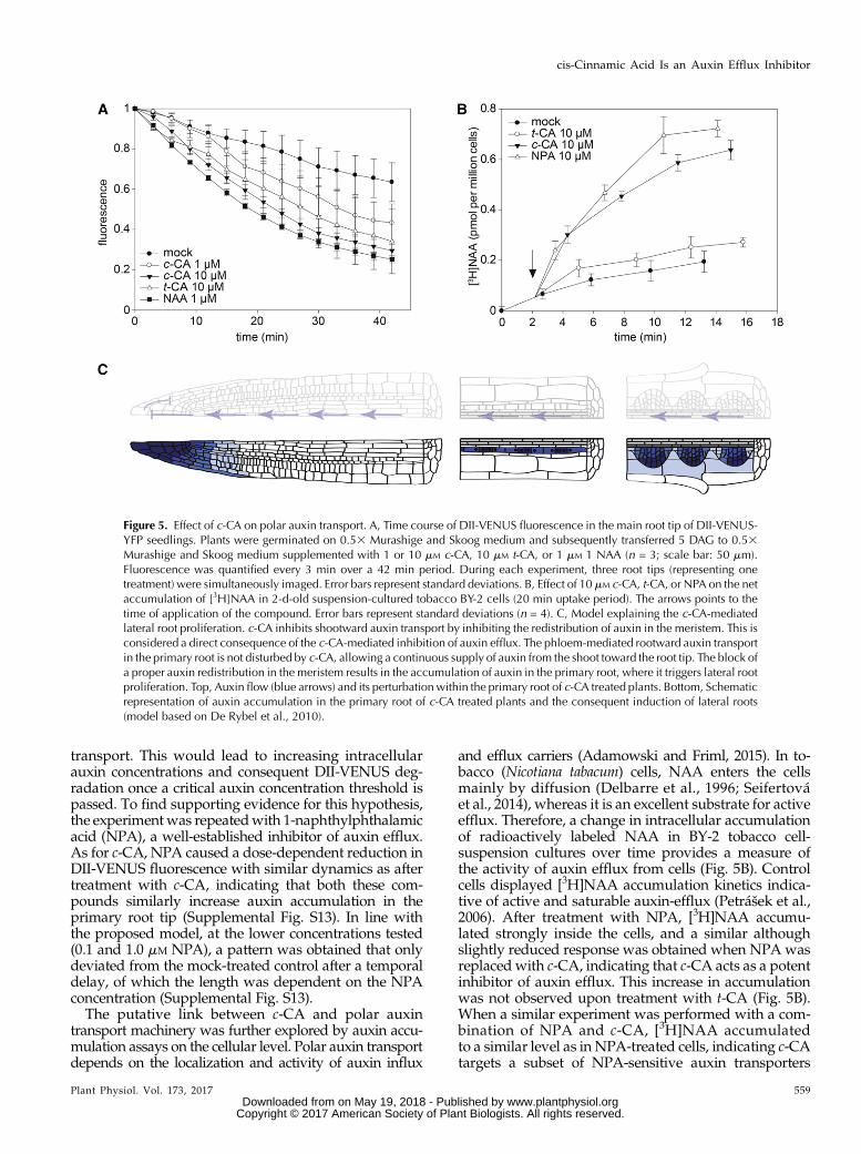
transport. This would lead to increasing intracellularauxin concentrations and consequent DII-VENUS deg-radation once a critical auxin concentration threshold ispassed. To find supporting evidence for this hypothesis,the experimentwas repeatedwith 1-naphthylphthalamicacid (NPA), a well-established inhibitor of auxin efflux.As for c-CA, NPA caused a dose-dependent reduction inDII-VENUS fluorescence with similar dynamics as aftertreatment with c-CA, indicating that both these com-pounds similarly increase auxin accumulation in theprimary root tip (Supplemental Fig. S13). In line withthe proposed model, at the lower concentrations tested(0.1 and 1.0 mM NPA), a pattern was obtained that onlydeviated from the mock-treated control after a temporaldelay, of which the length was dependent on the NPAconcentration (Supplemental Fig. S13).The putative link between c-CA and polar auxin
transport machinery was further explored by auxin accu-mulation assays on the cellular level. Polar auxin transportdepends on the localization and activity of auxin influx
and efflux carriers (Adamowski and Friml, 2015). In to-bacco (Nicotiana tabacum) cells, NAA enters the cellsmainly by diffusion (Delbarre et al., 1996; Seifertováet al., 2014), whereas it is an excellent substrate for activeefflux. Therefore, a change in intracellular accumulationof radioactively labeled NAA in BY-2 tobacco cell-suspension cultures over time provides a measure ofthe activity of auxin efflux from cells (Fig. 5B). Controlcells displayed [3H]NAA accumulation kinetics indica-tive of active and saturable auxin-efflux (Petrášek et al.,2006). After treatment with NPA, [3H]NAA accumu-lated strongly inside the cells, and a similar althoughslightly reduced response was obtained when NPA wasreplaced with c-CA, indicating that c-CA acts as a potentinhibitor of auxin efflux. This increase in accumulationwas not observed upon treatment with t-CA (Fig. 5B).When a similar experiment was performed with a com-bination of NPA and c-CA, [3H]NAA accumulatedto a similar level as in NPA-treated cells, indicating c-CAtargets a subset of NPA-sensitive auxin transporters
Figure 5. Effect of c-CA on polar auxin transport. A, Time course of DII-VENUS fluorescence in the main root tip of DII-VENUS-YFP seedlings. Plants were germinated on 0.53 Murashige and Skoog medium and subsequently transferred 5 DAG to 0.53Murashige and Skoog medium supplemented with 1 or 10 mM c-CA, 10 mM t-CA, or 1 mM 1 NAA (n = 3; scale bar: 50 mm).Fluorescence was quantified every 3 min over a 42 min period. During each experiment, three root tips (representing onetreatment) were simultaneously imaged. Error bars represent standard deviations. B, Effect of 10 mM c-CA, t-CA, or NPA on the netaccumulation of [3H]NAA in 2-d-old suspension-cultured tobacco BY-2 cells (20 min uptake period). The arrows points to thetime of application of the compound. Error bars represent standard deviations (n = 4). C, Model explaining the c-CA-mediatedlateral root proliferation. c-CA inhibits shootward auxin transport by inhibiting the redistribution of auxin in the meristem. This isconsidered a direct consequence of the c-CA-mediated inhibition of auxin efflux. The phloem-mediated rootward auxin transportin the primary root is not disturbed by c-CA, allowing a continuous supply of auxin from the shoot toward the root tip. The block ofa proper auxin redistribution in the meristem results in the accumulation of auxin in the primary root, where it triggers lateral rootproliferation. Top, Auxin flow (blue arrows) and its perturbation within the primary root of c-CA treated plants. Bottom, Schematicrepresentation of auxin accumulation in the primary root of c-CA treated plants and the consequent induction of lateral roots(model based on De Rybel et al., 2010).
Plant Physiol. Vol. 173, 2017 559
cis-Cinnamic Acid Is an Auxin Efflux Inhibitor
www.plantphysiol.orgon May 19, 2018 - Published by Downloaded from Copyright © 2017 American Society of Plant Biologists. All rights reserved.

(Supplemental Fig. S14), which could be either PIN-FORMED (PIN) or ATP-binding cassette-B (ABCB)transporters (Petrášek and Friml, 2009). To distinguishbetween both, NPA was substituted for the ABCB-specific inhibitor 2-[4-(diethylamino)-2-hydroxybenzoyl]benzoic acid (BUM; Kim et al., 2010). In contrast to NPA,BUM inhibited auxin efflux to the same extent as c-CA inthe auxin transport assay, and no additive effect wasobserved when BUM and c-CA were used simulta-neously (Supplemental Fig. S15). This strongly sug-gests c-CA targets predominantly the ABCB-auxintransport machinery. To test whether c-CA might alsoaffect auxin influx, [3H]NAA was replaced for [3H]2,4-dichlorophenoxyacetic acid ([3H]2,4-D), which is apreferred substrate for influx activity. When added tothe BY-2 cell suspension, [3H]2,4-D accumulated in thecells until a plateau was reached, representing equilib-rium between cellular influx and efflux of the labeledcompound (Supplemental Fig. S16). Using this experi-mental setup, we found no indication that either c-CA ort-CA affects cellular auxin influx.
Based on these experiments, we concluded that c-CA,but not t-CA, inhibits auxin efflux from cells, morespecifically theABCB-mediated part of auxin efflux. Theconsequent accumulation of intracellular auxin could beat the basis of the physiological and developmentaldefects observed in c-CA-treated Arabidopsis seedlings.
c-CA Does Not Inhibit Long-Distance RootwardAuxin Transport
Although both NPA and c-CA block cellular auxinefflux, their effects on Arabidopsis roots are entirelydifferent. NPA arrests (Casimero et al., 2001; Benkováet al., 2003) and c-CA induces lateral root formation.Supported by the spatiotemporal distribution of pDR5-driven luciferase activity, we hypothesized that a dif-ference in long-distance auxin transport could be theorigin of the phenotypic difference between these twoauxin efflux inhibitors. Whereas NPA affects bothrootward and shootward auxin transport in the pri-mary root (Casimero et al., 2001), the strong increase inluciferase activity in the tip of c-CA-treated roots sug-gested that rootward auxin transport is not disturbedby c-CA. To verify this hypothesis, we monitoredwhether local c-CA application could affect distantauxin-inducible luciferase activity using a split mediumapproach as described by Lewis and Muday (2009). Tothis end, seedlings were positioned on the medium ina way that either the upper or the lower half of theroot was in contact with c-CA. Dynamics of luciferaseactivity along the root were followed over time as de-scribed above. When the lower half of the root was incontact with c-CA, luciferase activity accumulated inthe root tip in line with earlier data (Supplemental Fig.S17). When only the upper part of the root was incontact with c-CA, the luciferase signal quickly ex-tended toward the non-treated zone (SupplementalFig. S17). This illustrates that auxin appears to be able
to pass through the c-CA-treated zone in a rootwarddirection.
Although the data support the hypothesis that c-CAallows long-distance rootward auxin transport, wecould not exclude an alternative explanation, namelythat c-CA itself is transported and triggers auxin sig-naling locally. To provide undisputed evidence forrootward transport of auxin in c-CA-treated roots, long-distance rootward auxin transport was assayed inprimary roots of Arabidopsis seedlings in which theroots were exposed to either mock or c-CA-treated0.53 Murashige and Skoog medium. In these assays,microdroplets of radiolabeled [3H]IAA were placedprecisely on the shoot apical meristems of Arabidopsisseedlings, and rootward auxin transport was measuredby harvesting a 4 mm segment centered on the root/shoot transition zone, as well as the entire root, in 2 mmsegments. Consistent with previous results, treatmentwith c-CA did not inhibit rootward auxin movement(Fig. 5C; Supplemental Fig. S18). The strong accumu-lation of the auxin-inducible luciferase in the root tip ischaracteristic of the inhibition of shootward auxintransport (Fig. 3A; Supplemental Figs. S6 and S17).Unfortunately, reliable data were not obtained forshootward auxin transport to support this hypothesis.
Taken together, our data supports a model (Fig. 5C)in which c-CA inhibits auxin efflux at the cellular levelin specific cells at or near the root apical meristem,while allowing long-distance rootward auxin transportat the organ level. The resultant accumulation of auxinin the root apical meristem might cause, at least in part,the observed growth defects induced by c-CA.
DISCUSSION
Being sessile organisms, plants cannot escape unfa-vorable growth conditions. This shortcoming is com-pensated by an extreme plasticity, allowing them toreact to changing environmental cues. Here, the phy-tohormone auxin has an important function, as it is keyin the regulation of many processes involved in growthand development (Vanneste and Friml, 2009). As for allbioactive compounds, tight regulation of its homeo-stasis and spatiotemporal distribution inside the plantis crucial, as suboptimal auxin concentrations will nottrigger the desired response, while high concentrationswill be harmful. The ability to control auxin levels is anecessity for plant survival and occurs at the cellularlevel by regulating biosynthesis, metabolic conversionsas well as degradation, whereas transport is essential totranslocate auxin between different cells and tissues.Synthetic inhibitors of auxin transport such as NPA andBUM have proven the importance of this process indiverse physiological actions, including embryogene-sis, tropisms, vascular patterning, and lateral root ini-tiation (Kim et al., 2010). Intriguingly, endogenousauxin transport inhibitors are scarce. Flavonols andflavonoids such as quercetin were considered to inhibitauxin transporters (Brown et al., 2001), although later
560 Plant Physiol. Vol. 173, 2017
Steenackers et al.
www.plantphysiol.orgon May 19, 2018 - Published by Downloaded from Copyright © 2017 American Society of Plant Biologists. All rights reserved.

work suggested that flavonoids also act by redirectingPIN efflux protein localization (Santelia et al., 2008).Certain flavonoid mutants display auxin-related defects(Buer et al., 2013) and an auxin-transport inhibiting ac-tivity was recently assigned to the flavonol glycosidekaempferol 3-O-rhamnoside-7-O-rhamnoside (Yin et al.,2014).Here, we introduce c-CA as a novel endogenous in-
hibitor of auxin transport. Intriguingly, the activity ofc-CA resembles that of NPA, but only at the cellularlevel. Although both NPA and c-CA block cellularauxin efflux, the effects of the two compounds onArabidopsis root architecture are entirely different,with c-CA inducing lateral root formation and NPAimpacting lateral root initiation. The exact mechanismof NPA action is still unknown, but according to onehypothesis the slr phenotype of NPA-treated plants is aconsequence of auxin depletion in the root due to theperturbation of basipetal and acropetal auxin transport(i.e. shootward and rootward, respectively; Casimiroet al., 2001). Although explaining the observed phenotype,the molecular mechanism underlying the inhibitionof phloem-based (and hence no-transporter-mediated)rootward auxin transport by an auxin efflux inhibitorremains unknown. Proceeding from this model, wehypothesized that a difference at the level of rootwardtransport (perturbed byNPA but not by c-CA) underliesthe phenotypic differences caused by the two com-pounds. Under mock conditions, auxin is redistributedin the root tip according to the “reverse-fountain”model,in which specific auxin transport proteins (PINs andABCB proteins) play distinct roles in establishing direc-tional movement of auxin (Benková et al., 2003; Blilouet al., 2005; Lewis and Muday, 2009). By inhibiting cel-lular auxin efflux, we hypothesize c-CA will affect theauxin reflux in themeristem, resulting in the inhibition ofshootward auxin transport. Consequently, auxin eithertransported from the shoot or synthesized in the pri-mary root tip will accumulate behind the root tipwhere it will trigger GATA23-expression and affectlateral root founder cell specification. Over time, theaccumulating auxin will enter pericycle cells, either bydiffusion or active influx where it will be trapped dueto the c-CA-mediated inhibition of auxin efflux, simi-lar to the situation in the primary root. Once the auxinconcentration passes a critical threshold, primed cellswill be triggered to develop into lateral root foundercells, which eventually will develop into new lateralroots, shaping the altered root architecture (Fig. 5C).Compared to NPA, c-CA was found slightly less ef-
ficient in the auxin accumulation assay. This differencemay result from the broader specificity of NPA, knownto affect different types of auxin efflux carriers. Basedon the absence of an additive effect of c-CA and BUM inthis assay, we concluded that c-CA targets the ABCBsubfamily of the multidrug-resistant/P-glycoproteinintegral membrane proteins. These transporters arewell known for their capacity to pump drugs out of thecell (Kang et al., 2011), increasing the resistance of thecell and hence the organism toward compounds that
are considered toxic under normal conditions. Inter-estingly, and in line with our observation, the cis-formof CA and not its trans-form raises a notable synergisticbactericidal activity against multiple-drug-resistantMycobacterium tuberculosis. It is tempting to speculatethat also in this case c-CA blocks theMDR-transporters,resulting in the intracellular accumulation of the sup-plied antibiotics to levels required to kill the bacteria(Chen et al., 2011).
Although the physiological role of endogenous c-CAis still unclear, the beauty of this bioactive molecule liesin the fact that it can be produced from a readily avail-able inactive compound (t-CA) by sunlight (Ding et al.,2011). This gives a tremendous opportunity to link en-vironmental conditions directly to developmental regu-lation without the need to activate gene expression toalter the auxin pool. In addition, we cannot exclude thata similar conversion can be obtained by a yet-to-be-discovered enzyme, further extending the possibilitiesto exploit this mechanism to steer plant developmentindependently of light. The question of whether c-CAhas an active role in the regulation of plant developmentremains an open and intriguing question; however, thefact that it was previously found in small but physio-logically relevant quantities in plants and that the effectson roots are evolutionary conserved only feeds thespeculation on its importance as an endogenous plantgrowth regulator (Yin et al., 2003;Wong et al., 2005). Thisfunction could be different from lateral root develop-ment, a system that we only used to elucidate the mo-lecular mechanism of c-CA action.
MATERIALS AND METHODS
Plant Material, Transgenic Lines, Chemicals, andGrowth Conditions
The effect of c/t-CA on plant growth and development was studied ina diverse set of plant species, comprising Physcomitrella patens, Selaginellamoellendorffii, Oryza sativa, Nicotiana benthamiana, Brachypodium distachyon,and Arabidopsis thaliana. Arabidopsis ecotype Columbia (Col-0) was used,unless stated elsewhere. Following transgenic lines were in the same eco-type: DII-VENUS, pDR5rev:GFP, pDR5:LUC, pGATA23:GUS, pGAZAT:GUS,pKNOLLE:KNOLLE-GFP, p35S:iaaL, slr, arf7 arf19, and tir1 afb2 afb3 (Romanoet al., 1991; Lukowitz et al., 1996; Fukaki et al., 2002; Friml et al., 2003;Dharmasiri et al., 2005; González-Carranza et al., 2007; Okushima et al., 2007;De Rybel et al., 2010; Moreno-Risueno et al., 2010; Brunoud et al., 2012).The transgenic line pCYCB1:GUS was in the ecotype Landsberg erecta (Ler;Colón-Carmona et al., 1999). Seeds were vapor-phase sterilized and grown on0.53 Murashige and Skoog medium. The 0.53 Murashige and Skoog medium(pH 5.7) contains per liter 1.5 gMurashige and Skoog basal salt mixture powder(Duchefa), 7.14 g Suc, 0.36 g MES monohydrate, 8 g plant tissue culture agar.The medium was supplemented with one of the following compounds: NAA(Sigma Aldrich), NPA (Sigma Aldrich), c-CA (Shanghai Specbiochem), andt-CA (Sigma Aldrich) from stock solutions in DMSO (final 0.1% DMSO) to theautoclaved medium prior to pouring the plates. After sowing, seeds were in-cubated at 4°C for at least 2 d, whereupon plates were placed in a vertical or-ientation in the tissue culture chamber room under a 16-h-light/8-h-darkphotoperiod at 21°C, except for the experiments done to reveal the pure c-CAand/or t-CA effect. Seedlings grown in darkness received a short 4 h red lightpulse to induce germination. Propidium-iodide (PI; Sigma Aldrich) was used tocounterstain the cell wall. The adventitious rooting assay was performed byplacing plates in darkness for 7 d (after a short light pulse with red light of 4 h).Plates were then exposed to light for 5 d. The root-bending assay was performed
Plant Physiol. Vol. 173, 2017 561
cis-Cinnamic Acid Is an Auxin Efflux Inhibitor
www.plantphysiol.orgon May 19, 2018 - Published by Downloaded from Copyright © 2017 American Society of Plant Biologists. All rights reserved.

on 5-d-old seedlings treated with different concentrations of c/t-CA. After 5 dplates were rotated 90 degrees and root gravitropismwas scored after 48 h. Scanswere made and the quantification of the response was performed withImageJ. Tobacco (Nicotiana tabacum ‘Bright Yellow-2’) cells of the cell line BY-2(Nagata et al., 1992) were cultivated according to Petrášek et al. (2006) and sub-cultured weekly. Bromophenol blue (Sigma-Aldrich) was used to stain the cellwall of Physcomitrella patens leaves.
Description of Plant Phenotype
To quantify growth parameters and check for aberrant phenotypes, seedswere grown on square plates placed in a vertical orientation in the growthchamber. Plateswere scannedusing the Scanmaker 9800XL, and root lengthwasmeasured using the ImageJ software. For each compound, the inhibitory con-centration (IC50) was calculated, plotting a dose-response curve in SigmaPlot.The dose-response curve resulting in the highest R2 value (coefficient of de-termination) was used. The number of plants used and the timing of thescanning depends on the plant species and the treatment. The number of ad-ventitious roots (above the root-shoot junction) and number of emerged lateralroots were counted using a stereomicroscope (CETI Binocular Zoom Stereo).
Histochemical Analysis and Confocal Microscopy
Root cell walls were stainedwith 30mMPI for pKNOLLE:KNOLLE-GFP at theonset of the experiment. The excitation energy of 488 nm was from an argonlaser. The PI fluorescence emission was collected between 550 and 650 nm andGFP/YFP between 500 and 550 nm. All images were captured with an invertedLSM 710 META confocal microscope equipped with 203-Air objectives (CarlZeiss). GUS assays were performed and inspected using differential interfer-ence contrast optics as described earlier in Beeckman and Engler (1994).
Time Lapse DII-VENUS
For analysis of chemically treated roots, 7-d-old DII-VENUS Arabidopsisseedlings were used. At the onset of the time lapse, three seedlings (biologicalrepeats)were placed in glass-bottomeddishes and coveredwith 0.53MS-mediacontaining NAA, NPA, c-CA, or t-CA. The time-lapse was started 5 min afterthe seedlings had been placed in contact with the media and captured over42 min (every 3 min) with an inverted LSM 710 META confocal microscopeequipped with 203-Air objectives (Carl Zeiss). Images were analyzed with theFiji software using the total signal from Z-projection of defined region (alwaysthe same area). Normalization was done by using the initial signal from theZ-projection of adefined region as the baseline.
Time Lapse pDR5rev:GFP
Seven-day-oldArabidopsis seedlingswere used to analyze the effect of c-CA,t-CA, NPA, and NAA on the expression of pDR5rev:GFP in the region betweentwo emerged lateral roots. At the start of the time lapse, seedlings were placedin glass-bottomed dishes and covered with media containing NAA, c-CA, ort-CA. The time lapse was started 5 min after the seedlings had been placed incontact with the media and captured over a period of 16h, every hour withan inverted LSM 710 META confocal microscope (Carl Zeiss) equipped with203-air objective (Carl Zeiss). Images were analyzed with the Volocity soft-ware. The accumulation projection spectrum was obtained by projecting theGFP intensity on a virtual line crossing the middle of the primary root over theimaged distance of the root. This way pDR5rev:GFP expression can be imagedand quantified in every cell type. Normalization was performed against theintensity to the highest obtained signal at the earliest time point.
Time Lapse pDR5:LUC
The pDR5:LUC images were taken by a Lumazone machine carrying a CCD(CCD) camera (Princeton Instruments). The CCD camera that is controlled by aWinView/32 software took movies of the LUC expression automatically every10 min (exposure time, 10 min) for 12 h. Before imaging, plates containing 0.53Murashige and Skoog medium were sprayed with 1 mM D-luciferin solution(Duchefa Biochemie). The picture series were saved as TIFF format for furtheranalysis. The luciferase signals were quantified by the measure of the analog-digital units per pixel by means of ImageJ. To visualize the spatiotemporalluciferase changes during treatment with the compound, a Kymograph
(http://www.embl.de/eamnet/html/body_kymograph.html) was generatedwith ImageJ.
Heterologous Expression of C4H and Microsome Assay
The Saccharomyces cerevisiae strain containing the Arabidopsis C4Hwas used(Van de Wouwer et al., 2016). One hundred microliters of recombinant yeast inglycerol was grown overnight at 30°C in 5 mL liquid DO medium (ClontechLaboratories). The yeast cells were pelleted (1 min at 4000 rpm), washed with5 mL sterile MQwater, pelleted again, and resuspended in another 5 mLwater.The amount of inoculum was calculated to reach an OD600 of 0.1 and subse-quently, the yeast cultures were grown for 16 h at 30°C with shaking (200 rpm)in DO medium (Clontech Laboratories) containing Gal to induce transcription.Microsomes were prepared according to Schalk et al. (1998). The microsomeassay was done with aliquots of 10 mL microsome, by adding 20 mM sodiumphosphate buffer (pH 7.4), 10 mL of the desired compound at final concentra-tions of 10 mM for c-CA and t-CA and equal amounts of DMSO as a control. Tostart the reaction, 10 mL of the 10mMNADP+ sodium phosphate buffer solutionwas added to the Eppendorf, briefly vortexed and immediately placed in theEppendorf thermomixer at 28°C for 20 min. The reaction was stopped byadding 150 mL ice-cold methanol. The pellet was resuspended in 500 mL 90%methanol and incubated in an Eppendorf thermomixer at 30°C for 10 min whileshaking at 1000 rpm. After centrifugation at 14,000 rpm for 5 min, the super-natant was transferred to a new Eppendorf tube and lyophilized. The pellet wastreated with 100 mL water and 100 mL cyclohexane. After 10 min of centrifu-gation (14,000 rpm), 80 mL of the aqueous phase was retained for UHPLC-MSanalysis. For reversed-phase LC, 10 mL of the aqueous phase was subjected toUHPLC-MS on a Waters Acquity system (Waters Corporation) connected to aThermo LTQ XL mass spectrometer (Thermo Scientific). Chromatographicgradient separation was carried out as described in the next paragraph. Theeluent was directed to the mass spectrometer via electrospray ionization innegative mode. MS source parameters were as follows: capillary temperature,300°C; capillary voltage, 24 V; source voltage, 3.5 V; source current, 100 A;sheath gas flow, 30; aux gas flow, 20; sweep gas flow, 5. The mass range was setbetween 100 and 1000 D. c-CA, t-CA, and p-coumaric acid were characterizedbased on the similarity of their masses and retention times with those of stan-dards. Peak detection and integration was done with Progenesis QI v2.1(Nonlinear Dynamics, a Waters Company). Product/substrate ratios werecalculated and P values were calculated using unpaired Student’s t-tests.
LC-MS/MS and LC-UV-Vis-MS to Determine c- andt-CA Photo-Isomerization
Exactly 2.5 mg of pure t-CA and c-CA was dissolved in 50.0 mL Milli-Q-H2O/DMSO (80/20). Solutions were subsequently incubated in the growthchamber and isomerization of both isomerswas followedover time byLC-MS/MS.For darkness, plates were covered with aluminum foil to exclude light, andsampling was performed in darkness. Deep-red and far-red illumination wasprovided by the GreenPower LED module (Philips).
For quantification of t-CA and c-CA, a 15mL aliquot was subjected to LC-MSanalysis performed on a Waters Acquity UPLC system equipped with a PDAdetector (lambda range from 190 to 500 nm;Waters Corporation) connected to aSynapt HDMS quadrupole time-of-flight mass spectrometer (Waters MSTechnologies). Chromatographic separation was performed on an AcquityUPLC BEHC18 column (2.1 mm3 150mm, 1.7mm;Waters Corporation) usinga water-acetonitrile gradient elution. Mobile phases were composed of (A)water containing 1% acetonitrile and 0.1% formic acid and (B) acetonitrilecontaining 1% water and 0.1% formic acid. The column temperature wasmaintained at 40°C, and the autosampler temperature was maintained at 10°C.A flow rate of 350 mL/min was applied during the gradient elution, with ini-tialization at time 0 min 5% (B), 30 min 50% (B), and 33 min 100% (B). ForUV-Vis detection, data were recorded between 210 and 500 nm. The eluant wasthen directed to the mass spectrometer equipped with an electrospray ioniza-tion source and lockspray interface for accurate mass measurements. The MSsource parameters were as follows: capillary voltage, 2.5 kV; sampling cone,37 V; extraction cone, 3.5 V; source temperature, 120°C; desolvation tempera-ture, 400°C; cone gas flow, 50 L h21; and desolvation gas flow, 550 L h21. Thecollision energy for the trap and transfer cells was 6 and 4 V, respectively. Fordata acquisition, the dynamic range enhancement mode was activated. Full-scan data were recorded in negative centroid V-mode; the mass range betweenm/z 100 and 1000,with a scan speedof 0.2 s scan21. Leucin-enkephalin (250 pgmL21;solubilized inwater: acetonitrile 1:1 [v/v]with 0.1% [v/v] formic acid)was used for
562 Plant Physiol. Vol. 173, 2017
Steenackers et al.
www.plantphysiol.orgon May 19, 2018 - Published by Downloaded from Copyright © 2017 American Society of Plant Biologists. All rights reserved.

lockmass calibration, with scanning every 10 s with a scan time of 0.5 s. All datawere recorded with Masslynx software (version 4.1, Waters). For the quantifi-cation of t-CA and c-CA, the UV-Vis chromatogram was extracted at 277 nm,and peaks were integrated automatically (automatic noise measurement; meansmoothing [window size, 3; no. of smooths, 2]). Peak areas were used to cal-culate the conversion of t-CA and c-CA.
Auxin Metabolite Profiling
Extraction and purification of auxin and its metabolites was done as de-scribed previously with minor modifications (Novák et al., 2012). Frozensamples were homogenized using aMixerMill (Retsch GmbH) and extracted in1 mL 50 mM sodium phosphate buffer (pH 7.0) containing antioxidant (1%sodium diethyldithiocarbamate) and a cocktail of deuterium and 13C6-labeledinternal standards of IAA and its metabolites. The pH was adjusted to 2.7 with1 M hydrochloric acid, and the extracts were purified on Oasis HLB columns(30mg,Waters Corporation), conditionedwith 1mLmethanol, 1mLwater, and0.5mL sodium phosphate buffer (pH 2.7). After sample application, the columnwas washed with 2 mL 5%methanol and then eluted with 2 mL 80%methanol.Eluates were evaporated to dryness and dissolved in 20 mL of mobile phaseprior to mass analysis using a 1290 Infinity LC system and 6460 Triple QuadLC-MS system (Agilent Technologies; Novák et al., 2012).
Auxin Accumulation Assays
Assays were performed according to Petrášek et al. (2003). Auxin accumu-lation was measured in tobacco BY-2 cells (Nagata et al., 1992) 48 h after sub-cultivation in 0.5 mL aliquots of cell suspension (target working cell density was7 3 105 cells 3 mL21, and it was determined precisely by counting in the Fuchs-Rosenthal hemocytometer). Cultivation medium was removed by filtration on20 mm mesh nylon filters, and cells were resuspended in uptake buffer (20 mM
MES, 10mMSuc, 0.5mMCaSO4, pHadjusted to 5.7withKOH) and equilibrated for45 min on the orbital shaker at 27°C in darkness. Equilibrated cells were collectedby filtration, resuspended in fresh uptake buffer, and incubated with continuousorbital shaking for another 90 min under the same conditions. Radiolabeled auxin([3H]NAA or [3H]2,4-D; specific [molar] radioactivity 20 Ci/mmol each; AmericanRadiolabeled Chemicals) was added to the cell suspension to a final concentrationof 2 nM. At certain time points, aliquots of the cell suspension were sampled, andaccumulation of radiolabeled auxins was terminated by rapid filtration under re-duced pressure on cellulose filters (22mm in diameter). Cell cakeswith filters weretransferred into scintillation vials, extractedwith ethanol (UV-spectroscopy grade)for 30 min, and radioactivity was determined by liquid scintillation counting(Packard Tri-Carb 2900TR scintillation counter, Packard Instrument). Countingefficiency was determined by automatic external standardization, and countswere corrected for quenching automatically. For remaining surface radioac-tivity, counts were corrected by subtracting counts of aliquots collectedimmediately after addition of radiolabeled auxin. Inhibitors were added asrequired from stock solutions to an appropriate final concentration, and propercontrols (solvent) were applied. Recorded accumulation values were recalcu-lated to 1 million cells.
Rootward Auxin Transport Assays
Rootward auxin transport assays were performed as described previously(Geisler et al., 2005). In brief, 0.1 mL microdroplets containing 500 nM [3H]IAA(American Radiolabeled Chemicals) and 500 nM cold IAA (SigmaAldrich) wereplaced on the shoot apical meristem of Arabidopsis seedlings, and rootwardauxin transport was measured by harvesting a 4 mm segment centered onthe root shoot transition zone, as well as the entire root, in 2 mm segments(beginning with root zone-1 just after the transition zone, and ending with themain root tip). Treatments with Murashige and Skoog media and 10 mM c-CAwere carried out by saturating the filter paper matrix on which the roots wereincubated during auxin transport assays with Murashige and Skoog mediasupplemented with either a water:methanol blank or c-CA.
Auxin Binding and Anti-auxin Experiments Using SPRand Docking
Auxin receptor proteins AtTIR1 and AtAFB5 were expressed in insect cells(T. ni High5) and purified as described previously (Calderón Villalobos et al.,2012; Lee et al., 2014). The biotinylated degron peptide representing Aux/IAA7
was purchased from ThermoFisher Scientific and immobilized on streptavidin-coated SPR chips (GE Healthcare). SPR experiments were run as describedpreviously (Calderón Villalobos et al., 2012; Lee et al., 2014). Briefly, com-pounds were added to purified receptor proteins from stock solutions in DMSOto give working concentrations, which were 50 mM unless stated otherwise(DMSO 0.1% final). Controls lacking auxin/compound and controls containingIAA (50 mM) were run as references at the start and end of every set of sen-sorgrams on every protein preparation. Compounds were run in three separateexperiments, with characteristic results shown. For anti-auxin runs, receptorproteins were mixed with 5 mM IAA plus compound at 50 mM. An anti-auxineffect was then determined if the compound competed with IAA, reducing theamplitude of TIR1/AFB5 binding on the sensorgram. Docking was performedusing the Vina docking algorithm (Morris et al., 2009; Trott and Olson, 2010).With the TIR1 crystal structure (PDB code 2P1P) from Tan et al. (2007). In silicomodeling, molecular graphics, and analyses were performed with the Univer-sity of California San Francisco Chimera package. Chimera is open sourceand developed by the Resource for Biocomputing, Visualization, and Infor-matics at the University of California, San Francisco (supported by NIGMSP41-GM103311; Pettersen et al., 2004). Marvin was used for drawing, dis-playing, and characterizing chemical structures, substructures, and reactions.Calculator plugins were used for structure property prediction and calcula-tion Marvin v15.10.12.0, 2015, ChemAxon (http://www.chemaxon.com).
Supplemental Figures
The following supplemental materials are available.
Supplemental Figure S1. The general phenylpropanoid pathway.
Supplemental Figure S2. Effect of c/t-CA on growth and development ofdifferent plant species.
Supplemental Figure S3. Photo-isomerization of c-CA and t-CA.
Supplemental Figure S4. Conversion of t-CA by C4H in Arabidopsis.
Supplemental Figure S5. Effect of c-CA on pGATA23-drivenGUS expression.
Supplemental Figure S6. Time-dependent pDR5-driven LUC expressionupon c-CA treatment.
Supplemental Figure S7. Time-dependent pDR5-driven GFP expressionupon c-CA treatment.
Supplemental Figure S8. Docking of c-CA and t-CA to the auxin bindingpocket of TIR1.
Supplemental Figure S9. Shift in IAA-related metabolites upon treatmentwith 10 mM c-CA and t-CA for 1 h.
Supplemental Figure S10. Shift in the IAA metabolome upon treatmentwith 10 mM c-CA and t-CA for 6 h.
Supplemental Figure S11. The effect on IAA reduction on c-CA-mediateddevelopmental defects in seedlings.
Supplemental Figure S12. DII-VENUS response to c-CA.
Supplemental Figure S13. DII-VENUS response to NPA.
Supplemental Figure S14. The effect of combined treatment with c-CA andNPA on auxin accumulation.
Supplemental Figure S15. The effect of combined treatment with c-CA andBUM on auxin accumulation.
Supplemental Figure S16. The effect of c-CA on polar auxin transport.
Supplemental Figure S17. Time-dependent DR5-driven LUC expressionupon local application of c-CA.
Supplemental Figure S18. The effect of c-CA on long distance rootwardauxin transport in Arabidopsis.
ACKNOWLEDGMENTS
We thank Ottoline Leyser (Sainsbury Laboratory, University of Cambridge)for providing us p35S:iaaL Arabidopsis seeds. We thank Karel Spruyt for imag-ingwherever needed, Toon Babylon for technical assistance,Wei Xua andDavy
Plant Physiol. Vol. 173, 2017 563
cis-Cinnamic Acid Is an Auxin Efflux Inhibitor
www.plantphysiol.orgon May 19, 2018 - Published by Downloaded from Copyright © 2017 American Society of Plant Biologists. All rights reserved.

Opdenacker for help with the pDR5:LUC experiments, and Tao Fang and HansMotte for providing us with the Selaginella moellendorffii plantlets. We also ap-preciated the help of the VIB Imaging Core facility, namely AmandaGonçalves,who helped with analyzing the imaging experiments, and Dominic Petrella forcritical reading of the manuscript. Finally, we thank Wim Grunewald, TomBeeckman, Steffen Vanneste, Bert De Rybel, and Jürgen Kleine-Vehn for scien-tific discussions.
Received September 29, 2016; accepted November 1, 2016; publishedNovember 11, 2016.
LITERATURE CITED
Åberg B (1961) Studies on plant growth regulator XVIII. Someb-substituted acrylic acids. Ann Roy Agric Coll Sweden 27: 99–123
Adamowski M, Friml J (2015) PIN-dependent auxin transport: action,regulation, and evolution. Plant Cell 27: 20–32
Beeckman T, Engler G (1994) An easy technique for the clearing of histo-chemically stained plant tissue. Plant Mol Biol Rep 12: 37–42
Benková E, Michniewicz M, Sauer M, Teichmann T, Seifertová D, Jürgens G,Friml J (2003) Local, efflux-dependent auxin gradients as a common modulefor plant organ formation. Cell 115: 591–602
Blilou I, Xu J, Wildwater M, Willemsen V, Paponov I, Friml J, Heidstra R,Aida M, Palme K, Scheres B (2005) The PIN auxin efflux facilitatornetwork controls growth and patterning in Arabidopsis roots. Nature433: 39–44
Boerjan W, Ralph J, Baucher M (2003) Lignin biosynthesis. Annu Rev PlantBiol 54: 519–546
Brown DE, Rashotte AM, Murphy AS, Normanly J, Tague BW, Peer WA,Taiz L, Muday GK (2001) Flavonoids act as negative regulators of auxintransport in vivo in arabidopsis. Plant Physiol 126: 524–535
Brunoud G, Wells DM, Oliva M, Larrieu A, Mirabet V, Burrow AH,Beeckman T, Kepinski S, Traas J, Bennett MJ, et al (2012) A novelsensor to map auxin response and distribution at high spatio-temporalresolution. Nature 482: 103–106
Buer CS, Kordbacheh F, Truong TT, Hocart CH, Djordjevic MA (2013)Alteration of flavonoid accumulation patterns in transparent testa mu-tants disturbs auxin transport, gravity responses, and imparts long-termeffects on root and shoot architecture. Planta 238: 171–189
Calderón Villalobos LI, Lee S, De Oliveira C, Ivetac A, Brandt W, Armitage L,Sheard LB, Tan X, Parry G, Mao H, et al (2012) A combinatorial TIR1/AFB-Aux/IAA co-receptor system for differential sensing of auxin. NatChem Biol 8: 477–485
Casimiro I, Marchant A, Bhalerao RP, Beeckman T, Shooge S, Swarup R,Graham N, Inzé D, Sandberg G, Casero PJ, Bennet M (2001) Auxintransport promotes Arabidopsis lateral root initiation. Plant Cell 13:843–852
Chen YL, Huang ST, Sun FM, Chiang YL, Chiang CJ, Tsai CM, Wang CJ(2011) Transformation of cinnamic acid from trans- to cis-form raises anotable bactericidal and synergistic activity against multiple-drug re-sistant Mycobacterium tuberculosis. Eur J Pharm Sci 43: 188–194
Colón-Carmona A, You R, Haimovitch-Gal T, Doerner P (1999) Technicaladvance: spatio-temporal analysis of mitotic activity with a labile cyclin-GUS fusion protein. Plant J 20: 503–508
De Rybel B, Vassileva V, Parizot B, Demeulenaere M, Grunewald W,Audenaert D, Van Campenhout J, Overvoorde P, Jansen L, Vanneste S,et al (2010) A novel aux/IAA28 signaling cascade activates GATA23-dependentspecification of lateral root founder cell identity. Curr Biol 20: 1697–1706
Delbarre A, Muller P, Imhoff V, Guern J (1996) Comparison of mecha-nisms controlling uptake and accumulation of 2,4-dichlorophenoxyacetic acid, naphthalene-1-acetic acid, and indole-3-acetic acid insuspension-cultured tobacco cells. Planta 198: 532–541
Dharmasiri N, Dharmasiri S, Weijers D, Lechner E, Yamada M, Hobbie L,Ehrismann JS, Jürgens G, Estelle M (2005) Plant development is reg-ulated by a family of auxin receptor F box proteins. Dev Cell 9: 109–119
Ding Z, Galván-Ampudia CS, Demarsy E, Łangowski Ł, Kleine-Vehn J,Fan Y, Morita MT, Tasaka M, Fankhauser C, Offringa R, et al (2011)Light-mediated polarization of the PIN3 auxin transporter for the pho-totropic response in Arabidopsis. Nat Cell Biol 13: 447–452
Friml J, Vieten A, Sauer M, Weijers D, Schwarz H, Hamann T, OffringaR, Jürgens G (2003) Efflux-dependent auxin gradients establish theapical-basal axis of Arabidopsis. Nature 426: 147–153
Fukaki H, Tameda S, Masuda H, Tasaka M (2002) Lateral root formation isblocked by a gain-of-function mutation in the SOLITARY-ROOT/IAA14gene of Arabidopsis. Plant J 29: 153–168
Geisler M, Blakeslee JJ, Bouchard R, Lee OR, Vincenzetti V, BandyopadhyayA, Titapiwatanakum B, Peer WA, Bailly A, Richards EL, et al (2005)Cellular efflux of auxin catalyzed by the Arabidopsis MDR/PGP trans-porter AtPGP1. Plant J 44: 179–194
González-Carranza ZH, Elliott KA, Roberts JA (2007) Expression ofpolygalacturonases and evidence to support their role during cellseparation processes in Arabidopsis thaliana. J Exp Bot 58: 3719–3730
Haagen-Smit SAJ, Went FW (1935) A physiological analysis of the growthsubstance. Proceedings. Koninklijke Akademie van Wetenschappen teAmsterdam 38: 852–857
Hitchcock AE (1935) Indole-3-n-propionic acid as a growth hormone andquantitative measurement of plant response. In Contributions of theBoyce Thompson Institute, Vol 7. Boyce Thompson Institute for PlantResearch, Yonkers, NY, pp 87–95
Hocking MB (1969) Photochemical and thermal isomerizations of cis- andtrans-cinnamic acids, and their photostationary state. Can J Chem 47:4567–4576
Kim JY, Henrichs S, Bailly A, Vincenzetti V, Sovero V, Mancuso S,Pollmann S, Kim D, Geisler M, Nam HG (2010) Identification of anABCB/P-glycoprotein-specific inhibitor of auxin transport by chemicalgenomics. J Biol Chem 285: 23309–23317
Kang J, Park J, Choi H, Burla B, Kretzschmar T, Lee Y, Marinoia E (2011)Plant ABC transporters. The Arabidopsis Book 9: e0153
Koepfli JB, Thimann KB, Went FW (1938) Plant hormones: structure andphysiological activity. I. J Biol Chem 122: 763–780
Kumpf RP, Shi CL, Larrieu A, Stø IM, Butenko MA, Péret B, Riiser ES,Bennett MJ, Aalen RB (2013) Floral organ abscission peptide IDA andits HAE/HSL2 receptors control cell separation during lateral rootemergence. Proc Natl Acad Sci USA 110: 5235–5240
Lee S, Sundaram S, Armitage L, Evans JP, Hawkes T, Kepinski S, Ferro N,Napier RM (2014) Defining binding efficiency and specificity of auxinsfor SCF(TIR1/AFB)-Aux/IAA co-receptor complex formation. ACSChem Biol 9: 673–682
Letham DS (1978) Naturally occurring plant growth regulators other thanthe principle hormones of higher plants. In DS Letham, PB Goodwin,TJV Higgins, eds, Phytohormones and Related Compounds: A Com-prehensive Treatise, Vol 1. Elsevier/North Holland Biomedical Press,Amsterdam, pp 349–417
Lewis DR, Muday GK (2009) Measurement of auxin transport in Arabidopsisthaliana. Nat Protoc 4: 437–451
Liu C, Xu Z, Chua NH (1993) Auxin polar transport is essential for theestablishment of bilateral symmetry during early plant embryogenesis.Plant Cell 5: 621–630
Lukowitz W, Mayer U, Jürgens G (1996) Cytokinesis in the Arabidopsisembryo involves the syntaxin-related KNOLLE gene product. Cell 84:61–71
Moreno-Risueno MA, Van Norman JM, Moreno A, Zhang J, Ahnert SE,Benfey PN (2010) Oscillating gene expression determines competencefor periodic Arabidopsis root branching. Science 329: 1306–1311
Morris GM, Huey R, Lindstrom W, Sanner MF, Belew RK, Goodsell DS,Olson AJ (2009) AutoDock4 and AutoDockTools4: Automated dockingwith selective receptor flexibility. J Comput Chem 30: 2785–2791
Nagata T, Nemoto Y, Hasezawa S (1992) Tobacco BY-2 cell-line as theHela-cell in the cell biology of higher-plants. Int Rev Cytol 132: 1–30
Novák O, Hényková E, Sairanen I, Kowalczyk M, Pospíšil T, Ljung K (2012)Tissue-specific profiling of the Arabidopsis thaliana auxin metabolome.Plant J 72: 523–536
Okushima Y, Fukaki H, Onoda M, Theologis A, Tasaka M (2007) ARF7 andARF19 regulate lateral root formation via direct activation of LBD/ASLgenes in Arabidopsis. Plant Cell 19: 118–130
Péret B, De Rybel B, Casimiro I, Benková E, Swarup R, Laplaze L,Beeckman T, Bennett MJ (2009) Arabidopsis lateral root development:an emerging story. Trends Plant Sci 14: 399–408
Petrášek J, Cerná A, Schwarzerová K, Elckner M, Morris DA, ZazímalováE (2003) Do phytotropins inhibit auxin efflux by impairing vesicle traf-fic? Plant Physiol 131: 254–263
Petrášek J, Friml J (2009) Auxin transport routes in plant development.Development 136: 2675–2688
Petrášek J, Mravec J, Bouchard R, Blakeslee JJ, Abas M, Seifertová D,Wisniewska J, Tadele Z, Kubes M, Covanová M, et al (2006) PIN
564 Plant Physiol. Vol. 173, 2017
Steenackers et al.
www.plantphysiol.orgon May 19, 2018 - Published by Downloaded from Copyright © 2017 American Society of Plant Biologists. All rights reserved.

proteins perform a rate-limiting function in cellular auxin efflux. Science312: 914–918
Pettersen EF, Goddard TD, Huang CC, Couch GS, Greenblatt DM, MengEC, Ferrin TE (2004) UCSF Chimera—a visualization system for ex-ploratory research and analysis. J Comput Chem 25: 1605–1612
Romano CP, Hein MB, Klee HJ (1991) Inactivation of auxin in tobacco trans-formed with the indoleacetic acid-lysine synthetase gene of Pseudomonassavastanoi. Genes Dev 5: 438–446
Santelia D, Henrichs S, Vincenzetti V, Sauer M, Bigler L, Klein M, BaillyA, Lee Y, Friml J, Geisler M, et al (2008) Flavonoids redirect PIN-mediated polar auxin fluxes during root gravitropic responses. J BiolChem 283: 31218–31226
Schalk M, Cabello-Hurtado F, Pierrel MA, Atanossova R, Saindrenan P,Werck-Reichhart D (1998) Piperonylic acid, a selective, mechanism-based inactivator of the trans-cinnamate 4-hydroxylase: a new tool tocontrol the flux of metabolites in the phenylpropanoid pathway. PlantPhysiol 118: 209–218
Seifertová D, Sk�upa P, Rychtá�r J, La�nková M, Pa�rezová M, Dobrev PI,Hoyerová K, Petrášek J, Za�zímalová E (2014) Characterization oftransmembrane auxin transport in Arabidopsis suspension-culturedcells. J Plant Physiol 171: 429–437
Tan Z, Calderon-Villalobos LIA, Sharon M, Zheng C, Robinson CV,Estelle M, Zheng N (2007) Mechanism of auxin perception by the TIR1ubiquitin ligase. Nature 446: 640–645
Trott O, Olson AJ (2010) AutoDock Vina: improving the speed and ac-curacy of docking with a new scoring function, efficient optimization,and multithreading. J Comput Chem 31: 455–461
Van de Wouwer D, Vanholme R, Decou R, Goeminne G, Audenaert D,Nguyen L, Höfer R, Pesquet E, Vanholme B, Boerjan W (2016) Chemicalgenetics uncovers novel inhibitors of lignification. Plant Physiol 172: 198–220
Van Overbeek J, Blondeau R, Horne V (1951) Trans-cinnamic acid as ananti-auxin. Am J Bot 38: 589–595
Vanneste S, Friml J (2009) Auxin: a trigger for change in plant development.Cell 136: 1005–1016
Vogt T (2010) Phenylpropanoid biosynthesis. Mol Plant 3: 2–20Went FW (1939) Analysis and integration of various auxin effects. II. Proceed-
ings. Koninklijke Akademie van Wetenschappen te Amsterdam 42: 731–739Wong WS, Guo D, Wang XL, Yin ZQ, Xia B, Li N (2005) Study of cis-
cinnamic acid in Arabidopsis thaliana. Plant Physiol Biochem 43: 929–937Yang XX, Choi HW, Yang SF, Li N (1999) A UV-light activated cinnamic
acid isomer regulates plant growth and gravitropism via an ethylenereceptor-independent pathway. Aust J Plant Physiol 26: 325–335
Yin R, Han K, Heller W, Albert A, Dobrev PI, Za�zímalová E, SchäffnerAR (2014) Kaempferol 3-O-rhamnoside-7-O-rhamnoside is an endoge-nous flavonol inhibitor of polar auxin transport in Arabidopsis shoots.New Phytol 201: 466–475
Yin ZQ, Wong WS, Ye WC, Li N (2003) Biologically active cis-cinnamicacid occurs naturally in Brassica parachinensis. Chin Sci Bull 48: 555–558
Plant Physiol. Vol. 173, 2017 565
cis-Cinnamic Acid Is an Auxin Efflux Inhibitor
www.plantphysiol.orgon May 19, 2018 - Published by Downloaded from Copyright © 2017 American Society of Plant Biologists. All rights reserved.