Circulatory System
58
+ Circulatory System Zoo 2 :Comparative Anatomy of the VERTEBRATE
-
Upload
gladys-jane-franken -
Category
Healthcare
-
view
113 -
download
1
Transcript of Circulatory System

+
Circulatory System
Zoo 2 :Comparative Anatomy of the
VERTEBRATE

+
CIRCULATORY
SYSTEM- Consist of Heart, Arteries,
Veins/Venous Sinuses,
Capillaries/Sinusoids and Blood, and
of Lymph channels and lymph.

+
CIRCULATORY SYSTEMis responsible for transporting materials throughout the entire body.
/circle of blood
Transport other wastes from cells
It helps maintain body temperature by transporting heat

+Arteries
-- Muscular and Elastic Walls capable of
distention with each intrusion of blood
-- Smaller Arteries with a length of 0.3mm or less
are Arterioles
-- Carries blood away from the heart
-Arterioles
--> dilate and and constrict reflexly and thereby
assist in regulating blood pressure.
-->Terminate in blood capillaries
-The arteries maintain pressure in the circulatory
system much like a balloon maintains pressure
on the air within it. The arteries therefore act as
pressure reservoirs by maintaining (storing)
pressure.

+ ARTERIESAorta: largest vessel (diameter of a garden
hose) –receives blood from left ventricle
Arteriole: smaller vessels connecting arteries to
capillaries

+Veins
-- Commence in capillaries
-- Carries blood towards the heart
-- Have proportionately less muscle
-- Elastic tissue and more fibrous tissues than
arteries therefore capable of less distention /
construction
-- Smallest are Venules
-The blood pressure in the veins is low so valves
in veins help prevent backflow.
-act as blood reservoirs because they contain
50% to 60% of the blood volume.
-Smooth muscle in the walls of veins can expand
or contract to adjust the flow volume returning to
the heart and make more blood available when
needed

+VEINS

+VEINS
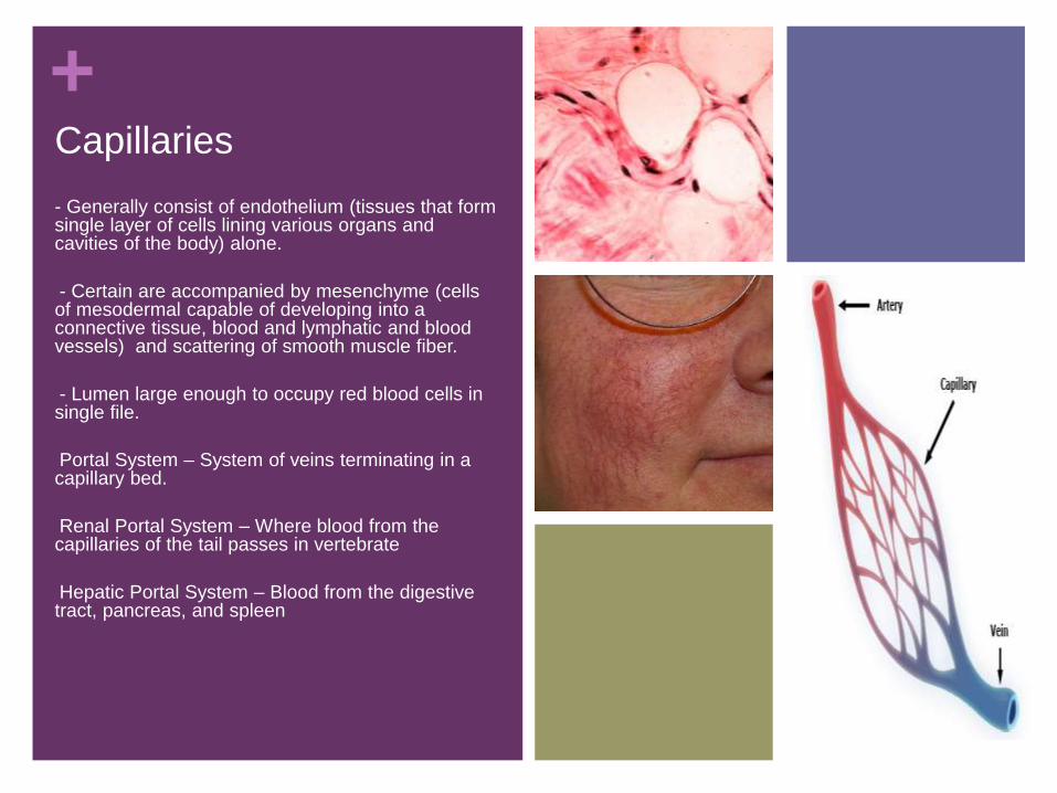
+Capillaries
- Generally consist of endothelium (tissues that form single layer of cells lining various organs and cavities of the body) alone.
-- Certain are accompanied by mesenchyme (cells of mesodermal capable of developing into a connective tissue, blood and lymphatic and blood vessels) and scattering of smooth muscle fiber.
-- Lumen large enough to occupy red blood cells in single file.
-Portal System – System of veins terminating in a capillary bed.
-Renal Portal System – Where blood from the capillaries of the tail passes in vertebrate
-Hepatic Portal System – Blood from the digestive tract, pancreas, and spleen
-

+PORTAL SYSTEM
Hepatic Portal System Portal System
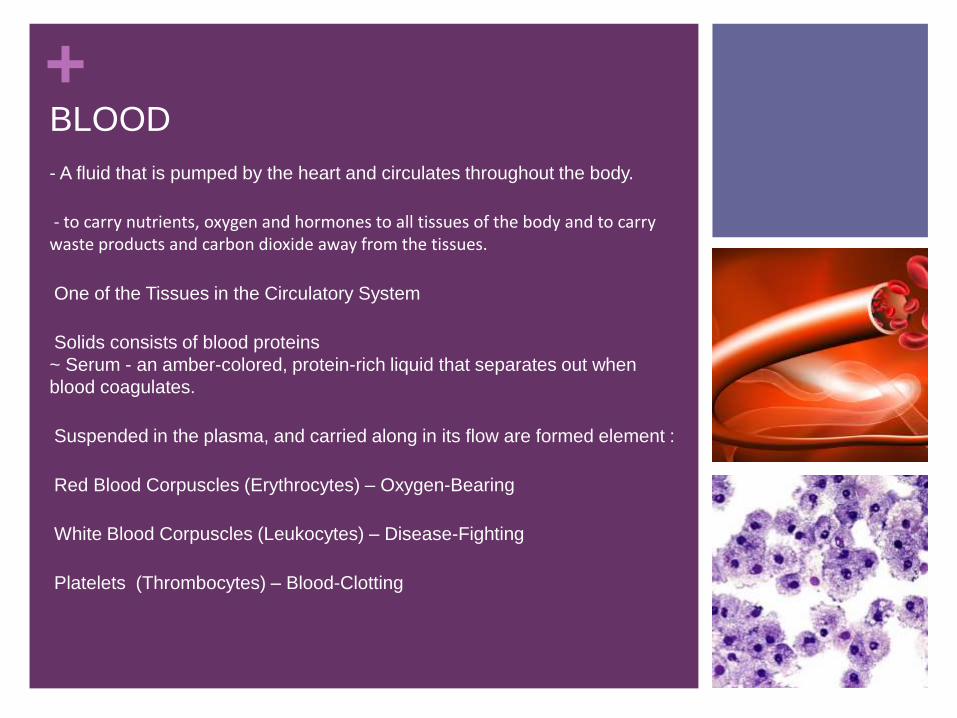
+BLOOD
- A fluid that is pumped by the heart and circulates throughout the body.
-- to carry nutrients, oxygen and hormones to all tissues of the body and to carry waste products and carbon dioxide away from the tissues.
-One of the Tissues in the Circulatory System
-Solids consists of blood proteins
~ Serum - an amber-colored, protein-rich liquid that separates out when
blood coagulates.
-Suspended in the plasma, and carried along in its flow are formed element :
-Red Blood Corpuscles (Erythrocytes) – Oxygen-Bearing
-White Blood Corpuscles (Leukocytes) – Disease-Fighting
-Platelets (Thrombocytes) – Blood-Clotting
-

+
BLOOD

+TYPES OF BLOOD
RED BLOOD -re responsible for carrying oxygen and carbon dioxide.
PLATELETS -are blood cells that help stop bleeding

+TYPES OF BLOOD WHITE BLOOD - help the body fight off
germs

+ WHITE BLOODGRANULOCYTES - help the body fight bacterial infections. The number
of granulocytes in the body goes up when there is a serious infection.
People with lower numbers of granulocytes are more likely to get bad
infections more often.
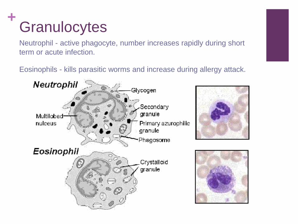
+GranulocytesNeutrophil - active phagocyte, number increases rapidly during short
term or acute infection.
Eosinophils - kills parasitic worms and increase during allergy attack.

+GranulocytesBasophils - contains histamine, which is discharged as site of
inflammation.
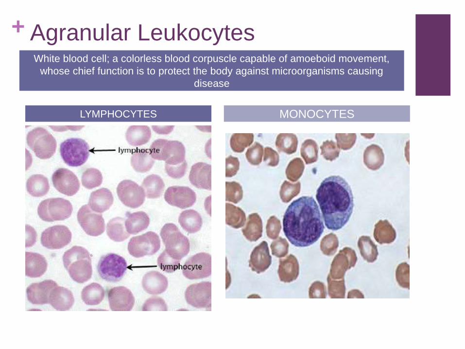
+ Agranular Leukocytes
LYMPHOCYTES MONOCYTES
White blood cell; a colorless blood corpuscle capable of amoeboid movement,
whose chief function is to protect the body against microorganisms causing
disease
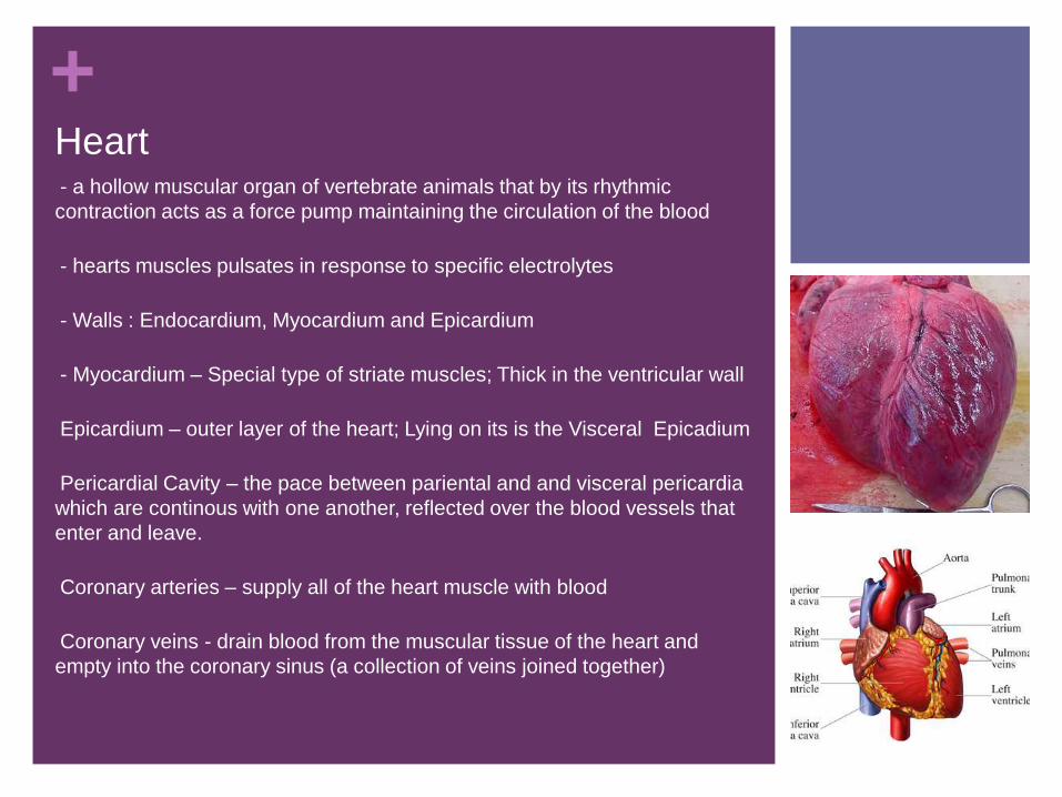
+Heart-- a hollow muscular organ of vertebrate animals that by its rhythmic
contraction acts as a force pump maintaining the circulation of the blood
-- hearts muscles pulsates in response to specific electrolytes
-- Walls : Endocardium, Myocardium and Epicardium
-- Myocardium – Special type of striate muscles; Thick in the ventricular wall
-Epicardium – outer layer of the heart; Lying on its is the Visceral Epicadium
-Pericardial Cavity – the pace between pariental and and visceral pericardia
which are continous with one another, reflected over the blood vessels that
enter and leave.
-Coronary arteries – supply all of the heart muscle with blood
-Coronary veins - drain blood from the muscular tissue of the heart and
empty into the coronary sinus (a collection of veins joined together)

+
HEART DIAGRAM
Hagfishes’s heart has
no nerve supply

+HEART
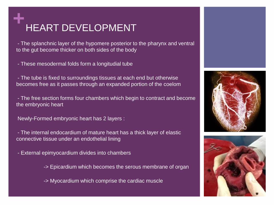
+HEART DEVELOPMENT
-- The splanchnic layer of the hypomere posterior to the pharynx and ventral
to the gut become thicker on both sides of the body
-- These mesodermal folds form a longitudial tube
-- The tube is fixed to surroundings tissues at each end but otherwise
becomes free as it passes through an expanded portion of the coelom
-- The free section forms four chambers which begin to contract and become
the embryonic heart
-Newly-Formed embryonic heart has 2 layers :
-- The internal endocardium of mature heart has a thick layer of elastic
connective tissue under an endothelial lining
-- External epimyocardium divides into chambers
- -> Epicardium which becomes the serous membrane of organ
- -> Myocardium which comprise the cardiac muscle
-
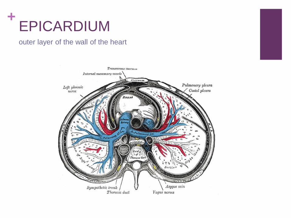
+EPICARDIUMouter layer of the wall of the heart

+MYOCARDIUMthe middle and thickest layer of the heart wall, composed of cardiac
muscle.

+
HEARTS OF GILL-
BREATHING FISHES
Fishes other than dipnoans have 4 chambers in
a series : Sinus Venosus, Atrium, ventricle, and
conus arteriosus.

+Heart of Gill-Breathing Fishes

+Atrium
Large thin-walled muscular sac that is
a sort of staging are for blood that Is
about to enter the ventricle to be
propelled toward the gills.
From the atrium, blood pours into the
relaxing ventricle through an
atrioventricular aperture that is
guarded by a pair of a one-way valve.
These prevents ventricular blood from
being pumped back into the atrium
when the ventricle conracts.

+
Ventricle
Has a very thick muscular walls and is
the actual pumping portion of the
heart.
The anterior end is prolonged as a
muscular tube of small diameter, the
conus arteriosus, which extends to the
extreme cephalic end of the pericardial
cavity, at which point it is continuous
with the ventral aorta.
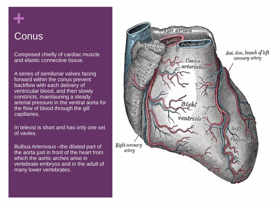
+Conus
Composed chiefly of cardiac muscle and elastic connective tissue.
A series of semilunar valves facing forward within the conus prevent backflow with each delivery of ventricular blood, and then slowly constricts, maintauning a steady arterial pressure in the ventral aorta for the flow of blood through the gill capillaries.
In teleost is short and has only one set of vavles.
Bulbus Arteriosus –the dilated part of the aorta just in front of the heart from which the aortic arches arise in vertebrate embryos and in the adult of many lower vertebrates.

+HEARTS OF LUNGFISHES AND
AMPHIBIANS
Modification in the heart of lungfishes and amphibians are correlated with
aerial respiration by means of swim bladders or lungs.
They are enable oxygenated blood returning from the lungs to be separated
from the deoxygenated blood returning from elsewhere.
Lungfishes and amphibians have bimodal gas exchange, but various
species employ air breathing with lungs to different degrees
Dynamics of inflow in the pulmonary and systemic veins
The extent and localization of atrial septation
The partial ventricular septum in lungfishes and the massive ventricular
trabeculation in both amphibians and lungfishes
Vasomotor reactions in the various outflow vessels from the heart.

+

+ LUNGFISHESHeart
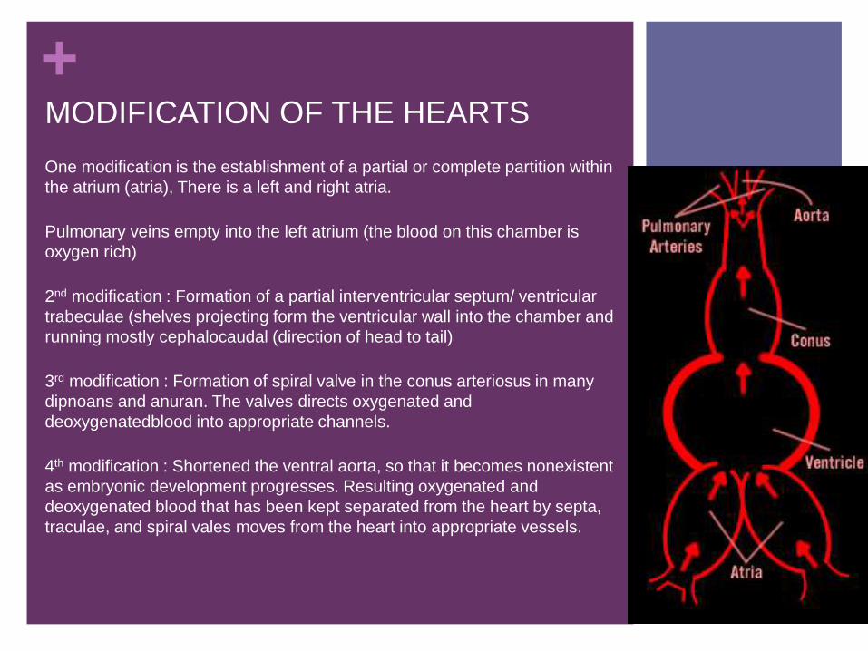
+MODIFICATION OF THE HEARTS
One modification is the establishment of a partial or complete partition within
the atrium (atria), There is a left and right atria.
Pulmonary veins empty into the left atrium (the blood on this chamber is
oxygen rich)
2nd modification : Formation of a partial interventricular septum/ ventricular
trabeculae (shelves projecting form the ventricular wall into the chamber and
running mostly cephalocaudal (direction of head to tail)
3rd modification : Formation of spiral valve in the conus arteriosus in many
dipnoans and anuran. The valves directs oxygenated and
deoxygenatedblood into appropriate channels.
4th modification : Shortened the ventral aorta, so that it becomes nonexistent
as embryonic development progresses. Resulting oxygenated and
deoxygenated blood that has been kept separated from the heart by septa,
traculae, and spiral vales moves from the heart into appropriate vessels.

+The Hearts of AmniotesAmniotes - are animals who shield the embryos of their offspring using
extensive membranes or keeping them inside the body, in contrast to most
other animals, which lay free-floating eggs in water.
- a group of limbed vertebrates that includes all living reptiles (class
Reptilia), birds (class Aves), mammals (class Mammalia)
-2 atria, 2 ventrticles, and a sinus venoses ( except in adult birds and
mammals)
-In crocodile sinus is partially incorporates into the wall of right atrium.
-Birds and mammals have a sinus venosus during early development but
fails to keep pace with the growth of the right atrium into which it empties
and finally incorporated into the wall of that chamber.
-Sinoatrial Node – section of nodal tissue that is loacted in the upper wall of
the right atrium
-Interatrial Foramen – An opening of the septum between the right and left
atria of the heart, present in the fetus but usually close soon after birth

+The Heart of AmniotesThe right and left atria of adult amniotes are completely separated by an
interatrial septum
Right atrium -> receives blood from the sinus venosus (reptiles) or blood
that previously emptied into the sinus venosus (Bird and Mammals) also
receives blood from the pulmonary veins
Left atrium -> receives blood from the pulmonary veins
Mammals each atrium has a earlike flap (auricle), containing a blind, saclike
chamber
2 ventricles are completely separated in crocodiliansc (Reptiles), birds, and
mammals
Other amniotes, the interventrucular septum is incomplete

+Heart of a Bird
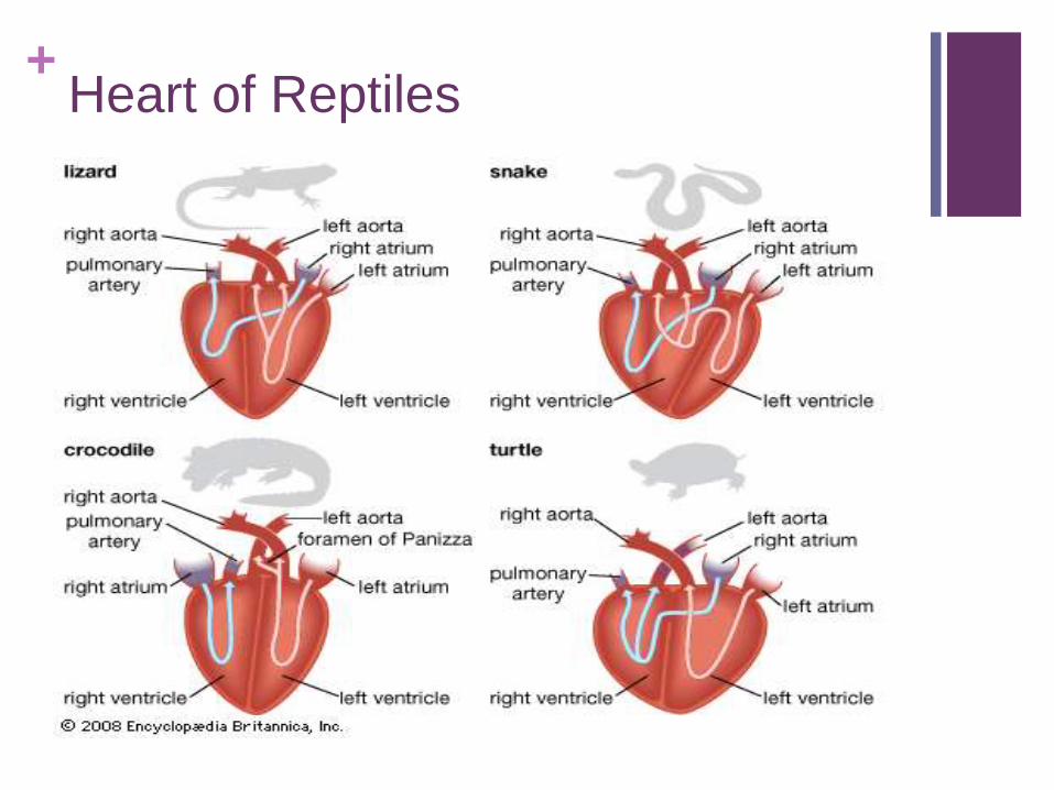
+Heart of Reptiles
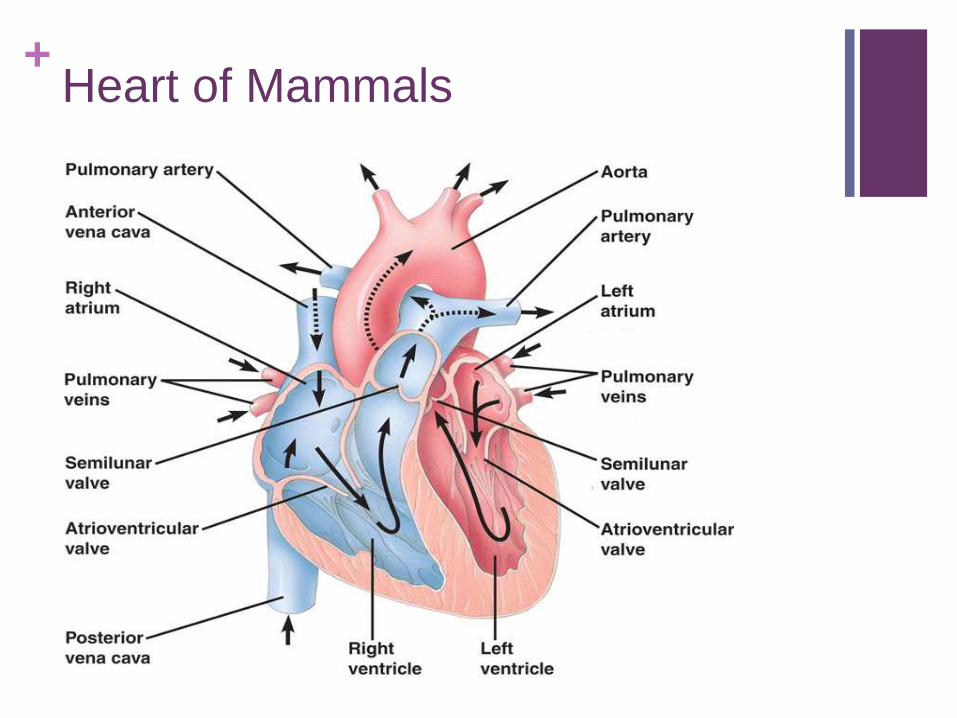
+Heart of Mammals

+SINOATRIAL NODE

+INTERARIAL FORAMEN

+Morphogenesis of the Heart
The heart of all vertebrate commence as a single, almost straight, pulsating
tube that receives incoming blood at the caudal end and empties into the
embryonic ventral aorta anteriorly.
The tube, whether of sharks or human beings, bends to the animal’s right,
then twists in to an “S” shape, so that the atrial region, previously at the
caudal end, is carried dorsad and cephalad until it lies where it is found in
adult fishes.
The twisting and bending is probably correlated with the confinement of the
rapidly growing heart in a less expansive pericardial cavity.
In Amphibians and Amniotes the twisting is carried further, for the atrial
region finally lies cephalad to the ventricular region., while internally an
interventricular septum completes the division of the amniote heart into right
and left sides.

+Morphogenesis of the Heart

+Morphogenesis of the Heart
As hatching approaches in birds, the sinus venosus is incorporated almost
completely into the wall of the right atrium.
In mammals, the sinus venosus fails early to keep up in growth with the rest
of the heart and its incorporation into the right atrial wall occurs sooner in
organogenesis
Oxygen and nutrients from the heart is the first organ to function and does
not even before any nerves have reached it to impose a cardiac rhythm.
Initial straight tube that will become the heart of sharks and amphibians
organizations from paired mesenchymal masses of lateral-plate somatic and
splanchnic mesenchyme that aggregate beneath the pharynx to form a
single tube.
In amniotes a pair already organized endothelial tube is brought together
beneath the pharynx, they fus, and a single tube results,
In either case, the heart is bilateral contribution of lately-plate mesoderm.

+Primitive Heart (Two-
Chambered) : Single
Circulation Pattern
-- They are nearly straight with 4 chambers,
pumping a single stream of deoxygenated blood
forward in the body
-- A thin walled sinus venosus receives blood
from the major veins and empties it through a
simple sinutrial valves in a large thick-walled
ventricle
-- The Ventricle pumps into the conus arteriosus
which looks like an enlarged artery and is lined
with several rows of semilunar valves preventing
backflow of blood as the ventricle fills.
-

+
AMPHIOXUSHas no heart only a homologous
pulsating vessels in the same position
where the heart evolved in vertebrate
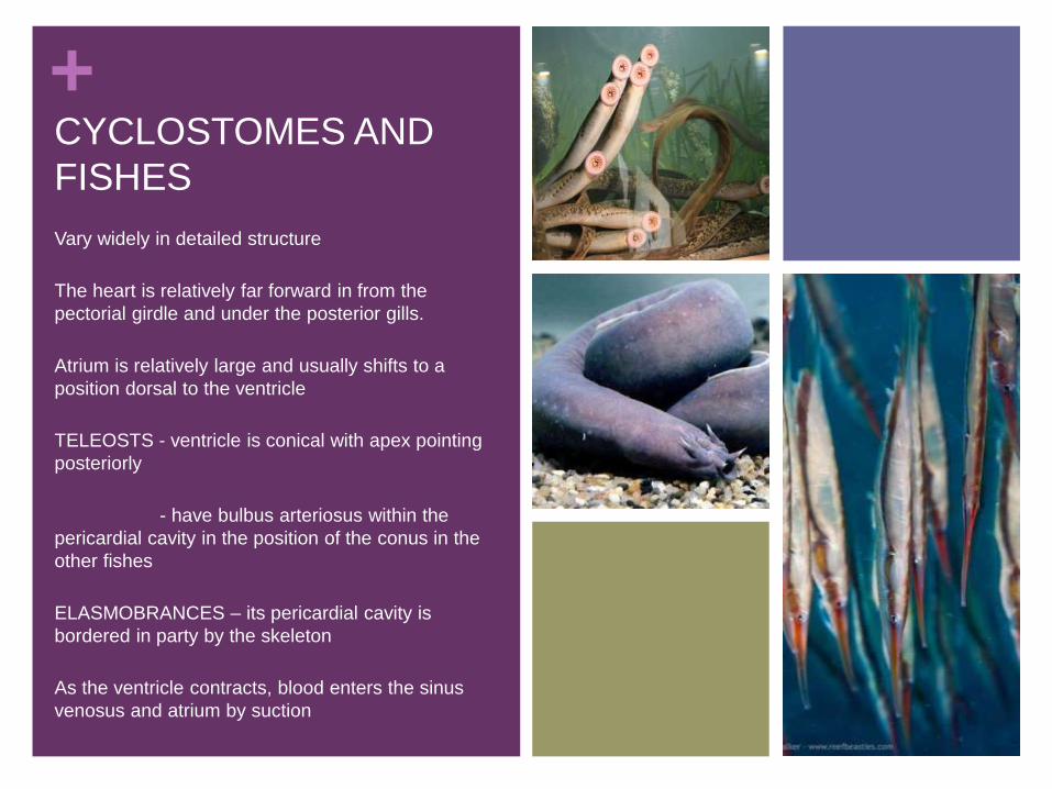
+CYCLOSTOMES AND
FISHES
Vary widely in detailed structure
The heart is relatively far forward in from the
pectorial girdle and under the posterior gills.
Atrium is relatively large and usually shifts to a
position dorsal to the ventricle
TELEOSTS - ventricle is conical with apex pointing
posteriorly
- have bulbus arteriosus within the
pericardial cavity in the position of the conus in the
other fishes
ELASMOBRANCES – its pericardial cavity is
bordered in party by the skeleton
As the ventricle contracts, blood enters the sinus
venosus and atrium by suction

+
CYCLOSTOMES AND
FISHES
Hearts of FISHES are relatively small due to
small volume of blood
- Cyclostomes and Fishes have accessory
hearts or pumping mechanisms
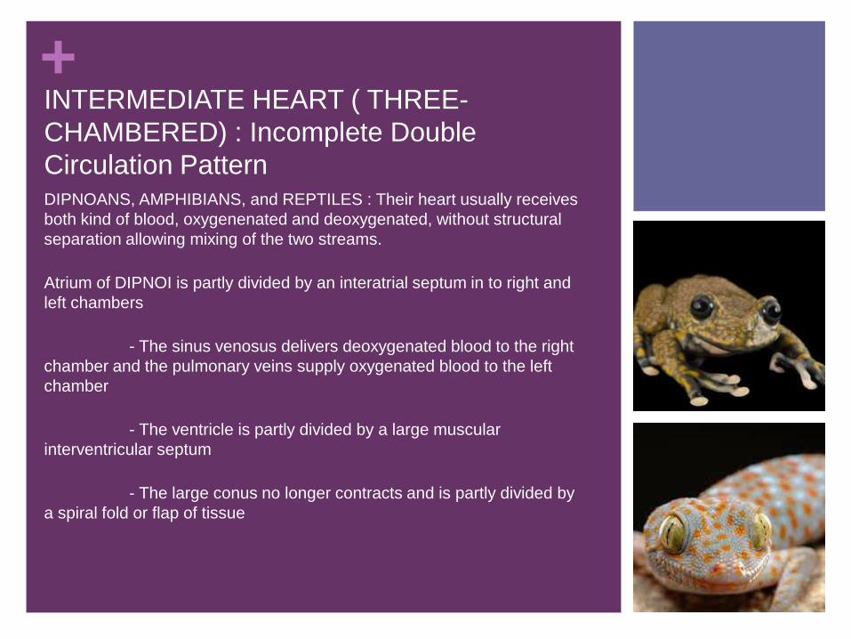
+INTERMEDIATE HEART ( THREE-
CHAMBERED) : Incomplete Double
Circulation PatternDIPNOANS, AMPHIBIANS, and REPTILES : Their heart usually receives
both kind of blood, oxygenenated and deoxygenated, without structural
separation allowing mixing of the two streams.
Atrium of DIPNOI is partly divided by an interatrial septum in to right and
left chambers
- The sinus venosus delivers deoxygenated blood to the right
chamber and the pulmonary veins supply oxygenated blood to the left
chamber
- The ventricle is partly divided by a large muscular
interventricular septum
- The large conus no longer contracts and is partly divided by
a spiral fold or flap of tissue

+
The Atrium of ANURANS is completely divided into right and left chambers
by the interatrial septum
- The right chamber received deoxygenated blood while the left
received blood oxygenated in the lungs
- Blood returning from the skin joint the systemic veins allowing
mixing of blood on the right side
- The ventricle is not divided but mixing of deoxygenated and
oxygenated blood is minimal
- Both blood may enter the left systematic arch and mix
depending on the resistance in the pulmonary circuit
INTERMEDIATE HEART ( THREE-
CHAMBERED) : Incomplete Double
Circulation Pattern

+
CHELOANS and SQUAMATE hearts appears during early embryonic
development but becomes divided in the adult to form the pulmonary
trunk and the independent right and left systemic trunk
- Atrium is completely divided into right and left atria
- The cavuum arteriosum received blood from the left atrium
but has no direct arterial output
- The cavuum pumonale does not receive blood directly from
the atria; It receives blood from the cavuum venosum through the
muscular ridge
- The cavuum venosum receives deoxygenated blood from
the right atrium
INTERMEDIATE HEART ( THREE-CHAMBERED) : Incomplete Double Circulation Pattern

+
CROCODILIAN heart is quite different from other reptiles
-The ventricle is divided by a complete interventicular septum into left and
right ventricle
-- The pulmonary trunk and left aortic arch open off the right ventricle
-- The right aortic arch open off the left ventricle
-- A narrow channel at the bases of the two systematic trunks, the foramen
of Panizza, connects
-- When CROCODILE dives, blood in the right ventricle travel through the
left aortic arch joining the systematic circulation and by passing lungs
-- Diversion of blood also happens when it is at rest on land going for long
intervals without taking a breath
INTERMEDIATE HEART ( THREE-CHAMBERED) : Incomplete Double Circulation Pattern

+CROCODILE HEART

+DOUBLE-CIRUIT HEART (FOUR-
CHAMBERED HEART): Complete Double
Circulation Pattern
ADULT BIRDS and MAMMALS – complete double circulation composed of : -Low-pressure pulmonary circuit on the right side of the heart
-> Needed to avoid edema and damage to delicate lung tissues
-- High-Pressure systemic circuit on the left side of the heart
- -> To drive blood through tissues that may have their own internal pressure like contracting muscles
-- Atrium is completely divided and smaller than in fishes
-- Ventricle is completely divide and stronger on the left side because of the resistance that is greater on the other side
-- Adult systematic arch is single
- -> It loops to the right in BIRDS and to the left in MAMMALS
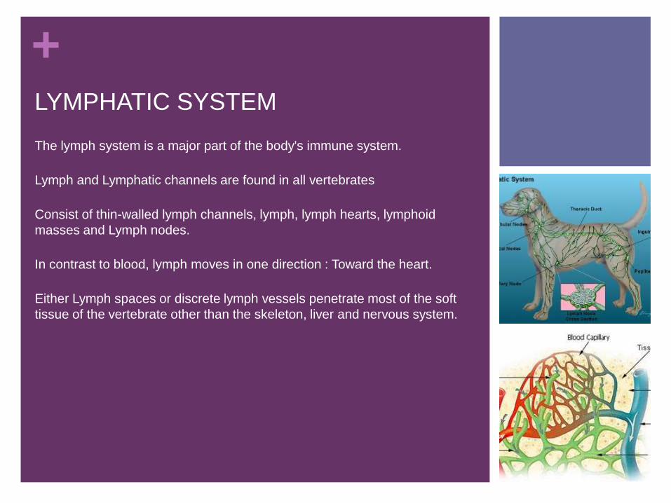
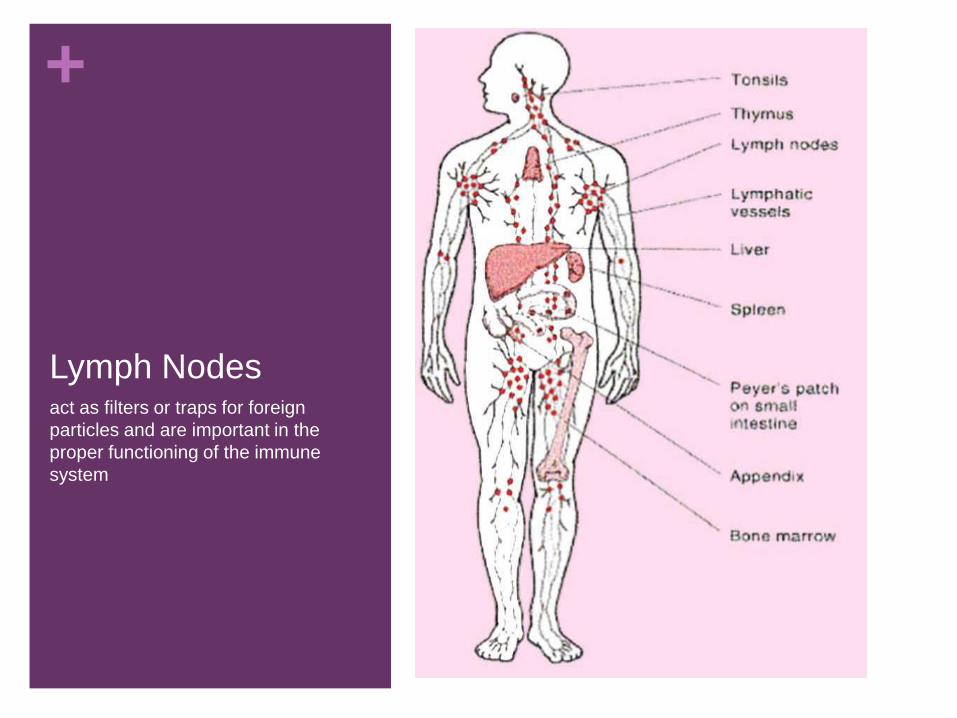
+LYMPHATIC SYSTEM
The lymph system is a major part of the body's immune system.
Lymph and Lymphatic channels are found in all vertebrates
Consist of thin-walled lymph channels, lymph, lymph hearts, lymphoid
masses and Lymph nodes.
In contrast to blood, lymph moves in one direction : Toward the heart.
Either Lymph spaces or discrete lymph vessels penetrate most of the soft
tissue of the vertebrate other than the skeleton, liver and nervous system.

+ LYMPHATIC SYSTEMa network of organs, lymph nodes, lymph
ducts, and lymph vessels that make and move
lymph from tissues to the bloodstream.
+
Lymph Nodes act as filters or traps for foreign
particles and are important in the
proper functioning of the immune
system
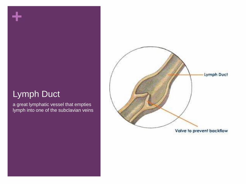
+
Lymph Ducta great lymphatic vessel that empties
lymph into one of the subclavian veins

+FLOW