Circulatory and Respiratory Systemsmssorensennghs.weebly.com/uploads/5/7/8/1/57815877/... · The...
51
Circulatory and Respiratory Systems By Monika Shpokayte, Andrea Lattanzio, and Christopher Tenorio
Transcript of Circulatory and Respiratory Systemsmssorensennghs.weebly.com/uploads/5/7/8/1/57815877/... · The...
Circulatory
and
Respiratory
Systems
By Monika Shpokayte, Andrea Lattanzio, and Christopher Tenorio
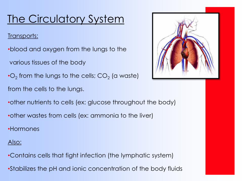
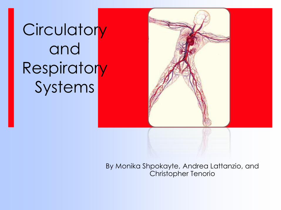
The Circulatory System
Transports:
•blood and oxygen from the lungs to the
various tissues of the body
•O2 from the lungs to the cells; CO2 (a waste)
from the cells to the lungs.
•other nutrients to cells (ex: glucose throughout the body)
•other wastes from cells (ex: ammonia to the liver)
•Hormones
Also:
•Contains cells that fight infection (the lymphatic system)
•Stabilizes the pH and ionic concentration of the body fluids
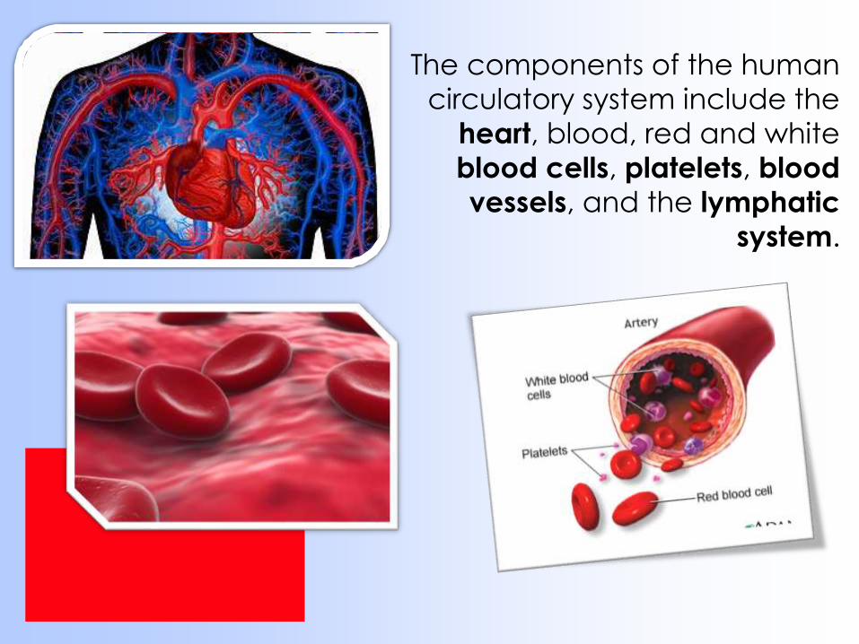
The components of the human
circulatory system include the
heart, blood, red and white
blood cells, platelets, blood
vessels, and the lymphatic
system.
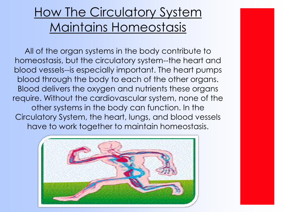
How The Circulatory System
Maintains Homeostasis
All of the organ systems in the body contribute to
homeostasis, but the circulatory system--the heart and blood vessels--is especially important. The heart pumps
blood through the body to each of the other organs.
Blood delivers the oxygen and nutrients these organs
require. Without the cardiovascular system, none of the other systems in the body can function. In the
Circulatory System, the heart, lungs, and blood vessels
have to work together to maintain homeostasis.
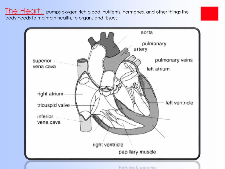
The Heart: pumps oxygen rich blood, nutrients, hormones, and other things the
body needs to maintain health, to organs and tissues.
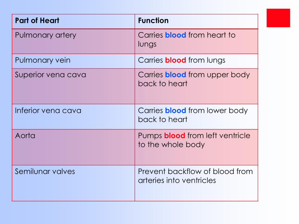
Part of Heart Function
Pulmonary artery Carries blood from heart to
lungs
Pulmonary vein Carries blood from lungs
Superior vena cava Carries blood from upper body
back to heart
Inferior vena cava Carries blood from lower body
back to heart
Aorta Pumps blood from left ventricle
to the whole body
Semilunar valves Prevent backflow of blood from
arteries into ventricles
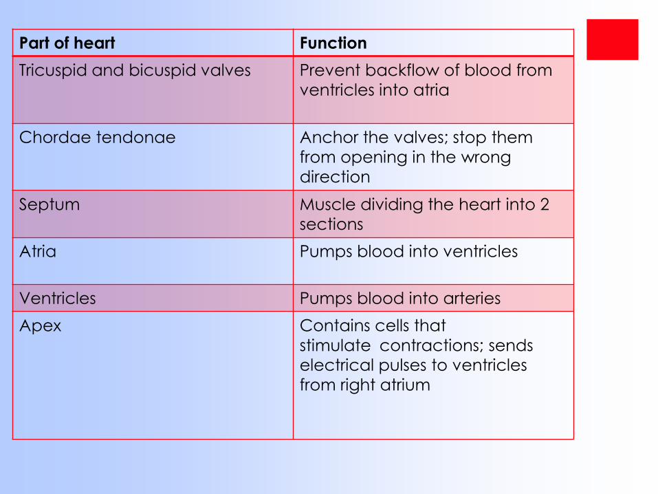
Part of heart Function
Tricuspid and bicuspid valves Prevent backflow of blood from
ventricles into atria
Chordae tendonae Anchor the valves; stop them
from opening in the wrong
direction
Septum Muscle dividing the heart into 2
sections
Atria Pumps blood into ventricles
Ventricles Pumps blood into arteries
Apex Contains cells that
stimulate contractions; sends electrical pulses to ventricles
from right atrium
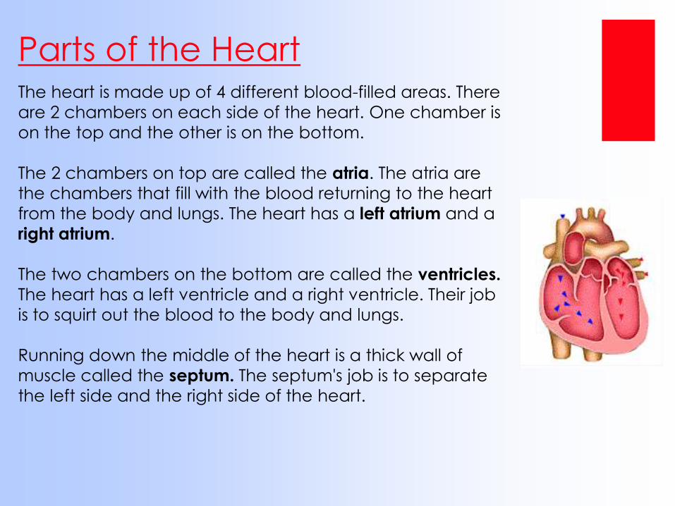
Parts of the HeartThe heart is made up of 4 different blood-filled areas. There
are 2 chambers on each side of the heart. One chamber is
on the top and the other is on the bottom.
The 2 chambers on top are called the atria. The atria are
the chambers that fill with the blood returning to the heart
from the body and lungs. The heart has a left atrium and a
right atrium.
The two chambers on the bottom are called the ventricles.
The heart has a left ventricle and a right ventricle. Their job
is to squirt out the blood to the body and lungs.
Running down the middle of the heart is a thick wall of
muscle called the septum. The septum's job is to separate
the left side and the right side of the heart.
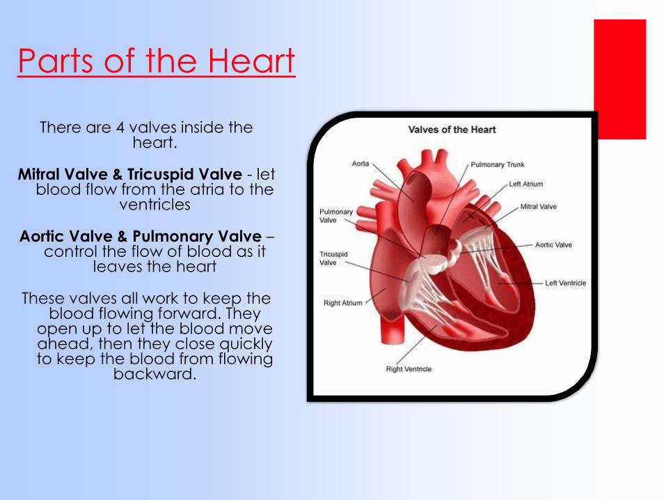
Parts of the Heart
There are 4 valves inside the heart.
Mitral Valve & Tricuspid Valve - let blood flow from the atria to the
ventricles
Aortic Valve & Pulmonary Valve –control the flow of blood as it
leaves the heart
These valves all work to keep the blood flowing forward. They
open up to let the blood move ahead, then they close quickly to keep the blood from flowing
backward.
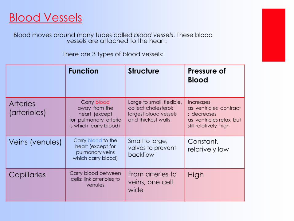
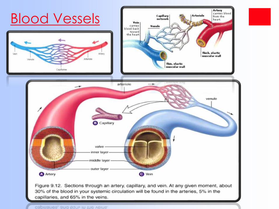
Blood moves around many tubes called blood vessels. These blood vessels are attached to the heart.
There are 3 types of blood vessels:
Blood Vessels
Function Structure Pressure of
Blood
Arteries
(arterioles)
Carry blood
away from the
heart (except
for pulmonary arterie
s which carry blood)
Large to small, flexible,
collect cholesterol;
largest blood vessels
and thickest walls
Increases
as ventricles contract
; decreases
as ventricles relax but
still relatively high
Veins (venules) Carry blood to the
heart (except for
pulmonary veins
which carry blood)
Small to large, valves to prevent backflow
Constant,
relatively low
Capillaries Carry blood between
cells; link arterioles to
venules
From arteries to
veins, one cell
wide
High
Blood Vessels
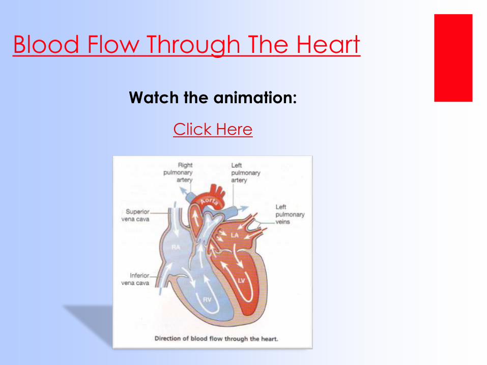
Watch the animation:
Click Here
Blood Flow Through The Heart
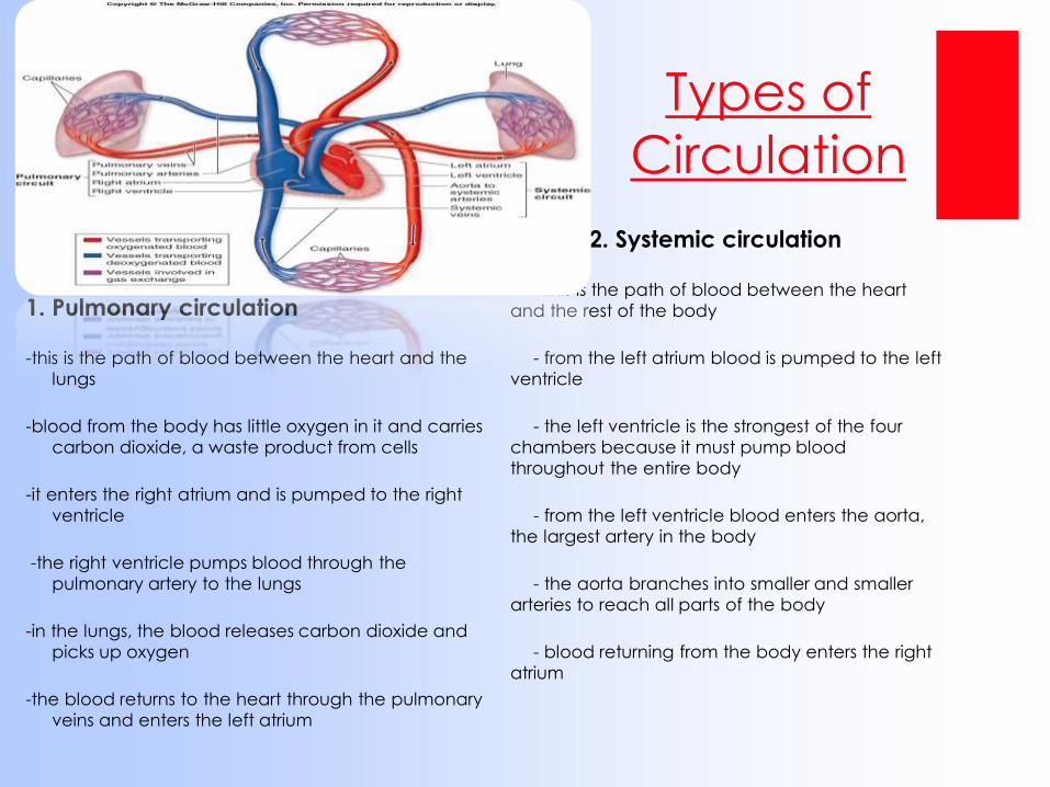
1. Pulmonary circulation
-this is the path of blood between the heart and the
lungs
-blood from the body has little oxygen in it and carries
carbon dioxide, a waste product from cells
-it enters the right atrium and is pumped to the right
ventricle
-the right ventricle pumps blood through the
pulmonary artery to the lungs
-in the lungs, the blood releases carbon dioxide and
picks up oxygen
-the blood returns to the heart through the pulmonary
veins and enters the left atrium
2. Systemic circulation
- this is the path of blood between the heart
and the rest of the body
- from the left atrium blood is pumped to the left
ventricle
- the left ventricle is the strongest of the four
chambers because it must pump blood
throughout the entire body
- from the left ventricle blood enters the aorta,
the largest artery in the body
- the aorta branches into smaller and smaller
arteries to reach all parts of the body
- blood returning from the body enters the right
atrium
Types of
Circulation
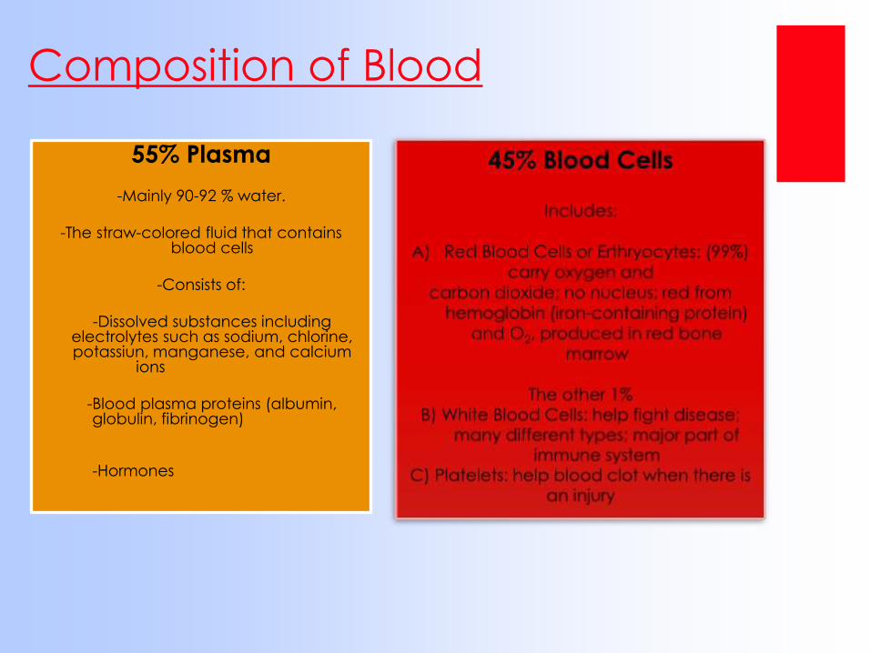
55% Plasma
-Mainly 90-92 % water.
-The straw-colored fluid that contains blood cells
-Consists of:
-Dissolved substances including electrolytes such as sodium, chlorine, potassiun, manganese, and calcium
ions
-Blood plasma proteins (albumin, globulin, fibrinogen)
-Hormones
Composition of Blood
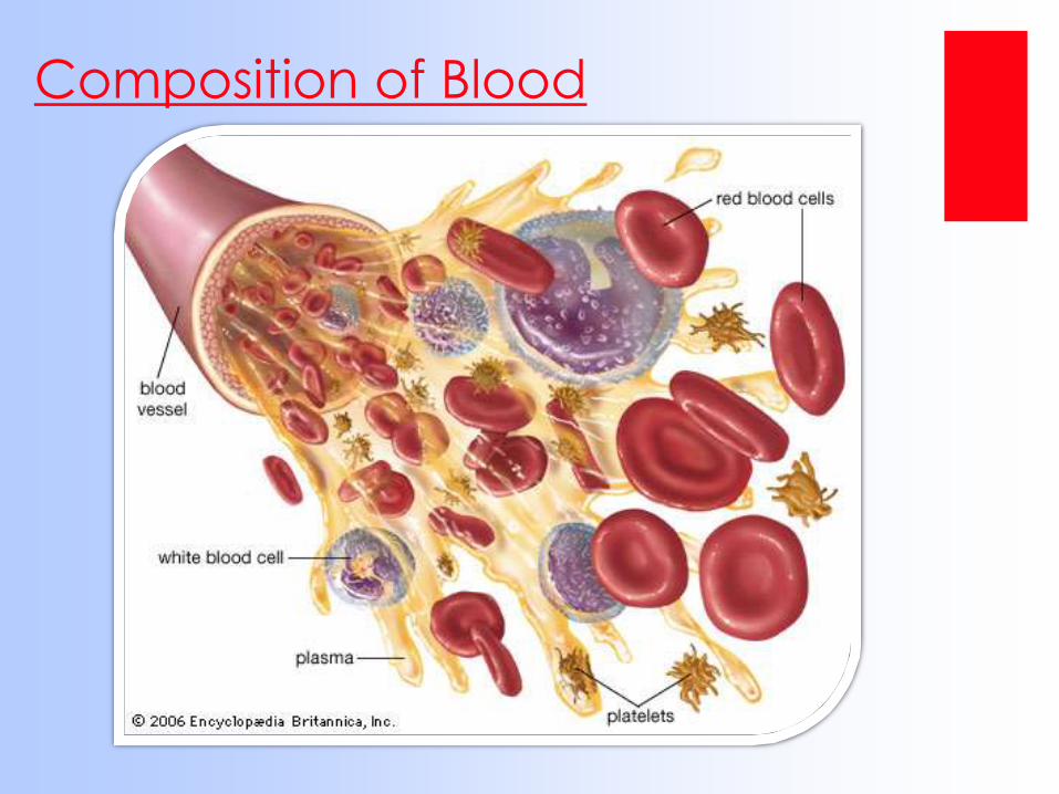
Composition of Blood
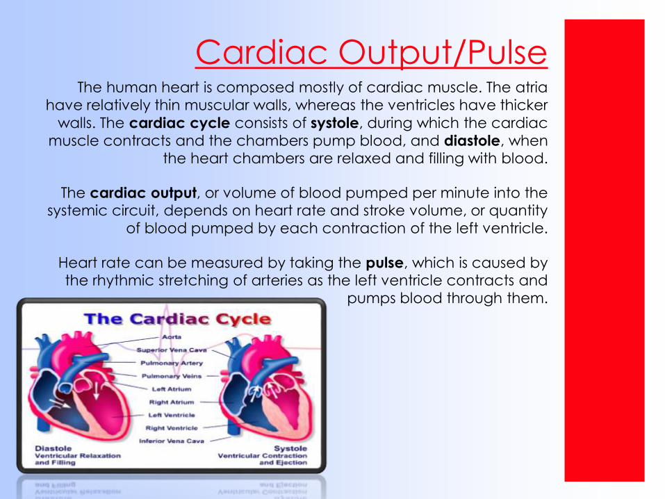
Cardiac Output/PulseThe human heart is composed mostly of cardiac muscle. The atria
have relatively thin muscular walls, whereas the ventricles have thicker
walls. The cardiac cycle consists of systole, during which the cardiac
muscle contracts and the chambers pump blood, and diastole, when the heart chambers are relaxed and filling with blood.
The cardiac output, or volume of blood pumped per minute into the systemic circuit, depends on heart rate and stroke volume, or quantity
of blood pumped by each contraction of the left ventricle.
Heart rate can be measured by taking the pulse, which is caused by the rhythmic stretching of arteries as the left ventricle contracts and
pumps blood through them.
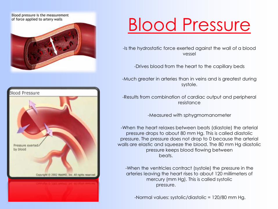
Blood Pressure-Is the hydrostatic force exerted against the wall of a blood
vessel
-Drives blood from the heart to the capillary beds
-Much greater in arteries than in veins and is greatest during
systole.
-Results from combination of cardiac output and peripheral
resistance
-Measured with sphygmomanometer
-When the heart relaxes between beats (diastole) the arterial
pressure drops to about 80 mm Hg. This is called diastolic
pressure. The pressure does not drop to 0 because the arterial
walls are elastic and squeeze the blood. The 80 mm Hg diastolic
pressure keeps blood flowing between
beats.
-When the ventricles contract (systole) the pressure in the
arteries leaving the heart rises to about 120 millimeters of
mercury (mm Hg). This is called systolic
pressure.
-Normal values: systolic/diastolic = 120/80 mm Hg.
Hypertension – aka high blood pressure
-damages the endothelium and initiates plaque formation
-promotes atherosclerosis; increases risk of heart attack or stroke
-caused by stress, poor diet, genetics, and smoking
-easily treated by drugs, diet, and exercise
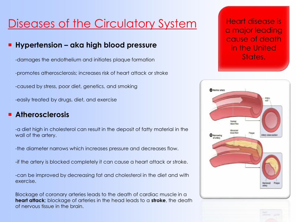
Atherosclerosis
-a diet high in cholesterol can result in the deposit of fatty material in the
wall of the artery.
-the diameter narrows which increases pressure and decreases flow.
-if the artery is blocked completely it can cause a heart attack or stroke.
-can be improved by decreasing fat and cholesterol in the diet and with
exercise.
Blockage of coronary arteries leads to the death of cardiac muscle in a
heart attack; blockage of arteries in the head leads to a stroke, the death
of nervous tissue in the brain.
Diseases of the Circulatory System Heart disease is
a major leading
cause of death
in the United
States.

Feedback MechanismAn example of a feedback mechanism in the human
circulatory system would be the increase in heart rate and
respiratory rate which occurs in response to increased
exercise or other increased muscle cell activity.
Heart rate is controlled via a bio-feedback loop in which
special receptors located in the brain known as chemo-
receptors monitor blood oxygen levels. As oxygen levels
fall, the chemo-receptors sense falling oxygen levels and
the brain sends electrical signals with increasing frequency
to the heart. This causes the heart to beat faster as well
and produce more cardiac contractions. As oxygen-rich
blood reaches the brain, the chemo-receptors sense this
restored oxygen level and the rate at which the heart
beats slows. In this way, oxygen levels to the brain remain
relatively constant and homeostasis is achieved.
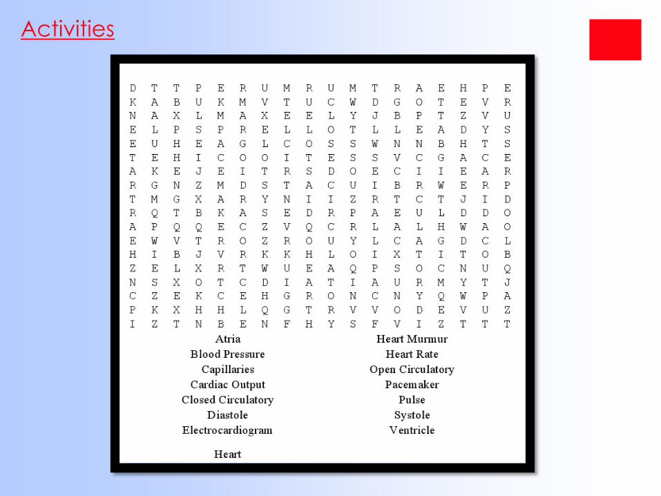
Activities
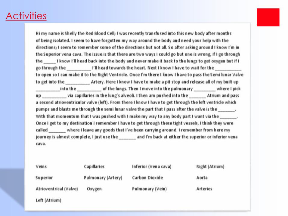
Activities
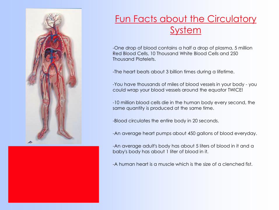
Fun Facts about the Circulatory
System
-One drop of blood contains a half a drop of plasma, 5 million
Red Blood Cells, 10 Thousand White Blood Cells and 250
Thousand Platelets.
-The heart beats about 3 billion times during a lifetime.
-You have thousands of miles of blood vessels in your body - you
could wrap your blood vessels around the equator TWICE!
-10 million blood cells die in the human body every second, the
same quantity is produced at the same time.
-Blood circulates the entire body in 20 seconds.
-An average heart pumps about 450 gallons of blood everyday.
-An average adult's body has about 5 liters of blood in it and a
baby's body has about 1 liter of blood in it.
-A human heart is a muscle which is the size of a clenched fist.
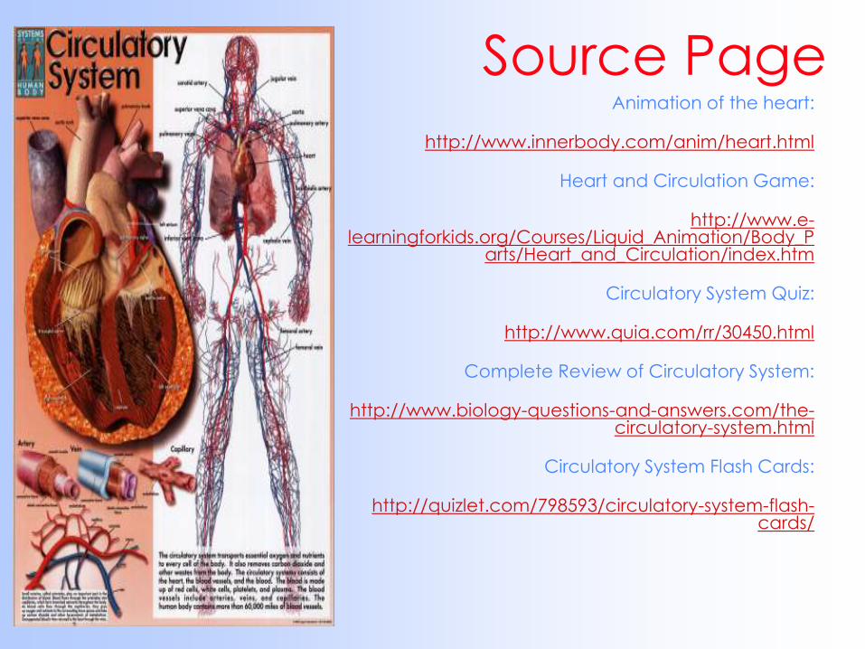
Source PageAnimation of the heart:
http://www.innerbody.com/anim/heart.html
Heart and Circulation Game:
http://www.e-learningforkids.org/Courses/Liquid_Animation/Body_P
arts/Heart_and_Circulation/index.htm
Circulatory System Quiz:
http://www.quia.com/rr/30450.html
Complete Review of Circulatory System:
http://www.biology-questions-and-answers.com/the-circulatory-system.html
Circulatory System Flash Cards:
http://quizlet.com/798593/circulatory-system-flash-cards/
References
•http://kvhs.nbed.nb.ca/gallant/biology/biology.html
•http://kidshealth.org/kid/htbw/heart.html
•http://regentsprep.org/regents/biology/units/homeostasis/feed
back.cfm
•http://www.ivy-
rose.co.uk/HumanBody/Blood/Blood_StructureandFunctions.php
•http://www.fi.edu/learn/heart/systems/circulation.html
•http://www.nlm.nih.gov/medlineplus/ency/article/003398.htm
•http://www.buzzle.com/articles/circulatory-system-facts.html
Next up is the RESPIRATORY SYSTEM……
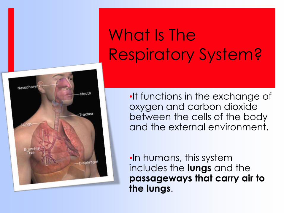
What Is The
Respiratory System?
•It functions in the exchange of oxygen and carbon dioxide between the cells of the body and the external environment.
•In humans, this system includes the lungs and the passageways that carry air to the lungs.
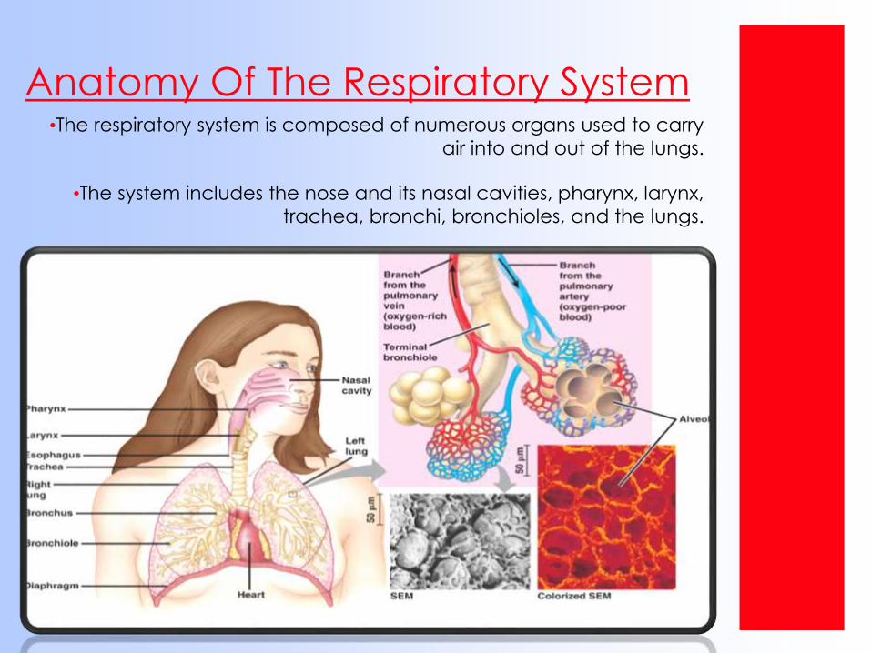
Anatomy Of The Respiratory System•The respiratory system is composed of numerous organs used to carry
air into and out of the lungs.
•The system includes the nose and its nasal cavities, pharynx, larynx,
trachea, bronchi, bronchioles, and the lungs.
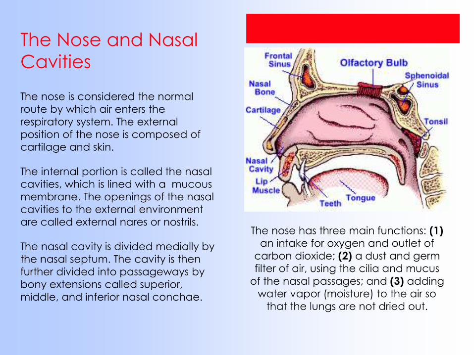
The Nose and Nasal
Cavities
The nose is considered the normal
route by which air enters the
respiratory system. The external
position of the nose is composed of
cartilage and skin.
The internal portion is called the nasal
cavities, which is lined with a mucous
membrane. The openings of the nasal
cavities to the external environment
are called external nares or nostrils.
The nasal cavity is divided medially by
the nasal septum. The cavity is then
further divided into passageways by
bony extensions called superior,
middle, and inferior nasal conchae.
The nose has three main functions: (1) an intake for oxygen and outlet of
carbon dioxide; (2) a dust and germ filter of air, using the cilia and mucus
of the nasal passages; and (3) adding water vapor (moisture) to the air so
that the lungs are not dried out.
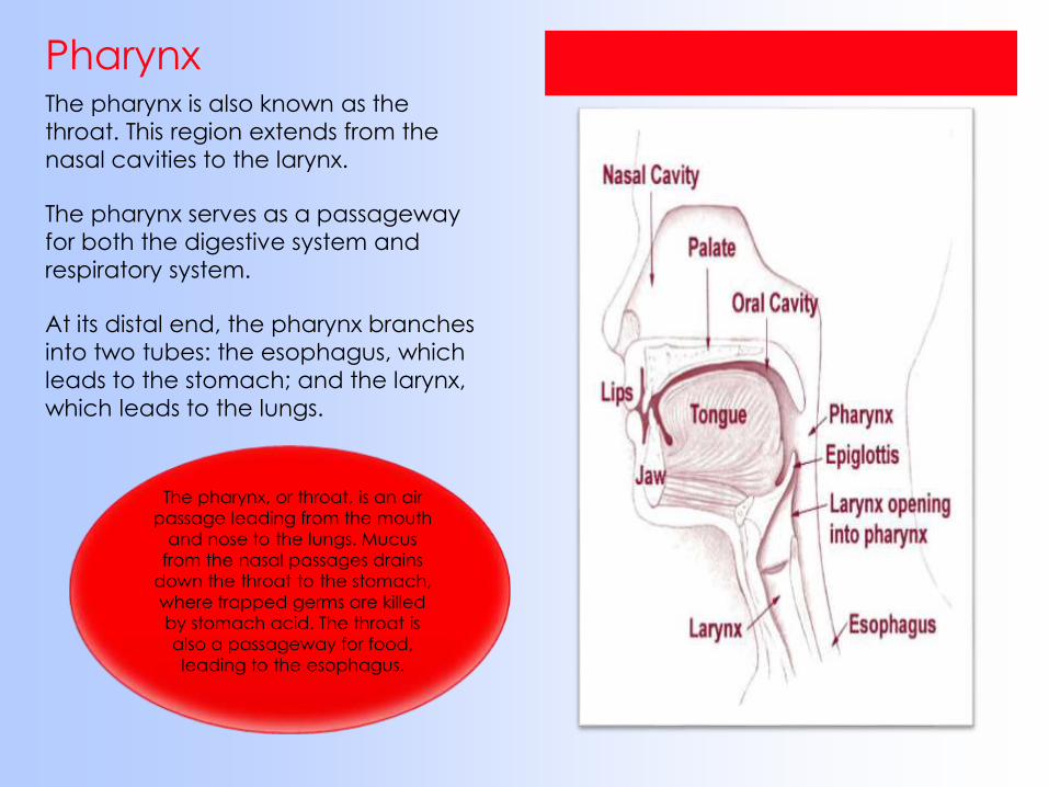
PharynxThe pharynx is also known as the
throat. This region extends from the
nasal cavities to the larynx.
The pharynx serves as a passageway
for both the digestive system and
respiratory system.
At its distal end, the pharynx branches
into two tubes: the esophagus, which
leads to the stomach; and the larynx,
which leads to the lungs.
The pharynx, or throat, is an air
passage leading from the mouth
and nose to the lungs. Mucus
from the nasal passages drains
down the throat to the stomach,
where trapped germs are killed
by stomach acid. The throat is
also a passageway for food,
leading to the esophagus.
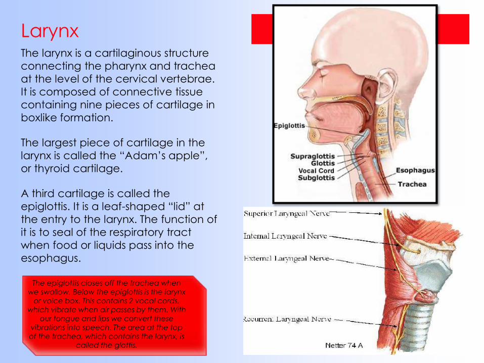
LarynxThe larynx is a cartilaginous structure
connecting the pharynx and trachea
at the level of the cervical vertebrae.
It is composed of connective tissue
containing nine pieces of cartilage in
boxlike formation.
The largest piece of cartilage in the
larynx is called the “Adam’s apple”,
or thyroid cartilage.
A third cartilage is called the
epiglottis. It is a leaf-shaped “lid” at
the entry to the larynx. The function of
it is to seal of the respiratory tract
when food or liquids pass into the
esophagus.
The epiglottis closes off the trachea when
we swallow. Below the epiglottis is the larynx or voice box. This contains 2 vocal cords,
which vibrate when air passes by them. With our tongue and lips we convert these
vibrations into speech. The area at the top of the trachea, which contains the larynx, is
called the glottis.
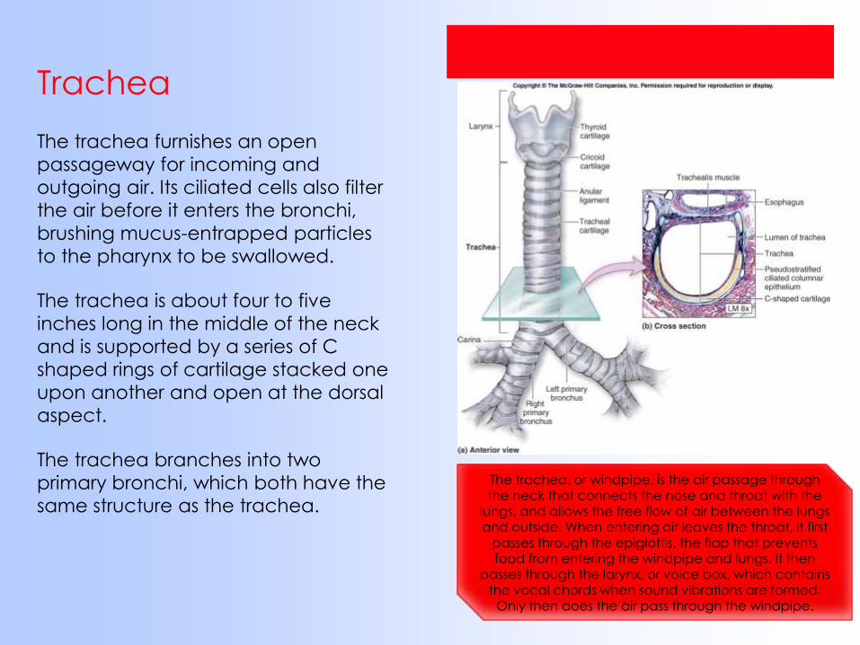
Trachea
The trachea furnishes an open
passageway for incoming and
outgoing air. Its ciliated cells also filter
the air before it enters the bronchi,
brushing mucus-entrapped particles
to the pharynx to be swallowed.
The trachea is about four to five
inches long in the middle of the neck
and is supported by a series of C
shaped rings of cartilage stacked one
upon another and open at the dorsal
aspect.
The trachea branches into two
primary bronchi, which both have the
same structure as the trachea.
The trachea, or windpipe, is the air passage through the neck that connects the nose and throat with the
lungs, and allows the free flow of air between the lungs and outside. When entering air leaves the throat, it first
passes through the epiglottis, the flap that prevents food from entering the windpipe and lungs. It then
passes through the larynx, or voice box, which contains the vocal chords when sound vibrations are formed.
Only then does the air pass through the windpipe.
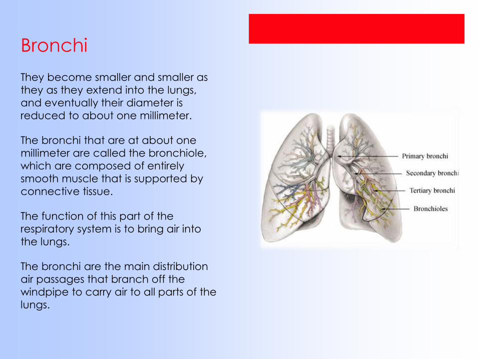
Bronchi
They become smaller and smaller as
they as they extend into the lungs,
and eventually their diameter is
reduced to about one millimeter.
The bronchi that are at about one
millimeter are called the bronchiole,
which are composed of entirely
smooth muscle that is supported by
connective tissue.
The function of this part of the
respiratory system is to bring air into
the lungs.
The bronchi are the main distribution
air passages that branch off the
windpipe to carry air to all parts of the
lungs.
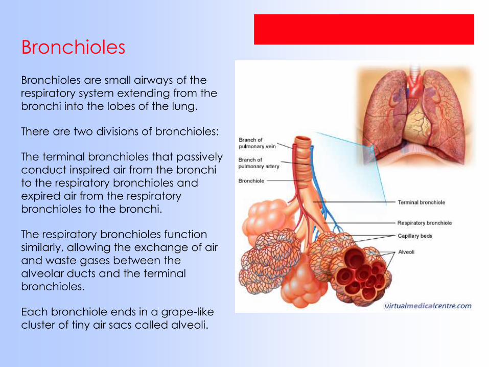
Bronchioles
Bronchioles are small airways of the
respiratory system extending from the
bronchi into the lobes of the lung.
There are two divisions of bronchioles:
The terminal bronchioles that passively
conduct inspired air from the bronchi
to the respiratory bronchioles and
expired air from the respiratory
bronchioles to the bronchi.
The respiratory bronchioles function
similarly, allowing the exchange of air
and waste gases between the
alveolar ducts and the terminal
bronchioles.
Each bronchiole ends in a grape-like
cluster of tiny air sacs called alveoli.
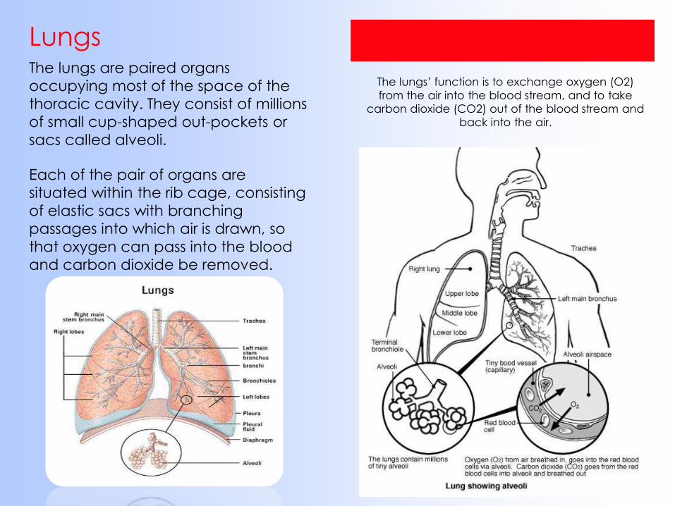
LungsThe lungs are paired organs
occupying most of the space of the
thoracic cavity. They consist of millions
of small cup-shaped out-pockets or
sacs called alveoli.
Each of the pair of organs are
situated within the rib cage, consisting
of elastic sacs with branching
passages into which air is drawn, so
that oxygen can pass into the blood
and carbon dioxide be removed.
The lungs’ function is to exchange oxygen (O2)
from the air into the blood stream, and to take
carbon dioxide (CO2) out of the blood stream and
back into the air.
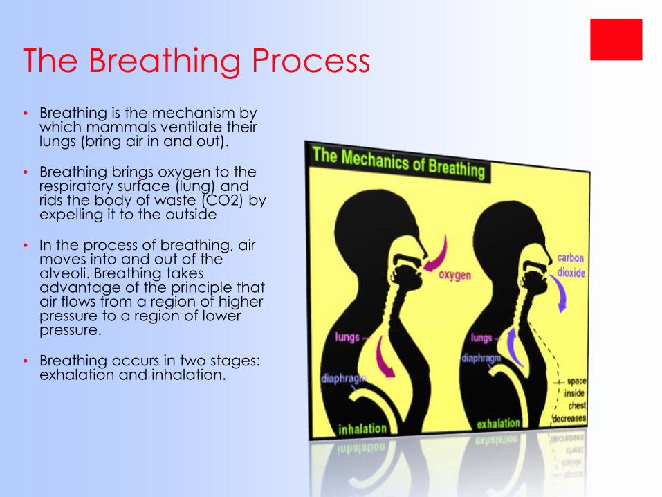
The Breathing Process
• Breathing is the mechanism by which mammals ventilate their lungs (bring air in and out).
• Breathing brings oxygen to the respiratory surface (lung) and rids the body of waste (CO2) by expelling it to the outside
• In the process of breathing, air moves into and out of the alveoli. Breathing takes advantage of the principle that air flows from a region of higher pressure to a region of lower pressure.
• Breathing occurs in two stages: exhalation and inhalation.

Inhalation
Inhalation is the process of
bringing air INTO the lungs.
During inhalation, the following
events occur:
1) The ribs move up and out
2) The diaphragm moves down.
3) The intercostal muscles contract
• When the above happens it
increases the volume of the
chest cavity. This creates a
low pressure inside the chest.
The pressure inside the chest is
less than the pressure outside
the body.
• Air “rushes” into the lungs from
the outside causing the lungs
to inflate.
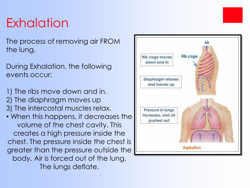
Exhalation
The process of removing air FROM
the lung.
During Exhalation, the following
events occur:
1) The ribs move down and in.2) The diaphragm moves up
3) The intercostal muscles relax.
• When this happens, it decreases the
volume of the chest cavity. This creates a high pressure inside the
chest. The pressure inside the chest is
greater than the pressure outside the
body. Air is forced out of the lung. The lungs deflate.
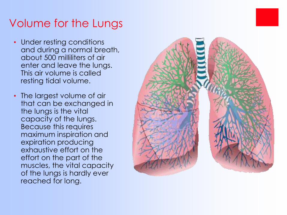
Volume for the Lungs
• Under resting conditions and during a normal breath, about 500 milliliters of air enter and leave the lungs. This air volume is called resting tidal volume.
• The largest volume of air that can be exchanged in the lungs is the vital capacity of the lungs. Because this requires maximum inspiration and expiration producing exhaustive effort on the effort on the part of the muscles, the vital capacity of the lungs is hardly ever reached for long.
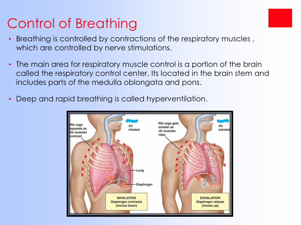
Control of Breathing• Breathing is controlled by contractions of the respiratory muscles ,
which are controlled by nerve stimulations.
• The main area for respiratory muscle control is a portion of the brain
called the respiratory control center. Its located in the brain stem and
includes parts of the medulla oblongata and pons.
• Deep and rapid breathing is called hyperventilation.
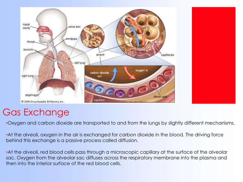
Gas Exchange•Oxygen and carbon dioxide are transported to and from the lungs by slightly different mechanisms.
•At the alveoli, oxygen in the air is exchanged for carbon dioxide in the blood. The driving force behind this exchange is a passive process called diffusion.
•At the alveoli, red blood cells pass through a microscopic capillary at the surface of the alveolar sac. Oxygen from the alveolar sac diffuses across the respiratory membrane into the plasma and then into the interior surface of the red blood cells.
Respiratory DisordersThe five major respiratory disorders:
•LUNG CANCER
•PEUMONIA
•ASTHMA
•BRONCHITIS
•EMPHASYEMA
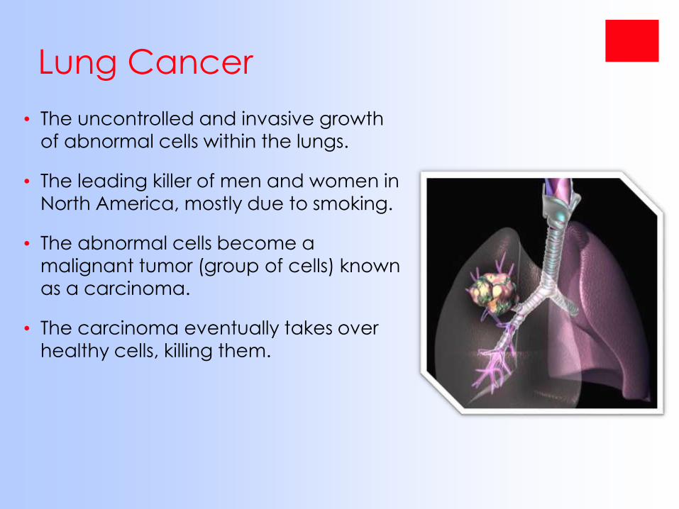
Lung Cancer
• The uncontrolled and invasive growth
of abnormal cells within the lungs.
• The leading killer of men and women in
North America, mostly due to smoking.
• The abnormal cells become a malignant tumor (group of cells) known
as a carcinoma.
• The carcinoma eventually takes over
healthy cells, killing them.
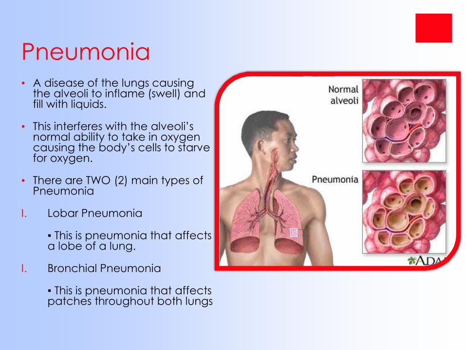
Pneumonia• A disease of the lungs causing
the alveoli to inflame (swell) and fill with liquids.
• This interferes with the alveoli’s normal ability to take in oxygen causing the body’s cells to starve for oxygen.
• There are TWO (2) main types of Pneumonia
I. Lobar Pneumonia
▪ This is pneumonia that affects a lobe of a lung.
I. Bronchial Pneumonia
▪ This is pneumonia that affects patches throughout both lungs
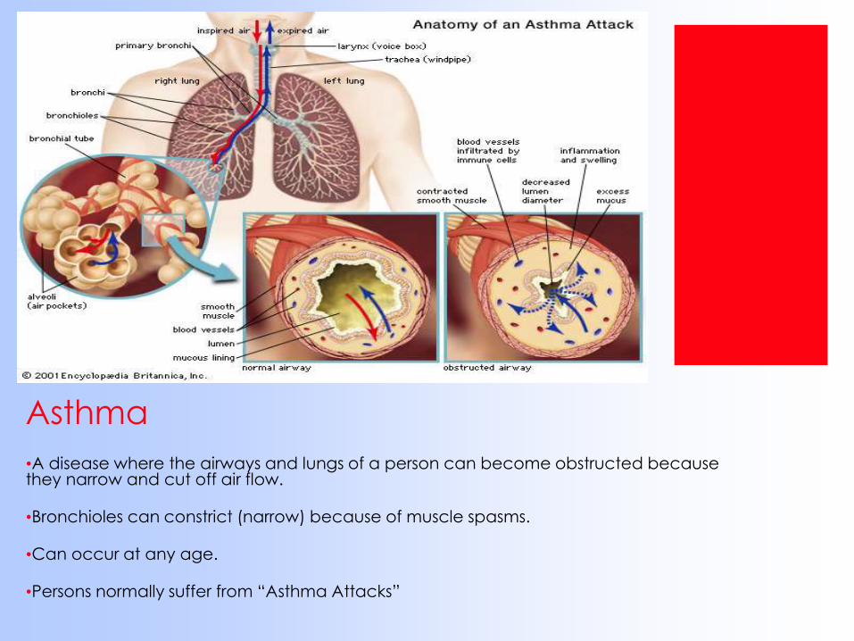
Asthma
•A disease where the airways and lungs of a person can become obstructed because they narrow and cut off air flow.
•Bronchioles can constrict (narrow) because of muscle spasms.
•Can occur at any age.
•Persons normally suffer from “Asthma Attacks”
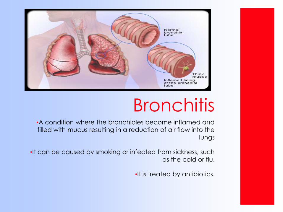
Bronchitis•A condition where the bronchioles become inflamed and
filled with mucus resulting in a reduction of air flow into the
lungs
•It can be caused by smoking or infected from sickness, such
as the cold or flu.
•It is treated by antibiotics.
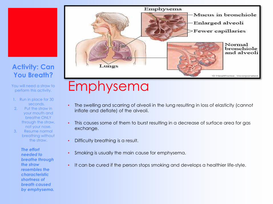
Emphysema• The swelling and scarring of alveoli in the lung resulting in loss of elasticity (cannot
inflate and deflate) of the alveoli.
• This causes some of them to burst resulting in a decrease of surface area for gas
exchange.
• Difficulty breathing is a result.
• Smoking is usually the main cause for emphysema.
• It can be cured if the person stops smoking and develops a healthier life-style.
Activity: Can
You Breath?
You will need a straw to
perform this activity.
1. Run in place for 30
seconds.
2. Put the straw in
your mouth and
breathe ONLY
through the straw,
not your nose.
3. Resume normal
breathing without
the straw.
The effort needed to breathe through
the straw resembles the characteristic shortness of breath caused by emphysema.
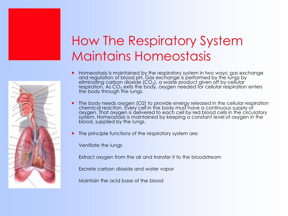
How The Respiratory System
Maintains Homeostasis Homeostasis is maintained by the respiratory system in two ways: gas exchange
and regulation of blood pH. Gas exchange is performed by the lungs by eliminating carbon dioxide (CO2), a waste product given off by cellular respiration. As CO2 exits the body, oxygen needed for cellular respiration enters the body through the lungs.
The body needs oxygen (O2) to provide energy released in the cellular respiration chemical reaction. Every cell in the body must have a continuous supply of oxygen. That oxygen is delivered to each cell by red blood cells in the circulatory system. Homeostasis is maintained by keeping a constant level of oxygen in the blood, supplied by the lungs.
The principle functions of the respiratory system are:
Ventilate the lungs
Extract oxygen from the air and transfer it to the bloodstream
Excrete carbon dioxide and water vapor
Maintain the acid base of the blood
Breathing/Feedback
Mechanism Breathing is clearly an involuntary process (you don't have to think
about it), and like many involuntary processes (such as heart rate) it is controlled by a region of the brain called the medulla. The medulla and its nerves are part of the autonomic nervous system (i.e. involuntary).
The region of the medulla that controls breathing is called the respiratory center. The respiratory center transmits regular nerve impulses to the diaphragm and intercostal muscles to cause inhalation. Stretch receptors in the alveoli and bronchioles detect inhalation and send inhibitory signals to the respiratory centre to cause exhalation. This negative feedback system is continuous and prevents damage to the lungs.
Ventilation is also under voluntary control from the cortex, the voluntary part of the brain. This allows you to hold your breath or blow out candles, but it can be overruled by the autonomic system in the event of danger. For example, if you hold your breath for a long time, the carbon dioxide concentration in the blood increases so much that the respiratory center forces you to gasp and take a breath.
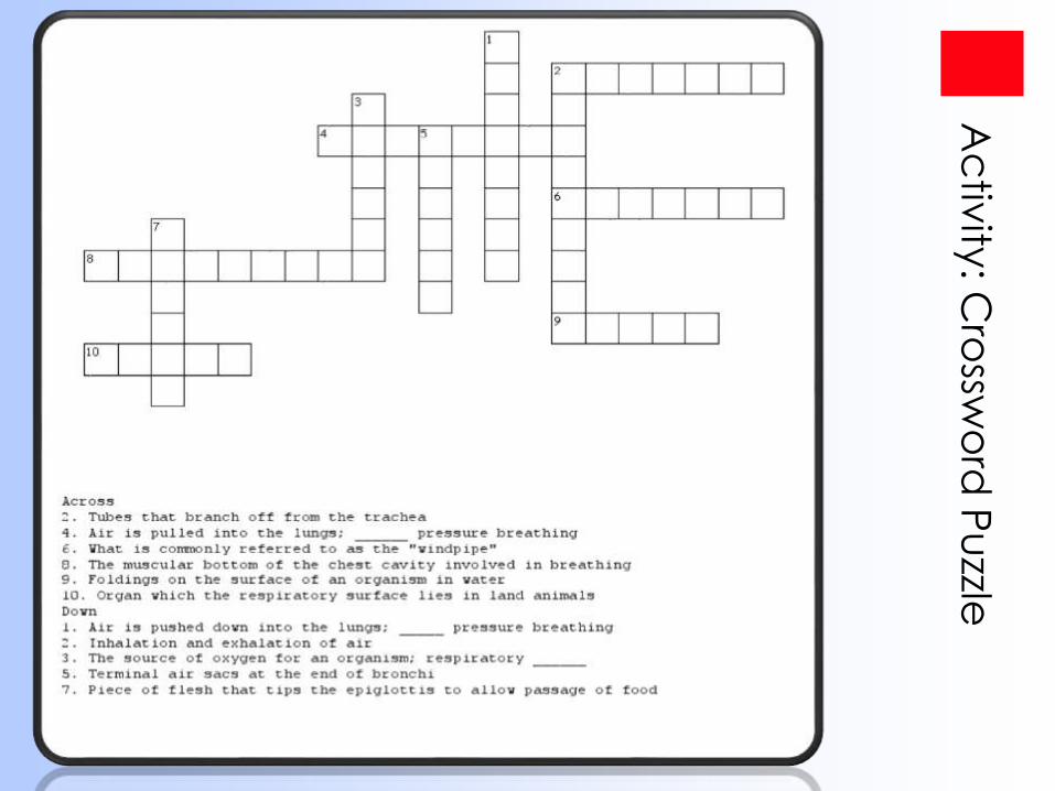
Ac
tivity
: Cro
sswo
rd P
uzzle
Fun Facts About The
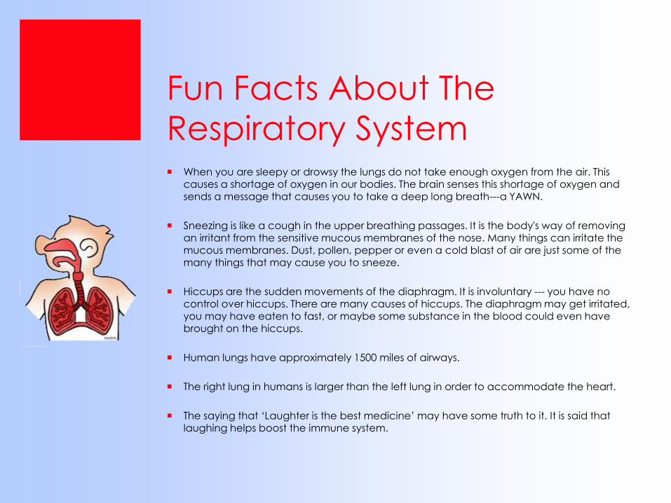
Respiratory System When you are sleepy or drowsy the lungs do not take enough oxygen from the air. This
causes a shortage of oxygen in our bodies. The brain senses this shortage of oxygen and sends a message that causes you to take a deep long breath---a YAWN.
Sneezing is like a cough in the upper breathing passages. It is the body's way of removing an irritant from the sensitive mucous membranes of the nose. Many things can irritate the mucous membranes. Dust, pollen, pepper or even a cold blast of air are just some of the many things that may cause you to sneeze.
Hiccups are the sudden movements of the diaphragm. It is involuntary --- you have no control over hiccups. There are many causes of hiccups. The diaphragm may get irritated,
you may have eaten to fast, or maybe some substance in the blood could even have brought on the hiccups.
Human lungs have approximately 1500 miles of airways.
The right lung in humans is larger than the left lung in order to accommodate the heart.
The saying that ‘Laughter is the best medicine’ may have some truth to it. It is said that laughing helps boost the immune system.
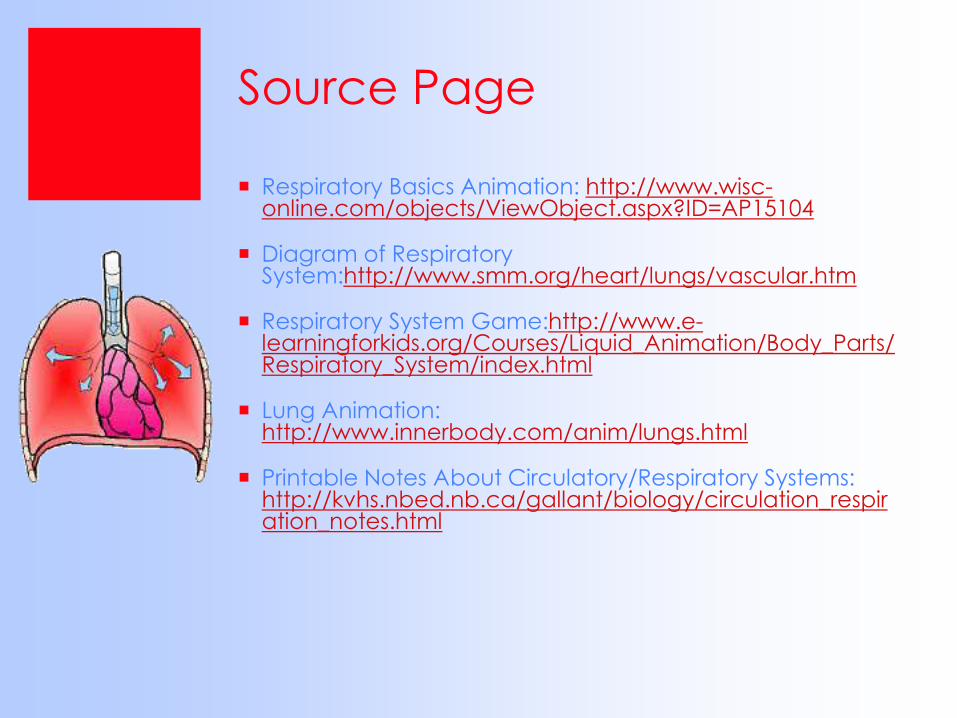
Source Page
Respiratory Basics Animation: http://www.wisc-online.com/objects/ViewObject.aspx?ID=AP15104
Diagram of Respiratory System:http://www.smm.org/heart/lungs/vascular.htm
Respiratory System Game:http://www.e-learningforkids.org/Courses/Liquid_Animation/Body_Parts/Respiratory_System/index.html
Lung Animation: http://www.innerbody.com/anim/lungs.html
Printable Notes About Circulatory/Respiratory Systems: http://kvhs.nbed.nb.ca/gallant/biology/circulation_respiration_notes.html
References
http://www.ambulancetechnicianstudy.co.uk/respsystem.html
http://hes.ucfsd.org/gclaypo/repiratorysys.html
http://www.brighthub.com/health/conditions-treatments/articles/57144.aspx
http://biology.clc.uc.edu/courses/bio105/respirat.htm
http://kidshealth.org/parent/general/body_basics/lungs.html
http://leavingbio.net/Respiratory%20System/THE%20RESPIRATORY%20SYSTEM.htm#parts