Circulation - conistonmrt.org.uk · Casualty Care syllabus 2009-13 Circulation Appreciate basic...
30
Circulation including Heart Attack and AED Revision notes for DFMRT Casualty Care Examination Course January 2013 Les Gordon
Transcript of Circulation - conistonmrt.org.uk · Casualty Care syllabus 2009-13 Circulation Appreciate basic...
Circulation including Heart Attack and AED
Revision notes for
DFMRT Casualty Care Examination Course January 2013
Les Gordon

Indicating special information in “Revision Notes” presentations
New information since Casualty Care in Mountain Rescue was published in 2006. NOTE: This presentation only includes essential information. To know the subject in greater depth, you must read Casualty Care in Mountain Rescue.

Casualty Care syllabus 2009-13 Circulation
Appreciate basic anatomy & physiology
Be able to assess circulation and recognise abnormalities
Understand types of shock and their causes
Be aware of the major causes of blood loss in the trauma
casualty
Understand general treatment principles
Be able to use appropriately the team’s AED

Components of the circulatory system – Circulating fluid (blood)
Fluid component (plasma) Contains chemicals called clotting factors to make blood clot.
Cellular component
Red blood cells carry oxygen Platelets help blood to clot White blood cells fight infection

Pulse (1)
What is it? Artery pulsating
Which arteries?
Radial Femoral Carotid Brachial Dorsalis pedis (foot)
Practical Tip Practice taking a pulse every opportunity you get (all casualties, family, friends, etc.). Then you will be able to do it reliably when faced with a sick casualty.

Pulse (2)
What to feel for Presence/absence
Rate (fast, normal or slow)
Rhythm (regular or irregular)
Strength (strong or weak)

Pulse (3) – Interpretation of findings Fast (>90-100 in adults)
Distress, Pain Bleeding Chest problems e.g. asthma, pneumothorax Abnormal heart rhythm Drugs e.g. amphetamines
Slow (<60 but could be much less) (adults) Spinal cord damage (upper thoracic or above) Cardiac arrhythmia Severe hypothermia Pre-existing due to drugs; someone who is very fit Severe brain injury
Irregular Irritable heart e.g. after MI
Weak Shock e.g. hypovolaemic (blood loss); anaphylaxis; cardiogenic (after MI) Dehydration Severe hypothermia

Blood Pressure Know the normal (approximately 120-140/70-90)
Know how to take it properly
Predicting the blood pressure from the palpable pulses
Radial + femoral + carotid = systolic BP probably 80 mm Hg or more Femoral + carotid only = systolic BP probably about 70 mm Hg Carotid only = systolic BP probably about 60 mm Hg
NB These are guides only. They are not 100% accurate

Capillary Refill Time Normal is <2 seconds.

Major causes of blood loss in trauma
Four major sites Chest Abdomen Pelvis Femur
The casualty can bleed to death from bleeding at any of these sites, particularly the first three.

Few practical points Be suspicious if history is compatible with major blood loss but there
is none/little to see. It may all be inside the body.
Typical blood loss after fractures: Upper arm 250 – 1000 ml Lower leg 500 – 1000 ml Shaft of femur 1000 – 2000 ml Rib fracture ± haemothorax 150 – 2000 ml Pelvis 500 – 3000 ml
(Q: Why is the range so great in rib and pelvic fractures?)
NOTE: several smaller injuries can cause as much blood loss as one big one.

General measures for managing moderate to major bleeding at any site
Oxygen
Lie casualty down
Keep casualty warm Hypothermia seriously impairs blood clotting – part of the “Triad of
Death”
If BP very low, can raise legs to return some blood from them back to the heart BUT if you do that, remember that if you put the legs down, the BP will fall again
Small amounts of IV fluids sufficient to keep radial artery palpable (for those people who can do this procedure).
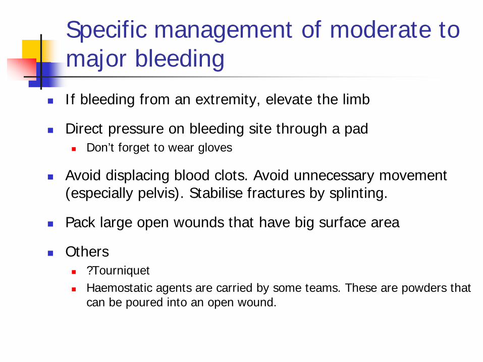
Specific management of moderate to major bleeding
If bleeding from an extremity, elevate the limb
Direct pressure on bleeding site through a pad Don’t forget to wear gloves
Avoid displacing blood clots. Avoid unnecessary movement (especially pelvis). Stabilise fractures by splinting.
Pack large open wounds that have big surface area
Others ?Tourniquet Haemostatic agents are carried by some teams. These are powders that
can be poured into an open wound.

Important ‘C’ questions when assessing a casualty
Is this casualty showing signs of shock?
Is there any external bleeding?
Is there possibility of concealed haemorrhage?
Is there potential for shock to develop?

Myocardial infarction (heart attack) The history (1)
Chest pain can be due to many causes including Angina Heart attack Pneumothorax Pulmonary embolism (‘clot on the lung’) Musculoskeletal Indigestion
Key to distinguishing MI from other causes of chest pain is to take a good history.
Important general points in the history Angina / Previous heart operation for angina / Anti-angina drugs Previous MI Smoker / diabetic / family history / obesity / high cholesterol

Myocardial infarction The history (2)
Typical MI history Crushing central chest pain lasting >20 minutes ± pain radiates to neck, jaw, L arm, upper abdomen Sweatiness Nausea / vomiting Breathlessness Palpitations Dizziness NB Can mimic bad indigestion

Myocardial infarction Examination
In pain Breathless when lying down (allow them to sit up) Breathing fast Colour (pale) Heart rate
Fast (distress, abnormal heart rhythm) Slow (abnormal heart rhythm; also, can be caused by some anti-angina
drugs patient is taking)
SpO2 May be low especially if heart failure or fluid collecting in lungs
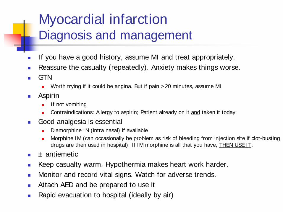
Myocardial infarction Diagnosis and management
If you have a good history, assume MI and treat appropriately. Reassure the casualty (repeatedly). Anxiety makes things worse. GTN
Worth trying if it could be angina. But if pain >20 minutes, assume MI
Aspirin If not vomiting Contraindications: Allergy to aspirin; Patient already on it and taken it today
Good analgesia is essential Diamorphine IN (intra nasal) if available Morphine IM (can occasionally be problem as risk of bleeding from injection site if clot-busting
drugs are then used in hospital). If IM morphine is all that you have, THEN USE IT.
± antiemetic Keep casualty warm. Hypothermia makes heart work harder. Monitor and record vital signs. Watch for adverse trends. Attach AED and be prepared to use it Rapid evacuation to hospital (ideally by air)

Principles of AED use AED means Automated External Defibrillator not Automatic
Uses a controlled electric shock passed through the heart to
change some cardiac arrest rhythms back to normal rhythms
The electricity is delivered to the patient via adhesive pads that are stuck on the chest wall
The machine will assess the cardiac rhythm and advise if a shock is needed

Principles of AED use – SAFETY Use in the presence of water (1)
“Remotely possible” to get shocked or to shock bystanders if pooled water around and under the patient (American Heart Association). However, there are no published case reports of this.
Studies of simulated patient (a turkey or a pig) lying in fresh water, salt water or with wet surface: No significant current leakage when defibrillating a wet pig Maximum voltage 15 cm from patient was 14V peak (14 mA peak) fresh water Maximum voltage 15 cm from patient was 30V peak (30 mA peak) salt water Everywhere else, <<1V 30V may result in some minor sensation but is unlikely to be hazardous
Defibrillators designed for out-of-hospital use have sealed cases so can be used in rain or snow.

Principles of AED use – SAFETY Use in the presence of water (2)
Practice points Move casualty to dry area if possible. Cut off wet clothing covering chest and dry the chest wall. Avoid getting the AED pads wet. Make sure no one is touching patient when shock button is pressed. Defibrillation on a metal surface is safe e.g. helicopter floor.
REFERENCES in case you want to read more. • Questions & Answers about AEDs and defibrillation. American Heart Association.
http://www.tvfr.com/safetytips/docs/AHA_aedqa.pdf (accessed 11/1/13). • Lyster T, et al. The safe use of Automated External Defibrillators in a wet environment. Prehosp Emerg Care 2003; 7: 307-11. • Medtronic LIFEPAK 500 Automated External Defibrillator – commonly asked questions (2003). • Philips Technical note HeartStart AED – Defibrillation on a wet or metal surface (2005). • Zoll Technical Report – Defibrillation on a wet or metal surface. • Schratter A, et al. Abstract 82: External cardiac defibrillation during wet surface cooling in pigs. Circulation 2006; 114:
II_1205.

Principles of AED use – Before placing the pads
The five P’s Perspiration and suntan oil
Dry the chest
Pendants Remove them
Pacemakers See later slide
Piercings Remove if possible. If not, place pad to side of piercing
Patches (e.g. nicotine, GTN) Some substances can explode Remove if near pad site

Principles of AED use – Pad positioning
The pads generally have a required orientation with the positive over the upper R side of the chest.
Pads usually have a picture indicating where they should be placed
Place the R pad above the R nipple but not over the clavicle
Place the L pad below the left breast and to the side
Press firmly to get good contact with skin (may need to shave chest)

Principles of AED use – The patient with a pacemaker
Aim is to place defibrillation pads in patients with pacemaker as far as possible from the pacemaker box.
When a pacemaker is located where a pad would normally be placed, position the pad at least 1” away from the device.
Ideal position Alternative anterior-posterior pad placement with positive on back of the chest

Principles of AED use – Safe operation of the device
Switch on AED (button 1) Voice prompt “Apply Electrodes”
Place electrodes as above
AED will check that pads are making good contact
If not, press pad firmly in centre to see if that helps
If necessary, remove pads, clean skin, ± shave hair and reapply
Press button 2 (blue) to start
analysis Stop chest compressions during
analysis

Principles of AED use – Safe operation of the device
Shock is needed Voice prompt –
“Do not touch the patient – Charging”
Remove any oxygen mask and
place >1m away to prevent reservoir of oxygen building up around patient’s face.
BEFORE DELIVERING THE SHOCK, THE PERSON WHO WILL PRESS THE BUTTON MUST VISUALLY CHECK THAT NO ONE IS TOUCHING THE PATIENT AND MUST SHOUT OUT “STAND CLEAR”
Press button 3 (orange) to deliver shock, and then immediately straight back on to the chest


Cardiac Arrest Basic Life Support Safe to approach Shake and shout Call for help Airway – chin lift/head tilt and look in mouth Breathing – count audibly for 10 seconds If no signs of life, straight on to chest NB must be IMMEDIATE CPR 30:2 Bag-valve-mask 2nd person can hold the mask with two hands whilst the person
doing chest compressions squeezes the bag Use one hand only to squeeze the bag (avoids squeezing too hard
and filling the stomach with air)
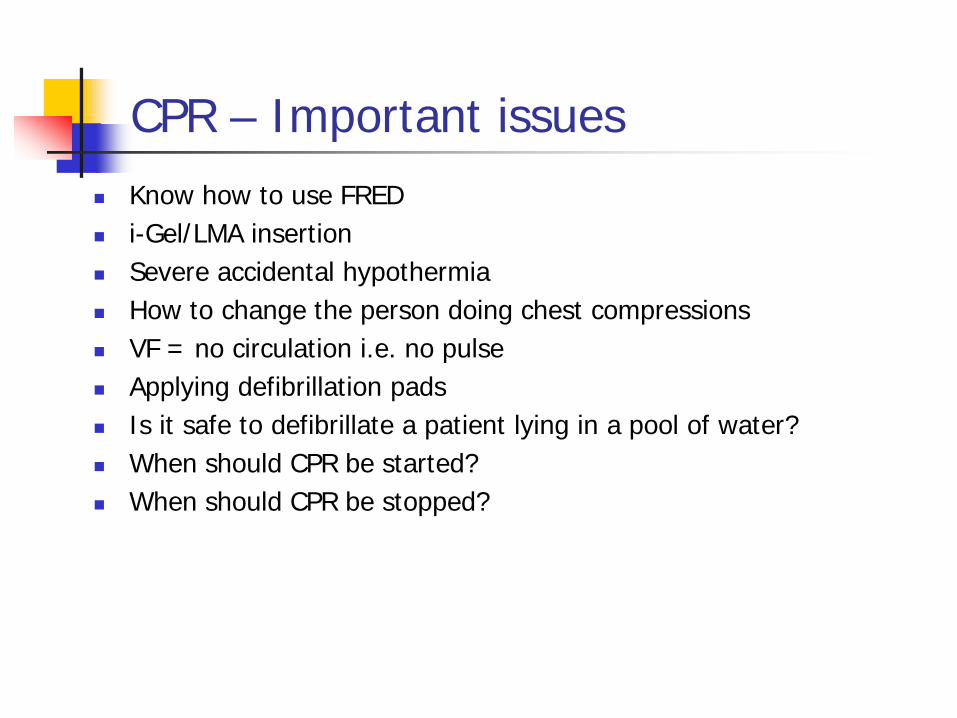
CPR – Important issues Know how to use FRED i-Gel/LMA insertion Severe accidental hypothermia How to change the person doing chest compressions VF = no circulation i.e. no pulse Applying defibrillation pads Is it safe to defibrillate a patient lying in a pool of water? When should CPR be started? When should CPR be stopped?

Training points Practice taking pulses, CRT and BP.