
CIRCULATION AND GAS EXCHANGE Chapter 42. Gas Exchange Systems of Different Animals Gastrovascular...
50
CIRCULATION AND GAS EXCHANGE Chapter 42
-
Upload
gabriel-thomas -
Category
Documents
-
view
228 -
download
0
Transcript of CIRCULATION AND GAS EXCHANGE Chapter 42. Gas Exchange Systems of Different Animals Gastrovascular...

CIRCULATION AND GAS EXCHANGE
Chapter 42

Gas Exchange Systems of Different Animals
• Gastrovascular Cavities – (Platyhelminthes, Cnidarians, simple animals)
• Open and Closed Circulatory Systems– Open – Arthropods such as insects, Molluscks– Closed – Annelids such as earthworms, Cephalopods such as squid
and octopi, vertebrates

Open vs. Closed

Various Closed Circulatory Systems
Fish2-chambered heart
Amphibian and Reptiles3-chambered heart
Mammal4-chambered heart

The Mammalian Heart
THE HEART•Made up of cardiac muscle (most of it)•4-chambered (2 atria on top of, 2 ventricles)•4 valves
-The 2 Atrioventricular valves are between the atria and ventricles
-The tricuspid on the right-The bicuspid on the left
-The 2 Semilunar valves are located at the 2 exits of the heart
-The Pulmonary valve that leads to the pulmonary artery-The Aortic valve that leads to the aorta
Endotherms (Hometherms) require 4-chambered hearts

(Aortic Valve)(Pulmonary Valve)
(Tricuspid Valve) (Bicuspid or Mitral Valve)


Heart Chambers and Valves• The heart has four internal chambers: two atria on top
and two ventricles below.
– Atria receive blood returning to the heart and have thin walls and ear-like auricles projecting from their exterior.
– The thick-muscled ventricles pump blood to the body.
• A septum divides the atrium and ventricle on each side. Each also has an atrioventricular (A-V) valve to ensure one way flow of blood.
– The right A-V valve (tricuspid) and left A-V valve (bicuspid or mitral valve) have cusps to which chordae tendinae attach.
– Chordae tendinae are, in turn, attached to papillary muscles in the inner heart wall that contract during ventricular contraction to prevent the backflow of blood through the A-V valves.
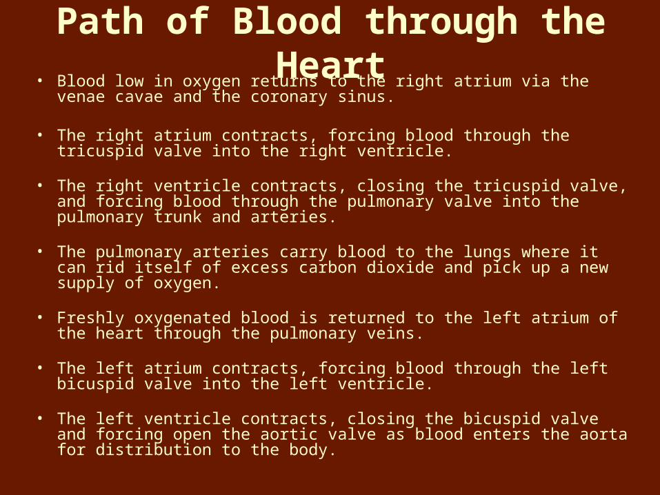
Path of Blood through the Heart• Blood low in oxygen returns to the right atrium via the venae cavae and
the coronary sinus.
• The right atrium contracts, forcing blood through the tricuspid valve into the right ventricle.
• The right ventricle contracts, closing the tricuspid valve, and forcing blood through the pulmonary valve into the pulmonary trunk and arteries.
• The pulmonary arteries carry blood to the lungs where it can rid itself of excess carbon dioxide and pick up a new supply of oxygen.
• Freshly oxygenated blood is returned to the left atrium of the heart through the pulmonary veins.
• The left atrium contracts, forcing blood through the left bicuspid valve into the left ventricle.
• The left ventricle contracts, closing the bicuspid valve and forcing open the aortic valve as blood enters the aorta for distribution to the body.

Blood Supply to the Heart
• The first branches off of the aorta, which carry freshly oxygenated blood, are the right and left coronary arteries that feed the heart muscle itself.
• Branches of the coronary arteries feed many capillaries of the myocardium.
• The heart muscle requires a continuous supply of freshly oxygenated blood, so smaller branches of arteries often have anastomoses as alternate pathways for blood, should one pathway become blocked.
• Cardiac veins drain blood from the heart muscle and carry it to the coronary sinus, which empties into the right atrium.

Direction of Blood Flow• The superior and inferior vena cavae bring blood from the
body to the right atrium.
• The right ventricle has a thinner wall than does the left ventricle because it must pump blood only as far as the lungs, compared to the left ventricle pumping to the entire body.
• At the base of the pulmonary trunk leading to the lungs is the pulmonary valve, which prevents a return flow of blood to the ventricle.
• The left atrium receives blood from four pulmonary veins.
• The left ventricle pumps blood into the entire body through the aorta, guarded by the aortic valve that prevents backflow of blood into the ventricle.
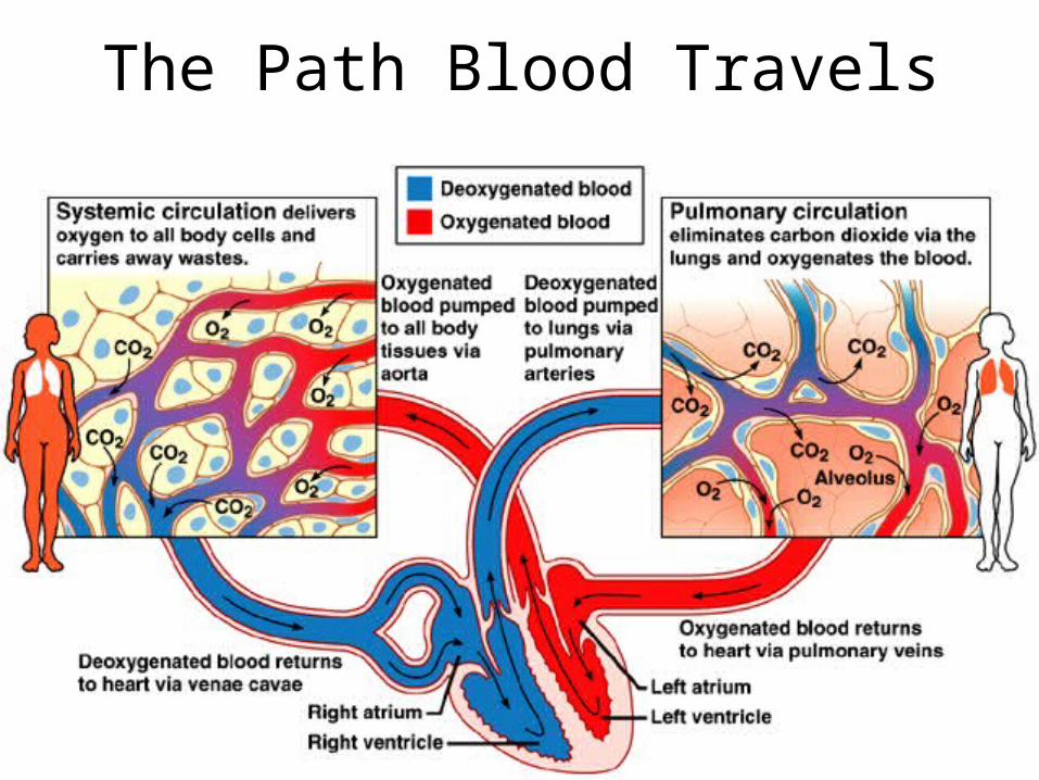
The Path Blood Travels

Terminology
1. Cardiac Cycle – one complete sequence of pumping out of blood and filling up with blood
2. Systole – the contraction of the heart chamber muscle
3. Diastole – the relaxation of the heart chamber muscle
4. Cardiac Output – the volume of blood pumped by the heart

The Rhythm is going to get you!
Specialized muscle tissue in the region of the heart called the sinoatrial node (SA) is the pacemaker. It maintains the heart’s pumping, by setting the pace at which cardiac muscle cells contract.
1. The SA node sends electric impulses anywhere between 60 and 100 times a minute
2. The impulses spread throughout the atria3. The atrioventricular (AV) node intercepts these signals
and relays them to the apex of the heart, via specialized muscle fibers called Bundle branches and Purkinje fibers
4. The signal then spreads throughout the ventricles

Artial Systole, Ventricular diastole
Atrial Diastole, Ventricular systole
“LUBB”
“DUPP”

Cardiac Cycle• During the cardiac cycle, pressure within the heart
chambers rises and falls with the contraction and relaxation of atria and ventricles.
• When the atria fill, pressure in the atria is greater than that of the ventricles, which forces the A-V valves open.
• Pressure inside atria rises further as they contract, forcing the remaining blood into the ventricles.
• When ventricles contract, pressure inside them increases sharply, causing A-V valves to close and the aortic and pulmonary valves to open.
– As the ventricles contract, papillary muscles contract, pulling on chordae tendinae and preventing the backflow of blood through the A-V valves.

Heart Sounds Heart Sounds
• Heart sounds are due to vibrations in heart tissues as Heart sounds are due to vibrations in heart tissues as blood rapidly changes velocity within the heart.blood rapidly changes velocity within the heart.
• Heart sounds can be described as a "lubb-dupp" sound.Heart sounds can be described as a "lubb-dupp" sound.
• The first sound (lubb) occurs as ventricles contract, atria The first sound (lubb) occurs as ventricles contract, atria relax. The A-V valves are closing and the pulmonary and relax. The A-V valves are closing and the pulmonary and aortic valves are opening.aortic valves are opening.
• The second sound (dupp) occurs as ventricles relax, The second sound (dupp) occurs as ventricles relax, atria contract. The aortic and pulmonary valves are atria contract. The aortic and pulmonary valves are closing and the A-V valves are opening.closing and the A-V valves are opening.

Cardiac Conduction System• Specialized cardiac muscle tissue conducts impulses throughout
the myocardium and comprises the cardiac conduction system.
• A self-exciting mass of specialized cardiac muscle called the sinoatrial node (S-A node or pacemaker), located on the posterior right atrium, generates the impulses for the heartbeat.
• Impulses spread next to the atrial syncytium, it contracts, and impulses travel to the junctional fibers leading to the atrioventricular node (A-V node) located in the septum.
– Junctional fibers are small, allowing the atria to contract before the impulse spreads rapidly over the ventricles.
• Branches of the A-V bundle give rise to Purkinje fibers leading to papillary muscles; these fibers stimulate contraction of the papillary muscles at the same time the ventricles contract.

Cardiac Conduction System

1
4
3
5
6
7
Atrial Syncytium2
Ventricular Syncytium8

The control of heart rhythm
p
q
r
s
t


The Structure of Blood Vessels

Blood Vessels

Arteries and Arterioles
• Arteries are strong, elastic vessels adapted for carrying high-pressure blood.
• Arteries become smaller as they divide and give rise to arterioles.
• The wall of an artery consists of an endothelium, tunica media (smooth muscle), and tunica externa (connective tissue).
• Arteries are capable of vasoconstriction as directed by the sympathetic impulses; when impulses are inhibited, vasodilation results.


Capillaries• Capillaries are the smallest vessels, consisting only of a layer of
endothelium through which substances are exchanged with tissue cells.
• Capillary permeability varies from one tissue to the next, generally with more permeability in the liver, intestines, and certain glands, and less in muscle and considerably less in the brain (blood-brain barrier).
• The pattern of capillary density also varies from one body part to the next.
• Areas with a great deal of metabolic activity (leg muscles, for example) have higher densities of capillaries.
• Precapillary sphincters can regulate the amount of blood entering a capillary bed and are controlled by oxygen concentration in the area.
– If blood is needed elsewhere in the body, the capillary beds in less important areas are shut down by the precapillary sphincters.


Exchanges in the CapillariesExchanges in the Capillaries• Blood entering capillaries contains high concentrations of Blood entering capillaries contains high concentrations of
oxygen and nutrients that diffuse out of the capillary wall and oxygen and nutrients that diffuse out of the capillary wall and into the tissues.into the tissues.
– Plasma proteins remain in the blood due to their large size.Plasma proteins remain in the blood due to their large size.
• Hydrostatic pressure drives the passage of fluids and very Hydrostatic pressure drives the passage of fluids and very small molecules out of the capillary at this point.small molecules out of the capillary at this point.
• At the venule end, osmosis, due to the osmotic pressure of the At the venule end, osmosis, due to the osmotic pressure of the blood, causes much of the tissue fluid to return to the blood, causes much of the tissue fluid to return to the bloodstream.bloodstream.
• Lymphatic vessels collect excess tissue fluid and return it to Lymphatic vessels collect excess tissue fluid and return it to circulation.circulation.

Venules and Veins
• Venules leading from capillaries merge to form veins that return blood to the heart.
• Veins have the same three layers as arteries have and have a flap-like valve inside to prevent backflow of blood.
– Veins are thinner and less muscular than arteries; they do not carry high-pressure blood.
– Veins also function as blood reservoirs.


Blood Pressure • Blood pressure is the force of blood against the inner walls
of blood vessels anywhere in the cardiovascular system, although the term "blood pressure" usually refers to arterial pressure.
• Arterial blood pressure rises and falls following a pattern established by the cardiac cycle.
– During ventricular contraction, arterial pressure is at its highest (systolic pressure).
– When ventricles are relaxing, arterial pressure is at its lowest (diastolic pressure).
• The surge of blood that occurs with ventricular contraction can be felt at certain points in the body as a pulse.

Blood Flow in capillary beds
•Capillaries are composed of a single layer of epithelium surrounding a lumen of a few micrometers. •The average capillary is only about 1 mm long. •The capillary beds are the region of exchange of materials with the tissues. O2 and nutrients are supplied to the cells, and CO2 and other waste products are taken away. •Some materials are transported across the endothelial membrane by diffusion, but material (especially water) also leaves through pores in the capillary walls. •Capillaries supply brain, heart, kidneys and liver with blood constantly, but their supply to other parts varies based on need.
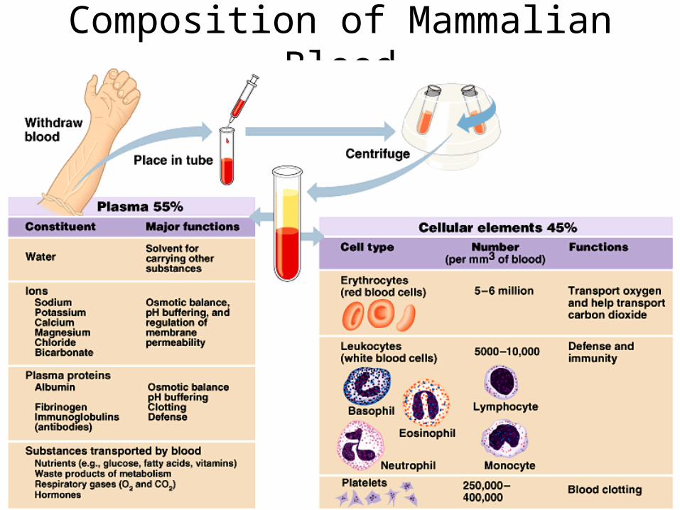
Composition of Mammalian Blood

Differentiation of blood cells
The Kidneys convert a plasma protein into a hormone called erythropoietin, which stimulates the bone marrow to produce erythrocytes
(Become macrophages“big eaters”)
(Phagocytes)
(Destroy larger invaders bySecreting destructive enzymes)
(release Histamine in response to tissue injury)

Blot Clotting

Atherosclerosis
Platelets and can clump and fibrin can coagulate within a blood vessel to form a clot. This clot is called a thrombus. These clots can cause cardiovascular diseases.
Myocardial Infarction (heart attack) is the death of cardiac muscle and a Stroke is the death of nervous tissue in the brain

Gas exchange in the lungs

SEM of Pulmonary Alveoli Alveoli are lung air sacs made
of simple squamous epithelial cells for diffusion of gases.
Capillaries plus alveoli form the respiratory membrane for the exchange of gases between the blood and the lungs.
Alveolus

Gas Exchange in the Alveoli
Capillary beds run around the outside of the entire alveolus. The capillary bed is from arteriole to venule, and is depicted as running from top left, down and to the right, and reaching the venule at the top right. Remember, pulmonary arterioles contain deoxyhemoglobin and are color coded in blue, while pulmonary venules contain oxyhemoglobin and are color coded in red.

Alveolar Gas Exchange
The gases only have to travel through a 2-cell thick region (one-cell thick capillary wall and a one-cell thick alveolar wall - with just the basement membrane between them). This 2-cell-thick-material is called the respiratory membrane.

CO2 TransportCarbon dioxide (CO2) combines with water forming carbonic acid, which dissociates into a hydrogen ion (H+) and as bicarbonate ions:
CO2 + H2O ↔ H2CO3 ↔ H+ + HCO3−
95% of the CO2 generated in the tissues is carried in the red blood cells:
It probably enters (and leaves) the cell by diffusing through transmembrane channels in the plasma membrane.
Once inside, about one-half of the CO2 is directly bound to hemoglobin (at a site different from the one that binds oxygen).
The rest is converted — following the equation above — by the enzyme carbonic anhydrase into bicarbonate ions that diffuse back out into the plasma and hydrogen ions (H+) bind to the protein portion of the hemoglobin (thus having no effect on pH).
Only about 5% of the CO2 generated in the tissues dissolves directly in the plasma. (A good thing, too: if all the CO2 we make were carried this way, the pH of the blood would drop from its normal 7.4 to an instantly-fatal 4.5!)
When the red cells reach the lungs, these reactions are reversed and CO2 is released to the air of the alveoli.

The Breathing Mechanism

What makes you take a breath?
Conclusion: Increased levels of Conclusion: Increased levels of COCO22 lead to taking a breath. lead to taking a breath.

In fish, the oxygenated blood from the gills does not need In fish, the oxygenated blood from the gills does not need to return to the heart to get pumped to the rest of the body. to return to the heart to get pumped to the rest of the body.
It can go to all body tissues directly from the gills.It can go to all body tissues directly from the gills.


When CO2 levels in he blood increase, blood pH drops. This causes an additional release of O2
from hemoglobin.
Conclusion: The greater the need for O2, the more it is released from hemoglobin.
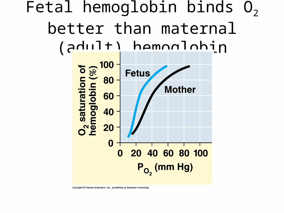
Fetal hemoglobin binds O2 better than maternal (adult) hemoglobin

THE END


