Circulating Sex Steroids during Pregnancy and Maternal...
14
Research Article Circulating Sex Steroids during Pregnancy and Maternal Risk of Non-epithelial Ovarian Cancer Tianhui Chen 1 , Helja-Marja Surcel 2 , Eva Lundin 3,4 , Marjo Kaasila 2 , Hans-Ake Lakso 3 , Helena Schock 1 , Rudolf Kaaks 1 , Pentti Koskela 2 , Kjell Grankvist 3 , Goran Hallmans 4 , Eero Pukkala 5,6 , Anne Zeleniuch-Jacquotte 7,8 , Paolo Toniolo 8,9,10 , Matti Lehtinen 2,6 , and Annekatrin Lukanova 1,9 Abstract Background: Sex steroid hormones have been proposed to play a role in the development of non-epithelial ovarian cancers (NEOC) but so far no direct epidemiologic data are available. Methods: A case–control study was nested within the Finnish Maternity Cohort, the world’s largest biorepository of serum specimens from pregnant women. Study subjects were selected among women who donated a blood sample during a singleton pregnancy that led to the birth of their last child preceding diagnosis of NEOC. Case subjects were 41 women with sex cord stromal tumors (SCST) and 21 with germ cell tumors (GCT). Three controls, matching the index case for age, parity at the index pregnancy, and date at blood donation were selected (n ¼ 171). OR and 95% CI associated with concentrations of testosterone, androstenedione, 17-OH-progesterone, progesterone, estradiol, and sex hormone–binding globulin (SHBG) were estimated through conditional logistic regression. Results: For SCST, doubling of testosterone, androstenedione, and 17-OH-progesterone concentrations were associated with about 2-fold higher risk of SCST [ORs and 95% CI of 2.16 (1.25–3.74), 2.16 (1.20–3.87), and 2.62 (1.27–5.38), respectively]. These associations remained largely unchanged after excluding women within 2-, 4-, or 6-year lag time between blood donation and cancer diagnosis. Sex steroid hormones concentrations were not related to maternal risk of GCT. Conclusions: This is the first prospective study providing initial evidence that elevated androgens play a role in the pathogenesis of SCST. Impact: Our study may note a particular need for larger confirmatory investigations on sex steroids and NEOC. Cancer Epidemiol Biomarkers Prev; 20(2); 324–36. Ó2010 AACR. Introduction Non-epithelial ovarian cancers (NEOC) account for approximately 10% of all ovarian tumors and about 7% of the invasive ones (1). They are divided into 2 major distinct subtypes, sex cord-stromal tumors (SCST) and germ cell tumors (GCT; refs. 1–3). SCST occur in women of all ages but increase in frequency during the fourth and fifth decades of age and have a median age at diagnosis of 52 years (4–6). In contrast, GCT occur predominantly in young women with the peak incidence around the age of 18 and are rarely observed after age 30 years (7). The incidence rates of the 2 subtypes of NEOC also differ by race: SCST are twice as frequent in women of European and American background as in women from Asian descent (4), whereas GCT are more frequent in Asian women (8, 9). Because of the low incidence of NEOC, very few studies have investigated risk factors for their develop- ment. So far, only 6 studies, 5 of which included between 10 and 72 cases of SCST or GCT, have reported on the association of these tumors with traditional reproductive risk factors, such as parity, oral contraceptive use, ages at the first and last births, and time since last birth (1, 10–14). Although the results from these studies are not entirely consistent, parous women appear to be at reduced risk of both SCST and GCT (1, 10, 12), but, there is some indica- tion that the effect of other reproductive factors (1, 11, 12, 15), particularly age at last birth (11, 12) may differ in the 2 subtypes. It has also been suggested that exposure to high Authors' Affiliations: 1 Division of Cancer Epidemiology, German Cancer Research Center, Heidelberg, Germany; 2 National Institute for Health and Welfare, Oulu, Finland; 3 Department of Medical Biosciences; 4 Public Health and Clinical Medicine: Nutritional Research, University of Umea, Umea, Sweden; 5 Finnish Cancer Registry, Institute for Statistical and Epidemiological Cancer Research, Helsinki; 6 University of Tampere, Tam- pere, Finland; 7 Institute of Environmental Medicine, New York University School of Medicine; 8 New York University Cancer Institute; 9 Department of Obstetrics and Gynecology, New York University School of Medicine, New York; and 10 Institute of Social and Preventive Medicine, Centre Hospitalier Universitaire Vaudois, University of Lausanne, Lausanne, Switzerland Corresponding Author: Annekatrin Lukanova, Division of Cancer Epide- miology, German Cancer Research Center, In Neuenheimer Feld 280, Heidelberg 69120, Germany. Phone: 496221-42-22-41; Fax: 496221-42- 22-03. E-mail: [email protected] doi: 10.1158/1055-9965.EPI-10-0857 Ó2010 American Association for Cancer Research. Cancer Epidemiology, Biomarkers & Prevention Cancer Epidemiol Biomarkers Prev; 20(2) February 2011 324 on July 4, 2018. © 2011 American Association for Cancer Research. cebp.aacrjournals.org Downloaded from Published OnlineFirst December 21, 2010; DOI: 10.1158/1055-9965.EPI-10-0857
Transcript of Circulating Sex Steroids during Pregnancy and Maternal...

Research Article
Circulating Sex Steroids during Pregnancy and MaternalRisk of Non-epithelial Ovarian Cancer
Tianhui Chen1, Helja-Marja Surcel2, Eva Lundin3,4, Marjo Kaasila2, Hans-Ake Lakso3, Helena Schock1, Rudolf Kaaks1,Pentti Koskela2, Kjell Grankvist3, Goran Hallmans4, Eero Pukkala5,6, Anne Zeleniuch-Jacquotte 7,8, Paolo Toniolo8,9,10,Matti Lehtinen2,6, and Annekatrin Lukanova1,9
AbstractBackground: Sex steroid hormones have been proposed to play a role in the development of non-epithelial
ovarian cancers (NEOC) but so far no direct epidemiologic data are available.
Methods: A case–control study was nested within the Finnish Maternity Cohort, the world’s largest
biorepository of serum specimens from pregnant women. Study subjects were selected among women who
donated a blood sample during a singleton pregnancy that led to the birth of their last child preceding
diagnosis of NEOC. Case subjects were 41 women with sex cord stromal tumors (SCST) and 21 with germ cell
tumors (GCT). Three controls, matching the index case for age, parity at the index pregnancy, and date at
blood donation were selected (n ¼ 171). OR and 95% CI associated with concentrations of testosterone,
androstenedione, 17-OH-progesterone, progesterone, estradiol, and sex hormone–binding globulin (SHBG)
were estimated through conditional logistic regression.
Results: For SCST, doubling of testosterone, androstenedione, and 17-OH-progesterone concentrations
were associatedwith about 2-fold higher risk of SCST [ORs and 95%CI of 2.16 (1.25–3.74), 2.16 (1.20–3.87), and
2.62 (1.27–5.38), respectively]. These associations remained largely unchanged after excluding women within
2-, 4-, or 6-year lag time between blood donation and cancer diagnosis. Sex steroid hormones concentrations
were not related to maternal risk of GCT.
Conclusions: This is the first prospective study providing initial evidence that elevated androgens play a
role in the pathogenesis of SCST.
Impact: Our study may note a particular need for larger confirmatory investigations on sex steroids
and NEOC. Cancer Epidemiol Biomarkers Prev; 20(2); 324–36. �2010 AACR.
Introduction
Non-epithelial ovarian cancers (NEOC) account forapproximately 10% of all ovarian tumors and about 7%of the invasive ones (1). They are divided into 2 majordistinct subtypes, sex cord-stromal tumors (SCST) andgerm cell tumors (GCT; refs. 1–3). SCST occur in women
of all ages but increase in frequency during the fourth andfifth decades of age and have amedian age at diagnosis of52 years (4–6). In contrast, GCT occur predominantly inyoung women with the peak incidence around the age of18 and are rarely observed after age 30 years (7). Theincidence rates of the 2 subtypes of NEOC also differ byrace: SCST are twice as frequent in women of Europeanand American background as in women from Asiandescent (4), whereas GCT are more frequent in Asianwomen (8, 9).
Because of the low incidence of NEOC, very fewstudies have investigated risk factors for their develop-ment. So far, only 6 studies, 5 of which included between10 and 72 cases of SCST or GCT, have reported on theassociation of these tumors with traditional reproductiverisk factors, such as parity, oral contraceptive use, ages atthe first and last births, and time since last birth (1, 10–14).Although the results from these studies are not entirelyconsistent, parous women appear to be at reduced risk ofboth SCST and GCT (1, 10, 12), but, there is some indica-tion that the effect of other reproductive factors (1, 11, 12,15), particularly age at last birth (11, 12)may differ in the 2subtypes. It has also been suggested that exposure to high
Authors' Affiliations: 1Division of Cancer Epidemiology, German CancerResearch Center, Heidelberg, Germany; 2National Institute for Health andWelfare, Oulu, Finland; 3Department of Medical Biosciences; 4PublicHealth and Clinical Medicine: Nutritional Research, University of Umea,Umea, Sweden; 5Finnish Cancer Registry, Institute for Statistical andEpidemiological Cancer Research, Helsinki; 6University of Tampere, Tam-pere, Finland; 7Institute of Environmental Medicine, New York UniversitySchool of Medicine; 8NewYork University Cancer Institute; 9Department ofObstetrics and Gynecology, New York University School of Medicine, NewYork; and 10Institute of Social and Preventive Medicine, Centre HospitalierUniversitaire Vaudois, University of Lausanne, Lausanne, Switzerland
Corresponding Author: Annekatrin Lukanova, Division of Cancer Epide-miology, German Cancer Research Center, In Neuenheimer Feld 280,Heidelberg 69120, Germany. Phone: 496221-42-22-41; Fax: 496221-42-22-03. E-mail: [email protected]
doi: 10.1158/1055-9965.EPI-10-0857
�2010 American Association for Cancer Research.
CancerEpidemiology,
Biomarkers& Prevention
Cancer Epidemiol Biomarkers Prev; 20(2) February 2011324
on July 4, 2018. © 2011 American Association for Cancer Research. cebp.aacrjournals.org Downloaded from
Published OnlineFirst December 21, 2010; DOI: 10.1158/1055-9965.EPI-10-0857
estrogens in utero may be associated with NEOC in theoffspring (16, 17) and, possibly, also in the mother (11).The association of NEOC with concentrations of sex
steroid hormones during the last pregnancy precedingthe index diagnosis was explored in a case–control study,nested within the large, nation-wide Finnish MaternityCohort (FMC). Early pregnancy (6–21 gestational weeks)concentrations of testosterone, androstenedione, 17-OH-progesterone (the precursor hormone for ovarian andadrenal synthesis of androgens; refs. 18, 19), progester-one, estradiol and sex hormone binding globulin (SHBG)were measured. To our knowledge, this is the first epi-demiologic investigation to directly assess the associa-tions of endogenous sex steroid hormones with maternalrisk of SCST and GCT.
Materials and Methods
Study populationThe FMC is the world’s largest biorepository of serum
specimens from pregnant women. It was established in1983 with the purpose of preserving for research serumsamples drawn in the latter part of the first trimester, orthe early weeks of the second trimester, from pregnantwomen during mandatory testing for systemic infections(20, 21). After testing, leftover sera are put away for long-term storage at �25�C in a central repository. The repo-sitory contains more than 1.6 million samples donated byover 850,000 women, from more than 98% of all preg-nancies in the country.
Selection of cases and controlsThe design was a case–control study nested within the
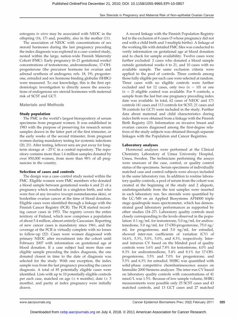
FMC. Eligible women were FMC members who donateda blood sample between gestational weeks 6 and 21 of apregnancy which resulted in a singleton birth, and whowere free of any invasive (except nonmelanoma skin), orborderline ovarian cancer at the time of blood donation.Eligible cases were identified through a linkage with theFinnish Cancer Registry (FCR). The FCR started record-ing cancer cases in 1953. The registry covers the entireterritory of Finland, which now comprises a populationof about 5.4 million, almost entirely Caucasian. Reportingof new cancer cases is mandatory since 1961, and thecoverage of the FCR is virtually complete with no lossesto follow-up (22). Cases were women diagnosed withprimary NEOC after recruitment into the cohort untilFebruary 2007 with information on gestational age atblood donation. If a case subject had more than oneeligible sample preceding the index diagnosis, the onedonated closest in time to the date of diagnosis wasselected for the study. With one exception, the indexsample was from the last pregnancy preceding the cancerdiagnosis. A total of 85 potentially eligible cases wereidentified. Lists with up to 10 potentially eligible controlsper each case, matched on age (� 6 months), date (� 3months), and parity at index pregnancy were initiallydrawn.
A record linkage with the Finnish Population Registryled to the exclusion of 8 cases (5 whose pregnancy did notend with a child birth and 3 multiple births). A linkage oftheworking file with detailed FMC fileswas conducted toverify information on gestational age at blood donationand to check for sample availability. Twelve cases werefurther excluded: 2 cases who donated a blood sampleoutside gestational weeks 6 to 21, and 10 cases with noavailable sample. The same exclusion criteria wereapplied to the pool of controls. Three controls amongthose fully eligible per each case were selected at random.Three cases with no eligible controls were furtherexcluded and for 12 cases, only two (n ¼ 10) or one(n ¼ 2) eligible control was available. For 9 controls, asample from the last but one pregnancy preceding indexdate was available. In total, 62 cases of NEOC and 171controls (41 cases and 113 controls for SCST; 21 cases and58 controls for GCT) were included in the study. Furtherdata about maternal and child characteristics duringindex birth were obtained from a linkagewith the FinnishBirth Registry (23). Information on invasive breast andovarian cancers diagnosed among the first-degree rela-tives of the study subjects was obtained through separatelinkages with the Population and Cancer Registries.
Laboratory analysesHormonal analyses were performed at the Clinical
Chemistry Laboratory of Umea University Hospital,Umea, Sweden. The technicians performing the assayswere unaware of the case, control, or quality controlstatus of the specimens. Serum specimens of individuallymatched case and control subjects were always includedin the same laboratory run. In addition to routine labora-tory quality controls, a pool of serum from the cohort wascreated at the beginning of the study and 2 aliquots,undistinguishable from the test samples were insertedin each laboratory run. Sex steroids were quantified bythe LC/MS on an Applied Biosystems API4000 triplestage quadrupole mass spectrometer, which has demon-strated good laboratory performances as supported byother studies (24–27). Laboratory quality controls mostclosely corresponding to the levels observed in the popu-lation: 0.1 ng/mL for testosterone, 5.0 ng/mL for andros-tenedione, 5.0 ng/mL for 17-OH-progesterone, 75.0 ng/mL for progesterone, and 5.0 ng/mL for estradiol,showed inter-run coefficients of variation (CV) of14.6%, 5.3%, 5.0%, 5.0%, and 8.3%, respectively. Inter-and intrarun CV based on the blinded pool of qualitycontrols were 3.6% and 7.8% for testosterone, 4.0% and8.3% for androstenedione, 5.4% and 8.1% for 17-OH-progesterone, 3.5% and 7.0% for progesterone, and5.5% and 6.3% for estradiol. SHBG was quantified withsolid-phase competitive chemiluminescence assays onImmulite 2000 Siemens analyzer. The inter-run CV basedon laboratory quality controls with concentrations of 41nmol/L was 1.5%. Because of low sample volume, SHBGmeasurements were possible only 25 SCST cases and 43matched controls, and 13 GCT cases and 27 matched
Sex Steroids in Pregnancy and Maternal Risk of Non-epithelial Ovarian Cancer
www.aacrjournals.org Cancer Epidemiol Biomarkers Prev; 20(2) February 2011 325
on July 4, 2018. © 2011 American Association for Cancer Research. cebp.aacrjournals.org Downloaded from
Published OnlineFirst December 21, 2010; DOI: 10.1158/1055-9965.EPI-10-0857

controls. Bioavailable fractions of testosterone and estra-diol concentrations were calculated by mass action mod-els based on concentrations of total hormones in bloodand their affinity constants for albumin and SHBG (28).No outliers, defined as concentrations exceeding 3 timesthe interquartile range, for any of the hormones wereidentified.
Statistical analysisPrior to analysis, all original hormone values were log2
transformed to normalize their distributions. Hormoneconcentrations varied linearly with gestational age(Fig. 1) and all statistical analyses were adjusted forgestational age.
Pearson partial correlation coefficients were used torelate hormone concentrations to specific characteristicsof interest [e.g., maternal age at sampling (last birth)].Mixed-effect models were used to compare mean hor-mone concentrations in cases and controls. The condi-tional logistic regression models (appropriate for theindividually matched design) were used to comparedifferences between cases and controls and to calculateORs and corresponding 95% CIs for SCST and GCT. Forthe hormonal variables, ORs were calculated for a unitchange of log2-transformed concentrations, which corre-sponds to a doubling of the concentrations. To ensure thatstudy results were not influenced by the presence of yetundiagnosed, but hormonally active tumor, sensitivityanalyses excluding women diagnosed within 2, 4, and 6years after blood donation were conducted. For subjectswith SHBG measurements, bioavailable fractions of tes-tosterone and estradiol were also related to risk. Furthersubgroup analyses by ages at sampling (<30 vs. �30years) and cancer diagnosis (below and above 40 years),limited to multiparous women, to those who donated aneligible sample during the last pregnancy precedingindex date, or with borderline tumors for SCST were alsoperformed. Tests of homogeneity between the ORs indifferent subgroups were based on c2 statistics, calcu-lated as the deviations of logistic regression coefficientsobserved in each of the subgroups, relative to the overallregression coefficient (29). The effect of potential con-founders (e.g., maternal age at first birth, family history ofbreast/ovarian cancers, maternal smoking, the child’ssex, birth length andweight) was evaluated and variablesthat changed point estimates by more than 5% for two ormore hormones were retained in the final models. Theeffect of adjustment for hormone concentrations includedadjustment of testosterone models for androstenedione(and vice versa) and adjustment of androgen and pro-gesterone models for 17-OH-progesterone (and viceversa) was also explored. All tests of statistical signifi-cance were 2 sided and the ORs were considered sig-nificant if the values of P < 0.05.
The study was approved by the ethical committees ofthe National Institute for Health and Welfare, Finland;University of Umea, Sweden; and German CancerResearch Center, Germany.
Results
SCST and GCT cases and their respective control sub-jects were comparable in most pregnancy, maternal, andchild characteristics (Table 1). The median age at diag-nosis was 39 years (range: 23.1–50.0) for SCST and 35years (range: 21.1–51.4) for GCT. The majority of SCSTwere borderline tumors (33 cases, 80%), whereas most ofGCT were invasive cancer (18 cases, 86%). The vastmajority of SCST (85%, 35 cases) were granulosa celltumors. The lag time between blood donation and diag-nosis was shorter for GCT than for SCST (4.1 vs. 6.8 years,respectively).
Correlations of sex steroids hormones with each otherand with gestational age and maternal age at sampling inthe all control subjects (N¼ 171) are presented in Table 2.The strongest correlations were observed between the 2androgens (r ¼ 0.87) which were also directly correlatedwith 17-OH-progesterone (r ¼ 0.54 and 0.63 for testoster-one and androstenedione, respectively). Progesteronewas weakly positively correlated with androgens (r ¼0.21 and 0.26 for testosterone and androstenedione,respectively) and moderately with 17-OH-progesterone(r ¼ 0.50). Interestingly, in the case group of women withSCST weak inverse correlations of progesterone withandrogens were observed (r ¼ �0.18 and r ¼ �0.10)and the correlation with 17-OH-progesterone was lesspronounced (r ¼ 0.20). Estradiol was moderately posi-tively correlated with other sex steroids in the controls(correlation coefficients ranging from 0.29 to 0.55), but inthe SCST cases, only very weak correlations of estradiolwith androgens and 17-OH-progesterone were observed(ranging from �0.06 to 0.13). There was no correlation ofmaternal age with any of the studied hormones amongcontrols.
Testosterone, androstenedione, and 17-OH-progester-one concentrations were significantly higher in SCSTcases than in their controls, but no other significantdifferences in hormone levels between SCST or GCTcases and their controls were observed (Table 1). SHBGconcentrations were marginally higher in SCST casesthan in their controls (189 vs. 162 nmol/L; P ¼ 0.08, withonly 25 cases and 43 controls). Correspondingly, in con-ditional regression analyses (Table 3), doubling of andro-gen and 17-OH-progesterone concentrations wereassociated with about 2-fold increase in risk of SCST.Adjustment for maternal age at first birth, family historyof breast/ovarian cancers, maternal smoking, and childsex increased risk estimates, with maternal age at firstbirth having the greatest impact (about 9% increase ofandrogen and 5% increase for 17-OH-progesterone esti-mates). The associations remained largely unchangedafter excluding women with 2-, 4-, or 6-year lag timebetween blood donation and cancer diagnosis (Table 3).Doubling of SHBG, bioavailable fractions of testosteroneand estradiol were not statistically significantly asso-ciated with risk of SCST [OR and 95% CI of 1.88 (0.70–5.04), 1.18 (0.52–2.64), and 0.97 (0.31–3.02), respectively].
Chen et al.
Cancer Epidemiol Biomarkers Prev; 20(2) February 2011 Cancer Epidemiology, Biomarkers & Prevention326
on July 4, 2018. © 2011 American Association for Cancer Research. cebp.aacrjournals.org Downloaded from
Published OnlineFirst December 21, 2010; DOI: 10.1158/1055-9965.EPI-10-0857

A B
C D
E F
Figure 1. Scatterplot of log2-scale testosterone (A), androstenedione (B), 17-OH-progesterone(C), progesterone (D), estradiol (E), and SHBG (F)concentrations (controls combined) by gestational age. The solid line shows the progression of hormone concentrations during pregnancy, estimated by linearregression.
Sex Steroids in Pregnancy and Maternal Risk of Non-epithelial Ovarian Cancer
www.aacrjournals.org Cancer Epidemiol Biomarkers Prev; 20(2) February 2011 327
on July 4, 2018. © 2011 American Association for Cancer Research. cebp.aacrjournals.org Downloaded from
Published OnlineFirst December 21, 2010; DOI: 10.1158/1055-9965.EPI-10-0857

Subgroup analyses by ages at sampling and diagnosisindicated somewhat stronger associations in womenwhowere diagnosed before age 40 than after that age, but theheterogeneity tests reached statistical significance onlyfor the effect of doubling of 17-OH-progesterone. In spiteof statistical significance, however, these results shouldbe interpreted with some caution as the heterogeneitytests were based on small numbers of subjects and weresensitive to selection of cutoff points. Analyses limited tomultiparous women (35 cases) or borderline tumors(33 cases, 80%) yielded similar results to those reportedoverall. Adjustment of testosterone models for andro-stenedione did not alter the magnitude of the risk esti-mates, whereas adjustment of androstenedione modelsfor testosterone completely abolished the associationof androstenedione with risk (data not shown). Adjust-ment of androgen models for 17-OH-progesterone or of
17-OH-progesterone models for androgens resulted insubstantial reduction of risk estimates (data not shown).
Sex steroids showedwere not associated with maternalrisk of GCT (Table 4). SHBG concentrations were asso-ciated with higher risk (not statistically significant); how-ever, only 13 case–control sets were included in thisanalysis, and the CI were large [OR and 95% CI of 2.01(0.39–10.27)]. Analyses excluding case–control sets withlag time to diagnosis of 2, 4, and 6 years and analyseslimited to multiparous women (15 cases) or invasivetumors (18 cases, 86%) were similarly unremarkable.
Discussion
This is the first prospective study to provide directepidemiologic evidence that elevated blood levels ofsex steroid hormones during pregnancy, androgens in
Table 1. Distribution of characteristics of NEOC cases and their matched controls, median (min, max),or n (%) from the FMC, 1983–2007
Characteristics SCST GCT
Cases (41) Controls (113) P value Cases (21) Controls (59) P value
Age at index (last) birth,a y 30.8 (22.2–40.0) 30.9 (21.7–40.9) – 29.7 (18.4–35.9) 29.5 (18.0–36.1) –
Parity at index pregnancy1 6 (15%) 18 (16%) – 6 (29%) 16 (27%) –
2 17 (41%) 49 (43%) 8 (38%) 28 (47%)3þ 18 (44%) 46 (41%) 7 (33%) 15 (26%)
Age at first birth, y 25.8 (18.6–40.5) 25.3 (16.4–41.4) 0.21 25.8 (19.0–32.5) 25.3 (17.9–33.3) 0.58Gestational age, wk 11 (7–16) 11 (6–20) 0.94 10 (7–15) 10 (6–18) 0.92Age at diagnosis, y 39.0 (23.1–50.0) – – 34.8 (21.1–51.4) – –
Lag time, y 6.8 (0.1–19.4) – – 4.1 (0.1–16.2) – –
Cancer typeInvasive 8 (20%) – 18 (86%) –
Borderline 33 (80%) – 3 (14%) –
Family history of ovary cancer 0 (0%) 1 (1%) 0.99 0 (0%) 1 (2%) 0.99Family history of breast cancer 1 (2%) 1 (1%) 0.71 1 (5%) 4 (7%) 0.59Smoking (yes) 4 (10%) 17 (16%) 0.38 3 (15%) 11 (20%) 0.69Child sex
Male 18 (45%) 62 (56%) 0.27 9 (45%) 31 (55%) 0.46Female 22 (55%) 48 (44%) 11 (55%) 25 (45%)
Child birth weight, g 3,535(2,080–4,650)
3,560(1,900–4,670)
0.29 3,775(2,470–4,400)
3,490(2,025–5,000)
0.21
Child birth length, cm 50 (42–55) 50 (43–57) 0.39 51 (46–55) 50 (44–55) 0.26Hormonesb
Testosterone, ng/mL 1.09 (0.95–1.24) 0.86 (0.79–0.93) 0.01 0.91 (0.75–1.10) 0.90 (0.80–1.01) 0.92Androstenedione, ng/mL 2.36 (2.08–2.67) 1.96 (1.81–2.11) 0.03 2.18 (1.86–2.55) 2.09 (1.90–2.30) 0.7217-OH-progesterone, ng/mL 2.86 (2.55–3.21) 2.42 (2.23–2.62) 0.03 2.47 (2.13–2.87) 2.67 (2.44–2.92) 0.45Progesterone, ng/mL 22.2 (20.4–24.2) 23.6 (22.4–25.0) 0.29 22.9 (20.3–25.9) 22.6 (20.9–24.3) 0.85Estradiol, ng/mL 2.05 (1.79–2.34) 2.00 (1.84–2.18) 0.81 1.91(1.53–2.40) 2.23 (1.95–2.56) 0.33
NOTE: Conditional logistic regression models were used to compare differences between cases and controls; mixed-effect modelswere used to compare mean hormone concentrations in cases and controls.aSamples not from the last pregnancy preceding cancer diagnosis (3 SCST controls, 1 GCT case, and 6 GCT controls).bGeometric means and (10th, 90th) percentile of hormone concentrations (adjustment for gestational age and maternal age).
Chen et al.
Cancer Epidemiol Biomarkers Prev; 20(2) February 2011 Cancer Epidemiology, Biomarkers & Prevention328
on July 4, 2018. © 2011 American Association for Cancer Research. cebp.aacrjournals.org Downloaded from
Published OnlineFirst December 21, 2010; DOI: 10.1158/1055-9965.EPI-10-0857

Tab
le2.
Pea
rson
partia
lcorrelatio
nsbetwee
nho
rmon
es,m
aterna
lage
atindex
pregn
ancy
,and
gestationa
lday
inallcon
trol
subjects(n
¼17
1)from
theFM
C,19
83–20
07
Horm
one
sTes
tosterone
And
rosten
edione
17-O
H-p
roges
terone
Proges
terone
Estradiol
SHBGa
Testos
terone
,ng
/mL
0.38
(P¼
0.00
2)And
rosten
edione
,ng
/mL
0.87
(P<0.00
01)
0.26
(P¼
0.01
)17
-OH-proge
steron
e,ng
/mL
0.54
(P<0.00
01)
0.63
(P<0.00
01)
0.12
(P¼
0.26
)Proge
steron
e,ng
/mL
0.21
(P¼
0.01
)0.26
(P¼
0.00
1)0.50
(P<0.00
01)
0.37
(P¼
0.00
02)
Estradiol,ng
/mL
0.55
(P<0.00
01)
0.49
(P<0.00
01)
0.29
(P¼
0.00
01)
0.50
(P<0.00
01)
0.67
(P<0.00
01)
Materna
lage
atsa
mpling,
y0.05
(P¼
0.56
)�0
.02(P
¼0.76
)0.03
(P¼
0.66
)0.06
(P¼
0.45
)�0
.003
(P¼
0.97
)�0
.002
(P¼
0.98
)Ges
tatio
nala
ge,d
�0.12(P
¼0.12
)�0
.25(P
¼0.00
1)�0
.32(P
<0.00
01)
0.51
(P<0.00
01)
0.67
(P<0.00
01)
0.63
(P<0.00
01)
NOTE
:Allco
rrelations
(exc
eptthos
ewith
gestationa
lday
)weread
justed
forge
stationa
lage
.aDataav
ailable
for95
subjectson
ly.
Sex Steroids in Pregnancy and Maternal Risk of Non-epithelial Ovarian Cancer
www.aacrjournals.org Cancer Epidemiol Biomarkers Prev; 20(2) February 2011 329
on July 4, 2018. © 2011 American Association for Cancer Research. cebp.aacrjournals.org Downloaded from
Published OnlineFirst December 21, 2010; DOI: 10.1158/1055-9965.EPI-10-0857

Tab
le3.
ORsan
d95
%CIs
forov
arianSCSTas
sociated
with
dou
blingof
horm
oneco
ncen
trations
from
theFM
C,19
83–20
07
Tes
tosterone
And
rosten
edione
17-O
H-proges
terone
Proges
terone
Estradiol
OR
(95%
CI)
POR
(95%
CI)
POR
(95%
CI)
POR
(95%
CI)
POR
(95%
CI)
P
SCST(41/11
3)1.90
(1.13–
3.21
)0.02
1.76
(1.03–
3.03
)0.04
2.07
(1.07–
4.02
)0.03
0.68
(0.31–
1.48
)0.33
1.07
(0.64–
1.78
)0.80
Multiv
ariate
mod
ela(41/11
3)2.16
(1.25–
3.74
)0.00
62.16
(1.20–
3.87
)0.01
2.62
(1.27–
5.38
)0.01
0.67
(0.29–
1.53
)0.34
1.04
(0.62–
1.77
)0.88
Sen
sitiv
ityan
alyses
Lagtim
e�
2y(34/96
)2.03
(1.13–
3.64
)0.02
1.85
(0.98–
3.48
)0.06
1.96
(0.90–
4.28
)0.09
0.58
(0.22–
1.54
)0.28
1.11
(0.62–
1.99
)0.72
Lagtim
e�
4y(31/88
)2.30
(1.17–
4.49
)0.02
1.99
(0.98–
4.08
)0.06
2.53
(1.04–
6.14
)0.04
0.68
(0.25–
1.86
)0.45
1.01
(0.55–
1.86
)0.97
Lagtim
e�
6y(23/65
)3.41
(1.43–
8.16
)0.01
2.54
(1.06–
6.10
)0.04
1.84
(0.67–
5.06
)0.24
0.47
(0.14–
1.63
)0.23
0.95
(0.46–
1.95
)0.89
Multip
arou
swom
en(35/95
)2.28
(1.26–
4.10
)0.01
2.17
(1.18–
4.00
)0.01
2.49
(1.11–
5.56
)0.03
0.53
(0.22–
1.29
)0.16
0.96
(0.53–
1.73
)0.89
Borde
rline
tumors(33/96
)2.01
(1.09–
3.72
)0.03
2.06
(1.09–
3.88
)0.03
2.58
(1.16–
5.70
)0.02
0.69
(0.27–
1.77
)0.44
1.09
(0.61–
1.94
)0.77
Sub
grou
pan
alyses
Age
atsa
mpling
<30y(15/40
)3.21
(0.86–
11.95)
0.08
2.09
(0.54–
8.07
)0.29
4.77
(0.86–
26.33)
0.07
0.55
(0.15–
1.99
)0.36
0.77
(0.31–
1.90
)0.57
�30y(26/73
)1.80
(0.92–
3.52
)0.09
1.89
(0.95–
3.76
)0.07
2.01
(0.88–
4.57
)0.10
0.68
(0.21–
2.28
)0.54
0.98
(0.50–
1.95
)0.96
Age
atdiagn
osisa
<40y(24/65
)2.91
(1.30–
6.50
)0.01
3.14
(1.30–
7.57
)0.01
7.68
(1.68–
35.13)
0.01
0.66
(0.21–
2.07
)0.48
1.15
(0.58–
2.29
)0.69
�40y(17/48
)1.45
(0.64–
3.26
)0.37
1.22
(0.51–
2.93
)0.66
1.00
(0.35–
2.82
)0.99
0.48
(0.11–
2.07
)0.33
0.88
(0.35–
2.24
)0.79
NOTE
:Adjustmen
tsforge
stationa
lage
,materna
lage
atfirst
birth,
family
historyof
breas
t/ov
arianca
ncers,
materna
lsmok
ing,
andch
ildse
x.aTh
ehe
teroge
neity
testsreac
hedstatistic
alsign
ifica
nceon
lyfor17
-OH-proge
steron
e(P
¼0.03
)byag
eat
diagn
osis,less
than
40ve
rsus
40ye
arsor
grea
ter.
Chen et al.
Cancer Epidemiol Biomarkers Prev; 20(2) February 2011 Cancer Epidemiology, Biomarkers & Prevention330
on July 4, 2018. © 2011 American Association for Cancer Research. cebp.aacrjournals.org Downloaded from
Published OnlineFirst December 21, 2010; DOI: 10.1158/1055-9965.EPI-10-0857

Tab
le4.
ORsan
d95
%CIs
forov
arianGCTas
sociated
with
dou
blingof
horm
oneco
ncen
trations
from
theFM
C,19
83–20
07
Tes
tosterone
And
rosten
edione
17-O
H-proges
terone
Proges
terone
Estradiol
OR
(95%
CI)
POR
(95%
CI)
POR
(95%
CI)
POR
(95%
CI)
POR
(95%
CI)
P
GCT(21/58
)1.05
(0.54–
2.08
)0.88
1.19
(0.54–
2.65
)0.66
0.67
(0.27–
1.67
)0.39
1.13
(0.42–
2.99
)0.81
0.75
(0.42–
1.36
)0.35
Multiv
ariate
mod
el(21/58
)1.04
(0.50–
2.17
)0.92
1.24
(0.53–
2.91
)0.62
0.66
(0.26–
1.66
)0.37
1.04
(0.38–
2.84
)0.94
0.68
(0.35–
1.33
)0.26
Sen
sitiv
ityan
alyses
Lagtim
e�
2y(16/45
)0.82
(0.34–
1.96
)0.66
1.05
(0.37–
2.94
)0.93
0.59
(0.21–
1.67
)0.32
1.08
(0.27–
4.24
)0.91
0.73
(0.31–
1.74
)0.48
Lagtim
e�
4y(11/31
)0.60
(0.15–
2.44
)0.47
0.66
(0.13–
3.29
)0.62
0.45
(0.09–
2.17
)0.32
0.86
(0.10–
7.32
)0.89
0.59
(0.15–
2.30
)0.45
Lagtim
e�
6y(9/26)
0.56
(0.13–
2.36
)0.43
0.59
(0.11–
3.06
)0.53
0.36
(0.06–
2.09
)0.25
0.89
(0.11–
7.56
)0.92
0.57
(0.13–
2.52
)0.42
Multip
arou
swom
en(15/42
)0.94
(0.41–
2.15
)0.88
1.23
(0.47–
3.22
)0.67
0.69
(0.21–
2.23
)0.53
1.71
(0.36–
8.20
)0.50
0.61
(0.25–
1.51
)0.29
Inva
sive
tumors(18/50
)1.07
(0.49–
2.35
)0.87
1.21
(0.50–
2.93
)0.68
0.77
(0.30–
2.01
)0.59
1.00
(0.34–
2.92
)0.99
0.65
(0.32–
1.32
)0.23
NOTE
:Non
eof
thehe
teroge
neity
testsreac
hedstatistic
alsign
ifica
ncean
dad
justmen
tforge
stationa
lage
,materna
lage
atfirst
birth,
family
historyof
brea
st/ova
rianca
ncers,
materna
lsmok
ing,
andch
ildse
x.
Sex Steroids in Pregnancy and Maternal Risk of Non-epithelial Ovarian Cancer
www.aacrjournals.org Cancer Epidemiol Biomarkers Prev; 20(2) February 2011 331
on July 4, 2018. © 2011 American Association for Cancer Research. cebp.aacrjournals.org Downloaded from
Published OnlineFirst December 21, 2010; DOI: 10.1158/1055-9965.EPI-10-0857

particular, may be related to an increased risk of develop-ing SCST. Doubling of testosterone, androstenedione, and17-OH-progesterone (the precursor hormone for andro-gen synthesis) during the early part of the last pregnancyleading to child birth and preceding cancer diagnosis wasassociated with an approximately 2-fold greater risk ofSCST. In contrast, none of the studied steroid hormonesshowed any association with risk of GCT.
Changes of sex steroids throughout pregnancy aresummarized as below. Concentrations of testosteroneare relatively stable throughout pregnancy, as demon-strated by longitudinal studies (30, 31). One longitudinalstudy (31) shows that total testosterone levels remainrelatively stable till the third trimester and increase62% from 6th to 34th week of gestation, and the otherlongitudinal study (30) reports that total testosteroneincreases approximately 29% from 5th to 40th week ofgestation. The free testosterone levels arewithin the rangeof nonpregnant women and gradually increase duringthe latter parts of pregnancy (31, 32). Androstenedionelevels only slightly increase throughout pregnancy in thelongitudinal studies (30, 31, 33). 17-OH-progesteronelevels reach their peak during the fifth to sixth week ofpregnancy and then gradually decrease thereafter, butincrease again during the latter parts of pregnancy (30,34). There are small variations in progesterone levels,similar to pre-pregnancy state (luteal phase of the men-strual cycle), from third to thirteenth week of gestation,and progesterone levels increase significantly thereafter(30, 34, 35). Estradiol levels rise steadily from third tothirteenth week of gestation (by eighth week of gestationits levels are above the range found during menstrualcycle) and then gradually increase thereafter (34–37).SHBG levels increase rapidly during the first half ofpregnancy (till 25th week of gestation), then remainedrelatively constant until delivery (30, 31). All studiedhormones levels at 1 year postpartum drop downto below the first trimester values, similar to the pre-pregnancy state (31, 33).
The 2-fold higher risk of SCST associated with elevatedprediagnostic androgen concentrations is consistent withresults from animal studies. Experiments in rodents havedemonstrated that androgensmay be involved in ovariancancer induction, though a long induction period may berequired (38–42). In particular, testosterone and dehy-droepiandrosterone strongly promote carcinogenesis ofspontaneous ovarian granulosa cell tumors (the predo-minant histologic subtype in this study) in SWXJ-9 strainmice (38) and continuous administration with testoster-one shortly after birth induced theca-cell ovarian tumors(also belonging to SCST) in the rat (39). Therefore, it isplausible that androgens are involved in the pathogenesisof SCST in humans.
The elevations in androgens and 17-OH-progesteronein the SCST case subjects are likely to be of maternalorigin. Although the placenta produces androstenedioneand testosterone from fetal precursors, these are rapidlyconverted to estrogens due to its potent aromatase
capacity (43). 17-OH-progesterone is considered to reflectactivities of corpus luteum, indicating ovarian steroido-genesis, particularly during the first trimester (36).Nevertheless, maternal 17-OH-progesterone during thelatter parts of pregnancy could be derived from maternaland fetal adrenal glands as well as from the placentasources (44), as the placenta does not secrete 17-OH-progesterone until 32 week of pregnancy (43, 45) dueto the lack of 17a-hydrolase activity. In contrast, theconcentrations of progesterone and estradiol, which arepredominantly of placental origin in the phase of preg-nancy during which study samples were collected (45),were similar in case and control subjects and were notrelated to risk of SCST. The divergent correlations ofandrogens and 17-OH-progesterone with estradiol andprogesterone concentrations in the SCST cases and in thecontrol group further underscores the possibility of dif-ferences in sex steroid hormones metabolism betweencases and controls.
Androgensmeasuredduring early pregnancy are likelyto reflect pre-pregnancy exposure to the hormone becauseconcentrations of androgens (testosterone and androste-nedione) during early pregnancy and in the nonpregnantstate are relatively similar (30, 31). It is plausible to assumethat women from general population, having normal pre-pregnancy androgens, continue to have normal andro-gensduring early pregnancy. Similarly, this assumption islikely generalizable to women with polycystic ovary syn-drome (PCOS) as evidence (37, 43) has demonstrated thatpregnant women with PCOS, having elevated pre-preg-nancy androgens, continue to maintain higher androgensalso during pregnancy, compared with normal pregnantwomen with normal androgen levels. Therefore, theobserved increase in risk of SCST associated with dou-bling of testosterone in our studymaybe applicable also tothe effect of elevated androgens/altered maternal andro-gen metabolism in the nonpregnant state.
The major strength of our study is its prospectivedesign, which largely avoids selection and inverse causa-tion biaseswhichplague studies of a classical case–controldesign. Case and control subjectswere tightlymatched forage at sampling (last birth), parity at the index pregnancy,and date of sampling, allowing careful control for theseimportant sources of potential confounding. SCSTdevelop from the theca and granulosa cells, which arethe major site of steroid hormones synthesis in the ovary(4, 46), and these tumors are often hormonally active(5, 47). To reduce the possibility that the observed associa-tions with risk of SCST were influenced by hormonalsecretion by a growing, but still undiagnosed tumor welimited the analyses to women whose samples weredonated 2, 4 and 6 years prior to their cancer diagnosis.For testosterone, themore potent androgen, risk estimatesremained strong and statistically significant, whereasthose for androstenedione were slightly reduced andbecame of borderline significance. Thus, it is unlikely thatthe observed associations in our studywere due to reversecausation bias.
Chen et al.
Cancer Epidemiol Biomarkers Prev; 20(2) February 2011 Cancer Epidemiology, Biomarkers & Prevention332
on July 4, 2018. © 2011 American Association for Cancer Research. cebp.aacrjournals.org Downloaded from
Published OnlineFirst December 21, 2010; DOI: 10.1158/1055-9965.EPI-10-0857

The very large overall size of Finnish Maternity Cohort(N � 850,000) made it possible to study the hormonaldeterminants of non-epithelial ovarian tumors, whichare rare forms of cancer; although, the very small num-bers of cases, particularly for GCT, was insufficient todetect moderate or weak hormone-risk associations. Alimitation of our study is that no data on maternal pre-pregnancy hyperadrogenism/testosterone concentra-tions were available and the lack of sufficient samplefor analyses of SHBG in all subjects. Study samples hadbeen stored at relatively high temperature (�25�C), butwe observed no correlations of hormone levels with timein storage and their concentrations varied with gesta-tional age as expected, as also shown previously (48).It is not feasible in the study design to select controls
matched to cases on gestational age, even in much largercohorts and will pose unreasonable, severe restrains oncontrol selection. Therefore, the only alternative is toadjust for gestational age in the phase of data analysisto minimize the intra-person variability in sex steroidslevels during pregnancy. One approach is adjusted for alinear term of gestational age, and the other is calculatingresidual values by the local linear regression (49). Weadopt the former approach in the article due to thefollowing 4 reasons. First, concentrations of all studiedhormone varied linearly with gestational age (Fig. 1).Second, the same approach has been extensively usedin various epidemiologic studies (50–53). Third, in factthe former approach provides more straightforwardinterpretation in comparison with the presentation ofresidual values by the local linear regression, as the latterapproach is required to calculate the difference (residual)between the assay value and the estimated mean hor-mone value determined for the day of gestation on whichthe blood sample was donated. Finally, results obtainedby both approaches give almost identical results. It is alsopossible that hormones levels adjusted for gestational agecould be subject to measurement error. However theintra-person variability in biomarker levels from a singlemeasurement is unlikely to be differential with respect tocases and controls and would most likely lead to anunderestimation of the effect estimates, as supportedby other studies (54–57).Another relative limitation of the study might be that
only a single blood sample was obtained for each preg-nant woman. A single measurement for endogenous sexsteroids in premenopausal women is suggested to reli-ably estimate average levels for serum androgen andSHBG over a 1- to 3-year period (58, 59, 55, 60, 61). Toour knowledge, so far no study on the reproducibility of asingle measurement for endogenous sex steroids in preg-nant women has been reported and little is known aboutthe intra-person variability in sex steroids during preg-nancy. Although a lot of work has been done in relation toDown syndrome markers, so far only 3 studies (62–64),after exhausting literature searches, on the correlationsbetween sex steroids levels during different period ofthe same pregnancy have been identified. Nevertheless, it
is plausible to conclude that sex steroids hormones trackduring pregnancy as there are moderate-to-strong corre-lations (more than 0.40) between concentrations of sexsteroids during the different period (62–64) of the samepregnancy. Firstly, correlations between the first andsecond trimester in both Caucasian women and Chinesewomen were strong for testosterone (0.83 for Bostonand 0.62 for Shanghai), SHBG (0.85 for Boston and 0.67for Shanghai), and estradiol (0.72 for Boston and 0.61 forShanghai), and modest for progesterone (0.45 for Bostonand 0.39 for Shanghai; ref. 62). Secondly, correlationsbetween the first and third trimester in white womenwere 0.53 for testosterone, 0.23 for SHBG, and 0.25for estradiol; whereas in black women were higher,0.67 for testosterone, 0.43 for SHBG, and 0.35 for estradiol(63). Thirdly, correlations between the second and thirdtrimester were 0.78 for estradiol and 0.60 for totalestrogen (64). In addition, strong correlations duringsuccessive pregnancies of the same women for estrogenhave been reported, for example, pregnancy estradiol,measured between eighth and seventeenth of gestationalweeks, were strongly correlated in successive pregnan-cies of the same women (0.78 for total estradiol and 0.73for free estradiol; ref. 65). It is plausible to assume thatthe correlations of hormone levels measured withinthe same pregnancy is most likely higher than thosecorrelations of hormone levels measured in successivepregnancies.
Taken together, a single measurement for androgens(testosterone in particular) during early pregnancy canreliably represent cumulative exposure to the hormone asstrong correlations during the different period of thesame pregnancy for testosterone have been reported,for example, correlations between the first and secondtrimester were 0.83 for Caucasian women in Boston and0.62 for Chinese women in Shanghai (62), and correla-tions between the first and third trimester were 0.67 inblack women and 0.53 in white women (63). In addition,concentrations of androgens are relatively stablethroughout pregnancy (30, 31, 33), and androgens levelsduring early pregnancy and in the nonpregnant state arealso relatively similar (30, 31). The samples of the FMCwere donated around the first trimester, between thesixth and twenty-first weeks of gestation, which is likelyto be the most relevant period for the exposure to earlypregnancy androgens (testosterone in particular), alsoreflecting pre-pregnancy exposure to the hormone.
A completion of the first full-term pregnancy mayinduce substantial alternations in a woman’s hormonalmilieu as most studies have shown that in pregnant state,parous women have lower concentrations of a number ofhormones, including estrogens and androgens (64, 66–69),compared with nulliparous women. Effects of pregnancyon hormone levels may sustain in the nonpregnant state,for example, before and aftermenopause. Although, somestudies report that nulliparouswomenhad similar (highertrend, but not statistically significant) androgens (testos-terone and androstenedione) levels, in comparison with
Sex Steroids in Pregnancy and Maternal Risk of Non-epithelial Ovarian Cancer
www.aacrjournals.org Cancer Epidemiol Biomarkers Prev; 20(2) February 2011 333
on July 4, 2018. © 2011 American Association for Cancer Research. cebp.aacrjournals.org Downloaded from
Published OnlineFirst December 21, 2010; DOI: 10.1158/1055-9965.EPI-10-0857

parous women, both in the pre-menopausal (70, 71) andpostmenopausal women (72–74). Most studies support ofreductions in concentrations of sex steroids after the firstfull-term pregnancy both in the pre-menopausal womenfor androgens [DHEA and DHEA sulfate (DHEAS); ref.75], progesterone (71), and estrogens (free estradiol; ref.76), andalso in thepostmenopausalwomen for androgens(testosterone; ref. 77), and estrogens (free estradiol; ref. 78)and estrone sulfate (79).
Therefore, the effect of pregnancy on maternal risk ofNEOC is most likely to be the result of the cumulativelong-term exposure of androgens (testosterone in parti-cular) throughout a woman’s life cycle till cancer diag-nosis, by promoting carcinogenesis of spontaneousovarian granulosa cell tumors or inducing theca-cellovarian tumors, as animal studies (38–42) has demon-strated that androgens may be involved in ovarian cancerinduction after a long induction period.
In summary, this study is the first prospective epide-miologic investigation providing initial direct evidencethat elevated androgens and testosterone in particular,play an important role in the pathogenesis of SCST longbefore the clinical onset of the disease. Our data suggestthat SCST and GCT have distinct hormonal risk profiles,
possibly reflecting differences in their pathogenesis. Ourstudy may note a particular need for larger confirmatoryinvestigations on sex steroids and NEOC.
Disclosure of Potential Conflict of Interest
No potential conflicts of interest were disclosed.
Acknowledgments
The authors are indebted to Yelena Afanasyeva, Pirjo Kontiokari,Annika Uimonen, and Sara Kuusiniemi for their excellent technicalassistance in the conduct of the study.
Grant Support
This research was supported by the National Cancer Institute at the NIH(R01-CA120061).
The costs of publication of this article were defrayed in part by thepayment of page charges. This article must therefore be hereby markedadvertisement in accordance with 18 U.S.C. Section 1734 solely to indicatethis fact.
Received August 16, 2010; revised November 26, 2010; acceptedDecember 2, 2010; published OnlineFirst December 21, 2010.
References1. Horn-Ross PL, Whittemore AS, Harris R, Itnyre J. Characteristics
relating to ovarian cancer risk: collaborative analysis of 12 U.S.case-control studies. VI. Non-epithelial cancers among adults. Col-laborative Ovarian Cancer Group. Epidemiology 1992;3:490–5.
2. Pectasides D, Pectasides E, Kassanos D. Germ cell tumors of theovary. Cancer Treat Rev 2008;34:427–41.
3. Guillem V, Poveda A. Germ cell tumours of the ovary. Clin Transl Oncol2007;9:237–43.
4. Pectasides D, Pectasides E, Psyrri A. Granulosa cell tumor of theovary. Cancer Treat Rev 2008;34:1–12.
5. Schumer ST, Cannistra SA. Granulosa cell tumor of the ovary. J ClinOncol 2003;21:1180–9.
6. Lee-Jones L. Ovary: sex cord-stromal tumors. Atlas Genet CytogenetOncol Haematol Nov 2003.
7. Lee-Jones L. Ovary: germ cell tumors. Atlas Genet Cytogenet OncolHaematol Aug 2003.
8. Koulouris CR, Penson RT. Ovarian stromal and germ cell tumors.Semin Oncol 2009;36:126–36.
9. Dallenbach P, Bonnefoi H, Pelte MF, Vlastos G. Yolk sac tumours ofthe ovary: an update. Eur J Surg Oncol 2006;32:1063–75.
10. Adami HO, Hsieh CC, Lambe M, Trichopoulos D, Leon D, Persson I,et al. Parity, age at first childbirth, and risk of ovarian cancer. Lancet1994;344:1250–4.
11. Albrektsen G, Heuch I, Kvale G. Full-term pregnancies and incidenceof ovarian cancer of stromal and germ cell origin: a Norwegianprospective study. Br J Cancer 1997;75:767–70.
12. Sanchez-Zamorano LM, Salazar-Martinez E, De Los Rios PE, Gon-zalez-Lira G, Flores-Luna L, Lazcano-Ponce EC. Factors associatedwith non-epithelial ovarian cancer amongMexican women: amatchedcase-control study. Int J Gynecol Cancer 2003;13:756–63.
13. dos SS I, Swerdlow AJ. Ovarian germ cell malignancies in England:epidemiological parallels with testicular cancer. Br J Cancer 1991;63:814–8.
14. Boyce EA, Costaggini I, Vitonis A, Feltmate C, Muto M, Berkowitz R,et al. The epidemiology of ovarian granulosa cell tumors: a case-control study. Gynecol Oncol 2009;115:221–5.
15. The reduction in risk of ovarian cancer associated with oral-contra-ceptive use. The Cancer and Steroid Hormone Study of the Centersfor Disease Control and the National Institute of Child Health andHuman Development. N Engl J Med 1987;316:650–5.
16. Henderson BE, Ross R, Bernstein L. Estrogens as a cause of humancancer: the Richard and Hinda Rosenthal Foundation award lecture.Cancer Res 1988;48:246–53.
17. Walker AH, Ross RK, Haile RW, Henderson BE. Hormonal factors andrisk of ovarian germ cell cancer in young women. Br J Cancer1988;57:418–22.
18. Nestler JE, Jakubowicz DJ. Decreases in ovarian cytochromeP450c17 alpha activity and serum free testosterone after reductionof insulin secretion in polycystic ovary syndrome. N Engl J Med1996;335:617–23.
19. Hirsutism. In: Speroff L, Glass RH, Kase NG, editors. Clinical Gyne-cologic Endocrinology and Infertility. 5th ed. Baltimore, MD: Williams& Wilkins;1994. p. 483–513.
20. Koskela P, Anttila T, Bjorge T, Brunsvig A, Dillner J, Hakama M, et al.Chlamydia trachomatis infection as a risk factor for invasive cervicalcancer. Int J Cancer 2000;85:35–9.
21. Lehtinen M, Koskela P, Ogmundsdottir HM, Bloigu A, Dillner J,Gudnadottir M, et al. Maternal herpesvirus infections and risk of acutelymphoblastic leukemia in the offspring. Am J Epidemiol 2003;158:207–13.
22. Teppo L, Pukkala E, Lehtonen M. Data quality and quality control of apopulation-based cancer registry. Experience in Finland. Acta Oncol1994;33:365–9.
23. Gissler M, Teperi J, Hemminki E, Merilainen J. Data quality afterrestructuring a national medical registry. Scand J Soc Med 1995;23:75–80.
24. Schlusener MP, Bester K. Determination of steroid hormones, hor-mone conjugates and macrolide antibiotics in influents and effluentsof sewage treatment plants utilising high-performance liquid chroma-tography/tandem mass spectrometry with electrospray and atmo-spheric pressure chemical ionisation. Rapid CommunMass Spectrom2005;19:3269–78.
Chen et al.
Cancer Epidemiol Biomarkers Prev; 20(2) February 2011 Cancer Epidemiology, Biomarkers & Prevention334
on July 4, 2018. © 2011 American Association for Cancer Research. cebp.aacrjournals.org Downloaded from
Published OnlineFirst December 21, 2010; DOI: 10.1158/1055-9965.EPI-10-0857

25. Lopez de Alda MJ, az-Cruz S, Petrovic M, Barcelo D. Liquidchromatography-(tandem) mass spectrometry of selected emer-ging pollutants (steroid sex hormones, drugs and alkylphenolicsurfactants) in the aquatic environment. J Chromatogr A 2003;1000:503–26.
26. Herrmann M, Harwood T, Gaston-Parry O, Kouzios D, Wong T, Lih A,et al. A new quantitative LC tandem mass spectrometry assay forserum 25-hydroxy vitamin D. Steroids 2010;75:1106–12.
27. Kunisue T, Fisher JW, Fatuyi B, Kannan K. Amethod for the analysis ofsix thyroid hormones in thyroid gland by liquid chromatography-tandem mass spectrometry. J Chromatogr B Analyt Technol BiomedLife Sci 2010;878:1725–30.
28. Vermeulen A, Verdonck L, Kaufman JM. A critical evaluation of simplemethods for the estimation of free testosterone in serum. J ClinEndocrinol Metab 1999;84:3666–72.
29. Whitehead A, Whitehead J. A general parametric approach to themeta-analysis of randomized clinical trials. Stat Med 1991;10:1665–77.
30. O’Leary P, Boyne P, Flett P, Beilby J, James I. Longitudinal assess-ment of changes in reproductive hormones during normal pregnancy.Clin Chem 1991;37:667–72.
31. Kerlan V, Nahoul K, Le Martelot MT, Bercovici JP. Longitudinalstudy of maternal plasma bioavailable testosterone and androsta-nediol glucuronide levels during pregnancy. Clin Endocrinol 1994;40:263–7.
32. Bammann BL, Coulam CB, Jiang NS. Total and free testosteroneduring pregnancy. Am J Obstet Gynecol 1980;137:293–8.
33. Soldin OP, Guo T, Weiderpass E, Tractenberg RE, Hilakivi-Clarke L,Soldin SJ. Steroid hormone levels in pregnancy and 1 year postpar-tum using isotope dilution tandem mass spectrometry. Fertil Steril2005;84:701–10.
34. Tulchinsky D, Hobel CJ, Yeager E, Marshall JR. Plasma estrone,estradiol, estriol, progesterone, and 17-hydroxyprogesterone inhuman pregnancy. I. Normal pregnancy. Am J Obstet Gynecol1972;112:1095–100.
35. Tulchinsky D, Hobel CJ. Plasma human chorionic gonadotropin,estrone, estradiol, estriol, progesterone, and 17 alpha-hydroxypro-gesterone in human pregnancy. 3. Early normal pregnancy. Am JObstet Gynecol 1973;117:884–93.
36. Yoshimi T, Strott CA, Marshall JR, Lipsett MB. Corpus luteum functionin early pregnancy. J Clin Endocrinol Metab 1969;29:225–30.
37. Lobo RA. Endocrinology of pregnancy. In:Lobo RA, Mishell Jr DR,Paulson RJ, Shoupe D, editors. Infertility, Contraception, and Repro-ductive Endocrinology. 4th ed. New York: Blackwell Science Inc.;1997. p. 183–207.
38. Beamer WG, Shultz KL, Tennent BJ. Induction of ovarian granulosacell tumors in SWXJ-9 mice with dehydroepiandrosterone. CancerRes 1988;48:2788–92.
39. Horning ES. Carcinogenic action of androgens. Br J Cancer1958;12:414–8.
40. Capen CC. Mechanisms of hormone-mediated carcinogenesis of theovary. Toxicol Pathol 2004;32Suppl 2:1–5.
41. Capen CC, Beamer WG, Tennent BJ, Stitzel KA. Mechanisms ofhormone-mediated carcinogenesis of the ovary in mice. Mutat Res1995;333:143–51.
42. Rainey WE, Sawetawan C, McCarthy JL, McGee EA, Bird IM, WordRA, et al. Human ovarian tumor cells: a potential model for thecal cellsteroidogenesis. J Clin Endocrinol Metab 1996;81:257–63.
43. Sir-Petermann T, Maliqueo M, Angel B, Lara HE, Perez-Bravo F,Recabarren SE. Maternal serum androgens in pregnant women withpolycystic ovarian syndrome: possible implications in prenatal andro-genization. Hum Reprod 2002;17:2573–9.
44. Tulchinsky D, Simmer HH. Sources of plasma 17alpha-hydroxypro-gesterone in human pregnancy. J Clin Endocrinol Metab 1972;35:799–808.
45. The endocrinology of pregnancy. In: Speroff L, FritzM, editors. ClinicalGynecologic Endocrinology and Infertility. 7th ed. Philadelphia, PA:Lippincott Williams & Wilkins; 2005. p. 259–318.
46. Gougeon A. Regulation of ovarian follicular development in primates:facts and hypotheses. Endocr Rev 1996;17:121–55.
47. Young RH, Scully RE. Endocrine tumors of the ovary. Curr Top Pathol1992;85:113–64.
48. Holl K, Lundin E, Kaasila M, Grankvist K, Afanasyeva Y, Hallmans G,et al. Effect of long-term storage on hormone measurements insamples from pregnant women: the experience of the Finnish Mater-nity Cohort. Acta Oncol 2008;47:406–12.
49. Cleveland WS, Loader C. Smoothing by local regression: principlesand methods. In:Schimek MG, editor. Statistical Theory and Compu-tational Aspects of Smoothing. 1st ed. New York: Springer; 1996. p.113–20.
50. Agborsangaya CB, Surcel HM, Toriola AT, Pukkala E, Parkkila S,Tuohimaa P, et al. Serum 25-hydroxyvitamin D at pregnancy and riskof breast cancer in a prospective study. Eur J Cancer 2010;46:467–70.
51. Toriola AT, Surcel HM, Calypse A, et al. Independent and joint effectsof serum 25-hydroxyvitamin D and calcium on ovarian cancer risk: aprospective nested case-control study. Eur J Cancer 2010;46:2799–805.
52. Holl K, Lundin E, Surcel HM, Grankvist K, Koskela P, Dillner J, et al.Endogenous steroid hormone levels in early pregnancy and risk oftesticular cancer in the offspring: a nested case-referent study. Int JCancer 2009;124:2923–8.
53. Chen T, Lundin E, Grankvist K, Zeleniuch-Jacquotte A, Wulff M,Afanasyeva Y, et al. Maternal hormones during early pregnancy: across-sectional study. Cancer Causes Control 2010;21:719–27.
54. Rosner B, Spiegelman D, Willett WC. Correction of logistic regressionrelative risk estimates and confidence intervals for random within-person measurement error. Am J Epidemiol 1992;136:1400–13.
55. Kotsopoulos J, Tworoger SS, Campos H, Chung FL, Clevenger CV,Franke AA, et al. Reproducibility of plasma and urine biomarkersamong premenopausal and postmenopausal women from theNurses’ Health Studies. Cancer Epidemiol Biomarkers Prev2010;19:938–46.
56. Tworoger SS, Hankinson SE. Use of biomarkers in epidemiologicstudies: minimizing the influence of measurement error in thestudy design and analysis. Cancer Causes Control 2006;17:889–99.
57. Hankinson SE, Manson JE, London SJ, Willett WC, Speizer FE.Laboratory reproducibility of endogenous hormone levels in post-menopausal women. Cancer Epidemiol Biomarkers Prev 1994;3:51–6.
58. Muti P, Trevisan M, Micheli A, Krogh V, Bolelli G, Sciajno R, et al.Reliability of serum hormones in premenopausal and postmenopausalwomen over a one-year period. Cancer Epidemiol Biomarkers Prev1996;5:917–22.
59. Micheli A, Muti P, Pisani P, Secreto G, Recchione C, Totis A, et al.Repeated serumandurinary androgenmeasurements in premenopau-sal and postmenopausal women. J Clin Epidemiol 1991;44:1055–61.
60. Missmer SA, Spiegelman D, Bertone-Johnson ER, Barbieri RL, PollakMN, Hankinson SE. Reproducibility of plasma steroid hormones,prolactin, and insulin-like growth factor levels among premenopausalwomen over a 2- to 3-year period. Cancer Epidemiol Biomarkers Prev2006;15:972–8.
61. Lukanova A, Lundin E, Akhmedkhanov A, Micheli A, Rinaldi S, Zele-niuch-Jacquotte A, et al. Circulating levels of sex steroid hormonesand risk of ovarian cancer. Int J Cancer 2003;104:636–42.
62. Lagiou P, Samoli E, Okulicz W, Xu B, Lagiou A, Lipworth L, et al.Maternal and cord blood hormone levels in the United States andChina and the intrauterine origin of breast cancer. Ann Oncol 2010 Oct13. [Epub ahead of print].
63. Zhang Y, Graubard BI, Klebanoff MA, Ronckers C, Stanczyk FZ,Longnecker MP, et al. Maternal hormone levels among populationsat high and low risk of testicular germ cell cancer. Br J Cancer2005;92:1787–93.
64. Panagiotopoulou K, Katsouyanni K, Petridou E, Garas Y, Tzonou A,Trichopoulos D. Maternal age, parity, and pregnancy estrogens.Cancer Causes Control 1990;1:119–24.
65. Bernstein L, Lipworth L, Ross RK, Trichopoulos D. Correlation ofestrogen levels between successive pregnancies. Am J Epidemiol1995;142:625–8.
66. Allen NE, RoddamAW, Allen DS, Fentiman IS, Dos Santos Silva I, PetoJ, et al. A prospective study of serum insulin-like growth factor-I (IGF-I),
Sex Steroids in Pregnancy and Maternal Risk of Non-epithelial Ovarian Cancer
www.aacrjournals.org Cancer Epidemiol Biomarkers Prev; 20(2) February 2011 335
on July 4, 2018. © 2011 American Association for Cancer Research. cebp.aacrjournals.org Downloaded from
Published OnlineFirst December 21, 2010; DOI: 10.1158/1055-9965.EPI-10-0857

IGF-II, IGF-binding protein-3 and breast cancer risk. Br J Cancer2005;92:1283–7.
67. Wuu J, Hellerstein S, Lipworth L, Wide L, Xu B, Yu GP, et al. Correlatesof pregnancy oestrogen, progesterone and sex hormone-bindingglobulin in the USA and China. Eur J Cancer Prev 2002;11:283–93.
68. Lof M, Hilakivi-Clarke L, Sandin SS, de AS, Yu W, Weiderpass E.Dietary fat intake and gestational weight gain in relation to estradioland progesterone plasma levels during pregnancy: a longitudinalstudy in Swedish women. BMC Womens Health 2009;9:10.
69. Troisi R, Hoover RN, Thadhani R, Hsieh CC, Sluss P, Ballard-BarbashR, et al. Maternal, prenatal and perinatal characteristics and firsttrimester maternal serum hormone concentrations. Br J Cancer2008;99:1161–4.
70. Hill P, Garbaczewski L, Kasumi K, Wynder EL. Plasma hormones inparous, nulliparous and postmenopausal Japanese women. CancerLett 1986;33:131–6.
71. Garcia-Closas M, Herbstman J, Schiffman M, Glass A, Dorgan JF.Relationship between serum hormone concentrations, reproductivehistory, alcohol consumption and genetic polymorphisms in pre-menopausal women. Int J Cancer 2002;102:172–8.
72. Chubak J, Tworoger SS, Yasui Y, Ulrich CM, Stanczyk FZ, McTiernanA. Associations between reproductive and menstrual factors andpostmenopausal androgen concentrations. J Womens Health2005;14:704–12.
73. McTiernan A, Wu L, Barnabei VM, Chen C, Hendrix S, Modugno F,et al. Relation of demographic factors, menstrual history, reproduction
and medication use to sex hormone levels in postmenopausalwomen. Breast Cancer Res Treat 2008;108:217–31.
74. Chavez-MacGregor M, Van Gils CH, Van Der Schouw YT, MonninkhofE, van Noord PA, Peeters PH. Lifetime cumulative number of men-strual cycles and serum sex hormone levels in postmenopausalwomen. Breast Cancer Res Treat 2008;108:101–12.
75. Musey VC, Collins DC, Brogan DR, Santos VR, Musey PI, Martino-Saltzman D, et al. Long term effects of a first pregnancy on thehormonal environment: estrogens and androgens. J Clin EndocrinolMetab 1987;64:111–8.
76. Bernstein L, Pike MC, Ross RK, Judd HL, Brown JB, Henderson BE.Estrogen and sex hormone-binding globulin levels in nulliparous andparous women. J Natl Cancer Inst 1985;74:741–5.
77. Lamar CA, Dorgan JF, Longcope C, Stanczyk FZ, Falk RT, Stephen-son HE Jr. Serum sex hormones and breast cancer risk factors inpostmenopausal women. Cancer Epidemiol Biomarkers Prev 2003;12:380–3.
78. Chubak J, Tworoger SS, Yasui Y, Ulrich CM, Stanczyk FZ, McTiernanA. Associations between reproductive and menstrual factors andpostmenopausal sex hormone concentrations. Cancer EpidemiolBiomarkers Prev 2004;13:1296–301.
79. Hankinson SE, Colditz GA, Hunter DJ, Manson JE, Willett WC,Stampfer MJ, et al. Reproductive factors and family history of breastcancer in relation to plasma estrogen and prolactin levels in post-menopausal women in the Nurses’ Health Study (United States).Cancer Causes Control 1995;6:217–24.
Chen et al.
Cancer Epidemiol Biomarkers Prev; 20(2) February 2011 Cancer Epidemiology, Biomarkers & Prevention336
on July 4, 2018. © 2011 American Association for Cancer Research. cebp.aacrjournals.org Downloaded from
Published OnlineFirst December 21, 2010; DOI: 10.1158/1055-9965.EPI-10-0857

2011;20:324-336. Published OnlineFirst December 21, 2010.Cancer Epidemiol Biomarkers Prev Tianhui Chen, Helja-Marja Surcel, Eva Lundin, et al. Risk of Non-epithelial Ovarian CancerCirculating Sex Steroids during Pregnancy and Maternal
Updated version
10.1158/1055-9965.EPI-10-0857doi:
Access the most recent version of this article at:
Cited articles
http://cebp.aacrjournals.org/content/20/2/324.full#ref-list-1
This article cites 72 articles, 10 of which you can access for free at:
E-mail alerts related to this article or journal.Sign up to receive free email-alerts
SubscriptionsReprints and
To order reprints of this article or to subscribe to the journal, contact the AACR Publications
Permissions
Rightslink site. (CCC)Click on "Request Permissions" which will take you to the Copyright Clearance Center's
.http://cebp.aacrjournals.org/content/20/2/324To request permission to re-use all or part of this article, use this link
on July 4, 2018. © 2011 American Association for Cancer Research. cebp.aacrjournals.org Downloaded from
Published OnlineFirst December 21, 2010; DOI: 10.1158/1055-9965.EPI-10-0857