Ciguatoxicity of Gambierdiscus and Fukuyoa species from...
19
RESEARCH ARTICLE Ciguatoxicity of Gambierdiscus and Fukuyoa species from the Caribbean and Gulf of Mexico R. Wayne Litaker 1 *, William C. Holland 1 , D. Ransom Hardison 1 , Francesco Pisapia 2 , Philipp Hess 2 , Steven R. Kibler 1 , Patricia A. Tester 3 1 National Ocean Service, National Centers for Coastal Ocean Science, Center for Coastal Fisheries and Habitat Research, Beaufort, North Carolina, United States of America, 2 L’Institut Franc ¸ ais de Recherche pour l’Exploitation de la Mer, Laboratoire Phycotoxines, Nantes, France, 3 Ocean Tester, LLC, Beaufort, North Carolina, United States of America * [email protected] Abstract Dinoflagellate species belonging to the genera Gambierdiscus and Fukuyoa produce cigua- toxins (CTXs), potent neurotoxins that concentrate in fish causing ciguatera fish poisoning (CFP) in humans. While the structures and toxicities of ciguatoxins isolated from fish in the Pacific and Caribbean are known, there are few data on the variation in toxicity between and among species of Gambierdiscus and Fukuyoa. Quantifying the differences in species-spe- cific toxicity is especially important to developing an effective cell-based risk assessment strategy for CFP. This study analyzed the ciguatoxicity of 33 strains representing seven Gambierdiscus and one Fukuyoa species using a cell based Neuro-2a cytotoxicity assay. All strains were isolated from either the Caribbean or Gulf of Mexico. The average toxicity of each species was inversely proportional to growth rate, suggesting an evolutionary trade-off between an investment in growth versus the production of defensive compounds. While there is 2- to 27-fold variation in toxicity within species, there was a 1740-fold difference between the least and most toxic species. Consequently, production of CTX or CTX-like compounds is more dependent on the species present than on the random occurrence of high or low toxicity strains. Seven of the eight species tested (G. belizeanus, G. caribaeus, G. carolinianus, G. carpenteri, Gambierdiscus ribotype 2, G. silvae and F. ruetzleri) exhib- ited low toxicities, ranging from 0 to 24.5 fg CTX3C equivalents cell -1 , relative to G. excentri- cus, which had a toxicity of 469 fg CTX3C eq. cell -1 . Isolates of G. excentricus from other regions have shown similarly high toxicities. If the hypothesis that G. excentricus is the pri- mary source of ciguatoxins in the Atlantic is confirmed, it should be possible to identify areas where CFP risk is greatest by monitoring only G. excentricus abundance using species-spe- cific molecular assays. PLOS ONE | https://doi.org/10.1371/journal.pone.0185776 October 18, 2017 1 / 19 a1111111111 a1111111111 a1111111111 a1111111111 a1111111111 OPEN ACCESS Citation: Litaker RW, Holland WC, Hardison DR, Pisapia F, Hess P, Kibler SR, et al. (2017) Ciguatoxicity of Gambierdiscus and Fukuyoa species from the Caribbean and Gulf of Mexico. PLoS ONE 12(10): e0185776. https://doi.org/ 10.1371/journal.pone.0185776 Editor: Senjie Lin, University of Connecticut, UNITED STATES Received: April 16, 2017 Accepted: September 19, 2017 Published: October 18, 2017 Copyright: This is an open access article, free of all copyright, and may be freely reproduced, distributed, transmitted, modified, built upon, or otherwise used by anyone for any lawful purpose. The work is made available under the Creative Commons CC0 public domain dedication. Data Availability Statement: The full data set is provided in the figures and tables in the manuscript or in the associated supplemental materials. Funding: This work was supported by the NOAA Ecology and Oceanography of Harmful Algal Blooms Program, ECOHAB contribution no. 900 and by grants from the Regional Council of the Pays de la Loire (Conventions COSELMAR 2012- 09684 and Francesco Pisapia PhD contract 2014- 08222) and L’Institut Franc ¸ais de Recherche pour l’Exploitation de la Mer (DRH/AA/JC n˚2014-C258)
Transcript of Ciguatoxicity of Gambierdiscus and Fukuyoa species from...

RESEARCH ARTICLE
Ciguatoxicity of Gambierdiscus and Fukuyoa
species from the Caribbean and Gulf of
Mexico
R. Wayne Litaker1*, William C. Holland1, D. Ransom Hardison1, Francesco Pisapia2,
Philipp Hess2, Steven R. Kibler1, Patricia A. Tester3
1 National Ocean Service, National Centers for Coastal Ocean Science, Center for Coastal Fisheries and
Habitat Research, Beaufort, North Carolina, United States of America, 2 L’Institut Francais de Recherche
pour l’Exploitation de la Mer, Laboratoire Phycotoxines, Nantes, France, 3 Ocean Tester, LLC, Beaufort,
North Carolina, United States of America
Abstract
Dinoflagellate species belonging to the genera Gambierdiscus and Fukuyoa produce cigua-
toxins (CTXs), potent neurotoxins that concentrate in fish causing ciguatera fish poisoning
(CFP) in humans. While the structures and toxicities of ciguatoxins isolated from fish in the
Pacific and Caribbean are known, there are few data on the variation in toxicity between and
among species of Gambierdiscus and Fukuyoa. Quantifying the differences in species-spe-
cific toxicity is especially important to developing an effective cell-based risk assessment
strategy for CFP. This study analyzed the ciguatoxicity of 33 strains representing seven
Gambierdiscus and one Fukuyoa species using a cell based Neuro-2a cytotoxicity assay.
All strains were isolated from either the Caribbean or Gulf of Mexico. The average toxicity of
each species was inversely proportional to growth rate, suggesting an evolutionary trade-off
between an investment in growth versus the production of defensive compounds. While
there is 2- to 27-fold variation in toxicity within species, there was a 1740-fold difference
between the least and most toxic species. Consequently, production of CTX or CTX-like
compounds is more dependent on the species present than on the random occurrence of
high or low toxicity strains. Seven of the eight species tested (G. belizeanus, G. caribaeus,
G. carolinianus, G. carpenteri, Gambierdiscus ribotype 2, G. silvae and F. ruetzleri) exhib-
ited low toxicities, ranging from 0 to 24.5 fg CTX3C equivalents cell-1, relative to G. excentri-
cus, which had a toxicity of 469 fg CTX3C eq. cell-1. Isolates of G. excentricus from other
regions have shown similarly high toxicities. If the hypothesis that G. excentricus is the pri-
mary source of ciguatoxins in the Atlantic is confirmed, it should be possible to identify areas
where CFP risk is greatest by monitoring only G. excentricus abundance using species-spe-
cific molecular assays.
PLOS ONE | https://doi.org/10.1371/journal.pone.0185776 October 18, 2017 1 / 19
a1111111111
a1111111111
a1111111111
a1111111111
a1111111111
OPENACCESS
Citation: Litaker RW, Holland WC, Hardison DR,
Pisapia F, Hess P, Kibler SR, et al. (2017)
Ciguatoxicity of Gambierdiscus and Fukuyoa
species from the Caribbean and Gulf of Mexico.
PLoS ONE 12(10): e0185776. https://doi.org/
10.1371/journal.pone.0185776
Editor: Senjie Lin, University of Connecticut,
UNITED STATES
Received: April 16, 2017
Accepted: September 19, 2017
Published: October 18, 2017
Copyright: This is an open access article, free of all
copyright, and may be freely reproduced,
distributed, transmitted, modified, built upon, or
otherwise used by anyone for any lawful purpose.
The work is made available under the Creative
Commons CC0 public domain dedication.
Data Availability Statement: The full data set is
provided in the figures and tables in the manuscript
or in the associated supplemental materials.
Funding: This work was supported by the NOAA
Ecology and Oceanography of Harmful Algal
Blooms Program, ECOHAB contribution no. 900
and by grants from the Regional Council of the
Pays de la Loire (Conventions COSELMAR 2012-
09684 and Francesco Pisapia PhD contract 2014-
08222) and L’Institut Francais de Recherche pour
l’Exploitation de la Mer (DRH/AA/JC n˚2014-C258)

Introduction
Species in the dinoflagellate genera Gambierdiscus and Fukuyoa produce cyclic polyether
toxins known as ciguatoxins (CTXs) and maitotoxins (MTXs). These compounds are among
the most potent naturally occurring toxins known [1]. CTXs activate voltage-gated sodium
channels and disrupt normal cellular function, with nerve cells being particularly susceptible
[2–6]. These toxins are lipophylic and accumulate in the food webs of many tropical, shallow
water marine ecosystems reaching their highest concentrations in fish [7–9]. The consump-
tion of fish containing sufficient CTX results in an illness known as ciguatera fish poisoning
(CFP) in humans. It is the most common non-bacterial seafood-related illness and character-
ized by a variety of gastrointestinal and neurological symptoms, and on rare occasions, death
[10, 11]. This illness is not only a concern for local populations in the tropics dependent on
fish as a protein source, but also for consumers of reef fish worldwide [12, 13]. There is con-
cern that increasing ocean temperatures in coming decades may promote range extensions
of CTX-producing dinoflagellates into higher latitudes not currently impacted by CFP [14,
15]. This range expansion is supported by recent studies documenting the occurrence of
Gambierdiscus species in more temperate waters surrounding the main islands of Japan, the
Mediterranean Sea, the Canary Islands and along the eastern coasts of North and South
America [14–23].
While only some Gambierdiscus and Fukuyoa isolates produce CTX or CTX-like com-
pounds as measured by mouse, cytotoxicity or LC-MS assays, most produce varying amounts
of water soluble MTXs (S1 Table). Though MTXs are slightly more toxic than CTXs when
measured by mouse bioassay using intra-peritoneal injections, they are only found in the
digestive tract and liver of fish, and are unlikely to contribute to CFP unless these tisses are
consumed [24–26]. Consequently, this study focused on characterizing CTX toxicity among
Gambierdiscus and Fukuyoa species as these toxins pose the predominant threat to human
health.
Currently there is no systematic screening protocol for testing fish for ciguatoxins. This is
due largely to the expense of running the analytical assays and the limited availability of certi-
fied standards [27]. Given this situation, estimating CFP risk is problematic. CFP frequently
occurs in tropical archipelagos well away from metropolitan centers, so the ability to test for
the toxins is limited. One approach for estimating CFP risk is to develop a cell abundance-
based monitoring effort to guide the need for toxin measurements. For this approach to be
effective, it is necessary that fluxes of toxins into the food web be proportional to the abun-
dances of Gambierdiscus and Fukuyoa species [8]. The data from a five-year survey in the
Pacific by Chinain et al. [28] indicate this is not necessarily the case there. While the two
years with the highest Gambierdiscus abundances exhibited higher than normal toxicity,
across all years the relationship between Gambierdiscus abundance and measured toxicity
was poor. Chinain et al. [28] hypothesized the variation was due to the presence of more
toxic isolates or species whose relative abundances varied from year to year. Subsequent
studies in the Pacific demonstrated that G. polynesiensis was considerably more toxic than
the other species tested; suggesting changes in the relative abundance of just one species may
significantly increase the CFP risk [29, 30]. The extent to which similarly toxic species or
strains occur in the Caribbean and Gulf of Mexico (GOM) is the topic of this study. Thirty-
three strains representing eight species of Gambierdiscus or Fukuyoa from the Caribbean
were assessed using the cell based neuro-2a assay (CBA-N2a). The results showed G. excen-tricus had much higher toxicity than other co-occurring Gambierdiscus or Fukuyoa species,
indicating it may be the dominant producer of CTX or CTX-like compounds in the Carib-
bean and GOM.
Toxicity of Gambierdiscus and Fukuyoa species
PLOS ONE | https://doi.org/10.1371/journal.pone.0185776 October 18, 2017 2 / 19
which partly funded Francesco Pisapia’s PhD
research.
Competing interests: The Ocean Tester
commercial affiliation did not alter our adherence
to PLOS ONE policies on sharing data and
materials, as detailed in the online guide for
authors http://journals.plos.org/plosone/s/
competing-interests, or did the Ocean Tester
impose any restrictions on sharing of data and/or
materials.

Materials and methods
Ethics statement
The material in this manuscript has not been published in whole or in part elsewhere nor is
currently being considered for publication in another journal. All the authors have been per-
sonally and actively involved in substantive work leading to the manuscript, and will hold
themselves jointly and individually responsible for its content. This research used only isolates
of microalgal species belonging to the genera Fukuyoa and Gambierdiscus. No human or ani-
mal subjects were involved and no collection permits were required.
Strain and culture conditions
Strains of seven Gambierdiscus (G. belizeanus n = 6, G. caribaeus n = 7, G. carolinianus n = 5,
G. carpenteri n = 5, G. excentricus n = 1, Gambierdiscus ribotype 2 n = 5, G. silvae n = 1) and
one Fukuyoa (F. ruetzleri n = 3) species obtained from the Caribbean and GOM were used to
determine specific growth rates and toxicity. Four of the strains (CCMP 1655, CCMP 399,
CCMP 1733, and CCMP 1651) were obtained from the National Centre for Marine Algae and
Microbiota (East Boothbay, Maine, USA). All other strains were established as single cell iso-
lates from field material as described previously [31] (Table 1). Where possible, isolates of the
species tested were selected from geographically disparate locations.
Cells were cultured in a Percival Scientific incubator (Perry, IA, USA) maintained at 27˚C
with a 12:12 h light:dark cycle. Photosynthetically active radiation (PAR) was maintained at
90–100 μmol photons m-2 s-1 by horizontally mounted fluorescent lamps (Full Spectrum Solu-
tions, Jackson, MI, USA). Light intensity was measured using a model QSL-100 4π wand
meter (Biospherical Instruments Inc., San Diego, CA, USA).
Growth medium consisted of 0.2 μm filtered Gulf Stream seawater (salinity 33) in 250 mL
tissue culture flasks with vented caps (BD Biosciences, Bedford, MA, USA). Vitamins and
nutrients were added according to a modified K-medium protocol [39]. Phosphate was added
in the form of Na2 β-glycerophosphoric acid, 5-hydrate at twice the concentration called for by
K-medium protocol. An EDTA-trace metal buffer system was used with the omission of cop-
per [40, 41]. Microwave treatment was used to sterilize the medium [42]. Culture pH was
monitored using a Thermo Orion 3-Star pH meter with a Ross ultra-combination pH elec-
trode (Thermo Fisher Scientific, Waltham, MA, USA) to ensure pH throughout experiments
remained between 8.1 and 8.4. Cell densities were maintained at relatively low levels (100 to
1000 cells ml-1) to avoid nutrient or CO2 limitation.
Growth rate analysis
For each isolate examined, three independent subcultures were established and the growth
rate was determined for each. These batch subcultures were grown semi-continuously by
removing calculated volumes based on cell density and adding fresh media to prevent cells
from entering late log phase growth. Maximal steady state growth rates were maintained for
the duration of each experiment, which ranged from a minimum of 18 days to a maximum of
200 days following a period of a month or more where cells were acclimated to exponential
growth conditions. Cells were counted and their biovolume was measured every three to four
days using a Beckman Coulter Multisizer™ 3 particle counter (Beckman Coulter Inc., Brea,
CA) equipped with a 280 μm aperture and using 1.0 mL sample volumes. Samples were mixed
thoroughly to ensure the cells were evenly distributed prior to counting. Specific growth rates
(d-1) were calculated after accounting for dilutions using a linear regression of the ln cells mL-1
vs. time curve [41] (Fig 1). This specific growth rate method provides a better estimate of
Toxicity of Gambierdiscus and Fukuyoa species
PLOS ONE | https://doi.org/10.1371/journal.pone.0185776 October 18, 2017 3 / 19
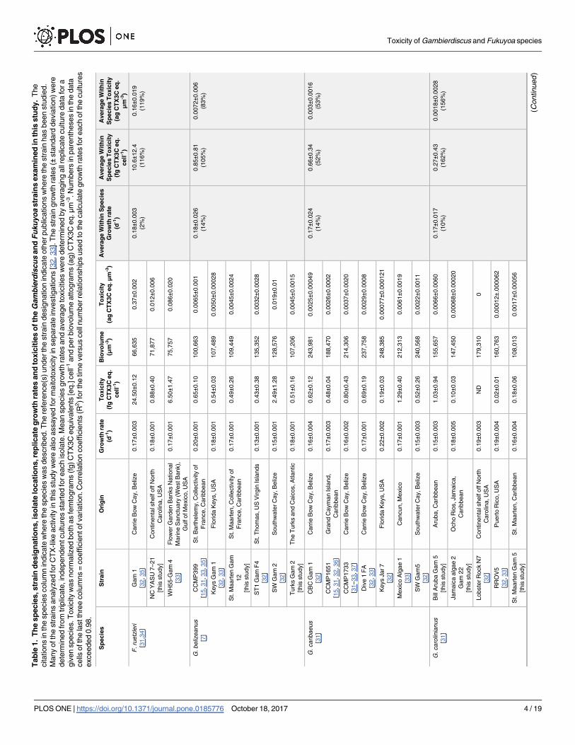
Tab
le1.
Th
esp
ecie
s,str
ain
desig
nati
on
s,is
ola
telo
cati
on
s,re
plicate
gro
wth
rate
san
dto
xic
itie
so
fth
eG
am
bie
rdis
cu
san
dF
uku
yo
astr
ain
sexam
ined
inth
isstu
dy.
The
citations
inth
especie
scolu
mn
indic
ate
where
the
specie
sw
as
described.T
he
refe
rence(s
)underth
estr
ain
desig
nation
indic
ate
oth
erpublic
ations
where
the
str
ain
has
been
stu
die
d.
Many
ofth
estr
ain
sanaly
zed
forC
TX
-lik
eactivity
inth
isstu
dy
were
als
oassayed
form
aitoto
xic
ity
insepara
tein
vestigations
[32,33].
The
str
ain
gro
wth
rate
s(±
sta
ndard
devia
tion)w
ere
dete
rmin
ed
from
trip
licate
,in
dependentculture
ssta
rted
foreach
isola
te.M
ean
specie
sgro
wth
rate
sand
avera
ge
toxic
itie
sw
ere
dete
rmin
ed
by
avera
gin
gall
replic
ate
culture
data
fora
giv
en
specie
s.T
oxic
ity
was
norm
aliz
ed
both
as
fem
togra
ms
(fg)C
TX
3C
equiv
ale
nts
[eq.]
cell-1
and
perbio
volu
me
attogra
ms
(ag)C
TX
3C
eq.μm
-3.N
um
bers
inpare
nth
eses
inth
edata
cells
ofth
ela
stth
ree
colu
mns
=coeffic
ientofvariation.C
orr
ela
tion
coeffic
ients
(R2)fo
rth
etim
evers
us
cell
num
berre
lationship
sused
toth
ecalc
ula
tegro
wth
rate
sfo
reach
ofth
eculture
s
exceeded
0.9
8.
Sp
ecie
sS
train
Ori
gin
Gro
wth
rate
(d-1
)
To
xic
ity
(fg
CT
X3C
eq
.
cell
-1)
Bio
vo
lum
e
(μm
-3)
To
xic
ity
(ag
CT
X3C
eq
.μm
-3)
Avera
ge
Wit
hin
Sp
ecie
s
Gro
wth
rate
(d-1
)
Avera
ge
Wit
hin
Sp
ecie
sT
oxic
ity
(fg
CT
X3C
eq
.
cell
-1)
Avera
ge
Wit
hin
Sp
ecie
sT
oxic
ity
(ag
CT
X3C
eq
.
μm-3
)
F.ru
etz
leri
[31,3
4]
Gam
1
[32,35]
Carr
ieB
ow
Cay,B
eliz
e0.1
7±0
.003
24.5
0±0
.12
66,6
35
0.3
7±0
.002
0.1
8±0
.003
(2%
)
10.6±1
2.4
(116%
)
0.1
6±0
.019
(119%
)
NC
YA
SU
7–21
[this
stu
dy]
Continenta
lshelf
off
Nort
h
Caro
lina,U
SA
0.1
8±0
.001
0.8
8±0
.40
71,8
77
0.0
12±0
.006
WH
55-G
am
4
[33]
Flo
wer
Gard
en
Banks
National
Marine
Sanctu
ary
(WestB
ank),
Gulf
ofM
exic
o,U
SA
0.1
7±0
.001
6.5
0±1
.47
75,7
57
0.0
86±0
.020
G.beliz
eanus
[7]
CC
MP
399
[15,31,33,35]
St.
Bart
hele
my,C
olle
ctivity
of
Fra
nce,C
aribbean
0.2
0±0
.001
0.6
5±0
.10
100,6
63
0.0
065±0
.001
0.1
8±0
.026
(14%
)
0.8
5±0
.81
(105%
)
0.0
072±0
.006
(83%
)
Keys
Gam
1
[32,33]
Flo
rida
Keys,U
SA
0.1
8±0
.001
0.5
4±0
.03
107,4
89
0.0
050±0
.00028
St.
Maart
en
Gam
12
[this
stu
dy]
St.
Maart
en,C
olle
ctivity
of
Fra
nce,C
aribbean
0.1
7±0
.001
0.4
9±0
.26
109,4
49
0.0
045±0
.0024
ST
1G
am
F4
[32]
St.
Thom
as,U
SV
irgin
Isla
nds
0.1
3±0
.001
0.4
3±0
.38
135,3
52
0.0
032±0
.0028
SW
Gam
2
[32]
South
wate
rC
ay,B
eliz
e0.1
5±0
.001
2.4
9±1
.28
128,5
76
0.0
19±0
.01
Turk
sG
am
2
[this
stu
dy]
The
Turk
sand
Caic
os,A
tlantic
0.1
8±0
.001
0.5
1±0
.16
107,2
06
0.0
045±0
.0015
G.caribaeus
[31]
CB
CG
am
1
[32]
Carr
ieB
ow
Cay,B
eliz
e0.1
6±0
.004
0.6
2±0
.12
243,9
81
0.0
025±0
.00049
0.1
7±0
.024
(14%
)
0.6
6±0
.34
(52%
)
0.0
03±0
.0016
(53%
)
CC
MP
1651
[15,31,32,36]
Gra
nd
Caym
an
Isla
nd,
Caribbean
0.1
7±0
.003
0.4
8±0
.04
188,4
70
0.0
026±0
.0002
CC
MP
1733
[31–33,37]
Carr
ieB
ow
Cay,B
eliz
e0.1
6±0
.002
0.8
0±0
.43
214,3
06
0.0
037±0
.0020
Div
e1
FA
[32,33]
Carr
ieB
ow
Cay,B
eliz
e0.1
7±0
.001
0.6
9±0
.19
237,7
58
0.0
029±0
.0008
Keys
Jar7
[32]
Flo
rida
Keys,U
SA
0.2
2±0
.002
0.1
9±0
.03
248,3
85
0.0
0077±0
.000121
Mexic
oA
lgae
1
[33]
Cancun,M
exic
o0.1
7±0
.001
1.2
9±0
.40
212,3
13
0.0
061±0
.0019
SW
Gam
5
[32]
South
wate
rC
ay,B
eliz
e0.1
5±0
.003
0.5
2±0
.26
240,5
68
0.0
022±0
.0011
G.caro
linia
nus
[31]
Bill
Aru
ba
Gam
5
[this
stu
dy]
Aru
ba,C
aribbean
0.1
5±0
.003
1.0
3±0
.94
155,6
57
0.0
066±0
.0060
0.1
7±0
.017
(10%
)
0.2
7±0
.43
(162%
)
0.0
018±0
.0028
(156%
)
Jam
aic
aalg
ae
2
Gam
22
[this
stu
dy]
Ocho
Rio
s,Jam
aic
a,
Caribbean
0.1
8±0
.005
0.1
0±0
.03
147,4
50
0.0
0068±0
.00020
Lobste
rR
ock
N7
[32]
Continenta
lshelf
off
Nort
h
Caro
lina,U
SA
0.1
9±0
.003
ND
179,3
10
0
RR
OV
5
[32,33]
Puert
oR
ico,U
SA
0.1
9±0
.004
0.0
2±0
.01
160,7
63
0.0
0012±.
000062
St.
Maart
en
Gam
5
[this
stu
dy]
St.
Maart
en,C
aribbean
0.1
6±0
.004
0.1
8±0
.06
108,0
13
0.0
017±0
.00056
(Continued
)
Toxicity of Gambierdiscus and Fukuyoa species
PLOS ONE | https://doi.org/10.1371/journal.pone.0185776 October 18, 2017 4 / 19
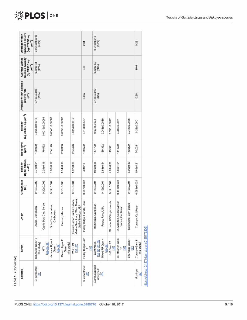
Tab
le1.
(Continued
)
Sp
ecie
sS
train
Ori
gin
Gro
wth
rate
(d-1
)
To
xic
ity
(fg
CT
X3C
eq
.
cell
-1)
Bio
vo
lum
e
(μm
-3)
To
xic
ity
(ag
CT
X3C
eq
.μm
-3)
Avera
ge
Wit
hin
Sp
ecie
s
Gro
wth
rate
(d-1
)
Avera
ge
Wit
hin
Sp
ecie
sT
oxic
ity
(fg
CT
X3C
eq
.
cell
-1)
Avera
ge
Wit
hin
Sp
ecie
sT
oxic
ity
(ag
CT
X3C
eq
.
μm-3
)
G.carp
ente
ri
[31]
Bill
Aru
ba
Gam
15
[this
stu
dy]
Aru
ba,C
aribbean
0.1
3±0
.002
0.7
1±0
.21
130,6
30
0.0
054±0
.0016
0.1
63±0
.026
(16%
)
0.8
9±0
.41
(47%
)
0.0
045±0
.0018
(45%
)
GT
4
[32,33,35]
Carr
ieB
ow
Cay,B
eliz
e0.2
0±0
.003
0.2
9±0
.16
179,5
22
0.0
016±0
.00089
Jam
aic
aA
lgae
2
Gam
1
[32,33]
Ocho
Rio
s,Jam
aic
a,
Caribbean
0.1
7±0
.002
0.9
3±0
.17
204,1
40
0.0
046±0
.00083
Mexic
oA
lgae
2
Gam
1
[this
stu
dy]
Cancun,M
exic
o0.1
5±0
.003
1.1
4±0
.18
206,3
06
0.0
055±0
.00087
WB
HR
21
[32,33]
Flo
wer
Gard
en
Banks
National
Marine
Sanctu
ary
(WestB
ank),
Gulf
ofM
exic
o,U
SA
0.1
8±0
.004
1.3
7±0
.30
254,4
76
0.0
054±0
.0012
G.excentr
icus
[38]
Pulle
yR
idge
Gam
2
[33]
Pulle
yR
idge,F
lorida,U
SA
0.0
57±0
.002
469±1
0179,5
22
2.6
1±0
.00557
0.0
57
469
2.6
1
Gam
bie
rdis
cus
riboty
pe
2
[31]
CC
MP
1655
[15,32,33,36]
Mart
iniq
ue,C
aribbean
0.1
5±0
.001
10.9±0
.36
147,7
00
0.0
74±.
0024
0.1
28±0
.010
(8%
)
6.6
2±2
.52
(38%
)
0.0
46±0
.016
(35%
)
Mix
ed
PR
Gam
4
[32,33]
Puert
oR
ico,U
SA
0.1
2±0
.001
6.6
3±0
.54
139,3
22
0.0
48±0
.0039
SJ3
Gam
F2
[32]
St.
John,U
SV
irgin
Isla
nds
0.1
2±0
.001
4.9
9±0
.38
143,5
11
0.0
35±0
.0027
St.
Maart
en
Gam
10
[33]
St.
Maart
en,C
olle
ctivity
of
Fra
nce,C
aribbean
0.1
41±0
.002
4.6
6±1
.01
141,5
70
0.0
33±0
.0071
SW
Alg
ae
Gam
1
[33]
South
wate
rC
ay,B
eliz
e0.1
3±0
.001
5.9
0±0
.80
143,2
09
0.0
41±0
.0056
G.silv
ae
[20]
Cura
cao
Gam
11
[this
stu
dy]
Cura
cao,C
aribbean
0.0
98±0
.024
19.6±4
.21
70,0
28
0.2
8±0
.060
0.9
819.6
0.2
8
htt
ps:
//doi.o
rg/1
0.1
371/jo
urn
al.p
one.
0185776.t001
Toxicity of Gambierdiscus and Fukuyoa species
PLOS ONE | https://doi.org/10.1371/journal.pone.0185776 October 18, 2017 5 / 19
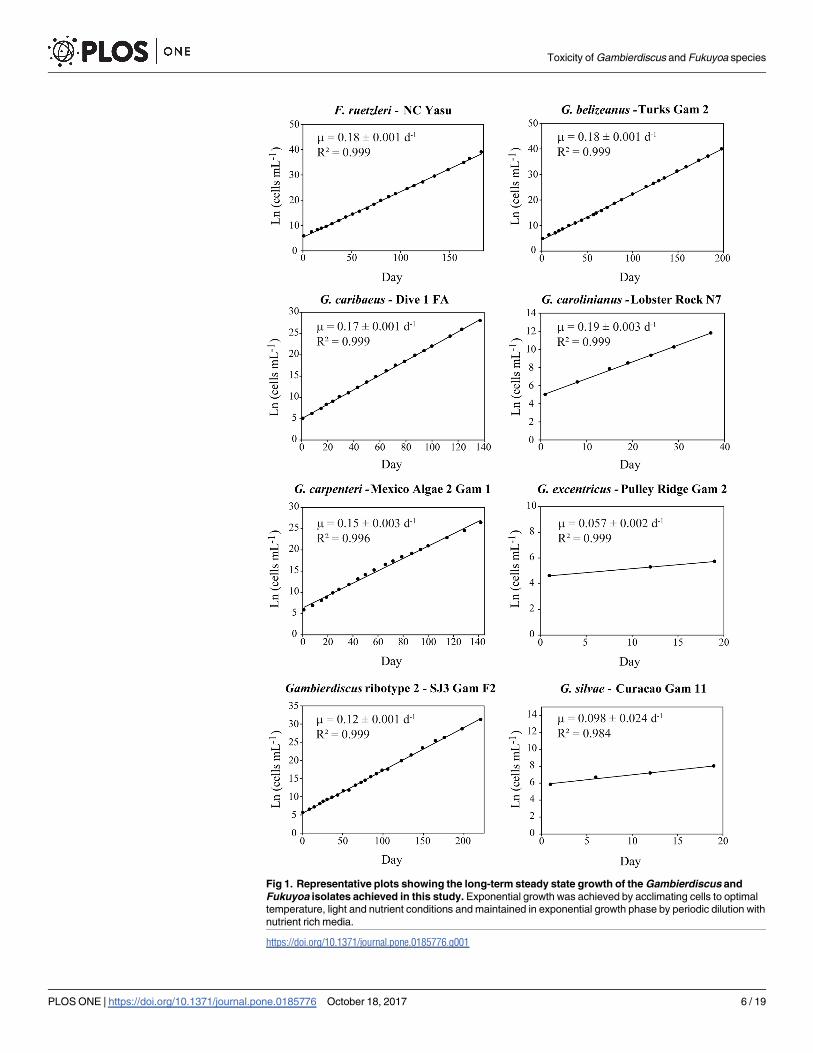
Fig 1. Representative plots showing the long-term steady state growth of the Gambierdiscus and
Fukuyoa isolates achieved in this study. Exponential growth was achieved by acclimating cells to optimal
temperature, light and nutrient conditions and maintained in exponential growth phase by periodic dilution with
nutrient rich media.
https://doi.org/10.1371/journal.pone.0185776.g001
Toxicity of Gambierdiscus and Fukuyoa species
PLOS ONE | https://doi.org/10.1371/journal.pone.0185776 October 18, 2017 6 / 19
average growth rate than the common practice of choosing the three steepest growth points
for a growth rate determination.
When cell densities were high enough, cells were harvested for toxicity by collecting a
known number of cells on a 20 μm sieve and washing them with filtered seawater (Salinity = 33)
into a 50 mL centrifuge tube. The cells were pelleted using centrifugation at 3200g for 10 min,
the supernatant carefully decanted, and the pellet was processed immediately or stored at
–20˚C prior to extraction. Because ciguatoxicity varies with growth phase, the decision was
made to ensure all the cultures were maintained in steady log phase growth prior to collection
of cells for toxin analysis [29, 36, 43]. This assured that the intra-strain and inter-specific toxic-
ity measurements were not biased due to harvesting cells in different growth phases.
Reagents
All reagents used in this study were ACS grade or higher. Solvents were HPLC grade or higher
purity. Pacific ciguatoxin-3C (CTX3C) was purchased from Wako Chemicals, USA, Inc.
(Richmond, Virginia, USA) and provided by Institut Louis Malarde, Tahiti, French Polynesia
(ILM). In this manuscript, we use the CTX nomenclature used by Yogi et al. [44] for the Pacific
ciguatoxins (e.g. CTX3C rather than P-CTX-3C). References to Caribbean ciguatoxins are pre-
ceded by a C (e.g. C-CTX1). CTX3C standards were stored at –20˚C and dissolved in 100%
methanol prior to utilization in the CBA-N2a. All water used was Milli-Q Ultra-pure grade
with 18.2 MO resistivity.
Toxin extraction
Cell pellets were sonicated for 1 min in 100% methanol at 3 mL per 100,000 cells using a Qso-
nica, Q700 unit (Thermo Fisher Scientific Inc., Waltham, Massachusetts) with the tip ampli-
tude setting at 50. Once cells were disrupted, the sample was centrifuged at 3,200g for 10 min
and the supernatant was transferred to a 20 mL glass scintillation vial. This was repeated two
more times and the methanol was collected and dried under N2 gas at 40˚C. The dried extract
was resuspended in dichloromethane (DCM) (5mL per 100,000 cells) and washed twice in a
separatory funnel with 60:40 methanol:water (2.5 mL per 100,000 cells). The dichloromethane
phases (bottom layer) were then collected and dried under N2 gas at 20˚C. The dried extract
was stored at –20˚C. When ready to process, the DCM extract was resuspended in a volume of
methanol that yielded a final concentration of 250–500 cells μL-1 [45].
Neuro-2a cell based assay (CBA-N2a)
The CBA-N2a assay allows estimation of the concentration of CTXs or CTX-like compounds
in extracts from fish or phytoplankton [38, 46–49]. The CBA-N2a assay measures bioactive
compounds that bind voltage gated-sodium channels, not all of which are ciguatoxins [45].
Previous studies of Gambierdiscus and Fukuyoa species using LC-MS, and the same dichloro-
methane extraction protocol as this study, however, have shown CTX or "CTX-like" com-
pound account for a majority of total cellular toxicity [29, 35, 43]. The consistency of these
data support CTX or CTX-like compounds as the primary toxins measured in the isolates
from this study.
The neuro-2a Mus musculus neuroblastoma cell line (N2a) used for the assay was obtained
from the American Type Culture Collection (ATCC1 CCL-131™). Cells were grown and
maintained in Eagle’s Minimum Essential Medium (EMEM; ATCC1 30–2003) containing 2
mM L-glutamine, 1 mM sodium pyruvate, 100 μg mL-1 streptomycin, 100 units mL-1 penicil-
lin, and 10% fetal bovine serum. Growth conditions were kept at 37˚C using a humidified 5%
CO2-enriched atmosphere. To prepare for toxicity analysis, the N2a cells were harvested with a
Toxicity of Gambierdiscus and Fukuyoa species
PLOS ONE | https://doi.org/10.1371/journal.pone.0185776 October 18, 2017 7 / 19

trypsin-ethylenediaminetetraacetic acid (trypsin-EDTA) solution and seeded into each well of
a 96-well microtiter plate at 30,000 cells per 100 μL of growth medium. The cells were subse-
quently incubated under the same growth conditions as above [36]. The plated N2a cells were
allowed to settle and grow 20–24 h until they were>90% confluent at the bottom of each well.
The standards, controls and samples were then added and the plates were incubated for 24 h.
Each plate included control wells containing buffer only or buffer plus 5% methanol, the
equivalent of the final methanol concentration when extracts were added. If the assay is work-
ing properly, both the buffer only and 5% methanol controls should contain a comparable
number of live cells after the 24-hour incubation period. The CTX3C standard curves used in
this assay ranged from 0.001–2,000 pg mL-1 and were suspended in the same 5% methanol
buffer solution as the samples. Aliquots of each sample were added to six wells. Three of these
wells contained 100 μM ouabain (O) and 10 μM veratridine (V) (O+/V+) to sensitize the
CBA-N2a cells to CTX, and the other three contained no O/V (O-/V-). The O-/V- wells served
to identify other non-specific toxins present in the samples. Cell viability in the control wells,
standard curve, sample O-/V- and the O+/V+ wells were assessed after 20–24 hours of toxin
exposure at 37˚C using the colorimetric 3-(4,5-dimethylthiazol-2-yl)-2,5-diphenyl tetrazolium
bromide (MTT) assay [49]. Cell mortality in the O+/V+ wells was converted to CTX estimates
based on the CTX3C standard curve. The limit of detecton was 0.2 pg CTX3C eq. mL-1.
The resulting toxicity measurements were expressed as both femtogram CTX3C eq. per cell
(fg cell-1) and attogram per μm3 cell volume (ag μm-3). The latter normalization employed the
average cell volumes determined using the Multisizer when the cells were harvested. This
approach determined if the variations in toxicity among isolates and among species were
attributable to differences in cell size or toxicity per unit biomass.
For six of the eight species, multiple isolates were examined making it possible to estimate
mean, standard deviaiton and coefficiants of variation in toxicity. To determine if the among
species toxicity differences were statistically significant, a Kruskal-Wallis nonparametric one
factor ANOVA was performed due to unequal variances. Gambierdiscus excentricus and G. sil-vae were excluded from the analysis because only a single clone was examined [50]. A Dunn’s
test, which estimates median toxicities, was used to determine if species toxicities fell into dis-
tinct groups.
The extent of interspecific variation was also estimated by calculating the ratio between the
average toxicitites for each species. In the case of G. excentricus and G. silvae the single toxicty
estimate for each isolate was used to represent the mean value. Still another way to assess varia-
tions in toxicity used the mean growth rates and approximate toxicity per cell to estimate toxin
production rates as fg CTX3C eq. cell-1 d-1. The results were plotted as species versus toxin
production rates and the ratio of the least to the most prolific toxin producing species was
calculated.
Results
Five of the eight Gambierdiscus and Fukuyoa species studied had similar average growth rates
ranging from 0.16 to 0.17 d-1. Gambierdiscus ribotype 2 (0.13 ± 0.01 d-1), G. silvae (0.098 d-1)
and G. excentricus (0.057 d-1) grew more slowly (Fig 1) (Table 1). The observed growth rates
were compared to those reported in other studies for the same species (S2 Table).
Gambierdiscus excentricus was the most toxic (469 fg CTX3C eq. cell-1) of the species exam-
ined (Table 1). The next most toxic species were G. silvae (19.6 fg CTX3C eq. cell-1) and Gam-bierdiscus ribotype 2 (4.7 to 10.9 fg CTX3C eq. cell-1). The remaining five species exhibited the
following range of toxicities: F. ruetzleri (0.9 to 24.5 fg CTX3C eq. cell-1), G. belizeanus (0.4 to
2.5 fg CTX3C eq. cell-1); G. caribaeus (0.2 to 1.3 fg CTX3C eq. cell-1); G. carolinianus (non-
Toxicity of Gambierdiscus and Fukuyoa species
PLOS ONE | https://doi.org/10.1371/journal.pone.0185776 October 18, 2017 8 / 19

detectable to 1.0 fg CTX3C eq. cell-1); and G. carpenteri (0.3 to 1.4 fg CTX3C eq. cell-1). The
within species coefficient of variation in toxicity for the species where multiple isolates were
tested ranged from 33% (Gambierdiscus ribotype 2) to 162% (G. carolinianus) (Table 1).
Within a species, the highest toxicity isolate was ~2- to 27-fold more toxic than the least toxic
isolate (S3 Table).
The results of a one factor ANOVA (non-parametric Kruskal-Wallis test) using the species
for which multiple isolates were available revealed toxicities among F. ruetzleri, G. belizeanus,G. caribaeus, G. carolinianus, G. carpenteri and Gambierdiscus ribotype 2 were significantly dif-
ferent (H = 18.76, p = 0.002) (Fig 2). A Dunn’s test indicated the six species were divided into
three groups according to their median toxicities. Group 1 included F. ruetzleri and Gambier-discus ribotype 2 (Fig 2). Group 2 was G. carpenteri, G. caribaeus and G. belizeanus while
Group 3 contained only G. carolinianus. It should be noted that while each of the species
included in the preceding analysis exhibited low toxicity relative to G. excentricus, significant
differences in toxicity were found among the lower toxicity species (Fig 2) (Table 1).
A plot of average Gambierdiscus growth rate versus average toxicity normalized on both a
per cell and per biovolume basis showed the slower growing Gambierdiscus species were more
toxic (Fig 3, S1 Fig). This increasing toxicity with declining growth rate followed an exponen-
tial relationship. Toxicity expressed as average toxin production rate fg CTX3C eq. cell-1 d-1
showed the same pattern of toxicity among species (Fig 4, S1 Fig). Based on the observed pro-
duction rates, the difference between the most (G. excentricus) and least (G. carolinianus) toxic
species was 613-fold. The equivalent difference between the most toxic and least toxic species
based on toxicity per cell was ~1740-fold.
Only one Fukuyoa species was examined, so it was impossible to say if a similar relationship
between growth rate and toxicity exists within this genus. It was apparent that toxicity for the
Fig 2. Results of a Kruskal-Wallis nonparametric one factor ANOVA for differences in CTX toxicity among
Gambierdiscus and Fukuyoa species. Gambierdiscus excentricus and G. silvae were excluded from the analysis
because only a single clone was examined. Abbreviations: n = sample size, M = median toxicity (fg CTX3C eq. cell-1), H =
Kruskal-Wallis test statistic, df = degrees of freedom. Brackets denote result of the Dunn’s follow up test. The statistic is
designed to estimate median toxicities to determine if the species partitioned into distinct groups.
https://doi.org/10.1371/journal.pone.0185776.g002
Toxicity of Gambierdiscus and Fukuyoa species
PLOS ONE | https://doi.org/10.1371/journal.pone.0185776 October 18, 2017 9 / 19

Fig 3. Ciguatoxicity versus growth rate. Natural log of cellular toxicity versus growth rate for each of the
Gambierdiscus and Fukuyoa species normalized (A) to femtograms (fg) CTX3C eq. cell-1 and (B) attograms (ag)
CTX3C eq. per μm-3 biovolume. Error bars = ± 1 standard deviation. The red arrows indicate data for F. ruetzleri, which
had a higher toxicity than the Gambierdiscus species growing at a similar rate.
https://doi.org/10.1371/journal.pone.0185776.g003
Toxicity of Gambierdiscus and Fukuyoa species
PLOS ONE | https://doi.org/10.1371/journal.pone.0185776 October 18, 2017 10 / 19

Fukuyoa isolates tested was higher on a per cell and a per biomass basis compared to the Gam-bierdiscus species growing at a similar rate (Fig 3).
Discussion
Relative toxicity of Gambierdiscus excentricus
The Gambierdiscus excentricus isolate tested in this study was ~44- to>1,740-fold more toxic
than the other species examined (~469 fg CTX3C eq. cell-1; Table 1, S3 Table). This result is
consistent with the high toxicities reported for G. excentricus isolates from the Canary Islands
(370–1,100 CTX1 eq. cell-1 and 1,425 CTX3C eq. cell-1; [38, 49]) and is similar to G. polynesien-sis, the dominant toxin-producing species in the Pacific [29, 30]. To date, G. polynesiensis has
not been identified from the eastern Atlantic, Caribbean or Gulf of Mexico (GOM), signifying
that G. excentricus is the dominant CTX producer in the temperate and tropical regions of the
eastern Atlantic, Caribbean and Gulf of Mexico (GOM) [8, 38, 49] (Table 1). In contrast, the
range of toxicities exhibited by the other six Gambierdiscus and one Fukuyoa species examined
varied from non-detectible to 24.5 fg CTX3C eq. cell-1.
The extent to which G. excentricus may dominate the CTX flux in Caribbean and GOM will
depend on both its abundance and distribution. The tenant that environments fostering higher
abundances of G. excentricus are more likely to produce ciguatoxic fish is put forward as a
working hypothesis. Obtaining the data on abundance and distribution necessary to test this
hypothesis will depend on quantitative species-specific molecular assays since Gambierdiscusspecies are not readily distinguished using light microscopy [31, 51]. Quantitative species-spe-
cific polymerase chain reaction (qPCR) assays are available for many Caribbean Gambierdiscusspecies, but not G. excentricus and the next most toxic species, G. silvae. Recently, PCR assays
Fig 4. Toxin production rates. This figure shows the estimated toxin production (fg CTX3C eq. cell-1 d-1) rate for each species.
https://doi.org/10.1371/journal.pone.0185776.g004
Toxicity of Gambierdiscus and Fukuyoa species
PLOS ONE | https://doi.org/10.1371/journal.pone.0185776 October 18, 2017 11 / 19

for G. excentricus and G. silvae were developed in our laboratory, but they have not yet been
validated for quantitative (qPCR) estimation of cell abundances (unpublished). However, PCR
screening on a limited number of field samples and newly isolated cultures allowed us to begin
defining the geographic ranges of these species. Gambierdiscus excentricus was found in the
Florida Keys, USA and the Bahamas, while G. silvae was present in the Bahamas, Saint Croix,
and the U.S. Virgin Islands. Combining these data with those from the literature confirmed
the minimum geographic range of G. excentricus extends from the northwest coast of Africa to
southern Florida, USA and the southeast coast of Brazil [52, 53]. Gambierdiscus silvae ranges
from the Canary Islands through the eastern and western Caribbean [8, 20]. More extensive
sampling using species-specific qPCR assays has shown that F. ruetzleri, G. belizeanus, G. cari-baeus, G. carolinianus, G. carpenteri and Gambierdiscus ribotype 2 are ubiquitously distributed
throughout the Caribbean and GOM [8]. It is likely G. excentricus and G. silvae share an
equally wide distribution. This suggests the contribution of G. excentricus to the overall toxin
flux depends primarily on their relative abundance. The average toxin rate is 28.1 fg CTX3C
eq. cell-1 d-1 for G. excentricus the most toxic species, 1.9 for the next most toxic species, G. sil-vae and F. ruetzleri, and 0.05 for G. carolinianus, the lest toxic species (Fig 3). If a population
consisted of only G. carolinianus and G. excentricus, G. excentricus need only make up 0.16%
of the total population to produce as much toxin as G. carolinianus. If the population con-
tained only G. silvae, F. ruetzleri and G. excentricus, G. excentricus would have to make up 6.3%
of the population on average to produce as many CTX equivalents as the other two species
combined.
If G. excentricus is confirmed as the primary CTX producing species in the Atlantic, fully
investigating its role in causing CFP may require careful chemical characterization of the spe-
cific CTX congeners it produces. That characterization would help facilitate development of
LC-MS toxin-specific analytical methods capable of answering whether the low toxicity Atlan-
tic Gambierdiscus and Fukuyoa species produce the same analogs in lesser quantities than
G. excentricus, or only analogs of lower toxicity [26, 38, 49].
Within species versus among species differences in CTX toxicity
A long-standing question in ciguatera research is the extent to which CFP risk is dependent on
variations in toxicity among species versus between species [8]. Results of a Kruskal-Wallis
nonparametric one factor ANOVA showed significant differences in CTX toxicity exist among
the various Gambierdiscus species tested (Fig 2), confirming between species differences in tox-
icity are, on average, greater than among isolates of the same species (S3 Table). Though the
within species variation for G. excentricus toxicity was not measured in this study, comparison
with estimates in Fraga et al. [38] indicate within species variation is ~3-fold (370 to 1,100 fg
P-CTX-1B eq. cell-1; n = 3). Other studies using CBA-N2a showed a similar within species var-
iation in toxicity—0.6–2.7 fg CTX3C eq. cell-1 (n = 3) for G. australes [49], 0–19.9 fg P-CTX-1
eq. cell-1 (n = 4) for G. balechii [54], 2.6–6.0 fg P-CTX-1 eq. cell-1 (n = 4) for Gambierdiscus sp.
type 4 [55] and 10.3–12.4 fg CTX3C eq. cell-1 (n = 2) for G. silvae [49] (S1 Table). Cumula-
tively, these data are consistent with CTX risk being primarily dependent on species
composition.
Relationship between growth rate and toxicity
Chinain [29] proposed that slower Gambierdiscus cell growth was associated with higher toxin
content per cell. Indeed, G. polynesiensis, the slowest growing Pacific species tested to date, is
by far the most toxic. The trend holds true for the Gambierdiscus species measured in this
study with the slowest growing species, G. excentricus exhibiting the highest toxicity
Toxicity of Gambierdiscus and Fukuyoa species
PLOS ONE | https://doi.org/10.1371/journal.pone.0185776 October 18, 2017 12 / 19
(Figs 3 and 4; Table 1). These data are consistent with an evolutionary tradeoff between an
investment in growth versus the production of defensive compounds as observed in other
harmful algal species [41, 56–58]. It is also noteworthy that relationship between toxicity and
growth is exponential and not linear (Fig 3; [49]).
Estimating CTX fluxes in the environment
Quantifying the contribution of various Gambierdiscus and Fukuyoa species to the flux of
CTXs in the environment requires simultaneous determination of the species abundances and
the amount of CTX being produced by each species. Undertaking such studies would be both
expensive and technically challenging, especially since the full suite of species and the toxins
they produce is unknown. A potentially, more tractable approach to understanding how differ-
ent species may contribute to overall toxin fluxes is to incorporate the average toxicities into a
physiologically-based Gambierdiscus growth rate model [15]. This approach would identify
regions in the Caribbean and GOM where CTX fluxes may be highest. Model runs could also
be adjusted to estimate how different relative abundances of low and high toxicity species
would affect the magnitude of toxin flux. Explicit assumptions underlying this approach are
that 1) average toxin concentrations represent the toxicity of the population as a whole and, 2)
CFP risk is proportional to the toxicities of the Gambierdiscus and Fukuyoa species themselves.
The use of average toxicities in models is consistent with our knowledge of microalgal pop-
ulation genetics. Numerous studies have shown that algal populations maintain a high diver-
sity of genotypes even during intense blooms, i.e. they are not dominated by only a few
genotypes [59–64]. Averaging the toxicities of different isolates approximates population level
toxicities. The relevance of using the toxicity of CTX-producing species to predict risk is sup-
ported by studies showing that as CTX congeners bioaccumulate in the food chain, some
remain the same while others are biologically modified to have higher toxicities than their par-
ent compounds [44, 65–67]. As a result, the toxicities remain the same or increase in the food
chain so Gambierdiscus and Fukuyoa toxicities provide minimum estimates of CFP risk.
Management implications
The results of this study have implications for managing CFP risk. Ideally, risk would be rou-
tinely assessed in an institutionalized surveillance system by quantitatively measuring a stan-
dard suite of CTXs in fish using LC-MS. Unfortunately, this is not practical because of the lack
of certified standards and the high cost of analytical methods [47]. Until these obstacles are
overcome, the problem requires a two-tiered approach. The first tier includes monitoring for
increased cell abundances to determine elevated CFP risk and understanding the environmen-
tal conditions conducive to high Gambierdiscus/Fukuyoa abundance. The second tier includes
the use of qPCR assays to determine the Gambierdiscus/Fukuyoa species composition with a
focus on the relative numbers of G. excentricus in the Caribbean.
With respect to the first approach, it is known that CFP events can occur from one month
to a year following a significant increase in Gambierdiscus cell densities [28, 68–70]. Conse-
quently, genus-level cell counts using light microscopy [71] can provide first order estimates of
CFP risk, but cannot predict severity. Despite this limitation, using this approach can provide
managers an indication of when and where CFP risk may be elevated [72].
Interpretation of microscopic Gambierdiscus and Fukuyoa cell abundances can be further
informed by understanding the environmental conditions that promote growth. Laboratory
and field studies indicate temperature is the primary environmental factor regulating growth
of Gambierdiscus and Fukuyoa species [15, 73, 74]. Modeling studies have also shown that in
terms of broad patterns, annual temperature cycles can predict the regions where CFP risk is
Toxicity of Gambierdiscus and Fukuyoa species
PLOS ONE | https://doi.org/10.1371/journal.pone.0185776 October 18, 2017 13 / 19
highest [75]. It is also known that CFP causing dinoflagellate species prefer habitats with low
turbulence, appropriate substrate (macrophytes, algal turfs, coral rubble, seagrasses, etc.),
nutrients supplied from the benthos or other sources, little or no direct runoff from land, and
light levels >10 and< 200–700 μmol photons m-2 s-1 depending on species [8, 14, 18, 53, 71,
73, 74, 76–79]. The low light requirements of these species mean that habitats down to 50
meters or more may be capable of supporting substantial populations [79]. As GIS databases
detailing habitat types throughout the Caribbean and Gulf of Mexico improve, they can be
used in combination with the physiologically-based models to predict areas of higher CFP risk.
The second tier approach would use qPCR assays and focus on G. excentricus if it is con-
firmed as the dominant source of CTX in the Caribbean [23, 51]. Quantitative PCR assays are
routinely used to monitor harmful algae in many regions of the world [80–83]. Only lack of
resources keeps this from being possible throughout the Caribbean. Ultimately, as LC-MS
methods become more cost effective, and high CFP risk areas are identified, the logical course
is to use cell-based monitoring to focus on samples that need to be tested for toxins.
Conclusions
Gambierdiscus excentricus was significantly more toxic than the other Gambierdiscus and the
single species of Fukuyoa examined in this study from the Caribbean and GOM. Even with its
slow growth rate, it is likely G. excentricus contributes disproportionally large fluxes of CTXs
in the food chain. Overall, toxicity was inversely related to growth rate, indicating a tradeoff
between investments of cellular resources in growth versus defensive compounds. Monitoring
overall Gambierdiscus and Fukuyoa cell densities using genus-specific light microcopy may
provide insight into when CFP risks are of concern, but cannot predict the severity of events.
Despite this limitation, a cell-based approach can be used to predict first order risk assessment
when no other method is available. If research confirms the hypothesis that one or a relatively
few species produce most of the ciguatoxins entering the food web, then monitoring of those
species using species-specific qPCR or other molecular assays will support more accurate
assessments of CFP risk. Ecological models based on the physiological and ecological prefer-
ences of the key toxin producing species, also offer a way to cost effectively identify time peri-
ods and locations when CFP risk is the highest and when more expensive testing using LC-MS
methods are warranted.
Supporting information
S1 Table. Comprehensive table showing what is known about CTX and MTX production
by Gambierdiscus and Fukuyoa isolates not included in this study.
(DOCX)
S2 Table. Comparison of the Gambierdiscus and Fukuyoa growth rate estimates deter-
mined in this study versus rates published in other studies.
(DOCX)
S3 Table. A. Ratio of the highest toxicity isolate divided by the lowest isolate in fg CTX3C
eq. cell-1.
(DOCX)
S1 Fig. Ciguatoxicity versus growth rate plotted on a linear scale. Cellular toxicity versus
growth rate for each of the Gambierdiscus and Fukuyoa species normalized (A) to femtograms
(fg) CTX3C eq. cell-1 and (B) attograms (ag) CTX3C eq. per μm-3 biovolume. Error bars = ± 1
standard deviation. This graph visually demonstrates the large difference in variation in
Toxicity of Gambierdiscus and Fukuyoa species
PLOS ONE | https://doi.org/10.1371/journal.pone.0185776 October 18, 2017 14 / 19
toxicity of G. excentricus relative to the other species.
(TIF)
Acknowledgments
Mireille Chinain and Taiana Darius provided critical toxin standards and methodological
advice and Mark Vanddersee provided constructive criticisms of the manuscript. We also wish
to thank the following individuals who have assisted in collecting field samples from numerous
locations in the Caribbean and Gulf of Mexico: Christine Addison, John Burke, Dave Cerino,
Brian Degan, Michael Dowgiallo, Wilson Freshwater, Brett Harrison, David Johnson, Doug
Kesling, William Lee, Roger Mays, James Morris, Roldan Muñoz, Brandon Puckett, Sherry
Reed, Bill Sunda, Jenny Vander Plum and Paula Whitfield. We thank the three anonymous
reviewers of the manuscript for their constructive edits and suggestions. This study was
inspired by the GEOHAB, HABs in Benthic Systems Core Research Program.
Author Contributions
Conceptualization: R. Wayne Litaker, Patricia A. Tester.
Data curation: William C. Holland, D. Ransom Hardison, Steven R. Kibler.
Formal analysis: R. Wayne Litaker, William C. Holland, D. Ransom Hardison, Philipp Hess.
Funding acquisition: R. Wayne Litaker, Philipp Hess, Patricia A. Tester.
Investigation: William C. Holland, D. Ransom Hardison, Francesco Pisapia.
Methodology: R. Wayne Litaker, William C. Holland, D. Ransom Hardison, Francesco
Pisapia.
Project administration: R. Wayne Litaker, Patricia A. Tester.
Supervision: R. Wayne Litaker.
Visualization: R. Wayne Litaker, D. Ransom Hardison.
Writing – original draft: R. Wayne Litaker, William C. Holland, D. Ransom Hardison, Phi-
lipp Hess, Steven R. Kibler, Patricia A. Tester.
Writing – review & editing: R. Wayne Litaker, Steven R. Kibler, Patricia A. Tester.
References
1. Lehane L. Ciguatera update. Med J Aust. 2000; 172(4): 176–9. PMID: 10772591
2. Ohizumi Y, Shibata S, Tachibana K. Mode of the excitatory and inhibitory actions of ciguatoxin in the
guinea-pig vas-deferens. J Pharmacol Exp Ther. 1981; 217(2): 475–80. PMID: 7229984
3. Bidard JN, Vijverberg HPM, Frelin C, Chungue E, Legrand AM, Bagnis R, et al. Ciguatoxin is a novel
type of NA+ channel toxin. J Biol Chem. 1984; 259(13): 8353–7. PMID: 6330108
4. Benoit E, Legrand AM, Dubois JM. Effects of ciguatoxin on current and voltage clamped frog myelinated
nerve-fiber. Toxicon. 1986; 24(4): 357–64. https://doi.org/10.1016/0041-0101(86)90195-9 PMID:
2424144
5. Seino A, Kobayashi M, Momose K, Yasumoto T, Ohizumi Y. The mode of inotropic action of ciguatoxin
on guinea-pig cardiac muscle. Br J Pharmacol. 1988; 95(3): 876–82. PMID: 3207997
6. Molgo J, Comella JX, Legrand AM. Ciguatoxin enhances quantal transmitter release from frog motor-
nerve terminals. Br J Pharmacol. 1990; 99(4): 695–700. PMID: 1972891
7. Faust MA. Observation of sand-dwelling toxic dinoflagellates (Dinophyceae) from widely differing sites,
including two new species. J Phycol. 1995; 31(6): 996–1003. https://doi.org/10.1111/j.0022-3646.1995.
00996.x
Toxicity of Gambierdiscus and Fukuyoa species
PLOS ONE | https://doi.org/10.1371/journal.pone.0185776 October 18, 2017 15 / 19
8. Litaker RW, Vandersea MW, Faust MA, Kibler SR, Nau AW, Holland WC, et al. Global distribution of
ciguatera causing dinoflagellates in the genus Gambierdiscus. Toxicon. 2010; 56(5): 711–30. https://
doi.org/10.1016/j.toxicon.2010.05.017 PMID: 20561539
9. Lewis RJ. The changing face of ciguatera. Toxicon. 2001; 39(1): 97–106. https://doi.org/10.1016/
s0041-0101(00)00161-6 PMID: 10936626
10. Chan TYK. Epidemiology and clinical features of ciguatera fish poisoning in Hong Kong. Toxins. 2014;
6(10): 2989–97. https://doi.org/10.3390/toxins6102989 PMID: 25333356
11. Chan TYK. Characteristic features and contributory factors in fatal ciguatera fish poisoning-implications
for prevention and public education. Am J Trop Med Hyg. 2016; 94(4): 704–9. https://doi.org/10.4269/
ajtmh.15-0686 PMID: 26787145
12. Asaeda G. The transport of ciguatoxin: A case report. J Emerg Med. 2001; 20(3): 263–5. https://doi.org/
10.1016/s0736-4679(00)00319-x PMID: 11267814
13. Epelboin L, Perignon A, Hossen V, Vincent R, Krys S, Caumes E. Two clusters of ciguatera fish poison-
ing in Paris, France, related to tropical fish imported from the French Caribbean by travelers. J Travel
Med. 2014; 21(6): 397–402. https://doi.org/10.1111/jtm.12161 PMID: 25345983
14. Yoshimatsu T, Yamaguchi H, Iwamoto H, Nishimura T, Adachi M. Effects of temperature, salinity and
their interaction on growth of Japanese Gambierdiscus spp. (Dinophyceae). Harmful Algae. 2014; 35:
29–37. https://doi.org/10.1016/j.hal.2014.03.007
15. Kibler SR, Tester PA, Kunkel KE, Moore SK, Litaker RW. Effects of ocean warming on growth and distri-
bution of dinoflagellates associated with ciguatera fish poisoning in the Caribbean. Ecol Model. 2015;
316: 194–210. https://doi.org/10.1016/j.ecolmodel.2015.08.020
16. Aligizaki K, Katikou P, Nikolaidis G, Panou A. First episode of shellfish contamination by palytoxin-like
compounds from Ostreopsis species (Aegean Sea, Greece). Toxicon. 2008; 51(3): 418–27. https://doi.
org/10.1016/j.toxicon.2007.10.016 PMID: 18067938
17. Llewellyn LE. Revisiting the association between sea surface temperature and the epidemiology of fish
poisoning in the South Pacific: Reassessing the link between ciguatera and climate change. Toxicon.
2010; 56(5): 691–7. https://doi.org/10.1016/j.toxicon.2009.08.011 PMID: 19706300
18. Tester PA, Feldman RL, Nau AW, Kibler SR, Litaker RW. Ciguatera fish poisoning and sea surface tem-
peratures in the Caribbean Sea and the West Indies. Toxicon. 2010; 56(5): 698–710. https://doi.org/10.
1016/j.toxicon.2010.02.026 PMID: 20206196
19. Jeong HJ, Lim AS, Jang SH, Yih WH, Kang NS, Lee SY, et al. First Report of the Epiphytic Dinoflagel-
late Gambierdiscus caribaeus in the Temperate Waters off Jeju Island, Korea: Morphology and Molecu-
lar Characterization. J Eukaryot Microbiol. 2012; 59(6): 637–50. https://doi.org/10.1111/j.1550-7408.
2012.00645.x PMID: 22897440
20. Fraga S, Rodriguez F. Genus Gambierdiscus in the Canary Islands (NE Atlantic Ocean) with description
of Gambierdiscus silvae sp nov., a new potentially toxic epiphytic benthic dinoflagellate. Protist. 2014;
165(6): 839–53. https://doi.org/10.1016/j.protis.2014.09.003 PMID: 25460234
21. Kohli GS, Murray SA, Neilan BA, Rhodes LL, Harwood DT, Smith KF, et al. High abundance of the
potentially maitotoxic dinoflagellate Gambierdiscus carpenteri in temperate waters of New South
Wales, Australia. Harmful Algae. 2014; 39: 134–45. https://doi.org/10.1016/j.hal.2014.07.007
22. Nishimura T, Sato S, Tawong W, Sakanari H, Yamaguchi H, Adachi M. Morphology of Gambierdiscus
scabrosus sp nov (Gonyaulacales): a new epiphytic toxic dinoflagellate from coastal areas of Japan. J
Phycol. 2014; 50(3): 506–14. https://doi.org/10.1111/jpy.12175 PMID: 26988323
23. Nishimura T, Hariganeya N, Tawong W, Sakanari H, Yamaguchi H, Adachi M. Quantitative PCR assay
for detection and enumeration of ciguatera-causing dinoflagellate Gambierdiscus spp. (Gonyaulacales)
in coastal areas of Japan. Harmful Algae. 2016; 52: 11–22. https://doi.org/10.1016/j.hal.2015.11.018
PMID: 28073467
24. Yasumoto T, Bagnis R, Vernoux JP. Toxicity of surgeonfishes—II. Properites of the prinicpal water-sol-
uble toxin. Bull Jap Soc Sci Fish. 1976; 43: 359–65.
25. Botana LM. Seafood and Freshwater Toxins: Pharmacology, Physiology, and Detection, Third Edition.
Boca Raton, Florida: CRC Press; 2014. 1215 p.
26. Kohli GS, Papiol GG, Rhodes LL, Harwood DT, Selwood A, Jerrett A, et al. A feeding study to probe the
uptake of maitotoxin by snapper (Pagrus auratus). Harmful Algae. 2014; 37: 125–32. https://doi.org/10.
1016/j.hal.2014.05.018
27. Reverte L, Solino L, Carnicer O, Diogene J, Campas M. Alternative methods for the detection of emerg-
ing marine toxins: biosensors, biochemical assays and cell-based assays. Mar Drugs. 2014; 12(12):
5719–63. https://doi.org/10.3390/md12125719 PMID: 25431968
Toxicity of Gambierdiscus and Fukuyoa species
PLOS ONE | https://doi.org/10.1371/journal.pone.0185776 October 18, 2017 16 / 19
28. Chinain M, Germain M, Deparis X, Pauillac S, Legrand AM. Seasonal abundance and toxicity of the
dinoflagellate Gambierdiscus spp (Dinophyceae), the causative agent of ciguatera in Tahiti, French
Polynesia. Mar Biol. 1999; 135(2): 259–67. https://doi.org/10.1007/s002270050623
29. Chinain M, Darius HT, Ung A, Cruchet P, Wang ZH, Ponton D, et al. Growth and toxin production in the
ciguatera-causing dinoflagellate Gambierdiscus polynesiensis (Dinophyceae) in culture. Toxicon. 2010;
56(5): 739–50. https://doi.org/10.1016/j.toxicon.2009.06.013 PMID: 19540257
30. Rhodes L, Harwood T, Smith K, Argyle P, Munday R. Production of ciguatoxin and maitotoxin by strains
of Gambierdiscus australes, G. pacificus and G. polynesiensis (Dinophyceae) isolated from Rarotonga,
Cook Islands. Harmful Algae. 2014; 39: 185–90. https://doi.org/10.1016/j.hal.2014.07.018
31. Litaker RW, Vandersea MW, Faust MA, Kibler SR, Chinain M, Holmes MJ, et al. Taxonomy of Gambier-
discus including four new species, Gambierdiscus caribaeus, Gambierdiscus carolinianus, Gambierdis-
cus carpenteri and Gambierdiscus ruetzleri (Gonyaulacales, Dinophyceae). Phycologia. 2009; 48(5):
344–90. https://doi.org/10.2216/07-15.1
32. Holland WC, Litaker RW, Tomas CR, Kibler SR, Place AR, Davenport ED, et al. Differences in the toxic-
ity of six Gambierdiscus (Dinophyceae) species measured using an in vitro human erythrocyte lysis
assay. Toxicon. 2013; 65: 15–33. https://doi.org/10.1016/j.toxicon.2012.12.016 PMID: 23313447
33. Pisapia F, Sibat M, Herrenknecht C, Lhaute K, Gaiani G, Ferron P-J, et al. Maitotoxin-4, a Novel MTX
Analog Produced by Gambierdiscus excentricus. Mar Drugs. 2017;Jul 11; 15(7). https://doi.org/10.
3390/md15070220 PMC5532662. PMID: 28696398
34. Gomez F, Qiu DJ, Lopes RM, Lin SJ. Fukuyoa paulensis gen. et sp nov., a new genus for the globular
species of the Dinoflagellate Gambierdiscus (Dinophyceae). Plos One. 2015; 10(4): 18. https://doi.org/
10.1371/journal.pone.0119676 PMID: 25831082
35. Lewis RJ, Inserra M, Vetter I, Holland WC, Hardison DR, Tester PA, et al. Rapid extraction and identifi-
cation of maitotoxin and ciguatoxin-like toxins from Caribbean and Pacific Gambierdiscus using a new
functional bioassay. PLoS One. 2016;Jul 28; 11(7):e0160006. https://doi.org/10.1371/journal.pone.
0160006 PMID: 27467390
36. Lartigue J, Jester ELE, Dickey RW, Villareal TA. Nitrogen source effects on the growth and toxicity of
two strains of the ciguatera-causing dinoflagellate Gambierdiscus toxicus. Harmful Algae. 2009; 8(5):
781–91. https://doi.org/10.1016/j.hal.2008.05.006
37. Roeder K, Erler K, Kibler S, Tester P, Ho VT, Lam NN, et al. Characteristic profiles of ciguatera toxins in
different strains of Gambierdiscus spp. Toxicon. 2010; 56(5): 731–8. https://doi.org/10.1016/j.toxicon.
2009.07.039 PMID: 19682482
38. Fraga S, Rodriguez F, Caillaud A, Diogene J, Raho N, Zapata M. Gambierdiscus excentricus sp. nov.
(Dinophyceae), a benthic toxic dinoflagellate from the Canary Islands (NE Atlantic Ocean). Harmful
Algae. 2011; 11: 10–22. https://doi.org/10.1016/j.hal.2011.06.013
39. Keller M, Guillard R. Factors significant to marine dinoflagellate culture. In: Anderson DM, White AW,
Baden DG, editors. Toxic Dinoflagellates proceedings of the Third International Conference on toxic
dinoflagellates. New York: Elsevier; 1985. p. 113–6.
40. Sunda WG, Price NM, Morel FM. Trace metal ion buffers and their use in culture studies. In: Anderson
RA, editor. Algal culturing techniques, 1st Edition: Academic Press; 2005. p. 35–63.
41. Hardison DR, Sunda WG, Litaker RW, Shea D, Tester PA. Nitrogen limitation increases brevetoxins in
Karenia brevis (Dinophyceae): implications for bloom toxicity. J Phycol. 2012; 48(4): 844–58. https://
doi.org/10.1111/j.1529-8817.2012.01186.x PMID: 27008996
42. Keller MD, Bellows WK, Guillard RRL. Microwave treatment for sterilization of phytoplankton culture
media. J Exp Mar Biol Ecol. 1988; 117(3): 279–83. https://doi.org/10.1016/0022-0981(88)90063-9
43. Caillaud A, de la Iglesia P, Barber E, Eixarch H, Mohammad-Noor N, Yasumoto T, et al. Monitoring of
dissolved ciguatoxin and maitotoxin using solid-phase adsorption toxin tracking devices: Application to
Gambierdiscus pacificus in culture. Harmful Algae. 2011; 10(5): 433–46. https://doi.org/10.1016/j.hal.
2011.02.004
44. Yogi K, Oshiro N, Matsuda S, Sakugawa S, Matsuo T, Yasumoto T. Toxin profiles in fish implicated in
ciguatera fish poisoning in Amami and Kakeroma Islands, Kagoshima Prefecture, Japan. Food Hygiene
and Safety Science. 2013; 54(6): 385–91.
45. Dickey RW, Plakas SM. Ciguatera: A public health perspective. Toxicon. 2010; 56(2): 123–36. https://
doi.org/10.1016/j.toxicon.2009.09.008 PMID: 19782098
46. Dechraoui MYB, Tiedeken JA, Persad R, Wang ZH, Granade HR, Dickey RW, et al. Use of two detec-
tion methods to discriminate ciguatoxins from brevetoxins: Application to great barracuda from Florida
Keys. Toxicon. 2005; 46(3): 261–70. https://doi.org/10.1016/j.toxicon.2005.04.006 PMID: 15982699
Toxicity of Gambierdiscus and Fukuyoa species
PLOS ONE | https://doi.org/10.1371/journal.pone.0185776 October 18, 2017 17 / 19
47. Caillaud A, de la Iglesia P, Darius HT, Pauillac S, Aligizaki K, Fraga S, et al. Update on methodologies
available for ciguatoxin determination: perspectives to confront the onset of ciguatera fish poisoning in
Europe. Mar Drugs. 2010; 8(6): 1838–907. https://doi.org/10.3390/md8061838 PMID: 20631873
48. Pawlowiez R, Darius HT, Cruchet P, Rossi F, Caillaud A, Laurent D, et al. Evaluation of seafood toxicity
in the Australes archipelago (French Polynesia) using the neuroblastoma cell-based assay. Food Addit
Contam Part A Chem Anal Control Expo Risk Assess. 2013; 30(3): 567–86. https://doi.org/10.1080/
19440049.2012.755644 PMID: 23286347
49. Pisapia F, Holland WC, Hardison DR, Litaker RW, Fraga S, Nishimura T, et al. Toxicity screening of 13
Gambierdiscus strains using neuro-2a and erythrocyte lysis bioassays. Harmful Algae. 2017; 63: 173–
83. https://doi.org/10.1016/j.hal.2017.02.005 PMID: 28366392
50. Zar JH. Biostatistical Analysis, Fifth Edition. New York, New York: Pearson; 2010. 960 p.
51. Vandersea MW, Kibler SR, Holland WC, Tester PA, Schultz TF, Faust MA, et al. Development of semi-
quantitative PCR assays for the detection and enumeration of Gambierdiscus species (Gonyaulacales,
Dinophyceae). J Phycol. 2012; 48(4): 902–15. https://doi.org/10.1111/j.1529-8817.2012.01146.x
PMID: 27009001
52. Ennaffah B, Chaira K. First report of Gambierdiscus in Moroccan Atlantic waters. Harmful Algae News
2015; 50: 20.
53. Nascimento SM, Melo G, Salgueiro F, Diniz BD, Fraga S. Morphology of Gambierdiscus excentricus
(Dinophyceae) with emphasis on sulcal plates. Phycologia. 2015; 54(6): 628–39. https://doi.org/10.
2216/15-61.1
54. Dai X, Mak YL, Lu CK, Mei HH, Wu JJ, Lee WH, et al. Taxonomic assignment of the benthic toxigenic
dinoflagellate Gambierdiscus sp. type 6 as Gambierdiscus balechii (Dinophyceae), including its distribu-
tion and ciguatoxicity. Harmful Algae. 2017; 67: 107–18. Epub 2017/08/02. https://doi.org/10.1016/j.hal.
2017.07.002 PMID: 28755713
55. Xu YX, Richlen ML, Morton SL, Mak YL, Chan LL, Tekiau A, et al. Distribution, abundance and diversity
of Gambierdiscus spp. from a ciguatera-endemic area in Marakei, Republic of Kiribati. Harmful Algae.
2014; 34: 56–68. https://doi.org/10.1016/j.hal.2014.02.007
56. Sunda WG, Graneli E, Gobler CJ. Positive feedback and the development and persistence of ecosys-
tem disruptive algal blooms. J Phycol. 2006; 42: 963–74.
57. Hardison DR, Sunda WG, Shea D, Litaker RW. Increased Toxicity of Karenia brevis during phosphate
limited growth: ecological and evolutionary implications. Plos One. 2013; 8(3). https://doi.org/10.1371/
journal.pone.0058545 PMID: 23554901
58. Hardison DR, Sunda WG, Tester PA, Shea D, Litaker RW. Increased cellular brevetoxins in the red tide
dinoflagellate Karenia brevis under CO2 limitation of growth rate: Evolutionary implications and potential
effects on bloom toxicity. Limnol Oceanogr. 2014; 59(2): 560–77. https://doi.org/10.4319/lo.2014.59.2.
0560
59. Hayhome BA, Whitten DJ, Harkins KR, Pfiester LA. Intraspecific variation in the dinoflagellate Peridi-
nium volzii. J Phycol. 1987; 23: 573–80.
60. Medlin LK, Lange M, Nothing E-M. Genetic diversity in the marine phytoplankton: a review and a consid-
eration of Antarctic phytoplankton. Antarct Sci. 2000; 12(3): 325–33.
61. John U, Groben R, Beszteri B, Medlin LK. Utility of amplified fragment length polymorphisms (AFLP) to
analyse genetic structure within the Alexandrium tamarense species complex. Protist. 2004; 155: 169–
79. PMID: 15305794
62. Rynearson TA, Armbrust EV. Maintenance of clonal diversity during a spring bloom of the centric diatom
Ditylum brightwellii. Mol Ecol. 2005; 14: 1631–40. https://doi.org/10.1111/j.1365-294X.2005.02526.x
PMID: 15836638
63. Alpermann TJ, Beszteri B, John U, Tillmann U, Cembella AD. Implications of life-history transitions on
the population genetic structure of the toxigenic marine dinoflagellate Alexandrium tamarense. Mol
Ecol. 2009; 18: 2122–33. https://doi.org/10.1111/j.1365-294X.2009.04165.x PMID: 19389181
64. Lebret K, Kritzberg ES, Figueroa R, Rengefors K. Genetic diversity within and genetic differentiation
between blooms of a microalgal species. Environ Microbiol. 2012; 14(9): 2395–404. https://doi.org/10.
1111/j.1462-2920.2012.02769.x Epub 2012 May 9. PMID: 22568551
65. Lehane L, Lewis RJ. Ciguatera: recent advances but the risk remains. Int J Food Microbiol. 2000; 61(2–
3): 91–125. https://doi.org/10.1016/s0168-1605(00)00382-2 PMID: 11078162
66. Yogi K, Oshiro N, Inafuku Y, Hirama M, Yasumoto T. Detailed LC-MS/MS Analysis of Ciguatoxins
Revealing Distinct Regional and Species Characteristics in Fish and Causative Alga from the Pacific.
Anal Chem. 2011; 83(23): 8886–91. https://doi.org/10.1021/ac200799j PMID: 22010820
Toxicity of Gambierdiscus and Fukuyoa species
PLOS ONE | https://doi.org/10.1371/journal.pone.0185776 October 18, 2017 18 / 19
67. Yogi K, Sakugawa S, Oshiro N, Ikehara T, Sugiyama K, Yasumoto T. Determination of Toxins Involved
in Ciguatera Fish Poisoning in the Pacific by LC/MS. J AOAC Int. 2014; 97(2): 398–402. https://doi.org/
10.5740/jaoacint.SGEYogi PMID: 24830151
68. Carlson RD. Distribution, periodicity and culture of benthic//epiphytic dinoflagelaltes in a ciguatera
endemic region of the Carribean: Southern Illinois University; 1984.
69. Kaly UL, Jones GP. Test of the effect of disturbance on ciguatera in Tuvalu. Mem Queensl Mus. 1994;
34(3): 523–32.
70. Chateau-Degat ML, Chinain M, Cerf N, Gingras S, Hubert B, Dewailly E. Seawater temperature, Gam-
bierdiscus spp. variability and incidence of ciguatera poisoning in French Polynesia. Harmful Algae.
2005; 4(6): 1053–62. https://doi.org/10.1016/j.hal.2005.03.003
71. Tester PA, Kibler SR, Holland WC, Usup G, Vandersea MW, Leaw CP, et al. Sampling harmful benthic
dinoflagellates: Comparison of artificial and natural substrate methods. Harmful Algae. 2014; 39: 8–25.
https://doi.org/10.1016/j.hal.2014.06.009
72. Tan T-H, Lim P-T, Mujahid A, Usup G, Leaw C-P. Benthic harmful dinoflagellate assemblages in a fring-
ing reef of Sampadi Island, Sarawak, Malaysia. Mar Res Indonesia 2013; 38: 11–21.
73. Kibler SR, Litaker RW, Holland WC, Vandersea MW, Tester PA. Growth of eight Gambierdiscus (Dino-
phyceae) species: Effects of temperature, salinity and irradiance. Harmful Algae. 2012; 19: 1–14.
https://doi.org/10.1016/j.hal.2012.04.007
74. Xu YX, Richlen ML, Liefer JD, Robertson A, Kulis D, Smith TB, et al. Influence of environmental vari-
ables on Gambierdiscus spp. (Dinophyceae) growth and distribution. Plos One. 2016; 11(4): 30. https://
doi.org/10.1371/journal.pone.0153197 PMID: 27074134
75. Kibler SR, Davenport ED, Tester PA, Hardison DR, Holland WC, Litaker RW. Gambierdiscus and
Fukuyoa species in the greater Caribbean: Regional growth projections for ciguatera-associated dino-
flagellates. Ecol Model. 2017; 360: 204–18.
76. Parsons ML, Preskitt LB. A survey of epiphytic dinoflagellates from the coastal waters of the island of
Hawai’i. Harmful Algae. 2007; 6(5): 658–69. https://doi.org/10.1016/j.hal.2007.01.001
77. Solino L, Widgy S, Pautonnier A, Turquet J, Loeffler CR, Quintana HAF, et al. Prevalence of ciguatoxins
in lionfish (Pterois spp.) from Guadeloupe, Saint Martin, and Saint Barthelmy Islands (Caribbean). Toxi-
con. 2015; 102: 62–8. https://doi.org/10.1016/j.toxicon.2015.05.015 PMID: 26026621
78. Tawong W, Yoshimatsu T, Yamaguchi H, Adachi M. Temperature and salinity effects and toxicity of
Gambierdiscus caribaeus (Dinophyceae) from Thailand. Phycologia. 2016; 55(3): 274–8. https://doi.
org/10.2216/15-111.1
79. Yoshimatsu T, Tie C, Yamaguchi H, Funaki H, Honma C, Tanaka K, et al. The effects of light intensity
on the growth of Japanese Gambierdiscus spp. (Dinophyceae). Harmful Algae. 2016; 60: 107–15.
http://dx.doi.org/10.1016/j.hal.2016.10.009. PMID: 28073553
80. Dyhrman ST, Erdner DL, La Du J, Galac M, Anderson DM. Molecular quantification of toxic Alexandrium
fundyense in the Gulf of Maine using real-time PCR. Harmful Algae. 2006; 5 242–50. https://doi.org/10.
1016/j.hal.2005.07.005
81. Antonella P, Luca G. The quantitative real-time PCR applications in the monitoring of marine harmful
algal bloom (HAB) species. Environmental Science and Pollution Research International. 2013; 20(10):
6851–62. https://doi.org/10.1007/s11356-012-1377-z PMID: 23247526
82. Wood SA, Smith KF, Banks JC, Tremblay LA, Rhodes L, Mountfort D, et al. Molecular genetic tools for
environmental monitoring of New Zealand’s aquatic habitats, past, present and the future. N Z J Mar
Freshwat Res. 2013; 47(1): 90–119. https://doi.org/10.1080/00288330.2012.745885
83. Smith KF, Biessy L, Argyle PA, Trnski T, Halafihi T, Rhodes LL. Molecular Identification of Gambierdis-
cus and Fukuyoa (Dinophyceae) from Environmental Samples. Mar Drugs. 2017;Aug 2; 15(8). https://
doi.org/10.3390/md15080243 PMID: 28767092
Toxicity of Gambierdiscus and Fukuyoa species
PLOS ONE | https://doi.org/10.1371/journal.pone.0185776 October 18, 2017 19 / 19