Cigarette Smoking Increases Copy Number Alterations in Non ... · Cigarette smoking and focal copy...
31
1 Cigarette Smoking Increases Copy Number Alterations in Non-Small Cell Lung Cancer Yen-Tsung Huang 1,2 , Xihong Lin 2 , Yan Liu 3 , Lucian R. Chirieac 4 , Ray McGovern 2 , John C. Wain 5,6 , Rebecca S. Heist 5,7 , Vidar Skaug 8 , Shanbeh Zienolddiny 8 , Aage Haugen 8 , Li Su 7 , 9 Edward A. Fox, Kwok-Kin Wong 3 and David C. Christiani 1,7,10 Affiliations: Departments of 1 Epidemiology, 2 Biostatistics and 7 Environmental Health, Harvard School of Public Health, 667 Huntington Avenue, Boston, MA 02115; 3 Department of Medical Oncology and 9 Molecular Diagnostics Laboratory, Dana-Farber Cancer Institute, 44 Binney Street Boston, MA 02115; 4 Department of Pathology, Brigham and Women’s Hospital, 75 Francis Street, Boston, MA 02115; 5 Cancer Center, 6 Thoracic Surgery Unit and 10 Pulmonary and Critical Care Unit, Massachusetts General Hospital, 55 Fruit Street, Boston, MA 02114; and 8 Department of Biological and Chemical Working Environment, National Institute of Occupational Health, P.O. Box 8149 Dep, N-0033 Oslo, Norway. Corresponding author: David C. Christiani, MD, MPH, Department of Environmental Health, Harvard School of Public Health, 665 Huntington Avenue, Boston, MA 02115; e- mail: [email protected] Classification: Major: Biological Sciences; Minor: Genetics
Transcript of Cigarette Smoking Increases Copy Number Alterations in Non ... · Cigarette smoking and focal copy...

1
Cigarette Smoking Increases Copy Number Alterations in Non-Small Cell Lung
Cancer
Yen-Tsung Huang1,2, Xihong Lin2, Yan Liu3, Lucian R. Chirieac4, Ray McGovern2, John
C. Wain5,6, Rebecca S. Heist5,7, Vidar Skaug8, Shanbeh Zienolddiny8, Aage Haugen8, Li
Su7, 9Edward A. Fox, Kwok-Kin Wong3 and David C. Christiani1,7,10
Affiliations: Departments of 1Epidemiology, 2Biostatistics and 7Environmental Health,
Harvard School of Public Health, 667 Huntington Avenue, Boston, MA 02115;
3Department of Medical Oncology and 9Molecular Diagnostics Laboratory, Dana-Farber
Cancer Institute, 44 Binney Street Boston, MA 02115; 4Department of Pathology,
Brigham and Women’s Hospital, 75 Francis Street, Boston, MA 02115; 5Cancer Center,
6Thoracic Surgery Unit and 10Pulmonary and Critical Care Unit, Massachusetts General
Hospital, 55 Fruit Street, Boston, MA 02114; and 8Department of Biological and
Chemical Working Environment, National Institute of Occupational Health, P.O. Box
8149 Dep, N-0033 Oslo, Norway.
Corresponding author: David C. Christiani, MD, MPH, Department of Environmental
Health, Harvard School of Public Health, 665 Huntington Avenue, Boston, MA 02115; e-
mail: [email protected]
Classification:
Major: Biological Sciences; Minor: Genetics

2
ABSTRACT
Cigarette smoking has been a well-established risk factor of lung cancer for decades.
How smoking contributes to tumorigenesis in lung remains not fully understood. Here we
report the results of a genome-wide study of DNA copy number and smoking pack-years
in a large collection of non-small cell lung cancer (NSCLC) tumors. Genome-wide
analyses of DNA copy number and pack-years of cigarette smoking were performed on
264 NSCLC tumors, which were divided into discovery and validation sets. The copy
numbers-smoking associations were investigated in three scales: whole-genome,
chromosome/arm and focal regions. We found that heavy cigarette smokers (>60 pack-
years) have significantly more copy number gains than non-/light smokers (≤60 pack-
years) (p=2.46×10⁻⁴), especially in 8q and 12q. Copy number losses tend to occur away
from genes in non-/light smokers (p=5.15×10⁻⁵) but not in heavy smokers (p=0.52). Focal
copy number analyses show that there are strong associations of copy number and
cigarette smoking pack-years in 12q23 (p=9.69×10⁻¹⁰) where IGF1 (insulin-like growth
factor 1) is located. All of the above analyses were tested in the discovery set and
confirmed in the validation set. DNA double-strand break assays using human bronchial
epithelial cell lines treated with cigarette smoke condensate (CSC) were also performed,
and indicated that CSC leads to genome instability in human bronchial epithelial cells.
We conclude that cigarette smoking leads to more copy number alterations, which may
be mediated by the genome instability.

3
\body INTRODUCTION
Lung cancer, of which 85% is non-small cell lung carcinoma (NSCLC), is the second
most common cancer and the leading cause of cancer-related death in the United States.(1)
The epidemiologic evidence supporting that cigarette smoking is an important factor in
causing lung cancer was reported almost six decades ago.(2-4) Moreover, lung cancer
mortality mirrors trends in tobacco use(5). Carcinogens derived from cigarette smoking
damage lung epithelium by oxidative stress and direct DNA damage.(6) Although there
has been progress in our understanding of lung carcinogenesis over the past few decades,
the knowledge of mechanisms by which cigarette smoking causes lung cancer remains
incomplete.
Profiles of copy number alterations (CNAs) in NSCLC have been studied.(7, 8)
However, what causes copy number (CN) changes remain unknown. Several mechanisms
of copy number changes have been proposed including homologous recombinations and
non-homologous mechanisms.(9, 10) Bacteria, yeast and human seem to share similar
mechanisms.(10) In bacteria, copy number alterations can be induced by environmental
stress to enable swifter evolution in response to such stress. In the cell population within
a tumor or precancerous lesion, similar stress such as hypoxia may induce copy number
change. Thus, it is plausible to hypothesize that cigarette smoking serves as an
environmental stress on the cells that leads to tumorigenesis by means of copy number
alterations.
Using the tumor cells separated from malignant pleural effusions, it was found
that gains of 11p were more frequent in smoking men than non-smoking men.(11)
Furthermore, another study identified a copy number-based genomic signature in resected

4
lung tumors for current smokers as compared to never smokers.(12) However, these
studies had significant limitations. First, discrete smoking status (smokers vs. non-
smokers) may not be an optimal indicator to capture the dose-response relationship
between cigarette smoking and copy number changes. Second, smoking may have
different implications on copy number depending on whether it induces gains or losses.
Third, the conclusions in the previous studies were drawn based on modest sample sizes.
Lastly, none of previous studies provide a biological explanation on how cigarette
smoking causes CNAs. To better investigate the relationship between cigarette smoking
and copy number alterations, we conducted a genome-wide study of copy numbers and
smoking pack-years in a large collection of resected NSCLC tumors. Our analyses cover
the association of cigarette smoking with copy numbers on three different scales: whole-
genome, chromosome/arm and focal copy numbers. The causal mechanism behind such
smoking-CNAs association was further explored in a human non-tumorigenic bronchial
cell line.
RESULTS
A total of 264 subjects were randomly divided into two data sets: discovery and
validation sets. The characteristics of the populations are similar (Table 1), indicating the
balance of the two sets. Two alternative data splittings were pursued to prevent from the
possibility that the results presented here are simply due to chance or to multiple
comparisons. (SI Appendix (Tables S1-S3)) To account for batch effects, we also
performed batch-adjusted analyses by normalization and explicitly adjusting for the batch
identity as a covariate in the regression. The batch effect-adjusted analyses showed

5
similar patterns to those without adjustment. (SI Appendix (Tables S4 and S5; Figure S1))
The analyses of smoking vs. copy number associations are outlined as three parts: on the
genome-wide scale, on the chromosome/arm specific scale and on the focal region scale.
Cigarette smoking and whole-genome copy number pattern
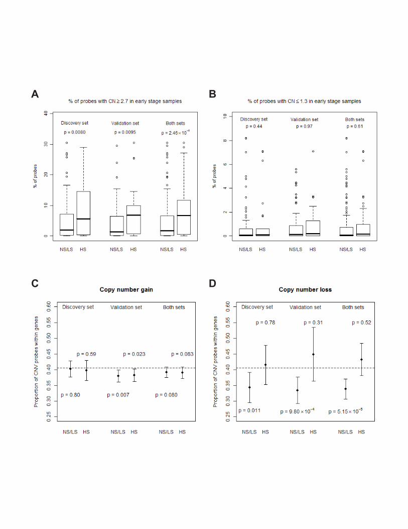
There is a significant increase in total events of copy number gains among heavy smokers
(>60 pack-years) (p=0.0080, 0.0095, and 2.5×10⁻⁴ for discovery, validation and both sets,
respectively), but no difference in copy number losses (Figure 1A and 1B). No significant
difference was observed in age, clinical stage, histology and gender between heavy and
light/non-smokers.
For copy number losses, G/T ratios in light/non-smokers (≤60 pack-years) are
significantly lower than the null ratio (i.e., the ratio when CNAs occur at random with
respect to the gene location) (p=0.011, 9.80×10⁻⁴, and 5.15×10⁻⁵ for discovery, validation
and both sets, respectively) but heavy smokers (>60 pack-years) show no difference
(p=0.78, 0.31 and 0.52, respectively) (Figure 1C and 1D). These results suggest that copy
number losses tend to occur away from genes but such tendency disappears in heavy
smokers. In contrast, there is no consistent pattern for copy number gains. Heavy smokers
seem to have more genes with copy number changes, especially in gains. (SI Appendix
(Figure S2))
Cigarette smoking and copy number pattern by chromosome/arm
The chromosome/arm-specific analyses suggest the majority responsible for the genome-
wide difference comes from chromosomes 8q (p =1.19×10⁻⁵ for total events of CN gains
between light and heavy smokers) and 12q (p =2.1×10⁻⁴) (Figure 2) as well as many
others (chromosomes 1, 3, 7, 10, 11, 16 and 17) (SI Appendix (Figure S3)). Similar

6
results were observed when genomic location was taken into account, especially in 8q
and 12q. (SI Appendix (Figure S4A)) The dose-response relationships between
continuous copy numbers and smoking pack-years are also significant in 8q (p=0.015)
and 12q (p=0.0025). (SI Appendix (Figure S4B)) These two regions are also found the
most signals in focal copy number analyses, as will be shown in the following.
Cigarette smoking and focal copy numbers
As stated in Materials and Methods, we performed single- and multiple-marker analyses
to investigate the association of cigarette smoking and focal copy numbers. In the moving
window 10-marker analyses, we selected the top 50 sets with smallest p values in the
discovery set and tested the 50 sets using the validation set (p<0.05). Using such criteria,
we identified one 10-marker set in 12q23 with p values of 9.69×10⁻¹⁰, which reached the
genome-wide significance. (Figure 3A) The region harbors a gene, IGF1 (insulin-like
growth factor 1) that plays an important role in tumorigenesis. (Figure 3B) In the single-
marker analyses, the most significant signals are also in the same region of 12q23: two
loci are in the intron between the last two exons of IGF1 and two loci are located
downstream of IGF1. (SI Appendix (Figure S5A and S5B, and Table S6)) The p value of
the 10-marker set identified in the 10-marker analyses and the corresponding p values and
R2 from the single-marker analyses were shown in Table 2. Compared with the single-
marker analyses, statistical power was gained from the 10-marker analyses by borrowing
information in the neighboring markers, accounting for correlation among the CNVs in
the marker set, reducing degrees of freedom of the test, and reducing the total number of
tests across the genome.

7
The dose-response relationship of copy number and smoking pack-years for the
four loci in 12q23.2 is shown in SI Appendix (Figure S5C-F), indicating a J-shape curve.
That is, beyond a certain threshold, the higher smoking pack-years, the more departure
from the neutral copy number. Notably, the threshold, about 60 pack-years, is consistent
with the cut-off used in the above analyses of whole-genome CNAs pattern.
In addition to 12q23, 3q24 and 8q24 are two additional regions that are potentially
associated with the pack-years of cigarette smoking from single-marker analyses. (SI
Appendix (Figures S6 and S7)) We also performed the analyses in the dichotomous
version, detail of which can be found in SI Appendix. (Table S7 and Figure S8)
DNA double-strand break assay
To investigate further the results of our statistical analyses, we determined whether
cigarette smoke could induce DNA double-stand breaks in cultured cells. To mimic
longer and heavier cigarette smoking conditions, we treated human nontumorigenic
bronchial epithelial cell HBEC 3KT with 0.04 and 0.4 μg/ml CSC for 24 hours. Under
these conditions, the survival rates are 96.9% and 95.7%, respectively, indicating the dose
of CSC and the length of treatment used in this study are not toxic to the cells (Figure
4A). To minimize background DNA double-strand breakage, we treated cells with CSC
right after the growth had reached confluence. Under these conditions, ~5% of non-CSC
treated cells still display double stand breaks (Figure 4C). When treated with 0.04 μg/ml
CSC for 24 hours, the percentage of cells with double strand breaks increased to 15%.
This percentage doubled with the application of more concentrated 0.4 μg/ml CSC
(Figure 4C). We also treated the cells with 0.4 μg/ml CSC for 2 hours, and observed a

8
similar DNA double-strand break ratio as that of the non-CSC treated control cells,
suggesting DNA double-strand break occurring after a longer time of CSC treatment.
To determine the effects of CSC on induction of cellular apoptosis which
indirectly contributes to DNA double-strand breaks, the same set of cells (as used in
Figure 4B and 4C) were lysed for apoptotic-specific Caspase-3/7 activity. As shown in
Figure 4D, there is a basal level of Caspase-3/7 activity in non-CSC treatment cells. Upon
CSC treatment, the value of relative fluorescence unit (RFU) increased in a dose-
dependent matter. However, the extent to which the RFU value increased in response to
CSC treatment was much less than the corresponding increase in DNA double strand
breaks in Figure 4C. Collectively, these results indicate that higher CSC leads to genome
instability in bronchial epithelial cells. As such, theses data provide biological evidence
to bridge the associations between CNAs and smoking observed in the above human data.
DISCUSSION
We show that heavy smokers (>60 pack-years) have more copy number gains than
light/non-smokers but not copy number losses and that light/non-smokers (≤60 pack-
years) have copy number losses away from the gene location, in contrast to heavy
smokers. The discrepancy between gains and losses suggests that different mechanisms
may exist for the genome impact of cigarette smoking. For gains, smoking executes its
oncogenic effect by increasing the event of copy number changes. For losses, in contrast,
smoking does not increase CNAs events but increase the proportion of genes being
affected. Because losing a fragment of DNA is less favorable than gaining one(13), two
separate mechanisms may be developed to hit the genes responsible for tumorigenesis.

9
The phenomenon may be a consequence of selection during cancer development and cell
proliferation. Because different cells possess different CNAs, selection by a nutrient-
limited environment makes those clones that can grow without regulatory control become
dominant.
For heavy smokers, there were more copy number gains compared to non- or light
smokers and no tendency for copy number losses to occur away from the gene location.
We have also found that genes with gains are more likely to be oncogenes or to be
involved in pathways that are associated with tumor growth, which suggests that lung
cancer cells in heavy smokers tend to acquire the growth advantage via copy number
gains.(14) As a result, copy number losses within genes have less unfavorable impact on
such cells since it is compensated by the fact that they can grow without regulation. This
explains our observation that the proportion of losses within gene among heavy smokers
is not different from that at random.
Previous studies have shown that copy number alterations are more frequent in
smokers than in non-smokers,(11, 12) consistent with our findings based on pack-years.
Copy number-based genomic signature has also been identified to discriminate current
smokers and never smokers(12), which, however, does not include IGF1. Smoking status
may not necessarily reflect the same oncogenic feature as pack-years of smoking, a
measure of cumulative exposure. Furthermore, the large sample size and discovery-
validation process in this study increase robustness of the findings.
Smoking causes lung cancer through numerous carcinogens derived from
cigarette combustion. There are two parts of the carcinogenic effect: early damage of
oxidative stress by reactive oxygen species and late damage by DNA adduct and DNA

10
mutation.(6) Both kinds of damage can serve as initiators of copy number changes,
especially oxidative stress. It has recently been proposed that cellular stress coming from
environmental agents can induce copy number changes, which seems to be a common
mechanism in bacteria, yeast and humans.(10)
The most significant region on 12q23 is at the junction of the last two exons and
the downstream of IGF1. The two loci within IGF1 are located in the intron between the
last two exons of IGF1. The protein product of the aberrant genomic DNA can exert its
undue influence on the cellular physiology. On the other hand, if the key player is the
downstream rather than the coding region of IGF1, it is still possible that IGF1 function
is affected because the downstream fragment can serve as a regulatory element of IGF1
transcription. That is, the alterations of the regulatory element can lead to the abnormal
gene expression of IGF1.
IGF1/IGF1R signaling pathway can induce many effects, including cell
proliferation, differentiation, transformation and inhibition of apoptosis.(15) Because of
the overlap with downstream signaling pathways of epidermal growth factor receptor
(EGFR) signaling, IGF1/IGF1R signaling may modulate the EGFR pathway, a critical
pathway in lung tumorigenesis(16), and it may explain, in part, clinical resistances to
EGFR inhibitors(17).
Several studies have provided the links among smoking, IGF1 and cancer. For
example, it has been reported that smoking may affect IGF1 serum level and its
signaling.(18, 19) On the other hand, IGF1 and the risk of developing cancer have also
been extensively studied in lung cancer(20-23), breast cancer(24), prostate cancer(25)
and colorectal cancer(26). Our analysis supports the hypothesis that smoking can act

11
through increasing the copy number of IGF1 to induce its over-expression and
subsequent oncogenesis.
MATERIALS AND METHODS
Study population, specimens and data collection
A series of 264 snap-frozen tumor samples from NSCLC patients with complete
information on cigarette smoking was collected during surgery or biopsy from the
Massachusetts General Hospital (MGH), Boston, MA, and the National Institute of
Occupational Health, Oslo, Norway. An additional 50 paired specimens of non-neoplastic
lung parenchyma and 63 paired blood samples were included as the reference group for
copy number estimation. Demographic and smoking information was collected by a
trained research assistant using a modified standardized American Thoracic Society
respiratory questionnaire.(27) A similar approach was used for the Norwegian cohort.(28)
Written informed consents were obtained from all patients. The study was approved by
the institutional review boards of MGH, the Harvard School of Public Health, and the
Norwegian Data Inspectorate, and Local Regional Committee for Medical Research
Ethics.
DNA quality, histopathology and genechip
DNA was extracted from tumor and non-neoplastic lung parenchyma after manual
microdissection from 5-μm thick histopathologic sections. Each specimen was evaluated
for amount and quality of tumor cells. Tumors were reviewed and classified using the
WHO criteria. Specimens with lower than 70% tumor cellularity, inadequate DNA

12
concentration, or not intact genomic DNA were not included for chip hybridization. The
platform of genechip is Affymetrix 250K Nsp GeneChip®.
Data preprocessing
Copy numbers were obtained with dChip software by invariant set normalization and
median smoothing with the window of 11 loci.(29) Only 256,554 probes on somatic
chromosomes were analyzed. We further classified the continuous inferred copy number
into a discrete variable of CNAs: copy number gains defined as copy numbers ≥2.7 and
copy number losses defined as copy numbers ≤1.3, to detect copy number ≥3 and ≤1 by
tolerating 30% normal tissue contamination. The probes were mapped to the RefSeq
genes with 2 kb extension both upstream and downstream using the UCSC Genome
Browser. Among the 256,554 probes on somatic chromosomes, 104,256 (40.64%) were
mapped to 11,700 genes.
Statistical analysis
Only early stage tumors were analyzed here because we have found that late stage tumors
have more CNAs. The number of pack-years is defined as the packs of cigarette smoked
per day multiplied by the years of smoking. 60 pack-years of cigarette smoking was
chosen as the cut-off for heavy and light/non-smokers according to the observation of
total CNAs events by the interval of 10 pack-years in both discovery and validation sets
(SI Appendix (Figure S9)). Using the cut-off, we had 203 light/non-smokers and 61
heavy smokers. We developed three methods to test the genome-wide or
chromosome/arm-specific copy number patterns between heavy and light/non-smokers
and one method to test the
association of the chromosome/arm-specific or focal CNs and smoking pack-years.

13
First, we calculated the total events of copy number gains and losses and
compared them between the two smoking groups by the two-sided t test, which provides
a convenient summary index but collapses CNAs information over the genomic locations.
The second method is to apply two-sample tests for continuous copy number by
calculating the standardized difference of two average copy numbers for each locus as:
( ) 221121 nvnvmmc iiiii +−= where mji and vji is the estimated mean and variance,
respectively, of copy number for group j at locus i, and nj is the sample size in group j.
We summed up ci 2 over i across the loci in the arm of a chromosome to calculate the
observed total standardized squared difference (Cobserved). By permuting the two groups
and carrying out the above procedure for 10,000 times, we obtained a non-parametric null
distribution (Cnull). Then p values were obtained by comparing Cobserved and Cnull. This
permutation procedure provides a valid global test for the overall difference by
accounting for multiple comparisons and correlation of CNAs between different loci.
The third is a similar method extended to the discrete variable of CNAs (CN≥2.7
or not; CN≤1.3 or not) as mentioned above, but with advantage of testing gains and losses
separately. We applied two-sample tests for binomial data by calculating the standardized
difference of two proportions for each locus as:
( ) ( ) ( ) 22211121 11 nppnppppd iiiiiii −+−−= where pji is the estimated proportion
(stabilized by adding 0.5 in the numerator) of CN gains (or losses) for group j at locus i
and nj is the sample size in group j. We summed up di 2 over i across the arm of a
chromosome to calculate the observed total standardized squared difference (Dobserved).
Non-parametric null distribution (Dnull) was approximated by 10,000 permutations, and p
values were obtained by comparing Dobserved and Dnull.

14
The above three methods require the smoking exposure to be dichotomized. To
fully capture the continuous dose-response relationship between copy numbers and pack-
years of cigarette smoking, we also developed a test to summarize such association in a
chromosome/arm-specific fashion. We obtained the test statistics, =i iobserved fF where
if is the F statistics of regressing continuous copy numbers on the smoking pack-years
(square-root transformed) up to the quadratic term at locus i. Again, the non-parametric
null distribution (Fnull) was generated by 10,000 permutations and p values were obtained
as the tail probability of Fobserved in Fnull.
The proposed test is equivalent to the powerful score test for testing the variance
of coefficients in a multivariate regression by assuming regression coefficients have an
arbitrary distribution with mean 0 and variance τ (30), in which copy numbers of a region
or chromosome (as a vector) are regressed on smoking pack-years. The null hypothesis of
our proposed test is that all the coefficients relating pack-years to copy numbers are zero,
or equivalently, copy numbers at all loci have no association with smoking pack-years,
which is equivalent to H0: τ=0. The alternative hypothesis would be that copy numbers at
some loci have association with smoking pack-years. This variance component test is a
powerful test by borrowing information in multiple markers and effectively accounting
for correlation among the CNVs in a marker set, and reducing the degrees of freedom of
the test.
We used another method to investigate gene selection of CNAs between heavy
and light/non-smokers. Both the total probes (T) in which CNAs were detected and the
probes locating within genes (G) in which CNAs were detected were calculated for each
individual. We proposed a ratio of G vs. T (termed as G/T ratio) to estimate the selection

15
of CNAs with respect to the gene location. Under the null hypothesis that CNAs occur
randomly relative to where genes locate, we would expect the null ratio of 40.64%
(104,256/256,554), where 104,256 is the number of probes located within genes on the
chip. By comparing the G/T ratios to the null ratio, 40.64%, with two-sided t test, we
were able to test whether CNAs occur preferentially away from genes.
To investigate the association of focal copy numbers and smoking pack-years, we
analyzed copy number >2 and ≤2 separately. Two outliers (>2.5 standard deviations) of
smoking pack-years were excluded to eliminate the potential spurious result driven by
them. Square root was taken for smoking pack-years to transform a right skewed
distribution into an approximately normal distribution.(31) Both multiple-marker and
single-marker analyses were performed. In the multiple-marker analyses, we grouped the
consecutive ten SNPs (markers) as a set and calculated Fobserved and Fnull by the methods
mentioned above to obtain the p values for each set of markers. For those with Fobserved
much greater than Fnull (from 10,000 permutations), the null distributions were obtained
by the Satterthwaite approximation(32), in which the first two moments of scaled χ2
distribution were matched with those of Fnull. In total, we performed 25,655 hypothesis
tests for copy number >2 and ≤2 separately. Such multiple-marker analyses had better
statistical power than the single-marker analyses when the markers were correlated,
which is the case in the copy number data.
For single-marker analyses, 256,554 regressions for both copy number >2 and ≤2
were performed in the discovery set with continuous copy number at each locus as a
dependent variable and square root of smoking pack-years and its quadratic term as
independent covariates. For the validated candidates (p<0.05 in the validation set), pooled

16
results were generated with linear regressions (with up to quadratic term of square root of
smoking pack-years), spline regressions (with spline of square root of smoking pack-
years) and locally weighted scatter plot smoothing (LOWESS). Adjusted linear and
spline regressions were performed with adjustment of age at diagnosis, gender, two
cohorts, clinical stage and histology.
DNA double-strand break assay
For the cytotoxicity analysis of cigarette smoke condensate, a human nontumorigenic
bronchial epithelial cell line HBEC 3KT was cultured in 12-well plates to confluence and
then treated with indicated concentration of cigarette smoke condensate (CSC) for 24
hours. Viable cells were monitored by MTT assay using the CellTiter 96 AQueous One
Solution Cell Proliferation Assay kit (Promega, Madison, WI). All assays were
performed in triplicate. For the neutral comet assay, HBEC 3KT was cultured in 100-mm
plates to confluence and then treated with indicated concentration of CSC for 24 hours.
Cells having DNA double-stand break were analyzed by Neutral comet assay using
CometAssay kit (TREVIGEN, Gaithersburg, MD). About 600~800 cells were viewed per
treatment. For the apoptosis analysis, HBEC 3KT was cultured in 100-mm plates to
confluence and then treated with indicated concentration of CSC for 24 hours. The status
of cellular apoptosis was determined using SendoLyteTM Homogeneous Rh110 Caspase-
3/7 Assay kit (ANASPEC, Fremont, CA). All apoptosis assays were performed in
triplicate.

17
ACKNOWLEDGEMENTS
We are indebted to the participants of Molecular and Genetic Analysis of Lung Cancer
Study; to Dr. Cheng Li of HSPH for his advice on data preprocessing; to the HSPH Lung
Cancer Study Group: Dr. Kofi Asomaning, Dr. Eugene Mark, Dr. Matthew Kulke, Dr.
Wei Zhou, Dr. Geoffrey Liu, Marcia Chertok, Andrea Shafer, Lauren Cassidy, Maureen
Convery, Salvatore Mucci; to Drs. Panos Fidias and Bruce A. Chabner and the physicians
and surgeons of the Massachusetts General Hospital Cancer Center; and to Dr. Lodve
Stangeland of Haukeland University Hospital, Norway for recruiting patients. This study
is supported by US National Institutes of Health Grants No. CA076404 and CA134294
(Y.-T. H. and X.L.), CA092824 (D.C.C.), CA074386 (D.C.C.) and CA090578 (D.C.C.),
and Norwegian Cancer Society (A.H.).

18
REFERENCES
1. Jemal A, Siegel R, Ward E, Hao Y, Xu J, et al. (2009) Cancer statistics, 2009 CA Cancer J Clin 59, 225-249.
2. Bach PB (2009) Smoking as a factor in causing lung cancer JAMA 301, 539-541. 3. Wynder EL & Graham EA (1950) Tobacco smoking as a possible etiologic factor
in bronchiogenic carcinoma; a study of 684 proved cases J Am Med Assoc 143, 329-336.
4. Doll R & Hill AB (1954) The mortality of doctors in relation to their smoking habits; a preliminary report Br Med J 1, 1451-1455.
5. Dubey S & Powell CA (2009) Update in lung cancer 2008 Am J Respir Crit Care Med 179, 860-868.
6. Alavanja MC (2002) Biologic damage resulting from exposure to tobacco smoke and from radon: implication for preventive interventions Oncogene 21, 7365-7375.
7. Kim TM, Yim SH, Lee JS, Kwon MS, Ryu JW, et al. (2005) Genome-wide screening of genomic alterations and their clinicopathologic implications in non-small cell lung cancers Clin Cancer Res 11, 8235-8242.
8. Weir BA, Woo MS, Getz G, Perner S, Ding L, et al. (2007) Characterizing the cancer genome in lung adenocarcinoma Nature 450, 893-898.
9. van Gent DC & van der Burg M (2007) Non-homologous end-joining, a sticky affair Oncogene 26, 7731-7740.
10. Hastings PJ, Lupski JR, Rosenberg SM, & Ira G (2009) Mechanisms of change in gene copy number Nat Rev Genet 10, 551-564.
11. Yen CC, Liang SC, Jong YJ, Chen YJ, Lin CH, et al. (2007) Chromosomal aberrations of malignant pleural effusions of lung adenocarcinoma: different cytogenetic changes are correlated with genders and smoking habits Lung Cancer 57, 292-301.
12. Massion PP, Zou Y, Chen H, Jiang A, Coulson P, et al. (2008) Smoking-related genomic signatures in non-small cell lung cancer Am J Respir Crit Care Med 178, 1164-1172.
13. Emerson JJ, Cardoso-Moreira M, Borevitz JO, & Long M (2008) Natural selection shapes genome-wide patterns of copy-number polymorphism in Drosophila melanogaster Science 320, 1629-1631.
14. Huang Y-T, Lin X, Chirieac LR, McGovern R, Wain JC, et al. (2011) Impact of Disease Development, Genomic Location and Biological Function on Copy Number Alterations of Non-Small Cell Lung Cancer PLoS One (in press).
15. Furstenberger G & Senn HJ (2002) Insulin-like growth factors and cancer Lancet Oncol 3, 298-302.
16. Herbst RS, Heymach JV, & Lippman SM (2008) Lung cancer N Engl J Med 359, 1367-1380.
17. Morgillo F, Kim WY, Kim ES, Ciardiello F, Hong WK, et al. (2007) Implication of the insulin-like growth factor-IR pathway in the resistance of non-small cell lung cancer cells to treatment with gefitinib Clin Cancer Res 13, 2795-2803.
18. Kaklamani VG, Linos A, Kaklamani E, Markaki I, & Mantzoros C (1999) Age, sex, and smoking are predictors of circulating insulin-like growth factor 1 and insulin-like growth factor-binding protein 3 J Clin Oncol 17, 813-817.

19
19. Tannheimer SL, Ethier SP, Caldwell KK, & Burchiel SW (1998) Benzo[a]pyrene- and TCDD-induced alterations in tyrosine phosphorylation and insulin-like growth factor signaling pathways in the MCF-10A human mammary epithelial cell line Carcinogenesis 19, 1291-1297.
20. Yu H, Spitz MR, Mistry J, Gu J, Hong WK, et al. (1999) Plasma levels of insulin-like growth factor-I and lung cancer risk: a case-control analysis J Natl Cancer Inst 91, 151-156.
21. Wu X, Yu H, Amos CI, Hong WK, & Spitz MR (2000) Joint effect of insulin-like growth factors and mutagen sensitivity in lung cancer risk J Natl Cancer Inst 92, 737-743.
22. Lukanova A, Toniolo P, Akhmedkhanov A, Biessy C, Haley NJ, et al. (2001) A prospective study of insulin-like growth factor-I, IGF-binding proteins-1, -2 and -3 and lung cancer risk in women Int J Cancer 92, 888-892.
23. Spitz MR, Barnett MJ, Goodman GE, Thornquist MD, Wu X, et al. (2002) Serum insulin-like growth factor (IGF) and IGF-binding protein levels and risk of lung cancer: a case-control study nested in the beta-Carotene and Retinol Efficacy Trial Cohort Cancer Epidemiol Biomarkers Prev 11, 1413-1418.
24. Hankinson SE, Willett WC, Colditz GA, Hunter DJ, Michaud DS, et al. (1998) Circulating concentrations of insulin-like growth factor-I and risk of breast cancer Lancet 351, 1393-1396.
25. Chan JM, Stampfer MJ, Giovannucci E, Gann PH, Ma J, et al. (1998) Plasma insulin-like growth factor-I and prostate cancer risk: a prospective study Science 279, 563-566.
26. Ma J, Pollak MN, Giovannucci E, Chan JM, Tao Y, et al. (1999) Prospective study of colorectal cancer risk in men and plasma levels of insulin-like growth factor (IGF)-I and IGF-binding protein-3 J Natl Cancer Inst 91, 620-625.
27. Zhou W, Heist RS, Liu G, Asomaning K, Miller DP, et al. (2006) Second hand smoke exposure and survival in early-stage non-small-cell lung cancer patients Clin Cancer Res 12, 7187-7193.
28. Zienolddiny S, Campa D, Lind H, Ryberg D, Skaug V, et al. (2008) A comprehensive analysis of phase I and phase II metabolism gene polymorphisms and risk of non-small cell lung cancer in smokers Carcinogenesis 29, 1164-1169.
29. Zhao X, Li C, Paez JG, Chin K, Janne PA, et al. (2004) An integrated view of copy number and allelic alterations in the cancer genome using single nucleotide polymorphism arrays Cancer Res 64, 3060-3071.
30. Liu D, Lin X, & Ghosh D (2007) Semiparametric regression of multidimensional genetic pathway data: least-squares kernel machines and linear mixed models Biometrics 63, 1079-1088.
31. Zhou W, Liu G, Park S, Wang Z, Wain JC, et al. (2005) Gene-smoking interaction associations for the ERCC1 polymorphisms in the risk of lung cancer Cancer Epidemiol Biomarkers Prev 14, 491-496.
32. Satterthwaite FE (1946) An Approximate Distribution of Estimates of Variance Components Biometrics Bulletin 2, 110-114.
33. Kwee L, Liu D, Lin X, Ghosh D, and Epstein M. (2008) A powerful and flexible multilocus association test for quantitative traits Am J of Human Genetics, 82, 386-397.

20
FIGURE LEGENDS
Figure 1. Association of cigarette smoking and whole-genome copy numbers.
A and B, Among the 256,554 total probes, the proportion (%) with CNAs (A, gains; B,
losses) events by pack-years of cigarette smoking (NS/LS: non-/light smokers, HS: heavy
smokers). C and D, Mean and its 95% confidence interval of G/T ratios in the heavy
smokers (HS) and non-/light smokers (NS/LS) for copy number gains (C) and losses (D);
and the dashed lines represent the null G/T ratio on the chip (104,256/256,554=40.64%).
P values were used to test the indicated indices between HS and NS/LS with methods
described in Methods.

21
Figure 2. Association of cigarette smoking and chromosome/arm-specific copy numbers.
A, the p values are from analyses testing the association of copy number-gain events with
the smoking group (heavy vs. light-/non-smokers). B, the p values are from analyses
testing the association of copy numbers (>2) with pack-years of cigarette smoking. The
dashed line indicates p=0.05.

22
Figure 3. Association of cigarette smoking and 25,655 moving window10-marker focal
copy numbers.
A. A plot of -log10P of the association between smoking pack-years and 10-marker set
focal copy number, which is analyzed for copy number >2 (upper half) and ≤2 (lower
half), separately. B, P values of focal copy number analyses in 12q23. The black dots and
line indicate p values from 10-marker analyses, and the superimposed gray dots and line
indicate the corresponding ones from single-marker analyses.

23
Figure 4. Effects of cigarette smoke condensate (CSC) treatment on HBEC 3KT DNA
double-stand breaks and apoptosis. A, Cytotoxic effect of CSC on HBEC3KT survival.
HBEC3KT cells were cultured in 12 well plates to confluence and then incubated with 0,
0.04, 0.4, 0.8, 4, and 40 μg/mL CSC for 24 hours. Viable cells were monitored with MTT
assay. Live cells treated with 0 μg/mL CSC were defined as 100%. Percentage of live
cells verses CSC concentration was plotted. B and C, CSC treatment induces DNA
single/double-stand breaks in HBEC 3KT cells. HBEC 3KT cells were culture to
confluence in 100-mm plates and then treated with 0, 0.04, and 0.4 μg/mL CSC for 24
hours. Cells were harvested by trypsinization and DNA single/double-stand breaks were
analyzed by neutral comet assay. A representative photo with undamaged-DNA (bright
dot) and DNA with single/double-strand breaks (bright dot with an elongated tail) was
shown (B). About 600~800 cells per treatment were viewed, and percentage of DNA
single/double-strand breaks verses CSC dose was plotted (C). D, CSC treatment induces
apoptosis in HBEC 3KT cells. The same set of cells used in B and C was also analyzed
for caspase-3/7 activity. Columns are mean value of relative fluorescence unit (RFU). A
larger RFU value represents a higher caspase-3/7 activity and thus a stronger apoptotic
response. Standard deviations are provided in C and D.

24
TABLE LEGENDS
Table 1. Characteristics of study populations
Table 2. Summary of the ten candidate loci at 12q23 from both 10-marker and single-
marker analyses.

1
Table 1. Characteristics of study populations
Discovery set Validation set P value*
Sample size 134 130
Male (%) 65.67 56.92 0.18
Age,
Mean ± standard deviation 67.27 ± 8.17 67.59 ± 8.39 0.75
Cigarette smoking pack-years,
Median ± interquartile range 34.25 ± 39.64 38 ± 35.93 0.28
Clinical stage 0.43
Stage 1 (%) 77.27 70.00
Stage 2 (%) 15.15 19.23
Stage 3 or 4 (%) 7.58 10.77
Cigarette smoking status 0.23
Never smokers (%) 7.46 6.15
Ex-smokers (%) 43.28 53.85
Current smokers (%) 49.25 40.00
Adenocarcinoma (%) 67.91 64.62 0.66
*P values were calculated with X2 test for percentage of male (1 degree of freedom, d.f.), adenocarcinoma,
patentis from MGH (1 d.f.), clinical stage (2 d.f.) and cigarette smoking status (2 d.f.); with t test for age;
and with Wilcoxon test for cigarette smoking pack-years.

2

1
Table 2. Summary of the ten candidate loci at 12q23 from both 10-marker and single-marker analyses.
Affy ID dbSNP
Cyto-
band
Position
(Mb) Gene
Focal copy number-smoking association
10-marker analyses Single-marker analyses
p value,
discovery set
p value,
validation set
p value,
pooled
p value,
pooled R2
p value,
adjusted*
SNP_A-2002985 rs5011687 12q23 101.157 -
3.17×10⁻⁸ 0.0291 9.69×10⁻¹⁰
0.0152 0.065 0.0168
SNP_A-2125858 rs17439974 12q23 101.171 - 0.0239 0.060 0.0285
SNP_A-4222341 rs17032384 12q23 101.179 - 0.000110 0.126 0.000263
SNP_A-1899321 rs1520223 12q23 101.229 - 2.92×10⁻⁶ 0.175 8.07×10⁻⁶
SNP_A-4222344 rs4764695 12q23 101.260 - 4.55×10⁻⁶ 0.167 1.33×10⁻⁵
SNP_A-4228436 rs10860860 12q23 101.283 - 1.79×10⁻⁸ 0.223 9.78×10⁻⁸
SNP_A-2106083 rs2946831 12q23 101.289 - 1.29×10⁻⁸ 0.235 2.63×10⁻⁸
SNP_A-2255731 rs10745940 12q23 101.300 IGF1 3.26×10⁻⁶ 0.163 8.20×10⁻⁶
SNP_A-2092658 rs9308315 12q23 101.306 IGF1 2.10×10⁻⁷ 0.202 7.74×10⁻⁷
SNP_A-2271065 rs2072592 12q23 101.316 IGF1 6.17×10⁻⁷ 0.200 4.42×10⁻⁶
*P values of smoking pack-years were calculated from linear models with up to quadratic term of square root-transformed smoking pack-years, adjusting for age,
gender, clinical stage, and cell type.
A B
C D

A
B

A
B

A B
C D