ci~ 0() 65 MASTER - UNT Digital Library
83
') BECEIVE .Q .SY DTIE AUG 30 19 I I. DENATURATION OF ENZYMES BY THE DIRECT ACTION OF IONIZING RADIATION • II. ENZYMICALLY ACTIVE COMPONENTS OF THE CRYSTALLINE CHYMOTRYPSINS by Louis Edwin Henderson B.A., University of Omaha, 1956 A Thesis to the Faculty of the Graduate School of the University of Colorado in partial fulfillment of the requirements for'the Degree Doctor of Philosophy Department of Chemistry 1966 0() 65 MASTER
Transcript of ci~ 0() 65 MASTER - UNT Digital Library

')
~ BECEIVE.Q .SY DTIE AUG 30 19 I
I. DENATURATION OF ENZYMES BY THE DIRECT
ACTION OF IONIZING RADIATION •
II. ENZYMICALLY ACTIVE COMPONENTS OF
THE CRYSTALLINE CHYMOTRYPSINS
by
Louis Edwin Henderson
B.A., University of Omaha, 1956
A Thesis submi~ted to the Faculty of the Graduate
School of the University of Colorado in partial
fulfillment of the requirements for'the Degree
Doctor of Philosophy
Department of Chemistry
1966
ci~ 0() 65
MASTER

DISCLAIMER
This report was prepared as an account of work sponsored by an agency of the United States Government. Neither the United States Government nor any agency Thereof, nor any of their employees, makes any warranty, express or implied, or assumes any legal liability or responsibility for the accuracy, completeness, or usefulness of any information, apparatus, product, or process disclosed, or represents that its use would not infringe privately owned rights. Reference herein to any specific commercial product, process, or service by trade name, trademark, manufacturer, or otherwise does not necessarily constitute or imply its endorsement, recommendation, or favoring by the United States Government or any agency thereof. The views and opinions of authors expressed herein do not necessarily state or reflect those of the United States Government or any agency thereof.

DISCLAIMER
Portions of this document may be illegible in electronic image products. Images are produced from the best available original document.

\ /
This Thesis for the Ph. D. degree by
Louis Edwin Henderson
·i . ' ' \
has been approved for the
Department of
Chemistry
by
Date C X~ t· · · \~~ $"" ·
,·.--::·
(I
I , I

. \
~···-···~· ~· .... ·~~-~
Acknowledgment
Sincere thanks are extended to Dr. Bert Tolbert,· Dr. Pe~er
Albersheim, and Dr. Mancourt Downing for their guidance and
encouragement.
;
' •, ) .
' . '. ' \
i

Henderson, Louis Edwio (Ph.D. Chemistry)
I. Denaturation of Enzymes by the Direct Action of Ionizing Radiation
and II. Enzymically Active Components of the Crystalline
Chymotrypsins
Thesis Directed by Professor Bert M. Tolbert
Part I. The inactivation of alpha-chymotrypsin by the direct -
action of gamma radiation was shown to be an all-or-none process.
The enzymic specific activity and the operational active sites of the
enzyme were lost with the same D37
dose. No chemically altered but
enzymically active molecules produced by irradiation could be detected
by ion exchange chromatography or gel ·electrophoresis. Alpha
Chymotrypsin requires an average dose of 42.1 eV/molecule to inacti
vate the enzyme in the solid state.
The direct action of gamma radiation on alpha-chymotrypsin
· results in sufficient alterations of the ordered conformation of the
molecule to account for loss of enzymic activity. The A. determined . 0
by optical rototary dispersion deqreases from 236. 6 mJ.l for the native
protein to about 224 mJ.l for the radiation denatured protein. The
radiation denatured protein shows no "buried" chromophores which can
. be exposed by heating the protein through the heat induced transition
of alpha-chymotrypsin.
The "hard to exchange amide hydrogens" (HEAH) of irradiated
alpha-chymotrypsin are lost by a multi-hit ·process. About 1. 65.eV are
required to expose one HEAH. This suggests that about 19 HEAH are
exposed per hit molecule.
The protein "irradiated to 50% inactivation showed no change in
·the amino acid analysis or disulfide analysis. There was no evidence
for specific chemical changes produced by radiation in the electro-. phoretic ·patterns of the reduced and non-reduced !)rote in.
ii

The doses required to inactivate a molecule of chymotrypsin-:
ogen1 delta-chymotrypsin and papain are also reported. These doses
do not differ significantly from the dose required to inactivate alpha
chymotrypsin. ·
Part II. An ion exchange column is described which is capable
of resolving the crystalline chymotrypsins into their respective com
ponents. Each crystalline chymotrypsin contains contamination of the
other chymotrypsins. 3X crystallized alpha-chymotrypsin contains
about 1 o% beta or gamma-chymotrypsin.
''. ~isc" gel electrophoresis of flurodiisopropyl phosphate p32
labeled chymotrypsins in 8M urea reveals. a different electrophoretic
mobility for alpha 1 ·beta I and gamma-chymotrypsin. However 1 these
proteins all yield the same electrophoretic pattern following reduction
with thioethanol. This suggests that these proteins may differ only
in their disulfide arrangement.
This abstract of about 600 words is approved as to fonn and content. I recommend its publication.
!
\ \
Signed __ ~;;.___..,_· _'M:...;..c_:?\----:.-=~:...;..1 __ _
Instructor in charge of dissertation
'··.
·.-:-,_
iii
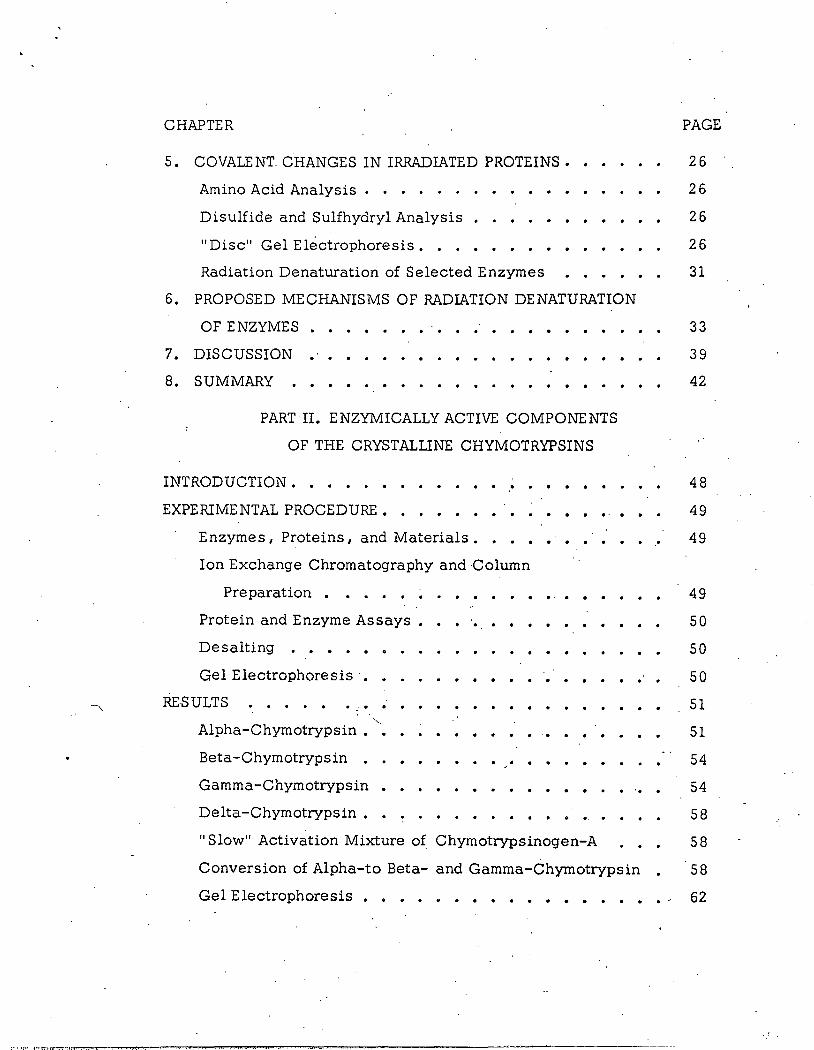
LIST OF FIGURES
FIGURE PAGE
PART I
1. Change in Specific Enzymic Activity of Irradiated
· Alpha-Chymotrypsin . . • . • . • . • • • 9
2. Loss of Active Sites of Irradiated Alpha-Chymotrypsin • 11
3-a. Chromatographic Elution Diagram of Enzymic Activity
from Irradiated and Native Alpha-Chymotrypsin • 13
3-b. Chromatographic Elution Diagram of Protein from
Irradiated and Native Alpha-Chymotrypsin
4. Change A of Irradiated Alpha-Chymotrypsin 0
5 OD 2 9 3 E . A t. . t . • vs nzym1c c 1v1 y . . • . • . . • • . . • •
6. Change in .6.E2 93
m!J. of Irradiated Alpha-Chymotrypsin
7. Change in HEAH of Irradiated Alpha-Chymotrypsin • • ·
8-a. Electrophoretic and Radioactive .Profile of Reduced
Alpha-Chymotrypsin . • • • • • • • .•
8-b. Electrophoretic and Radio~cti ve Profile of
Non-Reduced Alpha-Chymotrypsin
PART II
14
18
20
22
24
29
30
1. Chromatographic Elution Diagram of Alpha-Chymotrypsin 52
2 •. Chromatographic Elution Diagram of Component III from
3X crystallized Alpha-Chymotrypsin • • • ·• • • • 55
3. Chromatographic Elution Diagram of ~eta-Chymotrypsin.·· · 56
4. Chromatographic Elution Diagram of Gamma-
Chymotrypsin • • • • • • • . • • • • 57
5. Chromatographic Elution Diagram of Delta-Chymotrypsin 59
6. Chromatographic Elution Diagram of "Slow" Activation
Mixture of Chymotrypsinogen-A. 60

-------------------------------------------------------------------------------- .
FIGURE
7. Chromatographic Elution Diagram of Alpha
Chymotrypsin Stored for Two Weeks at
pH 7. 7 5 I 5° • . • • • . . • • • • •
8. Protein and Radioactivity Distributions in
PAGE
. . . . 61
·Electrophoretic Gels of DIP-32 Chymotrypsin • 63
9. Protein and Radioactivity Distributions in
Electrophoretic Gels of Thioethanol Reduced
DIP-32 Chymotrypsins • • • • • • • • • • 64
Expanded legends for Figures 1 through 9 7 0

UST OF TABLES
TABLES
PART I
I. .A of Irradiated Alpha-Chymotry-psin 0
II. Amino Acid Composition of Irradiated Alpha-
Chymotrypsin • • . • • • • • • • • •
III. .._ Sulfhydryl and Di~ulfide Groups of Irradiated '·
Alpha-Chymotrypt::in • • • • • • • • •
IV. The Average Energy Necessary to Inactivate Selected
Enzymes • . . . . .. . . . . . . . . . . PART II
I. Protein and Enzymic Composition of 3X Crystallized
Alpha-Chymotrypsin • . . . . ..
PAGE
16
27
28
32
53
CHAPTER
TABLE OF. CONTENTS
PART I. DENATURATION OF ENZYMES BY THE
DIRECT ACTION OF IONIZING RADIATION
PAGE
i·
i ACKNOWLEDGMENTS .•
ABSTRACT • . . . • . • • . . . . . . • • • • ii
1. INTRODUCTION
2. EXPERIMENTAL ••
Enzymes and Substrates
Reagents. . • • • •
Radiation Conditions
Enzyme Assay • • •
Active Site Titrations
. . . . .
. . . . . . . . . . .
. . . . . . . . . . . . . .
. . . . . . . . . . . . . . . . . . .
Ion-Exchange Chromatography
Hard-to-Exchange Amide Hydrogens •
. . . . . . .
. •. . . . . Heat Perturbation Spectrum • . . . . . . . . . . . .
1
3
3
3
3
3
4
4
5
5
6
6
7
7
Optical Rotatory Dis persiori
Amino Acid Analysis • • • •
Sulfhydryl and Disulfide Analysis
"Disc" Gel Electrophoresis. • .
. . . . . . . . . .
3. NATURE OF ENZYME,INACTIVATION BY IONIZING
RADIATION ••••
Enzymic Ac::ti vity
Titration of Active Sites
C <;>lumn Chromatography
. . . . .. . . ·.-:-. ... . . . .
4. CONFORMATIONAL INTEGRITY·, ENZYMIC ACTIVITY
AND IONIZING RADIATION • •
Optical Rotatory Dispersion . .
Ultraviolet light Absorption Effects
Hard-to-Exchange Amide Hydrogens
. . 8
8
10
• • 1 0
• 15
15
• • 19
• • 23
/
CHAPTER
5. CO VALE NT. CHANGES IN IRRADIATED PROTEINS •
Amino Acid Analysis . • • . . •
Disulfide and Sulfhydryl Analysis •
PAGE
26
26
26
"Disc" Gel Electrophoresis • • . • • • • • • • 2 6
Radiation Denaturation of Selected Enzymes 31
6. PROPOSED MECHANISMS OF RADIATION DENATURATION
OF ENZYMES . . . . . . . . 7. DISCUSSION
8. SUMMARY . . . . . PART II. ENZYMICALLY ACTIVE COMPONENTS
OF THE CRYSTALLINE CHYMOTRYPSINS
INTRODUCTION • • • • • • . . . EXPERIMENTAL PROCEDURE • . . . . . .
Enzymes 1 Proteins 1 and Materials . • . . . . . .•
Ion Exchange Chromatography and Column
Preparation • • • • • ~ •
Protein and Enzyme Assays •
Desalting . • • • • •
Gel Electrophoresis·. . . . . RESULTS
. . . . . . .
. . . . . . . .
. . . . . . . .
Alpha-Chymotrypsin •
Beta-Chymotrypsin •
Gamma -Chymotrypsin
Delta-Chymotrypsin • . . . . . . . . . . .
33
39
42
48
49
49
49
50
50
50
51
51
54
54
58
"Slow" Activation Mixture of Chymotrypsinogen-A 58
Conversion of Alpha-to Beta- and Gamma-Chymotrypsin • 58
Gel Electrophoresis eee•••••••e••••~ 62

r:x: r...:l 1:--i A
. .::t; ::c 0
ll)
l.O
z 0 1-1
tf.) tf.) p 0 tf.) 1
-1
Q
&: .::t; ~ ~
p tf.)
co l.O
tf.)
.r...:l 0 z ~
r...:l j:.J..,
~
·.-/
(

PART I
DENATURATION OF ENZYMES BY
THE DIRECT ACTION OF IONIZING RADIATION
1. INTRODUCTION
Solid state enzymes lose enzymic activity when subjected to
ionizing radiation. The quantitative aspects of the process indicate a
single ionization anywhere withfn the: .molecule inactivates the enzym~
(1). This concept has been used to estimate the molecular weight of
enzyme from radiation inactivation data (2 I 3 1 4) .·
The experimental evidence to support the single ionization ·
theor-Y is that many enzymes absorb about 30 to 40 eV of ioniz.i.ng energy
per molecule inactivated (5). In gaseous systems an average of 3 5 eV
of ionizing energy are required to create an ion pair (6). If these data
can be compared 1 it would appear that a single ionization anywhere
within the solid state enzyme results in enzyme inactivation. The model
suggests that enzyme inactivation by the direct action of ionizing . .
radiation is an all-,QE-~ process; the energy absorbing molecules are
-·' always inactivated and ·no altered but enzymically active molecules are
produced.
This nee<:l not be true 1 some ionizations outside the molecule may
inactivate the enzyme 1 while other ionizations 1 inside the molecule may _
not. To determine .the point I experimental evidence on irradiated samp'!es
shoulq show:
(A) All molecules with enzymic activity have not been ionized;
and
(B) All enzymically inactive molecules have been ionized.
·-· - ... ·-------......--.---..;,-...-...,. ___________ _._/ --

"'-----·~----1..:. ..
2
Since one cannot obs.erve ionizations in the solid state I it is necessary
to assume each ionization will result in some chemical or physical
change. Therefore 1 it must be shown that· all enzymically active
molecules have not changed physically or chemically I and all
enzymically inactive molecules have changed physically or chemically.
Many enzymes are inactivated by what appears to be a single
ionization process (5}. Evidently some general feature of enzymes
necessary for enzymic activity is altered by the absorbed energy. It is
generally suggested that the absorbed energy alters the conformation of
hit molecules (7). However 1 it has never been conclusively shown that
the conformation of hit molecules is sufficiently altered to account for
loss of biological activity.
The experimental work presented in this report is directed to
ward investigating aspects of the one ionization model. Alpha-chymo
trypsin is shown to be inactivated by. an all-or-none process and the
ordered structure of inactivated Juulecules is altered sufficiently to
account for loss of biological activity.
Alpha-chymotrypsin was s.elected as a model enzyme for this
study because previous _work by Pollard (3) indicated this enzyme is
inactivated by a single ionization process. The enzyme has a molecu
lar weight of 2413001 a known amino acid sequence and disulfide
arrangement (8) 1 a well-defined substrate (9 I 1 0) 1 and ·is available
commercially. Amino acid residues directly involved in the active site
are known and thei~ position in the sequence has been determined (11 1
•"
12} .. The enzyme is composed of three peptide chains held together by
disulfide bonds. Amino acids of the active site are located in two or
three chains. More detailed· information about the enzyme can be_
found in references cited.

2. EXPERIMENTAL
Enz),:mes and Substrates. Alpha-chymotrypsin 1 ·papain 1 trypsin
and chymotrypsinogen were obtained from Worthington Biochemical as
....... --· ···- -· --
the 3X crystallized enzymes and used without further purific.ation. Beta- 1
gamma- 1 and delta-chymotrypsins were obtained from Sigma Biochemical
as the 2X crystallized preparations and used without further purification.
N-acetyl-L-tyrosine ethyl ester (ATEE) I N-benzoyl-L-argenine ethyl
ester (BAEE) and N-trans~cinnamoyl imidazole (TCI) were obtained from 32
California Biochemical. Fluorodiisopropyl phosphate- P (FDIP-32) was
obtained from New England Nuclear Corp. Unlabeled FDIP was obtained
from Sigma Biochemical. I
Reagents. Deuterium oxide {99. 8%) and Bio-Rex 70 were obtained
from Bio-Rad Company. The Sephadex was obtained from Pharmacia Corp.
The urea I obtained from Sigma Biochemical was de-ionized and recrystal
lized immediately before use. Tris-hydroxymethylaminomethane (Tris)
was obtained from Sigma Biochemical. All other chemicals were reagent
grade preparations.
Radiation Conditions. All samples to be irradiated were sealed
under 10 micron vacuum in ampules made of 3 mm pyrex glass tubing.
The samples were weighed into the tubes 1 attached to a vacuum line 1
evacuated overnight I and sealed off. The samples were then irradiated
in the University of Colorado kilo-curie cs13 7
.source. The dose rate
was calibrated at 2. 65 x 1 o19 ev/g/hr with a Fricka dosimeter solution.
The temperature. in the source was constant at 24 o. The irradiated
samples were allowed to stand for at least 48 hours at -2 0 oc before
opening for analysis.
Enzyme Assay. Chymotryptic activity was determined by the
spectrophotometric method of Schwert and Takenaka (1 0) using ATEE as

----------··· ====~====-=·-===··:!,:' =~-~
the substrate. The assay solution was 1 x 1o-3M ATEE in 0. OS M Tris
HCl buffer at pH 7. 90; 0. 01 to 0. 001 mg of enzyme was added to the
test cell in about 100 .A of solution. The test cuvette was read at _time
zero and the time required to produce a 0. 050 decrease in absorbance
at 23 7 mJ-1 was recorded. The exact concentration of enzyme was
determined by weight and dilution techniques or by its 2 80 mJ-1 absorp- ·
tion (taking. E ~ ~m = 19 ~ 8} The change in the absorbance per minute
per mg of protein was taken as a measure of the specific enzyme
activity.
Papain activity was determined by the same procedure; only . -3
BAEE was used as the substrate. The substrate solution was 1 x 10 M - .
BAEE, 2 x 10⁻²
sodium citrate, pH 6. 00, 1 x 10⁻³M EDTA and 5--x -3
10 M cysteine.
Chymotrypsinogen was assayed as chymotrypsin following a
rapid tryptic activation by methods de scribed in ·the literature (13).
Active Site Titrati6ns. Chymotrypsin active site titrations were
performed by the method ·of. Schondaum (18). A solution of chymotrypsin
was allowed to react with TCI at pH 5. 00. The cinnamoylated derivative
was measured spectrophotometricaliy at 315 mJ-1. The concentration of
cinnamoylated enzyme was calculated using AE 31 5 = 1. 0 x 1 o4 •. This
value was determined on unirradiated 3X crystallized alpha-chymotrypsin
and agrees with the 1. 09 x 104
literature value.
Spectroph9tometdc measurements were made on a Cary Model
14 recording spectrophotometer. Recording was started 30 seconds after
adding the enzyme and extrapolated back to zero time to correct for ·
hydrolysis. ·Absorbance at extrapolated zero time was 'used to calculate
the precent remaining operationally active sites.
Ion-exchange Chromatography. Separation of the irradiated
samples of alpha-chymotrypsin by iqn exchange chromatography was
performed by the techniques· described in detail in Part II of this thesis.
/'.·

-,
--~--------·-··-·-- .. ·-····--·--·- ··- ·····-··-
5
Hard-to-Exchange Amide Hydrogen (HEAH). The HEAH were . .
determined by the method of Blout et al~ (25). The irradiated and
unirradiated protein was dissolved in 99. 8% D2
0 to make a 5% solution.
This solution was introduced into a o:os mm CaF 2
cell and the infrared
spectrum was taken between 5. 750 and 7.250 microns at such a rate as
to observe the 6. 400 micron region to 10 minutes after solution~
Cell blanks were run before each determination. Separate
samples of unirradiated protein were incubated in D2 0 for 24 hours at
room temperature and from 8 to 12 hours at 45 oc. These samples were
used to establish the base line for completely deuterated protein. The
amide I band at 6. 150 microns remained constant during exchange
reaction and was used as an internal standard to determine protein
concentrations. The amide II band at 6. 500 decreased with deuteration.
Blout determined that the ratio of ami.~e III was 0. 40 for the undeuterated am1 e
protein.
The amide II to amide I band ratio for the completely deuterated
alpha-chymotry:psin was 0.196. The HEAH were defined as all the
hydrogen which did not exchange in 1 0. 0 min. The percent remaining
HEAH were calculated from absorbance of the 6. 500 band after base line
and concentrations corrections were made taking the unirradiated
specific absorbance as 100%.
Heat Perturbation Spectrum. The change in the extinction co-
efficient at 2 93 m}l produced by heating the protein above its denatura-
tion temperature (about 55 "C) was determined by the following .procedure
(22). The irradiated or unirradiated sample's were dissolved in suf
ficient 0. OS M sodium acetate-acetic acid buffer pH 3. 52 (0. 1. M in
KCl) to bring the protein concentration to 1. 68 mg/ml. This solution
was placed in a 1·. 00 em quartz cell and read in a thermostated
Beckmann D. U. spectrophotometer. A pyrex glass cell filled with
buffer was used as a blank. The instrument was adjusted to read zero
at 293 mJl with the blank cell, the protein solution read about 0. 440 0. D.

- ---------~····-fl~··· .. • -------·--:·---- .... -----------""-'-~-------~---------- ---··-· ....................... ------
6
The temperature was ·regulated with a water bath. A thermometer was
fitted into the cell compartment· so that the cell compartment tempera
ture could be read directly. . The water bath was first cooled then
heated at 1 °/min. The 0. D. at 2 93 mJ-1. of the sample was determined
every 1 o fr.om 5 o to 6 5 o .
The change in absorbance from 30 o to the denaturation tempera
ture (about 55 oc) was taken as a measure of enzyme conformation and
reported as percent of the change in absorbance for the unirradiated
enzyme.
Optical Rotatory Dispersion. All measurements were made on a
Rudolph Model 2 OOA Oscillating Polarizer with a zirconium arc light
source. Measurements were made on 1. 0% protein solution in 0. OS
sodium acetate-ace.tic acid buffer pH 3. 52 (0. 1 M in KCl) at 2 5o.
Cells were 1. 6 mm bore x 5 em length with quartz end plates. Readings
were taken at nine wave lengths from 650 mJ-1. to 340 mJ). and the A. 0
calculntP.d by means of the Drudc equation
where [a ] is the specific rotation at a wave length A.., and a is a con
stant. "'A.0
is obtained from a plot of-1/[a]i\2
versus 1/A..2
.
Amino Acid Analysis. The amino acid analyses were performed
by the method of Moore (14). Five mg of protein was dissolved. in 5 ml
of a constant boiling HCl, frozen and sealed under vacuum. The sealed
tubes were heated at 11 0 o_ for 12 , 2 4 and 4 8 hours. Each determination
was run in duplicate. The hydrolyzates were lyophilized to dryness
and analyzed on a Beckmann automatic amino acid analyzer. The value
for serine and threonine was determined by extrapolating back to zero
time of hydrolysis. Tryptophan was determined separately by spectro
photometry (1 5).
'·
· ..

~-·----~-~-~--~.~-c .. -,_,~.-._,.._--------'-------_........,_ ____ ~-------
7
Sulfhydryl and Disulfide Analysis. Sulfhydryl and disulfide
analyses were performed by a modification of the metho~s of Boyer (16)
and Brown (1 7).
Approximately 3 mg of protein was wei~hed. accurately and
dissolved in 0. 5 ml of l. 0 M sodium borohydride in 8 M urea. After 3
hours at room temperature, the reaction was stopped with 0. 5 ml 3 M
acetic acid. The reduced protein was titrated spectrophotometrically
" with p-chloromercuribenzoate. (16) in the presence of 0. 05% sodium
dodecyl sulfate. Sulfhydryl groups were determined by omitting the
reduction step. This method consistently gave results for lysozyme
and chymotrypsin which were 2 0% too high. The source of this error
was undetermined.
Disc Gel Electrophoresis. Gel electrophoresis was performed.
by methods described in Part II of this thesis.
\
. ,·.

............ , ..... ;........;:..~-- .,.~~"'!tr._:;.J~~.;;._ ....... ~ .......... _~~---=---=.,...._..,.._..,-.,.(.,..J ---- -~-·:·-·-·- ······-·-· .. . .......... -... ······- -···-·1
3. NATURE OF ENZYME INACTIVATION
BY IONIZING RADIATION
tn·zymic· Activity. Samples of alpha-chymotrypsin were · '
weighed into tubes 1 sealed under vacuum and irradiated in a kilocurie 137 19 .
Cs source at 2. 65 x 10 eV/hr/g. Samples were assayed for
enzymic activity with N-acetyl-L-tyrosine ethyl ester (ATEE') as a
substrate by methods described in the experimental section (1 0). ·
Specific enzymic activities were based on the weight of the
sample and rate of ATEE hydrolysis. Percent remaining specific activi
ties were based on the specific activities of unirradiated controls.
These data are presented in Figure 1. The log percent r_emaining
specific activity is plotted versus dose in eV. The linear relationship
predicted by the one ionization theory is obtained (1). The inactivation f ll h l . , . l A ..:.. 2 . 2 0 3 · Wh - 2 . 2 0 3 process o ows t e re atwnsmp og A = D D: ere D
0 0 0 is a constant 1 A and A are the specific enzymic activities before and
0
after dose D. D is the average radiation energy required to inactivate · 0
a molecule. The D 3 7
value is the energy required to inactivate 6 3% of
the original enzyme and represents the amount of energy required to
inactivate all the molecules if repeat hits did not occur. The D 3 7
value
for alpha-chymotrypsin i~ 98 x 1 o19 eV/g which corresponds to 42. 1
eV/molecule. This value is consistent with the present data on the
energy required to create an ionization in organic solids.
In order to equate the number of inactivated molecules with loss
of enzymic activity it mus.t be assumed that all active molecules after
irradiation have the same turnover number.
It should be shown that the operationally active sites of the
irradiated protein have decreased by the same ~mount as enzymic
activity.
/

0')
' i.; ."I ,. ,, i ~ , .. \'
0
\ ·. ~ f-4 z
u H H > Cl)
ll H P-4 f-4 ;:....
u 0 ~ < f-4
0 I: 0
~ i H l· r-1 ~ 0 i: N I f,: (l) z
~ i: N ril Jl =' i! bD 0
H •.-1 H < ~ ~
Cl u H
ji 0 ~1 ril f-4 P-t
~ Cl)
I Cl
,, z ~
I H !
i ril H
I C) z I ~ l
I 0 I
oz
0£
/
Oi]
OS'
09 I ; i OL
@ 09
06
001
-· ' '

10
Titration of Active Sites. Operationally active sites of alpha
chymotrypsin can be titrated by the methods detailed in the experi
mental section (18). The method takes advantage of the fact that
catalysis by alpha-chymotrypsin occurs in several steps (19).
Enz-OH + RCOOR' ~ (Enz-OH · RCOOR') (1) (Enzyme) (Substrate) (Enzyme-Substrate Complex)
· (Enz-OH · RCOOR') ~ Enz-OOCR + HOR' (2) (Acylated Enzyme)
.E nz-OOCR + H2 0 Enz-OH + RCOOH (3)
Steps 1 and 2 are independent of pH between pH 3 and 8. ,Step
3 is dependent upon an un-ionized imidazole and is very slow below
pH 6. At pH 5 the reaction with N-!..@!}_§-cinnamoyl imidazole is
blocked at step 3. All molecules which undergo step 2 form a stable I
acylated enzyme. The cinnamoylated enzyme can be measured
spectrophotometrically and used to titrate the "operationally active
sites."
Figure 2 is a plot of the log percent remaining active sites vs
dose. The D3 7
is in good agreement with the D3 7
for the hydrolysis of
ATEE. It can be concluded that all enzymically active molecules after
irradiation have the same turn-over number.
Column Chromatography. The possible production of altered
molecules which retain enzymic integrity was investigated with cation
exchange chromatography. The procedure is.·the most sensit.ive method
available for purification of alpha-chymotrypsin. In the experiment,·
1 0 mg of irradiated protein was eluted from the column.
Protein wa~ detected by absorbance at 2 80 mj..J. and enzyinic
activity was measured by ATEE hydrolysis. Specific activity was
calculated assuming all protein to have a specific absorbance at 2 80
mj..l equivalent to alpha-chymotrypsin.
·'

0£1 0~1 011 001 06 08 0!.. 09 OS" 0~ 0£ oc: 01
z ·~ H J::r.. Cl)
·~ 0 p.. ;.,
~ ' ~ Cl) p:: s w .E-i II> .E-i 0 oc:
1-'• H
~ ::s
.CI) 1-'• ('lj ::s
~ 0 oq
(l) I !X> ~ H
~ ::l ·.E-i ()
bO 0 ·~ rt •r-l <X; 0£ 1-'• J::r.. <X; <
J::r.. ('0
0 Cl w rn
Cl) .E-i 1-'· Cl)
;:s 0~ rt
3 ~
Cl Q)
~ 'Bj.t\o6101X001=!.. '0 p:: ~ H OS' '1
oq @· 09 '1
I II> ,. s i 0!.. t
08
06
/ I .

12
Figures 3a and 3b are elution diagrams of 3X crystallized alpha
chymotrypsin and irradiated alpha-chymotrypsin. Figure 3a is an
enzymic activity elution diagram and Figure 3b .is a protein elution
diagram. No new enzymically active components are evident in the
irradiated protein. Most of the radiation-inactivated protein is eluted
in an unresolved slur before the enzymically active protein. The
specific enzymic activity of the major enzymically active component
in the irradiated sample is equal to the specific enzymic activity of
unirradiated alpha-chymotrypsin. All of the enzymic activity of the
irradiated protein was recovered from the column.
'"· ..

150
,......f
s C>
>,C> ._,....-t •.-I . :>C>
t~ 100 .:( ·;g u-....... ...... ::i s s >. ['.. N M ~ N JJ,:In
0 50 <l
I ' I . ,. l I
)
1\ Figure 3-a Irradiated to II
50% inactivation I 1
' I I I .•..•.... Native ' l
I I I I I
I I I
I I I
I I , I I r I
I I I I I I I
' ' ~· . . I
' I \
r \
I \ \
I \ I \.
I \ ~'"
I "~
100 200 300 400 500
ml of Elulate Enzymic Activity Elution Diagram
of Irradiated and Native Alpha-Chymotrypsin
600
....... w

.150
Q)
0 s:: l'(j
,..Q .100 1-< 0 C/l
..0 .::1! ::i. 8
0 co N
• OS
I /
(\
Irradiated to 50% I I Figure 3-b Inactivation t I
I I
.••...•.. l'Jative I I
... •?"
100
, I
I I I , I
r I
f I
f I ~ I r
I I I
I I I I I I I
I I
:I I I
~ I I I I I
300
ml of Elulate / Protein Elution Diagram of Irradiated and
Native Alpha-Chymotrypsin
./

'
4. CONFOR1V1ATIONAL INTEGRITY, ENZYMIC ACTIVITY
AND IONIZING RADIATION
Several experimental parameters are currently used to study
protein conformation. They include optical rotatory dispersions,
spectral shifts of ultra-violet light absorption, and the rate of hydrogen
exchange at amide bonds of the protein. Theories and principles of the
techniques are discussed in the literature and only a brief treatment
of each technique will be presented here. These techniques were used
to compare radiation inactivated alpha-chymotrypsin with a native
enzyme.
Optical Rotatory Dispersion. Proteins exhibit a dispersion of
optical rotation as a function of the wave length of light used (2 0, 21).
At shorter wave lengths specific rotation decreases through a minimum,
then increases. The wave length of the minimum has been termed the
A of the protein and may be related to the alpha-helix of the protein. 0
The A of alpha-chymotrypsin irradiated to various dose levels was 0 .
determined, these,values are given in Table I. The concentration of
the unirradiated protein was determined by absorbance at 2 80 mJ,l.
Radiation produces a chromophore which absorbs slightly in the 2 80
mJ,l region. Therefore, the concentration of irradiated samples was
determined by weight of the protein, this introduced a random error.
It is possible to obtain information about the effect of radiation
denaturation on the conformation of enzymes by assuming the D37
value for change in conformation to be equal to the D 3 7
for inactivation
of the molecule. This assumption will permit a quantitative descrip
tion of conformational parameter associated with radiation inactivated
molecules. The all-or-none nature of radiation inactivation of alpha
chymotrypsin makes this a tenable assumption.

Dose eV/g
0
., 31.8 X 10¹⁹
64. 8 x 1 o19
-,_
95.5xl019
Heated to 60"C
TABLE I
A of Irradiated Alpha-Chymotrypsin 0
A mJJ. Average A mJJ. 0 0
244 235 232 239 234 236
237 .), 233
232
232 228 232
231 228 226
224 224
236.6
234.0
230.6
......
228.3
224.0
16

w• •• .---- ~-• - ~--.--~ •••• ~-·~--- ~...,---__,...-....·,-------------------··------···------.
The following is a general scheme for relating change in
conformational parameters of irradiated .alpha-chymotrypsin to the
conformational parameters of a single hit molecule.
then
and
Alpha-chymotrypsin Enzymically active Native conformation
[Unknown conformation]
[Native conformation J =
Protein Enzymically inactive Unknown conformation
[Inactive protein J (Active alpha-chymotrypsin]
[Unknown conformation] = [Native conformation J e -kD
;.
17
The average A. for the radiation ·inactivated molecules was calculated 0
from data in Table I using the following equation and bas~d on the
above scheme.
(A.o) n~tive - (A.o)D -kd
( \) native - (\) irr · = e
where (\)D is the\ experimentq.lly determined for a sample which had
received a dose D 1 k is the slope of the loss of enzymic activity Y.§.
dose curve I (A. ) t' is the A. of the native alpha-chymotrypsin and · o na 1ve o (A. ) . · is the average A. of radiation. denatured molecules. The average
0 liT 0 . .
(A.). was 224 mJl:.r OliT
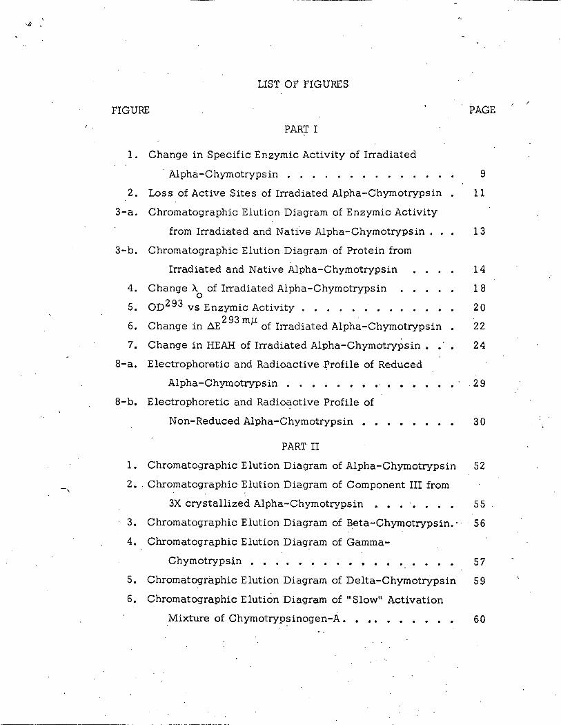
The log percent change of A. between 236. 6 mJl and ·224. 0 mJl 0
with increasing dose is shown in Figure 4. The value of-236. 6. mJl . .
was taken as 100% and 224 mJl was taken as zero % A. • The average 0
value of three independent determinations was taken to calculate the
percent remaining A. for the irradiated protein. .Points fall very close 0
to the straight line for loss of enzymic activity.
Alpha-chymotrypsin undergoes a heat induced conformational
transition at about 54 o which inactivates the molecule (22). The A. . 0
of alpha-chymotrypsin which has been heated to 60° was determined
·'

I.
~ 0
Cl)
+J ·r-f
-~ ~
~ Q)
..c: ~ 0 . s:: ...;t Q) N Q) N ~ +J '0 Q) s::
..a t1l·
0 ~ f-. IC .
bO.IC s:: tl'l
•r-f . N s::
•r-f
~ Q)
~
~
80
70
50
40
30
20
)
............ '
....... '
-$-=.The average value· of· the three determinations
Dose {xio-19ev/g)
. f. __ Enzymic ' ... Activity
0
> ~ '"d
~ I C)
~ 0 1-3
~ '"d Cll H z
C)
-~ Q [%1
H z
7 0
0 ":tj
H
~ t:l
~ 1-3 tr1 t:l
":tj 1-'•
(JQ. c 11 (1)
.p.
}-'
co
'' i ~
19
-and found to be 224m/). (Table I). The equivalence of A for the two 0
states of the protein indicates that radiation denatured protein has
assumed a conformation in solution which is similar to the conforma-
tion of heat denatured protein.
Ultraviolet Light Absorption Effects. The heat induced conforma
tional transition of alpha-chymotrypsin is accompanied by a blue shift
in the UV absorbtion spectra with a maximum change in the absorbtion
· at 2 93 m/). (22). This spectral shift is presumably associated with
movement of a number of tryptophan residues from a less polar to a
more polar environment (2 3).
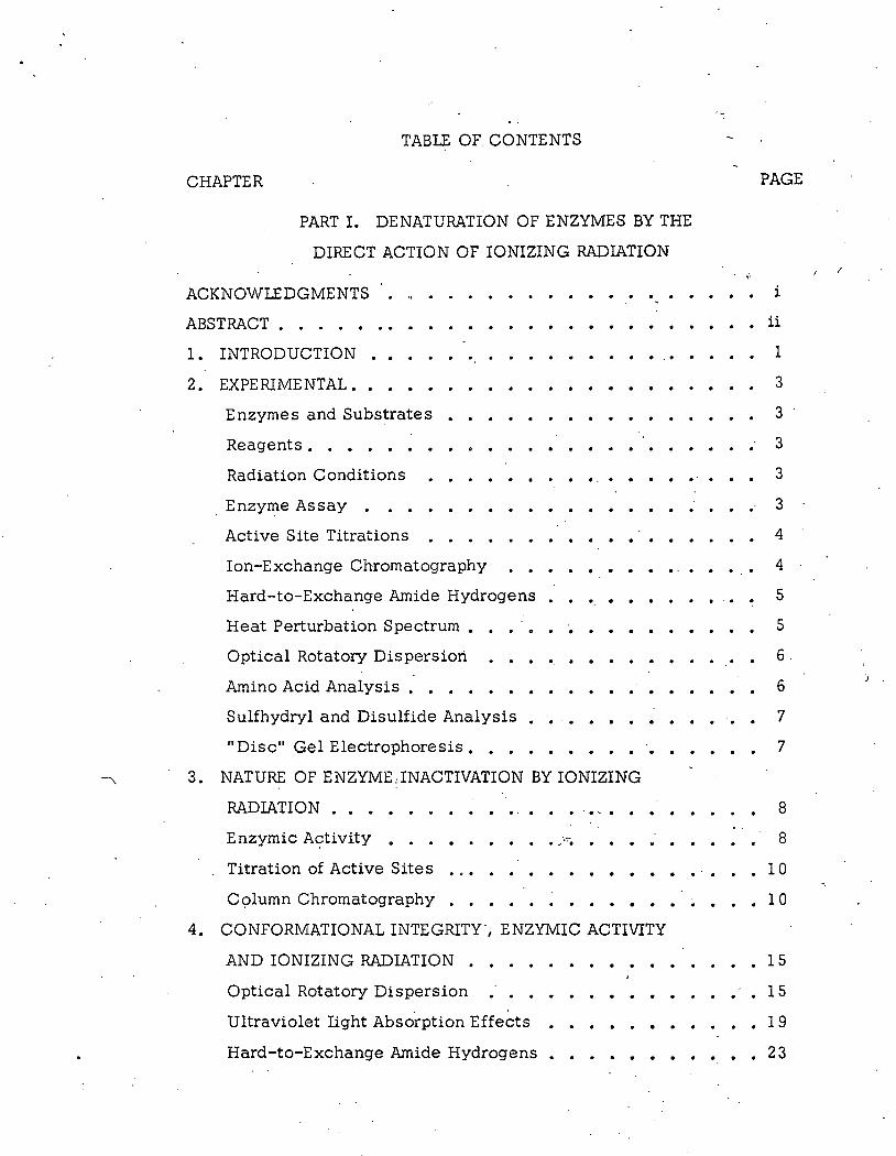
This spectral shift can be directly correlated with the loss of
enzymic activity of the heated sample. Figure 5 shows the change in . .
the 2 93 m/). absorbance for a sample of alpha-chymotrypsin heated
through this temperature transition. Samples were periodically with
drawn and assayed by the rate of ATEE hydrolysis.
The change in specific activity with temperature is also plotted
on the same temperature coordinate. As the enzyme undergoes the
structural transition the enzymic activity is lost. In this solvent
system the change in conformation is irreversible.
Since radiation denatured protein and heat denatured protein
have a A. of about 224 m/). it is reasonable to suspect that radiation 0
denatured protein will be: unable to undergo further temperature induced
spectral transitions. Hovvcver I this is not necessarily true. A is 0
presumably a measure of alpha-helix and the spectral transition reflects
a change in environment of tryptophan residues. The two properties
need not be directly related. The ~293 for radiation denatured protein
could be equal to 1 greater than 1 or less than that of native protein.
If AE293
for radiation denatured protein is zero~ the protein may be in
a conformational state similar to heat denatured protein.

tl'\ 0\ C'l
0 0
'
. I /
.;
0 f--I-- --t--e t-- .. i, Specific Enzymic Activity~~·
.... ·'
.100 on293 ~->
\ \ .\
\
100-l
7.5-!1 c.-a ('/)
"C1 n> n 1-'• H> 1-'•
50__1 n !l> n rt 1-'• < 1-'• rt
'<: 25
. . \i . ... , 0 200L I g l ~ - .... J. I J ~
30 35 40 45 50 55 60 65
TEHPERATURE
on293 vs. Enzymic Activity
1-Ij 1-'•
()Q c 'i n>
\.11
N 0
i t ! ·;
··l i < ,.
~ ;; ( •
;[ ~- F: {,
<'.{ n .•. \I 11 ;f
ll '1 .. I'
t I I
i
if i
I ! ( I I :

~--------------~-'~-------------------~---------------------
In the speciai case where .6.E 293 for the radiation denatured
protein is zero;
.6.E293 native
-kD =e . = ...};_
A 0
· where A and A refer to the specific enzymic activities of protein 0
irradiated to dose D and unirradiated protein respectively.
21
Figure 6 is a plot of the log percent remaining AE 2 9 3
v s dose.
Th~ D37 is identical to the n37
for loss of enzymic activity indicating
AE . 93
for radiation denatu~ed protein is zero. These data were
obtained by methods outlined· in the experimental section.
The temperature induced transition of alpha-chymotrypsin
·shown in Figure 5 is relatively sharp 1 occurring over a temperature
range of about 5o. The temperature induced transition for irradiated
samples was less s~arp. The samples which received high doses
began their transition at temperatures as low as 35 o. To obtain the 293 . 293
total AE. it was necessary to take the difference between E at liT 2 93
. 3 0 o C and the E at the peak temperature. This change in slope of '
the temperature transition curve implies some enzymically active but
heat labile molecules are present (24). This evidence is contr.adictive '
-, to the conclusion that raqiation inactivation of alpha-chymotrypsin
is an all-or-none process.
Temperature induced structural transitions of proteins may be
thought of as melting out ordered conformation of some parts of the
molecule. This probably involves breaking some hydrogen and hydro
phobic bonds. The temperature of transition can be lowered by a
number of additives. For example 1 in separate experiments with alpha
chymotrypsin it was found that 5 x 10⁻³M cyclohexanol was sufficient
to reduce the transitions temperature by 1 0 o. Irradiated protein repre
sents a heterogeneous mixture as evidenced by chromatography. The
t,.., .. -· I

100
90
80
70
60
50 ::t s.
(")
en . 40 N
j:Ll
<1 t)l
.~ ..... 30 ~ .... l'tl s Q) ~·
* 20
I /
0
/
Figure 6 Change in .D.E2 93J..L of Irradiated Al!)ha Chymotrypsin .
19 n37
=98x10 ey/g
N N
f:.
I
l I

-- ---- ----. ~----- ---~---- ·-·-······· ·-- .-------------------1
23
effect of the products of radiation upon the temperature induced transi
tion of unhit molecules cannot be neglected. Therefore, the observa
tion of heat labile enzymic activity in irradiated protein does not
necessarily reflect the presence of altered but enzymically active.
molecules.
Hard-to-Exchange Amide Hydrogens. It is possible to obtain
an estimate of the number of hydrogen bonds still intact after irradiation
by the method of Blout (25) using n2o. Blout has noted that the
infrared N-H deformation band of proteins undergoes a shift to longer
wave lengths when the hydrogen is exchanged for deuterium. There is
an initial ra.pid exchange of protons which is complete in 3 to 5 minutes
followed by a slow exchange which takes over 24 hours to complete.
The hard-to-exchange amide hydrogens (HEAH) are believed to he
involved in hydrogen bonds and hydrophobic centers. A change in
conformation of the protein may permit the rapid exchange of ths
exposed amide hydrogens. -
Figure 7 is a plot of log percent remaining HEAH .Y.§. dose for
irradiated alpha -chymotrypsin. All hydrogens which do not exchange
in 10 minutes have been designated as HEAH. The total HEAH of the
native enzyme was found to be 12 8 and was taken as 1 00 percent. ,
These data do not yield a. straight line. The initial ionizing
event resulted in the loss of some but not all of the HEAH of the protein.
Subsequent ionizations a~e necessary to furth~r reduce the HEAH of
inactive molecules •. The initial slope of the curve was· extrapolated to
the D 3 7
for the first ionizing event and the number of electro!' volts
absorbed per HEAH lost was found to be 1. 65 eV/HEAH. Since an
average of 42.1 eV are absorbed per molecule inactivated, the inactiva
ted molecules have lost an average of 19 HEAH/molecule.

' _)
100~~=-----======~=-==~====~=======-~-=-=--=======-======~~
90 80
70
60
50
40
30
20
' ' '
·.
,e ·' '
/ /
'
.. ..
' '
· ······ •... ~irst Ionization HEAH ..
. . . . . . . . . (37 .. . ·~.
... .. ...
' @' :' '~Enzymic Activity
' ' '
Dose (xlo-19ev/g)
(')
~ z Q
> [<:1
t-f 1-d H
~ z I ~ ~ (') ~ 1-'•
t ~ aq
::X:: c:: a t1 I 0 (t) i 1-3 "'j
~ ~ ,. ~ H I
~-(/)
H z t:l
~ 1-3 [<:1 t:l

25
This figure represents the avei_"age number of HEAH exposed by
conformational alterations in hit molecules. These hydrogens may have
been involved in hydrogen bonds or hydrophobic centers. Hit molecules
may not have completed their conformational change in the solid state.
Further change could take place after solution which may permit the
exchange of additional hydrogen atoms. For these reasons the
figure 1. 65 eV/HEAH must be considered a rough estimate.
/ . 1'·

5. COVALENT CHANGES IN IRRADIATED PROTEINS
Amino Acid Analysis: Table II shows the amino acid analysis
for-alpha-chymotrypsin before and after irradiation to 50% inactivation.
The samples were run in duplicate by methods outlined in the experi
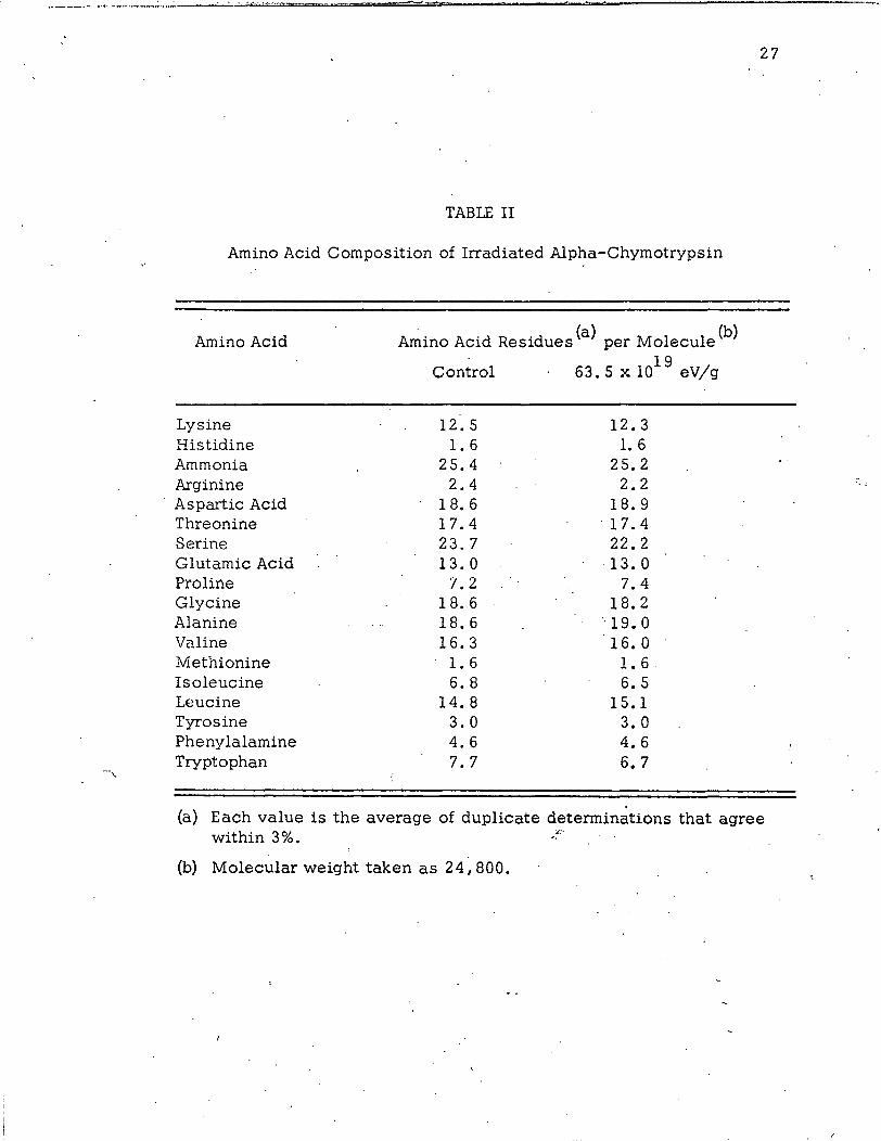
mental section. There is no significant loss of any amino acid residue.
Gamma radiation is not highly specific for the destruction of amino
acid residues in solid state proteins {27}. Chemical changes may take
place at any of several amino acid residues.
Disulfide and Sulfhydryl Analysis: Table III shows the disulfide
and sulfhydryl analysis of alpha-chymotrypsin before and after
irradiation. These figures are averages of triplicate runs performed by
' methods outlined in the experimental section. T.he method routinely
gave values for alpha-chymotrypsin and lysozyme which were 2 0% too
high. The reason for this discrepancy is unknown but the method is of
value for comparing the product and starting materials. There is an
increase of titratable sulfhydral groups following reduction of irradiated
protein but the increase is too small to account for the total enzyme
inactivation.
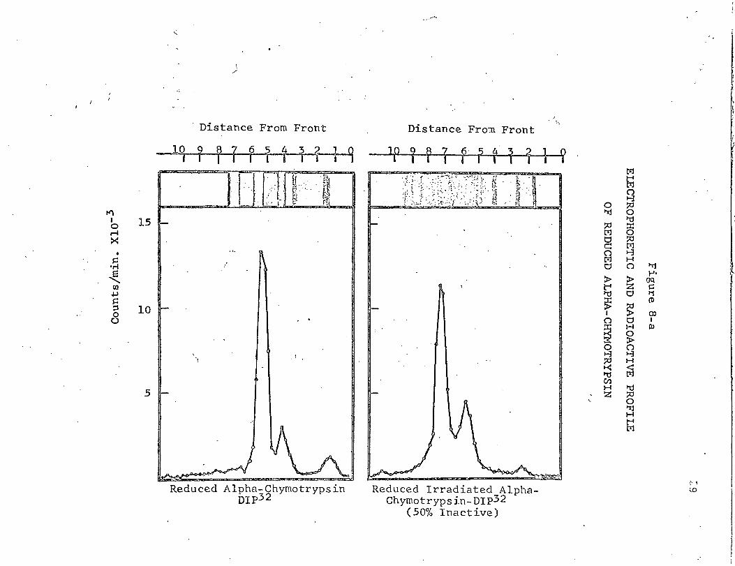
"Disc" Gel Electrophoresis: Irradiated and native alpha-chymo
trypsin was labeled at the active site with FDIP-32.
Thioethanol-reduced and non-reduced DIP-32 labeled protein
was electrophoresed in 8 M urea through polyacrylamide gels. Figure
8-a shows the distribution of radioactivity in the non-reduced gels.
The significance of each band and the distribution of radio
activity in the unirradiated protein is discussed in Part II of this
thesis. The techniques take advantage of the fact that enzymically
active alpha-chymotrypsin· will react with FDIP-32 at the active site
·----·---------------
- -~ --------~""---'-...:...-""'"'· -=·-=---· =-=--===-:=:!..:.=--------------------
27
TABLE II
Amino Acid Composition of Irradiated Alpha-Chymotrypsin
Amino Acid Amino Acid Residues (a) per Molecule (b)
Control 19
63. 5 X 10 eV/g
Lysine 12.5 12.3 Histidine 1.6 1.6 Ammonia 25.4 25.2 Arginine 2.4 2.2
· Aspartic Acid 18. 6 18.9 Threonine 17.4 . 17.4 Serine 23.7 22.2 Glutamic Acid 13.0 ·13.0 Proline 1.2 7.4 Glycine 18. 6 18. 2 Alanine 18.6 '·19 .. 0 Valine 16.3 16.0 Methionine 1.6 1.6 Isoleucine 6.8 6. 5 Leucine 14.8 15.1 Tyrosine 3.0 3.0 Phenylalamine 4.6 4.6 Tryptophan 7.7 6. 7 ---,
(a) Each value is the average of duplicate determinations that agree within 3%. .:-
(b) Molecular weight taken as 24, 800.

Dose· eV/g
0
79. 5 x 1 ol9
278xlo1 9
TABLE III
Sulfhydryl and Disulfide Groups of
Irradiated Alpha-Chymotrypsin
Groups (a) pe·r Molecule (b).
Sulfhydryl
0
0
0
. .
Disulfide
6.4 ,--,
7. 5
7. 0
(a) Each value is the average of triplicate determinations.
(b) Molecular weight taken as 24, 800.
28
tl't I 0 15 r-1 :>< • s:: ....... ~
c.o +.!
§ 10 0 0
5
"
I /
Distance From Front
10 ? f 7 6 1 1 1 2 1 q f I I I
l l' ! ,, .. . . .. ' it ... 'f~ I .. .I I ...... · . t! . · ·· - . ~--~ . r .. ' · ~; f;;~ t ... ~ . • •..
, .. I ,.. t'.<
Reduced Alpha-Chymotrypsin nrp32
Distance Fro~ Front : ..
]P 1 ~ 1 r· ? r· 1 1 1 ?
-HJ<fJ:r:ul t
Reduced Irradiated AlphaChyrnotrypsin-DIP32
(50% Inactive)
(• ~
~0
I
I : f !.·.

tf\ I 0 r-1 :><
• ~
•.-l
~ Cll
+.J ~ :::3 0 u
15
10
5
I /
Distance From Front Distance From Front
10 9 8 7 6 ? 1 3 2 } 9 1p ? ~ 7 6 ? $ ? ? 1 Q I I I I I I i I -
~~-~-::> [] l t
Alpha-Chymotrypsin-Dip32
· f -~JTim~ r ·
---.. Irradiated Alpha-Chymotrypsin
Dip32 (50% Inactive)
ttl t;1
0 ~ t'Ij ~
0 z "d 0 ::r:
~ 0
~ 0 1-J c:: H
&1 () t'Ij
0 :x> ..... z ()"Q :X:. 0 c: ~ li
~ (i)
~ 0 co I H I () ~ o'
~ () 1-J
0 H 1-J ~ ~ I'd I'd (/) ~ H· .0 z "rj
H
~ (/)
(..)
0
(
' i:
~
' (
li l
I
j t ·.1
I i i;
'
31
serine to form the serylphosphate ester·.· In the non-reduced electro
phoretic pattern I the radioactivity is associated with enzymically
active protein. In the reduced pattern it is associated with the C-chain. ~ I . ,
Comparison of the non-reduced 1 irradiated and native 1 protein ·
electrophoretic patterns and distributions of radioactivity shows no
new protein bands and no new radioactive bands in the irradiated
protein. The absence of distinct new protein bands indicates no
specific chemical changes occurred. There is a protein background
over the entire electrophoretic region .in the irradiated samples which
may be due to nonspecific chemical changes in the irradiated .protein.
The reduced protein always gave a much)clearer electrophoretic
pattern than unreduced material; if any significant amount of specific
chemical change did occur they should be· more evident in the reduced
electrophoretic pattern. Comparison of the reduced patterns of the
irradiated and native protein reveals no new electrophoretic band in
the irradiated protein. . ;.
Radiation Denaturation of Selected Enzymes. Information
regarding the mechanism of radiation denaturation of ·enzymes may be . .
gained by examining the energy required to inactivate different .enzymes.
Table. IV lists the energies required to inactivate several enzymes.
These data were determined by the method already discussed. Irradia
tions were conducted in the University of Colorado cesium-13 7 source.
This is a consistent set of data that eliminate calibration and many
analytical errors involved in comparing such valu-es from literature ,:~":'.
references. '
/

. - ~.-·----=-~.:.-.-___ -, __ _-__ __::::_-_ .----~-·---.~----~
TABLE IV
The Average Energy Necessary to Inactivate
Selected Enzymes
Enzyme
Alpha-Chymotrypsin
Chymotrypsinogen
Delta-Chymotrypsin
Papain
Lysozyme (a)
Ribonucleas~b)
(a) Data taken from reference no. 45
Inactivation Energy eV/Molecule
42
35
30
49
86
GG
(b) Data taken from reference no. 39, 50
' I
32

-:,
---~--·· . ·. ----~---......... -=-----==-;:;..;;;::=====.::.:==---------------
6. PROPOSED MECHANISMS OF RADIATION
DENATURATION OF ENZYMES
An enzyme may be inactivated by altering amino acid residues
directly or indirectly involved in the active site or by altering the
ordered conformation necessary for maintaining the catalytic center.
The inactivation of enzymes by ionizing radiation may proceed by
either one or both of these general mechanisms. In dilute solution the
radiolytic· products of the solvent may re-act with amino acid residues
on the surface of the enzyme and promote inactivation by chemical
changes (7). This proces~ is generally referred to as inactivation by
indirect effect of ionizing radiation. The inactivation of enzymes
by direct effects of ionizing radiation would not be expected to show a
preference for the surface amino acid residues. Most authors have
described enzyme inactivation by direct effect of ionizing radiation in
terms of non-:specific changes in the ordered conformation of the
molecule.
Augenstine has recently reviewed the proposed mechanisms for
enzyme inactivations by direct effects of ionizing radiation (7). All of
the proposed mechanisms consider the loss of ·ordered conformations
as the main mode of enzyme inactivation by a single ionizing event.
He and other workers are unable to define whether such chang.es are
the result of radiation induced covalent changes I or are directly
induced by the absorption of radiatio.~ energy.
. Pollard (5) suggests that "excitations" may move -along the
peptide. backbone 1 occasionally opening a covalent bond and disrupting
a number of hydrogen bonds. The end result would be a degraded
peptide chain with random configuration.
... ---··· =c.::==
34
There is little-or-no evidence for a degraded peptide chain in
irradiated lysozyme (2 E) and bovine serum albumin (2 7). The one 1 two
and three chain systems of the chymotrypsins are inactivated by about
the same energy per molecule. This suggests that much of the inacti
vation energy does not travel along the entire length of the peptide
chain.
Platzman and Franck (2 8) presented an analysis of the electronic
events which should. occur in a protein· immediately following its
ionization. They concluded that the formation of a single point charge
in a region of high dielectric constant should result in a polarization
wave through the medium of a sufficient magnitude to disrupt as many
as 14 secondary bonds. Their postulate would predict the formation of
many altered but enzymically active molecules 1 greater radiation .
sensitivity at elevated temperatures and a dependency of the radiation
sensitivity upon the conformation of the molec~le.
The HEAH of alpha-chymotrypsin are lost with an energy require
ment of 1. 65 eV/HEAH. 42. 1 eV are required (on the average) to ·in
activate the molecule. Thus I the actual number of HEAH lost per
single ionization would be 19 HEAH/single ionized molecule. This
figure is surprisingly close to the 14 secondary bonds predict~d by
Platzman and Franck. However I no altered but enzymically active
"""' molecules were found. :This suggests that the breakage of a few
hydrogen bonds in the solid state .is sufficient to produce a confopna-:
tiona! rearrangemerit which is not reversible.
G. Bugge! working in this laboratory showed that the D3 7
for
trypsin remained constant though the A for the protein was varied from . 0
219 mf.l to 236 mJl. The simplest interpretation of these data is that
the denaturation reaction is independent of the c.onformation of the
molecule ..

35
Augenstine (7) proposes that the breakage of specific convalent
bonds necessary for the maintenance of the ordered conformation may
play an important role in enzyme inactivation. He proposes that
disulfide bonds may be particularly radiation-sensitive and that the
energy once deposited in the molecule may migrate to the disulfide bond
causing its rupture. Data supporting this theory comes from solution
U. V. studies and it has been suggested this effect could extend to
solid state gamma radiation.
There is no evidence of disulfide cleavage in irradiated alpha
chymotrypsin, lysozyme or bovine serum albumin. Disulfide cleavages
have been noted in irradiated ribonuclease(29) but the effect could only
account for a small fraction of the denatured molecules. Other workers
fail to find any evidence of disulfide cleavage in irradiated ribonuclease
(3 0).
Norman and Speigler (31) have proposed ·that energies associated
with an ionizu.tion should be transferred into vibrational energies of the
molecule. This would result in a "hot" molecule which may denature
before the energy can be dissipated to the surrounding medium. One
can only estimate the temperature of the molecule because the rate of
heat loss is unknown for a molecule in the crystalline state. Our
experiments with alpha-chymotrypsin indicate that the enzyme can be
denatured in the solid state by heating to 155 o. The ionizing event
supplies more than enough energy to thermally denature the enzyme in
the solid state. However, the rate of heat loss in the solid state may
be much faster than the time necessary for a conformational rearrange
ment to take place. There is no data available which will decide this
point.
Several authors have proposed ra:iiation produced free radicals
as important intermediates in radiation denaturation of enzymes. Free
radicals produced in proteins by ionizing radiation have been studied
with ESR measurements (32, 35). These radicals seem to be composed

- -~--..J-,:...A...., ... ·t,.,.,,_..t_vl ~~,:"._......., __________ -'-------'-----------~-
36
of three classes: 1. a non-specific radical detected at very low
temperatures; 2. a free radical on an alpha-carbon called a glycine
like radical; and 3. a radical on a sulfur atom called a cystine like
radical. If the irradiated system is observed at 77 oK only type 1
radicals are observed, as the sample is warmed to room temperature
the type 1 radicals· are converted to a mixture of types 2 and 3 with
some loss in signal strength presumably due to recombination. ·This
is direct evidence for free radical migration through the solid state
system. The activation energy for migration has been estimated at 2
to 3 kcal/mole (36). The presence of cystine like radicals at room
temperature provides direct support for Augenstine' s hypothesis that
the disulfide bonds may be especially·radiation sensitive.
Several authors have noted a temperature dependence for the
inactivation of enzymes by ionizing radiation (37 ,38). This temperature
dependence yields an apparent activation energy for the denaturation
reaction of about 2 to 3 kcal/mole (36). The initial ionizing event is
not expected to show a temperature dependence but chemical or physical
changes induced by the ionization would be expected to have an
activation energy. This activation energy is difficult to interpret
because each ionized molecule has received an amount of vibrational
energy which may in some way aid the denaturation process. Free
radical migrations and th~ denaturation reaction have comparable
apparent activation energies. This has been interpreted as evidence
for a free radical mediated denaturation reaction (3 6).
Any proposed mechanism for a reaction must take into account
the nature of the starting material., the products and the known effects
of the reaction environment. At the_ sta_rt of this work very little was
known about the nature of the products of inactivation of enzymes by
ionizing radiation. Alexander (2 7) had shown that none of the compon
ent amino acids of bovine serum albumin were particularly radiation
-- _________________________ _.___... ______ _ 37
sensitive. He did note an increase in the ammonia yield from hydro
lyzates of irradia~ed protein. Later Garris-on (39, 40) followed up this
work and showed that the increased ammonia was accompanied by an
increased yield of carbonyl groups. These observations led him to
postulate the formation of an acid labile ·intermediate at the peptide
bond which subsequently hydrolizes to NH3
and alpha-keto acids. He
has proposed a substituted imino acid (a dehydropeptide) as the acid
labile intermediate but has failed to observe it directly in irradiated
proteins.
The evidence for some acid labile intermediates which can
hydrolize to yield ammonia and carbonyl groups is good. However, its
relevance to the denaturation reaction has not been established. It
is difficult to believe that dehydropeptide formation will inevitably.
result in enzyme inactivation, since this would imply that the proper
bond angles and distances must be maintained at every peptide bond.
Sedimentation experiments on irradiated bovine serum albumin
indicate that the products have the same molecular weight as the
starting material but a lower diffusion constant. Alexander (2 7) inter
preted this as evidence for ionized mole-cules having an expended
structure which one would expect if the ionization induced significant
changes in the ordered conformation of the molecule. These data were
not correlated with biolog_ical activity and represent a single observa
tion which may be subject to many errors of interpretation. The data
presented in this report supports the conclus_ions of Alexander a:hd
relate the conformational effects to the loss of biological activity.
;Numerous investigations have been conducted into the possi
bility of altering the radie:~.tion s·ensitivity of enzymes by adding selected
compounds to the system. In general it has been found that the addi
tion of copious quantities of sulfur containing compound to a lyophilized
preparation of protein will cause some radiation 11 protection .. for the
_.._--'-"'......._ ____ ......._::.::.;;; ......... ~...;.·~-""'"".._ ______ ........ ~ ........ -~-------------------·-~--
'
38
protein. These data and ESR measur.ements indicate that some energy·
transfer process is involved whereby energy is transferred from the
protein to the additive (3 5).
Other compounds, particularly biradical gases such. as oxygen,
will serve as radiation "sensitizers" when added to the system (41, 42}.
These compounds have the effect of lowering the average energy
necessary to inactivate an enzyme molecule and will quench the stable
free radicals of the system. It is difficult to interpret these results
without first knowing more about the fundamental principles underlying
the radiation .denaturation of proteins.
. ' ~',
·· ..
/

---------------------------------------------------- ---------------1
7. DISCUSSION
The single ionization theory of enzyme inactivation by ionizing
radiation states that a single ionization anywhere within the molecule
is sufficient to inactivate the enzyme (117). This theory predicts enzyme
inactivation should be an all-or-none process. Alpha-chymotrypsin is
inactivated in the solid state by the 'direct action of gamma radiation
with an average dose of 42.1 eV per molecule. This dose is compatible
with previous studies on this enzyme (43) 1 and is consistent with
average energies required to create an ion pair in the solid state. All
enzyme activity present in irradiated samples can be attributed to
molecules which are indistinguishable from native alpha-chymotrypsin.
Comparison of the n37
for enzymic ac_tivity and active sites shows all
enzymically active molecules have a turnover number equivalent to that
of native enzyme. Chromatographic separation of irradiated mixtures
shows all molecules with enzymic activity have chromatographic
mobilities equivalent to the native enzyme.- Most of the enzymically
inactive protein is chromatographically distinguishable from native
alpha-chymotrypsin. Apparently each ioni~ation in a native molecule
results in an alteration of chromatographic mobility I and loss of the
functional active site.
Optical rotc;ttory dispersion studies r~veal that radiation·
denatured alpha-chymotrypsin has a A equivalent to that of heat \ 0
denatured enzyme. Heat denatured enzyme has undergone an irrevers-
ible UV absorption spectra shift. The D3 7
for this spectra shift in
irradiated enzyme is equivalent to the n37
for loss of enzymic activity.
These data show that radiation denatured enzyme assumes a

·---------
'
40
conformation in solut'ion which is comparable to that of heat denatured
enzyme. A single ionization in ·the solid state enzyme results in a
confonnational change of sufficient magnitude to inactivate the enzyme.
Platzman and Franck (2 8) theorized that a single ionization in
a protein should result in a breakage of· about 14 secondary bonds.
The HEAH ·of irradiated alpha-chymotrypsin are lost by a multi-hit
process requiring 1. 65 eV/HEAH. This corresponds to a loss of about
19 HEAH per· ionization. This is in surprisingly good agreement with
· the Platzman and Franck hypothesis. Evidently the exposure of 19
HEAH is sufficient to cause a conformational change which will
inactivate alpha-chymotrypsin.
Although irradiated alpha-chymotrypsin showed no change in
the amino acid composition, or disulfide content and the electrophoretic
experiments showed no evidence of specific peptide bond breakage,
this does not rule out general chemical changes.· Both the reduced and
non-reduced irradiated samples showed a protein slur over the entire
electrophoretic regfon. This may be due to non-specific chemical
changes in irradiated protein.
The energy required to inactivate selected enzymes was
detennined. Chymotrypsinogen, delta-chymotrypsin and alpha
chymotrypsin have almost the same amino acid analysis and sequence
but represent one, two, and three peptide chains respectively (9).
They are all inactivated with energies which cluster around 35 eV per
. molecule.· There is no apparent dependence upon the number of _peptide \ .
chains. Papain can be degraded with ·leucine amino peptidase to a
third of its original molecular weight without loss of enzymic activity
(44). Although papain is more stable to -radiation denaturation than
alpha-chymotrypsin it is not three times more stable. An ionization in
any part of the molecule will apparently cause inactivation.

41
Ribonuclease and lysozyme are unusually stable to radiation
denaturation. These enzymes lose HEAH with about the same energy
requirement per HEAR as alpha-chymotrypsin (30 I 45). Altered but
enzymically active molecules have been detected in the radiation
products of both enzymes (2 9 1 46). Both ribonuclease and lysozyme
are anomalOUS enzymes in that they are Stable t0 heat (4 71 48)
denaturation. Conformational alterations in these enzymes are partly
or completely reversible. Conforrriational changes in radiation
denatured ribonuclease and lysozyme may be reversible ·provided
chemical changes do not restrict the process.
The evidence presented in this report indicates that a single
ionization in a solid state protein will result in conformational changes
which are sufficient to inactivate the enzyme. These conformational
changes may or may not be reversible depending on the nature of the
enzyme and any covalent changes induced by the radiation. The free
radicals produced in radiation denatured proteins (32 1 34) may initiate
covalent changes such as dehydopeptide (40) formation or migrate to
other molecules (35). If the free-radicals are transferred to other
molecules the denatured enzyme may have an increased probability of
, reforming an enzymically actlve conformation. The free radical acceptor
'would then act as a radiation·" protection". molecule {41, 49). The
\• migratory" free radicals of the system may not normally inactivate the \ . .
ionized molecules 1 but may ·do so w~en they react with oxygen or
other gases added to the system.
~---,

\
8. SUMMARY
Inactivation of alpha-chymotrypsin by direct action of gamma
radiation was shown to be an aU-or-none process. All enzymically
active molecules present after irradiation were shown to be indistin
guishable from native alpha-chymotrypsin by enzymic properties,
column chromatography, and gel electrophoresis. This evidence sup
ports the single ionization theory for enzyme inactivation by ionizing
radiation.
The radiation inactivated molecules were sho~n to have an
altered conformation. The change in the conformational parameters
would be correlated with the loss of enzymic activity. The conforma
. tional changes were detected by optical rotary dispersion, ultraviolet
light absorption and hard-to-exchange amide hydrogen content. The . :.
·first ionization appears to result in an inactive molecule with a A. of 0
224 mJ.l, no "buried" chromophores. , and the exposure of about 19
hard-to-exchange amide hydrogens.
Radiation denatured molecules showed no~.specific loss of .
amino acid residues or disulfide bounds .. There was no evidence of
specific chain cleavage. ·These data are discussed in terms of
currently proposed mechanisms of enzyme inactivation by direct action
of ionizing radiation. ) \
/ ·'

- -- --- ------ -·------~----_........., ___ __,._....u-. ... --... ""' .. _ _...._......,, __ .,-.. ...... _______________ ........
9. REFERENCES
1. Lea, D .. E. , Action o~ Radiations .Q!l Living Cells, Cambridge Press (1962), pp. 69-99.
· 2. Pollard, E. C. , Buzzell, A. , Jeffreys'· C. and Forro, F. , Arch. · Biochem. Biophys.,n_, 9 (1951).
3. Pollard, E. C., Adv. Biol. Med. Phys., ]., 153 (1953).
4. Adams, E.,J. Biol. Chern., 209,829 (1954),··Ibid., 217,325 (1955) ..
· 5. Pollard, E. C. , Guild, W. R. , Hutchinson, F. , and Setlow, R. B., Progr. Biophys. and Biophys. Chern.,.§., 72 (1955). ·'
6. Friedlander, G. I and Kennedy I J. ·W. I Nuclear and Radiochemistry, Wiley and Sons, Inc. , N. Y. (1955), pp •. 186.
7. Augenstine, L. G., Adv. Enzymology, 24, 359 (1962).
8. Hartley, B.S., Nature, 201,1284 (1964).
9. Desnuelle, P., inTheEnzymes, Vol. 4, Boyer, P. D., Lardy, H., Myrback, K. , (Editors), Academic Press, Inc. , New York, (1960), pp. 93.
10. Schwert, G. W., Takenaka, Y., Biochim. Biophys. Acta., .1.§., --, 5 70 (1955).
11. Schoellmann, G., Shaw, E., Biochemistry, ~~ 252 (1963) ...
12. Koshland, D. E., Strumeyer, D. H., Ray, W. J., Brookhaven Symp. Biol. ,,ll, 101 (1962).
")
13. Laskowski, M., Methods in Enzymology, Vol. 2, Colowisk, S. P., and Kaplan, N. 0. , (Editors), Academic Press, Inc. , New .York, (1955) 1 P• 8.
,r
44
14. Moore, s. I Spackman, D. H. and Stein, w.· H.,. Anal. Chern~, 30, 1185, 1190 (1958). - ..
15. Goodwin, T. W. and Morton, R. A., Biochem. L, 40, 628 (l946).
•16. Boyer, P. D., L Am. Chern. Soc.,~~ 4331 (1954).
17. Brown, W. D., Biochim. Biophys. Acta., 44, 365 (1960).
18. Schonbaum, G. R. , Zerner, B. , Bender, M. L. , 1· Biol. Chern., 2 3 6 1 2 9 3 0 (1 9 61) o
19. Bender, M. L. ,J. Am. Chern. Soc~·,84, 2582 (1962).
20. Moffitt, W., L Chern. Phys.,~, 467 (1956).
21. Moffitt, W., and Yang, J. T., Proc. Natl. Acad. Sci. U.S., 42, .. 596 (1956); Moffitt, W., Ibid., .11_, 736, (1956); Fitts, D.,
/
/- Kirkwood, J. G., Ibid., _11, 33 (1957); Moffitt, W., Fitts,· D., / · and Kirkwood, J. G., Ibid., Q,· 723 (1957).
22. Foss, J, G.; Biochim. Biophyc. Actu.., 47; 569 n9G1).
23. Donovan, J. w. I Laskowski, M. and s.cheraga, H. A. I 1· Am. Chern. Soc., ]1, 2686 (1961).
24. Braams, R., Inter. L Radiation Biol., .§., 297 (1963).
25. B1out, E. R., Daloze, G. and Asadourian, A., L Am. Cherri. Soc., g 1 1 59 5 (1 9 61 ) o
26. Stevens, C. 0., Personal Communication (1963).
27. Alexander, P. and Hamilton·, L. D. G., Radiation Res., g, 510 (1960); et al. , Ibid. , .u_, 214 (1960).
2'8. Platzman, R. and·Franck, J., §m. Information Theory Biol., 262-270, Paragon Press (1958).
29. Hunt, J. W. and Williams, J. F., Radiation Res. 1 ll~ 26 (1964).
30. Hayden, G. A. and Friedberg, F., Radiation Res., g, 130 (1964).
31. Norman, A. and Speig1er, P. I L ~ Phvs., 2]_, 2658 {1962).

·····---·-·-----==-~· ---·=·-._:,·,...,.., ... ,..,.,....._ ______ ..a.4.._....._ _________________________ _
32. Gordy, W. and Shields, :£i., Radiation Res., .fl, 29 (1964).
33. Patten, R. A. and Gordy, W., Radiation Res., g, 29 (1964).
34. Henriksen, T. , Sanner, T. , and Pihl, A. , Radiation Res. , ]J!, 147 (1963).
3 5. Henriksen, T. , Sanner, T. , and Pihl, A. , Radiation Res. , 1.§., 163 (1963).
3 6. Augenstine, L. G. , and Mason, R. , Biological Effects of Ionizing Radiation at the Molecular Level, Published by International Atomic Energy Agency (1962).
_.../
3 7. Setlow ~ R. and Doyle, B. , Arch, Biochem. Biophys. , i§., (1953}.
38. Pollard, E. G., ·Powell, W. F., Reaume, S. H., Prac. Natl. Acad. Sci.. U. S.,J.§., 173 (1952).
45
39. Garrison,·W. M., and Weeks, B. M. 1 Radiation~· ll~ 341,. (1962).
40. Garrison, W. M1 Jayko, M. E., and Bennett-Corniea, W. Science, 146 1 250 (1965).
41. Hutchinson 1 F. 1 Radiation Res. , Supplement, ,g_, 4 9 (19 60).
42. Butler, J. A. V. and Robins, A. B., Radiation~·, 12, 63 (1962).
43. Butler, J. A. V., Robins, A. B. and Rotblat 1 J. Proc. Roy. Soc. A, 2 5 6 1 1 (1 9 6 0) •
.,
44. Smith, E. L. , Hill, R. L. and Kimmel, J. R. 1 Symp. Q!l Protein Structure, Nueberger, A~ {Editor), London Methuen and Co. LTD. (1958) pp. 182.
45. Stevens, C. 0., Tolbert, B. M. and Reese, F. E. 1 &£h.. Biochem. · Biophys., 102, 423 (1963).

46. Stevens, C. 0., Henderson, L.E. andTolbert, B. M., Arch. Biochem. Biophys., 107, 367 (1964).
47. Anfinsen, C. B., and Haber, E. ,1. Biol. Chern~, 236, 1361 (1961).
. 46
48. Hayashi, K. , Hamaguck, Z. and Funatsu, ··M. , 1· Biochem. (Tokyo), . g, 374 (1963).
49. Braams, R., Radiation Res. ·g, 113 ,(1S60).
50. Blat, G. , Unpublished Data (1964).
'
----------------------------·~------~-----------
·. ··- ·-------
Part II
Enzymically Active Components
of the Crystalline Chymotrypsins
by
Louis Edwin Henderson
'· ..
/
------·--------~----------~--------------~----------------·--------...
48
C[lymotrypsinogen-A is an enzymically inactive protein which
can be crystallized in good yields from beef pancreas. The amino acid
sequence and disulfide arrangement of chymotrypsinogen-A has recently
been published (2). This protein is a single peptide chain with five
internal disulfide bonds. The action of trypsin on this protein results
in a mixture of proteolytic enzymes known as the chymotrypsins (3).
By selecting the proper conditions, this mixture of enzymes can be
fractionated into four crystalline enzyme preparations. These prepara
tions have been designated as alpha- 1 beta- 1 gamma- and delta
chymotrypsin.
Delta-chymotryp·sin is an enzymically active two chain molecule
derived from chymotrypsinogen-A by the enzymic release of a dipeptide
unit. Alpha-chymotcypsin is an enzymically active three chain molecule
derived from delta-chymotrypsin by the enzymic release of a second
dipeptide unit. Beta- and gamma-chymotrypsin ·are derived from alpha
chymotrypsin with no apparent change in their molecular weights (4) 1
amino acid analysis or 1 N- and C-terminal groups (5-7). They differ ih
sensitivity to urea denaturation I. crystal structure and conditions for
crystallization. The salient features of the chymotrypsins have recently
been reviewed (8).
In this report a chromatographic procedure capable of separating
some of the chymotrypsins is described. The technique is used to
investigate the heterogeneity of crystalline chymotrypsin preparations.
Some of the chymotrypsins can be also disti!lguished by their electro
phoretic mobilities through polyacrylamide gels in 8 M urea. Alpha- 1
beta- and gamma-chymotrypsin have different electrophoretic mobilities.
in this system but their reduced chains have identical electrophoretic
mobilities. This suggests that the three forms of the enzyme differ in
their disulfide arrangements.
;.t

------- _____________ ...._ ______________________ ~------------
EXPERIMENTAL PROCEDURE
Enzymes, Proteins and Materials: All chemicals employed were
reagent grade. Alpha-chymotrypsin, chymotrypsinogen-A and trypsin
were obtained from Worthington Biochemical Company as 3X crystallized
salt-free preparations. Beta-, gamma-, and delta-chymotrypsin were
obtained from Mann Biochemical Company as 2X crystallized preparations
N-acetyl-L-tyrosine ethyl ester (ATEE) was obtained from California
Biochemical Company. Bio-Rex 70 (100-200 mesh) was from Bio-Rad
Laboratories. Fluorodiisopropyl phosphate (FDIP}·was obtained from
Mann ·Biochemical and 32
P labeled FDIP (FDIP-32} was obtained from
New England Nuclear Corp. ·
lQn_Exchange Chromatography and Column Preparation: Bio-Rex
70 (1 00-2 QO mesh} is a polyacrylic acid cation exchange resin. The
resin was first washed with 1 N NaOH, then with distilled water, then
with 1 N HCl and again with distilled water. The washed resin was
suspended in 0. 10 M citric acid and titrated to pH 5. 60 ~ 0. 02 at 2 5o
with 50% NaOH. The resin was allowed to settle and the fines were
removed by decanting the supernatant and resuspending the resin in
fresh elution buffer. This operation was repeated several times. The pH
of the resin in the elution buffer was closely adjusted to 5~ 60 ~ 0. 02 at
25o~ The washed, equilibrated resin was packed into 220 em pyrex glass
columns. The packing was continued until a 200 em resin bed was ob
tained. The packed column was placed in a 4 o room, and the citrate
elution buffer was allowed to flow through the column overnight. The pH
of the elulate should be 5. 60 ~ 0. 02 at 25°. The eiution buffer JNas pre
pared by titrating 51. of 0.10 M citric acid to a pH of·5. 60 ~ o.-02 at 25°C
·with 50% NaOH. Analytical (0. 8 x 22 0 em} columns will take up to 3 00 mg
of protein although the runs reported here did not exceed 50 mg. The
protein was' applied to the column in 1. 0 ml of elution buffer and washed . .
. i /
r • .

-------------------------------------------------------·-------------1
50
in with 2 to 3 ml of buffer. The columns were run at flow ·rates not
exceeding 6 ml/hr. Three ml fractions were collected. A 4 x 220 em
column prepared by the above technique was used to separate 3 grams
of protein.
Protein and enzYme assays: The protein eluted from the columns . . ~·
was detected by its 280 mj.l absorbance. Relative enzymic specific
activities were calculated from the rate of hydrolysis of ATEE and the
protein concentration calculated using 1. 9 8 for the absorbance of 1
mg/ml alpha-chymotrypsin. All enzymic specific activities are reported
as relative values taking the specific activity of 3X crystallized alpha
chymotrypsin as 1. 00. Enzyme assays were performed by the method of
Schwert and Takenaka (9) using ATEE as substrate and following the rate
of hydrolysis in a thermostated Beckmann DU spectrophotometer.
Desalting: The fraction collector tubes containing a single
protein component were pooled arid desalted on a 4 x 50 em Sephadex
G-2 5 column at 4 o. The eluted protein was detected by 2 8 0 mj.l abs or- ·
bance. The desalted protein was lyophylized to dryness. One pass
through the column usually reduced the citrate in the protein fraction to
less than 1% by weight. Citrate determinations were performed by
measuring the absorbance at 500 mj.l of the red complex formed when
citric acid is dissolved in a 50:50 mixture of pyridine: acetic anhydride.
~Electrophoresis: "Disc" gel electrophoresis experiments
were performed by the method of Reisfeld, Lewis and Williams (1 0) as
modified by Maize! (11). All electrophoretic .runs were performed in
7. 5% polyacrylamide· gels which were 8 M in deionized urea. One mg of
protein in 0.1 ml of spacer gel buffer 12 M in urea was layered on top
of the spacer gel. Electrophoresis was carried out at 4_0 for four hours
at 300 V.
·----------------
I
·'

. --·--····-~------------------...._ _______ ___,_ ______ ,
51
Each enzyme sample was reacted with FDIP-32 to label the
serine at the active site (12-14). The sample was then made 12 M in
deionized urea. One half of the sample was set aside, the other half
was reduced with thioethanol. These s.a.mples, labeled "reduced" and
"non-reduced" were electrophoresed by the above technique. The gels
were removed from the tubes and stained in a 0. 5% solution of amino
Schwartz black for 18 hours. Excess stain was removed by soaking the
gels in 10% acetic acid over a bed of activated charcoal for 3 6 hours.
The developed gels were placed next to a meter stick and photographed.
The gels were then frozen with dry ice-acetone and sliced into discs
0. 8 mm thick. The radioactivity in each disc was determined in a
windowless planchet counter.
The tops of some of the gels were destroyed in removing them
from the glass tubes. Therefore, it was necessary to measure the dis
tance of each band from the "front." This "front 1f is visible during the
electrophoretic run as a sharp ditfraction band, and appears as a sharp
blue band after staining. The position of this band was essentially
constant from run to run. A scale sketch of each ·gel was prepared from
the photographs and the distribution of radioactivity plotted under the
sketch on the same·scale. These data appear in figures 8 and 9.
RESULTS
Alpha_-Chymotrypsin. Fig. 1 is the elution diagram obtained by
chromatographing 3X crystallized alpha-chymotrypsin by the· techniques
described in the experimental section. The dotted line representing· the
enzymic activity has been adjusted to fit the 2 80 mf.l curve at the indicated ·
point. Three enzymically active components are evident, these have been
designated as components I, II and III in order of elution. ·Table I gives
the percent of each component present and their relative specific
activities.

Q) • 4
u s:: 10
,.Q 1-t 0 Ul
,.Q
.::t: ::t s 0 co N
• 2
i /
Figure 1 . Chromatographic Elution Diagram of Alpha-Chymotrypsin
-------Absorbance at 2 80 mf.l
·--· --· Enzyf!liC Activity
100 200 . 300
ml of Elul.ate
400 en N
I I
I :

Percent of Total Protein ..
Relative Specific Enzymic Activity
TABLE I
Protein and Enzymic Composition of
3x Crystallized Alpha-Chymotrypsin
Component
I II III Inactive Protein
..
4.4 ·74. 5 16.3 2. 8
0. 72 1. 00 0.93 0 ,
..
' \
53
..

~~-···..-:...>.:;;>.·:..-* ··:.:..;:::. .... :·.r., '""'--------"'---------------.:.=====:=::::~~-
\
54
Protein corresponding to component III was isolated from a 3g
preparative run I d~salted 1 lyophilized and rechromatographed on an
analytical column. Fig. 2 shows the elution diagram of the "purified"
component III. The small amount of component II is probably due to
the incomplete separation of these components on the preparative column.
A substantial ~mount of component I is present indicating that some
protein was converted to this component during the purification process.
The major protein component of "purified" component III rechromato
graphed as component III. This is taken as evidence that this component
of alpha-chymotrypsin is not a chromatographic artifact.
Beta-ChYmotrypsin. Fig. 3 shows the elution di-agram of the
soluble fraction of 2X crystallized beta-chymotrypsin. A considerable
amount of inactive protein is eluted in the early fractions. The major
enzymically active fraction of beta-chymotrypsin is eluted in the same
volume as component III of 3X crystallized alpha-chymotrypsin. ·This
result was confirmed by co-chromatographing a mixture of crystalline
alpha- and beta-chymotrypsin~ The remainder of the protein and enzymic 1
activity is eluted in broad brand ~overing the elution volumes for compon-
. ents I and II of crystalline alpha-chymotrypsin.;· This broad region may
represent many different enzymically active species.
Gamma-Chymotrvpsin. Fig. 4 shows the elution diagram of
crystalline gamma-chymotrypsin. The major active component appears
in the same elution volume as compon·ent III of crystalline alpha
chymotrypsin. The remainder of the protein is eluted in a series of over
lapping components covering the elution volumes of components I and II.
All of the eluted protein from crystalline gamma-chymotrypsin was
enzymically active. Elution volumes were checked by co-chromatograph
ing a mixture of crystalline alpha- and gamma-chymotrypsin. The major
active component of gamma-chymotrypsin appears to have a higher
specific enzymic activity than the major active species of alpha-chymo
trypsin. This result could be due to a lower specific absorbance at
2 80 mJ.l for. this component of gamma-chymotrypsin.

. 4 <I> 0 c rtl
..Q· 1-1 0 Cll
..0
.::c ::i. s
0 (X) • 2 N .
100 .
)
Figure 2 Chromatographic Elution Diagram of Component III from 3x crystallized Alpha-chymotrypsin
200 300
ml of Elulate
0.91
c.n c.n

Q) 0 ~ ttl
..Q s-. 0 (/)
..Q r::t:
0 4
~ 0 2 0· co N
)
Figure 3 Chromatographic Elution Diagram of Beta-Chymotrypsin
Absorbance at 2 80 mf.l
o o o o 0 o; ••• -;Enzymic Activity
' •;
I /
,. \: . ..
r
l
ml of Elulate
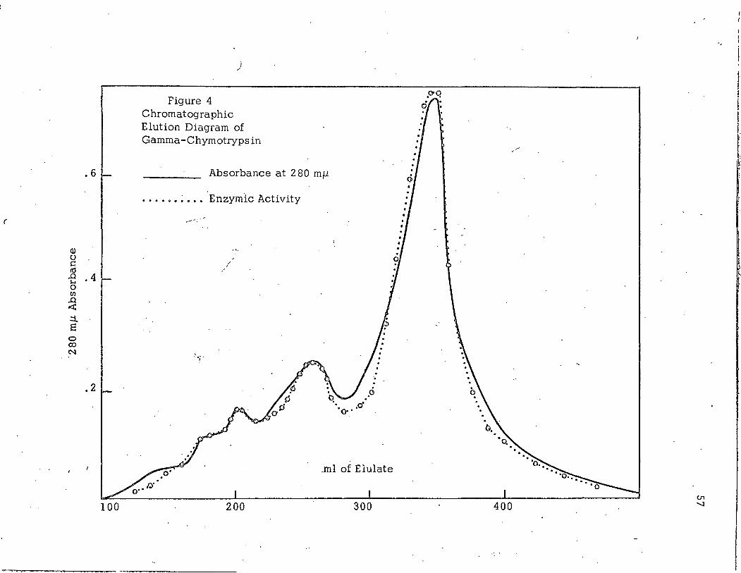
. 6
(
Q)
0 c ~ .0 . 4 s,... 0 (/)
.0 .::X: .::!.. s
0 <X> N ..
. 2
100
i /
Figure 4 Chromatographic Elution Diagram of Gamma-Chymotrypsin
Absorbance at 2 80 mJl
••.••• ~ ••. Enzymic Activity
/
.o
.ml of Elulate
200 300
...
1 l I
I ~
400
1.

----·--~- 1
58
Delta-Chymotrypsin. Fig. 5 shows ,the chromatographic elution
pattern for crystalline delta-chymotrypsin. The major active component
is eluted between components I and II of crystalline alpha-chymotrypsin.
Approximately 50% of the eluted protein was present as components I 1 II
and III. All of the eluted protein was enzymically active. The number
over each component in Fig. 5 represent the relative specific enzymic·
activities taking the specific enzymic activity of 3X crystallized alpha
chymotrypsin as 1. 00.
On the basis of the foregoing data we conclude that component II
(Fig. 1) is alpha-chymotrypsin. Component III is either gamma- or
beta-chymotrypsin and component I remains unidentified.
"Slow" Activation Mixture of Chymotrypsinogen-A. Fig. 6 shows
the e'lution diagram obtained by chromatographing the mixture of enzymes
produced when chymotrypsinogen-A is acted upon by trypsin under the
conditions of "slow" activation (15). Under these conditions most of
the protein should be alpha-chymotrypsin. Components corresponding
to delta- I alpha-, and beta- or gamma-chymotrypsin are present. Alpha
chymotrypsin represents the major enzymically active component. The
numbers over the peaks .refer to the relative enzymic specific activities
based on the 2 80 mfJ. absorbance and the rate of ATEE hydrolysis.
Conversion of Alpha- to Beta- and Gamma-Chymotrypsin. A
solution of 3X crystallized alpha-chymotrypsin was allowed to incubate . .
for two weeks under the condition for the conversion. of alpha-chymotryp-
sin to beta- and gamma-chymotrypsin (2 I 15). The. entire incubation
mixture was chromatographed on an analytical column. Fig. 7 shows
the elution diagram obtained. An appreciable fraction of the protein
has been converted to the beta- and gamma- forms but most of the protein
remains as alpha-chymotrypsin.
Components II and III (Fig. 1) of crystalline alpha-chymotrypsin
were isolated from a 3g' run on a preparative column, desalted and . .
lyophilized. These two components had the same amino acid analysis
/
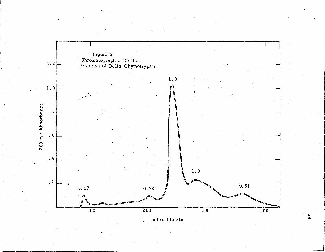
1.2
1.0
Q) 0 I:!
. s I ctl ·,..Q ~ 0 (I)
,..Q ~
::t . 6 E
0 co N .L
• 4
~ 2
i /
Figure 5 Chromatographic Elution ~iagram of Delta-Chymotrypsin
--
/ I
~,1
0.97
200
1.0
ml of Elulate
/
30C 400 U1 w
·-
. f
I
I J I'
fl II

• 6
Q) 0 s:: ttl
..0 r.... 0 (/)
.. 4 ..0 .::t: ::i. 8
0 ro N
~; i
. 2
100
:
I ._,
mi. Elulate
200
Figure 6 Chromotagraphic Elution Diagram of a "Slow·• Activation Mixture
, of Chymotrypsinogen-A
300 400
-.. ...... ::x .,..~ .-· ,'4 .. >.
O'l 0
·-

Q) u t:: 10
• 6
-E • 4 -0 Ul
..0 .:X: :i s
0 CX)
.·· N
• 2
100
I /
Figure 7 Chromatographic Elution Diagram of Alpha-Chymotrypsin Stored for 2 weeks at pH7. 75, 5°.
Absorbancy at 2 80 mj.l
Enzymic Activity
' . ~;. .
... ·.
200
. - -·-- ----------·
300 ml Elulate
..
400
f
I I I. I i ~

1,·
62
which agreed well with the published amino acid analysis for alpha
chymotrypsin (8). They both gave tyrosine and leucine as the major
products of carboxypeptidase-A digestion I and alanine and isoleucine
as N-terminal groups by the method of Sanger (16). They had identical
specific absorbance at 2 80 mJ.l and identical K for ATEE. However I . m the V for component III appeared to be about 10% less than the V
max . max of component II with ATEE as a substrate. There was no reliable differ-
ence in the rates of casein hydrolysis. These data agreed with that
puhJi shed previously on alpha- I beta-and gamma-chymotrypsin.
Gel Electrophoresis. All electrophoretic runs were done in 8 M
urea. Enzyme preparations were labeled in solutions of FDIP-32 at pH
7. 00 {12). The solutions of chymotrypsin-DIP-32 were divided into
two parts. One part was applied directly to the electrophoresis column.
The other was reduced with thioethanol before electrophoresis. Since
FDIP reacts selectively with the active site serine any protein in the
electrophoresis gel which contained appreciable radioactivity must have
originated from an active molecule.
Fig. 8 shows the distrib~tion of radioactivity and non-reduced
protein in the gels. Crystalline preparations of alpha- and gamma
chymotrypsin yield two radioactive bands. Most of the radioactivity
incorporated into beta-chymotrypsin is associated with one broa~ band.
Crystalline alpha-chymotrypsin contains appreciable amounts of either
beta- or gamma-chymotrypsin. Crystalline gamma-chymotrypsin
contains appreciable amounts of alpha-chymotrypsin while cryst!3-lline
beta-chymotrypsin contains only small amounts of alpha-chymotrypsin
but may contain other enzymically active species •.
Fig. 9 shows the distribution of radioactivity on the electro
phoretic gels of reduced crystalline alpha- 1 beta-, and gamma-chymo
trypsin-DIP-32. Relative distance of each band from the front band
wa·s used to construct the sketches. The radioactivity associated with
the third electrophoretic band may represent non-specific labeling and

)
Distance from the Front
(") ·--=-_ ... -., 0 r-i
>< s::
·.-I
I E
"'-.. Ul .... s:: ;::l 0 0
Alpha-Chymotrypsin-DIP-32
Distance from the Front
- -
Beta-Chymotrypsin-DIP- 32 Figure 8
Distance from the Front
Gamma-Chymotrypsin-DIP-32
.,
I I l !
·I
f I
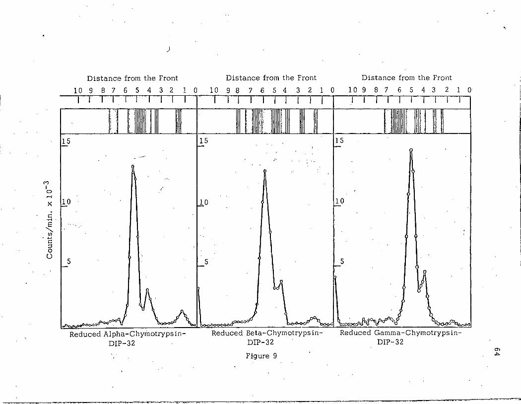
(\')
I 0 ......
. s:: .......
~ U) - ...... s:: ;::s 0 0
)
Distance from the Front Distance from the Front Distance from the Front
10 9 8 7 6 5 4 3 2 1 0 10 9 8 7 6 5 4 3 2 1 0 10 9 8 7 6 5 4 3 2 1 0
I I I I I I I I I I '·,
' i . .)
II \ ..
~5
·-·--
.. !
10 f-
·-.
.
' 5 -
1'>. A ~ ~ Reduced Alpha-Chymotrypsin
DIP-32
15 ~
~0
~5
l I I I I I I I I I m f : ~~ 'I I
r ( ·I. '.
'
- -
' lJl
.J ~ LA~
Reduced Beta-ChymotrypsinDIP-32
Figure 9
.I I I I I I I I I I
,!~ r I
til
It t
I f
fl . ..
15 r--
..
10 -
5 -
~ .~ ~
Reduced Gamma-ChymotrypsinDIP-32
' 65
will not be considered further in this discussion'.' The major radioactivity
is assumed to be associated with the C-chain. Thus 1 the second major
band in each electrophoretic pattern represents the C-chain.
Polyacrylamide gels act as molecular sieves and larger protein
molecules move more slowly under the influence of an applied field.
Thus 1 the slowest moving major component in each of th"e gels should
be the B-chain 1 the fastest major component should be the A-chain and
the C-chain should have an intermediate mobility. The position of the
C-chain is compatible with this scheme and we have tentatively assigned
the order of B 1 C 1 A to the separated chains in order of increasing
electrophoretic mobility.
The major reduced chains of alpha-:- 1 beta- and gamma-chymotryp
sin have essentially identical electrophoretic mobpities in this system.
Each preparation shows more than the expected three bands. Reduced
alpha-chymotrypsin shows five bands suggesting that some of the
molecules of this preparation may have an additional vwteolytic clcw.vage
in one of the three chains. Reduced beta- and gamma-chymotrypsin show
at least six bands each and possibly more.
DISCUSSION
The heterogeneity of crystalline alpha-chymotrypsin has received
--, renewed attention in rece11-t publications (18-21). This report describes
the first separation of the enzymically active components of chymotrypsin.
by ion exchange chromatography. The system described has been used
to separate the enzymically active components of crystalline alpha 1
beta- 1 gamma- and delta-chymotrypsin. Beta- and gamma-chymotrypsin
have the same chromatographic mobilities in this system. Alpha- and
delta-chymotrypsin can be separated from each other, and from beta-
·and gamma-chymotrypsin.
/

-- ----- --··--- -· '" ·--· -· _ ......... "-""------ ·~·-...~.o·-------~~--------------------1
\
\ 66
The major enzymically active components in the alpha family
of chymotrypsins (alpha 8 beta and gamma) seem to be common to all
preparations. Crystalline delta-chymotrypsin also contains contam
inations by components of the alpha family. Each crystalline prepara
tion investigated contained about 5% of the total protein as an
enzymically active component which did not correspond to any of the
known chymotrypsin. This heterogeneity of the alpha family is
particularly interesting when one considers that the preparations have
been shown to have the same amino acid analysis 1 N- and C- terminal
end groups (5-7) 1 molecular weight and enzymic properties (22). In
non-denaturing solvents ·they have nearly the same isoelectric points
(18). They differ in rate of urea denaturation and 1 as we have shown 1
in chromatographic mobilities.
The electrophoretic study shows that non-reduced protein contain-
ing the active site serine has different mobilities for alpha- 1 beta- a.nd
gamma-chymotrypsin. Th~s observation could be explained by two
possibilities. (a) Beta- and gamma-:-chymotrypsin may have additio'nal
proteolytic cleavages which result in the separation of part of the
molecule from the remainder of the molecule in 8 M urea or (b) these
molecules may have a disulfide arrangement which allows separation of
one or more oi the chains of the molecule from the C-chain in 8 M urea.
The first hypothesis should result in hewN- and C-terminal
groups and an altered electrophoretic mobility of one or more of the
reduced chain of the molecules·. This possibility is inconsistent with
published results. The second hypothesis would not introduce new end '
· groups and the reduced chains of these molecules would ·have identical~
electrophoretic mobilities. The reduced chains of alpha- 1 beta- and
gamma-chymotrypsin have nearly identical electrophoretic mobilities.
Therefore 1 beta- and gamma-chymotrypsin probably differ from alpha
chymotrypsin in the disulfide arrangements. Beta- and gamma-chymotryp
sin may have a peptide chain which is associated with the remainder of
the molecule only through non-covalent interactions.

'· 67
It has been assumed that the disulfide arrangement of any protein
molecule is determined by the amino acid sequence of the molecule I and
represents the ·most thermodynamically stable state (2 3-2 5). In this
discussion we have assumed that alpha-chymotrypsin has the same
disulfide arrangement as chymotrypsinogen-A. Alpha-chymotrypsin does
not have the same amino acid sequence as chymotrypsinogen-A. Two
dipeptides have been removed resulting in the conversion of a· single
chain molecule to a three chain molecule. It may be that the disulfide
arrangement of alpha-chymotrypsin is not the most thermodynamically
stable state for the three chain molecule. More stable disulfide
arrangements of the three chain molecule may be represented by beta
and gamr.1a chymotrypsin.
SUMMARY
An ion exchange chromatographic system has been develop~d
which is capable of separating various crystalline chymotrypsins into
enzymically active components. This technique was utilized to
investigate heterogeneity of varl.oU:s crystalline chymotrypsins.
Crystalline alpha- I beta- I gamma- 1 and delta-chymotrypsin were shown
to contain several enzymically active components. Each crystalline
preparation contained contaminations of other chymotrypsins.
Crystalline alpha- 1 beta- 1 and gamma-chymotrypsin were
investigated with "disc" gel electrophoresis in 8 M urea. The major
enzymically active component of these preparations have different
electrophoretic mobilities in this system. The reduced chains of
crystalline alpha- 1 beta-, and gamma-chymotrypsin have identical
electrophoretic mobilities. These data suggest that these enzymes
may differ from each other in their disulfide arrangements.

,., .
j ., 'l I l . ,
i I
l i
, r ~.
REFERENCES
1. Henderson, L. E., and Tolbert, B. M., Fed. Proc., 23, 314,(1964).
2. Hartley, B. S., Nature, ~26, 1284,(1964).
3. Kt:.-:-J.tz, ~·L 1 L Ger:. P:--ss:cl., 221 207 ,(1938}.
4. Bartl, P., Sedlacek, B., andKacena, V., Chern. Listy, 4·9, 4601 (1955).
5. Gladner, J. A., and Neurath, H., L Biol. Chern., 205, 345,(1955); Ibid, 206, 911,(1954).
6. Rovery, M., Fabre, C., and Desnuelle, P., Biochim. Biophys. Acta. 1
. lQ 1 4 8lt (l ~53) o
7. Rovery, M .. , Gabeloteau, C., de Vernejoul, P., Guidoni; A.; and Desnuelle, P., Biochim. Biophys. Acta., 32, 256,(1958) •
. . 8. Desnuelle, P., in Boyer, P. D., Lardy, H., and Myrback, K.,
(Editors), The Enzymes, Vol. 4, Academic Press, Inc. , New York, 1960, p. 93.
9. Schwert, G. W., and Takenaka, Y., Biochim~ Biophys. Acta ,1.6, 5 7 0 1 (1 9 55) o
10. Reisfeld, R. A., Lewis, U. J., and-Williams, D. E., Nature, 195, 281,(1962}.
11. Maize!, J. V. Jr., Ann. NewYorkAcad. Sci., 121, 382,(1964). -------- - -- .
12. Jansen, E. F. ,·and Balls, A. K., L Biol. Chern., 194, 721,(1952).
13. Jansen, E. F., Jang, R., and Balls, A. K. ,j. Biol. Chern., 196, 2 4 7 ,(1 9 52) •
14. Schaffer, N. K., May, S.C. Jr., and Summerson, W. H., L Biol., Chern., 202, 67
1 (1953); Ibid, 206, 201,(1954}.
15. Laskowski, M.·, inColowisk, S. P., andKaplan, N. 0., (Editors}, Methods· in Enzymology, Vol. 2, Academic Press, Inc. , New York, 195~ 1 p. 8.
---·~--- '~

--- . -----· ----~--------------....._ _____________________________ _
69
16. Sanger, F. , Biochem. L , 45, 563,(1949).
17. Raacke, L D., Arch. Biochem. Biophys., .6.2.., 184 ~1956).
18. Egan, R., Michel, H. 0., Schlueter, R., andJandorf, Biochem·. Biophys .. , .§.§_, 354 ~1957).
B. J., Arch. --~
19. Hofstee, B. H. J,, L Biol. Chern., 238, 3235i(l963).
20. Erlanger, B. F., Cooper, A. G., and Bendich, A. J., Bioche.mistry, 3., 1880,(1964).
21. Kezdy, F. J,, and Bender, M. L.,Biochemistr¥, ,1, 104,(1965).
22. Cunningham, L. W., and Brown, C. S., L Biol. Chern., 221, 287, (1956).
23. Anfinsen, C. B., Comp. Biochem. Physiol., _1,229,(1962).
24. Epstein, C. J., Goldberger, R. F., and Anfinsen, C. B., Cold Spring Harbor .§.v!:!!p. Quant. Biol. , 1§_, 43 9 ,(1963).
25. Schachman, H. K., Cold Spring Harbor Symp. Quant. Biol., 1§_, 409, · (1963).
\. ;
\
'.
,. ' . '
···~

·-.,
70
Figure 1. Column Chromatographic Elution Diagram of 3X Crystallized
Alpha-Chymotrypsin. The elution diagram was obtained by
eluting 25 mg of protein from a 0.8 x 200 em columi] of Bio
Rex 70 with 0. 1 M sodium citrate pH 5. 60 at 4 o. The enzymic
activity (• -· -·-·)was fitted to the 280 mJ.J. absorbance (-}
at point (0) .
. Figure 2. Column Chromatography Elution Diagram of Purified Component
III of 3X Crystallized Alpha-Chymotrypsin: The same column , /
conditions were employed here. as for Figure 1 to elute 2 5 mg
of protein. Numbers over the components represent the
relative specific enzymic activity taking that 3X crystallized
Alpha-Chymotrypsin as L 00.
Figure 3. Column Chromatographic Elution Diagram of the Soluble Fraction
of 50 mg of 2X crystallized Beta-Chymotrypsin: The same
column conditions were employed here as for Figure 1. Enzymic
·activity (· -· -·) and 2 80 mJ.l absorbance (-) are plotted on the
same coordinates as in Figure 1.
Figure 4. Column Chromatographic Elution Diagram of 50 mg of Gamma
Chymotrypsin: The same column conditions were employed
here as for Figure 1. Enzymic activity (· -· -·) and 280 mJ.J.
absoroance (_:.._) are plotted on the same coordinates as in
Figure 1.
Figure 5. Column Chromatographic Elution Diagram at 50 mg of Delta
Chymotrypsin: The same column conditions were employed
here as for Figure 1. Absorbance at 2 80 mJ.l (-) is plotted
on a different scale than in Figure 1. Numbers over the
components represent relative specific enzymic activity as
in Figure 2.
I ~