chvH Locus of Agrobacterium Encodes a Homologue of an ...
10
JOURNAL OF BACTERIOLOGY, 0021-9193/01/$04.000 DOI: 10.1128/JB.183.1.36–45.2001 Jan. 2001, p. 36–45 Vol. 183, No. 1 Copyright © 2001, American Society for Microbiology. All Rights Reserved. The chvH Locus of Agrobacterium Encodes a Homologue of an Elongation Factor Involved in Protein Synthesis WEN-TAO PENG, 1 † LOIS M. BANTA, 2 ‡ TREVOR C. CHARLES, 3 AND EUGENE W. NESTER 1 * Department of Microbiology, University of Washington, Seattle, Washington 98195-7242 1 ; Department of Biology, Haverford College, Haverford, Pennsylvania 19041 2 ; and Department of Biology, University of Waterloo, Waterloo, Ontario N2L 3G1, Canada 3 Received 13 July 2000/Accepted 6 October 2000 The virulence of Agrobacterium tumefaciens depends on both chromosome- and Ti plasmid-encoded gene products. In this study, we characterize a chromosomal locus, chvH, previously identified by TnphoA mutagen- esis and shown to be required for tumor formation. Through DNA sequencing and comparison of the sequence with identified sequences in the database, we show that this locus encodes a protein similar in sequence to elongation factor P, a protein thought to be involved in peptide bond synthesis in Escherichia coli. The analysis of vir-lacZ and vir-phoA translational fusions as well as Western immunoblotting revealed that the expression of Vir proteins such as VirE2 was significantly reduced in the chvH mutant compared with the wild-type strain. The E. coli efp gene complemented detergent sensitivity, virulence, and expression of VirE2 in the chvH mutant, suggesting that chvH and efp are functionally homologous. As expected, ChvH exerts its activity at the posttranscriptional level. Southern analysis suggests that the gene encoding this elongation factor is present as a single copy in A. tumefaciens. We constructed a chvH deletion mutant in which a 445-bp fragment within its coding sequence was deleted and replaced with an omega fragment. On complex medium, this mutant grew more slowly than the wild-type strain, indicating that elongation factor P is important but not essential for the growth of Agrobacterium. Agrobacterium tumefaciens causes crown gall disease in a wide range of dicotyledonous plants. The disease, character- ized by neoplastic transformation at the site of infection, re- sults from the transfer and expression of oncogenes from the bacterium to susceptible plant cells (for a review, see reference 27). This transfer process is governed primarily by the products of the vir genes located on the Ti plasmid. These genes are tightly regulated and are expressed to a significant level only in the presence of plant signal molecules synthesized by wounded plant cells. The vir genes are transcriptionally regulated by the Ti plasmid-encoded VirA/VirG two-component regulatory sys- tem (26, 52). The VirA protein senses the plant signal mole- cules and then transduces the signal by phosphate transfer to the response regulator, the VirG protein. The activated VirG protein is a positive transcriptional activator of itself and all other Ti plasmid-encoded vir operons. Numerous chromosomal virulence genes (chv) have also been shown to play important roles in the ability of Agrobac- terium to transform plants (for a review, see reference 39). In general, the functions of chromosomal virulence genes have not been well elucidated, and mutations in these genes are pleiotropic. Consequently, their precise roles in tumor forma- tion have been difficult to assess. An analysis of a limited number of chv mutants suggests that whereas vir genes on the Ti plasmid are dedicated solely to specific steps in the inter- action of Agrobacterium with host plants, the chromosomal virulence genes play important roles in the general physiology of Agrobacterium and have been conscripted to play ancillary but significant roles in the interaction of this bacterium with its hosts. The best-understood chromosomal virulence gene is chvE, which codes for a glucose-galactose periplasmic binding pro- tein. It normally functions in the uptake of a number of monosaccharides into the bacterial cell and is also involved in chemotaxis towards these sugars. In the transformation pro- cess, this periplasmic protein interacts with these same monosaccharides, all of which are components of the plant cell wall. It then binds to the periplasmic domain of the VirA sensor molecule (26), a requirement for maximum activation of VirG and the subsequent activation of all Ti plasmid-en- coded vir genes. Depending on the strain, chvE mutants are either avirulent or severely attenuated in tumor formation on a wide variety of host plants. Another chv locus, acvB, is unusual in that some strains of A. tumefaciens have a functional copy (virJ) on the Ti plasmid. Only by studying a strain that lacks a copy on the Ti plasmid was this chv locus identified as one that is required for T-DNA transfer (29, 41, 54). The relationship between the chromo- somal locus and its Ti plasmid counterpart is unknown, as is the precise role that this locus plays in tumor formation. However, the identification of functionally redundant loci raises the pos- sibility that a similar situation may hold for other Ti plasmid or chv loci, which complicates their isolation and identification. Some chv genes have homologues in other bacteria that also display close interactions with host cells, either plant or animal. A. tumefaciens, Brucella abortus, and Sinorhizobium meliloti all belong to the same -2 subdivision of the proteobacteria ac- cording to 16S rRNA sequence analysis. These three genera require similar chromosomal loci to establish a relationship * Corresponding author. Mailing address: Department of Microbi- ology, Box 357242, University of Washington, Seattle, WA 98195-7242. Phone: (206) 616-8588. Fax: (206) 543-8297. E-mail: gnester@u .washington.edu. † Present address: Department of Biological Sciences, University of Calgary, AB, Canada T2N 1N4. ‡ Present address: Department of Biology, Williams College, Wil- liamstown, MA 01267. 36
Transcript of chvH Locus of Agrobacterium Encodes a Homologue of an ...

JOURNAL OF BACTERIOLOGY,0021-9193/01/$04.00!0 DOI: 10.1128/JB.183.1.36–45.2001
Jan. 2001, p. 36–45 Vol. 183, No. 1
Copyright © 2001, American Society for Microbiology. All Rights Reserved.
The chvH Locus of Agrobacterium Encodes a Homologue of anElongation Factor Involved in Protein Synthesis
WEN-TAO PENG,1† LOIS M. BANTA,2‡ TREVOR C. CHARLES,3 AND EUGENE W. NESTER1*Department of Microbiology, University of Washington, Seattle, Washington 98195-72421; Department of Biology,
Haverford College, Haverford, Pennsylvania 190412; and Department of Biology, University of Waterloo, Waterloo,Ontario N2L 3G1, Canada3
Received 13 July 2000/Accepted 6 October 2000
The virulence of Agrobacterium tumefaciens depends on both chromosome- and Ti plasmid-encoded geneproducts. In this study, we characterize a chromosomal locus, chvH, previously identified by TnphoA mutagen-esis and shown to be required for tumor formation. Through DNA sequencing and comparison of the sequencewith identified sequences in the database, we show that this locus encodes a protein similar in sequence toelongation factor P, a protein thought to be involved in peptide bond synthesis in Escherichia coli. The analysisof vir-lacZ and vir-phoA translational fusions as well as Western immunoblotting revealed that the expressionof Vir proteins such as VirE2 was significantly reduced in the chvH mutant compared with the wild-type strain.The E. coli efp gene complemented detergent sensitivity, virulence, and expression of VirE2 in the chvH mutant,suggesting that chvH and efp are functionally homologous. As expected, ChvH exerts its activity at theposttranscriptional level. Southern analysis suggests that the gene encoding this elongation factor is presentas a single copy in A. tumefaciens. We constructed a chvH deletion mutant in which a 445-bp fragment withinits coding sequence was deleted and replaced with an omega fragment. On complex medium, this mutant grewmore slowly than the wild-type strain, indicating that elongation factor P is important but not essential for thegrowth of Agrobacterium.
Agrobacterium tumefaciens causes crown gall disease in awide range of dicotyledonous plants. The disease, character-ized by neoplastic transformation at the site of infection, re-sults from the transfer and expression of oncogenes from thebacterium to susceptible plant cells (for a review, see reference27). This transfer process is governed primarily by the productsof the vir genes located on the Ti plasmid. These genes aretightly regulated and are expressed to a significant level only inthe presence of plant signal molecules synthesized by woundedplant cells. The vir genes are transcriptionally regulated by theTi plasmid-encoded VirA/VirG two-component regulatory sys-tem (26, 52). The VirA protein senses the plant signal mole-cules and then transduces the signal by phosphate transfer tothe response regulator, the VirG protein. The activated VirGprotein is a positive transcriptional activator of itself and allother Ti plasmid-encoded vir operons.
Numerous chromosomal virulence genes (chv) have alsobeen shown to play important roles in the ability of Agrobac-terium to transform plants (for a review, see reference 39). Ingeneral, the functions of chromosomal virulence genes havenot been well elucidated, and mutations in these genes arepleiotropic. Consequently, their precise roles in tumor forma-tion have been difficult to assess. An analysis of a limitednumber of chv mutants suggests that whereas vir genes on theTi plasmid are dedicated solely to specific steps in the inter-
action of Agrobacterium with host plants, the chromosomalvirulence genes play important roles in the general physiologyof Agrobacterium and have been conscripted to play ancillary butsignificant roles in the interaction of this bacterium with its hosts.
The best-understood chromosomal virulence gene is chvE,which codes for a glucose-galactose periplasmic binding pro-tein. It normally functions in the uptake of a number ofmonosaccharides into the bacterial cell and is also involved inchemotaxis towards these sugars. In the transformation pro-cess, this periplasmic protein interacts with these samemonosaccharides, all of which are components of the plant cellwall. It then binds to the periplasmic domain of the VirAsensor molecule (26), a requirement for maximum activationof VirG and the subsequent activation of all Ti plasmid-en-coded vir genes. Depending on the strain, chvE mutants areeither avirulent or severely attenuated in tumor formation ona wide variety of host plants.
Another chv locus, acvB, is unusual in that some strains of A.tumefaciens have a functional copy (virJ) on the Ti plasmid.Only by studying a strain that lacks a copy on the Ti plasmidwas this chv locus identified as one that is required for T-DNAtransfer (29, 41, 54). The relationship between the chromo-somal locus and its Ti plasmid counterpart is unknown, as is theprecise role that this locus plays in tumor formation. However,the identification of functionally redundant loci raises the pos-sibility that a similar situation may hold for other Ti plasmid orchv loci, which complicates their isolation and identification.
Some chv genes have homologues in other bacteria that alsodisplay close interactions with host cells, either plant or animal.A. tumefaciens, Brucella abortus, and Sinorhizobium meliloti allbelong to the same "-2 subdivision of the proteobacteria ac-cording to 16S rRNA sequence analysis. These three generarequire similar chromosomal loci to establish a relationship
* Corresponding author. Mailing address: Department of Microbi-ology, Box 357242, University of Washington, Seattle, WA 98195-7242.Phone: (206) 616-8588. Fax: (206) 543-8297. E-mail: [email protected].
† Present address: Department of Biological Sciences, University ofCalgary, AB, Canada T2N 1N4.
‡ Present address: Department of Biology, Williams College, Wil-liamstown, MA 01267.
36
between the bacteria and their hosts. One set of such genesrequired for the virulence of A. tumefaciens is a chromosomallyencoded two-component regulatory system, chvG and chvI (12,38). Insertion mutations in either chvG (the sensor histidineprotein kinase) or chvI (the response regulator) render A.tumefaciens avirulent. Similar two-component regulatory sys-tems critical for endosymbiosis or virulence were found in S.meliloti (15, 40) and B. abortus (48). The similarity of thesetwo-component regulatory systems is accentuated further bythe contiguous phosphoenol-pyruvate carboxykinase gene inall three species.
Another set of genes required for tumor formation by A.tumefaciens is chvA/chvB. These two genes are concerned witheither the synthesis (chvB) or the transport (chvA) of a cyclic
polysaccharide, #-1,2 glucan, into the periplasm. Both S. me-liloti and B. abortus synthesize #-1,2 glucan, and both ndvA andndvB are required for effective nodule invasion by S. meliloti(20). For B. abortus, the cgs gene, which complements an S.meliloti ndvB mutant and an A. tumefaciens chvB mutant, isalso required for virulence (28).
In this report we characterize another chromosomal locus,chvH, previously shown to be required for tumor formation.We show that this locus encodes a homologue of the Esche-richia coli elongation factor P (2, 3, 4).
MATERIALS AND METHODS
Bacterial strains, plasmids, and media. The bacterial strains and plasmidsused in this study are listed in Table 1. A. tumefaciens strains were grown in
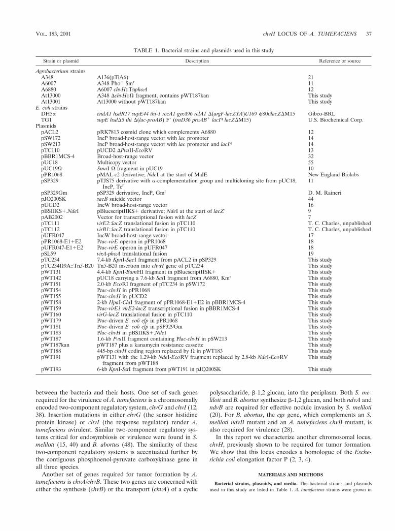
TABLE 1. Bacterial strains and plasmids used in this study
Strain or plasmid Description Reference or source
Agrobacterium strainsA348 A136(pTiA6) 21A6007 A348 Pho$ Smr 11A6880 A6007 chvH::TnphoA 12At13000 A348 %chvH::& fragment, contains pWT187kan This studyAt13001 At13000 without pWT187kan This study
E. coli strainsDH5" endA1 hsdR17 supE44 thi-1 recA1 gyrA96 relA1 %(argF-lacZYA)U169 '80dlacZ%M15 Gibco-BRLTG1 supE hsd%5 thi %(lac-proAB) F( (traD36 proAB! lacIq lacZ%M15) U.S. Biochemical Corp.
PlasmidspACL2 pRK7813 cosmid clone which complements A6880 12pSW172 IncP broad-host-range vector with lac promoter 14pSW213 IncP broad-host-range vector with lac promoter and lacIq 14pTC110 pUCD2 %PvuII-EcoRV 13pBBR1MCS-4 Broad-host-range vector 32pUC18 Multicopy vector 55pUC19& SmaI & fragment in pUC19 10pPR1068 pMAL-c2 derivative; NdeI at the start of MalE New England BiolabspSP329 pTJS75 derivative with "-complementation group and multicloning site from pUC18,
IncP, Tcr11
pSP329Gm pSP329 derivative, IncP, Gmr D. M. RaineripJQ200SK sacB suicide vector 44pUCD2 IncW broad-host-range vector 16pBSIIKS!.NdeI pBluescriptIIKS! derivative; NdeI at the start of lacZ( 9pAB2002 Vector for transcriptional fusion with lacZ 7pTC111 virE2::lacZ translational fusion in pTC110 T. C. Charles, unpublishedpTC112 virB1::lacZ translational fusion in pTC110 T. C. Charles, unpublishedpUFR047 IncW broad-host-range vector 17pPR1068-E1!E2 Ptac-virE operon in pPR1068 18pUFR047-E1!E2 Ptac-virE operon in pUFR047 18pSL59 virA-phoA translational fusion 19pTC234 7.4-kb KpnI-SacI fragment from pACL2 in pSP329 This studypTC234&9A::Tn5-B20 Tn5-B20 insertion into chvH gene of pTC234 This studypWT131 4.4-kb KpnI-BamHI fragment in pBluescriptIISK! This studypWT142 pUC18 carrying a 7.6-kb SalI fragment from A6880, Kmr This studypWT151 2.0-kb EcoRI fragment of pTC234 in pSW172 This studypWT154 Ptac-chvH in pPR1068 This studypWT155 Ptac-chvH in pUCD2 This studypWT158 2-kb HpaI-ClaI fragment of pPR1068-E1!E2 in pBBR1MCS-4 This studypWT159 Ptac-virE1 virE2-lacZ transcriptional fusion in pBBR1MCS-4 This studypWT160 virG-lacZ translational fusion in pTC110 This studypWT179 Ptac-driven E. coli efp in pPR1068 This studypWT181 Ptac-driven E. coli efp in pSP329Gm This studypWT183 Plac-chvH in pBSIIKS!.NdeI This studypWT187 1.6-kb PvuII fragment containing Plac-chvH in pSW213 This studypWT187kan pWT187 plus a kanamycin resistance cassette This studypWT188 445-bp chvH coding region replaced by & in pWT183 This studypWT191 pWT131 with the 1.29-kb NdeI-EcoRV fragment replaced by 2.8-kb NdeI-EcoRV
fragment from pWT188This study
pWT193 6-kb KpnI-SstI fragment from pWT191 in pJQ200SK This study
VOL. 183, 2001 chvH LOCUS OF A. TUMEFACIENS 37

MG/L (35) or induction medium (IM) (11) at either 22 or 28°C. E. coli strainswere grown in Luria-Bertani medium (46) at 37°C. The following antibiotics wereused at the indicated concentrations when added to solid medium (in micro-grams per millimeter): for A. tumefaciens, kanamycin (100), gentamicin (100),and spectinomycin (250); and for E. coli, carbenicillin (100), kanamycin (30),gentamicin (5), spectinomycin (50), and tetracycline (15). These concentrationswere reduced by one-half for liquid medium.
Construction of plasmids. Several plasmids were constructed to demonstratethat chvH is the only gene responsible for the defects of the chvH mutant. A2.0-kb EcoRI fragment from pTC234 containing the chvH gene was cloned intopSW172 to give pWT151. A 1.4-kb NdeI-EcoRI fragment starting from thepredicted start site of the chvH gene was ligated to a 5.3-kb NdeI-EcoRI fragmentof pPR1068 to create pWT154. In this construct, the chvH gene is under thecontrol of the Ptac promoter. A 2-kb EcoRV fragment of pWT154 containing thechvH gene was cloned into pUCD2 to create pWT155.
To test whether the E. coli efp gene can complement the defects of strainA6880, we placed the E. coli efp gene under the control of the Ptac promoter.Two primers, efp-1, GGCCATATGGCAACGTACTATAGCAAC, and efp-2,ACACTGCAGTTACTTCACGCGAGAGAC, were used to amplify the efp cod-ing region from DH5" genomic DNA with Pfu. NdeI and PstI restriction siteswere introduced into primers efp-1 and efp-2, respectively. PCR amplificationfollowed the usual methods using Pfu: denaturation temperature, 95°C for 45 s;annealing temperature, 55°C for 60 min; and polymerization temperature, 72°Cfor 2 min. The 0.57-kb E. coli efp PCR product (digested with NdeI and PstI) wasligated with a 5.3-kb NdeI-PstI fragment of pPR1068 to create pWT179. A 1.2-kbEcoRV-PstI fragment of pWT179 containing the Ptac-driven efp gene was clonedinto pSP329Gm which had been digested with SmaI and PstI to create pWT181.
To determine the transcription of the Ptac-virE operon, pWT159 was con-structed as follows. A 2-kb HpaI-ClaI fragment of pPR1068-E1!E2 was clonedinto SmaI- and ClaI-digested pBBR1MCS-4, giving pWT158. A 4.5-kb EcoRIfragment containing the lacZ-Gmr cassette from pAB2002 was ligated withEcoRI-digested pWT158 to create pWT159.
DNA sequencing and analysis. Restriction fragments were subcloned intopBluescript II KS! or pBluescript II SK!. All double-stranded DNA templateswere prepared with a Qiagen kit. Sequencing was completed with universalforward and reverse primers as well as synthetic oligonucleotides deduced fromalready determined sequences (BRL). DNA sequencing was performed with aBigDye Terminator cycle sequencing ready reaction kit (PE Applied Biosystems,Foster City, Calif.), and the reactions were run on an ABI 377 automated DNAsequencer at the DNA sequence facility of the Biochemistry Department, Uni-versity of Washington. Both strands were sequenced. DNA sequences wereanalyzed with GeneJockey. Protein homology searches were performed using theBlastp program at the National Center for Biotechnology Information.
To determine the precise insertion site of TnphoA in the original chvH mutantA6880, we cloned the phoA and flanking chvH region. Total chromosomal DNAof A6880 was digested with SalI, ligated with SalI-digested pUC18, and trans-formed into strain DH5", selecting for simultaneous resistance to carbenicillinand kanamycin. The resulting plasmid, pWT142, was used as the template tosequence across the insertion junction by using an oligonucleotide primer, 5(-ACCCGTTAAACGGCGAGCACCGCCGGG-3( (part of the phoA sequence).
vir gene expression assays. The reporter plasmid pTC111 is derived frompSM358cd (49, 50), which contains a Tn3HoHo1 insertion in the virE2 gene. Thisresulted in the production of a VirE2-LacZ fusion protein. The reporter plasmidpTC112 is derived from pSM243cd (49, 50), which contains a Tn3HoHo1 inser-tion in the virB1 gene, resulting in the production of a VirB1-LacZ fusionprotein. pWT160 is derived from pSM321cd (49, 50) and contains a Tn3HoHo1insertion in the virG gene, resulting in a chimeric VirG-LacZ fusion protein thathas approximately 130 amino acids of the VirG protein located at the aminoterminus. pSL59 is a virA-phoA translational fusion plasmid (19). The re-porter plasmids were introduced into A348 and A6880 by electroporation. virgene expression assays were performed basically according to publishedmethods (43). Alkaline phosphatase assays were performed as describedpreviously (19).
Protein gels and Western immunoblotting. Protein analysis using sodiumdodecyl sulfate-polyacrylamide gel electrophoresis (SDS-PAGE) was performedaccording to standard protocols (1). Gels were either stained with Coomassieblue or processed for Western blot analysis. Proteins were transferred to poly-vinylidene difluoride membranes (Millipore) using the Trans-Blot SD semidryelectrophoretic transfer cell (Bio-Rad Laboratories) and detected with the ECLWestern blotting analysis system (Amersham Life Science). To detect VirA andVirB proteins, proteins were transferred to nitrocellulose in a Tris-glycine-meth-anol transfer buffer using a Transblot apparatus (Hofer, San Francisco, Calif.).vir genes in A. tumefaciens were induced as described previously (11). A348 and
A6880 cells were harvested from overnight cultures grown in MG/L medium andwashed once with induction medium containing 200 )M AS. The cells were thendiluted into fresh induction medium containing 200 )M acetosyringone (AS) toan optical density at 600 nm (OD600) of 0.1 and then induced for 20 h at 28°C (or16 h at 22°C for VirA and VirB proteins). Total crude extracts were prepared andsubjected to electrophoresis (the polyacrylamide concentrations were 7% forVirA; 10% for VirB, VirE2, and VirD2; and 12% for VirG, VirJ, ChvE, and Ros)and immunoblotting analysis as described previously (6, 43). Polyclonal an-tibodies against proteins of VirA (a gift from S. Winans) or VirB (6), VirE2and VirD2 (18), and VirJ (41) were used to detect Vir proteins. A monoclonalVirG antibody (a gift from S. Jin) was used to detect VirG. Antiserum specificto ChvE (43) or Ros (34) was used to detect ChvE and Ros, respectively.Protein concentrations were determined by using the Bio-Rad protein assaykit (Bio-Rad Laboratories, Richmond, Calif.) with bovine serum albumin asthe standard.
Southern blot analysis. A. tumefaciens genomic DNA for hybridization anal-ysis was prepared by a published method (12). Genomic DNA was digested withrestriction enzymes, subjected to gel electrophoresis, and transferred to a Hy-bond-N! membrane under alkaline conditions. The membranes were hybridizedin Church buffer (0.5 M NaHPO4, 7% SDS, 1 mM EDTA) at 65°C. Two 30-minwashes were performed in 2* SSC–0.1% SDS (1* SSC is 0.15 M NaCl plus0.015 M sodium citrate) at 65°C for low-stringency conditions. For high-strin-gency washes, two 30-min washes in 0.5* SSC–0.1% SDS and one 30-min washin 0.1* SSC–0.1% SDS were performed. Radiolabeled probes were prepared bya random oligonucleotide labeling procedure using the Ready To Go DNAlabeling beads (without dCTP) from Amersham Pharmacia Biotech Inc.
Construction of strain At13000. To determine whether chvH is essential forviability of Agrobacterium, strains were constructed that contained a single func-tional copy of chvH under the control of the inducible lac promoter. The 1.4-kbNdeI-EcoRI fragment starting from the predicted start site of chvH was ligatedwith pBSIIKS!.NdeI digested with NdeI and EcoRI to create pWT183. In thisconstruct the expression of chvH was under the control of the lac promoter. A1.6-kb PvuII fragment containing the Plac-driven chvH was cloned into blunt-ended EcoRI-digested pSW213, which can replicate in Agrobacterium and con-tains lacIq, resulting in pWT187. A 1.6-kb BamHI fragment containing a kana-mycin resistance cassette was inserted at the BamHI site of pWT187 to givepWT187kan.
To construct a deleted version of the chvH mutation, pWT183 was digestedwith HindIII and ClaI, deleting a 445-bp fragment within the chvH coding region.A 2-kb SmaI fragment containing the spectinomycin-resistant & cassette wasinserted between these sites to give pWT188. The 2.8-kb NdeI-EcoRV fragmentcontaining & was subcloned into the large NdeI-EcoRV fragment of pWT131 tocreate pWT191. The 6-kb KpnI-SstI fragment of pWT191 was treated with T4DNA polymerase and cloned into SmaI-digested pJQ200SK, which resulted inconstruct pWT193. pJQ200SK contains two selection markers for the subsequenthomologous recombination step: aacC1, conferring gentamicin (Gm) resistance,and sacB, conferring sucrose sensitivity. In construct pWT193, a 445-bpHindIII-ClaI fragment within the coding sequence of chvH was replaced by the& fragment. Both pWT187kan and pWT193 were transformed into A348, andKanr Spr colonies were selected (single crossover). The resulting strains had atotal of three chvH copies: a functional chvH copy controlled by the lac promoter,a chvH wild-type gene, and a deleted version of the chvH gene replaced by the& fragment. To isolate strains that carried a single functional chvH gene con-trolled by the lac promoter, sucrose selection was carried out on these Kanr Spr
colonies. The Kanr Spr colonies were grown overnight in MG/L medium con-taining 1 mM IPTG (isopropyl-#-D-thiogalactopyranoside) and then spread ontoAB plates (11) containing 5% sucrose, 1 mM IPTG, spectinomycin, and kana-mycin. The Sucr Kanr Spr colonies were then tested for the loss of the Gmr
marker on plates containing IPTG. The Sucr Kanr Spr Gms colonies were furthercharacterized by Southern blotting to identify the strain (At13000) which had asingle functional chvH gene controlled by the lac promoter in a plasmid and adeleted version of chvH in the chromosome.
Assays for virulence. Virulence assays were performed on Kalanchoe daigre-montiana leaves. A. tumefaciens cells were grown in liquid MG/L to mid-logphase, then pelleted by centrifugation, concentrated, and deposited onto leaveswounded with a toothpick. Inoculated plants were grown for 2 to 5 weeks beforetumor formation was scored.
Nucleotide sequence accession number. The nucleotide sequence of the 4.4-kbKpnI-BamHI fragment carried on pTC234 has been submitted to the GenBankdatabase and assigned accession number AF177860.
38 PENG ET AL. J. BACTERIOL.

RESULTS
General features of chvH mutant A6880. The chvH mutantA6880, obtained by TnphoA mutagenesis of A6007 (11, 12), isan avirulent, pleiotropic mutant. It is far more sensitive todetergents such as SDS, sodium deoxycholate, and Sarkosylthan the parent strain (12) and also to carbenicillin comparedto the parental strain (data not shown). This suggests that theintegrity of the outer membrane is impaired. The growth rateof the mutant on an enriched medium is somewhat slower thanthat of the wild-type strain (Fig. 1).
chvH locus encodes a protein similar to elongation factor P.To gain insight into the possible function of chvH, we first isolateda cosmid clone, pACL2, which complemented the detergentsensitivity and restored virulence to chvH mutant A6880 (12).We subcloned a 7.4-kb KpnI-SacI fragment from pACL2 into
pSP329 to make plasmid pTC234. This plasmid also comple-mented the chvH mutant. A partial restriction map of pTC234was then constructed (Fig. 2). TnphoA (36, 37) and Tn5-B20(47) were used to mutagenize the 7.4-kb fragment in order tolocalize the region required for complementation of strain A6880.Several insertions in a 2.0-kb EcoRI fragment (the insert inpWT151) (Fig. 2) abolished complementation (data not shown).
The 4.4-kb KpnI-BamHI fragment of pTC234 was se-quenced (GenBank accession number AF177860), and threeopen reading frames (ORFs) were identified (ORF1, ORF2,and ORF3) (Fig. 2). The first one, extending from nucleotides771 to 2225, is preceded by a ribosome-binding site (GGGAAA) and encodes a putative protein of 484 amino acids with apredicted molecular mass of 51,000 Da. The second ORF(nucleotides 2346 to 2915, complementary strand) with its po-
FIG. 1. Growth curves. At time zero, overnight cultures were diluted in MG/L at a starting OD600 of 0.06 and incubated at 28°C with shaking.At the indicated times, the OD600 was measured and plotted against time. The growth curves shown represent a typical experiment.
FIG. 2. Restriction map of A. tumefaciens chromosomal DNA inserts in pTC234, pWT151, and pWT155. Restriction sites are labeled as follows:B, BamHI; E, EcoRI; EV, EcoRV; H, HindIII; K, KpnI; N, NdeI; S, SacI; X, XhoI. The arrowhead under ORF2 indicates the site of the TnphoAinsertion, while the arrow underneath indicates its orientation.
VOL. 183, 2001 chvH LOCUS OF A. TUMEFACIENS 39

tential ribosome-binding site (AGGAAG) encodes a putativeprotein of 189 amino acids with a predicted molecular mass of21,000 Da. A third ORF was identified between nucleotides3167 and 4231, which codes for a predicted protein of 354amino acids with a molecular mass of 39,000 Da.
A database search of the predicted amino acid sequence ofthe three polypeptides revealed that ORF1 has a high level ofidentity with NodT from Rhizobium leguminosarum (52% iden-tity and 68% similarity) (51). NodT is a member of a growingfamily of outer membrane proteins found in a wide variety ofgram-negative bacteria, including several pathogens (42).ORF3 shows a high degree of similarity to lysyl-tRNA syn-thetase from a large number of bacteria.
ORF2 contains the site of the TnphoA insertion of the avir-ulent mutant A6880, which has been designated chvH. Com-paring the predicted amino acid sequence of ChvH with theprotein database revealed that ChvH is similar at the aminoacid sequence level to a range of elongation factor P proteins.Elongation factor P has been implicated in peptide bond syn-thesis (2). The predicted amino acid sequences for the A.tumefaciens ChvH and the elongation factor P proteins forRickettsia prowazekii and E. coli are aligned in Fig. 3 using theMalign program. The identity and similarity at the amino acidlevel are 39 and 62% for R. prowazekii and 36 and 57% for E.coli, respectively.
TnphoA is a transposon that can fuse alkaline phosphataselacking a signal peptide to the amino-terminal sequences of theproteins into whose genes it inserts. Active fusions expressingalkaline phosphatase can arise only when this transposon in-serts in genes encoding secreted or membrane-spanning pro-teins. Sequence analysis suggests that ChvH is a soluble pro-tein. The TnphoA insertion in A6880 is located between bp 123and 124 of the chvH ORF, and chvH is in the opposite orien-tation relative to the phoA gene of TnphoA (Fig. 2), althoughthe insertion was initially identified after screening a collectionof mutants that expressed alkaline phosphatase activity. How-ever, when we checked colonies of A6880 growing on MG/Lplates containing XP (5-bromo-4-chloro-3-indolylphosphate),they were white, as would be predicted. Two potential rho-independent transcriptional terminators (nucleotides 2244 to2279 and 2299 to 2326) were identified, one downstream of thechvH gene and the other downstream of the nodT homologue.The isoelectric point of ChvH, calculated from the sequence, is5.13.
Complementation of the chvH mutation. To prove that theTnphoA insertion is actually responsible for the several defectsobserved in strain A6880, we complemented the mutation withchvH-containing subclones of the complementing cosmid. Twoplasmids, pWT151 and pWT155, were constructed (see Fig. 2and Materials and Methods). pWT151 contains the chvH geneunder the control of its native promoter and flanking DNAsequences, while pWT155 contains the chvH gene under thecontrol of the tac promoter. Both pWT151 and pWT155 com-plemented the detergent sensitivity (Fig. 4A) and restoredvirulence of A6880 on K. daigremontiana (for complementa-tion by pWT155, see Fig. 5). These data strongly suggest thatdisruption of chvH is responsible for both the avirulence anddetergent sensitivity of A6880.
We also tested whether the E. coli efp gene could comple-ment the chvH mutation. The efp gene from E. coli was clonedunder the control of the tac promoter to form plasmidpWT181. This gene (pWT181) complemented the detergentsensitivity of the chvH mutant strain (Fig. 4A) and increasedthe growth rate (data not shown). Most importantly, the E. coli
FIG. 3. Amino acid alignment of the A. tumefaciens (Atu) elonga-tion factor P protein with the elongation factor P proteins from R.prowazekii (Rpr) (AJ235271) and E. coli (Eco) (X61676). Amino acidsare represented by the single-letter code. The Malign program wasused for the comparison. Amino acid positions are indicated on theright. Amino acid residues that are identical in two of the sequencesare shaded.
FIG. 4. (A) Functional complementation of chvH mutant A6880.Cells were grown on MG/L plates containing 0.2 g of SDS per liter at28°C and observed after 3 days. (B) Measurement of VirE2 expression.A. tumefaciens strains were grown in IM containing 200 )M AS at 28°Cfor 20 h. Cells were pelleted, and total crude extracts were subjected toimmunoblotting analysis as described in Materials and Methods.Strains: 1, A6007; 2, A6880(pWT155); 3, A6880(pWT181); 4,A6880(pSP329Gm).
40 PENG ET AL. J. BACTERIOL.

efp gene (pWT181) restored virulence to A6880 (Fig. 5), al-though it took longer for tumors to appear and the tumorswere smaller than those formed by the construct with the tacpromoter-driven chvH itself (pWT155). Thus, the tac promot-er-driven E. coli efp can phenotypically complement the chvHmutant, although the E. coli gene does not function optimallyin A. tumefaciens. chvH and efp are functionally homologous.
Regulation of the chvH gene. Plasmid pTC234&9A::Tn5-B20is a derivative of pTC234 in which Tn5-B20 has inserted intothe 2.0-kb EcoRI fragment containing the chvH gene. TheTn5-B20 insertion also abolished the complementation abilityof pTC234. Tn5-B20 forms an operon fusion with the gene intowhich it inserts. We transformed A348 with pTC234&9A::Tn5-B20. The cells were grown in (i) AB (pH 7.5) or AB plus50 mM MES (morpholineethanesulfonic acid, pH 5.5) or IMwith and without AS. The #-galactosidase activities were de-
termined. The results showed that chvH gene is constitutivelyexpressed, independent of acid conditions and phenolic com-pounds (data not shown).
Reduced expression of virulence genes in chvH mutant.Since the chvH mutation rendered cells avirulent, we investi-gated how well the Ti plasmid-encoded vir genes were ex-pressed in the mutant. An appropriate level of expression ofthe vir genes is critical for tumor formation. Plasmid pTC112containing the virB1::lacZ translational fusion was introducedinto the chvH mutant and the wild-type strain. As shown inTable 2, expression of the virB-lacZ fusion was reduced 80% inthe chvH mutant compared with the wild-type strain under thesame conditions. We also introduced a virE2-lacZ translationalfusion on a plasmid (pTC111) into the same strains. TheVirE2-LacZ fusion protein in the chvH mutant was assayedunder inducing and noninducing conditions. Under noninduc-ing conditions, we observed that the basal level of VirE2-LacZexpression was reduced approximately 70% in the chvH mu-tant compared with the wild-type strain. Under inducing con-ditions, the expression of VirE2-LacZ was reduced approxi-mately 85%.
The above results clearly show that the expression of VirBand VirE2 is reduced in the chvH mutant. Because the viroperons are under the control of the VirA/VirG two-compo-nent system, chvH might reduce the level of VirB and VirE2proteins indirectly by affecting the expression of the two-com-ponent system. Therefore, the level of VirA was determined byassaying the phoA activity of a virA-phoA translational fusion(pSL59) in the two backgrounds. As shown in Table 2, theinduction of VirA expression by AS in the wild-type strainagrees with previous observations (45, 53), while the expres-sion of VirA was only induced slightly less in the chvH mutant.To measure VirG expression, we used virG-lacZ translationalfusion plasmid pWT160. Under noninducing conditions, VirG-LacZ expression was reduced 40% in the chvH mutant (Table2). In the presence of AS, the reduction was substantiallygreater (approximately 75%), probably in part because of thepositive autoregulation of VirG (52) (Table 2).
We also analyzed the expression of vir genes by measuringthe accumulation of several Vir proteins by Western immuno-blotting. As shown in Fig. 6, the levels of VirB8, -9, -10, and -11were reduced dramatically in the mutant cells compared withthose of the wild-type strain. VirE2 and VirJ were undetect-able in the chvH mutant. The level of VirD2 was markedlyreduced but still detectable. The levels of both VirA and VirGwere significantly reduced. The expression of VirE2 was alsomeasured in the chvH mutant transformed with the Ptac-driven
FIG. 5. Tumorigenesis assay on Kalanchoe leaves. The assay wasperformed as described in Materials and Methods.
TABLE 2. Effect of chvH mutation on expression of lacZ and phoA translational fusions to vir genesa
Fusion (plasmid)
#-Galactosidase expression (Miller units)
chvH strain (A348) chvH::TnphoA strain (A6880)
IM IM ! 100 )M AS IM IM ! 100 )M AS
virB-lacZ (pTC112) ND 1,357 + 5 ND 285 + 6 (80%)virE-lacZ (pTC111) 46.6 + 1.0 3,208 + 46 15.1 + 1.0 (70%) 519 + 15 (85%)virG-lacZ (pWT160) 77.5 + 1.5 1,536 + 39 48.0 + 2.4 (40%) 360 + 10 (75%)virA-phoA (pSL59) 6.9 + 0.7 59 + 1.5 (8.6) 6.9 + 0.2 42 + 1.0 (6)
a Data are means + standard errors of the mean for three samples. The percentages in parentheses represent the reduction in expression in the chvH mutant relativeto the wild-type strain. The numbers in parentheses are the fold increases in induction upon adding AS. The units are Miller units. ND, not determined.
VOL. 183, 2001 chvH LOCUS OF A. TUMEFACIENS 41
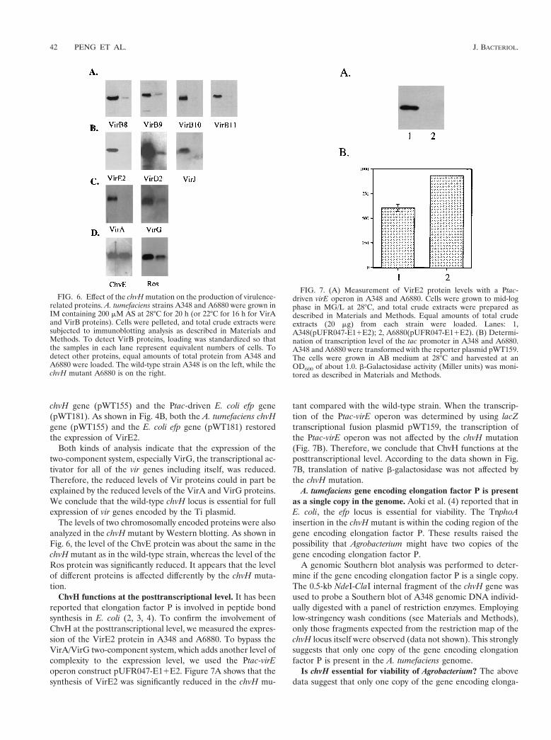
chvH gene (pWT155) and the Ptac-driven E. coli efp gene(pWT181). As shown in Fig. 4B, both the A. tumefaciens chvHgene (pWT155) and the E. coli efp gene (pWT181) restoredthe expression of VirE2.
Both kinds of analysis indicate that the expression of thetwo-component system, especially VirG, the transcriptional ac-tivator for all of the vir genes including itself, was reduced.Therefore, the reduced levels of Vir proteins could in part beexplained by the reduced levels of the VirA and VirG proteins.We conclude that the wild-type chvH locus is essential for fullexpression of vir genes encoded by the Ti plasmid.
The levels of two chromosomally encoded proteins were alsoanalyzed in the chvH mutant by Western blotting. As shown inFig. 6, the level of the ChvE protein was about the same in thechvH mutant as in the wild-type strain, whereas the level of theRos protein was significantly reduced. It appears that the levelof different proteins is affected differently by the chvH muta-tion.
ChvH functions at the posttranscriptional level. It has beenreported that elongation factor P is involved in peptide bondsynthesis in E. coli (2, 3, 4). To confirm the involvement ofChvH at the posttranscriptional level, we measured the expres-sion of the VirE2 protein in A348 and A6880. To bypass theVirA/VirG two-component system, which adds another level ofcomplexity to the expression level, we used the Ptac-virEoperon construct pUFR047-E1!E2. Figure 7A shows that thesynthesis of VirE2 was significantly reduced in the chvH mu-
tant compared with the wild-type strain. When the transcrip-tion of the Ptac-virE operon was determined by using lacZtranscriptional fusion plasmid pWT159, the transcription ofthe Ptac-virE operon was not affected by the chvH mutation(Fig. 7B). Therefore, we conclude that ChvH functions at theposttranscriptional level. According to the data shown in Fig.7B, translation of native #-galactosidase was not affected bythe chvH mutation.
A. tumefaciens gene encoding elongation factor P is presentas a single copy in the genome. Aoki et al. (4) reported that inE. coli, the efp locus is essential for viability. The TnphoAinsertion in the chvH mutant is within the coding region of thegene encoding elongation factor P. These results raised thepossibility that Agrobacterium might have two copies of thegene encoding elongation factor P.
A genomic Southern blot analysis was performed to deter-mine if the gene encoding elongation factor P is a single copy.The 0.5-kb NdeI-ClaI internal fragment of the chvH gene wasused to probe a Southern blot of A348 genomic DNA individ-ually digested with a panel of restriction enzymes. Employinglow-stringency wash conditions (see Materials and Methods),only those fragments expected from the restriction map of thechvH locus itself were observed (data not shown). This stronglysuggests that only one copy of the gene encoding elongationfactor P is present in the A. tumefaciens genome.
Is chvH essential for viability of Agrobacterium? The abovedata suggest that only one copy of the gene encoding elonga-
FIG. 6. Effect of the chvH mutation on the production of virulence-related proteins. A. tumefaciens strains A348 and A6880 were grown inIM containing 200 )M AS at 28°C for 20 h (or 22°C for 16 h for VirAand VirB proteins). Cells were pelleted, and total crude extracts weresubjected to immunoblotting analysis as described in Materials andMethods. To detect VirB proteins, loading was standardized so thatthe samples in each lane represent equivalent numbers of cells. Todetect other proteins, equal amounts of total protein from A348 andA6880 were loaded. The wild-type strain A348 is on the left, while thechvH mutant A6880 is on the right.
FIG. 7. (A) Measurement of VirE2 protein levels with a Ptac-driven virE operon in A348 and A6880. Cells were grown to mid-logphase in MG/L at 28°C, and total crude extracts were prepared asdescribed in Materials and Methods. Equal amounts of total crudeextracts (20 )g) from each strain were loaded. Lanes: 1,A348(pUFR047-E1!E2); 2, A6880(pUFR047-E1!E2). (B) Determi-nation of transcription level of the tac promoter in A348 and A6880.A348 and A6880 were transformed with the reporter plasmid pWT159.The cells were grown in AB medium at 28°C and harvested at anOD600 of about 1.0. #-Galactosidase activity (Miller units) was moni-tored as described in Materials and Methods.
42 PENG ET AL. J. BACTERIOL.

tion factor P is present in Agrobacterium. Therefore, two pos-sibilities exist: (i) the gene encoding elongation factor P isessential for the viability of Agrobacterium but the TnphoAinsertion is leaky, and (ii) the gene is not essential for theviability of Agrobacterium. To distinguish between these twopossibilities, we constructed a deletion mutant of chvH andcharacterized the resulting strain.
We first placed the chvH gene under the control of theIPTG-inducible lac promoter (in plasmid pWT187kan; see Ma-terials and Methods). This construct was then transferred into A.tumefaciens A348 by electroporation. We then exchanged thewild-type copy of chvH with the deleted version of the chvH gene(a 445-bp HindIII-ClaI fragment was deleted) in the presence ofIPTG, giving strain At13000 (see Materials and Methods).
To determine whether chvH is an essential gene, we firststreaked out the At13000 cells on MG/L plates lacking IPTGto see whether the depletion of the ChvH protein stopped cellgrowth. The depletion did not stop cell growth. The cellsformed colonies on solid medium and also increased in opticaldensity in liquid medium, suggesting that chvH is not an es-sential gene. If this is indeed the case, it should be possible tocure strain At13000 of plasmid pWT187kan without affectingcell viability. To this end, we grew strain At13000 under non-selective conditions overnight in liquid medium and plated outthe culture for single colonies on MG/L plates containing spec-tinomycin. Among 300 colonies, 20 colonies were kanamycinsensitive. These were candidates for cells that had lost theplasmid. Southern blot analysis of 10 of these colonies con-firmed that they indeed did not contain pWT187kan (data notshown). The cured strain was named At13001. Since a strainwhich lacked the chvH gene could be isolated, the gene prod-uct is not essential for cell viability.
Elongation factor P is necessary for optimum growth of A.tumefaciens. We investigated the effect of elimination of thechvH locus on cell growth in rich medium, MG/L. As shown inFig. 1, the deletion mutant At13001 grew significantly morepoorly than the TnphoA insertion mutant A6880, suggestingthat the original mutant was leaky for elongation factor Pactivity. The deletion mutant also exhibited a longer lag timethan the TnphoA insertion mutant. The effect of an introducedwild-type chvH gene and the E. coli efp gene on the growth ofstrain At13001 was also studied. As expected, the chvH geneexpressed from the Ptac promoter (pWT155) increased thegrowth rate to nearly the level of the wild-type strain A348(data not shown). Interestingly, the E. coli efp gene expressedfrom the Ptac promoter (pWT181) increased the growth rateto about the same extent as the introduced chvH gene (datanot shown). As expected, strain At13001 is avirulent. BothpWT155 and pWT181 restored virulence to strain At13001 (datanot shown).
A 32-kDa protein accumulates in the chvH mutant. Theeffect of the chvH mutation in A6880 on the protein profile wasexamined on an SDS-PAGE gel. Figure 8 shows representativedata for the patterns of soluble proteins in the chvH mutantA6880 and its parental strain A6007 when equal amounts ofprotein were loaded on the gel. The most striking difference isthat a 32-kDa protein accumulated in the chvH mutant. Sev-eral other less striking differences were observed between themutant and parental strains. The 32-kDa protein also accumu-
lated in the chvH deletion mutant At13001 (data not shown).We are in the process of identifying this protein.
DISCUSSION
In this study, we have characterized the chvH chromosomalvirulence locus, which was originally identified by TnphoA mu-tagenesis as being required for virulence. Sequence analysisindicates that chvH encodes a protein homologous to the elon-gation factor P protein of E. coli (2, 3, 4). This sequencehomology is strongly supported by the observation that expres-sion of the efp gene of E. coli can complement the chvH mutantand restore all of the phenotypic alterations resulting from themutation in Agrobacterium. This is the first demonstration thatan E. coli gene can complement the avirulent phenotype of anA. tumefaciens mutant and speaks to the highly conservednature of this protein.
The exact role of elongation factor P in protein synthesis isnot clear. It was originally isolated from a complex of 70Sribosome-AUG-formyl[35S]Met-tRNA and added puromycin(22, 23). This factor stimulated the synthesis of N-formyl-me-thionyl-puromycin and was thought to be involved in peptidebond synthesis. Further studies indicated that elongation fac-tor P was more effective in increasing the efficiency of peptidebond formation between formyl[35S]Met-tRNA and amino ac-ids with small R groups, such as glycine (24). A recent studyindicates that bacterial elongation factor P is homologous tothe eukaryal/archaeal eIF-5A (33), with the highest homologyat the N-terminal 90 residues. Factor eIF-5A was originallyisolated from a high-salt wash of rabbit reticulocyte ribosomesand was thought to be involved in translation initiation (31).eIF-5A enhanced the synthesis of methionyl-puromycin invitro, suggesting that eIF-5A is involved in the formation of thefirst peptide bond in translation (8). In Saccharomyces cerevi-siae, two genes (TIF51A and TIF51B) code for eIF-5A. Al-though deletion of both eIF-5A genes was lethal, the completedepletion of eIF-5A in the cell led to only a 30% drop in thefirst round of protein synthesis (30). These authors suggestedthat eIF-5A is not absolutely necessary for general proteinsynthesis in eukaryotic cells but is essential for the translationof a subset of specific mRNAs.
FIG. 8. Protein profiles of chvH mutant A6880 and its parentalstrain A6007. A. tumefaciens A6007 and A6880 were grown in IMcontaining 200 )M AS at 28°C for 20 h. Cells were pelleted, and totalcrude extracts were subjected to SDS-PAGE analysis. Equal amountsof total crude extracts (20 )g) from each strain were loaded. Lanes: 1,A6007; 2, A6880. The arrow indicates the 32-kDa protein.
VOL. 183, 2001 chvH LOCUS OF A. TUMEFACIENS 43
A recent study concluded that efp was essential for viabilityand was required for protein synthesis in E. coli (4). We havenot found this to be the case in A. tumefaciens. We have clearlyshown that A. tumefaciens has a single copy of chvH, which canbe disrupted without loss of viability, although the cells growmore slowly and are no longer virulent. Our observations areconsistent with the supposition that elongation factor P in-creases the efficiency of formation of peptide bonds involvingaminoacyl acceptors that bind poorly to the ribosome in itsabsence (25) and argue against the notion that elongationfactor P is required for general protein synthesis.
The virulence genes of A. tumefaciens for which chvH-de-pendent expression was examined in the present study appearto belong to a class of genes whose optimal translation dependson elongation factor P. Expression of other genes that weretested was not affected to the same extent. The elongationfactor P-dependent genes might code for particular sequencesof amino acids, perhaps near the start of translation, that areexceptionally dependent on elongation factor P for translation.An example of this type of specificity is the observation thatwhen the Bacteroides fragilis efp gene was introduced into E.coli, the B. fragilis glutamine synthetase activity increased in E.coli but the activity of B. fragilis sucrase was unaffected (1). Thesignificance of the control of A. tumefaciens virulence geneexpression at the posttranscriptional level is not immediatelyapparent. Further experimental work is necessary to charac-terize the role of specific amino acid sequences on the elon-gation factor P dependence of translation.
We have demonstrated that at least some of the vir genes areregulated posttranscriptionally in a chvH-dependent manner.When virE2 transcription was driven by the tac promoter andthus uncoupled from VirG control, the level of VirE2 proteinwas drastically reduced in the chvH mutant strain, although thelevels of virE2 transcription in the chvH mutant and the wild-type strain were comparable (Fig. 7). The reduction in thelevels of Vir proteins in the chvH mutant thus appears to bedetermined at two stages. In addition to the direct effects ontranslation of vir mRNA, the reduction in the levels of all Virproteins could be partly due to the reduced levels of VirA andespecially VirG, which is rate limiting for vir gene induction(14). The effect of the chvH mutation on the expression ofVirG is likely due to a direct effect on the translation of VirG.However, it is possible that this reduction results from over-production of the 32-kDa protein acting on the translation ofthe vir genes. In this case, the role of chvH would be indirect.The reduction in Vir protein levels at the posttranscriptionallevel raises the possibility that elongation factor P serves as aposttranscriptional regulator of vir gene expression, perhaps inconcert with a second regulatory factor. Identification of the32-kDa protein that accumulated in the chvH mutant mightprovide an insight into this possibility.
The simplest explanation for the avirulence of the chvHmutant is that the levels of key proteins required for T-DNAtransfer are reduced profoundly. These include VirB8, VirB9,VirB10, and VirB11 as well as the single-stranded-DNA-bind-ing protein VirE2. We would predict that the levels of otherVir proteins would also be depressed. However, the possibilitythat the chvH gene product contributes in other ways to tu-morigenesis cannot be ruled out. In this regard, it may be signif-
icant that a number of chromosomal avirulent mutants have incommon a lack of integrity in their outer membrane (12).
ACKNOWLEDGMENTS
This work was supported by Public Health Service grant GM32618from the National Institutes of Health to E. W. Nester and by grantMCB9513662 from the National Science Foundation to L. M. Banta.
We thank S. Jin, Y. Machida, and C. I. Kado for antibodies to VirG,CheE, and Ros, respectively. A. Becker, P. Christie, M. Hynes, andK. M. Peterson kindly provided plasmids. Lishan Chen, Derek Wood,Rad Roberts, Mario Pantoja, and Paul de Figueiredo were helpful ingiving suggestions and engaging in critical discussions of the data. Thetechnical assistance of T. Jackson and M. Stremlau is gratefully ac-knowledged. B. R. Glick is thanked for enlightening discussions onearly elongation factor P research.
REFERENCES
1. Abratt, V. R., M. Mbewe, and D. R. Woods. 1998. Cloning of an EF-Phomologue from Bacteroides fragilis that increases B. fragilis glutamine syn-thetase activity in Escherichia coli. Mol. Gen. Genet. 258:363–372.
2. Aoki, H., S. L. Adams, D. G. Chung, M. Yaguchi, S. E. Chuang, and M. C.Ganoza. 1991. Cloning, sequencing and overexpression of the gene for pro-karyotic factor EF-P involved in peptide bond synthesis. Nucleic Acids Res.19:6215–6220.
3. Aoki, H., S. L. Adams, M. A. Turner, and M. C. Ganoza. 1997. Molecularcharacterization of the prokaryotic efp gene product involved in a peptidyl-transferase reaction. Biochimie 79:7–11.
4. Aoki, H., K. Dekany, S. L. Adams, and M. C. Ganoza. 1997. The geneencoding the elongation factor P protein is essential for viability and isrequired for protein synthesis. J. Biol. Chem. 272:32254–32259.
5. Ausubel, F. M., R. Brent, R. E. Kingston, D. D. Moore, J. G. Seidman, J. A.Smith, and K. Struhl (ed.). 1996. Current protocols in molecular biology.John Wiley & Sons, New York, N.Y.
6. Banta, L., M. J. Bohne, S. D. Lovejoy, and K. Dostal. 1998. Stability of theAgrobacterium tumefaciens VirB10 protein is modulated by growth temper-ature and periplasmic osmoadaption. J. Bacteriol. 180:6597–6606.
7. Becker, A., M. Schmidt, M. Jager, and A. Puhler. 1995. New gentamicin-resistance and lacZ promoter-probe cassettes suitable for insertion mutagen-esis and generation of transcriptional fusions. Gene 162:37–39.
8. Benne, R., M. L. Broen-Luedi, and J. W. Hershey. 1978. Purification andcharacterization of protein synthesis initiation factors eIF-1, eIF-4C, eIF-4D,and eIF-5 from rabbit reticulocytes. J. Biol. Chem. 253:3070–3077.
9. Berger, B. R., and P. J. Christie. 1994. Genetic complementation analysis ofthe Agrobacterium tumefaciens virB operon: virB2 through virB11 are essen-tial virulence genes. J. Bacteriol. 176:3646–3660.
10. Bilge, S. S., J. M. Apostol, Jr., K. J. Fullner, and S. L. Moseley. 1993.Transcriptional organization of the F1845 fimbrial adhesin determinant ofEscherichia coli. Mol. Microbiol. 7:993–1006.
11. Cangelosi, G. A., E. A. Best, G. Martinetti, and E. W. Nester. 1991. Geneticanalysis of Agrobacterium. Methods Enzymol. 204:384–397.
12. Charles, T. C., and E. W. Nester. 1993. A chromosomally encoded two-component sensory transduction system is required for virulence of Agrobac-terium tumefaciens. J. Bacteriol. 175:6614–6625.
13. Charles, T. C., S. L. Doty, and E. W. Nester. 1994. Construction of Agrobac-terium strains by electroporation of genomic DNA and its utility in analysisof chromosomal virulence mutations. Appl. Environ. Microbiol. 60:4192–4194.
14. Chen, C.-Y., and S. C. Winans. 1991. Controlled expression of the transcrip-tional activator gene virG in Agrobacterium tumefaciens by using the Esche-richia coli lac promoter. J. Bacteriol. 173:1139–1144.
15. Cheng, H. P., and G. C. Walker. 1998. Succinoglycan production by Rhizo-bium meliloti is regulated through the ExoS-ChvI two-component regulatorysystem. J. Bacteriol. 180:20–26.
16. Close, T. J., D. Zaitlin, and C. I. Kado. 1984. Design and development ofamplifiable broad-host-range cloning vectors: analysis of the vir region ofAgrobacterium tumefaciens plasmid pTiC58. Plasmid 12:111–118.
17. DeFeyter, R. D., Y. Yang, and D. W. Gabriel. 1993. Gene-for-genes interac-tions between cotton R genes and Xanthomonas campestris pv. malvacearumavr genes. Mol. Plant-Microbe Interact. 6:225–237.
18. Deng, W., L. Chen, W. T. Peng, X. Liang, S. Sekiguchi, M. P. Gordon, L.Comai, and E. W. Nester. 1999. VirE1 is a specific molecular chaperone forthe exported single-stranded-DNA-binding protein VirE2 in Agrobacterium.Mol. Microbiol. 31:1795–1807.
19. Doty, S. L., M. C. Yu, J. I. Lundin, J. D. Heath, and E. W. Nester. 1996.Mutational analysis of the input domain of the VirA protein of Agrobacte-rium tumefaciens. J. Bacteriol. 178:961–970.
20. Dylan, T., L. Ielpi, S. Stanfield, L. Kashyap, C. Douglas, M. Yanofsky, et al.1986. Rhizobium meliloti genes required for nodule development are related
44 PENG ET AL. J. BACTERIOL.

to chromosomal virulence genes of Agrobacterium tumefaciens. Proc. Natl.Acad. Sci. USA 83:4403–4407.
21. Garfinkel, D. J., and E. W. Nester. 1980. Agrobacterium tumefaciens mutantsaffected in crown gall tumorigenesis and octopine catabolism. J. Bacteriol.144:732–743.
22. Glick, B. R., and M. C. Ganoza. 1975. Identification of a soluble protein thatstimulates peptide bond synthesis. Proc. Nat. Acad. Sci. USA 72:4257–4260.
23. Glick, B. R., and M. C. Ganoza. 1976. Characterization and site of action ofa soluble protein that stimulates peptide-bond synthesis. Eur. J. Biochem.71:483–491.
24. Glick, B. R., S. Chladek, and M. C. Ganoza. 1979. Peptide bond formationstimulated by protein synthesis factor EF-P depends on the aminoacyl moi-ety of the acceptor. Eur. J. Biochem. 97:23–28.
25. Glick, B. R. 1980. A molecular model of peptide chain propogation. J. Theor.Biol. 82:149–155.
26. Heath, J. D., T. C. Charles, and E. W. Nester. 1995. Ti plasmid and chro-mosomally encoded two-component systems important in plant cell trans-formation by Agrobacterium species, p. 367–385. In J. A. Hoch and T. J.Silhavy (ed.), Two-component signal transduction. American Society forMicrobiology, Washington, D.C.
27. Hooykaas, P. J. J., and A. G. M. Beijersbergen. 1994. The virulence systemof Agrobacterium tumefaciens. Annu. Rev. Phytopathol. 32:157–179.
28. Inon de Iannino, N., G. Briones, M. Tolmasky, and R. A. Ugalde. 1998.Molecular cloning and characterization of cgs, the Brucella abortus cyclic#(1-2) glucan synthetase gene: genetic complementation of Rhizobium me-liloti ndvB and Agrobacterium tumefaciens chvB mutants. J. Bacteriol. 180:4392–4400.
29. Kalogeraki, V. S., and S. C. Winans. 1995. The octopine Ti plasmid pTiA6of Agrobacterium tumefaciens contains a gene homologous to the chromo-somal virulence gene acvB. J. Bacteriol. 177:892–897.
30. Kang, H. A., and J. W. Hershey. 1994. Effect of initiation factor eIF-5Adepletion on protein synthesis and proliferation of Saccharomyces cerevisiae.J. Biol. Chem. 269:3934–3940.
31. Kemper, W. M., K. W. Berry, and W. C. Merrick. 1976. Purification andproperties of rabbit reticulocyte protein synthesis initiation factors M2B"and M2B#. J. Biol. Chem. 251:5551–5557.
32. Kovach, M. E., P. H. Elzer, D. S. Hill, G. T. Robertson, M. A. Farris, andR. M. Roop II. 1995. Four new derivatives of the broad-host-range cloningvector pBBR1MCS, carrying different antibiotic-resistance cassettes. Gene166:175–176.
33. Kyrpides, N. C., and C. R. Woese. 1998. Universally conserved translationinitiation factors. Proc. Natl. Acad. Sci. USA 95:224–228.
34. Lai, E. M., and C. I. Kado. 1998. Processed VirB2 is the major subunit of thepromiscuous pilus of Agrobacterium tumefaciens. J. Bacteriol. 180:2711–2717.
35. Lichtenstein, C. P., and J. Draper. 1985. Genetic engineering of plants, p.67–119. In D. M. Glover (ed.), DNA cloning: a practical approach, vol. II.IRL Press, Washington, D.C.
36. Long, S., S. McCune, and G. C. Walker. 1988. Symbiotic loci of Rhizobiummeliloti identified by random TnphoA mutagenesis. J. Bacteriol. 170:4257–4265.
37. Manoil, C., and J. Beckwith. 1985. TnphoA: a transposon probe for proteinexport signals. Proc. Natl. Acad. Sci. USA 82:8129–8133.
38. Mantis, N. J., and S. C. Winans. 1993. The chromosomal response regula-tory gene chvI of Agrobacterium tumefaciens complements an Escherichia coliphoB mutation and is required for virulence. J. Bacteriol. 175:6625–6636.
39. Nester, E. W., J. Kemner, W. Deng, Y.-W. Lee, K. Fullner, and X. Liang.
1996. Agrobacterium: a natural genetic engineer exploited for plant biotech-nology, p. 111–120. In G. Stacey, B. Mullin, and P. M. Gresshoff (ed.),Biology of plant-microbe interactions. International Society for MolecularPlant-Microbe Interactions, Minneapolis, Minn.
40. Østerås, M., J. Stanley, and T. M. Finan. 1995. Identification of rhizobium-specific intergenic mosaic elements within an essential two-component reg-ulatory system of Rhizobium species. J. Bacteriol. 177:5485–5494.
41. Pan, S. Q., S. Jin, M. I. Boulton, M. Hawes, M. P. Gordon, and E. W. Nester.1995. An Agrobacterium virulence factor encoded by a Ti plasmid gene or achromosomal gene is required for T-DNA transfer into plants. Mol. Micro-biol. 17:259–69.
42. Paulsen, I. T., J. H. Park, P. S. Choi, and M. H. Saier, Jr. 1997. A family ofgram-negative bacterial outer membrane factors that function in the exportof proteins, carbohydrates, drugs and heavy metals from gram-negative bac-teria. FEMS Microbiol. Lett. 156:1–8.
43. Peng, W.-T., Y.-W. Lee, and E. W. Nester. 1998. The phenolic recognitionprofiles of the Agrobacterium tumefaciens VirA protein are broadened by ahigh level of the sugar binding protein ChvE. J. Bacteriol. 180:5632–5638.
44. Quandt, J., and M. F. Hynes. 1993. Versatile suicide vectors which allowdirect selection for gene replacement in gram-negative bacteria. Gene 127:15–21.
45. Rogowsky, P. M., T. J. Close, J. A. Chimera, J. J. Shaw, and C. I. Kado. 1987.Regulation of the vir genes of Agrobacterium tumefaciens plasmid pTiC58. J.Bacteriol. 169:5101–5112.
46. Sambrook, J., E. F. Fritsch, and T. Maniatis. 1989. Molecular cloning: alaboratory manual, 2nd ed. Cold Spring Harbor Laboratory Press, ColdSpring Harbor, N.Y.
47. Simon, R., J. Quandt, and W. Klipp. 1989. New derivatives of transposonTn5 suitable for mobilization of replicons, generation of operon fusions andinduction of genes in gram-negative bacteria. Gene 80:161–169.
48. Sola-Landa, A., J. Pizarro-Cerda, M. J. Grillo, E. Moreno, I. Moriyon, J. M.Blasco, J. P. Gorvel, and I. Lopez-Goni. 1998. A two-component regulatorysystem playing a critical role in plant pathogens and endosymbionts ispresent in Brucella abortus and controls cell invasion and virulence. Mol.Microbiol. 29:125–38.
49. Stachel, S. E., G. An, C. Flores, and E. W. Nester. 1985. A Tn3-lacZ trans-poson for the random generation of #-galactosidase gene fusions: applica-tions to the analysis of gene expression in Agrobacterium. EMBO J. 4:891–898.
50. Stachel, S. E., and P. C. Zambryski. 1986. virA and virG control the plant-induced activation of the T-DNA transfer process of A. tumefaciens. Cell46:325–333.
51. Surin, B. P., J. M. Watson, W. D. O. Hamilton, A. Economou, and J. A.Downie. 1990. Molecular characterization of the nodulation gene nodT fromtwo biovars of Rhizobium leguminosarum. Mol. Microbiol. 4:245–252.
52. Winans, S. C. 1992. Two-way chemical signaling in Agrobacterium-plantinteractions. Microbiol. Rev. 56:12–31.
53. Winans, S. C., R. A. Kerstetter, and E. W. Nester. 1988. Transcriptionalregulation of the virA and virG genes of Agrobacterium tumefaciens. J. Bac-teriol. 170:4047–4054.
54. Wirawan, I. G., H. W. Kang, and M. Kojima. 1993. Isolation and character-ization of a new chromosomal virulence gene of Agrobacterium tumefaciens.J. Bacteriol. 175:3208–3212
55. Yanisch-Perron, C., J. Vieira, and J. Messing. 1985. Improved M13 phagecloning vectors and host strains: nucleotide sequences of the M13mp18 andpUC19 vectors. Gene 33:103–119.
VOL. 183, 2001 chvH LOCUS OF A. TUMEFACIENS 45