Chromoblastomycosis and sporotrichosis, two endemic but ......Summary Chromoblastomycosis and...
13
GENERAL REVIEW/REVUE GE ´ NE ´ RALE Chromoblastomycosis and sporotrichosis, two endemic but neglected fungal infections in Madagascar T. Rasamoelina a , O. Raharolahy b , N. Rakotozandrindrainy a,c , I. Ranaivo b , M. Andrianarison b , B. Rakotonirina d , D. Maubon e,f , F.A. Rakotomalala a , M. Rakoto Andrianarivelo a , A. Andriantsimahavandy g , F. Rapelanoro Rabenja b , L.S. Ramarozatovo b,h , M. Cornet e,f, * a Centre d’infectiologie Charles-Me ´ rieux, Ankatso, universite ´ d’Antananarivo, BP 4299, 101 Antananarivo, Madagascar b USFR dermatologie-rhumatologie, HUJRB Antananarivo, Madagascar BP 14bis, 101 Antananarivo, Mada- gascar c UPFR parasitologie-mycologie, HUJRA Antananarivo, BP4150, 101 Antananarivo, Madagascar d Institut malgache de recherches applique ´ es, fondation Rakoto-Ratsimamanga, Avarabohitra-Itaosy, BP3833, 101 Antananarivo, Madagascar e Laboratoire de parasitologie-mycologie, CHU de Grenoble-Alpes, CS 10217, 38043 Grenoble cedex 9, France f Laboratoire TIMC-IMAG TheREx, universite ´ de Grenoble-Alpes, domaine de la Merci, 38706 La Tronche cedex, France g Laboratoire d’immunologie, immunopathologie et immunodiagnostic, faculte ´ des sciences, universite ´ d’Antananarivo, BP 906, 101 Antananarivo, Madagascar h Me ´ decine interne-dermatologie pavillon spe ´ cial A CHU de Befelatanana, BP 14bis, 101 Antananarivo, Madagascar Received 3 April 2017; accepted 8 August 2017 Available online 25 August 2017 KEYWORDS Chromoblastomycosis; Sporotrichosis; Madagascar Summary Chromoblastomycosis and sporotrichosis are endemic fungal infections of tropical and subtropical regions, including Madagascar. The causal fungi develop in the soil or on plants and infect humans through wounds, either directly (wounding by the plant, through thorns, for example), or through the contact of an existing wound with contaminated soil. For this reason, the lesions predominantly occur on the limbs, and these fungi principally infect people working Journal de Mycologie Médicale (2017) 27, 312—324 * Corresponding author. Laboratoire de parasitologie-mycologie, CHU de Grenoble-Alpes, CS 10217, 38043 Grenoble cedex 9, France. E-mail address: [email protected] (M. Cornet). Available online at ScienceDirect www.sciencedirect.com http://dx.doi.org/10.1016/j.mycmed.2017.08.003 1156-5233/# 2017 Elsevier Masson SAS. All rights reserved.
Transcript of Chromoblastomycosis and sporotrichosis, two endemic but ......Summary Chromoblastomycosis and...

Journal de Mycologie Médicale (2017) 27, 312—324
Available online at
ScienceDirectwww.sciencedirect.com
GENERAL REVIEW/REVUE GENERALE
Chromoblastomycosis and sporotrichosis,
two endemic but neglected fungal infectionsin MadagascarT. Rasamoelina a, O. Raharolahy b, N. Rakotozandrindrainy a,c,I. Ranaivo b, M. Andrianarison b, B. Rakotonirina d, D. Maubon e,f,F.A. Rakotomalala a, M. Rakoto Andrianarivelo a,A. Andriantsimahavandy g, F. Rapelanoro Rabenja b,L.S. Ramarozatovo b,h, M. Cornet e,f,*
aCentre d’infectiologie Charles-Merieux, Ankatso, universite d’Antananarivo, BP 4299, 101 Antananarivo,MadagascarbUSFR dermatologie-rhumatologie, HUJRB Antananarivo, Madagascar BP 14bis, 101 Antananarivo, Mada-gascarcUPFR parasitologie-mycologie, HUJRA Antananarivo, BP4150, 101 Antananarivo, Madagascard Institut malgache de recherches appliquees, fondation Rakoto-Ratsimamanga, Avarabohitra-Itaosy, BP3833,101 Antananarivo, Madagascare Laboratoire de parasitologie-mycologie, CHU de Grenoble-Alpes, CS 10217, 38043 Grenoble cedex 9, Francef Laboratoire TIMC-IMAG TheREx, universite de Grenoble-Alpes, domaine de la Merci, 38706 La Tronchecedex, Franceg Laboratoire d’immunologie, immunopathologie et immunodiagnostic, faculte des sciences, universited’Antananarivo, BP 906, 101 Antananarivo, MadagascarhMedecine interne-dermatologie pavillon special A CHU de Befelatanana, BP 14bis, 101 Antananarivo,Madagascar
Received 3 April 2017; accepted 8 August 2017Available online 25 August 2017
KEYWORDSChromoblastomycosis;Sporotrichosis;Madagascar
* Corresponding author. LaboratoireE-mail address: mcornet@chu-gren
http://dx.doi.org/10.1016/j.mycmed.1156-5233/# 2017 Elsevier Masson SA
Summary Chromoblastomycosis and sporotrichosis are endemic fungal infections of tropicaland subtropical regions, including Madagascar. The causal fungi develop in the soil or on plantsand infect humans through wounds, either directly (wounding by the plant, through thorns, forexample), or through the contact of an existing wound with contaminated soil. For this reason,the lesions predominantly occur on the limbs, and these fungi principally infect people working
de parasitologie-mycologie, CHU de Grenoble-Alpes, CS 10217, 38043 Grenoble cedex 9, France.oble.fr (M. Cornet).
2017.08.003S. All rights reserved.
Chromoblastomycosis and sporotrichosis, fungal infections in Madagascar 313
outside with bare hands and/or feet. The subcutaneous lesions of chromoblastomycosis areinitially nodular, subsequently becoming warty, tumoral, cauliflower-like and pruriginous, whichpromotes dissemination. The chronic nature of the infection and its progression over long periodslead to highly disabling lesions in essentially rural and agricultural populations. The lesions ofsporotrichosis are also nodular, but more ulcerous, and they form an extended chain followingthe route of the lymph vessels. Pus, squamous or skin biopsy specimens are used for themycological examination of these mycoses. Treatment depends on the severity and form of thelesions and is based on antifungal drugs sometimes combined with physical methods. There hasbeen no study of these infections for more than two decades in Madagascar, despite the largenumbers of cases seen by doctors in all parts of the island. The nature, diversity and distributionof the plants responsible for contamination have not been described in Madagascar. In thisreview, we described these two endemic mycoses in terms of their epidemiological, mycologi-cal, clinical and therapeutic characteristics, focusing particularly on Madagascar, which is one ofthe leading foci of these two infections worldwide.# 2017 Elsevier Masson SAS. All rights reserved.
Introduction
Chromomycosis or chromoblastomycosis (CBM) and sporo-trichosis (SPT) are chronic subcutaneous or cutaneolym-phatic infections found mostly in tropical and subtropicalregions [1—4]. Studies carried out by the Institut Pasteur ofMadagascar between 1955 and 1994 provided an inventoryof the number of cases of CBM and identified this country asthe leading focus of this mycosis worldwide. Mean incidencewas estimated at about 1/200,000 inhabitants at the time[5]. Since 1994, no further studies on CBM have been per-formed in Madagascar and no new data have been obtainedto update the epidemiological situation [6]. CBM is usuallycaused by dematiaceous fungi (also known as black yeasts),principally Fonsecaea pedrosoi and Cladophialophora car-rionii. The causal agent of SPT is Sporothrix schenckii, adimorphic hyphomycete [4,7]. Only sporadic cases havebeen reported in Madagascar, due to the absence of specificsurveillance. SPT may be polymorphic and disseminated,with pulmonary forms.
These fungal infections are typical of so-called ‘‘myco-ses of implantation or inoculation’’, because they arelinked to traumatic inoculation with the fungus throughwounding by the plant or through soil contamination of anexisting wound [8,9]. The chronic course and long durationof lesions may result in considerable disability in theabsence of treatment. Due to the large number of casesworldwide, their geographic distribution, professional ori-gin, preponderance among the poorest populations in theworld and refractory nature, these infections are candida-tes for inclusion in the WHO list of neglected tropicaldiseases. In Madagascar, it is rarely possible to confirmclinically suspected infections or to identify the fungalspecies responsible, because health structures are undere-quipped and resources are limited. Given the large numberof cases currently being seen by doctors throughout theisland, the prevalence of these infections appears to belargely underestimated. In this review, we describe thesetwo fungal infections, focusing on the most recent studiesand the situation in Madagascar. We also report preliminaryresults for the first 28 months of a prospective studycurrently underway, involving the recruitment of patients
with suspect lesions. The confirmation of infection bymycological and molecular biology methods in this studysuggests that these two infections remain highly prevalentin this country.
Chromoblastomycosis
CBM is a phaeohyphomycosis, but it has a number of clinicaland diagnostic features that are pathognomonic and distin-guish it from the other diseases of this group, such asmycetoma. Indeed, CBM is a chronic fungal infection ofthe subcutaneous tissues characterized by warty hyperkera-tosic plaques and the presence of fumagoid cells on micro-scopic examination of squamous samples. The causal fungidevelop on living plants or on decomposing plant material inthe soils of tropical and subtropical regions.
Pathogenic agents
The pathogenic agents responsible for CBM are fungi from thephylum Ascomycetes, mostly from the order Chaetotyriales,and familyHerpotrichiellaceae.This family includes theblackyeasts of the genus Exophiala and dematiaceous filamentousfungi, including the causal agents of CBM: Fonsecaea spp.,Phialophora verrucosa, Cladophialophora carrionii and Rhi-nocladiella aquaspersa [10—12]. F. pedrosoi is particularlyabundant in the Amazon rainforest in Brazil [13]. It was firstisolated by Alexandrino Pedroso in 1911 [14] and was sub-sequently described as Hormodendrum pedrosoi by Brumpt in1922. The genus Fonsecaea was established in 1936, byNegroni [15]. Several species of this genus other thanF. pedrosoi have been implicated in CBM: F. monophora,F. nubica and F. pugnacius. Another species, F. compacta,hasbeen reported tobeamorphological variant ofF.pedrosoi[16,17]. A recent study described two new species isolatedfrom thorny plants: F. erecta and F. minima [18].
P. verrucosa was first described by Medlar in 1915 [10].Several authors have described this fungus as a causal agentof CBM [19] and of phaeohyphomycotic cysts [20].
C. carrionii was first identified in the arid regions of SouthAmerica, Africa and Australia. Trejos described it under the

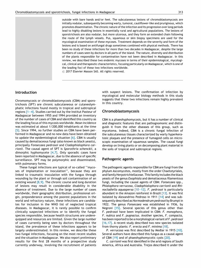
[(Figure_1)TD$FIG]
Figure 1 Thorny plants of Solanum erythrecanthum (A) and Rubus rosifolius (B) growing in the north-north east region ofMadagascar.
314 T. Rasamoelina et al.
name of Cladosporium carrionii [21]. Recent taxonomic stu-dies of pathogenic species have led to a change in the name ofthe genus from Cladosporium to Cladophialophora [22].
R. aquaspersa was described by Borelli, Schell et al. in1972 and has been reported to be responsible for rare casesof warty infections of the skin in Mexico and Brazil [23].
In the environment, the fungi responsible for CBM arefound in soil organic matter, water and on plants [24,25].F. pedrosoi has been isolated principally from decomposingplant matter and soil [26]. According to Salgado et al.,Mimosa pudica, a plant with thorny stems, is a source ofF. pedrosoi infections in Brazil [25]. However, all thesestudies were based on morphological rather than molecularidentification techniques. C. carrionii was reported to bepresent on cactus thorns and fragments, but this finding wassubsequently corrected by de Hoog et al., who identified thespecies present as C. yegresii [27,28]. Another potentialsource of the dematiaceous agents responsible for CBM isthe Palmaceae family (palms), which is widely exploited intropical zones [12,29]. It has also been shown that theidentification of fungi as F. pedrosoi and Cladophialophorasp. on the basis of their morphological features in theenvironment, and on the basis of physiological and fermen-tation tests in the laboratory, may not be confirmed inmolecular biology tests. Caution is therefore required whenattributing responsibility to a particular plant species as thesource of infection [18].
Epidemiology and geographic distribution
CBM is present mostly in the tropical and subtropical regionsof America, Asia and Africa [12]. High prevalences have beenreported in Mexico, Cuba, Venezuela, Colombia and Braziland in Central, Sub-Saharan and North Africa. In his review,Queiroz-Telles described cases in South Africa and Algeria[3,12,30—35]. In Asia, CBM is most frequent in Japan, SriLanka, India and China [1,36—38].
In Madagascar, Guillet and Radaody-Ralarosy reported thefirst probable case of CBM in 1940 [39]. The arrival ofE.R. Brygoo at the Institut Pasteur of Madagascar in 1950
made it possible to authenticate new cases of CBM [11,39]caused by F. pedrosoi and C. carrionii. Studies carried out atthe Institut Pasteur between 1955 and 1994 identified 1343cases of CBM, making this country the leading focus of thissubcutaneous mycosis worldwide, with an estimated preva-lence of 1/200,000 inhabitants at the time. The epidemio-logical data for this disease in Madagascar have not beenupdated since 1994 [5].
Most of the infected patients are men working in agri-culture [5,6,25]. Most are involved in the cropping, by hand,of vanilla, coffee or sugar cane, but some are involved inhusbandry and the rearing of livestock (e.g. pigs or zebucattle) close to plantations. These farmers also engage inwoodcutting and charcoal-producing activities. Due to theinfluence of the trade winds to the east and monsoons to thenorth, Madagascar has two distinctly different climates: ahumid tropical climate, in which F. pedrosoi is found (in theeast, north and north-west) and a semi-arid climate in whichC. carrionii is found (in the south) [5,40]. The humid climateof the north and east is marked by a mean annual precipita-tion of more than 1500 mm and a mean temperature of morethan 15 8C, conditions favoring the multiplication ofF. pedrosoi. There is a considerable natural diversity of plantspecies in this region, including orchids (Orchidaceae) andpalms (Arecaceae). In the northeast, vanilla (Vanilla plani-folia) plantations, woody perfume tree forests, bamboo(Bambuseae), palms and trees of the genus Tambourissapredominate. Thorny plants responsible for transferringthe fungi to humans, such as ‘‘sakoa’’ (Solanum macrocar-pum) and ‘‘angivibe’’ (Solaneum erythrecanthum) andbrambles or ‘‘voarô’’ (Rubus rosifolius) are also abundantin this region (Fig. 1).
By contrast, in the south, precipitation is much lower, at300 to 600 mm, with a mean temperature of more than 20 8Cthroughout the year. Eight to nine months per year are dry. Inthese conditions, C. carrionii predominates. This thornforest zone is characterized by a forest of thorny plants ofthe Didieraceae, together with euphorbias and plants fromthe Apocyanaceae. It also contains sisal (Agave sisalana[41]), which is used for the manufacture of rope, eucalyptus

Chromoblastomycosis and sporotrichosis, fungal infections in Madagascar 315
(Eucalyptus), which is used for charcoal production, andcacti (Alluaudia procera) used in construction materials [6].
In 2012, the Charles Mérieux Infectiology Centre (CMIC)of the University of Antananarivo and the dermatologydepartment of Joseph Raseta Befelatanana University Hos-pital Antananarivo, together with Grenoble-Alpes Univer-sity, initiated a collaborative study designed to updateprevalence data, and to describe the distributions of casesand causal agents in Madagascar. Preliminary results for thefirst 28 months of our prospective study, for the period fromMarch 2013 to June 2015 [42], suggest that the prevalence ofCBM is high, and confirm that F. pedrosoi and C. carrionii arethe principal causal species. Eight cases of CBM wereconfirmed among the 55 patients (15%) included on thebasis of suspicious lesions at a dermatology consultationin the capital or at district hospitals. The species F. pedrosoi(7 cases) in the south and southeast and C. carrionii(1 case) were identified by microscopy, and confirmed bysequencing.
Clinical presentation
Following inoculation, a primary lesion appears as a smallpapule that develops and forms papulosquamous plaques orbecomes nodular within a few months. The proliferation ofthe fungus may then result in a warty florid appearance, orlead to the centrifugal extension of plaques or the formationof large plaques or extended circumferential lesions of theaffected limb at advanced stages.
Plaque forms are generally violet in color, with a well-delimited raised border. The central part of the plaque mayhave healed and may present an infiltrated, squamousappearance (Fig. 2A). In nodular forms, the nodules maybe isolated or multiple and may resemble large soft warts(Fig. 2B). The warty forms are characterized by pink pimpleswith a typical ‘‘cauliflower’’ appearance (Fig. 2C).
[(Figure_2)TD$FIG]
Figure 2 Clinical appearance of chromoblastomycosis on the lokeratosic, weeping, scaly and pruriginous, after more than two yearswith blackish spots, after seven years of progression (B); papulosqupimples, after more than four years of progression (C).
In most cases, the CBM lesions are localised on the lowerlimbs,particularlyon thedorsal surfaceof the feet, theanklesand the legs [43]. These parts of the body are those mostexposed to contamination from plants or soil. Surprisingly,lesions do not occur on the soles of the feet, even though thesubjects at risk walk and work barefoot. The upper limbs andthebuttocks arealso frequently affected. Lesions on theears,face and trunk are less common [44]. The lesion progressesslowly, over a period of two to 20 years. If treatment is absentor insufficient, the lesion extends and becomes highly dis-abling due to the development of elephantiasis-type oedemasor superinfections. Itching and scratching of the lesion favordissemination. The lesions may vary in appearance, but nodistinct forms associated with individual species (C. carrioniior F. pedrosoi) have been described [39]. Malignant trans-formation into epidermoid carcinoma is rare and concernslong-standing, advanced lesions [12,45].
Diagnosis
Direct microscopic examination of specimens
Clinical specimens (biopsy, squamous or pus specimens) areexamined in chlorazol black solution or in a potassium solu-tion (10 to 40% KOH). The presence of characteristic muri-form cells in superficial samples is used to confirm thediagnosis of CBM. These cells are grouped into compartmen-ted ‘‘clusters’’ of 4 to 12 mm in diameter, which are brown incolor and have a thick wall. These clusters consist of acombination of skin cell debris, dried blood and fungalstructures. Some cells may display the first signs of filamen-tation but should nevertheless be distinguished from thefilamentous forms encountered in cases of disseminatedphaeohyphomycosis [12]. These elements have also beendescribed in microabscesses, dermal tissues and infectedepidermis (Fig. 3).
wer limb. Single lesion, 8 cm in diameter, tumorous, scabby,of progression (A); single nodular lesion with a raised border, pinkamous plaque lesions, breaking out into ‘‘cauliflower-shaped’’

[(Figure_3)TD$FIG]
Figure 3 Muriform cells present in skin biopsy specimens from patients with chromoblastomycosis caused by Cladophialophoracarrionii (A) and Fonsecaea pedrosoi (B, C). Photomicrographs ( � 400 magnification) obtained after staining with chlorazol black andmounting of the specimen on a slide with a coverslip.
316 T. Rasamoelina et al.
Histology
Histological sections of the epidermis display hyperplasticlesions and papillomatous proliferation. Hyperacanthosis isobserved much more frequently than dyskeratosis. Muriformcells are present in a granulomatous reaction involving lym-phocytes, plasmocytes and inflammatory cells, includinggiant Langhans cells.
Culture
The detection of muriform cells can be used as the basis ofthe diagnosis, but isolation of the fungus by culture isindispensable for identification of the causal agent andsubsequent treatment, because F. pedrosoi may be lesssusceptible to antifungal drugs than C. carrionii [46,47].The causal agents of CBM grow on the usual culture media,but their growth is slow, and up to six weeks of incubation at27 to 30 8C may be required [1,13]. Selective media contain-ing chloramphenicol and/or cycloheximide do not inhibittheir growth.
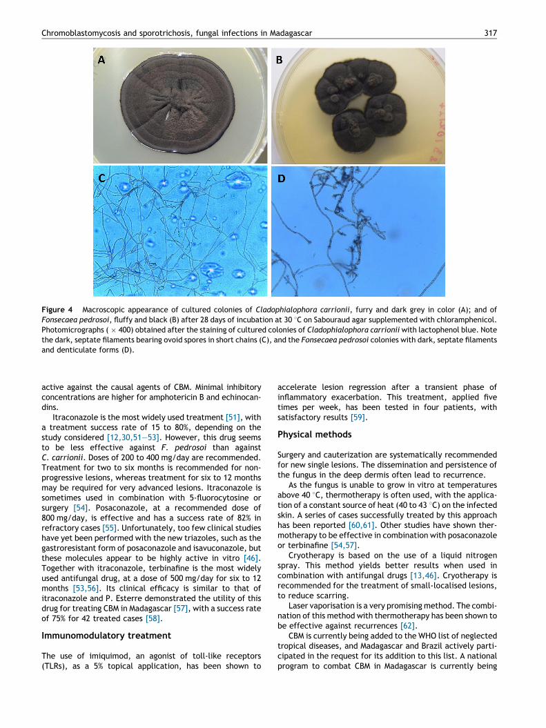
Cladophialophora spp. form powdery black or olive greencolonies. Under the microscope, the conidiophores are long,branched or unbranched and bear long chains of smooth-walled or slightly rough conidia (Fig. 4A, C). Fonsecaea spp.form very hard black colonies. Under the microscope, sep-tate hyphae, cylindrical conidiophores, short phialides withand without collars, denticulate forms and fine-walled coni-dia are visible (Fig. 4B, D).
Microscopic examination can provide identification to thegenus level, but sequencing of the ITS regions of rDNA isessential for identification to species level.
In the ongoing prospective study, diagnosis is confirmedby direct examination and culture at the CMIC and histolo-
gical examination at Joseph Raseta Befelatanana UniversityHospital, Antananarivo. Molecular methods (PCR followed bysequencing and specific PCR) are performed at the CMIC andat Grenoble-Alpes University.
Treatment
Depending on disease severity and the form of the lesions,treatment may be based on antifungal drugs, surgery, laservaporization, thermotherapy or cryotherapy. However,other than for lesions that have progressed very little andare excised very early by surgery, complete cure, withsterilization of the lesions, is rarely possible. The refractorynature of this infection generally renders very long-termtreatment necessary.
Antifungal treatments
The susceptibility of the strains responsible for CBM to anti-fungal drugs in vitro is only partially predictive of treatmentsuccess. Such analyses provide no information about theefficacy of these drugs against the fungal forms present inthe muriform cells, which are probably not very accessible tothese molecules [12].
With this caveat concerning the interpretation of in vitrotests, most systemic antifungal drugs are effective againstFonsecaea spp. and Cladophialophora spp., which are highlysusceptible to triazoles, such as itraconazole, posaconazole,voriconazole and isavuconazole, but not to fluconazole [47—50]. Posaconazole seems to be the most active of the triazolesagainst these fungi. F. pedrosoi has proved less susceptiblethan F. nubica and F. monophora to voriconazole and isavu-conazole [46]. Terbinafine, a non-triazole compound, is also
[(Figure_4)TD$FIG]
Figure 4 Macroscopic appearance of cultured colonies of Cladophialophora carrionii, furry and dark grey in color (A); and ofFonsecaea pedrosoi, fluffy and black (B) after 28 days of incubation at 30 8C on Sabouraud agar supplemented with chloramphenicol.Photomicrographs ( � 400) obtained after the staining of cultured colonies of Cladophialophora carrionii with lactophenol blue. Notethe dark, septate filaments bearing ovoid spores in short chains (C), and the Fonsecaea pedrosoi colonies with dark, septate filamentsand denticulate forms (D).
Chromoblastomycosis and sporotrichosis, fungal infections in Madagascar 317
active against the causal agents of CBM. Minimal inhibitoryconcentrations are higher for amphotericin B and echinocan-dins.
Itraconazole is the most widely used treatment [51], witha treatment success rate of 15 to 80%, depending on thestudy considered [12,30,51—53]. However, this drug seemsto be less effective against F. pedrosoi than againstC. carrionii. Doses of 200 to 400 mg/day are recommended.Treatment for two to six months is recommended for non-progressive lesions, whereas treatment for six to 12 monthsmay be required for very advanced lesions. Itraconazole issometimes used in combination with 5-fluorocytosine orsurgery [54]. Posaconazole, at a recommended dose of800 mg/day, is effective and has a success rate of 82% inrefractory cases [55]. Unfortunately, too few clinical studieshave yet been performed with the new triazoles, such as thegastroresistant form of posaconazole and isavuconazole, butthese molecules appear to be highly active in vitro [46].Together with itraconazole, terbinafine is the most widelyused antifungal drug, at a dose of 500 mg/day for six to 12months [53,56]. Its clinical efficacy is similar to that ofitraconazole and P. Esterre demonstrated the utility of thisdrug for treating CBM in Madagascar [57], with a success rateof 75% for 42 treated cases [58].
Immunomodulatory treatment
The use of imiquimod, an agonist of toll-like receptors(TLRs), as a 5% topical application, has been shown to
accelerate lesion regression after a transient phase ofinflammatory exacerbation. This treatment, applied fivetimes per week, has been tested in four patients, withsatisfactory results [59].
Physical methods
Surgery and cauterization are systematically recommendedfor new single lesions. The dissemination and persistence ofthe fungus in the deep dermis often lead to recurrence.
As the fungus is unable to grow in vitro at temperaturesabove 40 8C, thermotherapy is often used, with the applica-tion of a constant source of heat (40 to 43 8C) on the infectedskin. A series of cases successfully treated by this approachhas been reported [60,61]. Other studies have shown ther-motherapy to be effective in combination with posaconazoleor terbinafine [54,57].
Cryotherapy is based on the use of a liquid nitrogenspray. This method yields better results when used incombination with antifungal drugs [13,46]. Cryotherapy isrecommended for the treatment of small-localised lesions,to reduce scarring.
Laser vaporisation is a very promising method. The combi-nation of this method with thermotherapy has been shown tobe effective against recurrences [62].
CBM is currently being added to the WHO list of neglectedtropical diseases, and Madagascar and Brazil actively parti-cipated in the request for its addition to this list. A nationalprogram to combat CBM in Madagascar is currently being

318 T. Rasamoelina et al.
developed. The choice of treatment is currently dictated byaccess to drugs, with only itraconazole and terbinafine avai-lable, but at too high costs for the general public. In ourstudy, the treatments currently used in Madagascar aredelivered to the patients free-of-charge. The patientsreceive 200 mg itraconazole per day. Efficacy is variable,with very good treatment responses sometimes obtainedas early as the second month of treatment, whereasother patients show no signs of a clear improvement aftereight months of itraconazole treatment, leading to theaddition of terbinafine to the treatment regimen, at a doseof 100 mg/day.
Sporotrichosis
SPT is a mycosis of animals and humans. It may be subacuteor chronic and it develops following traumatic inoculation ofthe dermis with spores or mycelial fragments of the fungusSporothrix schenckii. Its typical clinical presentation is acutaneous/lymphatic form with chains of nodular ulcerous/scabby lesions on a limb.
Pathogenic agent
S. schenckii was first described by Benjamen Schenck in 1896,from a sample collected from the arm of a patient at JohnsHopkins Hospital in Baltimore. S. schenckii belongs to phylumAscomycetes, class Pyrenomycetes, order Ophiostomatalesand family Ophiostomataceae, which includes pathogenicfungi of trees in temperate regions [4,63].
However, SPT may also be caused by agents other thanS. schenckii. Marimon et al. [64] performed genotypic andphenotypic analyses leading to the description of four newspecies of the Sporothrix complex: the cosmopolitan speciesS. globosa [65]; S. brasiliensis in Brazil [64,66]; S. mexicana,found only in Mexico [64] and S. luriei, formerly known asS. schenckii var. luriei [67]. Meyr and coworkers have alsorecently described three other species of Sporothrix presentin the environment: S. stylites, S. humicola and S. lignivora.S. humicola has been described as the environmental form ofS. schenckii [4,68].
S. schenckii is a dimorphic fungus, with two differentmorphological appearances depending on temperature.When cultured at 25 8C on standard medium (Sabouraud agarsupplemented with chloramphenicol), S. schenckii adopts afilamentous form with diverse fruiting structures, includinghyaline conidia and pigmented spores. When this fungus iscultured at 37 8C on blood agar, globular or elongated yeast-like forms are observed [69]. As human body temperature isabout 37 8C, this fungus develops in the yeast form afterentering the body via the skin.
Epidemiology and geographic distribution
SPT is a widespread infection, but it is particularly prevalentin the tropical and subtropical zones of Brazil, India, Mexico,the United States, Japan and South Africa [70—75]. It is rarein Europe, but there has been no epidemiological study of itsprevalence. Only sporadic cases have been reported inFrance [76] and Italy [77].
SPT principally affects rural populations in frequentdirect contact with the environment (soil and plants), parti-cularly those working with bare hands and feet, such asfarmers (whether involved in agronomy or husbandry), car-penters and charcoal producers. However, it is also found inpeople simply walking outside without shoes, including stu-dents and traders.
Animal scratches and bites have also been reported asanother mode of S. schenckii transmission to humans. Theanimals most frequently recognised as responsible for thistransmission are armadillos, squirrels, dogs and cats[4,78,79]. One epidemiological study reported that 156 of178 cases in human concerned people in contact with infectedcats and that 97 of these cases followed scratches or bites [70].S. schenckii is present on the claws and in the buccal cavity ofcats [80]. Farmers and veterinary surgeons are the individualsmost exposed to this mode of transmission [4,81].
There are no epidemiological data concerning this myco-sis in Madagascar, despite the large number of cases seen inhospitals and in consultations with dermatologists and infec-tious biology specialists. In 2007, the dermatologydepartment of Antananarivo University Hospital confirmeda series of cases, one of which was dealt with in a detailedpublication [82,83]. A single case was identified in the reviewby Lima Barros et al. published in 2011, for which the dateand region were not specified [4]. It is therefore notimpossible that this case and the case published in 2007are one and the same.
The importance and history of sporotrichosis in Madagas-car are thus difficult to evaluate and this infection hasprobably been largely neglected due to a lack of diagnostictools. In our above mentioned prospective study, 18 cases(33%) of sporotrichosis have been confirmed by culture andmolecular biology methods. S. schenckii is the only species tohave been identified in these cases to date. The prevalenceof sporotrichosis in Madagascar, which has never before beenestimated, appears to be even greater than that of CBM.These results remain to be confirmed.
Distribution of cases according to climateand vegetation
In the environment, S. schenckii is found in the soil, onplants, on sphagnum moss, hay and tree bark and debris[73,84]. It has also been detected in air, water and othermatter contaminated with soil [85]. According to Vásquez-del-Mercado et al., S. schenckii is more frequent in zones inwhich the temperature varies between 15 and 25 8C, with arelative humidity of 90% [7].
Clinical presentation
The principal clinical forms of SPT are the cutaneous-lym-phatic form and the pulmonary and disseminated form.
Cutaneous-lymphatic form
This is the most frequent form. The SPT lesions are mostlylocated on the hands, feet and legs, but may also be found onthe face, particularly in children [86].
[(Figure_5)TD$FIG]
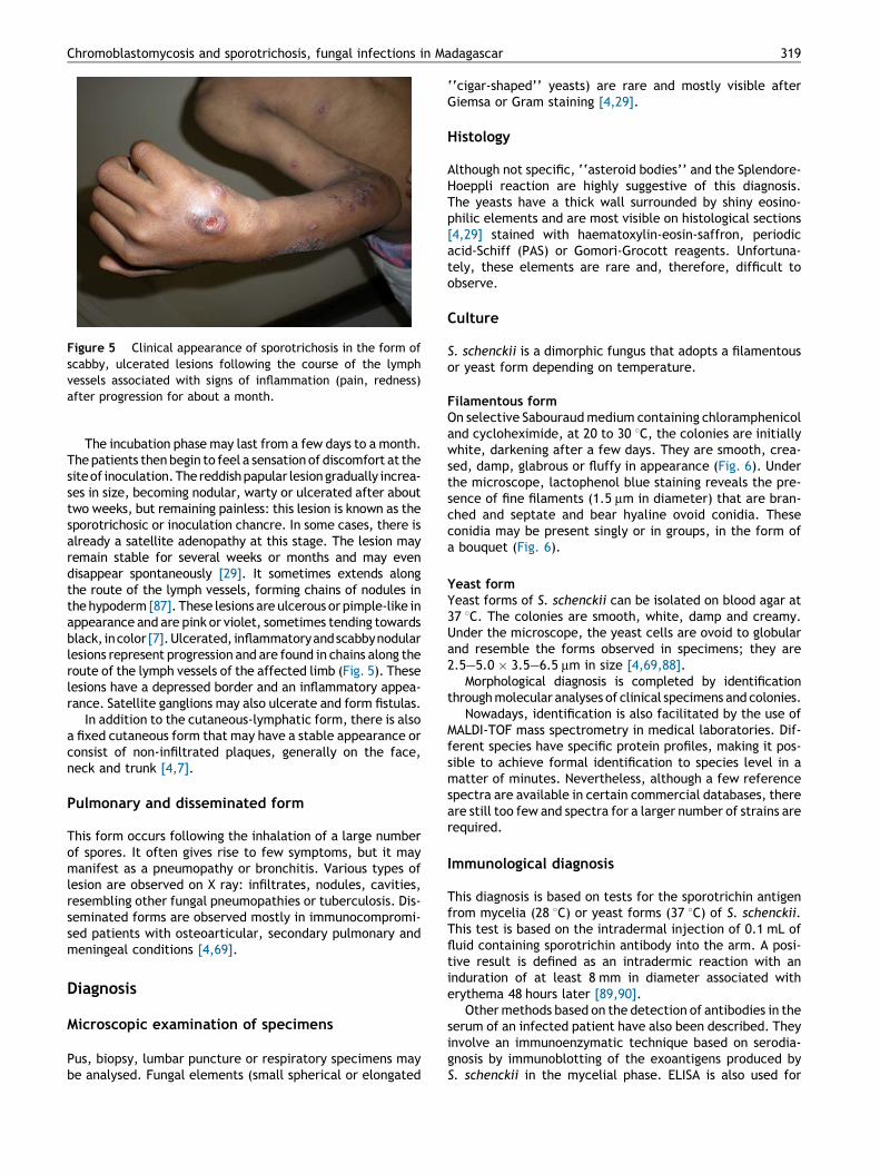
Figure 5 Clinical appearance of sporotrichosis in the form ofscabby, ulcerated lesions following the course of the lymphvessels associated with signs of inflammation (pain, redness)after progression for about a month.
Chromoblastomycosis and sporotrichosis, fungal infections in Madagascar 319
The incubation phase may last from a few days to a month.The patients then begin to feel a sensation of discomfort at thesiteof inoculation.The reddishpapular lesiongradually increa-ses in size, becoming nodular, warty or ulcerated after abouttwo weeks, but remaining painless: this lesion is known as thesporotrichosic or inoculation chancre. In some cases, there isalready a satellite adenopathy at this stage. The lesion mayremain stable for several weeks or months and may evendisappear spontaneously [29]. It sometimes extends alongthe route of the lymph vessels, forming chains of nodules inthe hypoderm [87]. These lesions are ulcerous or pimple-like inappearance and are pink or violet, sometimes tending towardsblack, incolor[7].Ulcerated, inflammatoryandscabbynodularlesions represent progression and are found in chains along theroute of the lymph vessels of the affected limb (Fig. 5). Theselesions have a depressed border and an inflammatory appea-rance. Satellite ganglions may also ulcerate and form fistulas.
In addition to the cutaneous-lymphatic form, there is alsoa fixed cutaneous form that may have a stable appearance orconsist of non-infiltrated plaques, generally on the face,neck and trunk [4,7].
Pulmonary and disseminated form
This form occurs following the inhalation of a large numberof spores. It often gives rise to few symptoms, but it maymanifest as a pneumopathy or bronchitis. Various types oflesion are observed on X ray: infiltrates, nodules, cavities,resembling other fungal pneumopathies or tuberculosis. Dis-seminated forms are observed mostly in immunocompromi-sed patients with osteoarticular, secondary pulmonary andmeningeal conditions [4,69].
Diagnosis
Microscopic examination of specimens
Pus, biopsy, lumbar puncture or respiratory specimens maybe analysed. Fungal elements (small spherical or elongated
‘‘cigar-shaped’’ yeasts) are rare and mostly visible afterGiemsa or Gram staining [4,29].
Histology
Although not specific, ‘‘asteroid bodies’’ and the Splendore-Hoeppli reaction are highly suggestive of this diagnosis.The yeasts have a thick wall surrounded by shiny eosino-philic elements and are most visible on histological sections[4,29] stained with haematoxylin-eosin-saffron, periodicacid-Schiff (PAS) or Gomori-Grocott reagents. Unfortuna-tely, these elements are rare and, therefore, difficult toobserve.
Culture
S. schenckii is a dimorphic fungus that adopts a filamentousor yeast form depending on temperature.
Filamentous formOn selective Sabouraud medium containing chloramphenicoland cycloheximide, at 20 to 30 8C, the colonies are initiallywhite, darkening after a few days. They are smooth, crea-sed, damp, glabrous or fluffy in appearance (Fig. 6). Underthe microscope, lactophenol blue staining reveals the pre-sence of fine filaments (1.5 mm in diameter) that are bran-ched and septate and bear hyaline ovoid conidia. Theseconidia may be present singly or in groups, in the form ofa bouquet (Fig. 6).
Yeast formYeast forms of S. schenckii can be isolated on blood agar at37 8C. The colonies are smooth, white, damp and creamy.Under the microscope, the yeast cells are ovoid to globularand resemble the forms observed in specimens; they are2.5—5.0 � 3.5—6.5 mm in size [4,69,88].
Morphological diagnosis is completed by identificationthrough molecular analyses of clinical specimens and colonies.
Nowadays, identification is also facilitated by the use ofMALDI-TOF mass spectrometry in medical laboratories. Dif-ferent species have specific protein profiles, making it pos-sible to achieve formal identification to species level in amatter of minutes. Nevertheless, although a few referencespectra are available in certain commercial databases, thereare still too few and spectra for a larger number of strains arerequired.
Immunological diagnosis
This diagnosis is based on tests for the sporotrichin antigenfrom mycelia (28 8C) or yeast forms (37 8C) of S. schenckii.This test is based on the intradermal injection of 0.1 mL offluid containing sporotrichin antibody into the arm. A posi-tive result is defined as an intradermic reaction with aninduration of at least 8 mm in diameter associated witherythema 48 hours later [89,90].
Other methods based on the detection of antibodies in theserum of an infected patient have also been described. Theyinvolve an immunoenzymatic technique based on serodia-gnosis by immunoblotting of the exoantigens produced byS. schenckii in the mycelial phase. ELISA is also used for

[(Figure_6)TD$FIG]
Figure 6 Macroscopic appearance of a cultured colony of Sporothrix schenckii after 24 days of incubation at 30 8C on Sabouraud-chloramphenicol medium (A). The colony is smooth, damp, white at the edges and brownish towards the centre. Photomicrograph( � 400) obtained after lactophenol blue staining of cultured colonies of Sporothrix schenckii (B). Note the fine, branched filamentssurrounded by ovoid spores.
320 T. Rasamoelina et al.
detection of the response to a peptide-rhamnomannancomplex from the cellular membranes of the yeast formof S. schenckii. Immunodiffusion and immunoelectrophoresismethods can also be used [91,92].
Immunological diagnosis methods are widely used in epi-demiological studies. However, diverse antigens are used,potentially leading to differences in the results obtained.Differences in specificity and sensitivity and cross-reactionsare also observed [4].
Treatment
Potassium iodide
Potassium iodide is the treatment most widely used incountries with limited resources. It is cheap and very welltolerated [93]. Potassium iodide is administered orally,mixed with a liquid, at an initial dose of 0.5 g/day, graduallyincreased to 4 to 6 g/day [11]. It modulates the immunesystem, but its mechanism of action remains unclear. In theusual cutaneous-lymphatic forms, the disease is cured aftertwo to three months. The treatment must then be stoppedgradually, with a progressive decrease in dose. The principalsecondary effects are uncommon: gastric intolerance,oedema, rash and erythema [7]. By contrast, treatment isineffective in cases of extracutaneous SPT [88].
Itraconazole
Since the introduction of azole compounds in the 1990s,itraconazole has been the drug of choice recommendedand used for the treatment of all forms of SPT [94]. Itsefficacy and tolerability, even during long-term treatment,have been demonstrated in many studies [95—97]. A curerate of 94.6% for a group of 645 patients treated with a doseof 50—400 mg/day was reported. Of the 610 patients cured,547 were treated with a dose of 100 mg/day, 59 were treatedwith a dose of 200—400 mg/day and four children weretreated with a dose of 50 mg/day [29,95].
Posaconazole and terbinafine
Studies of the activity of terbinafine and posaconazoleagainst S. schenckii in vitro have given encouraging results[98,99]. Terbinafine at a dose of 250 to 1000 mg/day wasfound to be effective in human cases of cutaneous SPT[91,100]. In vitro, posaconazole is among the most activeof the compounds tested and could constitute an alternativetreatment. However, clinical trials will be necessary beforethis molecule can be considered as a treatment option [99].
In vitro tests with fluconazole, voriconazole, flucytosineand micafungin used against various strains of S. schenckiihave shown minimal inhibitory concentrations to be high,suggesting that these antifungal drugs are not very effectiveagainst this fungus. There is currently no recommendationfor the use of these antifungal drugs [101,102].
Thermotherapy
Potassium iodide and itraconazole are contraindicated inpregnant women. In such cases, thermotherapy is used. Thistreatment is based on the application of a heat source (apouch of hot water or a source of infrared radiation) at 42 to43 8C on the lesions, one to three times per day, for a totalduration of 40 to 60 minutes today until the lesions resolve[69]. The heat acts on the cells of S. schenckii taken up byphagocytes. The efficacy of this treatment has not yet beenevaluated. This treatment can be combined with the use ofantifungal drugs [7].
The treatment currently used in Madagascar is 200 mgitraconazole per day for a total of six months. A very goodresponse to treatment is observed, from the second monthonwards.
Conclusion
CBM and SPT are chronic fungal infections occurring predo-minantly in the hot and humid zones of tropical and sub-tropical countries. In Madagascar, these two mycoses

Chromoblastomycosis and sporotrichosis, fungal infections in Madagascar 321
are frequently encountered but remain neglected anduntreated due to underdiagnosis. New active molecules,such as oral posaconazole and isavuconazole, are openingup new possibilities for the management of these diseases.Access to these treatments in countries with limited sourcesremains a major issue for international organizations in thedomain of health. The upcoming recognition, by the WHO, ofCBM as a neglected tropical disease may facilitate theimplementation of national programmes. Prevention isbased on knowledge of the sources of contamination. Thedistribution of the causal fungi in the environment has notbeen studied in Madagascar. Caution is required in theidentification of possible sources of contamination, andmolecular techniques should be used to prevent confusionwith saprophytic species.
Since 2012, the Charles Mérieux Infectiology Centre(CMIC) of Antananarivo, the dermatology department ofJoseph Raseta Befelatanana University Hospital and Gre-noble-Alpes University have been conducting a collaborativestudy to update the prevalences of these two mycoses and todescribe the distribution of the causal agents in the envi-ronment in Madagascar.
Disclosure of interest
The authors declare that they have no competing interest.
Acknowledgments
We are grateful to Fondation Mérieux, Lyon, France Institutde Recherche pour le Développement, IRD; Société françaisede mycologie médicale and Campus France that give supportfor the research on CBM and SPT in Madagascar.
References
[1] Fukushiro R. Chromomycosis in Japan. Int J Dermatol1983;22:221—9. http://dx.doi.org/10.1111/j.1365-4362.1983.tb03371.x.
[2] de Mouchalouat MF, Gutierrez Galhardo MC, Zancopé-OliveiraRM, Monteiro Fialho PC, de Oliveira Coelho JMC, Silva TavaresPM, et al. Chromoblastomycosis: a clinical and molecularstudy of 18 cases in Rio de Janeiro. Brazil Int J Dermatol2011;50:981—6. http://dx.doi.org/10.1111/j.1365-4632.2010.04729.x.
[3] Pérez-Blanco M, Hernández Valles R, García-Humbría L,Yegres F. Chromoblastomycosis in children and adolescentsin the endemic area of the Falcón State. Venezuela Med Mycol2006;44:467—71. http://dx.doi.org/10.1080/13693780500543238.
[4] Barros MB, de L, de Almeida Paes R, Schubach AO. Sporothrixschenckii and Sporotrichosis. Clin Microbiol Rev 2011;24:633—54. http://dx.doi.org/10.1128/CMR.00007-11.
[5] Esterre P, Andriantsimahavandy A, Ramarcel ER, Pecarrere JL.Forty years of chromoblastomycosis in Madagascar: a review.Am J Trop Med Hyg 1996;55:45—7.
[6] Esterre P, Andriantsimahavandy A, Raharisolo C. Naturalhistory of chromoblastomycosis in Madagascar and the IndianOcean. Natural history of chromoblastomycosis in Madagas-car and the Natural history of chromoblastomycosis inMadagascar and the Indian Ocean. Bull Soc Pathol Exot1997;90:312—7.
[7] Vásquez-del-Mercado E, Arenas R, Padilla-Desgarenes C. Spo-rotrichosis. Clin Dermatol 2012;30:437—43. http://dx.doi.org/10.1016/j.clindermatol.2011.09.017.
[8] Queiroz-Telles F, Nucci M, Colombo AL, Tobón A, Restrepo A.Mycoses of implantation in Latin America: an overviewof epidemiology, clinical manifestations, diagnosis andtreatment. Med Mycol 2011;49:225—36. http://dx.doi.org/10.3109/13693786.2010.539631.
[9] Reiss E, Shadomy HJ, Lyon GM. Fundamental medical mycolo-gy. John Wiley & Sons; 2011.
[10] Esterre P, Chabasse D. Chromomycose. In: Mal Infect. Paris:Encycl Med Chir Ed Sci Médicales Elsevier SAS; 2003: 6 [TousDroits Réservés].
[11] Aubry P. Mycoses sous-cutanées. Mycol. Médicale, Lavoisier;2013: 750.
[12] Queiroz-Telles F, de Hoog S, Santos DWCL, Salgado CG, VicenteVA, Bonifaz A, et al. Chromoblastomycosis. Clin Microbiol Rev2017;30:233—76. http://dx.doi.org/10.1128/CMR.00032-16.
[13] Silva JP, de Souza W, Rozental S. Chromoblastomycosis: aretrospective study of 325 cases on Amazonic Region (Brazil).Mycopathologia 1998;143:171—5.
[14] Elgart GW. Chromoblastomycosis. Dermatol Clin 1996;14:77—83.
[15] Arora. Humans, animals and insects. Handbook of appliedmycology, 2. CRC Press; 1991.
[16] Carolina Rojas O, León-Cachón RBR, Pérez-Maya AA, Aguirre-Garza M, Moreno-Treviño MG, González GM. Phenotypic andmolecular identification of Fonsecaea pedrosoi strains isolat-ed from chromoblastomycosis patients in Mexico and Vene-zuela. Mycoses 2015;58:267—72. http://dx.doi.org/10.1111/myc.12308.
[17] Krzysciak PM, Pindycka-Piaszczynska M, Piaszczynski M. Chro-moblastomycosis. Postepy Dermatol Alergol 2014;31:310—21.http://dx.doi.org/10.5114/pdia.2014.40949.
[18] Vicente VA, Najafzadeh MJ, Sun J, Gomes RR, Robl D, MarquesSG, et al. Environmental siblings of black agents of humanchromoblastomycosis. Fungal Divers 2014;65:47—63. http://dx.doi.org/10.1007/s13225-013-0246-5.
[19] El Mouhaddab M, Basraoui D, Ousehal A, Galuia F. Chronicfistula of the forefoot in a Moroccan man: bone chromomycosisdue to Phialophora verrucosa. Med Trop Rev Corps Sante Colon2010;70:181—3.
[20] Chabasse D. Mycoses à champignons noirs : chromoblastomy-coses et phæohyphomycoses. EM-Consulte n.d. 2011. http://www.em-consulte.com/article/285091/mycoses-a-champignons-noirs-chromoblastomycoses-et.[accessed January 2, 2017].
[21] Barde AK, Singh SM. Cladosporium carrionii Trejos 1954 infec-tion of human nail. Mycoses 1984;27:366—9. http://dx.doi.org/10.1111/j.1439-0507.1984.tb02044.x.
[22] de Hoog GS, Guého E, Masclaux F, Gerrits van den Ende AH,Kwon-Chung KJ, McGinnis MR. Nutritional physiology and tax-onomy of human-pathogenic Cladosporium-Xylohypha spe-cies. J Med Vet Mycol 1995;33:339—47.
[23] Badali H, Bonifaz A, Barrón-Tapia T, Vázquez-González D,Estrada-Aguilar L, Oliveira NMC, et al. Rhinocladiella aquas-persa, proven agent of verrucous skin infection and a noveltype of chromoblastomycosis. Med Mycol 2010;48:696—703.http://dx.doi.org/10.3109/13693780903471073.
[24] Marques SG, Silva C, de MP, Saldanha PC, Rezende MA, VicenteVA, et al. Isolation of Fonsecaea pedrosoi from the shell of thebabassu coconut (Orbignya phalerata Martius) in the Amazonregion of Maranhão Brazil. Nihon Ishinkin Gakkai Zasshi Jpn JMed Mycol 2006;47:305—11.
[25] Salgado CG, da Silva JP, Diniz JAP, da Silva MB, da Costa PF,Teixeira C, et al. Isolation of Fonsecaea pedrosoi from thornsof Mimosa pudica, a probable natural source of chromoblas-tomycosis. Rev Inst Med Trop Sao Paulo 2004;46:33—6.

322 T. Rasamoelina et al.
[26] Gezuele E, Mackinnon JE, Conti-Díaz IA. The frequent isolationof Phialophora verrucosa and Phialophora pedrosoi from nat-ural sources. Sabouraudia J Med Vet Mycol 1972;10:266—73.http://dx.doi.org/10.1080/00362177285190501.
[27] Rubin HA, Bruce S, Rosen T, McBride ME. Evidence for percu-taneous inoculation as the mode of transmission for chromo-blastomycosis. J Am Acad Dermatol 1991;25:951—4.
[28] de Hoog GS, Nishikaku AS, Fernandez-Zeppenfeldt G, Padín-González C, Burger E, Badali H, et al. Molecular analysis andpathogenicity of the Cladophialophora carrionii complex,with the description of a novel species. Stud Mycol2007;58:219—34. http://dx.doi.org/10.3114/sim.2007.58.08.
[29] Chabasse D, Delevoux M. Mycoses d’importation. ElsevierMasson; 2003.
[30] Bonifaz A, Carrasco-Gerard E, Saúl A. Chromoblastomycosis:clinical and mycologic experience of 51 cases. Mycoses2001;44:1—7.
[31] Diáz Almeida JG, Taboas González M, Dube Dube A.Chromoblastomycosis in Cuba, retrospective clinical andepidemiologic studies of 72 patients. Rev Cubana Med Trop1978;30:95—108.
[32] Restrepo-Moreno A, Calle-Velez G, Sanchez-Arbelaez J, Cor-rea-Gonzalez A. A review of medical mycology in ColombiaSouth America, including presentation of 309 originalcases of various mycoses. Mycopathol Mycol Appl1962;17:93—110.
[33] Develoux M, Dieng M-T, Ndiaye B, Raphenon G, Lepers J-P.Chromomycosis caused by Exophilia spinifera in SahelianAfrica. Ann Dermatol Venereol 2006;133:68—9.
[34] Hofmann H, Choi S-M, Wilsmann-Theis D, Horré R, de Hoog GS,Bieber T. Invasive chromoblastomycosis and sinusitis due toPhialophora verrucosa in a child from northern Africa. Mycoses2005;48:456—61. http://dx.doi.org/10.1111/j.1439-0507.2005.01150.x.
[35] Kombila M, Gomez de Diaz M, Richard-Lenoble D, Renders A,Walter P, Billiault X, et al. Chromoblastomycosis in Gabon.Study of 64 cases. Sante Montrouge Fr 1995;5:235—44.
[36] Attapattu MC. Chromoblastomycosis — a clinical and myco-logical study of 71 cases from Sri Lanka. Mycopathologia1997;137:145—51.
[37] Sharma N, Marfatia YS. Genital elephantiasis as a complicationof chromoblastomycosis: a diagnosis overlooked. Indian J SexTransm Dis 2009;30:43—5. http://dx.doi.org/10.4103/0253-7184.55486.
[38] Xi L, Sun J, Lu C, Liu H, Xie Z, Fukushima K, et al. Moleculardiversity of Fonsecaea (Chaetothyriales) causing chromoblas-tomycosis in southern China. Med Mycol 2009;47:27—33.http://dx.doi.org/10.1080/13693780802468209.
[39] Brygoo ER. La chromoblastomycose à Madagascar. Paris n.d:Mycol Médicale Inst Pasteur Madag Dir Courdurier Serv MycolInst Pasteur; 1956.
[40] Coulanges P, Locheron P. Chromomycosis in Madagascar.Epidemiological data on the most important reservoir cur-rently known in the world (apropos of 891 cases diagnosedfrom 1955 to 1978). Arch Inst Pasteur Madagascar1981;48:69—95.
[41] Chevalier A. La végétation a Madagascar d’après l’ouvrage deH. Perrier de la Bathie. Ann Geogr 1922;31:465—84.
[42] Rasamoelina T, Rakotozandrindrainy N, Raberahona M,Rabenja RF, Andrianarivelo RM, Andrianarison M, et al. Chro-moblastomycosis and sporotrichosis in Madagascar: epidemi-ology, molecular diagnostic and perspectives: P131. Mycoses2015;58:99—100.
[43] Chandenier J, Renard JC, Goma D, Nzounza R, De Bievre C,Ravisse P, et al. Mycoses tropicales diagnostiquées au CentreHospitalier Universitaire de Brazzaville (Congo), novembre1991—juillet 1995. Aspects cliniques, mycologiques et théra-peutiques. J Mycol Med 2000;10:67—77.
[44] Lari HB, Mirani N, Chu DS. Corneal chromoblastomycosis cau-sed by Cladophialophora carrionii after cataract surgery. JCataract Refract Surg 2011;37:963—6. http://dx.doi.org/10.1016/j.jcrs.2011.03.007.
[45] Esterre P, Pecarrere J-L, Raharisolo C, Huerre M. Carcinomeépidermoïde développé sur des lésions de chromomycose : àpropos de deux observations. Ann Pathol 1999;19:516—20[Masson].
[46] Najafzadeh MJ, Badali H, Illnait-Zaragozi MT, De Hoog GS, MeisJF. In vitro activities of eight antifungal drugs against 55clinical isolates of Fonsecaea spp.. Antimicrob Agents Chemo-ther 2010;54:1636—8. http://dx.doi.org/10.1128/AAC.01655-09.
[47] Feng P, Najafzadeh MJ, Sun J, Ahmed S, Xi L, de Hoog GS, et al.In vitro activities of nine antifungal drugs against 81Phialophora and Cyphellophora isolates. Antimicrob AgentsChemother 2012;56:6044—7. http://dx.doi.org/10.1128/AAC.01112-12.
[48] McGinnis MR, Pasarell L. In vitro evaluation of terbinafine anditraconazole against dematiaceous fungi. Med Mycol1998;36:243—6.
[49] Esterre P, Inzan C, Ratsioharana M, Andriantsimahavandy A,Raharisolo C, Randrianiaina E, et al. A multicentre trial ofterbinafine in patients with chromoblastomycosis: effect onclinical and biological criteria. J Dermatol Treat 1998;9:S29—34. http://dx.doi.org/10.3109/09546639809160714.
[50] Daboit TC, Massotti Magagnin C, Heidrich D, Czekster Anto-chevis L, Vigolo S, Collares Meirelles L, et al. In vitro suscep-tibility of chromoblastomycosis agents to five antifungal drugsand to the combination of terbinafine and amphotericin B.Mycoses 2014;57:116—20. http://dx.doi.org/10.1111/myc.12111.
[51] Garnica M, Nucci M, Queiroz-Telles F. Difficult mycoses of theskin: advances in the epidemiology and management of eumy-cetoma, phaeohyphomycosis and chromoblastomycosis. CurrOpin Infect Dis 2009;22:559—63. http://dx.doi.org/10.1097/QCO.0b013e328332bbc5.
[52] Queiroz-Telles F, Esterre P, Perez-Blanco M, Vitale RG, Sal-gado CG, Bonifaz A, et al. an overview of clinical manifesta-tions, diagnosis and treatment. Med Mycol 2009;47:3—15.
[53] Queiroz-Telles F, Purim KS, Fillus JN, Bordignon GF, LameiraRP, Van Cutsem J, et al. Itraconazole in the treatment ofchromoblastomycosis due to Fonsecaea pedrosoi. Int J Der-matol 1992;31:805—12.
[54] Bonifaz A, Martínez-Soto E, Carrasco-Gerard E, Peniche J.Treatment of chromoblastomycosis with itraconazole, cryo-surgery and a combination of both. Int J Dermatol1997;36:542—7.
[55] Keating GM. Posaconazole. Drugs 2005;65:1553—67 [discus-sion 1568-1569].
[56] Bonifaz A, Saúl A, Paredes-Solis V, Araiza J, Fierro-Arias L.Treatment of chromoblastomycosis with terbinafine: experi-ence with four cases. J Dermatol Treat 2005;16:47—51.http://dx.doi.org/10.1080/09546630410024538.
[57] Esterre P, Inzan CK, Ramarcel ER, Andriantsimahavandy A,Ratsioharana M, Pecarrere JL, et al. Treatment of chromo-mycosis with terbinafine: preliminary results of an open pilotstudy. Br J Dermatol 1996;134:33—6 [discussion 40].
[58] Esterre P, Raharisolo C, Inzan CK, Roig P. De la mycologiemédicale tropicale à une approche de santé publique : lachromoblastomycose à Madagascar. J Mycol Med2003;13:133—40.
[59] de Sousa M, da GT, Belda W, Spina R, Lota PR, Valente NS,et al. Topical application of imiquimod as a treatment forchromoblastomycosis. Clin Infect Dis 2014;58:1734—7. http://dx.doi.org/10.1093/cid/ciu168.
[60] Hiruma M, Kawada A, Yoshida M, Kouya M. Hyperthermictreatment of chromomycosis with disposable chemical pocket

Chromoblastomycosis and sporotrichosis, fungal infections in Madagascar 323
warmers. Report of a successfully treated case, with a reviewof the literature. Mycopathologia 1993;122:107—14.
[61] Yanase K, Yamada M. ‘‘Pocket-warmer’’ therapy of chromo-mycosis. Arch Dermatol 1978;114:1095.
[62] Kuttner BJ, Siegle RJ. Treatment of chromomycosis with a CO2
laser. J Dermatol Surg Oncol 1986;12:965—8.[63] Schenck BR. On refractory subcutaneous abscesses caused by a
fungus possibly related to the Sporotricha. Johns HopkinsPress; 1898.
[64] Marimon R, Cano J, Gené J, Sutton DA, Kawasaki M, Guarro J.Sporothrix brasiliensis, S. globosa, and S. mexicana,three new Sporothrix species of clinical interest. J Clin Micro-biol 2007;45:3198—206. http://dx.doi.org/10.1128/JCM.00808-07.
[65] Madrid H, Cano J, Gené J, Bonifaz A, Toriello C, Guarro J.Sporothrix globosa, a pathogenic fungus with widespreadgeographical distribution. Rev Iberoam Micol 2009;26:218—22. http://dx.doi.org/10.1016/j.riam.2009.02.005.
[66] Almeida-Paes R, de Oliveira MME, Freitas DFS, do Valle ACF,Zancopé-Oliveira RM, Gutierrez-Galhardo MC. Sporotrichosisin Rio de Janeiro, Brazil: Sporothrix brasiliensis is associatedwith atypical clinical presentations. PLoS Negl Trop Dis2014;8. http://dx.doi.org/10.1371/journal.pntd.0003094.
[67] Marimon R, Gené J, Cano J, Guarro J. Sporothrix luriei: a rarefungus from clinical origin. Med Mycol 2008;46:621—5. http://dx.doi.org/10.1080/13693780801992837.
[68] de Meyer EM, de Beer ZW, Summerbell RC, Moharram AM, deHoog GS, Vismer HF, et al. Taxonomy and phylogeny of newwood- and soil-inhabiting Sporothrix species in the Ophios-toma stenoceras-Sporothrix schenckii complex. Mycologia2008;100:647—61.
[69] Hiruma M, Katoh T, Yamamoto I, Kagawa S. Local hyperther-mia in the treatment of sporotrichosis. Mykosen1987;30:315—21.
[70] Barros MB, de L, de Schubach AO, do Valle ACF, GutierrezGalhardo MC, Conceição-Silva F, et al. Cat-transmitted spo-rotrichosis epidemic in Rio de Janeiro, Brazil: description of aseries of cases. Clin Infect Dis 2004;38:529—35. http://dx.doi.org/10.1086/381200.
[71] Agarwal S, Gopal K, Umesh null, Kumar B. Sporotrichosis inUttarakhand (India): a report of nine cases. Int J Dermatol2008;47:367—71. http://dx.doi.org/10.1111/j.1365-4632.2008.03538.x.
[72] Arenas R, Miller D, Campos-Macias P. Epidemiological data andmolecular characterization (mtDNA) of Sporothrix schenckii in13 cases from Mexico. Int J Dermatol 2007;46:177—9. http://dx.doi.org/10.1111/j.1365-4632.2006.03036.x.
[73] Dixon DM, Salkin IF, Duncan RA, Hurd NJ, Haines JH, KemnaME, et al. Isolation and characterization of Sporothrix schenc-kii from clinical and environmental sources associated withthe largest U.S. epidemic of sporotrichosis. J Clin Microbiol1991;29:1106—13.
[74] Fukushiro R. Epidemiology and ecology of sporotrichosis inJapan. Zentralbl Bakteriol Mikrobiol Hyg [A] 1984;257:228—33.
[75] Vismer HF, Hull PR. Prevalence, epidemiology and geographi-cal distribution of Sporothrix schenckii infections in Gauteng,South Africa. Mycopathologia 1997;137:137—43.
[76] Magand F, Perrot J-L, Cambazard F, Raberin M-H, Labeille B.Sporotrichose cutanée autochtone française. Ann DermatolVenereol 2009;136:273—5. http://dx.doi.org/10.1016/j.ann-der.2008.09.021.
[77] Criseo G, Malara G, Romeo O, Puglisi Guerra A. Lymphocuta-neous sporotrichosis in an immunocompetent patient: a casereport from extreme southern Italy. Mycopathologia2008;166:159—62. http://dx.doi.org/10.1007/s11046-008-9121-4.
[78] Kauffman CA. Sporotrichosis. Clin Infect Dis 1999;29:231—6.http://dx.doi.org/10.1086/520190 [quiz 237].
[79] Reed KD, Moore FM, Geiger GE, Stemper ME. Zoonotic trans-mission of sporotrichosis: case report and review. Clin InfectDis 1993;16:384—7.
[80] Reis RS, Almeida-Paes R, Muniz M, de M, Tavares PM, e S, et al.Molecular characterisation of Sporothrix schenckiiisolates from humans and cats involved in the sporotrichosisepidemic in Rio de Janeiro, Brazil. Mem Inst Oswaldo Cruz2009;104:769—74.
[81] Yegneswaran PP, Sripathi H, Bairy I, Lonikar V, Rao R, PrabhuS. Zoonotic sporotrichosis of lymphocutaneous type in a manacquired from a domesticated feline source: report of a firstcase in southern Karnataka, India. Int J Dermatol2009;48:1198—200. http://dx.doi.org/10.1111/j.1365-4632.2008.04049.x.
[82] Rapelanoro-Rabenja F, Ralandison S, Ramarozatovo L, Ran-drianasolo F, Ratrimoarivony C. Sporotrichose a Madagascar :une pathologie méconnue. Nouv Dermatol 2007;26:10.
[83] Carod JF, Ramarozatovo L, Randrianasolo P, Ratsima E, Ran-drianirina F, Rapelanoro FR. Cutaneous sporotricosis in aMalagasy patient. Med Trop Rev Corps Sante Colon 2007;67:18.
[84] O’Reilly LC, Altman SA. Macrorestriction analysis of clinicaland environmental isolates of Sporothrix schenckii. J ClinMicrobiol 2006;44:2547—52. http://dx.doi.org/10.1128/JCM.00078-06.
[85] Chabasse D, Therizol-Ferly M. Les mycoses d’importation. ResGate 2000;2000:51—65. http://dx.doi.org/10.1016/S0338-9898(00)80434-5.
[86] Tlougan BE, Podjasek JO, Patel SP, Nguyen XH, Hansen RC.Neonatal sporotrichosis. Pediatr Dermatol 2009;26:563—5.http://dx.doi.org/10.1111/j.1525-1470.2009.00986.x.
[87] Buot G, Lavalle P, Mariat F. Sporotrichose. Ed Tech. In: Fr MalInfect. Paris: Encycl Méd Chir; 1993: 4.
[88] Mahajan VK. Sporotrichosis: an overview and therapeuticoptions. Dermatol Res Pract 2014;2014:272376. http://dx.doi.org/10.1155/2014/272376.
[89] Itoh M, Okamoto S, Kariya H. Survey of 200 cases of sporotri-chosis. Dermatologica 1986;172:209—13.
[90] Carlos IZ. Sporotrichosis: new developments and future pros-pects. Springer; 2015.
[91] Mendoza M, Díaz AM, Hung MB, Zambrano EA, Díaz E,De Albornoz MC. Production of culture filtrates of Sporothrixschenckii in diverse culture media. Med Mycol 2002;40:447—54.
[92] Almeida-Paes R, Pimenta MA, Pizzini CV, Monteiro PCF, Per-alta JM, Nosanchuk JD, et al. Use of mycelial-phase Sporothrixschenckii exoantigens in an enzyme-linked immunosorbentassay for diagnosis of sporotrichosis by antibody detection.Clin Vaccine Immunol CVI 2007;14:244—9. http://dx.doi.org/10.1128/CVI.00430-06.
[93] Xue S-L, Li L. Oral potassium iodide for the treatment ofsporotrichosis. Mycopathologia 2009;167:355—6. http://dx.doi.org/10.1007/s11046-008-9178-0.
[94] Kauffman CA, Bustamante B, Chapman SW, Pappas PG. Infec-tious Diseases Society of America Clinical practice guidelinesfor the management of sporotrichosis: 2007 update by theInfectious Diseases Society of America. Clin Infect Dis2007;45:1255—65. http://dx.doi.org/10.1086/522765.
[95] de Lima Barros MB, Schubach AO, de Vasconcellos Carvalhaesde Oliveira R, Martins EB, Teixeira JL, Wanke B. Treatment ofcutaneous sporotrichosis with itraconazole—study of 645patients. Clin Infect Dis 2011;52:e200—6. http://dx.doi.org/10.1093/cid/cir245.
[96] Manohar A, Nizlan MN. Chronic nonhealing ulcer of the rightthumb with multiple subcutaneous nodules. Orthopedics2008;31:710.
[97] Milby AH, Pappas ND, O’Donnell J, Bozentka DJ. Sporotrichosisof the upper extremity. Orthopedics 2010;33. http://dx.doi.org/10.3928/01477447-20100225-27.
324 T. Rasamoelina et al.
[98] Marimon R, Serena C, Gené J, Cano J, Guarro J. In vitroantifungal susceptibilities of five species of sporothrix. Anti-microb Agents Chemother 2008;52:732—4. http://dx.doi.org/10.1128/AAC.01012-07.
[99] Silveira CP, Torres-Rodríguez JM, Alvarado-Ramírez E, Mur-ciano-Gonzalo F, Dolande M, Panizo M, et al. MICs and mini-mum fungicidal concentrations of amphotericin B,itraconazole, posaconazole and terbinafine in Sporothrixschenckii. J Med Microbiol 2009;58:1607—10. http://dx.doi.org/10.1099/jmm.0.007609-0.
[100] Francesconi G, Francesconi do Valle AC, Passos SL, de LimaBarros MB, de Almeida Paes R, Curi ALL, et al. Comparative
study of 250 mg/day terbinafine and 100 mg/day itraconazolefor the treatment of cutaneous sporotrichosis. Mycopatholo-gia 2011;171:349—54. http://dx.doi.org/10.1007/s11046-010-9380-8.
[101] Alvarado-Ramírez E, Torres-Rodríguez JM. In vitro susceptibili-ty of Sporothrix schenckii to six antifungal agents determinedusing three different methods. Antimicrob Agents Chemother2007;51:2420—3. http://dx.doi.org/10.1128/AAC.01176-06.
[102] Nakai T, Uno J, Ikeda F, Tawara S, Nishimura K, Miyaji M. Invitro antifungal activity of Micafungin (FK463) against dimor-phic fungi: comparison of yeast-like and mycelial forms. Anti-microb Agents Chemother 2003;47:1376—81.