Chromium(VI) as a novel MRI contrast agent for cerebral white matter: Preliminary results in mouse...
6
Chromium(VI) as a Novel MRI Contrast Agent for Cerebral White Matter: Preliminary Results in Mouse Brain In Vivo Takashi Watanabe, * Roland Tammer, Susann Boretius, Jens Frahm, and Thomas Michaelis This work demonstrates that intraventricular microinjections of a low dose of potassium dichromate (0.4 L of 10 mM solution) yield a specific contrast enhancement of white matter (WM) tracts in T 1 -weighted 3D MRI of mouse brain in vivo. Pro- nounced and persistent signal increases (40 –100% at 24 hr after injection) were observed in the corpus callosum, anterior commissure, fornix, and stria medullaris, as well as in the mam- millothalamic tract and fasciculus retroflexus. These results suggest that the extracellular diffusion of diamagnetic chromi- um(VI) (Cr(VI)) after injection is followed by a tissue-specific reduction to paramagnetic Cr(V) and (III), which relies predom- inantly on the oxidation of myelin lipids. Because Cr(VI)-induced contrast leads to only a mild unspecific enhancement (10 –20%) of gray matter (GM) structures, such as the hippocampal for- mation, the method reveals novel information that differs from that obtainable using other paramagnetic ions, such as manganese. Magn Reson Med 56:1– 6, 2006. © 2006 Wiley- Liss, Inc. Key words: myelinated nerve fibers; mouse brain; MRI; lipid oxidation; potassium dichromate In neuroanatomy, specific chemical reactions of lipids ex- posed to aqueous solutions of potassium dichromate (K 2 Cr 2 O 7 with chromium in redox state Cr(VI)) have been used for histological staining of myelin. For example, in a previous study based on Weigert-Smith-Dietrich proce- dures, myelin sheaths in white matter (WM) tracts in rat brain were identified by controlled chromation of lipids, which was accompanied by a simultaneous reduction of Cr(VI) (1). The present work attempts to exploit such prin- ciples for cerebral MRI of WM. The idea is based on the assumption that a reduction of diamagnetic Cr(VI) to para- magnetic Cr(V) and Cr(III) by lipid oxidation causes a shortening of the T 1 relaxation time of surrounding water and/or lipid protons. Using sodium dichromate to study the metabolism of chromium species in biochemical reactions occurring on the skin of living rats, Liu and coworkers (2) demonstrated the potential of Cr(VI) for in vivo EPR spectroscopy. Sim- ilarly, the formation of Cr(V) and Cr(III) was detected by MRI after injection of sodium dichromate in the kidney and liver of a mouse in vivo (3), and of potassium chro- mate in the lung of a rat postmortem (4). The purposes of this first Cr(VI)-enhanced MRI study of mouse brain in vivo were to 1) examine whether a direct injection of a small amount of Cr(VI) induces a contrast enhancement detectable by T 1 -weighted 3D MRI, 2) determine the struc- tures highlighted in the intact living brain, and 3) discuss putative histochemical and physiological mechanisms and how they differ from other paramagnetic MRI contrast agents. MATERIALS AND METHODS Animals and Chromium Administration Ten female mice (NMRI, 8 –11 weeks old, 28 –34 g) were studied in accordance with German animal protection laws after approval was granted by the responsible govern- mental authority. For intraventricular injection the ani- mals were anesthetized by intraperitoneal injection of ket- amine (200 mg/kg body weight) and xylazine (16 mg/kg body weight), and fixed to a stereotaxic instrument (TSE, Bad Homburg, Germany). The bregma, sagittal suture, and surface of the skull were used as references for the anteri- or–posterior, lateral, and ventral coordinates, respectively. A 33-gauge needle attached to a 2.5-L Hamilton microsy- ringe was placed into the anterior horn of the right lateral ventricle in accordance with MR images obtained before- hand. In animals 1–5 a solution (0.4 L, pH 5) of 10 mM potassium dichromate (K 2 Cr 2 O 7 ; Sigma, Taufkirchen, Ger- many) dissolved in physiological saline was slowly in- jected into the ventricle over an 8-min period. Animal 6 received a high dose of potassium dichromate (5.0 L, 100 mM). In these experiments Cr 2 O 7 2 ions carry two negative charges with chromium in redox state Cr(VI), which is diamagnetic. To assess the putative influences of charge and redox state on the distribution of chromium ions in brain tissue, we administered animals 7 and 8 a low dose (0.4 L, 20 mM) and a 10-fold higher dose (0.4 L, 200 mM) of chromium chloride hexahydrate (CrCl 3 6H 2 O; Sigma, Taufkirchen, Germany), respec- tively. In this case Cr 3 ions carry three positive charges and are in redox state Cr(III), which is paramagnetic. Fi- nally, animals 9 and 10 were intraventricularly injected with Gd-DTPA (5.0 L, 100 mM; Magnevist®, Schering, Berlin, Germany). Immediately after administration the Biomedizinische NMR Forschungs GmbH, Max Planck Institut fu ¨r Bio- physikalische Chemie, Go ¨ ttingen, Germany. *Correspondence to: T. Watanabe, MD, Biomedizinische NMR Forschungs GmbH, 37070 Go ¨ ttingen, Germany. E-mail: [email protected] Received 18 November 2005; revised 3 February 2006; accepted 22 March 2006. DOI 10.1002/mrm.20930 Published online 9 June 2006 in Wiley InterScience (www.interscience. wiley.com). 1 © 2006 Wiley-Liss, Inc. Magnetic Resonance in Medicine 56:1– 6 (2006) COMMUNICATION
-
Upload
takashi-watanabe -
Category
Documents
-
view
214 -
download
2
Transcript of Chromium(VI) as a novel MRI contrast agent for cerebral white matter: Preliminary results in mouse...

Chromium(VI) as a Novel MRI Contrast Agent forCerebral White Matter: Preliminary Results in MouseBrain In Vivo
Takashi Watanabe,* Roland Tammer, Susann Boretius, Jens Frahm, andThomas Michaelis
This work demonstrates that intraventricular microinjections ofa low dose of potassium dichromate (0.4 �L of 10 mM solution)yield a specific contrast enhancement of white matter (WM)tracts in T1-weighted 3D MRI of mouse brain in vivo. Pro-nounced and persistent signal increases (40–100% at 24 hrafter injection) were observed in the corpus callosum, anteriorcommissure, fornix, and stria medullaris, as well as in the mam-millothalamic tract and fasciculus retroflexus. These resultssuggest that the extracellular diffusion of diamagnetic chromi-um(VI) (Cr(VI)) after injection is followed by a tissue-specificreduction to paramagnetic Cr(V) and (III), which relies predom-inantly on the oxidation of myelin lipids. Because Cr(VI)-inducedcontrast leads to only a mild unspecific enhancement (10–20%)of gray matter (GM) structures, such as the hippocampal for-mation, the method reveals novel information that differs fromthat obtainable using other paramagnetic ions, such asmanganese. Magn Reson Med 56:1–6, 2006. © 2006 Wiley-Liss, Inc.
Key words: myelinated nerve fibers; mouse brain; MRI; lipidoxidation; potassium dichromate
In neuroanatomy, specific chemical reactions of lipids ex-posed to aqueous solutions of potassium dichromate(K2Cr2O7 with chromium in redox state Cr(VI)) have beenused for histological staining of myelin. For example, in aprevious study based on Weigert-Smith-Dietrich proce-dures, myelin sheaths in white matter (WM) tracts in ratbrain were identified by controlled chromation of lipids,which was accompanied by a simultaneous reduction ofCr(VI) (1). The present work attempts to exploit such prin-ciples for cerebral MRI of WM. The idea is based on theassumption that a reduction of diamagnetic Cr(VI) to para-magnetic Cr(V) and Cr(III) by lipid oxidation causes ashortening of the T1 relaxation time of surrounding waterand/or lipid protons.
Using sodium dichromate to study the metabolism ofchromium species in biochemical reactions occurring onthe skin of living rats, Liu and coworkers (2) demonstratedthe potential of Cr(VI) for in vivo EPR spectroscopy. Sim-
ilarly, the formation of Cr(V) and Cr(III) was detected byMRI after injection of sodium dichromate in the kidneyand liver of a mouse in vivo (3), and of potassium chro-mate in the lung of a rat postmortem (4). The purposes ofthis first Cr(VI)-enhanced MRI study of mouse brain invivo were to 1) examine whether a direct injection of asmall amount of Cr(VI) induces a contrast enhancementdetectable by T1-weighted 3D MRI, 2) determine the struc-tures highlighted in the intact living brain, and 3) discussputative histochemical and physiological mechanisms andhow they differ from other paramagnetic MRI contrastagents.
MATERIALS AND METHODS
Animals and Chromium Administration
Ten female mice (NMRI, 8–11 weeks old, 28–34 g) werestudied in accordance with German animal protectionlaws after approval was granted by the responsible govern-mental authority. For intraventricular injection the ani-mals were anesthetized by intraperitoneal injection of ket-amine (200 mg/kg body weight) and xylazine (16 mg/kgbody weight), and fixed to a stereotaxic instrument (TSE,Bad Homburg, Germany). The bregma, sagittal suture, andsurface of the skull were used as references for the anteri-or–posterior, lateral, and ventral coordinates, respectively.A 33-gauge needle attached to a 2.5-�L Hamilton microsy-ringe was placed into the anterior horn of the right lateralventricle in accordance with MR images obtained before-hand. In animals 1–5 a solution (0.4 �L, pH 5) of 10 mMpotassium dichromate (K2Cr2O7; Sigma, Taufkirchen, Ger-many) dissolved in physiological saline was slowly in-jected into the ventricle over an 8-min period. Animal 6received a high dose of potassium dichromate (5.0 �L,100 mM). In these experiments Cr2O7
2� ions carry twonegative charges with chromium in redox state Cr(VI),which is diamagnetic. To assess the putative influences ofcharge and redox state on the distribution of chromiumions in brain tissue, we administered animals 7 and 8 alow dose (0.4 �L, 20 mM) and a 10-fold higher dose(0.4 �L, 200 mM) of chromium chloride hexahydrate(CrCl3 � 6 H2O; Sigma, Taufkirchen, Germany), respec-tively. In this case Cr3� ions carry three positive chargesand are in redox state Cr(III), which is paramagnetic. Fi-nally, animals 9 and 10 were intraventricularly injectedwith Gd-DTPA (5.0 �L, 100 mM; Magnevist®, Schering,Berlin, Germany). Immediately after administration the
Biomedizinische NMR Forschungs GmbH, Max Planck Institut fur Bio-physikalische Chemie, Gottingen, Germany.*Correspondence to: T. Watanabe, MD, Biomedizinische NMR ForschungsGmbH, 37070 Gottingen, Germany. E-mail: [email protected] 18 November 2005; revised 3 February 2006; accepted 22 March2006.DOI 10.1002/mrm.20930Published online 9 June 2006 in Wiley InterScience (www.interscience.wiley.com).
1© 2006 Wiley-Liss, Inc.
Magnetic Resonance in Medicine 56:1–6 (2006)COMMUNICATION

skin incisions of the scalp were closed and covered withlidocaine hydrochloride (2% Xylocaine® gel). The ani-mals were recovered from anesthesia and returned to theircages with unlimited access to food and water.
MRI
MRI of the five animals with a low dose of potassiumdichromate was performed before and 24 hr after Cr(VI)administration. Three animals were also scanned at 3 and48 hr, and two animals were scanned at 72 hr. Animal 6did not survive for 24 hr and was studied only at 3 hr afterinjection. The four animals that received CrCl3 or Gd-DTPA were studied at 3 and 24 hr.
For MRI the animals were anesthetized and placed inthe magnet as previously described (5). All measurementswere carried out at 2.35 T using a MRBR 4.7/400 mmmagnet (Magnex Scientific, Abingdon, UK) and a DBXsystem (Bruker Biospin MRI GmbH, Ettlingen, Germany)equipped with B-GA20 gradients. Radiofrequency excita-tion and signal reception was accomplished with use of aHelmholtz coil (i.d. 100 mm) and an elliptical surface coil(i.d. 20 � 12 mm), respectively. T1-weighted 3D MRI datasets (RF-spoiled 3D fast low-angle shot (FLASH), TR/TE �17/7.6 ms, flip angle � 25°, field of view (FOV) � 19.2 �19.2 � 19.2 mm3, matrix � 128 � 128 � 128, 16 averages,measuring time � 75 min) were acquired at 150 �m iso-tropic resolution (6).
For quantitative evaluations, the signal-to-noise ratio(SNR, defined as the mean MRI signal intensity of a brainregion divided by the standard deviation (SD) of the noisein a part of the image outside the animal) was determinedusing software supplied by the manufacturer. The analysisfollowed a previously described strategy (5). Briefly, ana-tomic cross sections were obtained by multiplanar recon-
structions from the original 3D MRI data sets. Standard-ized regions of interest (ROIs) were selected in close ac-cordance with resolved anatomic structures. Relative SNRincreases were obtained in comparison to intraindividual3D MRI acquisitions before Cr(VI) administration.
RESULTS
Cr(VI) is diamagnetic and has no direct effect on the relax-ation rates of water protons. We confirmed this MRI si-lence by dissolving potassium dichromate in saline atdifferent concentrations (data not shown). The lack of anyMRI signal change reflects the fact that Cr(VI) remainsunreduced if no chemical reaction partners, such as oxi-dizable lipids, are available.
In mice, intraventricular injections of a low dose ofCr(VI) selectively enhanced brain structures in T1-weighted MRI. Figure 1 shows three sagittal sections of a3D MRI data set of the brain of a mouse acquired beforeand 24 hr after administration. In the mid-sagittal section(top), major commissural fiber structures, such as the cor-pus callosum, anterior commissure, ventral hippocampalcommissure, habenular commissure, and posterior com-missure, are strongly highlighted. The parasagittal sections(middle and bottom) reveal pronounced signal increases inmajor WM tracts, such as the fornix and stria medullaris.In addition, and despite the fact that these WM structuresare not directly exposed to cerebrospinal fluid (CSF), themammillothalamic tract and the fasciculus retroflexus arealso well delineated. Figure 2 shows corresponding trans-verse-to-coronal sections along these latter tracts from thesame data set. Apart from the strongly enhanced corpuscallosum, the images clearly identify increased signals inthe mammillothalamic tract and fasciculus retroflexus on
FIG. 1. (top) Midsagittal sectionand left-hemispheric parasagittalsections (middle) 0.3 mm and(bottom) 0.6 mm from the midlineof mouse 1 (left) before as well as(right) 24 hr after Cr(VI) adminis-tration (T1-weighted 3D FLASH,TR/TE � 17/7.6 ms, flip angle �25°, isotropic resolution �150 �m). Marked signal increasesare observed in the choroidplexus (CP) as well as in WMtracts such as the corpus callo-sum (CC), anterior commissure(AC), ventral hippocampal com-missure (VHC), habenular com-missure (HC), posterior commis-sure (PC), fornix (Fx), and striamedullaris (SM). In addition, thefasciculus retroflexus (FR) andmammillothalamic tract (MT) aredelineated.
2 Watanabe et al.
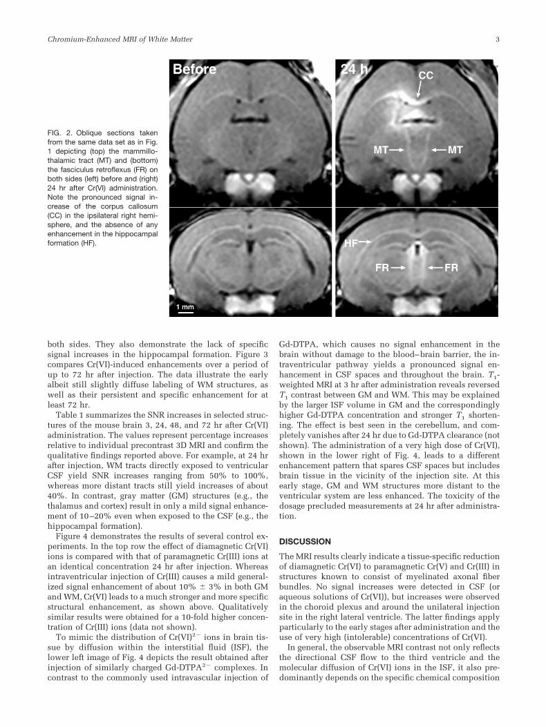
both sides. They also demonstrate the lack of specificsignal increases in the hippocampal formation. Figure 3compares Cr(VI)-induced enhancements over a period ofup to 72 hr after injection. The data illustrate the earlyalbeit still slightly diffuse labeling of WM structures, aswell as their persistent and specific enhancement for atleast 72 hr.
Table 1 summarizes the SNR increases in selected struc-tures of the mouse brain 3, 24, 48, and 72 hr after Cr(VI)administration. The values represent percentage increasesrelative to individual precontrast 3D MRI and confirm thequalitative findings reported above. For example, at 24 hrafter injection, WM tracts directly exposed to ventricularCSF yield SNR increases ranging from 50% to 100%,whereas more distant tracts still yield increases of about40%. In contrast, gray matter (GM) structures (e.g., thethalamus and cortex) result in only a mild signal enhance-ment of 10–20% even when exposed to the CSF (e.g., thehippocampal formation).
Figure 4 demonstrates the results of several control ex-periments. In the top row the effect of diamagnetic Cr(VI)ions is compared with that of paramagnetic Cr(III) ions atan identical concentration 24 hr after injection. Whereasintraventricular injection of Cr(III) causes a mild general-ized signal enhancement of about 10% � 3% in both GMand WM, Cr(VI) leads to a much stronger and more specificstructural enhancement, as shown above. Qualitativelysimilar results were obtained for a 10-fold higher concen-tration of Cr(III) ions (data not shown).
To mimic the distribution of Cr(VI)2� ions in brain tis-sue by diffusion within the interstitial fluid (ISF), thelower left image of Fig. 4 depicts the result obtained afterinjection of similarly charged Gd-DTPA2� complexes. Incontrast to the commonly used intravascular injection of
Gd-DTPA, which causes no signal enhancement in thebrain without damage to the blood–brain barrier, the in-traventricular pathway yields a pronounced signal en-hancement in CSF spaces and throughout the brain. T1-weighted MRI at 3 hr after administration reveals reversedT1 contrast between GM and WM. This may be explainedby the larger ISF volume in GM and the correspondinglyhigher Gd-DTPA concentration and stronger T1 shorten-ing. The effect is best seen in the cerebellum, and com-pletely vanishes after 24 hr due to Gd-DTPA clearance (notshown). The administration of a very high dose of Cr(VI),shown in the lower right of Fig. 4, leads to a differentenhancement pattern that spares CSF spaces but includesbrain tissue in the vicinity of the injection site. At thisearly stage, GM and WM structures more distant to theventricular system are less enhanced. The toxicity of thedosage precluded measurements at 24 hr after administra-tion.
DISCUSSION
The MRI results clearly indicate a tissue-specific reductionof diamagnetic Cr(VI) to paramagnetic Cr(V) and Cr(III) instructures known to consist of myelinated axonal fiberbundles. No signal increases were detected in CSF (oraqueous solutions of Cr(VI)), but increases were observedin the choroid plexus and around the unilateral injectionsite in the right lateral ventricle. The latter findings applyparticularly to the early stages after administration and theuse of very high (intolerable) concentrations of Cr(VI).
In general, the observable MRI contrast not only reflectsthe directional CSF flow to the third ventricle and themolecular diffusion of Cr(VI) ions in the ISF, it also pre-dominantly depends on the specific chemical composition
FIG. 2. Oblique sections takenfrom the same data set as in Fig.1 depicting (top) the mammillo-thalamic tract (MT) and (bottom)the fasciculus retroflexus (FR) onboth sides (left) before and (right)24 hr after Cr(VI) administration.Note the pronounced signal in-crease of the corpus callosum(CC) in the ipsilateral right hemi-sphere, and the absence of anyenhancement in the hippocampalformation (HF).
Chromium-Enhanced MRI of White Matter 3

of the tissue (i.e., the availability of oxidizable lipids andtheir reactivity toward a reduction of Cr(VI)). As far as thefirst mechanism is concerned, the generalized enhance-ment obtained after intraventricular injection and diffu-sion of Gd-DTPA2� complexes (Fig. 4, lower left) stronglysuggests that the much smaller Cr(VI)2� ions should alsohave reached the entire brain parenchyma after only 3 hr.Second, the observation of only a mild unspecific signalenhancement after administration of paramagnetic
Cr(III)3� ions (Fig. 4 upper right) further demonstrates thatin the case of diamagnetic Cr(VI) the initial diffusion isdominated by the chemical process required to generatecontrast. If Cr(VI) ions are in excess relative to availablelipids, then the concentration of the resulting paramag-netic reaction products may be sufficient to even enhancethe MRI signal of GM in regions proximal to the injection(Fig. 4, lower right). At a much lower and tolerable dose ofCr(VI), persistent enhancements were primarily observedin WM directly exposed to CSF, while smaller but stillstrong signal increases allowed for a delineation of moredistant fibers (Figs. 1�3; Fig. 4, upper left). These findingsmay be explained by the specific composition of WM. Thedensely packed and myelinated fiber bundles contain un-saturated phospholipids that have been reported to pos-sess a strong affinity to Cr(VI) ions (1,7). In addition, my-elin has a substantially higher lipid content (�70% in dryweight) than GM (about 30% in dry weight) (8). Finally, asevidenced by the 3–72-hr time course of signal enhance-ments in GM and WM (Fig. 3 and Table 1), the paramag-netic reaction products are assumed to stay in the multi-lamellar structure of myelinated axons. In fact, while Cr(V)still has a high redox potential, Cr(III) ions are stable andmost likely remain bound or complexed to oxidated com-pounds in myelinated fiber tracts. In agreement with ear-lier histochemical staining methods for visualizing chro-mium in tissue (1), this stability may open further appli-cations of Cr(VI) as a dual marker for in vivo MRI and lightmicroscopy.
Apart from the carcinogenic effect demonstrated by thehigh incidence of respiratory tract cancers in workers oc-cupationally exposed to Cr(VI) compounds (3), acute tox-icity of Cr(VI) due to the formation of reactive oxygenspecies poses a limitation of the present technique. Inparticular, the effect may reduce the levels of non-enzy-matic antioxidants, while antioxidant enzyme activitiesincrease as an adaptive brain response against the oxida-tive stress (9). Here the use of a very high dose in oneanimal led to clonic convulsions within 24 hr. In contrast,the injection of only 0.4 �g of Cr(VI), which is muchsmaller than the 1040 �g used in MRI studies of mouseliver and kidney (3), and 100 �g in rat lung (4), yielded asufficient contrast enhancement in the brain within 24 hrafter administration, with no observable behavioral alter-ations.
It should be noted that the need for a specific chemicalreaction that converts Cr(VI) into a paramagnetic MRI con-trast agent leads to marked differences in comparison withother paramagnetic ions used for contrast-enhanced MRI.For example, MRI observations in mouse brain after intra-cerebral injection (5) or subcutaneous application ofMnCl2 (10) resulted in a pattern of enhanced structuresthat was distinctly different from that seen here afterCr(VI) administration. Manganese is taken up by neuronsthrough calcium channels and is subsequently transportedalong connected axons. Manganese-enhanced MRI afterintracerebral MnCl2 injections therefore strongly increasesthe signal intensity of the densely packed cell body assem-blies in the hippocampal formation, as well as direct hip-pocampal projections (11). Conversely, Cr(VI)-enhancedMRI has no specific effect on GM structures, including thehippocampus, but unilateral administration strongly en-
FIG. 3. Left-hemispheric parasagittal sections 0.3 mm from themidline taken from 3D MRI data sets (parameters as in Fig. 1)obtained 3, 24, and 48 hr (mouse 3), and 72 hr (mouse 5) after Cr(VI)administration. While the early studies reveal some residual diffuseenhancement in brain tissue close to the injected ventricle, the laterstages highlight major WM tracts, such as the CC, AC, Fx, and FR,until at least 72 hr after administration.
4 Watanabe et al.

FIG. 4. Transverse sections takenfrom 3D MRI data sets (parame-ters as in Fig. 1) obtained (top)24 hr after administration of a lowdose of (left) diamagnetic Cr(VI)(mouse 4) and (right) paramag-netic Cr(III) (mouse 7) as well as(bottom) 3 hr after administrationof (left) Gd-DTPA (mouse 9) and(right) a very high dose of Cr(VI)(mouse 6). While intraventricularinjections of both Gd-DTPA2�
and Cr(III)3� ions yield a general-ized signal enhancement through-out the brain, the contrast afterCr(VI)2� predominantly reflectsthe reduction to paramagneticCr(V) and Cr(III) by oxidation ofavailable lipids.
Table 1SNR Increases in Mouse Brain In Vivo After Injection of a Low Dose of Chromium(VI) into the Right Lateral Ventricle*
Structure3 hours(n � 3)
24 hours(n � 4)
48 hours(n � 3)
72 hours(n � 2)
WM exposed to CSFCorpus callosum 68 � 53 80 � 29 51 � 29 50 � 25Anterior commissure 72 � 27 80 � 21 57 � 18 51 � 13Habenular commissure 115 � 30 98 � 13 75 � 15 77 � 12Stria medullaris R 85 � 17 74 � 16 57 � 11 46 � 12Stria medullaris L 60 � 17 57 � 9 38 � 6 37 � 13Fornix R 106 � 42 91 � 26 71 � 17 54 � 16Fornix L 52 � 13 54 � 11 36 � 14 38 � 14
WM not exposed to CSFMammillothalamic tract R 33 � 11 41 � 8 31 � 3 27 � 13Mammillothalamic tract L 30 � 9 37 � 7 28 � 2 26 � 10Fasciculus retroflexus R 41 � 14 40 � 5 30 � 2 27 � 11Fasciculus retroflexus L 38 � 10 41 � 4 31 � 3 26 � 8
GM exposed to CSFHippocampal formation R 22 � 9 21 � 10 3 � 9 8 � 10Hippocampal formation L 20 � 1 20 � 11 7 � 9 6 � 9
GM not exposed to CSFThalamus R 19 � 8 17 � 9 16 � 3 12 � 9Thalamus L 15 � 4 16 � 7 13 � 4 10 � 8Cerebral cortex R 20 � 7 13 � 9 10 � 4 5 � 11Cerebral cortex L 19 � 2 12 � 7 12 � 4 5 � 3
*SNR increases are given in percent relative to intraindividual precontrast MRI and represent mean values � SD averaged across animals.WM � white matter, GM � gray matter, R � right, L � left.
Chromium-Enhanced MRI of White Matter 5

hances the corpus callosum, which is not affected by in-traventricular injections of manganese. Thus, one mayconsider manganese as an intracellular neuronal contrastagent for functionally active brain systems, while Cr(VI)serves as an extracellular “surface” agent that labels themyelin lipids of all accessible WM structures. In view ofthe fact that no histochemical method is available to dem-onstrate manganese in tissues (12), functional mapping ofneural pathways by manganese-enhanced MRI (11,13) maynow be complemented by outlining WM tracts with use ofCr(VI)-induced MRI contrast.
CONCLUSIONS
In summary, the present results show that Cr(VI) can beused to specifically enhance the T1-weighted MRI signalintensity of myelinated WM tracts. These findings are inline with histological staining techniques and the highlipid content of myelin. The suggested mechanism as-sumes the molecular diffusion of Cr(VI) ions in the CSFand ISF to be followed by the oxidation of myelin lipids, areduction to paramagnetic forms, and a subsequent reten-tion of the reaction products in the multilamellar struc-ture. It may be concluded that Cr(VI)-enhanced MRI pro-vides new insights into the histochemistry of reactive tis-sue compounds that differ from the information gatheredby other paramagnetic ions, such as manganese. Applica-tions to neurodevelopment and animal models involvingbiochemical and structural alterations of myelin in thecentral nervous system are currently being investigated.
REFERENCES
1. Pearse AGE. Histochemistry, theoretical and applied. Vol. 2, 4th ed.New York: Churchill Livingstone; 1985. p 808–811.
2. Liu KJ, Mader K, Shi X, Swartz HM. Reduction of carcinogenic chro-mium(VI) on the skin of living rats. Magn Reson Med 1997;38:524–526.
3. Liu KJ, Shi X. In vivo reduction of chromium(VI) and its related freeradical generation. Mol Cell Biochem 2001;222:41–47.
4. Shayer R, Kinchesh P, Raffray M, Kortenkamp A. Biomonitoring ofchromium(VI) deposited in pulmonary tissues: pilot studies of a mag-netic resonance imaging technique in a post-mortem rodent model.Biomarkers 2004;9:32–46.
5. Watanabe T, Radulovic J, Spiess J, Natt O, Boretius S, Frahm J, Michae-lis T. In vivo 3D MRI staining of the mouse hippocampal system usingintracerebral injection of MnCl2. Neuroimage 2004;22:860–867.
6. Natt O, Watanabe T, Boretius S, Radulovic J, Frahm J, Michaelis T.High-resolution 3D MRI of mouse brain reveals small cerebral struc-tures in vivo. J Neurosci Methods 2002;120:203–209.
7. Morell P, Quarles RH, Norton WT. Myelin formation, structure, andbiochemistry. In: Siegel GJ, Agranoff BW, Albers RW, Molinoff PB,editors. Basic neurochemistry. 5th ed. New York: Raven Press; 1994.127 p.
8. Holczinger L. The reaction of unsaturated fats with the acid hemateintest. Histochemie 1964;4:120–122.
9. Travacio M, Polo JM, Llesuy S. Chromium(VI) induces oxidative stressin the mouse brain. Toxicology 2001;162:139–148.
10. Watanabe T, Natt O, Boretius S, Frahm J, Michaelis T. In vivo 3D MRIstaining of mouse brain after subcutaneous application of MnCl2. MagnReson Med 2002;48:852–859.
11. Watanabe T, Frahm J, Michaelis T. Functional mapping of neuralpathways in rodent brain in vivo using manganese-enhanced three-dimensional magnetic resonance imaging. NMR Biomed 2004;17:554–568.
12. Tiffany-Castiglioni E, Qian Y. Astroglia as metal deposits: molecularmechanisms for metal accumulation, storage and release. Neurotoxi-cology 2001;22:577–592.
13. Koretsky AP, Silva AC. Manganese-enhanced magnetic resonance im-aging (MEMRI). NMR Biomed 2004;17:527–531.
6 Watanabe et al.