Chromatography: Protein Characterization by Hydrogen/Deuterium Exchange Mass Spectrometry
59
Protein Characterization by Hydrogen/Deuterium Exchange Mass Spectrometry: Epitope Mapping and Higher Order Structure Yoshitomo Hamuro
-
Upload
chromatography-mass-spectrometry-solutions -
Category
Technology
-
view
616 -
download
4
description
Protein Characterization by Hydrogen/Deuterium Exchange Mass Spectrometry: Epitope Mapping and Higher Order Structure
Transcript of Chromatography: Protein Characterization by Hydrogen/Deuterium Exchange Mass Spectrometry
Protein Characterization by Hydrogen/Deuterium Exchange Mass Spectrometry:
Epitope Mapping and Higher Order Structure
Yoshitomo Hamuro
2
Protein Characterization by H/D-Exchange-MS
How H/D-Exchange-MS Works Higher Order Structure Characterization Epitope Mapping
How H/D-Exchange-MS Works Higher Order Structure Characterization Epitope Mapping
3
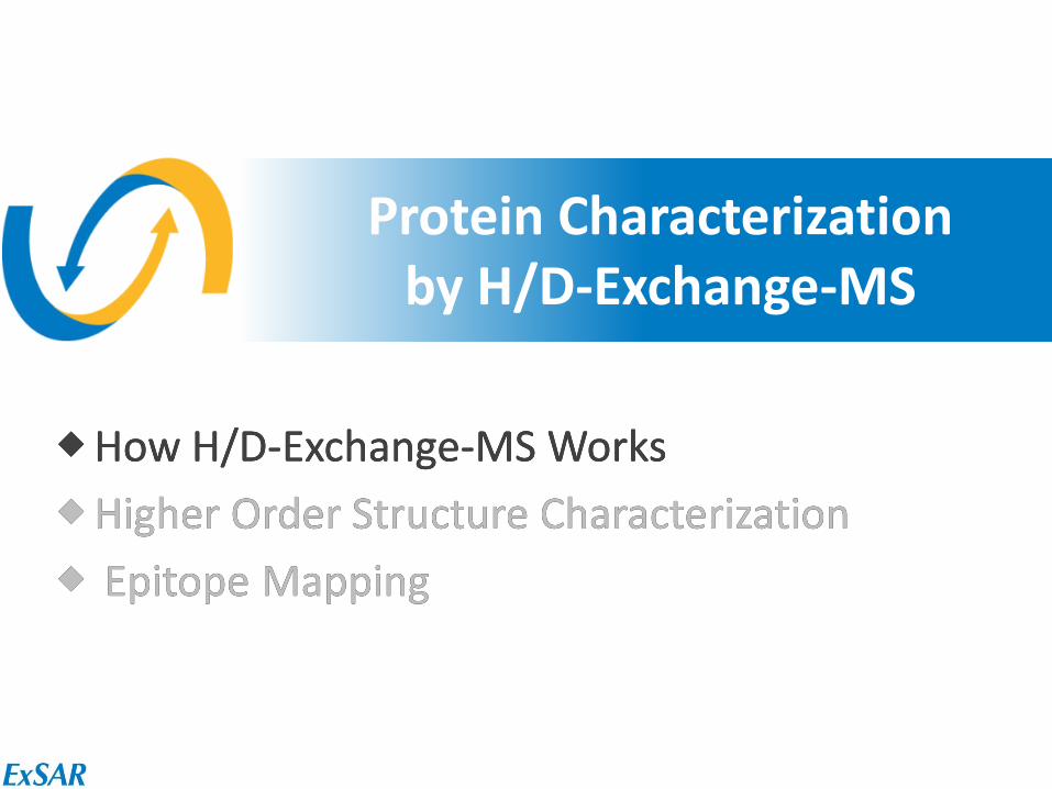
Exchange Rates of Hydrogens in Protein
H2N
O O
O O
OH NH
HN
HN
OH
O
OH
NH2
Hydrogen Exchange Rate OH, SH, NH2, CO2H, CONH2 fast Main chain CONH medium Aliphatic CH Aromatic CH slow
4
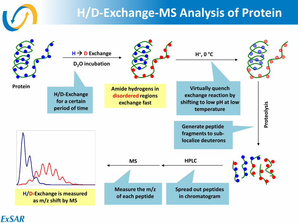
H/D-Exchange-MS Analysis of Protein
H D Exchange
D2O incubation
Protein
H+, 0 °C
Amide hydrogens in disordered regions
exchange fast
H/D-Exchange is measured as m/z shift by MS
Virtually quench exchange reaction by
shifting to low pH at low temperature
H/D-Exchange for a certain
period of time
Generate peptide fragments to sub- localize deuterons
Prot
eoly
sis
HPLC
Spread out peptides in chromatogram
MS
Measure the m/z of each peptide

5
What Does H/D-Exchange Rate Mean?
∆Gf
∆Gch‡
∆Gch‡ = – RT ln kch
∆Gex
‡ = – RT ln kex ∆Gf = ∆Gex
‡ – ∆Gch‡
= – RT ln (kex / kch)
∆Gex‡
NH
O
ND
O
NH
ND
Can be calculated
We measure this
6
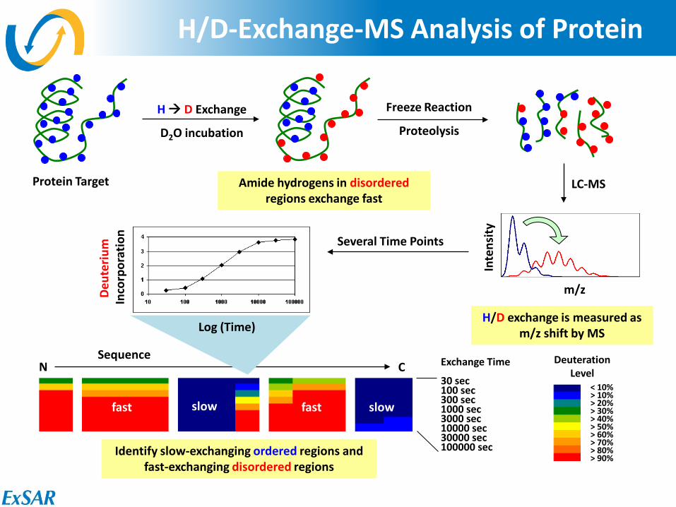
H/D-Exchange-MS Analysis of Protein
H D Exchange
D2O incubation
Protein Target
Freeze Reaction
Proteolysis
LC-MS Amide hydrogens in disordered regions exchange fast
H/D exchange is measured as m/z shift by MS
Inte
nsity
m/z
Exchange Time 30 sec 100 sec 300 sec 1000 sec 3000 sec 10000 sec 30000 sec 100000 sec
< 10% > 10% > 20% > 30% > 40% > 50% > 60% > 70% > 80% > 90%
Deuteration Level
Several Time Points
Sequence N C
Deut
eriu
m
Inco
rpor
atio
n
Log (Time)
Identify slow-exchanging ordered regions and fast-exchanging disordered regions
slow slow fast fast
7
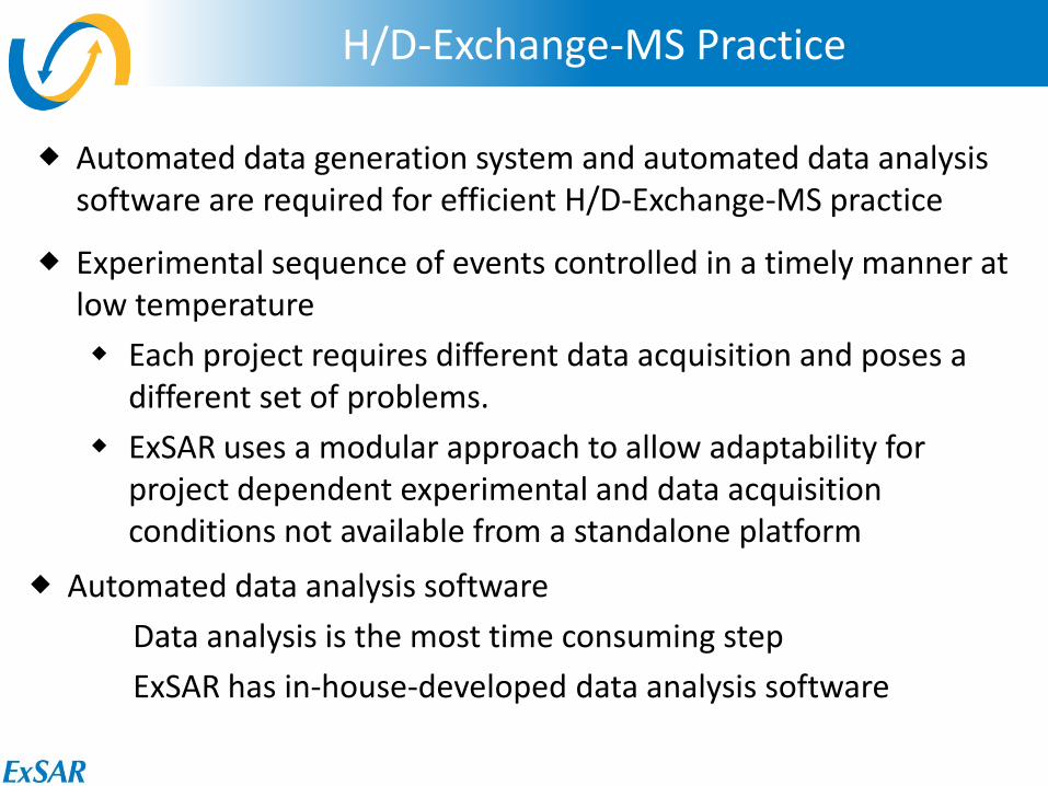
H/D-Exchange-MS Practice
Automated data generation system and automated data analysis software are required for efficient H/D-Exchange-MS practice
Practice of H/D-Exchange-MS
Experimental sequence of events controlled in a timely manner at low temperature Each project requires different data acquisition and poses a
different set of problems. ExSAR uses a modular approach to allow adaptability for
project dependent experimental and data acquisition conditions not available from a standalone platform
Automated data analysis software Data analysis is the most time consuming step ExSAR has in-house-developed data analysis software
8
Protein Characterization by H/D-Exchange-MS
How H/D-Exchange-MS Works Higher Order Structure Characterization Epitope Mapping
9
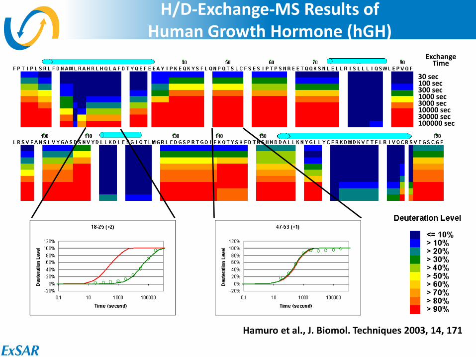
H/D-Exchange-MS Results of Human Growth Hormone (hGH)
Exchange Time
30 sec 100 sec 300 sec 1000 sec 3000 sec 10000 sec 30000 sec 100000 sec
Hamuro et al., J. Biomol. Techniques 2003, 14, 171
10
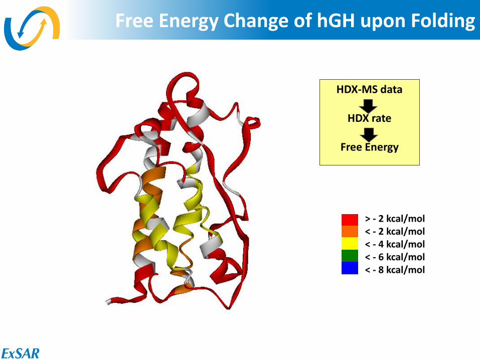
Free Energy Change of hGH upon Folding
> - 2 kcal/mol < - 2 kcal/mol < - 4 kcal/mol < - 6 kcal/mol < - 8 kcal/mol
HDX-MS data
HDX rate
Free Energy
11
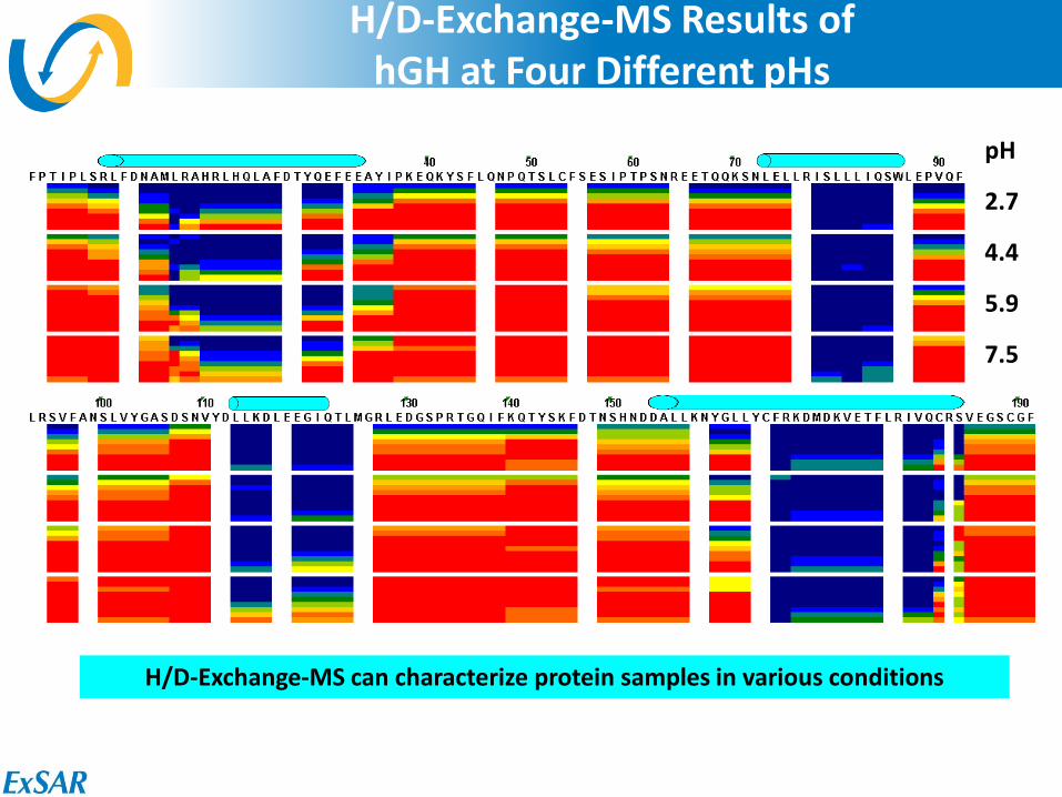
H/D-Exchange-MS Results of hGH at Four Different pHs
pH
2.7
4.4
5.9
7.5
H/D-Exchange-MS can characterize protein samples in various conditions
12
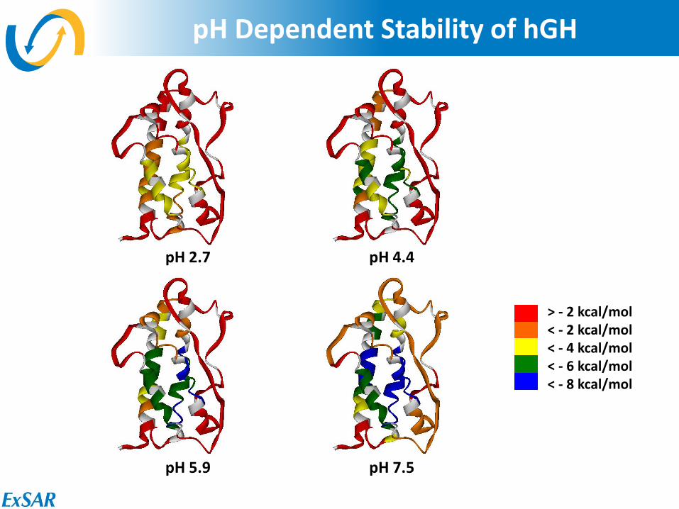
pH Dependent Stability of hGH
> - 2 kcal/mol < - 2 kcal/mol < - 4 kcal/mol < - 6 kcal/mol < - 8 kcal/mol
pH 2.7 pH 4.4
pH 5.9 pH 7.5
13
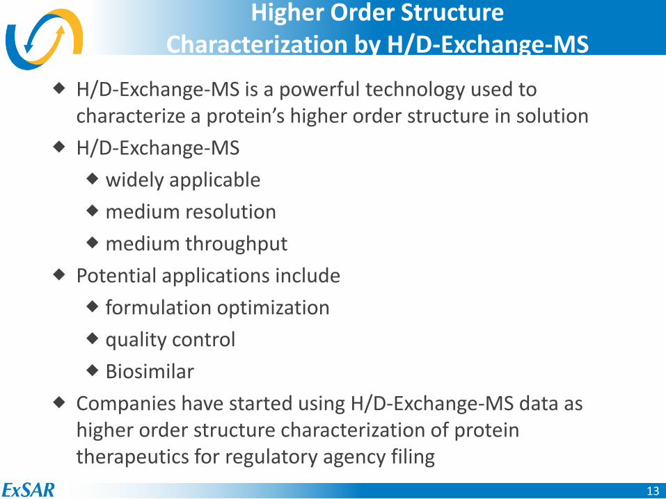
Higher Order Structure Characterization by H/D-Exchange-MS
H/D-Exchange-MS is a powerful technology used to characterize a protein’s higher order structure in solution
H/D-Exchange-MS widely applicable medium resolution medium throughput
Potential applications include formulation optimization quality control Biosimilar
Companies have started using H/D-Exchange-MS data as higher order structure characterization of protein therapeutics for regulatory agency filing
14
Protein Characterization by H/D-Exchange-MS
How H/D-Exchange-MS Works Higher Order Structure Characterization Epitope Mapping
15
http://knol.google.com/k/krishan-maggon/top-ten-twenty-best-selling-drugs-2010/3fy5eowy8suq3/141#
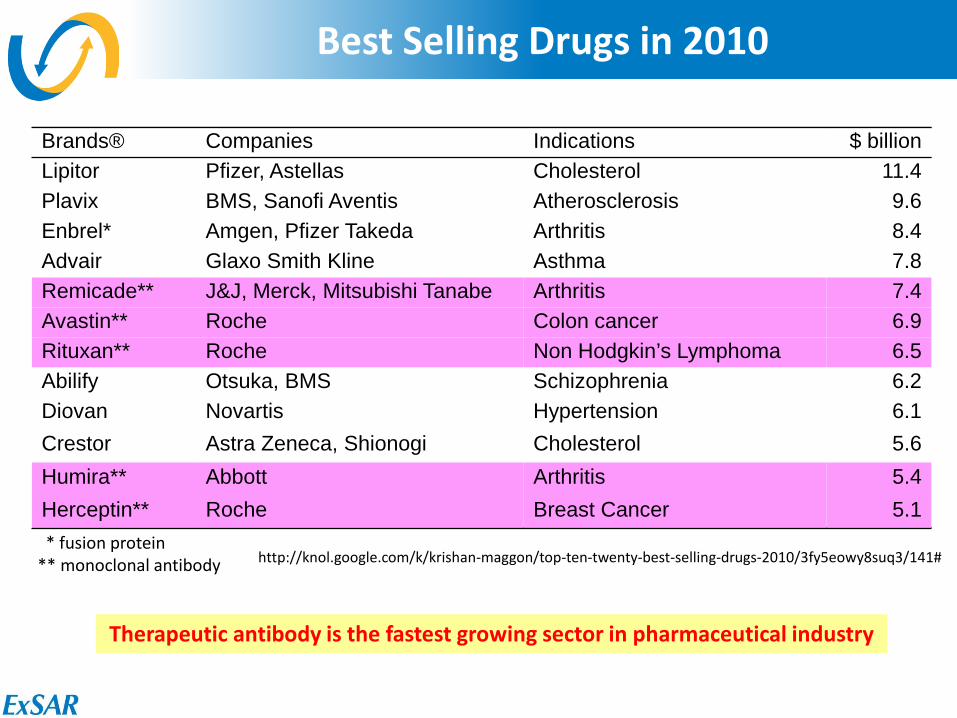
Best Selling Drugs in 2010
Brands® Companies Indications $ billion Lipitor Pfizer, Astellas Cholesterol 11.4 Plavix BMS, Sanofi Aventis Atherosclerosis 9.6 Enbrel* Amgen, Pfizer Takeda Arthritis 8.4 Advair Glaxo Smith Kline Asthma 7.8 Remicade** J&J, Merck, Mitsubishi Tanabe Arthritis 7.4 Avastin** Roche Colon cancer 6.9 Rituxan** Roche Non Hodgkin’s Lymphoma 6.5 Abilify Otsuka, BMS Schizophrenia 6.2 Diovan Novartis Hypertension 6.1 Crestor Astra Zeneca, Shionogi Cholesterol 5.6 Humira** Abbott Arthritis 5.4 Herceptin** Roche Breast Cancer 5.1
Brands® Companies Indications $ billion Lipitor Pfizer, Astellas Cholesterol 11.4 Plavix BMS, Sanofi Aventis Atherosclerosis 9.6 Enbrel* Amgen, Pfizer Takeda Arthritis 8.4 Advair Glaxo Smith Kline Asthma 7.8 Remicade** J&J, Merck, Mitsubishi Tanabe Arthritis 7.4 Avastin** Roche Colon cancer 6.9 Rituxan** Roche Non Hodgkin’s Lymphoma 6.5 Abilify Otsuka, BMS Schizophrenia 6.2 Diovan Novartis Hypertension 6.1 Crestor Astra Zeneca, Shionogi Cholesterol 5.6 Humira** Abbott Arthritis 5.4 Herceptin** Roche Breast Cancer 5.1
* fusion protein ** monoclonal antibody
Therapeutic antibody is the fastest growing sector in pharmaceutical industry
16
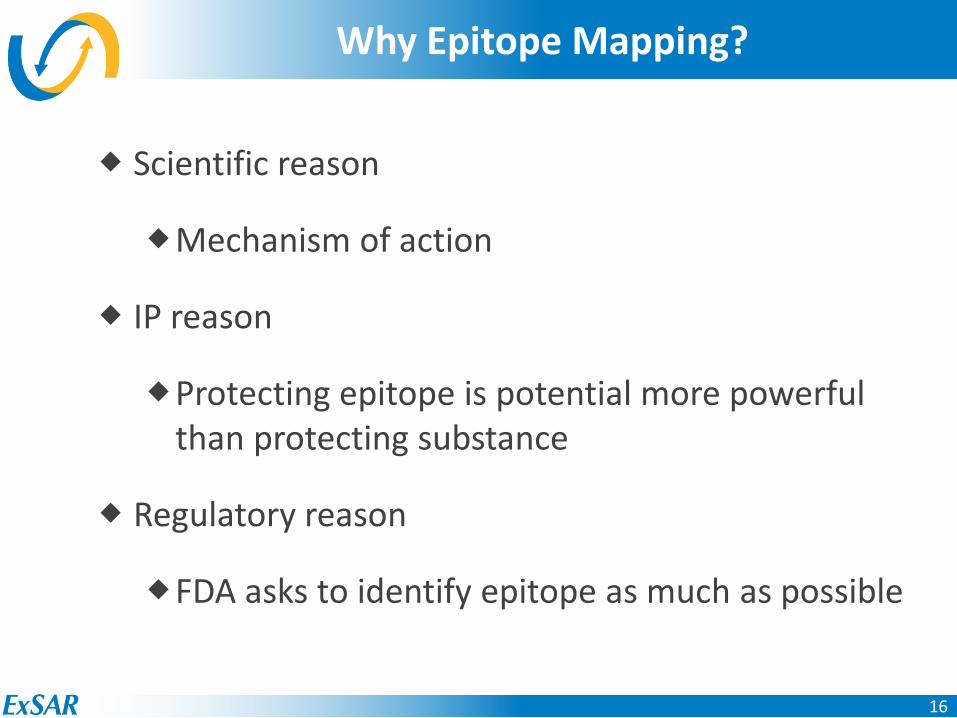
Why Epitope Mapping?
Scientific reason
Mechanism of action
IP reason
Protecting epitope is potential more powerful than protecting substance
Regulatory reason
FDA asks to identify epitope as much as possible
17
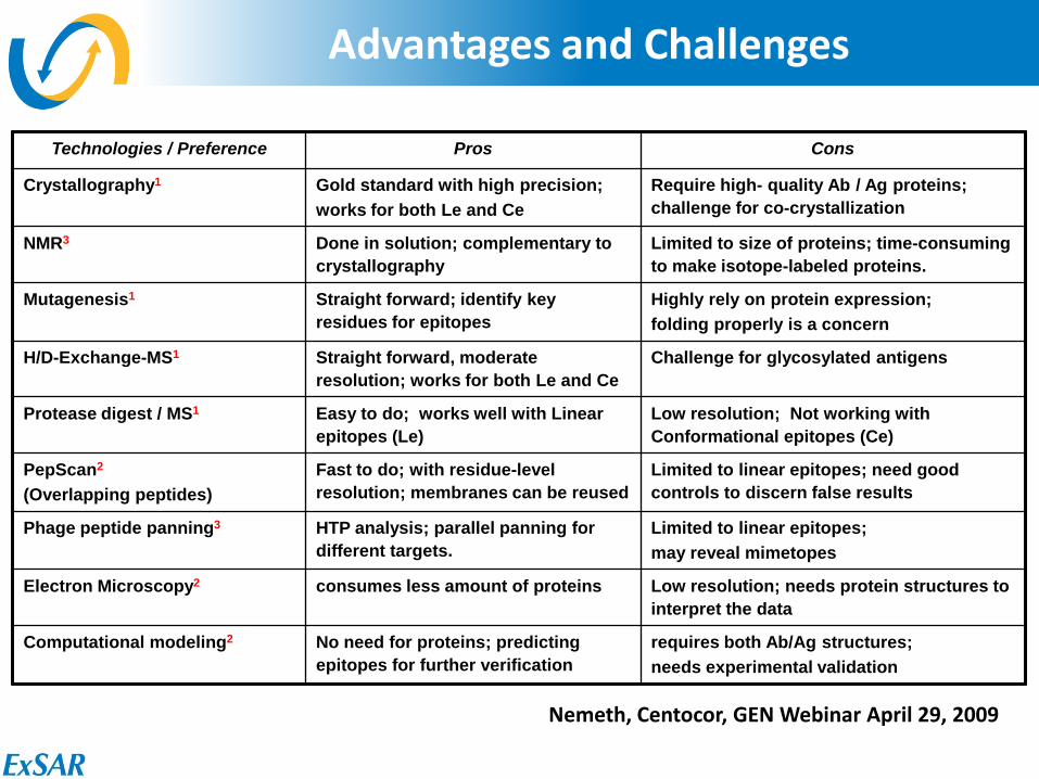
Nemeth, Centocor, GEN Webinar April 29, 2009
Advantages and Challenges
Technologies / Preference Pros Cons
Crystallography1 Gold standard with high precision; works for both Le and Ce
Require high- quality Ab / Ag proteins; challenge for co-crystallization
NMR3 Done in solution; complementary to crystallography
Limited to size of proteins; time-consuming to make isotope-labeled proteins.
Mutagenesis1 Straight forward; identify key residues for epitopes
Highly rely on protein expression; folding properly is a concern
H/D-Exchange-MS1 Straight forward, moderate resolution; works for both Le and Ce
Challenge for glycosylated antigens
Protease digest / MS1 Easy to do; works well with Linear epitopes (Le)
Low resolution; Not working with Conformational epitopes (Ce)
PepScan2 (Overlapping peptides)
Fast to do; with residue-level resolution; membranes can be reused
Limited to linear epitopes; need good controls to discern false results
Phage peptide panning3 HTP analysis; parallel panning for different targets.
Limited to linear epitopes; may reveal mimetopes
Electron Microscopy2 consumes less amount of proteins Low resolution; needs protein structures to interpret the data
Computational modeling2 No need for proteins; predicting epitopes for further verification
requires both Ab/Ag structures; needs experimental validation
18
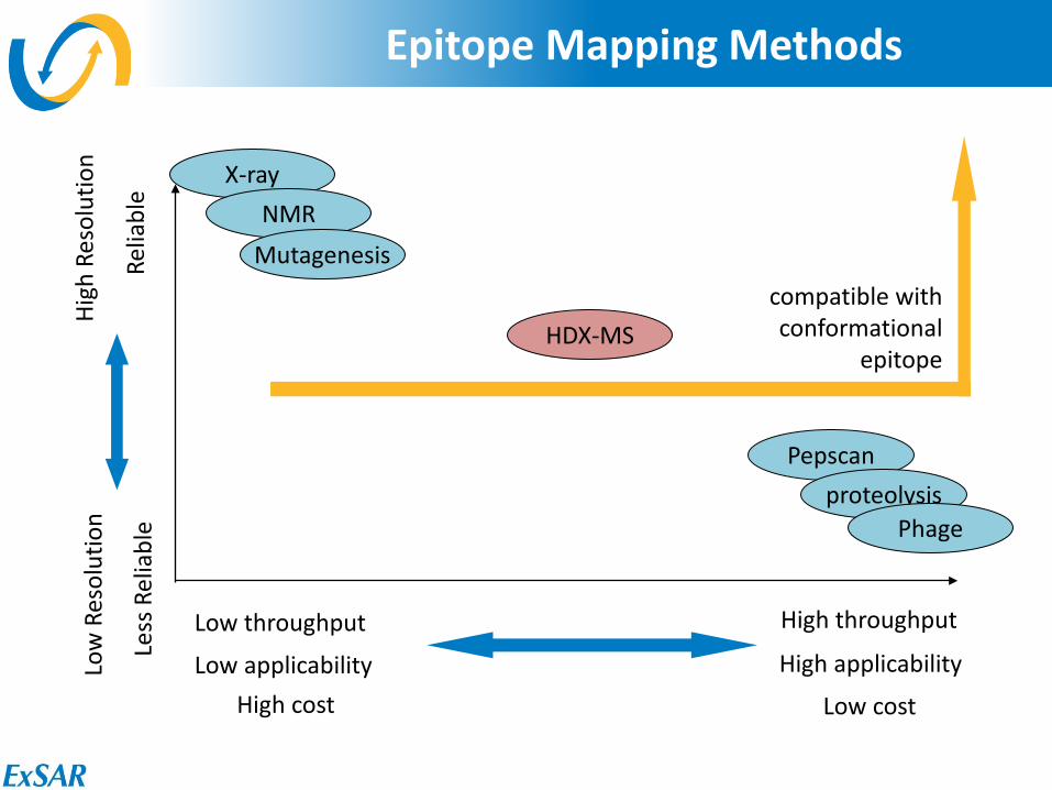
Epitope Mapping Methods
High throughput
Low cost
Low throughput
High cost
High
Res
olut
ion
Relia
ble
Low
Res
olut
ion
Less
Rel
iabl
e X-ray
NMR Mutagenesis
Pepscan proteolysis
Phage
HDX-MS compatible with conformational
epitope
Low applicability High applicability
19
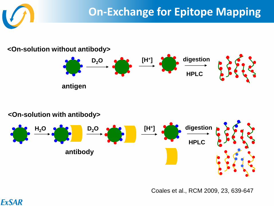
On-Exchange for Epitope Mapping
<On-solution with antibody>
<On-solution without antibody>
H2O
antigen
D2O
[H+] digestion HPLC
D2O
[H+] digestion HPLC
antibody
Coales et al., RCM 2009, 23, 639-647
20
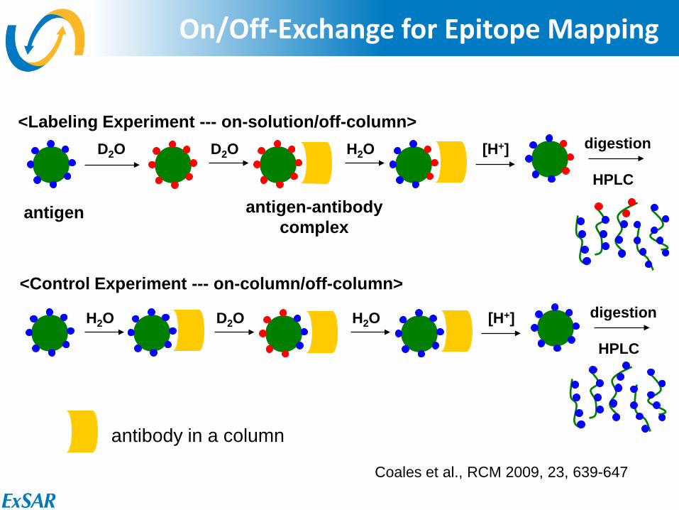
On/Off-Exchange for Epitope Mapping
<Control Experiment --- on-column/off-column>
<Labeling Experiment --- on-solution/off-column>
H2O
antigen antigen-antibody complex
D2O
[H+] digestion HPLC
D2O H2O
[H+] digestion HPLC
H2O D2O
Coales et al., RCM 2009, 23, 639-647
antibody in a column
21
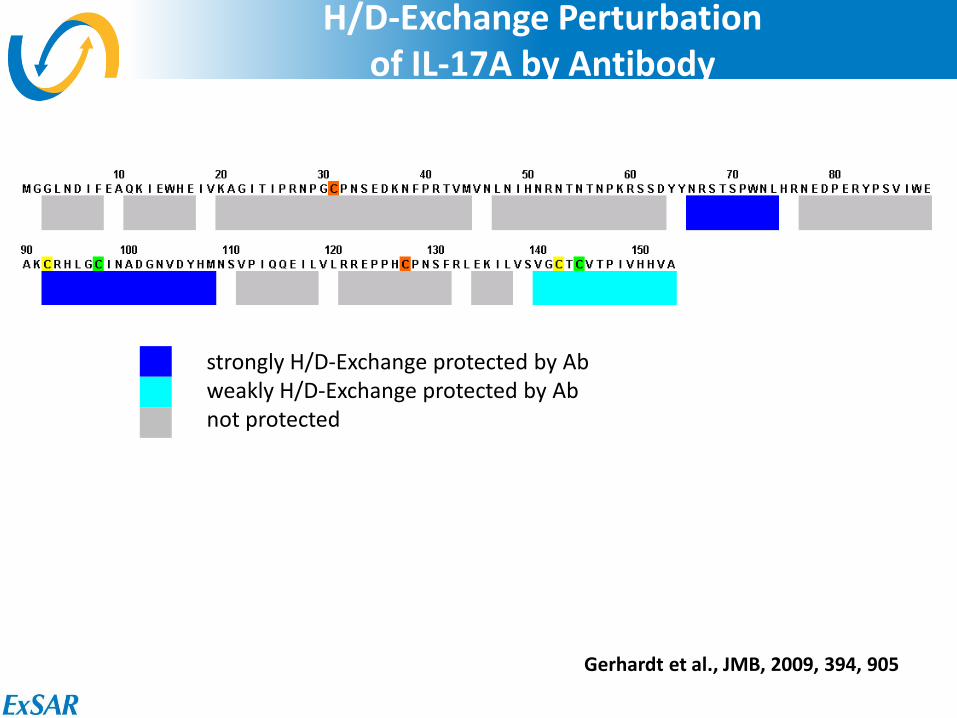
H/D-Exchange Perturbation of IL-17A by Antibody
strongly H/D-Exchange protected by Ab weakly H/D-Exchange protected by Ab not protected
Gerhardt et al., JMB, 2009, 394, 905
22
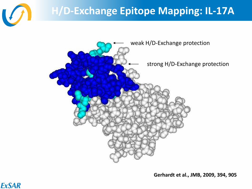
H/D-Exchange Epitope Mapping: IL-17A
strong H/D-Exchange protection
weak H/D-Exchange protection
Gerhardt et al., JMB, 2009, 394, 905
23
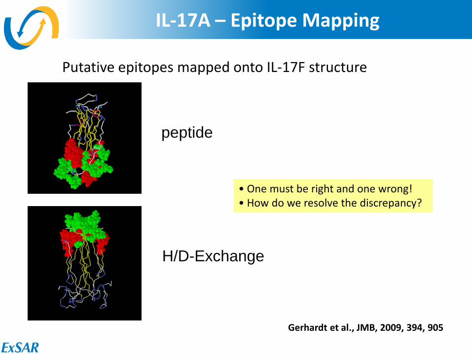
IL-17A – Epitope Mapping
• One must be right and one wrong! • How do we resolve the discrepancy?
Putative epitopes mapped onto IL-17F structure
peptide
H/D-Exchange
Gerhardt et al., JMB, 2009, 394, 905
24
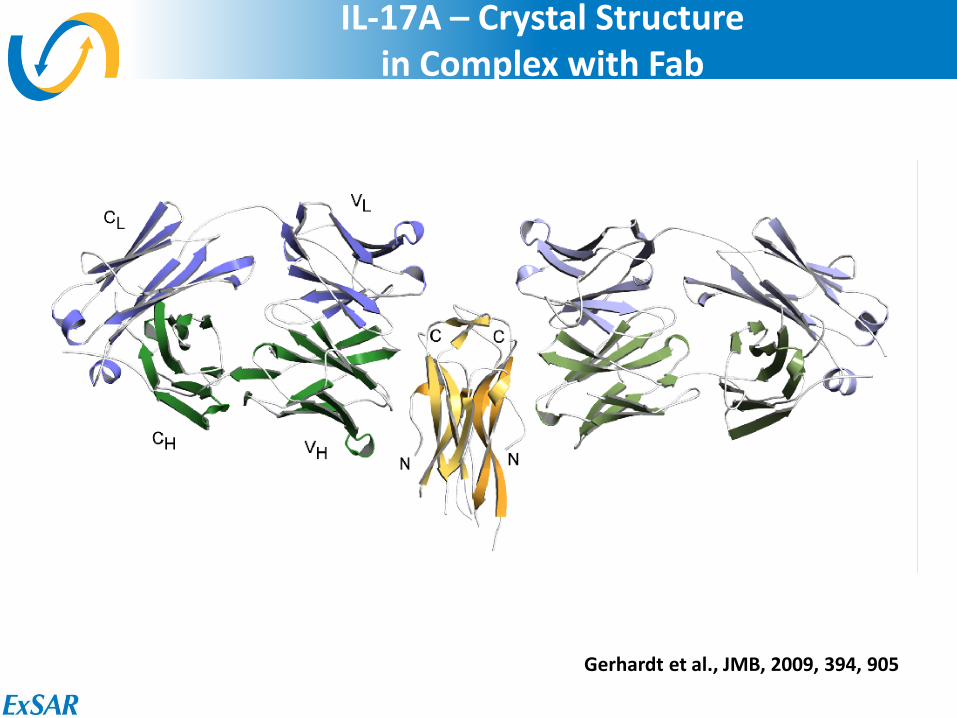
IL-17A – Crystal Structure in Complex with Fab
Gerhardt et al., JMB, 2009, 394, 905

25
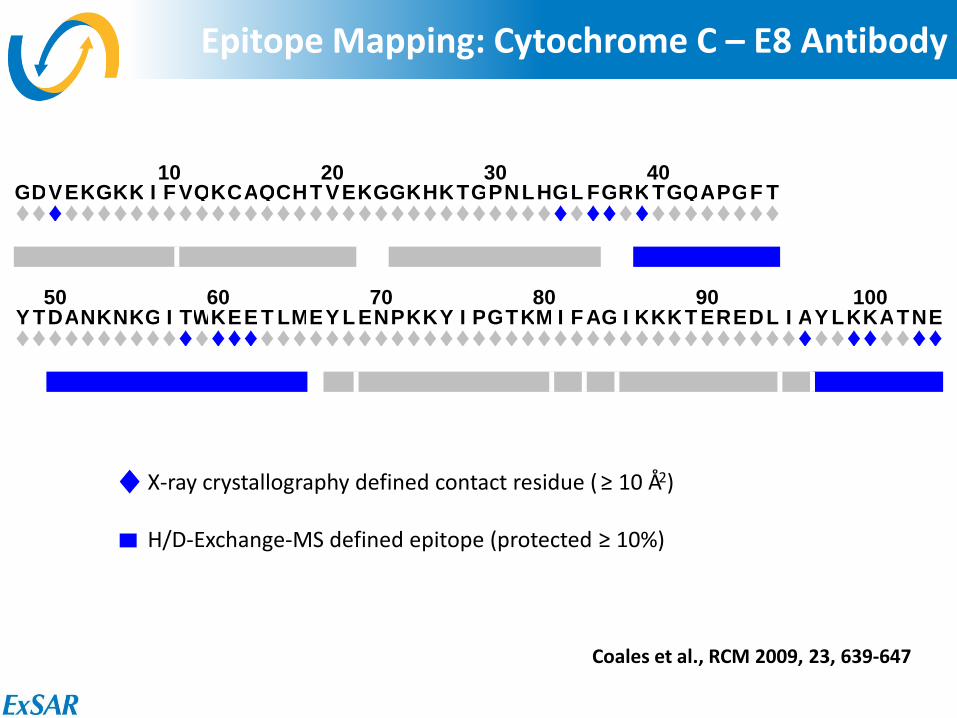
Epitope Mapping: Cytochrome C – E8 Antibody
X-ray crystallography defined contact residue ( ≥ 10 Å2) H/D-Exchange-MSMS defined epitope (protected ≥ 10%)
10 20 30 40GDVEKGKK I F VQKCAQCHT VEKGGKHKTGPNLHGL FGRKTGQAPGF T♦♦♦♦♦♦♦♦♦♦♦♦♦♦♦♦♦♦♦♦♦♦♦♦♦♦♦♦♦♦♦♦♦♦♦♦♦♦♦♦♦♦♦♦♦♦♦●●● ● ●●● ●● ● ●● ●●● ●
50 60 70 80 90 100Y TDANKNKG I TWKEE T LMEY L ENPKKY I PGTKM I FAG I KKKT EREDL I AY LKKATNE♦♦♦♦♦♦♦♦♦♦♦♦♦♦♦♦♦♦♦♦♦♦♦♦♦♦♦♦♦♦♦♦♦♦♦♦♦♦♦♦♦♦♦♦♦♦♦♦♦♦♦♦♦♦♦♦♦●● ●●●● ●● ●● ● ●● ●●●
Coales et al., RCM 2009, 23, 639-647
26
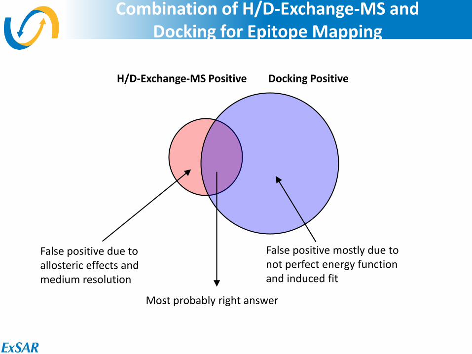
Combination of H/D-Exchange-MS and Docking for Epitope Mapping
H/D-Exchange-MS Positive Docking Positive
False positive due to allosteric effects and medium resolution
False positive mostly due to not perfect energy function and induced fit
Most probably right answer
27
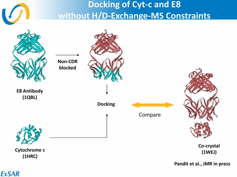
Docking of Cyt-c and E8 without H/D-Exchange-MS Constraints
Non-CDR blocked
Cytochrome c (1HRC)
E8 Antibody (1QBL)
Co-crystal (1WEJ)
Compare
Docking
Pandit et al., JMR in press
28
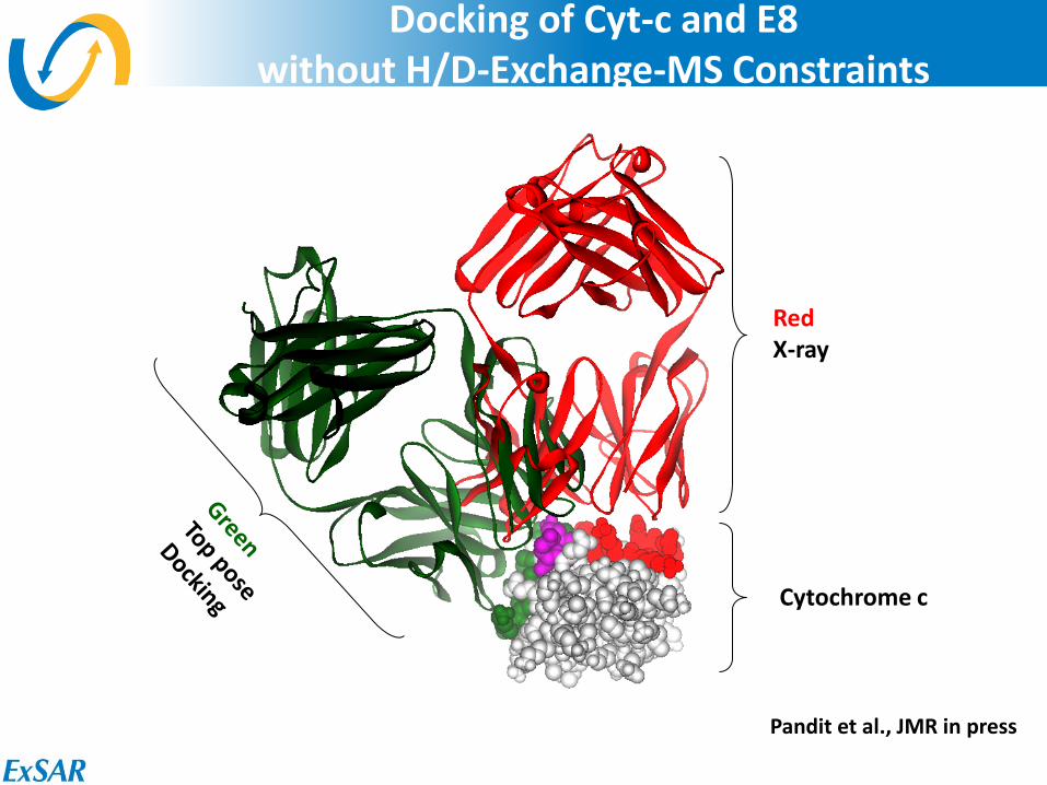
Docking of Cyt-c and E8 without H/D-Exchange-MS Constraints
Cytochrome c
Red X-ray
Pandit et al., JMR in press
29
Epitope Mapping: Cytochrome C – E8 Antibody
X-ray crystallography defined contact residue ( ≥ 10 Å2) H/D-Exchange-MS defined epitope (protected ≥ 10%)
10 20 30 40GDVEKGKK I F VQKCAQCHT VEKGGKHKTGPNLHGL FGRKTGQAPGF T♦♦♦♦♦♦♦♦♦♦♦♦♦♦♦♦♦♦♦♦♦♦♦♦♦♦♦♦♦♦♦♦♦♦♦♦♦♦♦♦♦♦♦♦♦♦♦●●● ● ●●● ●● ● ●● ●●● ●
50 60 70 80 90 100Y TDANKNKG I TWKEE T LMEY L ENPKKY I PGTKM I FAG I KKKT EREDL I AY LKKATNE♦♦♦♦♦♦♦♦♦♦♦♦♦♦♦♦♦♦♦♦♦♦♦♦♦♦♦♦♦♦♦♦♦♦♦♦♦♦♦♦♦♦♦♦♦♦♦♦♦♦♦♦♦♦♦♦♦●● ●●●● ●● ●● ● ●● ●●●
Coales et al., RCM 2009, 23, 639-647

30
Docking of Cyt-c and E8 with H/D-Exchange-MS Constraints
Non-CDR blocked
Non-H/D perturbed blocked
Docking
Cytochrome c (1HRC)
E8 Antibody (1QBL)
Co-crystal (1WEJ)
Compare
Pandit et al., JMR in press

31
Docking of Cyt-c and E8 without H/D-Exchange-MS Constraints
Cytochrome c
Red X-ray
Pandit et al., JMR in press
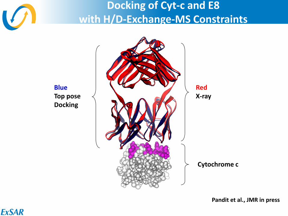
32
Docking of Cyt-c and E8 with H/D-Exchange-MS Constraints
Blue Top pose Docking
Cytochrome c
Red X-ray
Pandit et al., JMR in press
33
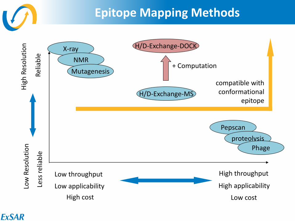
Epitope Mapping Methods
High throughput
Low cost
Low throughput
High cost
High
Res
olut
ion
Relia
ble
Low
Res
olut
ion
Less
relia
ble
X-ray NMR
Mutagenesis
Pepscan proteolysis
Phage
H/D-Exchange-MS
+ Computation
H/D-Exchange-DOCK
compatible with conformational
epitope
Low applicability High applicability
34
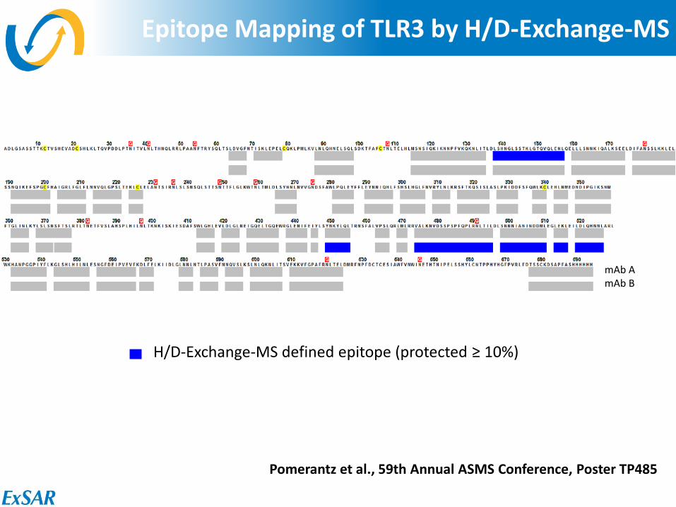
Epitope Mapping of TLR3 by H/D-Exchange-MS
H/D-Exchange-MS defined epitope (protected ≥ 10%)
mAb A mAb B
Pomerantz et al., 59th Annual ASMS Conference, Poster TP485
35
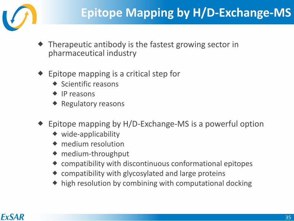
Epitope Mapping by H/D-Exchange-MS
Therapeutic antibody is the fastest growing sector in pharmaceutical industry
Epitope mapping is a critical step for
Scientific reasons IP reasons Regulatory reasons
Epitope mapping by H/D-Exchange-MS is a powerful option wide-applicability medium resolution medium-throughput compatibility with discontinuous conformational epitopes compatibility with glycosylated and large proteins high resolution by combining with computational docking
36
Acknowledgments
ExSAR Stephen J. Coales Kathleen S. Molnar Steven J. Tuske Kelly E Deepangi Pandit
Janssen Jennifer F. Nemeth
AstraZeneca Mark Abbott
UPenn Yvonne Paterson
Financial support NIH
Expanding the Utility of a Commercial HDX Automation Solution
Eric B. Monroe
University of Arizona
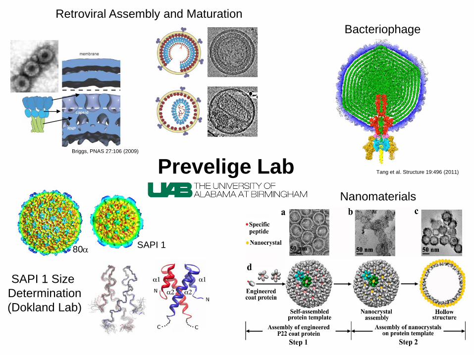
Prevelige Lab
Retroviral Assembly and Maturation
Nanomaterials
Bacteriophage
SAPI 1 Size Determination (Dokland Lab)
80α SAPI 1
Tang et al. Structure 19:496 (2011)
Briggs, PNAS 27:106 (2009)
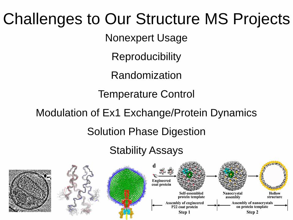
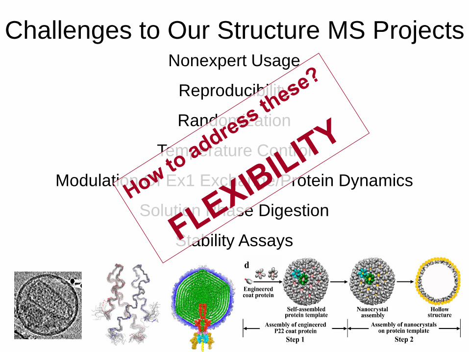
Challenges to Our Structure MS Projects Nonexpert Usage
Reproducibility
Randomization
Temperature Control
Modulation of Ex1 Exchange/Protein Dynamics
Solution Phase Digestion
Stability Assays
Nonexpert Usage
Reproducibility
Randomization
Temperature Control
Modulation Of Ex1 Exchange/Protein Dynamics
Solution Phase Digestion
Stability Assays
Challenges to Our Structure MS Projects
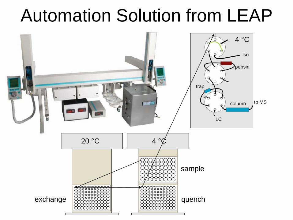
Automation Solution from LEAP
20 °C 4 °C
exchange
sample
quench
4 °C
LC
iso
pepsin
trap
column to MS
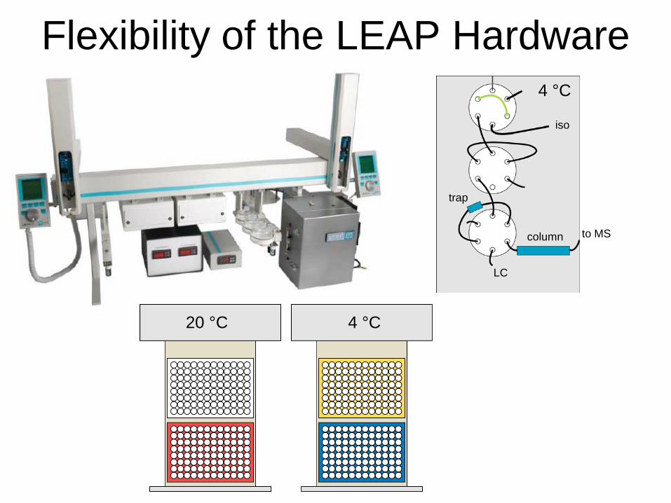
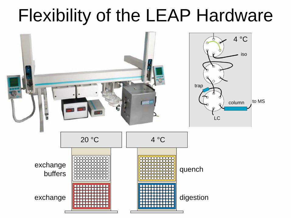
Flexibility of the LEAP Hardware
20 °C 4 °C
4 °C
LC
iso
trap
column to MS
Flexibility of the LEAP Hardware
20 °C 4 °C
exchange
quench
digestion
4 °C
LC
iso
trap
column to MS
exchange buffers
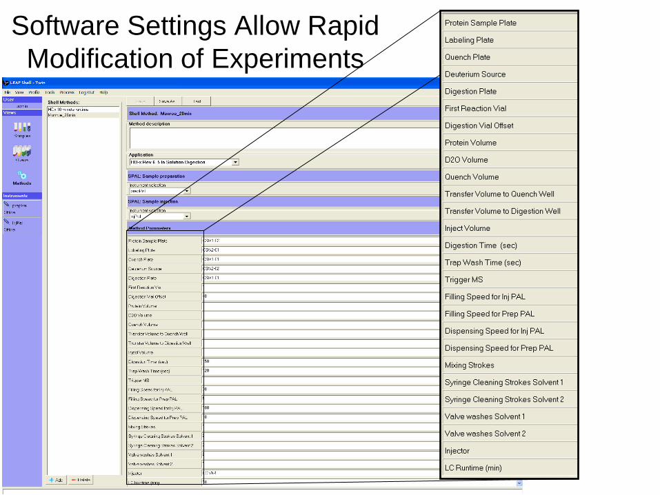
Software Settings Allow Rapid Modification of Experiments

Tweaking Workflow for Randomization • Software “requires” runs in increasing time of exchange • Solution—import list and queue
multiple runs
Excel File
0, 15s, 30s, 1m, 2m, 4m, 8m, 15m, 30m, 60m runs in triplicate
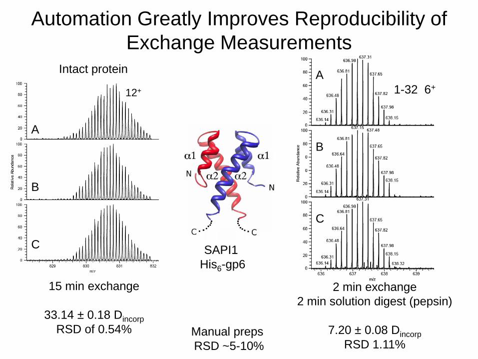
Automation Greatly Improves Reproducibility of Exchange Measurements
12+
15 min exchange
33.14 ± 0.18 Dincorp RSD of 0.54%
2 min exchange 2 min solution digest (pepsin)
7.20 ± 0.08 Dincorp
RSD 1.11%
1-32 6+
Intact protein
A B C
A B C
SAPI1 His6-gp6
Manual preps RSD ~5-10%
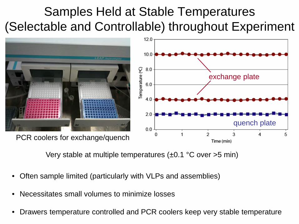
Samples Held at Stable Temperatures (Selectable and Controllable) throughout Experiment
quench plate
exchange plate
PCR coolers for exchange/quench
• Often sample limited (particularly with VLPs and assemblies)
• Necessitates small volumes to minimize losses
• Drawers temperature controlled and PCR coolers keep very stable temperature
Very stable at multiple temperatures (±0.1 °C over >5 min)
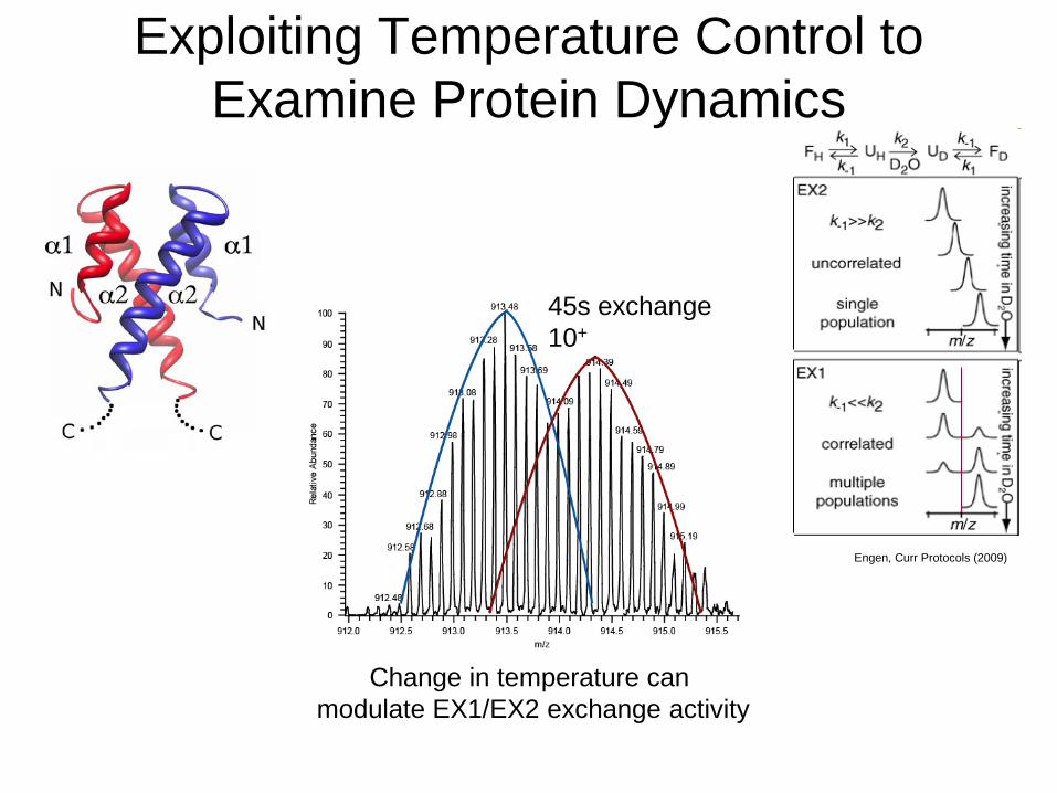
Exploiting Temperature Control to Examine Protein Dynamics
45s exchange 10+
Change in temperature can modulate EX1/EX2 exchange activity
Engen, Curr Protocols (2009)
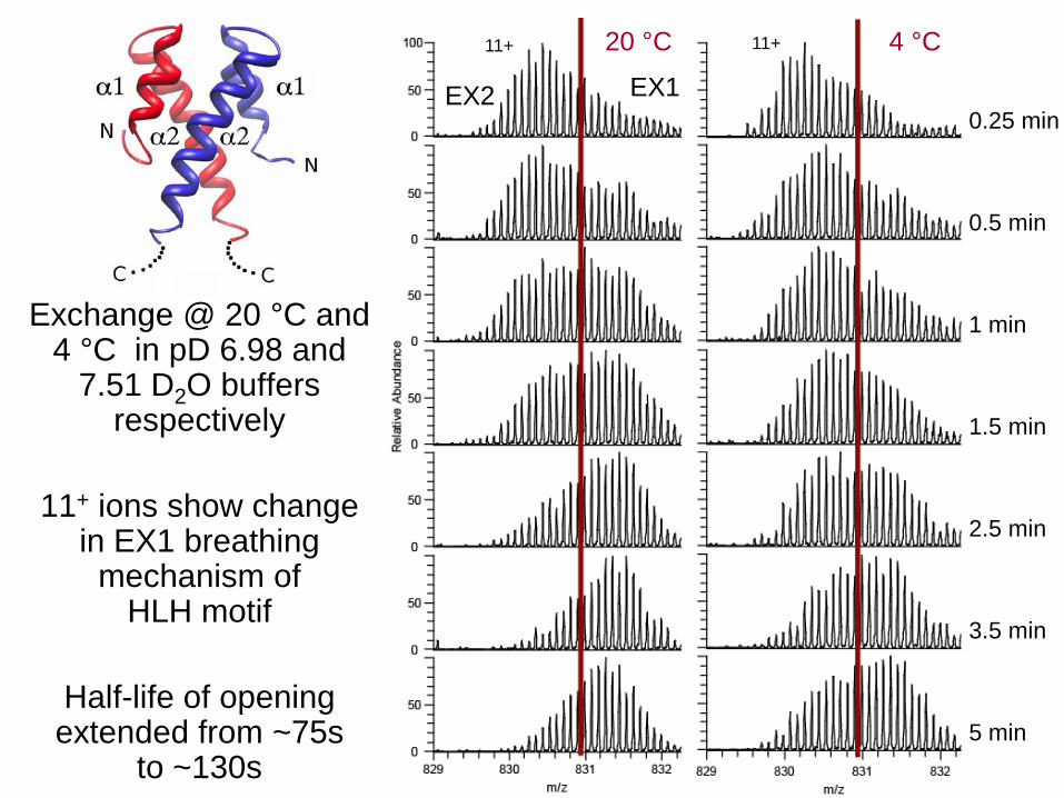
Exchange @ 20 °C and 4 °C in pD 6.98 and
7.51 D2O buffers respectively
11+ ions show change
in EX1 breathing mechanism of
HLH motif
Half-life of opening extended from ~75s
to ~130s
20 °C 4 °C
0.25 min
0.5 min
1 min
1.5 min
2.5 min
3.5 min
5 min
11+ 11+
EX1 EX2
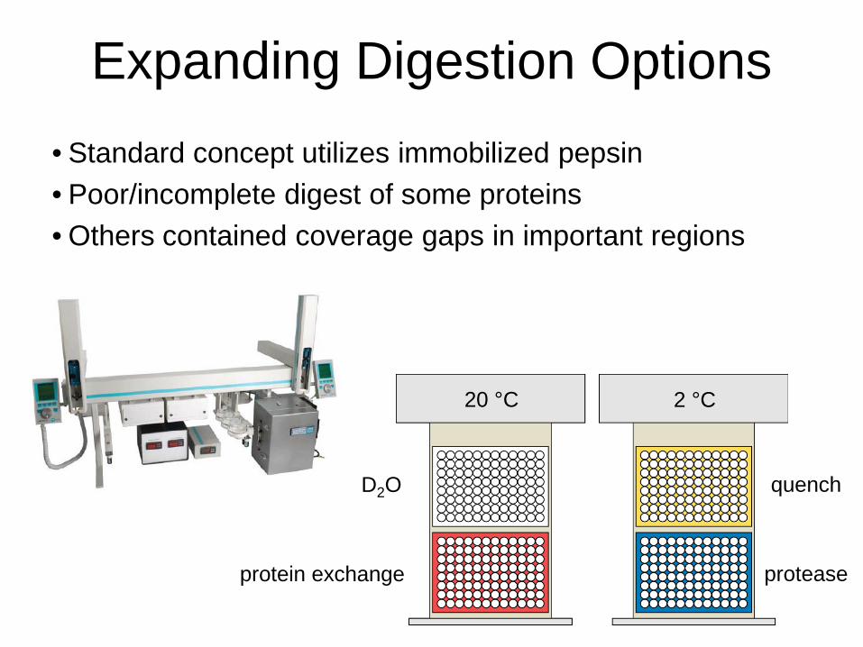
Expanding Digestion Options • Standard concept utilizes immobilized pepsin • Poor/incomplete digest of some proteins • Others contained coverage gaps in important regions
D2O quench
protease protein exchange
2 °C 20 °C
pepsin
XIII
pepsin
XIII
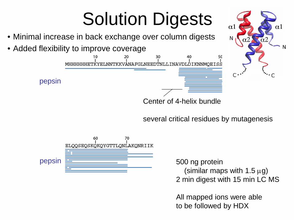
Solution Digests • Minimal increase in back exchange over column digests • Added flexibility to improve coverage
Center of 4-helix bundle several critical residues by mutagenesis
500 ng protein (similar maps with 1.5 µg) 2 min digest with 15 min LC MS All mapped ions were able to be followed by HDX
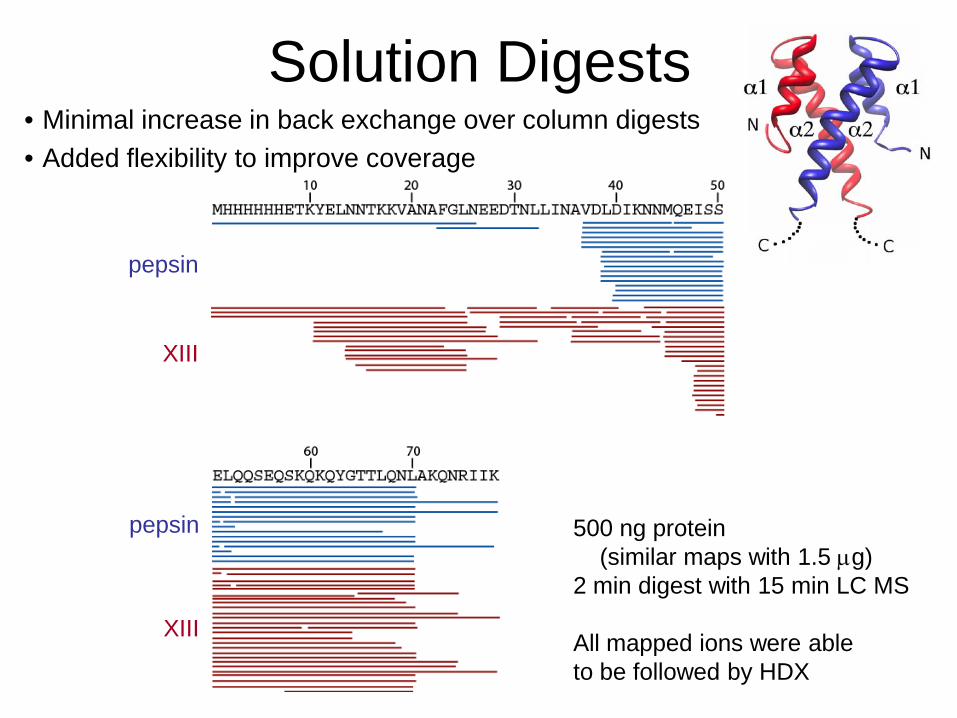
Solution Digests • Minimal increase in back exchange over column digests • Added flexibility to improve coverage
500 ng protein (similar maps with 1.5 µg) 2 min digest with 15 min LC MS All mapped ions were able to be followed by HDX
pepsin
XIII
pepsin
XIII
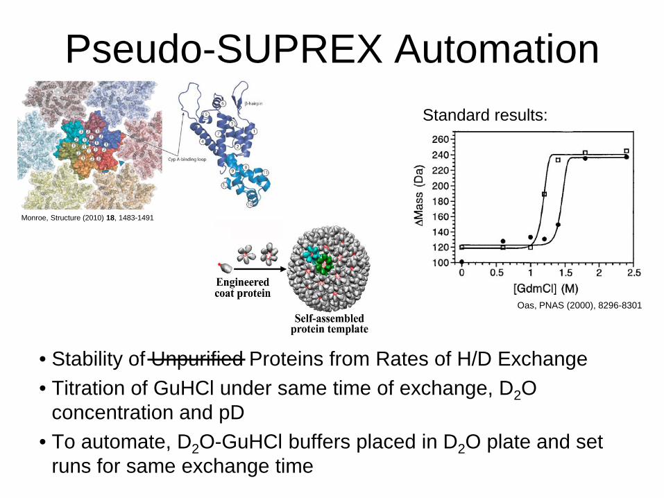
Pseudo-SUPREX Automation
• Stability of Unpurified Proteins from Rates of H/D Exchange • Titration of GuHCl under same time of exchange, D2O
concentration and pD • To automate, D2O-GuHCl buffers placed in D2O plate and set
runs for same exchange time
Monroe, Structure (2010) 18, 1483-1491
Oas, PNAS (2000), 8296-8301
Standard results:
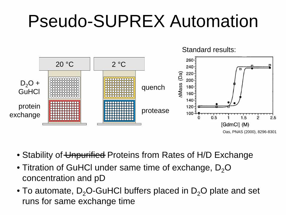
Pseudo-SUPREX Automation
• Stability of Unpurified Proteins from Rates of H/D Exchange • Titration of GuHCl under same time of exchange, D2O
concentration and pD • To automate, D2O-GuHCl buffers placed in D2O plate and set
runs for same exchange time
Oas, PNAS (2000), 8296-8301
Standard results:
D2O + GuHCl quench
protease protein exchange
2 °C 20 °C
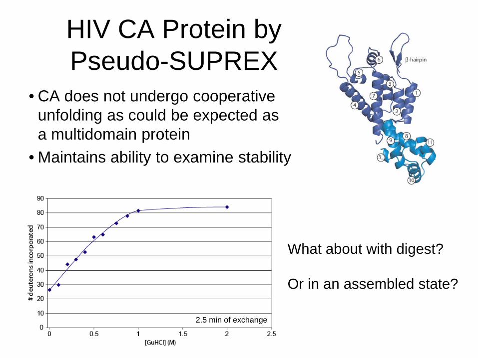
HIV CA Protein by Pseudo-SUPREX
• CA does not undergo cooperative unfolding as could be expected as a multidomain protein
• Maintains ability to examine stability
What about with digest? Or in an assembled state?
2.5 min of exchange
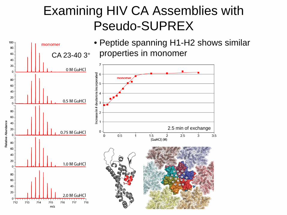
Examining HIV CA Assemblies with Pseudo-SUPREX • Peptide spanning H1-H2 shows similar
properties in monomer CA 23-40 3+
2.5 min of exchange
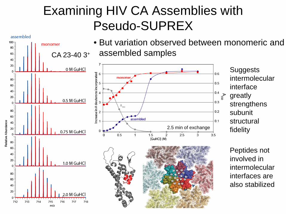
• But variation observed between monomeric and assembled samples
CA 23-40 3+
Suggests intermolecular interface greatly strengthens subunit structural fidelity Peptides not involved in intermolecular interfaces are also stabilized
2.5 min of exchange
Examining HIV CA Assemblies with Pseudo-SUPREX
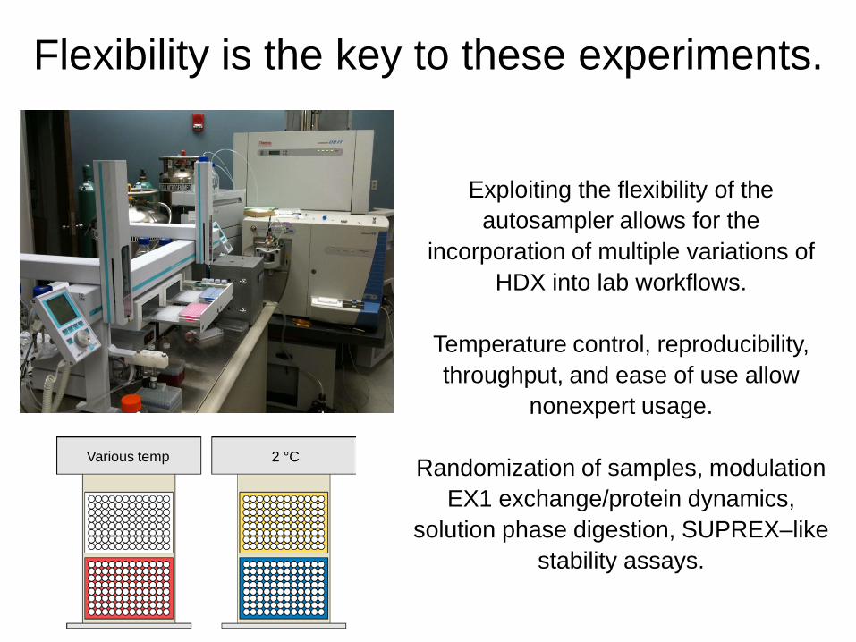
Flexibility is the key to these experiments.
Various temp 2 °C
Exploiting the flexibility of the autosampler allows for the
incorporation of multiple variations of HDX into lab workflows.
Temperature control, reproducibility, throughput, and ease of use allow
nonexpert usage.
Randomization of samples, modulation EX1 exchange/protein dynamics,
solution phase digestion, SUPREX–like stability assays.
Acknowledgements Peter Prevelige
David Morris James Cherwa
Rui Li
Matt Renfrow LeeAnn Boerma
Terje Dokland Altaira Dearborn Michael Spilman
Peter Smith
National Institutes of Health RR17261, DK077279-01,
R01AI044626, F32GM087994


![Analysis of Steroids using Solid Phase Microextraction-Gas Chromatography-Mass … · 2018-03-29 · spectrometry and tandem mass spectrometry[12], liquid chromatography-mass spectrometry](https://static.fdocuments.net/doc/165x107/5f47f27e21a760452d67e4a6/analysis-of-steroids-using-solid-phase-microextraction-gas-chromatography-mass-2018-03-29.jpg)