Chlorinated Benzenes Cause Concomitantly …...Chlorinated Benzenes Cause Concomitantly Oxidative...
16
Chlorinated Benzenes Cause Concomitantly Oxidative Stress and Induction of Apoptotic Markers in Lung Epithelial Cells (A549) at Nonacute Toxic Concentrations Nora Mo ¨rbt, † Janina Tomm, † Ralph Feltens, †,‡ Iljana Mo ¨gel, ‡ Stefan Kalkhof, † Kalaimathi Murugesan, † Henry Wirth, †,§ Carsten Vogt, | Hans Binder, § Irina Lehmann, ‡ and Martin von Bergen* ,†,⊥ Department of Proteomics, Department of Environmental Immunology, Department of Isotope Biogeochemistry, and Department of Metabolomics, Helmholtz Centre for Environmental Research - UFZ, Permoser Strasse 15, 04318 Leipzig, Germany, Interdisciplinary Centre for Bioinformatics of Leipzig University, D-4107 Leipzig, Haertelstrasse 16-18, Germany Received June 8, 2010 In industrialized countries, people spend more time indoors and are therefore increasingly exposed to volatile organic compounds that are emitted at working places and from consumer products, paintings, and furniture, with chlorobenzene (CB) and 1,2-dichlorobenzene (DCB) being representatives of the halogenated arenes. To unravel the molecular effects of low concentrations typical for indoor and occupational exposure, we exposed human lung epithelial cells to CB and DCB and analyzed the effects on the proteome level by 2-D DIGE, where 860 protein spots were detected. A set of 25 and 30 proteins were found to be significantly altered due to exposure to environmentally relevant concentrations of 10 -2 g/m 3 of CB or 10 -3 g/m 3 of DCB (2.2 and 0.17 ppm), respectively. The most enriched pathways were cell death signaling, oxidative stress response, protein quality control, and metabolism. The involvement of oxidative stress was validated by ROS measurement. Among the regulated proteins, 28, for example, voltage-dependent anion-selective channel protein 2, PDCD6IP protein, heat shock protein beta-1, proliferating cell nuclear antigen, nucleophosmin, seryl-tRNA synthetase, prohibitin, and protein arginine N-methyltransferase 1, could be correlated with the molecular pathway of cell death signaling. Caspase 3 activation by cleavage was confirmed for both CB and DCB by immuno- blotting. Treatment with CB or DCB also caused differential protein phosphorylation, for example, at the proteins HNRNP C1/C2, serine-threonine receptor associated protein, and transaldolase 1. Compared to previous results, where cells were exposed to styrene, for the chlorinated aromatic substances besides oxidative stress, apoptosis was found as the predominant cellular response mechanism. Keywords: 1,2-dichlorobenzene • apoptosis • chlorobenzene • lung epithelial cells • VOC 1. Introduction In the last fifty years, changes in lifestyle in the industrialized countries have led to prolonged times spent indoors, concur- rent with the cumulative use of consumer products containing volatile organic compounds (VOCs). Therefore, many people are increasingly exposed to these chemicals which in severe cases may cause symptoms that are commonly referred to as the sick building syndrome, a condition that is characterized by irritation of the eyes, nose, throat and skin and in extreme cases even results in neuronal disorders (for review, see refs 1 and 2). According to their structure, VOCs can be classified into aliphatic and aromatic hydrocarbons and additionally subdi- vided into nonhalogenated and halogenated compounds. Among the latter, chlorobenzene (CB) and dichlorobenzene (DCB) are among the most abundant representatives. 3 CB is used as an intermediate in chemical synthesis of herbicides and dyes and is also present in consumer products, such as rubber and paint. 4 In 1988, 88 500 t of DCB were produced in Western Europe. 5 Like chlorobenzene, it is mainly used as an intermediate in the chemical synthesis of pesticides but is also present as solvent in adhesives and painting material. Thus, halogenated compounds can cause severe problems since their biologically mediated degradation is rather slow and these * To whom correspondence should be addressed. PD Dr. Martin von Bergen, UFZ Helmholtz Centre for Environmental Research, Department of Proteomics, Permoser Str. 15, 04318 Leipzig, Germany. E-mail: [email protected]. Fax: +49-341-2351787. † Department of Proteomics, Helmholtz Centre for Environmental Re- search - UFZ. ‡ Department of Environmental Immunology, Helmholtz Centre for Environmental Research - UFZ. § Interdisciplinary Centre for Bioinformatics of Leipzig University. | Department of Isotope Biogeochemistry, Helmholtz Centre for Envi- ronmental Research - UFZ. ⊥ Department of Metabolomics, Helmholtz Centre for Environmental Research - UFZ. 10.1021/pr1005718 2011 American Chemical Society Journal of Proteome Research 2011, 10, 363–378 363 Published on Web 11/20/2010
Transcript of Chlorinated Benzenes Cause Concomitantly …...Chlorinated Benzenes Cause Concomitantly Oxidative...
Chlorinated Benzenes Cause Concomitantly Oxidative Stress and
Induction of Apoptotic Markers in Lung Epithelial Cells (A549) at
Nonacute Toxic Concentrations
Nora Morbt,† Janina Tomm,† Ralph Feltens,†,‡ Iljana Mogel,‡ Stefan Kalkhof,†
Kalaimathi Murugesan,† Henry Wirth,†,§ Carsten Vogt,| Hans Binder,§ Irina Lehmann,‡ andMartin von Bergen*,†,⊥
Department of Proteomics, Department of Environmental Immunology, Department of IsotopeBiogeochemistry, and Department of Metabolomics, Helmholtz Centre for Environmental Research - UFZ,
Permoser Strasse 15, 04318 Leipzig, Germany, Interdisciplinary Centre for Bioinformatics of Leipzig University,D-4107 Leipzig, Haertelstrasse 16-18, Germany
Received June 8, 2010
In industrialized countries, people spend more time indoors and are therefore increasingly exposed tovolatile organic compounds that are emitted at working places and from consumer products, paintings,and furniture, with chlorobenzene (CB) and 1,2-dichlorobenzene (DCB) being representatives of thehalogenated arenes. To unravel the molecular effects of low concentrations typical for indoor andoccupational exposure, we exposed human lung epithelial cells to CB and DCB and analyzed the effectson the proteome level by 2-D DIGE, where 860 protein spots were detected. A set of 25 and 30 proteinswere found to be significantly altered due to exposure to environmentally relevant concentrations of10-2 g/m3 of CB or 10-3 g/m3 of DCB (2.2 and 0.17 ppm), respectively. The most enriched pathwayswere cell death signaling, oxidative stress response, protein quality control, and metabolism. Theinvolvement of oxidative stress was validated by ROS measurement. Among the regulated proteins,28, for example, voltage-dependent anion-selective channel protein 2, PDCD6IP protein, heat shockprotein beta-1, proliferating cell nuclear antigen, nucleophosmin, seryl-tRNA synthetase, prohibitin,and protein arginine N-methyltransferase 1, could be correlated with the molecular pathway of celldeath signaling. Caspase 3 activation by cleavage was confirmed for both CB and DCB by immuno-blotting. Treatment with CB or DCB also caused differential protein phosphorylation, for example, atthe proteins HNRNP C1/C2, serine-threonine receptor associated protein, and transaldolase 1. Comparedto previous results, where cells were exposed to styrene, for the chlorinated aromatic substances besidesoxidative stress, apoptosis was found as the predominant cellular response mechanism.
Keywords: 1,2-dichlorobenzene • apoptosis • chlorobenzene • lung epithelial cells • VOC
1. Introduction
In the last fifty years, changes in lifestyle in the industrializedcountries have led to prolonged times spent indoors, concur-rent with the cumulative use of consumer products containingvolatile organic compounds (VOCs). Therefore, many peopleare increasingly exposed to these chemicals which in severecases may cause symptoms that are commonly referred to as
the sick building syndrome, a condition that is characterizedby irritation of the eyes, nose, throat and skin and in extremecases even results in neuronal disorders (for review, see refs 1and 2).
According to their structure, VOCs can be classified intoaliphatic and aromatic hydrocarbons and additionally subdi-vided into nonhalogenated and halogenated compounds.Among the latter, chlorobenzene (CB) and dichlorobenzene(DCB) are among the most abundant representatives.3 CB isused as an intermediate in chemical synthesis of herbicidesand dyes and is also present in consumer products, such asrubber and paint.4 In 1988, 88 500 t of DCB were produced inWestern Europe.5 Like chlorobenzene, it is mainly used as anintermediate in the chemical synthesis of pesticides but is alsopresent as solvent in adhesives and painting material. Thus,halogenated compounds can cause severe problems since theirbiologically mediated degradation is rather slow and these
* To whom correspondence should be addressed. PD Dr. Martin vonBergen, UFZ Helmholtz Centre for Environmental Research, Departmentof Proteomics, Permoser Str. 15, 04318 Leipzig, Germany. E-mail:[email protected]. Fax: +49-341-2351787.
† Department of Proteomics, Helmholtz Centre for Environmental Re-search - UFZ.
‡ Department of Environmental Immunology, Helmholtz Centre forEnvironmental Research - UFZ.
§ Interdisciplinary Centre for Bioinformatics of Leipzig University.| Department of Isotope Biogeochemistry, Helmholtz Centre for Envi-
ronmental Research - UFZ.⊥ Department of Metabolomics, Helmholtz Centre for Environmental
Research - UFZ.
10.1021/pr1005718 2011 American Chemical Society Journal of Proteome Research 2011, 10, 363–378 363Published on Web 11/20/2010
compounds are therefore classified as persistent organic pol-lutants (POPs6).
In industry, very high occupational concentration levels havebeen reported for workplaces associated with the productionand usage of volatile organic compounds. However, CB is notbioaccumulated to high levels and does not belong to the groupof persistent organic compounds.5,7 Only low amounts (1-9ng/g) of CB were found in 98% of human adipose tissuesamples from all regions of the United States. In addition, CBwas also detected in exhaled breath, urine (20-120 ng/L), andhuman breast milk.8,9
Measured DCB concentrations in blood of the general“unexposed” population are below 3 ppb.10 The content inwhole human milk ranged from 3 to 29 ppb while 38 µg of DCBper kg fat were detected in adipose tissue.11 Bioaccumulationis only expected in tissues with high fat content duringprolonged, continuous exposures as observed in rats exposedto high concentrations of 250 mg/kg.12
Occupational DCB levels up to 8.5 ppm (51 mg/m3) havebeen found in chlorobenzene factories.11 For chlorobenzene,concentrations between 18.7-488 mg/m3 were found.13 Atthese high concentrations chlorobenzene can cause adversehealth effects like irritation of the mucosa in nose and throatas well as headaches and dizziness.14 In order to protect hu-man health, German Ministry of Labor and Social Affairs(Arbeitsplatzgrenzwert, AGW) and the American Conference ofIndustrial Hygienists (Threshold Limit Value, TLV) adopted alimit of 10 ppm for the occupational exposure to CB. Thecurrent Permissible Exposure Limit (PEL) according to theOccupational Safety & Health Administration (OSHA) is 75ppm. Occupational Safety & Health Administration (OSHA)adopted a Permissible Exposure Limit of 50 ppm for DCB. In2001, the German MAK-value for DCB was lowered to 10 ppm(60 mg/m3).7
Under normal circumstances, concentrations in householdsare too low to cause acute toxic effects. In epidemiologic studiesperformed in the area of Leipzig (Germany), concentrations ofmono- and dichlorobenzene were found to be much lower thanthe reported workplace levels.15 In particular for monochlo-robenzene, concentrations in the range of 1-3.5 µg/m3 weremeasured in apartments, which is comparable to those foundin other studies from Germany.16 Similar results were obtainedin the United States of America.17 While the commonlyencountered indoor concentrations are not suspected to elicitacute toxic effects, epidemiological studies have shown thatlow VOC concentrations are correlated with an increased riskof developing atopic diseases such as eczema15 or obstructivebronchitis.18 Furthermore, it has been shown that low-levelexposure to VOCs can also add up in affecting the developingimmune system of the newborn child19 or infants.20 To unravelthe molecular mechanisms underlying the mode of action ofvolatile organic compounds, the application of in vitro modelsystems is required. Since the respiratory tract is a primary siteof exposure, the lung epithelial cell line A549 was selected asthe model of choice for our investigations.21 Despite themalignant nature, A549 possess typical properties of alveolarepithelial type II cells. They express multi drug resistance-associated proteins (MRP) 1 and 3 as well as most of the majorconstitutive and inducible CYP forms found in lung epithelialcells22 and are able to secrete surfactant proteins, especiallywhen cultured air-exposed.23 Moreover, A549 cells have beenshown to be competent for expressing and secreting inflam-
matory markers such as cytokines and chemokines in responseto volatile organic compounds.21,24
Toxicological data for mono-, 1,2-, and 1,4-dichlorobenzeneobtained in mice have indicated significant differences espe-cially in terms of hepatotoxicity, protein droplet formation inthe kidney and with respect to the serum level of thyroxine.25
These earlier observations point to differences in the toxico-logical effects of the two compounds. Thus, we decided tocompare these CB and DCB side by side using an in vitrosystem to study to which extent the degree of chlorinationaffects cellular response. Since the focus was set on nonacuteeffects of these two compounds, we applied a concentrationrange for which no significant toxicity had been observed in aprevious study using chlorobenzene and the here-applied invitro exposure system26 and tested it for effects on the globalproteome response.
In earlier exposure studies, the A549 cells were additionallystimulated by TNFR24,26-28 to simulate the cells resulting in asomewhat arbitrary situation presumably resembling the invivo situation of an inflammatory response occurring withinthe lung epithelium. However, under these conditions it isdifficult to distinguish between the synergistic stimulatoryeffects caused by TNFR and those caused by exposure to theVOCs alone. Therefore, we decided to omit TNFR from theculturing medium in this study.
For the analysis of molecular effects, we have chosen aproteomic approach. It is a fact that a complete coverage ofthe cellular proteome cannot be obtained using currentmethods, especially regarding those proteins that are expressedin low copy numbers is not achievable. Still it is possible touse changes in the expression level or posttranscriptionalmodification of the more abundant proteins to gain valuableinsights into the involved pathways of cellular reaction towardxenobiotic substances.29,30 On the technical side, we opted fora 2D DIGE approach. 2D gels are not only capable to displaychanges in protein expression but in addition in many casesconcomitantly reveal posttranscriptional modifications. Fur-thermore, DIGE exhibits the best sensitivity and reliability ofquantification among the gel based approaches.31 Small changesin expression are often crucial for the actual activity of proteinsand thereby affect the involved molecular pathway(s) asdescribed by the molecular species hypothesis.32 In an earlierstudy we have established a reference 2D map of A549 cells33
which proved to be helpful in this study. Although the proteinexpression concept has its shortcomings it has been proven tobe useful in many studies in conjunction with the dataevaluation following the concept of gene enrichment foridentifying the most prominent processes involved. Here weare primarily interested in the cellular reaction to mono- anddichlorbenzene at nonacute toxic concentrations.
Targeted proteomic and transcriptomic approaches haveshown that oxidative stress is a typical response to the VOCsmonochlorobenzene and styrene.26,34 A similar response couldbe identified in a global proteome approach analyzing theeffects of styrene.33 The degradation of aromatic compoundswas well analyzed in earlier studies, showing that a first stepcatalyzed by microsomal enzymes, namely cytochrome mon-oxygenases, is the formation of arene epoxides, which may exerttheir toxic effects by forming covalent bonds with proteins andDNA.33,35 Further transformation of the epoxides leads to thecorresponding dihydrodiols and dichlorophenols.36 Moreover,secondary metabolism of phenols may yield catechols, hydro-quinones and benzoquinones. Oxidized forms of these com-
research articles Morbt et al.
364 Journal of Proteome Research • Vol. 10, No. 2, 2011
pounds together with the epoxides are supposed to be respon-sible for the toxicity of chlorinated benzenes.35 The catecholswere detetected in urine from all workers exposed to DCB andCB.37,38 Moreover, secondary metabolism of phenols may yieldcatechols, hydroquinones and benzoquinones. Oxidized formsof these compounds together with the epoxides are supposedto be responsible for the toxicity of chlorinated benzenes.35 Theformation of catechols is caused by the activity of monooxy-genases, which as a side product can also form reactive oxygenspecies. Furthermore, catechols themselves cause oxidativestress through the redox cycling between the quinone and thecatechol with concomitant generation of damaging ROS, thatin turn can cause adducts at proteins or DNA-modification.39
Thus, the observed oxidative stress might be either a conse-quence of the side products from the increased activity of themonoxygenases that were induced to degrade the originalchemical substance or might result directly from the formedmetabolites.
However, it is still an open question to which extent aromaticVOC might drive the cells toward apoptosis. By choosingexposure conditions without obvious acute toxic effects, thedominant presence of necrotic processes can be excluded.However, proteomic changes involving components of thesepathways may still occur, providing early markers for anapoptotic response developing at a later time point. Thepotential causative role of oxidative stress for the induction ofapoptosis can be proven by the usage of antioxidative agents.26
The aim of this study was to detect the molecular changesin lung epithelial cells upon exposure to CB and DCB andthereby to identify the underlying molecular pathways with thefinal aim to define early marker proteins or sets thereof thatcan be used for improved precautionary testing of structurallyrelated substances.
2. Material and Methods
2.1. Analysis of CB and DCB. CB and DCB (both Merck,Darmstadt, Germany) concentrations were measured after 24 hof exposure (100 g and 10-1 g/m3) by automated headspace gaschromatography (GC) with a Varian 3800 gas chromatograph(Varian, Palo Alto, CA) equipped with a CP SIL 5 CB capillarycolumn and a flame ionization detector. The chromatographicconditions as well as sample preparation and measurementwere carried out in triplicates as described recently for styreneanalysis.33
For calibrations, seven diluted standards of CB (55 µg/L-5.5mg/L) and DCB (64.9 µg/L-6.49 mg/L) prepared from stocksolutions were treated in the same way as the samples. Thestock solutions were prepared in pure methanol. CB and DCBexhibited the following retention times: 2.70 and 4.44 min,respectively. The mean partition coefficients at each time pointwere calculated by dividing the corresponding VOC concentra-tions of the culture medium by the assumed VOC concentrationin the atmosphere of the exposure system.
2.2. Cell Culture and Exposure to VOC. Human lungepithelial cells (A549, ATCC No. CCL-185; LGC Promochem,Wesel, Germany) were cultured (250,000 cells seeded on eachtranswell insert and cultivated for 3 days) and exposed intriplicates to CB (10-2 g/m3) or DCB (10-3 g/m3) in tightly closedprewarmed glass flasks (600 mL volume) for 24 h at 37 °C asdescribed earlier.21,27 Controls were exposed to methanol (VOCsolvent).
2.3. Safety Measures. All operations were performed usingpersonal protection measures defined by German law (safety
glasses, gloves, and laboratory coat) as well as clean benchesof the security level 2 that were additionally equipped withactivated carbon filters to efficiently avoid VOC contaminationof the air in the laboratory. VOC containers were storedseparately in a cool place. Wastes have been collected anddisposal has been managed according to German law.
2.4. Cytotoxicity Measurements. Before and after exposure,cell viability and cell numbers were recorded by Trypan blueexclusion following trypsinization of cultured cells. Membraneintegrity was additionally measured using the CytotoxicityDetection Kit (Roche Applied Science, Mannheim, Germany),according to the supplier’s information. Leakage of lactatedehydrogenase (LDH) from exposed cells (triplicates) wasestimated following exposure to 10-3, 10-2, 10-1, 100, 101, 102
g/m3 of CB and DCB for 24 h as published earlier for styrene.LDH release was expressed as percent of total cell LDH(determined using a lysis control).
2.5. Detection of Reactive Oxygen Species. A549 cells(500 000 cells in each well) were grown on 25 mm transwellinserts (Nunc, Roskilde, Denmark) for 24 h. Cells were washedwith PBS (with Ca2+ and Mg2+) and loaded with 25 µM 2′,7′-dichlorofluorescein diacetate (DCFH2-DA; Fluka, Seelze,Germany) for 30 min at 37 °C in the dark. After an additionalwash cells were exposed to CB, DCB or methanol (control) for2 h. Exposed cells were washed again and were harvested byincubation in 250 µL trypsin for 3 min. After resuspension in250 µL 2% FCS-containing PBS, intracellular generation ofreactive oxygen species was quantified by monitoring offluorescence using flow cytometry (BD FACS Calibur, BDBiosciences, Heidelberg, Germany).
2.6. DIGE- 2DE. Three independent biological experimentshave been performed using A549 cells exposed to CB (10-2
g/m3) or DCB (10-3 g/m3). For each of these experiments, cellsof three transwell inserts exposed to CB or DCB in parallel inone flask were pooled as described elsewhere.33 This has beendone because of the limited cell number per transwell.
Cellular proteins of samples and controls (150 µg per sample)were precipitated with pure acetone at -20 °C for 15 min, andthe precipitates were centrifuged at 20 000× g for 15 min. Thepellets were resuspended in 30 µL of labeling buffer (30 mMTRIS pH 9.0, 7 M urea, 2 M thiourea, 4% CHAPS) as describedelsewhere. Solubilisation of the precipitates was facilitated bysonication for 30 s. 2/3 of all sample volumes were then labeledusing the fluorescent cyanine dyes (Cy3 for control samples,Cy5 for treated samples, 200 pmol/100 µg protein each). Theremaining 50 µg of all samples were pooled for the internalstandard and labeled with Cy2 (200 pmol/100 µg protein). Thelabeling reaction was carried out for 30 min on ice in the darkand stopped with 1 µL of 10 mM lysine per 100 µg of proteinfor 10 min. 100 µg of the labeled sample and control were mixedtogether with 100 µg of labeled internal standard to be run onone two-dimensional gel. 0.3 µL of the mixed protein solution(of each 2D-gel) were run on a 10 cm SDS-PAGE in order toverify the labeling efficiency. 340 µL of DeStreak rehydrationsolution containing 0.5% IPG-buffer 3-10 NL (both reagentsGE Healthcare, Freiburg, Germany) were added to the remain-ing protein solution (∼60 µL). Samples were centrifuged at20 000× g for 30 min at 20 °C. The soluble proteins in thesupernatant were applied to the wells of a rehydration tray.Isoelectric focusing (18 cm IPG-strips; pH 3-10 NL) andpreparations for second dimension were performed as de-scribed recently.40 Second dimension separations (18.5 × 20cm) were performed on PROTEAN II xi/XL system at 6 mA per
Chlorinated Benzenes research articles
Journal of Proteome Research • Vol. 10, No. 2, 2011 365
gel overnight (BIO-RAD Laboratories GmbH, Munich, Ger-many) until the dye front reached the bottom of the gel. Duringall stages of the work light exposure was carefully avoided.
2.7. Image Acquisition. Immediately after the run, DIGE gelswere scanned using Ettan DIGE Image Scanner (GE Healthcare)with the following excitation/emission filters (Cy2:480 nm/530nm; Cy3: 540 nm/595 nm; Cy5: 635 nm/680 nm). The pictureswere evaluated using Image Quant software, whereby thefluorescence intensities of the most intense spot and thebackground were compared. The exposure time was adjustedto a value with that the most intense spot reaches themaximum of acquired gray values. After scanning, gels werestained with CBB G250 (Merck, Darmstadt, Germany) accordingto Neuhoff et al.41 and dried between cellophane sheets (BIO-RAD, Munich, Germany).
2.8. Quantitative Gel Analysis. Gel pictures were quantita-tively analyzed in Delta 2D version 3.6 software (DecodonGmbH, Greifswald, Germany42). After warping all gels (bywarping the pictures of the internal standard), a fusion gel wascreated including all gel pictures of the experiment. Detectedspots were manually edited and transferred to all gel pictures.Spot volumes (integrated staining intensity) were normalizedon the total protein amount of each gel (excluding the biggestspots representing ∼5% of total intensity from the normaliza-tion). Relative volumes of the spots were determined incomparison to the same spots intensity in the internal standardchannel on each gel. Mean relative volumes of identical spotson triplicate gels were calculated and divided by the meanrelative volume of the corresponding spots in the controls,yielding the expression ratio. Differentially expressed proteinswere identified using the following parameters: expression ratiolower than 0.77 or higher than 1.3 and a p-value of p < 0.05, asobtained by the software’s integrated Student’s t test. Proteinswere cut from dried gels and identified by mass spectrometryif they were significantly up- or down-regulated. Differentiallyexpressed proteins were furthermore evaluated by multipletesting. Significantly regulated spots following multiple testingwere defined by a fold change of 1.3 or higher and a tail area-based Fdr43 < 0.1, as obtained by ”fdrtool”44 using p-values of“Delta2d’s” Student’s t test.
2.9. Detection of Differential Protein Phosphorylation.Differential protein phosphorylation was detected followingcell exposure to CB or DCB (both at exposure to 100 g/m3 and10-4 g/m3) for 1 h by Pro-Q Diamond Phosphoprotein Gelstaining. Cells on three transwell inserts were washed twice with1 mL of ice cold PBS, lyzed directly on the membranes of theinserts using 300 µL of the same lysis buffer as for the DIGEexperiment, pooled in one tube and stored at -70 °C followingfreezing in liquid nitrogen. Experiments were performed threetimes including controls exposed to methanol for 1 h. Cytosolpreparation and protein estimation were carried out as men-tioned above. 200 µg of cellular protein were precipitated foreach sample applying the described method of acetone pre-cipitation. 135 µL of DeStreak rehydration solution containing0.5% IPG-buffer 3-10 NL (both reagents GE Healthcare,Freiburg, Germany) were added to dried protein pellet. Sampleswere allowed to dissolve for 10 min under continuous shakingand they were centrifuged at 20 000× g for 30 min at 20 °C.The supernatant containing the soluble protein was appliedto the wells of a rehydration tray. Proteins were focused using7 cm IPG strips 3-10 NL (GE Healthcare, Freiburg, Germany)and prepared for second dimension as described recently.33
Immediately after the run of the second dimension, gels were
incubated in 100 mL of fixation solution (50% methanol, 10%acetic acid) for 30 min. Gels were stored in fresh fixationsolution overnight at 4 °C. In the next morning gels werewashed 3 times with 100 mL of doubly distilled water for 10min and incubated (with continuous shaking) for 90 min in 50mL of the Pro-Q Diamond Phosphoprotein Gel stain (MolecularProbes, Leiden, The Netherlands) in the dark. Afterward gelswere incubated 3 times for 30 min in 100 mL of Destainsolution (75% (v/v) doubly distilled water, 20% (v/v) ACN, 5%1 M sodium acetate solution (pH 4.0)). Before gel scanning onEttan DIGE Image Scanner (GE Healthcare), gels were rinsedtwice for 10 min in doubly distilled water. Scanner settings wereas following: excitation/emission filters: 540 nm/595 nm,exposure of 0.4 s per pixel. Gels were washed again in waterand exposed overnight to SYPRO Ruby total protein gel stain(50 mL; BIO-RAD Laboratories GmbH, Munich, Germany). Thestaining solution was removed and the gels were destained for30 min in a solution of 10% methanol and 7% acetic acid. Gelswere rinsed with water and scanned using the same fluores-cence scanner and the following settings: excitation/emissionfilters: 480 nm/595 nm, exposure of 1.0 s per pixel. Phospho-stain images were quantitatively analyzed in Delta 2D version3.6 software (Decodon GmbH, Greifswald, Germany42). First,all SYPRO Ruby images were warped manually and overlaidwith the corresponding Pro-Q Diamond signals of the identicalgels. A fusion gel (of all Pro-Q Diamond images) was created.Detected spots were manually edited and retransferred to allthe Pro-Q Diamond images. Spot volumes (integrated stainingintensity) were normalized on the cumulative signal on eachimage (excluding the biggest spots representing ∼5% of totalintensity from the normalization). Mean relative volumes ofidentical spots on triplicate gels were calculated and dividedby the mean relative volume of the corresponding spots in thecontrols, yielding the expression ratio. Differentially expressedproteins were identified using the following parameters: ex-pression ratio lower than 0.77 or higher than 1.3 and a p-valueof p < 0.05, as obtained by the software’s integrated Student’st test. Significantly regulated proteins were cut from wetCoomassie stained gels and identified by mass spectrometry.
2.10. Identification of Protein Spots. Tryptic digestion andprotein identification using nano-HPLC-Chip/nano-ESI-ion-trap-MS or MALDI-TOF/TOF MS was carried out as describedrecently by Jehmlich et al.45 In brief, the protein spots ofinterest were cut out of a 2D SDS-gel and digested overnightusing porcine trypsin (Sigma-Aldrich, Geisenhofen, Germany).Resulting peptides were extracted from the gel, concentratedby vacuum centrifugation and analyzed either by spotting analiquot on a MALDI stainless steel target using HCCA matrix(0.6 mg/mL) and analyzed using MALDI-TOF/TOF MS on anUltraflex III instrument (Bruker Daltonik, Bremen, Germany)or were separated by reversed-phase nano-HPLC-Chip (LC1100series, Agilent Technologies, Palo Alto, CA, USA; column:Zorbax 300SB-C18, 3.5 mm, 150_0.075 mm; eluent: 0.1% formicacid, 0-60% ACN) and analyzed by nano-ESI-ion trap-MS/MS(LC/MSD TRAP XCT mass spectrometer, Agilent Technologies).
For the identification of phosphorylated proteins the peptidemixtures were separated by nano-HPLC (NanoLC-Ultra 2D,Eksigent Technologies, Dublin, CA) and analyzed by an LTQ-Orbitrap hybrid mass spectrometer (LTQ-Orbitrap XL ETD,ThermoElectron, Bremen, Germany) as described by Mulleret al.46
Database searches were performed using the MS/MS ionsearch (MASCOT, http://www.matrixscience.com) against all
research articles Morbt et al.
366 Journal of Proteome Research • Vol. 10, No. 2, 2011
human protein sequences of the Swiss-Prot database (http://expasy.org/sprot/). As subsequent parameters, tryptic digestionwith up to one missed cleavage, carbamidomethylation ofcysteines (fixed modification) and oxidation of methionines(variable modification) were used.47 MALDI-TOF/TOF-MS datawere searched with peptide mass tolerances (100 ppm andMS/MS tolerances of (0.6 Da. For searches of nano-HPLC-Chip/nano-ESI-ion trap-MS data peptide mass tolerances of(1.2 Da and MS/MS fragment tolerances of (0.6 Da wereallowed, whereas peptide mass tolerances of (10 ppm and MS/MS fragment tolerances of (0.5 Da were used for nano-HPLC/nano-ESI-orbitrap-MS data analysis. Proteins were specified asunambiguously identified if the MOWSE score was found tobe higher than 100 and at least 2 different peptides (p < 0.05)were used for identification. Molecular weight and pI of theidentified protein were cross-checked with the gel position ofthe excised spot.
2.11. Immunoblot Analysis of Apoptosis Signaling. Ten µgof all cell lysates used for the DIGE experiment were analyzedfor Caspase 3 cleavage. Protein extracts were separated usingSDS-PAGE on 12% gels. Gels were electro-blotted by tankblotting on Optitran BA-S83 Reinforced Nitrocellulose (What-man, Dassel, Germany) for 1,5 h at 100 V and 12 °C in CAPSbuffer as described earlier48 (10 mM CAPS, pH 11, 10%methanol). Protein bands were stained with Ponceau S. Mem-branes were incubated for 1 h in 5% skimmed milk in TBS-including 0.1% Tween 20 (TBS-T), washed three times 10 minin TBS-T and incubated overnight in the respective primaryantibody dilution, containing 2% skimmed milk in TBS-T.Caspase 3-specific polyclonal rabbit antibody (#9662, CellSignaling Technology, 1:1000) or Cleaved Caspase 3-specificpolyclonal rabbit antibody (#9661, Cell Signaling Technology,1:1000) were used for overnight incubation of membranes. Aftersuccessive washing steps in TBS-T, the horseradish peroxidase-conjugated secondary antibody (goat antirabbit antibody (#170-6515, BIO-RAD Laboratories, 1:2000) was added andincubated for 1.5 h at RT. Chemiluminescence signal wasmeasured using Amersham ECL Advance Western Blottingdetection kit (GE Healthcare, Freiburg, Germany) and Fluo-rChem 8900 (Alpha Innotech).
2.12. Cluster Analysis of Protein Expression Data. Dif-ferential protein expression in response to CB (10-2 g/m3) orDCB (10-3 g/m3) as determined via 2D gel electrophoresis wascompared with data obtained in a previous study,33 wherecellular effects of styrene at different concentration (10-10-3
g/m3) were investigated using the same experimental setup anddata analysis procedure. For this, expression ratios of less than1 (corresponding to downregulation of the respective protein)were converted to their negative reciprocals; expression ratiosbetween the cutoff values of -1.3 and 1.3 (please see section2.7) were set to zero. Cluster analysis of the combined data setwas performed using the freely available software PAST49 usingthe WARD algorithm (based on Euclidean distance), and thecorresponding heatmap and dendrogram were exported. Thefinal layout was generated using the graphic software CorelDRAW(version X3). Additionally pairwise analysis of similarities inexpression changes via variance-covariance and Pearson cor-relation were performed for the different exposure setups(PAST). Resulting coefficients of the two analyses were com-bined into a single matrix (MS Excel), and from this a heatmapwas generated using the freely available Java program JColor-Grid.50
3. Results and Discussion
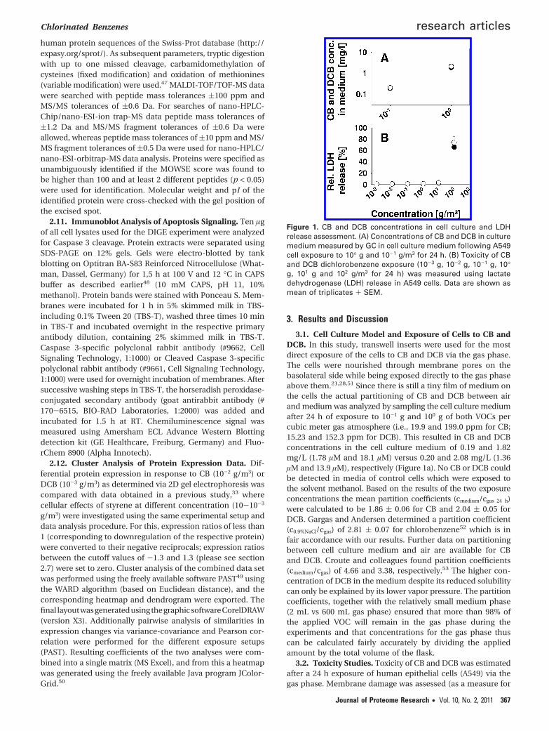
3.1. Cell Culture Model and Exposure of Cells to CB andDCB. In this study, transwell inserts were used for the mostdirect exposure of the cells to CB and DCB via the gas phase.The cells were nourished through membrane pores on thebasolateral side while being exposed directly to the gas phaseabove them.21,28,51 Since there is still a tiny film of medium onthe cells the actual partitioning of CB and DCB between airand medium was analyzed by sampling the cell culture mediumafter 24 h of exposure to 10-1 g and 100 g of both VOCs percubic meter gas atmosphere (i.e., 19.9 and 199.0 ppm for CB;15.23 and 152.3 ppm for DCB). This resulted in CB and DCBconcentrations in the cell culture medium of 0.19 and 1.82mg/L (1.78 µM and 18.1 µM) versus 0.20 and 2.08 mg/L (1.36µM and 13.9 µM), respectively (Figure 1a). No CB or DCB couldbe detected in media of control cells which were exposed tothe solvent methanol. Based on the results of the two exposureconcentrations the mean partition coefficients (cmedium/cgas 24 h)were calculated to be 1.86 ( 0.06 for CB and 2.04 ( 0.05 forDCB. Gargas and Andersen determined a partition coefficient(c0.9%NaCl/cgas) of 2.81 ( 0.07 for chlorobenzene52 which is infair accordance with our results. Further data on partitioningbetween cell culture medium and air are available for CBand DCB. Croute and colleagues found partition coefficients(cmedium/cgas) of 4.66 and 3.38, respectively.53 The higher con-centration of DCB in the medium despite its reduced solubilitycan only be explained by its lower vapor pressure. The partitioncoefficients, together with the relatively small medium phase(2 mL vs 600 mL gas phase) ensured that more than 98% ofthe applied VOC will remain in the gas phase during theexperiments and that concentrations for the gas phase thuscan be calculated fairly accurately by dividing the appliedamount by the total volume of the flask.
3.2. Toxicity Studies. Toxicity of CB and DCB was estimatedafter a 24 h exposure of human epithelial cells (A549) via thegas phase. Membrane damage was assessed (as a measure for
Figure 1. CB and DCB concentrations in cell culture and LDHrelease assessment. (A) Concentrations of CB and DCB in culturemedium measured by GC in cell culture medium following A549cell exposure to 10° g and 10-1 g/m3 for 24 h. (B) Toxicity of CBand DCB dichlorobenzene exposure (10-3 g, 10-2 g, 10-1 g, 10°g, 101 g and 102 g/m3 for 24 h) was measured using lactatedehydrogenase (LDH) release in A549 cells. Data are shown asmean of triplicates + SEM.
Chlorinated Benzenes research articles
Journal of Proteome Research • Vol. 10, No. 2, 2011 367
cell viability) by measuring cellular lactate dehydrogenase(LDH) release into the cell culture medium (Figure 1b). LDHrelease within 24 h increased up to 65.6% and 73.8% of totalcellular LDH when cells were exposed to 102 g/m3 of CB orDCB, respectively, compared to control cultures (only 0.6% ofLDHtotal). However, only a weak membrane damage of 2.41%versus 3.13% could be detected when exposing cells to 101 g/m3
of CB or DCB, respectively. In a recent study on differentialprotein expression following 24 h styrene exposure, less severecell membrane leakage of only 29% LDHtotal at 102 g/m3 wasfound. No membrane damage could be detected when cellswere exposed to concentrations less than 101 g/m3 of CB, DCBand styrene.33 Thus, using this experimental readout systemtoxicity of the tested VOCs increasessalbeit slightlyswith thedegree of chlorination, a result that is in line with experimentsperformed by Croute et al.53 In this study, A549 cells that wereexposed to 100 µM CB for 4 days showed a significant growthinhibition (-17%), whereas for the same inhibition only 10 µMof DCB were required. Even though our results do not implysuch a strong dependency on the number of chlorine substit-uents, they give a further example of differences in toxicitybetween mono- and dichlorobenzenes. Since the focus of thisstudy was to detect cellular responses upon exposition tononacute toxic conditions, we chose concentrations that wereat least 2 orders of magnitude below the lowest concentrationscausing significant effects on cell viability.
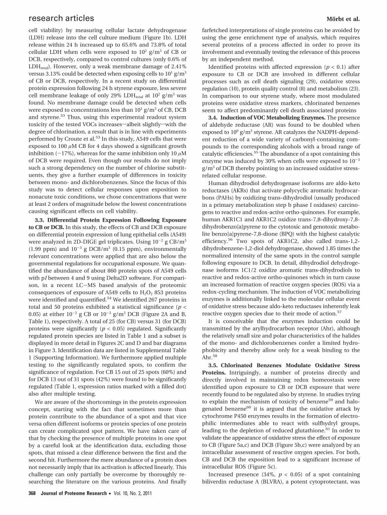
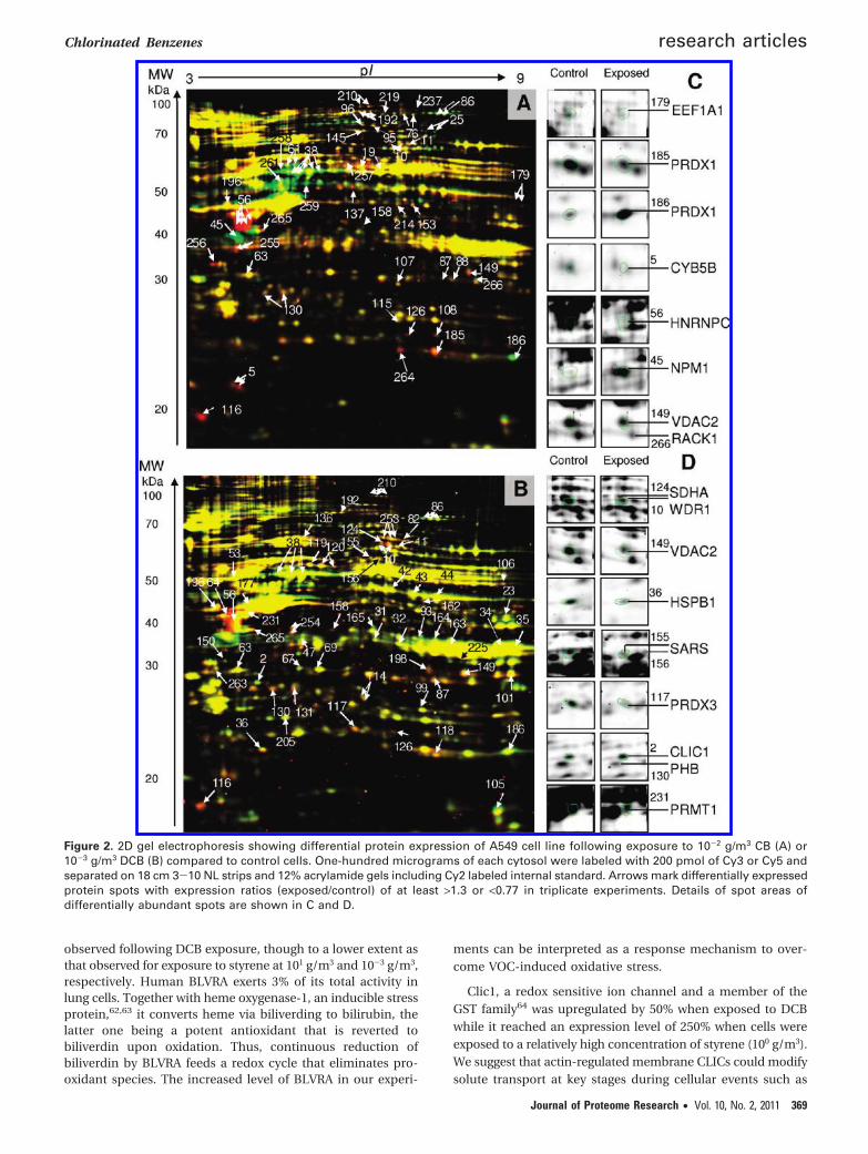
3.3. Differential Protein Expression Following Exposureto CB or DCB. In this study, the effects of CB and DCB exposureon differential protein expression of lung epithelial cells (A549)were analyzed in 2D-DIGE gel triplicates. Using 10-2 g CB/m3
(1.99 ppm) and 10-3 g DCB/m3 (0.15 ppm), environmentallyrelevant concentrations were applied that are also below thegovernmental regulations for occupational exposure. We quan-tified the abundance of about 860 protein spots of A549 cellswith pI between 4 and 9 using Delta2D software. For compari-son, in a recent LC-MS based analysis of the proteomicconsequences of exposure of A549 cells to H2O2 853 proteinswere identified and quantified.54 We identified 267 proteins intotal and 50 proteins exhibited a statistical significance (p <0.05) at either 10-2 g CB or 10-3 g/m3 DCB (Figure 2A and B,Table 1), respectively. A total of 25 (for CB) versus 31 (for DCB)proteins were significantly (p < 0.05) regulated. Significantlyregulated protein species are listed in Table 1 and a subset isdisplayed in more detail in Figures 2C and D and bar diagramsin Figure 3. Identification data are listed in Supplemental Table1 (Supporting Information). We furthermore applied multipletesting to the significantly regulated spots, to confirm thesignificance of regulation. For CB 15 out of 25 spots (60%) andfor DCB 13 out of 31 spots (42%) were found to be significantlyregulated (Table 1, expression ratios marked with a filled dot)also after multiple testing.
We are aware of the shortcomings in the protein expressionconcept, starting with the fact that sometimes more thanprotein contribute to the abundance of a spot and that viceversa often different isoforms or protein species of one proteincan create complicated spot pattern. We have taken care ofthat by checking the presence of multiple proteins in one spotby a careful look at the identification data, excluding thosespots, that missed a clear difference between the first and thesecond hit. Furthermore the mere abundance of a protein doesnot necessarily imply that its activation is affected linearly. Thischallenge can only partially be overcome by thoroughly re-searching the literature on the various proteins. And finally
farfetched interpretations of single proteins can be avoided byusing the gene enrichment type of analysis, which requiresseveral proteins of a process affected in order to prove itsinvolvement and eventually testing the relevance of this processby an independent method.
Identified proteins with affected expression (p < 0.1) afterexposure to CB or DCB are involved in different cellularprocesses such as cell death signaling (29), oxidative stressregulation (10), protein quality control (8) and metabolism (23).In comparison to our styrene study, where most modulatedproteins were oxidative stress markers, chlorinated benzenesseem to affect predominantly cell death associated proteins
3.4. Induction of VOC Metabolizing Enzymes. The presenceof aldehyde reductase (AR) was found to be doubled whenexposed to 100 g/m3 styrene. AR catalyzes the NADPH-depend-ent reduction of a wide variety of carbonyl-containing com-pounds to the corresponding alcohols with a broad range ofcatalytic efficiencies.55 The abundance of a spot containing thisenzyme was induced by 30% when cells were exposed to 10-3
g/m3 of DCB thereby pointing to an increased oxidative stress-related cellular response.
Human dihydrodiol dehydrogenase isoforms are aldo-ketoreductases (AKRs) that activate polycyclic aromatic hydrocar-bons (PAHs) by oxidizing trans-dihydrodiol (usually producedin a primary metabolization step b phase I oxidases) carcino-gens to reactive and redox-active ortho-quinones. For example,human AKR1C1 and AKR1C2 oxidize trans-7,8-dihydroxy-7,8-dihydrobenzo[a]pyrene to the cytotoxic and genotoxic metabo-lite benzo[a]pyrene-7,8-dione (BPQ) with the highest catalyticefficiency.56 Two spots of AKR1C2, also called trans-1,2-dihydrobenzene-1,2-diol dehydrogenase, showed 1.85 times thenormalized intensity of the same spots in the control samplefollowing exposure to DCB. In detail, dihydrodiol dehydroge-nase isoforms 1C1/2 oxidize aromatic trans-dihydrodiols toreactive and redox-active ortho-quinones which in turn causean increased formation of reactive oxygen species (ROS) via aredox-cycling mechanism. The induction of VOC metabolizingenzymes is additionally linked to the molecular cellular eventof oxidative stress because aldo-keto reductases inherently leakreactive oxygen species due to their mode of action.57
It is conceivable that the enzymes induction could betransmitted by the arylhydrocarbon receptor (Ahr), althoughthe relatively small size and polar characteristics of the halidesof the mono- and dichlorobenzenes confer a limited hydro-phobicity and thereby allow only for a weak binding to theAhr.58
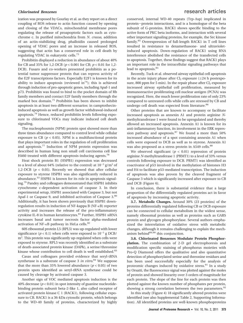
3.5. Chlorinated Benzenes Modulate Oxidative StressProteins. Intriguingly, a number of proteins directly anddirectly involved in maintaining redox homeostasis wereidentified upon exposure to CB or DCB exposure that wererecently found to be regulated also by styrene. In studies tryingto explain the mechanism of toxicity of benzene59 and halo-genated benzene60 it is argued that the oxidative attack bycytochrome P450 enzymes results in the formation of electro-philic intermediates able to react with sulfhydryl groups,leading to the depletion of reduced glutathione.61 In order tovalidate the appearance of oxidative stress the effect of exposureto CB (Figure 5a,c) and DCB (Figure 5b,c) were analyzed by anintracellular assessment of reactive oxygen species. For both,CB and DCB the exposition lead to a significant increase ofintracellular ROS (Figure 5c).
Increased presence (34%, p < 0.05) of a spot containingbiliverdin reductase A (BLVRA), a potent cytoprotectant, was
research articles Morbt et al.
368 Journal of Proteome Research • Vol. 10, No. 2, 2011
observed following DCB exposure, though to a lower extent asthat observed for exposure to styrene at 101 g/m3 and 10-3 g/m3,respectively. Human BLVRA exerts 3% of its total activity inlung cells. Together with heme oxygenase-1, an inducible stressprotein,62,63 it converts heme via biliverding to bilirubin, thelatter one being a potent antioxidant that is reverted tobiliverdin upon oxidation. Thus, continuous reduction ofbiliverdin by BLVRA feeds a redox cycle that eliminates pro-oxidant species. The increased level of BLVRA in our experi-
ments can be interpreted as a response mechanism to over-come VOC-induced oxidative stress.
Clic1, a redox sensitive ion channel and a member of theGST family64 was upregulated by 50% when exposed to DCBwhile it reached an expression level of 250% when cells wereexposed to a relatively high concentration of styrene (100 g/m3).We suggest that actin-regulated membrane CLICs could modifysolute transport at key stages during cellular events such as
Figure 2. 2D gel electrophoresis showing differential protein expression of A549 cell line following exposure to 10-2 g/m3 CB (A) or10-3 g/m3 DCB (B) compared to control cells. One-hundred micrograms of each cytosol were labeled with 200 pmol of Cy3 or Cy5 andseparated on 18 cm 3-10 NL strips and 12% acrylamide gels including Cy2 labeled internal standard. Arrows mark differentially expressedprotein spots with expression ratios (exposed/control) of at least >1.3 or <0.77 in triplicate experiments. Details of spot areas ofdifferentially abundant spots are shown in C and D.
Chlorinated Benzenes research articles
Journal of Proteome Research • Vol. 10, No. 2, 2011 369
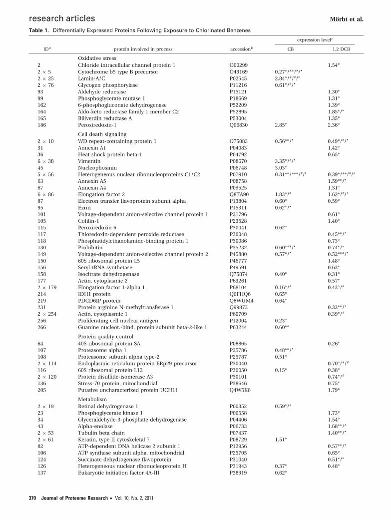
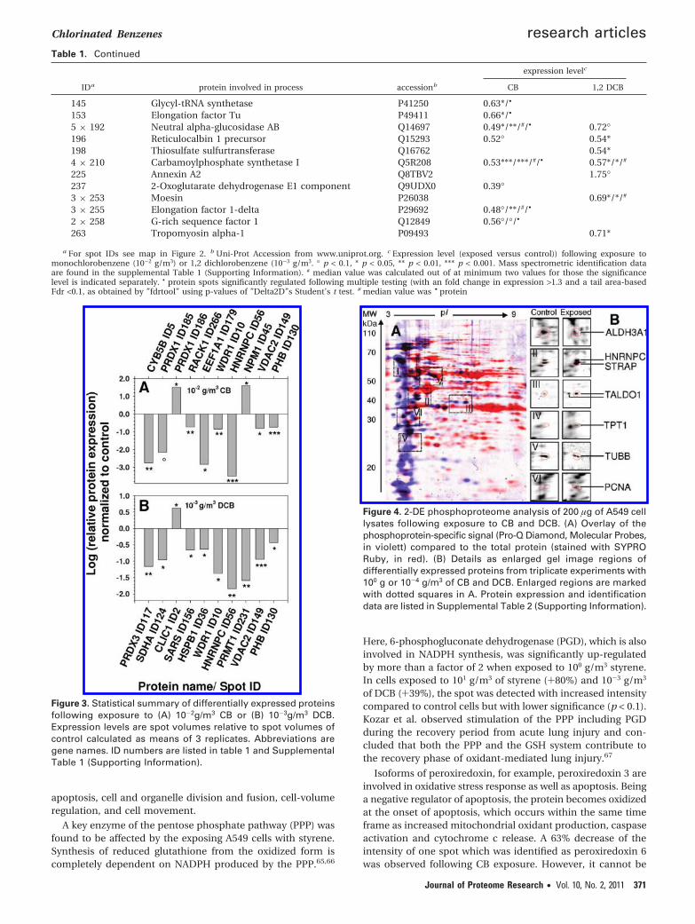
Table 1. Differentially Expressed Proteins Following Exposure to Chlorinated Benzenes
expression levelc
IDa protein involved in process accessionb CB 1,2 DCB
Oxidative stress2 Chloride intracellular channel protein 1 O00299 1.54*2 × 5 Cytochrome b5 type B precursor O43169 0.27*/**/#/•
2 × 25 Lamin-A/C P02545 2.84°/*/#/•
2 × 76 Glycogen phosphorylase P11216 0.61*/#/•
93 Aldehyde reductase P15121 1.30*99 Phosphoglycerate mutase 1 P18669 1.31°162 6-phosphogluconate dehydrogenase P52209 1.39°164 Aldo-keto reductase family 1 member C2 P52895 1.85*/•
165 Biliverdin reductase A P53004 1.35*186 Peroxiredoxin-1 Q06830 2.85* 2.36°
Cell death signaling2 × 10 WD repeat-containing protein 1 O75083 0.56**/• 0.49*/#/•
31 Annexin A1 P04083 1.42°36 Heat shock protein beta-1 P04792 0.65*6 × 38 Vimentin P08670 3.35*/#/•
45 Nucleophosmin P06748 3.03*5 × 56 Heterogeneous nuclear ribonucleoproteins C1/C2 P07910 0.31**/***/#/• 0.39*/**/#/•
63 Annexin A5 P08758 1.59**/•
67 Annexin A4 P09525 1.31°6 × 86 Elongation factor 2 Q8TA90 1.83°/# 1.62*/#/•
87 Electron transfer flavoprotein subunit alpha P13804 0.60° 0.59°95 Ezrin P15311 0.62*/•
101 Voltage-dependent anion-selective channel protein 1 P21796 0.61°105 Cofilin-1 P23528 1.40°115 Peroxiredoxin 6 P30041 0.62°117 Thioredoxin-dependent peroxide reductase P30048 0.45**/•
118 Phosphatidylethanolamine-binding protein 1 P30086 0.73°130 Prohibitin P35232 0.60***/• 0.74*/•
149 Voltage-dependent anion-selective channel protein 2 P45880 0.57*/• 0.52***/•
150 60S ribosomal protein L5 P46777 1.48°156 Seryl-tRNA synthetase P49591 0.63*158 Isocitrate dehydrogenase Q75874 0.40* 0.31*177 Actin, cytoplasmic 2 P63261 0.57*2 × 179 Elongation factor 1-alpha 1 P68104 0.16*/# 0.43°/#
214 IDH1 protein Q6FHQ6 0.65*219 PDCD6IP protein Q8WUM4 0.64*231 Protein arginine N-methyltransferase 1 Q99873 0.33**/•
2 × 254 Actin, cytoplasmic 1 P60709 0.39*/#
256 Proliferating cell nuclear antigen P12004 0.23°266 Guanine nucleot.-bind. protein subunit beta-2-like 1 P63244 0.60**
Protein quality control64 40S ribosomal protein SA P08865 0.26*107 Proteasome alpha 1 P25786 0.48**/•
108 Proteasome subunit alpha type-2 P25787 0.51°2 × 114 Endoplasmic reticulum protein ERp29 precursor P30040 0.70°/*/#
116 60S ribosomal protein L12 P30050 0.15* 0.38°2 × 120 Protein disulfide-isomerase A3 P30101 0.74*/#
136 Stress-70 protein, mitochondrial P38646 0.75*205 Putative uncharacterized protein UCHL1 Q4W5K6 1.79*
Metabolism2 × 19 Retinal dehydrogenase 1 P00352 0.59°/#
23 Phosphoglycerate kinase 1 P00558 1.73°34 Glyceraldehyde-3-phosphate dehydrogenase P04406 1.54°43 Alpha-enolase P06733 1.68**/#
2 × 53 Tubulin beta chain P07437 1.40**/•
2 × 61 Keratin, type II cytoskeletal 7 P08729 1.51*82 ATP-dependent DNA helicase 2 subunit 1 P12956 0.57**/•
106 ATP synthase subunit alpha, mitochondrial P25705 0.65°124 Succinate dehydrogenase flavoprotein P31040 0.51*/•
126 Heterogeneous nuclear ribonucleoprotein H P31943 0.37* 0.48°137 Eukaryotic initiation factor 4A-III P38919 0.62°
research articles Morbt et al.
370 Journal of Proteome Research • Vol. 10, No. 2, 2011
apoptosis, cell and organelle division and fusion, cell-volumeregulation, and cell movement.
A key enzyme of the pentose phosphate pathway (PPP) wasfound to be affected by the exposing A549 cells with styrene.Synthesis of reduced glutathione from the oxidized form iscompletely dependent on NADPH produced by the PPP.65,66
Here, 6-phosphogluconate dehydrogenase (PGD), which is alsoinvolved in NADPH synthesis, was significantly up-regulatedby more than a factor of 2 when exposed to 100 g/m3 styrene.In cells exposed to 101 g/m3 of styrene (+80%) and 10-3 g/m3
of DCB (+39%), the spot was detected with increased intensitycompared to control cells but with lower significance (p < 0.1).Kozar et al. observed stimulation of the PPP including PGDduring the recovery period from acute lung injury and con-cluded that both the PPP and the GSH system contribute tothe recovery phase of oxidant-mediated lung injury.67
Isoforms of peroxiredoxin, for example, peroxiredoxin 3 areinvolved in oxidative stress response as well as apoptosis. Beinga negative regulator of apoptosis, the protein becomes oxidizedat the onset of apoptosis, which occurs within the same timeframe as increased mitochondrial oxidant production, caspaseactivation and cytochrome c release. A 63% decrease of theintensity of one spot which was identified as peroxiredoxin 6was observed following CB exposure. However, it cannot be
Table 1. Continued
expression levelc
IDa protein involved in process accessionb CB 1,2 DCB
145 Glycyl-tRNA synthetase P41250 0.63*/•
153 Elongation factor Tu P49411 0.66*/•
5 × 192 Neutral alpha-glucosidase AB Q14697 0.49*/**/#/• 0.72°196 Reticulocalbin 1 precursor Q15293 0.52° 0.54*198 Thiosulfate sulfurtransferase Q16762 0.54*4 × 210 Carbamoylphosphate synthetase I Q5R208 0.53***/***/#/• 0.57*/*/#
225 Annexin A2 Q8TBV2 1.75°237 2-Oxoglutarate dehydrogenase E1 component Q9UDX0 0.39°3 × 253 Moesin P26038 0.69*/*/#
3 × 255 Elongation factor 1-delta P29692 0.48°/**/#/•
2 × 258 G-rich sequence factor 1 Q12849 0.56°/°/•
263 Tropomyosin alpha-1 P09493 0.71*
a For spot IDs see map in Figure 2. b Uni-Prot Accession from www.uniprot.org. c Expression level (exposed versus control)) following exposure tomonochlorobenzene (10-2 g/m3) or 1,2 dichlorobenzene (10-3 g/m3. ° p < 0.1, * p < 0.05, ** p < 0.01, *** p < 0.001. Mass spectrometric identification dataare found in the supplemental Table 1 (Supporting Information). # median value was calculated out of at minimum two values for those the significancelevel is indicated separately. • protein spots significantly regulated following multiple testing (with an fold change in expression >1.3 and a tail area-basedFdr <0.1, as obtained by ”fdrtool” using p-values of ”Delta2D”s Student’s t test. # median value was • protein
Figure 3. Statistical summary of differentially expressed proteinsfollowing exposure to (A) 10-2g/m3 CB or (B) 10-3g/m3 DCB.Expression levels are spot volumes relative to spot volumes ofcontrol calculated as means of 3 replicates. Abbreviations aregene names. ID numbers are listed in table 1 and SupplementalTable 1 (Supporting Information).
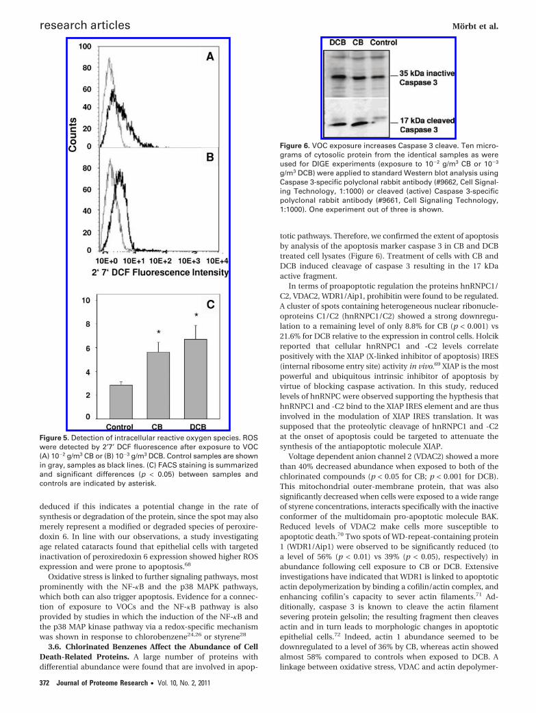
Figure 4. 2-DE phosphoproteome analysis of 200 µg of A549 celllysates following exposure to CB and DCB. (A) Overlay of thephosphoprotein-specific signal (Pro-Q Diamond, Molecular Probes,in violett) compared to the total protein (stained with SYPRORuby, in red). (B) Details as enlarged gel image regions ofdifferentially expressed proteins from triplicate experiments with100 g or 10-4 g/m3 of CB and DCB. Enlarged regions are markedwith dotted squares in A. Protein expression and identificationdata are listed in Supplemental Table 2 (Supporting Information).
Chlorinated Benzenes research articles
Journal of Proteome Research • Vol. 10, No. 2, 2011 371
deduced if this indicates a potential change in the rate ofsynthesis or degradation of the protein, since the spot may alsomerely represent a modified or degraded species of peroxire-doxin 6. In line with our observations, a study investigatingage related cataracts found that epithelial cells with targetedinactivation of peroxiredoxin 6 expression showed higher ROSexpression and were prone to apoptosis.68
Oxidative stress is linked to further signaling pathways, mostprominently with the NF-κB and the p38 MAPK pathways,which both can also trigger apoptosis. Evidence for a connec-tion of exposure to VOCs and the NF-κB pathway is alsoprovided by studies in which the induction of the NF-κB andthe p38 MAP kinase pathway via a redox-specific mechanismwas shown in response to chlorobenzene24,26 or styrene28
3.6. Chlorinated Benzenes Affect the Abundance of CellDeath-Related Proteins. A large number of proteins withdifferential abundance were found that are involved in apop-
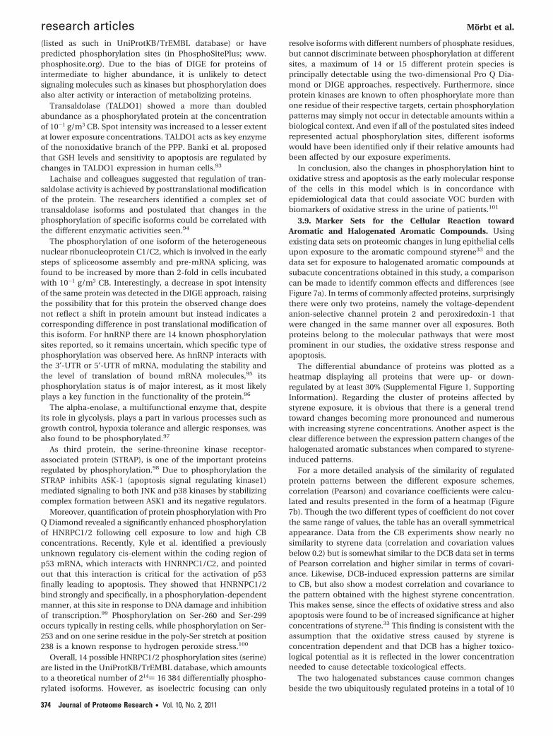
totic pathways. Therefore, we confirmed the extent of apoptosisby analysis of the apoptosis marker caspase 3 in CB and DCBtreated cell lysates (Figure 6). Treatment of cells with CB andDCB induced cleavage of caspase 3 resulting in the 17 kDaactive fragment.
In terms of proapoptotic regulation the proteins hnRNPC1/C2, VDAC2, WDR1/Aip1, prohibitin were found to be regulated.A cluster of spots containing heterogeneous nuclear ribonucle-oproteins C1/C2 (hnRNPC1/C2) showed a strong downregu-lation to a remaining level of only 8.8% for CB (p < 0.001) vs21.6% for DCB relative to the expression in control cells. Holcikreported that cellular hnRNPC1 and -C2 levels correlatepositively with the XIAP (X-linked inhibitor of apoptosis) IRES(internal ribosome entry site) activity in vivo.69 XIAP is the mostpowerful and ubiquitous intrinsic inhibitor of apoptosis byvirtue of blocking caspase activation. In this study, reducedlevels of hnRNPC were observed supporting the hypthesis thathnRNPC1 and -C2 bind to the XIAP IRES element and are thusinvolved in the modulation of XIAP IRES translation. It wassupposed that the proteolytic cleavage of hnRNPC1 and -C2at the onset of apoptosis could be targeted to attenuate thesynthesis of the antiapoptotic molecule XIAP.
Voltage dependent anion channel 2 (VDAC2) showed a morethan 40% decreased abundance when exposed to both of thechlorinated compounds (p < 0.05 for CB; p < 0.001 for DCB).This mitochondrial outer-membrane protein, that was alsosignificantly decreased when cells were exposed to a wide rangeof styrene concentrations, interacts specifically with the inactiveconformer of the multidomain pro-apoptotic molecule BAK.Reduced levels of VDAC2 make cells more susceptible toapoptotic death.70 Two spots of WD-repeat-containing protein1 (WDR1/Aip1) were observed to be significantly reduced (toa level of 56% (p < 0.01) vs 39% (p < 0.05), respectively) inabundance following cell exposure to CB or DCB. Extensiveinvestigations have indicated that WDR1 is linked to apoptoticactin depolymerization by binding a cofilin/actin complex, andenhancing cofilin’s capacity to sever actin filaments.71 Ad-ditionally, caspase 3 is known to cleave the actin filamentsevering protein gelsolin; the resulting fragment then cleavesactin and in turn leads to morphologic changes in apoptoticepithelial cells.72 Indeed, actin 1 abundance seemed to bedownregulated to a level of 36% by CB, whereas actin showedalmost 58% compared to controls when exposed to DCB. Alinkage between oxidative stress, VDAC and actin depolymer-
Figure 5. Detection of intracellular reactive oxygen species. ROSwere detected by 2′7′ DCF fluorescence after exposure to VOC(A) 10-2 g/m3 CB or (B) 10-3 g/m3 DCB. Control samples are shownin gray, samples as black lines. (C) FACS staining is summarizedand significant differences (p < 0.05) between samples andcontrols are indicated by asterisk.
Figure 6. VOC exposure increases Caspase 3 cleave. Ten micro-grams of cytosolic protein from the identical samples as wereused for DIGE experiments (exposure to 10-2 g/m3 CB or 10-3
g/m3 DCB) were applied to standard Western blot analysis usingCaspase 3-specific polyclonal rabbit antibody (#9662, Cell Signal-ing Technology, 1:1000) or cleaved (active) Caspase 3-specificpolyclonal rabbit antibody (#9661, Cell Signaling Technology,1:1000). One experiment out of three is shown.
research articles Morbt et al.
372 Journal of Proteome Research • Vol. 10, No. 2, 2011
ization was proposed by Gourlay et al. as they report on a directcoupling of ROS release to actin function caused by openingand closing of the VDACs, mitochondrial membrane poresregulating the release of proapoptotic factors such as cyto-chrome c. In purified mitochondria from N. crassa, additionof an actin-stabilizing drug, phalloidin caused prolongedopening of VDAC pores and an increase in released ROS,suggesting that actin has a conserved role in cell death byregulating VDAC in eukaryotic cells.73
Prohibitin displayed a reduction in abundance of about 40%for CB and 35% for 1,2-DCB (p < 0.001 for CB; p < 0.01 for 1,2-DCB). Fusaro and co-workers presented prohibitin as a po-tential tumor suppressor protein that can repress activity ofthe E2F transcriptions factors. Especially E2F1 is known for itsability to induce apoptosis (reviewed in74); this is achievedthrough induction of pro-apoptotic genes, including Apaf-1 andp73. Prohibitin was found to bind to the pocket domain of Rbfamily members and contact E2F family members through themarked box domain.75 Prohibitin has been shown to inhibitapoptosis in at least two different scenarios: in camptothecin-induced apoptosis as well as growth factor withdrawal-inducedapoptosis.76 Hence, reduced prohibitin levels following expo-sure to chlorinated VOCs may indicate induced cell deathsignaling.
The nucleophosmin (NPM) protein spot showed more thanthree times abundance compared to control level while cellularexposure to CB (p < 0.05). NPM is a multifunctional proteinthat plays important roles in the regulation of cell proliferationand apoptosis.77 Induction of NPM protein expression wasdescribed in human lung non small cell carcinoma cell lineH460 treated with different apoptosis-inducing agents.78
Heat shock protein B1 (HSPB1) expression was decreasedto a level of about 64% (relative to the control) at 10-3 g/m3 of1,2-DCB (p < 0.05). Recently we showed that after cellularexposure to styrene HSPB1 was also significantly reduced inabundance.33 HSPB1 is known for its role in apoptosis signal-ing.79 Pandey and colleagues demonstrated that HSPB1 inhibitscytochrome c-dependent activation of caspase 3. In theirexperimental setup, HSPB1 associated with Caspase 3, but notApaf-1 or Caspase 9, and inhibited activation of Caspase 3.80
Additionally, it has been shown previously that HSPB1 down-regulation results in induction of NF-kappa B (NF-κB) reporteractivity and increases the release of the pro-inflammatorycytokine IL-8 in human keratinocytes.81 Further, HSPB1 siRNAincreases basal and tumor necrosis factor alpha-mediatedactivation of NF-κB pathway in HeLa cells.82
60S ribosomal protein L5 (RPL5) was up-regulated with lowersignificance (p < 0.1) when cells were exposed to 10-3 g DCB/m3. This protein was significantly up-regulated when cells wereexposed to styrene. RPL5 was recently identified as a substrateof death-associated protein kinase (DAPk), a serine/threoninekinase whose contribution to cell death is well established.83
Casas and colleagues provided evidence that seryl-tRNAsynthetase is a substrate of caspase 3 in vitro.84 We supposethat the more than 35% lowered abundance (p < 0.05) of twoprotein spots identified as seryl-tRNA synthetase could becaused by cleavage by activated caspase 3.
Another sign of VOC mediated apoptosis induction is the40% decrease (p < 0.01) in spot intensity of guanine nucleotide-binding protein subunit beta-2-like 1, also called receptor ofactivated protein kinase C 1 (RACK1), following cellular expo-sure to CB. RACK1 is a 36-kDa cytosolic protein, which belongsto the WD-40 family of proteins, characterized by highly
conserved, internal WD-40 repeats (Trp-Asp) implicated inprotein-protein interactions, and is a homologue of the betasubunit of G-proteins. RACK1 shows specific binding to theactive form of PKC beta isoforms, and interaction with severalother important signaling proteins, for example, the Src kinasefamily.85 Overexpression of full-length RACK1 in T cell linesresulted in resistance to dexamethasone- and ultraviolet-induced apoptosis. Down-regulation of RACK1 using RNAinterference abolished the resistance of the transfected cellsto apoptosis. Together, these findings suggest that RACK1 playsan important role in the intracellular signaling pathways thatlead to apoptosis.85
Recently, Tuck et al. observed airway epithelial cell apoptosisin the acute injury phase after Cl2 exposure (e24 h postexpo-sure, 800 ppm for 5 min). In the repair phase after Cl2 exposureincreased airway epithelial cell proliferation, measured byimmunoreactive proliferating cell nuclear antigen (PCNA) wasrecognized. Here, the much lower proliferation rate of only 23%compared to untreated cells while cells are stressed by CB andundergo cell death was expected from literature.86
Other proteins that are known to accompany or facilitateincreased apoptosis as annexin A1 and protein arginine N-methyltransferase 1 were found to be upregulated and therebyallowed an increased apoptosis. Annexin A1 is known for itsanti-inflammatory function, its involvement in the ERK repres-sion pathway and apoptosis.87 We found a more than 50%increased abundance of a spot containing annexin A1 whencells were exposed to DCB as well as to styrene. Annexin A1was also proposed as a stress protein in A549 cells.88
We observed significant (p < 0.01) repression of proteinarginine N-methyltransferase 1 (PRMT1) to a level of 33% versuscontrols following exposure to DCB. PRMT1 was identified ascoactivator of p53 involved in the methylation of histones H3and H4 to facilitate p53-mediated transcription. The inductionof apoptosis was also proven by the cleaved fragment ofCaspase 3 which is significantly increased after exposure to CBand DCB (Figure 6).
In conclusion, there is substantial evidence that a largeproportion of the differentially regulated proteins act in favorof apoptosis by increasing apoptotic activity.
3.7. Metabolic Changes. Around 30% (23 proteins) of theproteins differentially regulated following CB or DCB exposurecan be connected to cellular metabolism in the widest sense,namely ribosomal proteins as well as proteins such as GARSprotein and glycogen phosphorylase. Several authors empha-sized the interrelation of oxidative stress with metabolicchanges, although it remains challenging to explain the mech-anism behind89,90 this conjunction.
3.8. Chlorinated Benzenes Modulate Protein Phosphor-ylation. The combination of 2-D gel electrophoresis andmodification specific staining of phosphorus moieties withPro-Q Diamond offers the qualitative and also quantitativedetection of phosphorylated serine and threonine residues andhas been used successfully especially for the analysis ofproteomic changes induced by oxidative stress.91 In a studyby Orsatti, the fluorescence signal was plotted against the molesof protein and showed linearity over 3 orders of magnitude foreach protein. The slope of the line for each protein was thenplotted against the known number of phosphates per protein,showing a strong correlation between the two parameters.92
In this study (Figure 4) 18 significantly altered proteins wereidentified (see also Supplemental Table 2, Supporting Informa-tion). All identified proteins are well-known phosphoproteins
Chlorinated Benzenes research articles
Journal of Proteome Research • Vol. 10, No. 2, 2011 373
(listed as such in UniProtKB/TrEMBL database) or havepredicted phosphorylation sites (in PhosphoSitePlus; www.phosphosite.org). Due to the bias of DIGE for proteins ofintermediate to higher abundance, it is unlikely to detectsignaling molecules such as kinases but phosphorylation doesalso alter activity or interaction of metabolizing proteins.
Transaldolase (TALDO1) showed a more than doubledabundance as a phosphorylated protein at the concentrationof 10-1 g/m3 CB. Spot intensity was increased to a lesser extentat lower exposure concentrations. TALDO1 acts as key enzymeof the nonoxidative branch of the PPP. Banki et al. proposedthat GSH levels and sensitivity to apoptosis are regulated bychanges in TALDO1 expression in human cells.93
Lachaise and colleagues suggested that regulation of tran-saldolase activity is achieved by posttranslational modificationof the protein. The researchers identified a complex set oftransaldolase isoforms and postulated that changes in thephosphorylation of specific isoforms could be correlated withthe different enzymatic activities seen.94
The phosphorylation of one isoform of the heterogeneousnuclear ribonucleoprotein C1/C2, which is involved in the earlysteps of spliceosome assembly and pre-mRNA splicing, wasfound to be increased by more than 2-fold in cells incubatedwith 10-1 g/m3 CB. Interestingly, a decrease in spot intensityof the same protein was detected in the DIGE approach, raisingthe possibility that for this protein the observed change doesnot reflect a shift in protein amount but instead indicates acorresponding difference in post translational modification ofthis isoform. For hnRNP there are 14 known phosphorylationsites reported, so it remains uncertain, which specific type ofphosphorylation was observed here. As hnRNP interacts withthe 3′-UTR or 5′-UTR of mRNA, modulating the stability andthe level of translation of bound mRNA molecules,95 itsphosphorylation status is of major interest, as it most likelyplays a key function in the functionality of the protein.96
The alpha-enolase, a multifunctional enzyme that, despiteits role in glycolysis, plays a part in various processes such asgrowth control, hypoxia tolerance and allergic responses, wasalso found to be phosphorylated.97
As third protein, the serine-threonine kinase receptor-associated protein (STRAP), is one of the important proteinsregulated by phosphorylation.98 Due to phosphorylation theSTRAP inhibits ASK-1 (apoptosis signal regulating kinase1)mediated signaling to both JNK and p38 kinases by stabilizingcomplex formation between ASK1 and its negative regulators.
Moreover, quantification of protein phosphorylation with ProQ Diamond revealed a significantly enhanced phosphorylationof HNRPC1/2 following cell exposure to low and high CBconcentrations. Recently, Kyle et al. identified a previouslyunknown regulatory cis-element within the coding region ofp53 mRNA, which interacts with HNRNPC1/C2, and pointedout that this interaction is critical for the activation of p53finally leading to apoptosis. They showed that HNRNPC1/2bind strongly and specifically, in a phosphorylation-dependentmanner, at this site in response to DNA damage and inhibitionof transcription.99 Phosphorylation on Ser-260 and Ser-299occurs typically in resting cells, while phosphorylation on Ser-253 and on one serine residue in the poly-Ser stretch at position238 is a known response to hydrogen peroxide stress.100
Overall, 14 possible HNRPC1/2 phosphorylation sites (serine)are listed in the UniProtKB/TrEMBL database, which amountsto a theoretical number of 214) 16 384 differentially phospho-rylated isoforms. However, as isoelectric focusing can only
resolve isoforms with different numbers of phosphate residues,but cannot discriminate between phosphorylation at differentsites, a maximum of 14 or 15 different protein species isprincipally detectable using the two-dimensional Pro Q Dia-mond or DIGE approaches, respectively. Furthermore, sinceprotein kinases are known to often phosphorylate more thanone residue of their respective targets, certain phosphorylationpatterns may simply not occur in detectable amounts within abiological context. And even if all of the postulated sites indeedrepresented actual phosphorylation sites, different isoformswould have been identified only if their relative amounts hadbeen affected by our exposure experiments.
In conclusion, also the changes in phosphorylation hint tooxidative stress and apoptosis as the early molecular responseof the cells in this model which is in concordance withepidemiological data that could associate VOC burden withbiomarkers of oxidative stress in the urine of patients.101
3.9. Marker Sets for the Cellular Reaction towardAromatic and Halogenated Aromatic Compounds. Usingexisting data sets on proteomic changes in lung epithelial cellsupon exposure to the aromatic compound styrene33 and thedata set for exposure to halogenated aromatic compounds atsubacute concentrations obtained in this study, a comparisoncan be made to identify common effects and differences (seeFigure 7a). In terms of commonly affected proteins, surprisinglythere were only two proteins, namely the voltage-dependentanion-selective channel protein 2 and peroxiredoxin-1 thatwere changed in the same manner over all exposures. Bothproteins belong to the molecular pathways that were mostprominent in our studies, the oxidative stress response andapoptosis.
The differential abundance of proteins was plotted as aheatmap displaying all proteins that were up- or down-regulated by at least 30% (Supplemental Figure 1, SupportingInformation). Regarding the cluster of proteins affected bystyrene exposure, it is obvious that there is a general trendtoward changes becoming more pronounced and numerouswith increasing styrene concentrations. Another aspect is theclear difference between the expression pattern changes of thehalogenated aromatic substances when compared to styrene-induced patterns.
For a more detailed analysis of the similarity of regulatedprotein patterns between the different exposure schemes,correlation (Pearson) and covariance coefficients were calcu-lated and results presented in the form of a heatmap (Figure7b). Though the two different types of coefficient do not coverthe same range of values, the table has an overall symmetricalappearance. Data from the CB experiments show nearly nosimilarity to styrene data (correlation and covariation valuesbelow 0.2) but is somewhat similar to the DCB data set in termsof Pearson correlation and higher similar in terms of covari-ance. Likewise, DCB-induced expression patterns are similarto CB, but also show a modest correlation and covariance tothe pattern obtained with the highest styrene concentration.This makes sense, since the effects of oxidative stress and alsoapoptosis were found to be of increased significance at higherconcentrations of styrene.33 This finding is consistent with theassumption that the oxidative stress caused by styrene isconcentration dependent and that DCB has a higher toxico-logical potential as it is reflected in the lower concentrationneeded to cause detectable toxicological effects.
The two halogenated substances cause common changesbeside the two ubiquitously regulated proteins in a total of 10
research articles Morbt et al.
374 Journal of Proteome Research • Vol. 10, No. 2, 2011
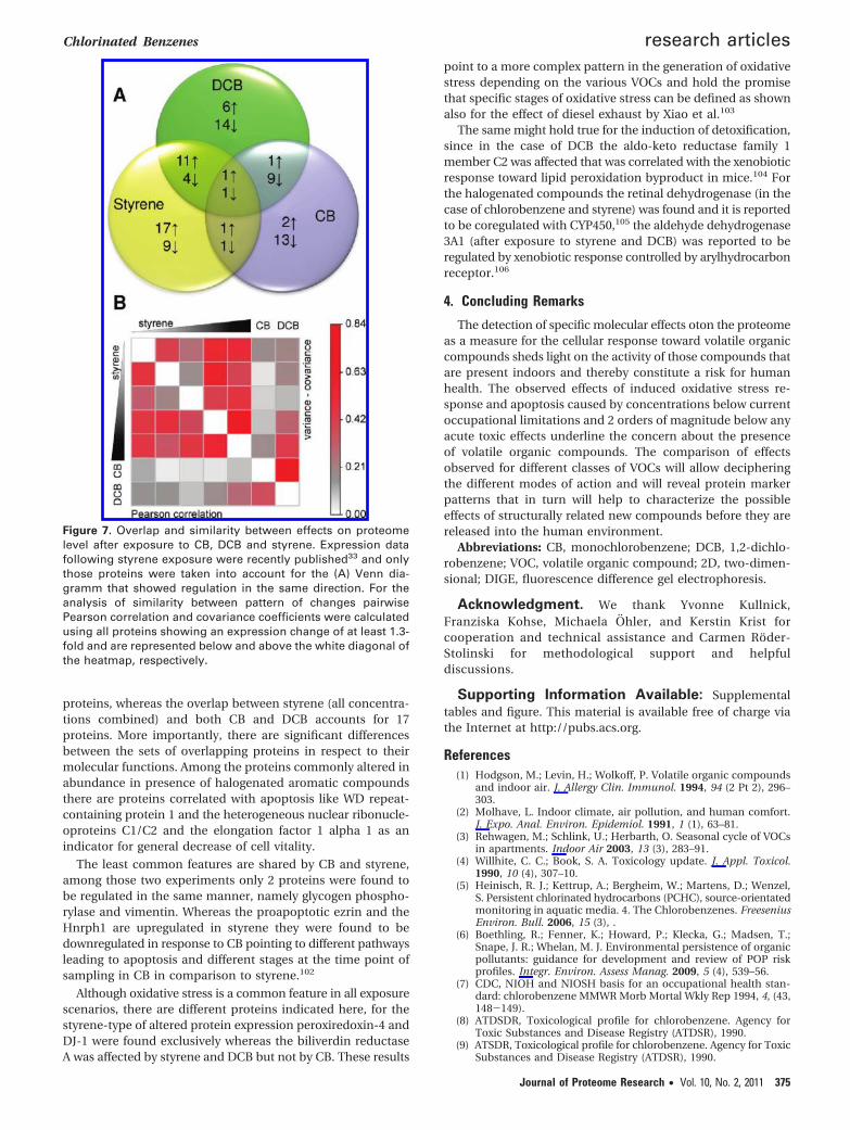
proteins, whereas the overlap between styrene (all concentra-tions combined) and both CB and DCB accounts for 17proteins. More importantly, there are significant differencesbetween the sets of overlapping proteins in respect to theirmolecular functions. Among the proteins commonly altered inabundance in presence of halogenated aromatic compoundsthere are proteins correlated with apoptosis like WD repeat-containing protein 1 and the heterogeneous nuclear ribonucle-oproteins C1/C2 and the elongation factor 1 alpha 1 as anindicator for general decrease of cell vitality.
The least common features are shared by CB and styrene,among those two experiments only 2 proteins were found tobe regulated in the same manner, namely glycogen phospho-rylase and vimentin. Whereas the proapoptotic ezrin and theHnrph1 are upregulated in styrene they were found to bedownregulated in response to CB pointing to different pathwaysleading to apoptosis and different stages at the time point ofsampling in CB in comparison to styrene.102
Although oxidative stress is a common feature in all exposurescenarios, there are different proteins indicated here, for thestyrene-type of altered protein expression peroxiredoxin-4 andDJ-1 were found exclusively whereas the biliverdin reductaseA was affected by styrene and DCB but not by CB. These results
point to a more complex pattern in the generation of oxidativestress depending on the various VOCs and hold the promisethat specific stages of oxidative stress can be defined as shownalso for the effect of diesel exhaust by Xiao et al.103
The same might hold true for the induction of detoxification,since in the case of DCB the aldo-keto reductase family 1member C2 was affected that was correlated with the xenobioticresponse toward lipid peroxidation byproduct in mice.104 Forthe halogenated compounds the retinal dehydrogenase (in thecase of chlorobenzene and styrene) was found and it is reportedto be coregulated with CYP450,105 the aldehyde dehydrogenase3A1 (after exposure to styrene and DCB) was reported to beregulated by xenobiotic response controlled by arylhydrocarbonreceptor.106
4. Concluding Remarks
The detection of specific molecular effects oton the proteomeas a measure for the cellular response toward volatile organiccompounds sheds light on the activity of those compounds thatare present indoors and thereby constitute a risk for humanhealth. The observed effects of induced oxidative stress re-sponse and apoptosis caused by concentrations below currentoccupational limitations and 2 orders of magnitude below anyacute toxic effects underline the concern about the presenceof volatile organic compounds. The comparison of effectsobserved for different classes of VOCs will allow decipheringthe different modes of action and will reveal protein markerpatterns that in turn will help to characterize the possibleeffects of structurally related new compounds before they arereleased into the human environment.
Abbreviations: CB, monochlorobenzene; DCB, 1,2-dichlo-robenzene; VOC, volatile organic compound; 2D, two-dimen-sional; DIGE, fluorescence difference gel electrophoresis.
Acknowledgment. We thank Yvonne Kullnick,Franziska Kohse, Michaela Ohler, and Kerstin Krist forcooperation and technical assistance and Carmen Roder-Stolinski for methodological support and helpfuldiscussions.
Supporting Information Available: Supplementaltables and figure. This material is available free of charge viathe Internet at http://pubs.acs.org.
References(1) Hodgson, M.; Levin, H.; Wolkoff, P. Volatile organic compounds
and indoor air. J. Allergy Clin. Immunol. 1994, 94 (2 Pt 2), 296–303.
(2) Molhave, L. Indoor climate, air pollution, and human comfort.J. Expo. Anal. Environ. Epidemiol. 1991, 1 (1), 63–81.
(3) Rehwagen, M.; Schlink, U.; Herbarth, O. Seasonal cycle of VOCsin apartments. Indoor Air 2003, 13 (3), 283–91.
(4) Willhite, C. C.; Book, S. A. Toxicology update. J. Appl. Toxicol.1990, 10 (4), 307–10.
(5) Heinisch, R. J.; Kettrup, A.; Bergheim, W.; Martens, D.; Wenzel,S. Persistent chlorinated hydrocarbons (PCHC), source-orientatedmonitoring in aquatic media. 4. The Chlorobenzenes. FreeseniusEnviron. Bull. 2006, 15 (3), .
(6) Boethling, R.; Fenner, K.; Howard, P.; Klecka, G.; Madsen, T.;Snape, J. R.; Whelan, M. J. Environmental persistence of organicpollutants: guidance for development and review of POP riskprofiles. Integr. Environ. Assess Manag. 2009, 5 (4), 539–56.
(7) CDC, NIOH and NIOSH basis for an occupational health stan-dard: chlorobenzene MMWR Morb Mortal Wkly Rep 1994, 4, (43,148-149).
(8) ATDSDR, Toxicological profile for chlorobenzene. Agency forToxic Substances and Disease Registry (ATDSR), 1990.
(9) ATSDR, Toxicological profile for chlorobenzene. Agency for ToxicSubstances and Disease Registry (ATDSR), 1990.
Figure 7. Overlap and similarity between effects on proteomelevel after exposure to CB, DCB and styrene. Expression datafollowing styrene exposure were recently published33 and onlythose proteins were taken into account for the (A) Venn dia-gramm that showed regulation in the same direction. For theanalysis of similarity between pattern of changes pairwisePearson correlation and covariance coefficients were calculatedusing all proteins showing an expression change of at least 1.3-fold and are represented below and above the white diagonal ofthe heatmap, respectively.
Chlorinated Benzenes research articles
Journal of Proteome Research • Vol. 10, No. 2, 2011 375
(10) ATSDR, Toxicological profile for Dichlorobenzene. Agency forToxic Substances and Disease Registry (ATDSR), 2006.
(11) Howard, P. Large Production and Priority Pollutants. Handbookof Environmental Fate and Exposure Data for Organic Chemicals;Lewis Publishers: 1989.
(12) Hissing, A. M.; Van Ommen, B.; Van Bladeren, P. J. Dosedependent kinetics and metabolism of 1,2-dichlorobenzene inrat; effect of pretreatment with phenobarbital. Xenobiotica 1996,26 (1), 89–105.
(13) Kusters, E.; Lauwerys, R. Biological monitoring of exposure tomonochlorobenzene. Int. Arch. Occup. Environ. Health 1990, 62(4), 329–31.
(14) von Burg, R. Toxicology updates. Monochlorobenzene. J. Appl.Physiol. 1981, 95 (6), 2444–52.
(15) Diez, U.; Kroessner, T.; Rehwagen, M.; Richter, M.; Wetzig, H.;Schulz, R.; Borte, M.; Metzner, G.; Krumbiegel, P.; Herbarth, O.Effects of indoor painting and smoking on airway symptoms inatopy risk children in the first year of life results of the LARS-study. Leipzig Allergy High-Risk Children Study. Int. J. Hyg.Environ. Health 2000, 203 (1), 23–8.
(16) Hippelein, M. Background concentrations of individual and totalvolatile organic compounds in residential indoor air of Schleswig-Holstein, Germany. J. Environ. Monit. 2004, 6 (9), 745–52.
(17) BUA, BUA-Report: Chlorobenzene CAS-No. 108-90-7. AdvisoryCommittee on Existing Chemicals of Environmental Relevance(BUA); Hirzel Verlag, S., Ed.; German Chemical Society, Gesell-schaft Deutscher Chemiker: Stuttgart, 1993.
(18) Diez, U.; Rehwagen, M.; Rolle-Kampczyk, U.; Wetzig, H.; Schulz,R.; Richter, M.; Lehmann, I.; Borte, M.; Herbarth, O. Redecorationof apartments promotes obstructive bronchitis in atopy riskinfants--results of the LARS Study. Int. J. Hyg. Environ. Health2003, 206 (3), 173–9.
(19) Lehmann, I.; Thoelke, A.; Weiss, M.; Schlink, U.; Schulz, R.; Diez,U.; Sierig, G.; Emmrich, F.; Jacob, B.; Belcredi, P.; Bolte, G.;Heinrich, J.; Herbarth, O.; Wichmann, H. E.; Borte, M. T cellreactivity in neonates from an East and a West German city--results of the LISA study. Allergy 2002, 57 (2), 129–36.
(20) Lehmann, I.; Rehwagen, M.; Diez, U.; Seiffart, A.; Rolle-Kampczyk,U.; Richter, M.; Wetzig, H.; Borte, M.; Herbarth, O. Enhanced invivo IgE production and T cell polarization toward the type 2phenotype in association with indoor exposure to VOC: resultsof the LARS study. Int. J. Hyg. Environ. Health 2001, 204 (4), 211–21.
(21) Fischader, G.; Roder-Stolinski, C.; Wichmann, G.; Nieber, K.;Lehmann, I. Release of MCP-1 and IL-8 from lung epithelial cellsexposed to volatile organic compounds. Toxicol. In Vitro 2008,22 (2), 359–66.
(22) Sheets, P. L.; Yost, G. S.; Carlson, G. P. Benzene metabolism inhuman lung cell lines BEAS-2B and A549 and cells overexpressingCYP2F1. J. Biochem. Mol. Toxicol. 2004, 18 (2), 92–9.
(23) Chuang, C. Y.; Chen, T. L.; Chen, R. M. Molecular mechanismsof lipopolysaccharide-caused induction of surfactant protein-Agene expression in human alveolar epithelial A549 cells. Toxicol.Lett. 2009, 191 (2-3), 132–9.
(24) Roder-Stolinski, C.; Fischader, G.; Oostingh, G. J.; Eder, K.; Duschl,A.; Lehmann, I. Chlorobenzene induces the NF-kappa B and p38MAP kinase pathways in lung epithelial cells. Inhal. Toxicol. 2008,20 (9), 813–20.
(25) den Besten, C.; Vet, J. J.; Besselink, H. T.; Kiel, G. S.; van Berkel,B. J.; Beems, R.; van Bladeren, P. J. The liver, kidney, and thyroidtoxicity of chlorinated benzenes. Toxicol. Appl. Pharmacol. 1991,111 (1), 69–81.
(26) Feltens, R.; Mogel, I.; Roder-Stolinski, C.; Simon, J. C.; Herberth,G.; Lehmann, I. Chlorobenzene induces oxidative stress in humanlung epithelial cells in vitro. Toxicol. Appl. Pharmacol. 2009, .
(27) Wichmann, G.; Muhlenberg, J.; Fischader, G.; Kulla, C.; Rehwagen,M.; Herbarth, O.; Lehmann, I. An experimental model for thedetermination of immunomodulating effects by volatile com-pounds. Toxicol. In Vitro 2005, 19 (5), 685–93.
(28) Roder-Stolinski, C.; Fischader, G.; Oostingh, G. J.; Feltens, R.;Kohse, F.; von Bergen, M.; Morbt, N.; Eder, K.; Duschl, A.;Lehmann, I. Styrene induces an inflammatory response in humanlung epithelial cells via oxidative stress and NF-kappaB activation.Toxicol. Appl. Pharmacol. 2008, 231 (2), 241–7.
(29) Kroger, M.; Hellmann, J.; Toldo, L.; Gluckmann, M.; von Eiff, B.;Fella, K.; Kramer, P. J. Toxicoproteomics: first experiences in aBMBF-study. Altex 2004, 21 (3), 28–40.
(30) Wetmore, B. A.; Merrick, B. A. Toxicoproteomics: proteomicsapplied to toxicology and pathology. Toxicol. Pathol. 2004, 32(6), 619–42.
(31) Marouga, R.; David, S.; Hawkins, E. The development of the DIGEsystem: 2D fluorescence difference gel analysis technology. Anal.Bioanal. Chem. 2005, 382 (3), 669–78.
(32) Jungblut, P. R.; Holzhutter, H. G.; Apweiler, R.; Schluter, H. Thespeciation of the proteome. Chem. Cent. J. 2008, 2, 16.
(33) Morbt, N.; Mogel, I.; Kalkhof, S.; Feltens, R.; Roder-Stolinski, C.;Zheng, J.; Vogt, C.; Lehmann, I.; von Bergen, M. Proteomechanges in human bronchoalveolar cells following styrene ex-posure indicate involvement of oxidative stress in the molecular-response mechanism. Proteomics 2009, 9 (21), 4920–33.
(34) Harvilchuck, J. A.; Pu, X.; Klaunig, J. E.; Carlson, G. P. Indicatorsof oxidative stress and apoptosis in mouse whole lung and Claracells following exposure to styrene and its metabolites. Toxicology2009, 264 (3), 171–8.
(35) Nedelcheva, V.; Gut, I.; Soucek, P.; Frantik, E. Cytochrome P450catalyzed oxidation of monochlorobenzene, 1,2- and 1,4-dichlo-robenzene in rat, mouse, and human liver microsomes. Chem.Biol. Interact. 1998, 115 (1), 53–70.
(36) den Besten, C.; Ellenbroek, M.; van der Ree, M. A.; Rietjens, I. M.;van Bladeren, P. J. The involvement of primary and secondarymetabolism in the covalent binding of 1,2- and 1,4-dichloroben-zenes. Chem. Biol. Interact. 1992, 84 (3), 259–75.
(37) Kumagai, S.; Matsunaga, I. Identification of urinary metabolitesof human subjects exposed to o-dichlorobenzene. Int. Arch.Occup. Environ. Health 1995, 67 (3), 207–9.
(38) Kumagai, S.; Matsunaga, I. Concentrations of urinary metabolitesin workers exposed to monochlorobenzene and variation in theconcentration during a workshift. Occup. Environ. Med. 1994, 51(2), 120–4.
(39) Mangal, D.; Vudathala, D.; Park, J. H.; Lee, S. H.; Penning, T. M.;Blair, I. A. Analysis of 7,8-dihydro-8-oxo-2′-deoxyguanosine incellular DNA during oxidative stress. Chem. Res. Toxicol. 2009,22 (5), 788–97.
(40) Georgieva, D.; Risch, M.; Kardas, A.; Buck, F.; von Bergen, M.;Betzel, C. Comparative analysis of the venom proteomes of Viperaammodytes ammodytes and Vipera ammodytes meridionalis. J.Proteome Res. 2008, 7 (3), 866–86.
(41) Neuhoff, V.; Arold, N.; Taube, D.; Ehrhardt, W. Improved stainingof proteins in polyacrylamide gels including isoelectric focusinggels with clear background at nanogram sensitivity using Coo-massie Brilliant Blue G-250 and R-250. Electrophoresis 1988, 9(6), 255–62.
(42) Berth, M.; Moser, F. M.; Kolbe, M.; Bernhardt, J. The state of theart in the analysis of two-dimensional gel electrophoresis images.Appl. Microbiol. Biotechnol. 2007, 76 (6), 1223–43.
(43) Strimmer, K. A unified approach to false discovery rate estimation.BMC Bioinform. 2008, 9, 303.
(44) Strimmer, K. fdrtool: a versatile R package for estimating localand tail area-based false discovery rates. Bioinformatics 2008, 24(12), 1461–2.
(45) Jehmlich, N.; Schmidt, F.; Taubert, M.; Seifert, J.; von Bergen, M.;Richnow, H. H.; Vogt, C. Comparison of methods for simulta-neous identification of bacterial species and determination ofmetabolic activity by protein-based stable isotope probing(Protein-SIP) experiments. Rapid Commun. Mass Spectrom. 2009,23 (12), 1871–8.
(46) Muller, S.; Kohajda, T.; Findeiss, S.; Stadler, P.; Washietl, S.; Kellis,M.; von Bergen, M.; Kalkhof, S. Optimization of parameters forcoverage of low molecular weight proteins. Anal. Bioanal. Chem., 1–15.
(47) Santos, P. M.; Roma, V.; Benndorf, D.; von Bergen, M.; Harms,H.; Sa-Correia, I. Mechanistic insights into the global responseto phenol in the phenol-biodegrading strain Pseudomonas sp.M1 revealed by quantitative proteomics. Omics 2007, 11 (3), 233–51.
(48) Benndorf, D.; Muller, A.; Bock, K.; Manuwald, O.; Herbarth, O.;von Bergen, M. Identification of spore allergens from the indoormould Aspergillus versicolor. Allergy 2008, 63 (4), 454–60.
(49) Hammer, O.; Harper, D. A. T.; Ryan, P. D. PAST: PaleontologicalStatistics Software Package for Education and Data Analysis.Palaeontologia Electronica 2001 4, (1), 9.
(50) Joachimiak, M. P.; Weisman, J. L.; May, B. JColorGrid: softwarefor the visualization of biological measurements. BMC Bioinform.2006, 7, 225.
(51) Lehmann, I.; Roder-Stolinski, C.; Nieber, K.; Fischader, G. In vitromodels for the assessment of inflammatory and immuno-modulatory effects of the volatile organic compound chloroben-zene. Exp. Toxicol. Pathol. 2008, 60 (2-3), 185–93.
(52) Gargas, M. L.; Burgess, R. J.; Voisard, D. E.; Cason, G. H.;Andersen, M. E. Partition coefficients of low-molecular-weight
research articles Morbt et al.
376 Journal of Proteome Research • Vol. 10, No. 2, 2011
volatile chemicals in various liquids and tissues. Toxicol. Appl.Pharmacol. 1989, 98 (1), 87–99.
(53) Croute, F.; Poinsot, J.; Gaubin, Y.; Beau, B.; Simon, V.; Murat,J. C.; Soleilhavoup, J. P. Volatile organic compounds cytotoxicityand expression of HSP72, HSP90 and GRP78 stress proteins incultured human cells. Biochim. Biophys. Acta 2002, 1591 (1-3),147–55.
(54) Duan, X.; Kelsen, S. G.; Clarkson, A. B., Jr.; Ji, R.; Merali, S. SILACanalysis of oxidative stress-mediated proteins in human pneu-mocytes: new role for treacle. Proteomics , .
(55) Martin, H. J.; Breyer-Pfaff, U.; Wsol, V.; Venz, S.; Block, S.; Maser,E. Purification and characterization of akr1b10 from human liver:role in carbonyl reduction of xenobiotics. Drug Metab. Dispos.2006, 34 (3), 464–70.
(56) Burczynski, M. E.; Lin, H. K.; Penning, T. M. Isoform-specificinduction of a human aldo-keto reductase by polycyclic aromatichydrocarbons (PAHs), electrophiles, and oxidative stress: implica-tions for the alternative pathway of PAH activation catalyzed byhuman dihydrodiol dehydrogenase. Cancer Res. 1999, 59 (3), 607–14.
(57) Penning, T. M.; Drury, J. E. Human aldo-keto reductases: Func-tion, gene regulation, and single nucleotide polymorphisms. Arch.Biochem. Biophys. 2007, 464 (2), 241–50.
(58) Takahashi, O.; Oishi, S.; Yoneyama, M.; Ogata, A.; Kamimura, H.Antiestrogenic effect of paradichlorobenzene in immature miceand rats. Arch. Toxicol. 2007, 81 (7), 505–17.
(59) Ross, D. Metabolic basis of benzene toxicity. Eur. J. Haematol.Suppl. 1996, 60, 111–8.
(60) Rietjens, I. M.; den Besten, C.; Hanzlik, R. P.; van Bladeren, P. J.Cytochrome P450-catalyzed oxidation of halobenzene derivatives.Chem. Res. Toxicol. 1997, 10 (6), 629–35.
(61) Croute, F.; Beau, B.; Arrabit, C.; Gaubin, Y.; Delmas, F.; Murat,J. C.; Soleilhavoup, J. P. Pattern of stress protein expression inhuman lung cell-line A549 after short- or long-term exposure tocadmium. Environ. Health Perspect. 2000, 108 (1), 55–60.
(62) Slebos, D. J.; Ryter, S. W.; Choi, A. M. Heme oxygenase-1 andcarbon monoxide in pulmonary medicine. Respir Res. 2003, 4, 7.
(63) Slebos, D. J.; Ryter, S. W.; van der Toorn, M.; Liu, F.; Guo, F.;Baty, C. J.; Karlsson, J. M.; Watkins, S. C.; Kim, H. P.; Wang, X.;Lee, J. S.; Postma, D. S.; Kauffman, H. F.; Choi, A. M. Mitochon-drial localization and function of heme oxygenase-1 in cigarettesmoke-induced cell death. Am. J. Respir. Cell Mol. Biol. 2007, 36(4), 409–17.
(64) Singh, H.; Ashley, R. H. Redox regulation of CLIC1 by cysteineresidues associated with the putative channel pore. Biophys. J.2006, 90 (5), 1628–38.
(65) Banki, K.; Hutter, E.; Colombo, E.; Gonchoroff, N. J.; Perl, A.Glutathione levels and sensitivity to apoptosis are regulated bychanges in transaldolase expression. J. Biol. Chem. 1996, 271 (51),32994–3001.
(66) Rho, H. K.; Park, J.; Suh, J. H.; Kim, J. B. Transcriptional regulationof mouse 6-phosphogluconate dehydrogenase by ADD1/SREBP1c.Biochem. Biophys. Res. Commun. 2005, 332 (1), 288–96.
(67) Kozar, R. A.; Weibel, C. J.; Cipolla, J.; Klein, A. J.; Haber, M. M.;Abedin, M. Z.; Trooskin, S. Z. Antioxidant enzymes are inducedduring recovery from acute lung injury. Crit. Care Med. 2000, 28(7), 2486–91.
(68) Kubo, E.; Hasanova, N.; Tanaka, Y.; Fatma, N.; Takamura, Y.;Singh, D. P.; Akagi, Y. Protein expression profiling of lensepithelial cells from Prdx6-depleted mice and their vulnerabilityto UV radiation exposure. Am. J. Physiol. Cell Physiol. , 298 (2),C342–54.
(69) Holcik, M.; Gordon, B. W.; Korneluk, R. G. The internal ribosomeentry site-mediated translation of antiapoptotic protein XIAP ismodulated by the heterogeneous nuclear ribonucleoproteins C1and C2. Mol. Cell. Biol. 2003, 23 (1), 280–8.
(70) Cheng, E. H.; Sheiko, T. V.; Fisher, J. K.; Craigen, W. J.; Korsmeyer,S. J. VDAC2 inhibits BAK activation and mitochondrial apoptosis.Science 2003, 301 (5632), 513–7.
(71) Kile, B. T.; Panopoulos, A. D.; Stirzaker, R. A.; Hacking, D. F.;Tahtamouni, L. H.; Willson, T. A.; Mielke, L. A.; Henley, K. J.;Zhang, J. G.; Wicks, I. P.; Stevenson, W. S.; Nurden, P.; Watowich,S. S.; Justice, M. J. Mutations in the cofilin partner Aip1/Wdr1cause autoinflammatory disease and macrothrombocytopenia.Blood 2007, 110 (7), 2371–80.
(72) White, S. R.; Williams, P.; Wojcik, K. R.; Sun, S.; Hiemstra, P. S.;Rabe, K. F.; Dorscheid, D. R. Initiation of apoptosis by actincytoskeletal derangement in human airway epithelial cells. Am. J.Respir. Cell Mol. Biol. 2001, 24 (3), 282–94.
(73) Gourlay, C. W.; Ayscough, K. R. The actin cytoskeleton: a keyregulator of apoptosis and ageing. Nat. Rev. Mol. Cell Biol. 2005,6 (7), 583–9.
(74) Phillips, A. C.; Vousden, K. H. E2F-1 induced apoptosis. Apoptosis2001, 6 (3), 173–82.
(75) Wang, S.; Nath, N.; Fusaro, G.; Chellappan, S. Rb and prohibitintarget distinct regions of E2F1 for repression and respond todifferent upstream signals. Mol. Cell. Biol. 1999, 19 (11), 7447–60.
(76) Fusaro, G.; Wang, S.; Chellappan, S. Differential regulation of Rbfamily proteins and prohibitin during camptothecin-inducedapoptosis. Oncogene 2002, 21 (29), 4539–48.
(77) Grisendi, S.; Mecucci, C.; Falini, B.; Pandolfi, P. P. Nucleophosminand cancer. Nat. Rev. Cancer 2006, 6 (7), 493–505.
(78) Lee, H. Z.; Wu, C. H.; Chang, S. P. Release of nucleophosmin fromthe nucleus: Involvement in aloe-emodin-induced human lungnon small carcinoma cell apoptosis. Int. J. Cancer 2005, 113 (6),971–6.
(79) Mehlen, P.; Coronas, V.; Ljubic-Thibal, V.; Ducasse, C.; Granger,L.; Jourdan, F.; Arrigo, A. P. Small stress protein Hsp27 accumula-tion during dopamine-mediated differentiation of rat olfactoryneurons counteracts apoptosis. Cell Death Differ. 1999, 6 (3), 227–33.
(80) Pandey, P.; Saleh, A.; Nakazawa, A.; Kumar, S.; Srinivasula, S. M.;Kumar, V.; Weichselbaum, R.; Nalin, C.; Alnemri, E. S.; Kufe, D.;Kharbanda, S. Negative regulation of cytochrome c-mediatedoligomerization of Apaf-1 and activation of procaspase-9 by heatshock protein 90. EMBO J. 2000, 19 (16), 4310–22.
(81) Sur, R.; Lyte, P. A.; Southall, M. D. Hsp27 regulates pro-inflammatory mediator release in keratinocytes by modulatingNF-kappaB signaling. J. Invest. Dermatol. 2008, 128 (5), 1116–22.
(82) Park, K. J.; Gaynor, R. B.; Kwak, Y. T. Heat shock protein 27association with the I kappa B kinase complex regulates tumornecrosis factor alpha-induced NF-kappa B activation. J. Biol.Chem. 2003, 278 (37), 35272–8.
(83) Bialik, S.; Berissi, H.; Kimchi, A. A high throughput proteomicsscreen identifies novel substrates of death-associated proteinkinase. Mol. Cell. Proteomics 2008, 7 (6), 1089–98.
(84) Casas, C.; Ribera, J.; Esquerda, J. E. Antibodies against c-JunN-terminal peptide cross-react with neo-epitopes emerging aftercaspase-mediated proteolysis during apoptosis. J. Neurochem.2001, 77 (3), 904–15.
(85) Mourtada-Maarabouni, M.; Kirkham, L.; Farzaneh, F.; Williams,G. T. Functional expression cloning reveals a central role for thereceptor for activated protein kinase C 1 (RACK1) in T cellapoptosis. J. Leukoc. Biol. 2005, 78 (2), 503–14.
(86) Bardales, R. H.; Xie, S. S.; Schaefer, R. F.; Hsu, S. M. Apoptosis isa major pathway responsible for the resolution of type IIpneumocytes in acute lung injury. Am. J. Pathol. 1996, 149 (3),845–52.
(87) Debret, R.; El Btaouri, H.; Duca, L.; Rahman, I.; Radke, S.; Haye,B.; Sallenave, J. M.; Antonicelli, F. Annexin A1 processing isassociated with caspase-dependent apoptosis in BZR cells. FEBSLett. 2003, 546 (2-3), 195–202.
(88) Rhee, H. J.; Kim, G. Y.; Huh, J. W.; Kim, S. W.; Na, D. S. AnnexinI is a stress protein induced by heat, oxidative stress and asulfhydryl-reactive agent. Eur. J. Biochem. 2000, 267 (11), 3220–5.
(89) Kondoh, H.; Lleonart, M. E.; Bernard, D.; Gil, J. Protection fromoxidative stress by enhanced glycolysis; a possible mechanismof cellular immortalization. Histol. Histopathol. 2007, 22 (1), 85–90.
(90) Lelli, S. M.; San Martin de Viale, L. C.; Mazzetti, M. B. Responseof glucose metabolism enzymes in an acute porphyria model.Role of reactive oxygen species. Toxicology 2005, 216 (1), 49–58.
(91) Wang, M.; Xiao, G. G.; Li, N.; Xie, Y.; Loo, J. A.; Nel, A. E. Use ofa fluorescent phosphoprotein dye to characterize oxidative stress-induced signaling pathway components in macrophage andepithelial cultures exposed to diesel exhaust particle chemicals.Electrophoresis 2005, 26 (11), 2092–108.
(92) Orsatti, L.; Forte, E.; Tomei, L.; Caterino, M.; Pessi, A.; Talamo,F. 2-D Difference in gel electrophoresis combined with Pro-QDiamond staining: a successful approach for the identificationof kinase/phosphatase targets. Electrophoresis 2009, 30 (14), 2469–76.
(93) Banki, K.; Hutter, E.; Gonchoroff, N. J.; Perl, A. Molecular orderingin HIV-induced apoptosis. Oxidative stress, activation of caspases,and cell survival are regulated by transaldolase. J. Biol. Chem.1998, 273 (19), 11944–53.
Chlorinated Benzenes research articles
Journal of Proteome Research • Vol. 10, No. 2, 2011 377
(94) Lachaise, F.; Martin, G.; Drougard, C.; Perl, A.; Vuillaume, M.;Wegnez, M.; Sarasin, A.; Daya-Grosjean, L. Relationship betweenposttranslational modification of transaldolase and catalasedeficiency in UV-sensitive repair-deficient xeroderma pigmen-tosum fibroblasts and SV40-transformed human cells. Free Radic.Biol. Med. 2001, 30 (12), 1365–73.
(95) Shetty, S. Regulation of urokinase receptor mRNA stability byhnRNP C in lung epithelial cells. Mol. Cell. Biochem. 2005, 272(1-2), 107–18.
(96) Velusamy, T.; Shetty, P.; Bhandary, Y. P.; Liu, M. C.; Shetty, S.;Posttranscriptional regulation of urokinase receptor expressionby heterogeneous nuclear ribonuclear protein, C. Biochemistry2008, 47 (24), 6508–17.
(97) Jin, X.; Wang, L. S.; Xia, L.; Zheng, Y.; Meng, C.; Yu, Y.; Chen,G. Q.; Fang, N. Y. Hyper-phosphorylation of alpha-enolase inhypertrophied left ventricle of spontaneously hypertensive rat.Biochem. Biophys. Res. Commun. 2008, 371 (4), 804–9.
(98) Jung, H.; Seong, H. A.; Manoharan, R.; Ha, H. Serine-threoninekinase receptor-associated protein inhibits apoptosis signal-regulating kinase 1 function through direct interaction. J. Biol.Chem. , 285 (1), 54–70.
(99) Christian, K. J.; Lang, M. A.; Raffalli-Mathieu, F. Interaction ofheterogeneous nuclear ribonucleoprotein C1/C2 with a novel cis-regulatory element within p53 mRNA as a response to cytostaticdrug treatment. Mol. Pharmacol. 2008, 73 (5), 1558–67.
(100) Stone, J. R.; Maki, J. L.; Collins, T. Basal and hydrogen peroxidestimulated sites of phosphorylation in heterogeneous nuclearribonucleoprotein C1/C2. Biochemistry 2003, 42 (5), 1301–8.
(101) Rossner, P., Jr.; Svecova, V.; Milcova, A.; Lnenickova, Z.; Solansky,I.; Sram, R. J. Seasonal variability of oxidative stress markers incity bus drivers Part II. Oxidative damage to lipids and proteins.Mutat. Res. 2008, 642 (1-2), 21–7.
(102) Adams, J. M. Ways of dying: multiple pathways to apoptosis.Genes Dev. 2003, 17 (20), 2481–95.
(103) Xiao, G. G.; Wang, M.; Li, N.; Loo, J. A.; Nel, A. E. Use ofproteomics to demonstrate a hierarchical oxidative stress re-sponse to diesel exhaust particle chemicals in a macrophage cellline. J. Biol. Chem. 2003, 278 (50), 50781–90.
(104) Liu, M. J.; Takahashi, Y.; Wada, T.; He, J.; Gao, J.; Tian, Y.; Li, S.;Xie, W. The aldo-keto reductase Akr1b7 gene is a commontranscriptional target of xenobiotic receptors pregnane X receptorand constitutive androstane receptor. Mol. Pharmacol. 2009, 76(3), 604–11.
(105) Stoilov, I.; Jansson, I.; Sarfarazi, M.; Schenkman, J. B. Roles ofcytochrome p450 in development. Drug Metabol. Drug Interact.2001, 18 (1), 33–55.
(106) Sladek, N. E. Transient induction of increased aldehyde dehy-drogenase 3A1 levels in cultured human breast (adeno)carcinomacell lines via 5′-upstream xenobiotic, and electrophile, responsiveelements is, respectively, estrogen receptor-dependent and -in-dependent. Chem. Biol. Interact. 2003, 143 (144), 63–74.
PR1005718
research articles Morbt et al.
378 Journal of Proteome Research • Vol. 10, No. 2, 2011