
Chloride-induced shape transformation of silver ...staff.ustc.edu.cn/~zengj/paper/2015-Lan-EP-Ag...
7
Chloride-induced shape transformation of silver nanoparticles in a water environment Lan Zhang a, b , Xin Li a , Rong He b , Lijun Wu a , Liyun Zhang a, * , Jie Zeng b a Institute of Technical Biology and Agriculture Engineering, Key Laboratory of Ion Beam Bioengineering, Hefei Institutes of Physical Science, Chinese Academy of Sciences, Hefei, Anhui 230031, PR China b Hefei National Laboratory for Physical Sciences at the Microscale & Collaborative Innovation Center of Suzhou Nano Science and Technology, Center of Advanced Nanocatalysis (CAN-USTC) & Department of Chemical Physics, University of Science and Technology of China, Hefei, Anhui 230026, PR China article info Article history: Received 6 February 2015 Received in revised form 18 April 2015 Accepted 20 April 2015 Available online Keywords: Silver nanoparticles Environmental transformation Chloride Heterostructure Water environments abstract The effects of chloride on dissolution and toxicity of silver nanoparticles (AgNPs) have been well studied. However, their intermediate shapes during the transition have not been illustrated to-date. Herein, the chloride-induced shape transformation process of AgNPs under long-term, low-concentration conditions is explored. A unique triangular AgeAgCl heterostructure is observed. The structure then evolves into a symmetric hexapod and finally into a smaller AgNP. This transformation process could be affected by other environmental conditions, such as 0.4 mg/mL humic acid, 5% surfactants and 1 mg/mL bovine serum albumin protein. Our results offer new knowledge regarding the shape transformation process of AgNPs in the presence of chloride, which can be valuable in relevant studies concerning the effect of water chemistry on the behavior of AgNPs. © 2015 Elsevier Ltd. All rights reserved. 1. Introduction Due to their inherent antimicrobial properties, silver nano- particles (AgNPs) are widely used in a variety of products, including textiles, bandages, deodorants, baby products, toothpaste, air filters and household appliances (El-Temsah and Joner, 2012; Kumari et al., 2009). The increasing number of products containing AgNPs will lead to a larger release of AgNPs into the environment during the manufacture, use, cleaning and disposal of the products (Chen et al., 2013). This release could cause a mass interaction between AgNPs and many environmental factors (inorganic anions, metal cations, and even natural organic matters), leading to alter- ations in the composition, structure, and surface properties of the AgNPs (Chambers et al., 2013; Levard et al., 2013a, 2013b; Liu et al., 2013; Ma et al., 2013; Wirth et al., 2012). Accordingly, these alter- ations could in-turn affect the toxicity and transportation of AgNPs in natural water. Therefore, the environmental transformations of AgNPs should be investigated to determine their changes in morphology and size. However, many of the former toxicity studies that have been conducted regarding AgNPs are not environmentally relevant (Burchardt et al., 2012; Poynton et al., 2012; Schultz et al., 2012; van Aerle et al., 2013; Yu et al., 2013). The shape transformations of the nanoparticles under various en- vironments conditions have been scarcely evaluated. In particular, knowledge of the intermediate products during the transforming process, which significantly affects the stability of AgNPs and consequently their bioavailability and toxicity, is limited. Structurally, well-defined silver nanoparticles include nano- spheres, nanocubes, nanoplates, nanoprisms, nanorods, nanowires and nanobelts, etc. (Jana et al., 2001; Jin et al., 2003, 2001; Liu et al., 2009; Sun et al., 2003; Zhang et al., 2009), yet most of these shapes are not thermodynamically stable in the presence of certain etching agents and tend to develop into new morphologies with lower surface free energies (An et al., 2008). This instability offers a great opportunity to utilize nanocrystals as environmental tracers by studying their transformation process under environmental expo- sure. Here, we chose Ag triangular nanoplates (Ag-TNPs) with relatively sharp corners as a typical example to demonstrate the susceptibility of AgNPs to environmental transformations based on the well-established localized surface plasmon resonance (LSPR) of Ag-TNPs (Jin et al., 2001; Kelly et al., 2003). Ag triangular nanoplates, which exhibit striking color changes during structural transformations, along with the intrinsic strong * Corresponding author. E-mail address: [email protected] (L. Zhang). Contents lists available at ScienceDirect Environmental Pollution journal homepage: www.elsevier.com/locate/envpol http://dx.doi.org/10.1016/j.envpol.2015.04.018 0269-7491/© 2015 Elsevier Ltd. All rights reserved. Environmental Pollution 204 (2015) 145e151
Transcript of Chloride-induced shape transformation of silver ...staff.ustc.edu.cn/~zengj/paper/2015-Lan-EP-Ag...

lable at ScienceDirect
Environmental Pollution 204 (2015) 145e151
Contents lists avai
Environmental Pollution
journal homepage: www.elsevier .com/locate/envpol
Chloride-induced shape transformation of silver nanoparticles in awater environment
Lan Zhang a, b, Xin Li a, Rong He b, Lijun Wu a, Liyun Zhang a, *, Jie Zeng b
a Institute of Technical Biology and Agriculture Engineering, Key Laboratory of Ion Beam Bioengineering, Hefei Institutes of Physical Science, ChineseAcademy of Sciences, Hefei, Anhui 230031, PR Chinab Hefei National Laboratory for Physical Sciences at the Microscale & Collaborative Innovation Center of Suzhou Nano Science and Technology, Center ofAdvanced Nanocatalysis (CAN-USTC) & Department of Chemical Physics, University of Science and Technology of China, Hefei, Anhui 230026, PR China
a r t i c l e i n f o
Article history:Received 6 February 2015Received in revised form18 April 2015Accepted 20 April 2015Available online
Keywords:Silver nanoparticlesEnvironmental transformationChlorideHeterostructureWater environments
* Corresponding author.E-mail address: [email protected] (L. Zhang).
http://dx.doi.org/10.1016/j.envpol.2015.04.0180269-7491/© 2015 Elsevier Ltd. All rights reserved.
a b s t r a c t
The effects of chloride on dissolution and toxicity of silver nanoparticles (AgNPs) have been well studied.However, their intermediate shapes during the transition have not been illustrated to-date. Herein, thechloride-induced shape transformation process of AgNPs under long-term, low-concentration conditionsis explored. A unique triangular AgeAgCl heterostructure is observed. The structure then evolves into asymmetric hexapod and finally into a smaller AgNP. This transformation process could be affected byother environmental conditions, such as 0.4 mg/mL humic acid, 5% surfactants and 1 mg/mL bovineserum albumin protein. Our results offer new knowledge regarding the shape transformation process ofAgNPs in the presence of chloride, which can be valuable in relevant studies concerning the effect ofwater chemistry on the behavior of AgNPs.
© 2015 Elsevier Ltd. All rights reserved.
1. Introduction
Due to their inherent antimicrobial properties, silver nano-particles (AgNPs) are widely used in a variety of products, includingtextiles, bandages, deodorants, baby products, toothpaste, air filtersand household appliances (El-Temsah and Joner, 2012; Kumariet al., 2009). The increasing number of products containingAgNPs will lead to a larger release of AgNPs into the environmentduring the manufacture, use, cleaning and disposal of the products(Chen et al., 2013). This release could cause a mass interactionbetween AgNPs andmany environmental factors (inorganic anions,metal cations, and even natural organic matters), leading to alter-ations in the composition, structure, and surface properties of theAgNPs (Chambers et al., 2013; Levard et al., 2013a, 2013b; Liu et al.,2013; Ma et al., 2013; Wirth et al., 2012). Accordingly, these alter-ations could in-turn affect the toxicity and transportation of AgNPsin natural water. Therefore, the environmental transformations ofAgNPs should be investigated to determine their changes inmorphology and size. However, many of the former toxicity studiesthat have been conducted regarding AgNPs are not
environmentally relevant (Burchardt et al., 2012; Poynton et al.,2012; Schultz et al., 2012; van Aerle et al., 2013; Yu et al., 2013).The shape transformations of the nanoparticles under various en-vironments conditions have been scarcely evaluated. In particular,knowledge of the intermediate products during the transformingprocess, which significantly affects the stability of AgNPs andconsequently their bioavailability and toxicity, is limited.
Structurally, well-defined silver nanoparticles include nano-spheres, nanocubes, nanoplates, nanoprisms, nanorods, nanowiresand nanobelts, etc. (Jana et al., 2001; Jin et al., 2003, 2001; Liu et al.,2009; Sun et al., 2003; Zhang et al., 2009), yet most of these shapesare not thermodynamically stable in the presence of certain etchingagents and tend to develop into new morphologies with lowersurface free energies (An et al., 2008). This instability offers a greatopportunity to utilize nanocrystals as environmental tracers bystudying their transformation process under environmental expo-sure. Here, we chose Ag triangular nanoplates (Ag-TNPs) withrelatively sharp corners as a typical example to demonstrate thesusceptibility of AgNPs to environmental transformations based onthe well-established localized surface plasmon resonance (LSPR) ofAg-TNPs (Jin et al., 2001; Kelly et al., 2003).
Ag triangular nanoplates, which exhibit striking color changesduring structural transformations, along with the intrinsic strong
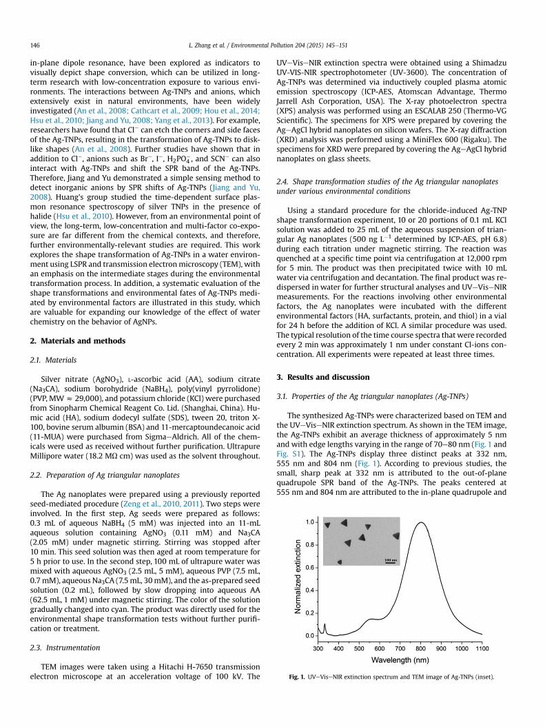
Fig. 1. UVeViseNIR extinction spectrum and TEM image of Ag-TNPs (inset).
L. Zhang et al. / Environmental Pollution 204 (2015) 145e151146
in-plane dipole resonance, have been explored as indicators tovisually depict shape conversion, which can be utilized in long-term research with low-concentration exposure to various envi-ronments. The interactions between Ag-TNPs and anions, whichextensively exist in natural environments, have been widelyinvestigated (An et al., 2008; Cathcart et al., 2009; Hou et al., 2014;Hsu et al., 2010; Jiang and Yu, 2008; Yang et al., 2013). For example,researchers have found that Cl� can etch the corners and side facesof the Ag-TNPs, resulting in the transformation of Ag-TNPs to disk-like shapes (An et al., 2008). Further studies have shown that inaddition to Cl�, anions such as Br�, I�, H2PO
�4 , and SCN� can also
interact with Ag-TNPs and shift the SPR band of the Ag-TNPs.Therefore, Jiang and Yu demonstrated a simple sensing method todetect inorganic anions by SPR shifts of Ag-TNPs (Jiang and Yu,2008). Huang's group studied the time-dependent surface plas-mon resonance spectroscopy of silver TNPs in the presence ofhalide (Hsu et al., 2010). However, from an environmental point ofview, the long-term, low-concentration and multi-factor co-expo-sure are far different from the chemical contexts, and therefore,further environmentally-relevant studies are required. This workexplores the shape transformation of Ag-TNPs in a water environ-ment using LSPR and transmission electronmicroscopy (TEM), withan emphasis on the intermediate stages during the environmentaltransformation process. In addition, a systematic evaluation of theshape transformations and environmental fates of Ag-TNPs medi-ated by environmental factors are illustrated in this study, whichare valuable for expanding our knowledge of the effect of waterchemistry on the behavior of AgNPs.
2. Materials and methods
2.1. Materials
Silver nitrate (AgNO3), L-ascorbic acid (AA), sodium citrate(Na3CA), sodium borohydride (NaBH4), poly(vinyl pyrrolidone)(PVP, MWz 29,000), and potassium chloride (KCl) were purchasedfrom Sinopharm Chemical Reagent Co. Lid. (Shanghai, China). Hu-mic acid (HA), sodium dodecyl sulfate (SDS), tween 20, triton X-100, bovine serum albumin (BSA) and 11-mercaptoundecanoic acid(11-MUA) were purchased from SigmaeAldrich. All of the chem-icals were used as received without further purification. UltrapureMillipore water (18.2 MU cm) was used as the solvent throughout.
2.2. Preparation of Ag triangular nanoplates
The Ag nanoplates were prepared using a previously reportedseed-mediated procedure (Zeng et al., 2010, 2011). Two steps wereinvolved. In the first step, Ag seeds were prepared as follows:0.3 mL of aqueous NaBH4 (5 mM) was injected into an 11-mLaqueous solution containing AgNO3 (0.11 mM) and Na3CA(2.05 mM) under magnetic stirring. Stirring was stopped after10 min. This seed solution was then aged at room temperature for5 h prior to use. In the second step, 100 mL of ultrapure water wasmixed with aqueous AgNO3 (2.5 mL, 5 mM), aqueous PVP (7.5 mL,0.7mM), aqueous Na3CA (7.5mL, 30mM), and the as-prepared seedsolution (0.2 mL), followed by slow dropping into aqueous AA(62.5 mL, 1 mM) under magnetic stirring. The color of the solutiongradually changed into cyan. The product was directly used for theenvironmental shape transformation tests without further purifi-cation or treatment.
2.3. Instrumentation
TEM images were taken using a Hitachi H-7650 transmissionelectron microscope at an acceleration voltage of 100 kV. The
UVeViseNIR extinction spectra were obtained using a ShimadzuUV-VIS-NIR spectrophotometer (UV-3600). The concentration ofAg-TNPs was determined via inductively coupled plasma atomicemission spectroscopy (ICP-AES, Atomscan Advantage, ThermoJarrell Ash Corporation, USA). The X-ray photoelectron spectra(XPS) analysis was performed using an ESCALAB 250 (Thermo-VGScientific). The specimens for XPS were prepared by covering theAgeAgCl hybrid nanoplates on silicon wafers. The X-ray diffraction(XRD) analysis was performed using a MiniFlex 600 (Rigaku). Thespecimens for XRD were prepared by covering the AgeAgCl hybridnanoplates on glass sheets.
2.4. Shape transformation studies of the Ag triangular nanoplatesunder various environmental conditions
Using a standard procedure for the chloride-induced Ag-TNPshape transformation experiment, 10 or 20 portions of 0.1 mL KClsolution was added to 25 mL of the aqueous suspension of trian-gular Ag nanoplates (500 ng L�1 determined by ICP-AES, pH 6.8)during each titration under magnetic stirring. The reaction wasquenched at a specific time point via centrifugation at 12,000 rpmfor 5 min. The product was then precipitated twice with 10 mLwater via centrifugation and decantation. The final product was re-dispersed in water for further structural analyses and UVeViseNIRmeasurements. For the reactions involving other environmentalfactors, the Ag nanoplates were incubated with the differentenvironmental factors (HA, surfactants, protein, and thiol) in a vialfor 24 h before the addition of KCl. A similar procedure was used.The typical resolution of the time course spectra that were recordedevery 2 min was approximately 1 nm under constant Cl-ions con-centration. All experiments were repeated at least three times.
3. Results and discussion
3.1. Properties of the Ag triangular nanoplates (Ag-TNPs)
The synthesized Ag-TNPs were characterized based on TEM andthe UVeViseNIR extinction spectrum. As shown in the TEM image,the Ag-TNPs exhibit an average thickness of approximately 5 nmand with edge lengths varying in the range of 70e80 nm (Fig. 1 andFig. S1). The Ag-TNPs display three distinct peaks at 332 nm,555 nm and 804 nm (Fig. 1). According to previous studies, thesmall, sharp peak at 332 nm is attributed to the out-of-planequadrupole SPR band of the Ag-TNPs. The peaks centered at555 nm and 804 nm are attributed to the in-plane quadrupole and
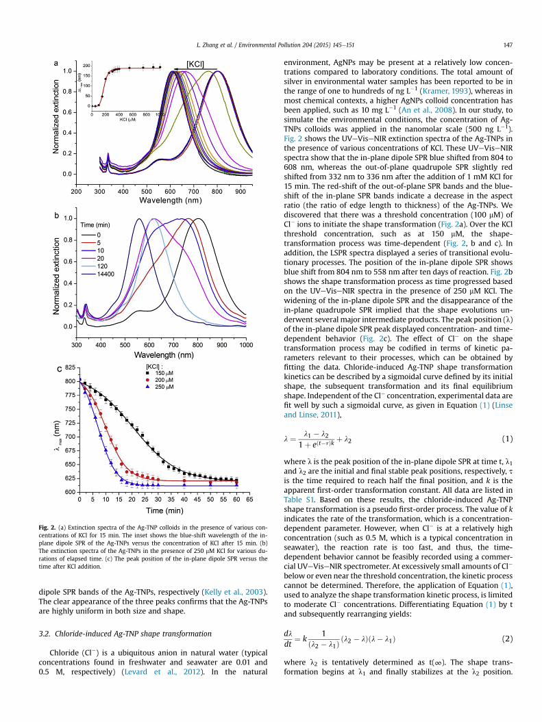
Fig. 2. (a) Extinction spectra of the Ag-TNP colloids in the presence of various con-centrations of KCl for 15 min. The inset shows the blue-shift wavelength of the in-plane dipole SPR of the Ag-TNPs versus the concentration of KCl after 15 min. (b)The extinction spectra of the Ag-TNPs in the presence of 250 mM KCl for various du-rations of elapsed time. (c) The peak position of the in-plane dipole SPR versus thetime after KCl addition.
L. Zhang et al. / Environmental Pollution 204 (2015) 145e151 147
dipole SPR bands of the Ag-TNPs, respectively (Kelly et al., 2003).The clear appearance of the three peaks confirms that the Ag-TNPsare highly uniform in both size and shape.
3.2. Chloride-induced Ag-TNP shape transformation
Chloride (Cl�) is a ubiquitous anion in natural water (typicalconcentrations found in freshwater and seawater are 0.01 and0.5 M, respectively) (Levard et al., 2012). In the natural
environment, AgNPs may be present at a relatively low concen-trations compared to laboratory conditions. The total amount ofsilver in environmental water samples has been reported to be inthe range of one to hundreds of ng L�1 (Kramer, 1993), whereas inmost chemical contexts, a higher AgNPs colloid concentration hasbeen applied, such as 10 mg L�1 (An et al., 2008). In our study, tosimulate the environmental conditions, the concentration of Ag-TNPs colloids was applied in the nanomolar scale (500 ng L�1).Fig. 2 shows the UVeViseNIR extinction spectra of the Ag-TNPs inthe presence of various concentrations of KCl. These UVeViseNIRspectra show that the in-plane dipole SPR blue shifted from 804 to608 nm, whereas the out-of-plane quadrupole SPR slightly redshifted from 332 nm to 336 nm after the addition of 1 mM KCl for15 min. The red-shift of the out-of-plane SPR bands and the blue-shift of the in-plane SPR bands indicate a decrease in the aspectratio (the ratio of edge length to thickness) of the Ag-TNPs. Wediscovered that there was a threshold concentration (100 mM) ofCl� ions to initiate the shape transformation (Fig. 2a). Over the KClthreshold concentration, such as at 150 mM, the shape-transformation process was time-dependent (Fig. 2, b and c). Inaddition, the LSPR spectra displayed a series of transitional evolu-tionary processes. The position of the in-plane dipole SPR showsblue shift from 804 nm to 558 nm after ten days of reaction. Fig. 2bshows the shape transformation process as time progressed basedon the UVeViseNIR spectra in the presence of 250 mM KCl. Thewidening of the in-plane dipole SPR and the disappearance of thein-plane quadrupole SPR implied that the shape evolutions un-derwent several major intermediate products. The peak position (l)of the in-plane dipole SPR peak displayed concentration- and time-dependent behavior (Fig. 2c). The effect of Cle on the shapetransformation process may be codified in terms of kinetic pa-rameters relevant to their processes, which can be obtained byfitting the data. Chloride-induced Ag-TNP shape transformationkinetics can be described by a sigmoidal curve defined by its initialshape, the subsequent transformation and its final equilibriumshape. Independent of the Cl� concentration, experimental data arefit well by such a sigmoidal curve, as given in Equation (1) (Linseand Linse, 2011),
l ¼ l1 � l2
1þ eðt�tÞk þ l2 (1)
where l is the peak position of the in-plane dipole SPR at time t, l1and l2 are the initial and final stable peak positions, respectively, tis the time required to reach half the final position, and k is theapparent first-order transformation constant. All data are listed inTable S1. Based on these results, the chloride-induced Ag-TNPshape transformation is a pseudo first-order process. The value of kindicates the rate of the transformation, which is a concentration-dependent parameter. However, when Cl� is at a relatively highconcentration (such as 0.5 M, which is a typical concentration inseawater), the reaction rate is too fast, and thus, the time-dependent behavior cannot be feasibly recorded using a commer-cial UVeViseNIR spectrometer. At excessively small amounts of Cl�
below or even near the threshold concentration, the kinetic processcannot be determined. Therefore, the application of Equation (1),used to analyze the shape transformation kinetic process, is limitedto moderate Cl� concentrations. Differentiating Equation (1) by tand subsequently rearranging yields:
dldt
¼ k1
ðl2 � l1Þðl2 � lÞðl� l1Þ (2)
where l2 is tentatively determined as t(∞). The shape trans-formation begins at l1 and finally stabilizes at the l2 position.
L. Zhang et al. / Environmental Pollution 204 (2015) 145e151148
Equation (2) is a logistic differential equation and clearly shows thatthe above-described sigmoidal curve is a logistic curve for ourexperimental results. The Equation (2) may imply that Cl� couldfunction as a stabilizing agent. The long-term transformation pro-cess is tracked using LSPR (Fig. S2), as demonstrated by thedisappearance of the blue shift after several months. Previousstudies have also reported similar stabilization of Cl� (An et al.,2008; Im et al., 2005). However, other stabilizing agents, such asPVP, which is included in the synthesis, are still present on thesurface of the nanostructures after the exposure to chloride.Therefore, the capping agents and chloride are likely responsiblefor the stability of the newly formed disk-like structures. Huanget al. explained that the change in peak positions of the in-planedipole SPR of silver colloids in the presence of Cl� ions are corre-lated to the truncating effect on the corners of the silver nanoplatesand to the increase in the nanoplate thickness (Hsu et al., 2010).However, a thickness change was not observed in our data (Fig. S3).Therefore, a new mechanism regarding the role of Cl� in shapetransformation must be proposed.
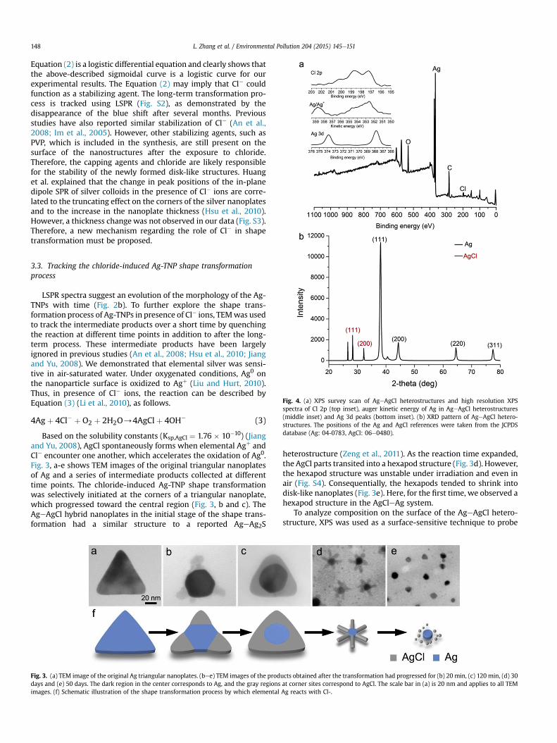
Fig. 4. (a) XPS survey scan of AgeAgCl heterostructures and high resolution XPSspectra of Cl 2p (top inset), auger kinetic energy of Ag in AgeAgCl heterostructures(middle inset) and Ag 3d peaks (bottom inset). (b) XRD pattern of AgeAgCl hetero-structures. The positions of the Ag and AgCl references were taken from the JCPDSdatabase (Ag: 04-0783, AgCl: 06e0480).
3.3. Tracking the chloride-induced Ag-TNP shape transformationprocess
LSPR spectra suggest an evolution of the morphology of the Ag-TNPs with time (Fig. 2b). To further explore the shape trans-formation process of Ag-TNPs in presence of Cl� ions, TEMwas usedto track the intermediate products over a short time by quenchingthe reaction at different time points in addition to after the long-term process. These intermediate products have been largelyignored in previous studies (An et al., 2008; Hsu et al., 2010; Jiangand Yu, 2008). We demonstrated that elemental silver was sensi-tive in air-saturated water. Under oxygenated conditions, Ag0 onthe nanoparticle surface is oxidized to Agþ (Liu and Hurt, 2010).Thus, in presence of Cl� ions, the reaction can be described byEquation (3) (Li et al., 2010), as follows.
4Agþ 4Cl� þ O2 þ 2H2O/4AgClþ 4OH� (3)
Based on the solubility constants (Ksp,AgCl ¼ 1.76 � 10�10) (Jiangand Yu, 2008), AgCl spontaneously forms when elemental Agþ andCl� encounter one another, which accelerates the oxidation of Ag0.Fig. 3, a-e shows TEM images of the original triangular nanoplatesof Ag and a series of intermediate products collected at differenttime points. The chloride-induced Ag-TNP shape transformationwas selectively initiated at the corners of a triangular nanoplate,which progressed toward the central region (Fig. 3, b and c). TheAgeAgCl hybrid nanoplates in the initial stage of the shape trans-formation had a similar structure to a reported AgeAg2S
Fig. 3. (a) TEM image of the original Ag triangular nanoplates. (bee) TEM images of the proddays and (e) 50 days. The dark region in the center corresponds to Ag, and the gray regionsimages. (f) Schematic illustration of the shape transformation process by which elemental
heterostructure (Zeng et al., 2011). As the reaction time expanded,the AgCl parts transited into a hexapod structure (Fig. 3d). However,the hexapod structure was unstable under irradiation and even inair (Fig. S4). Consequentially, the hexapods tended to shrink intodisk-like nanoplates (Fig. 3e). Here, for the first time, we observed ahexapod structure in the AgCleAg system.
To analyze composition on the surface of the AgeAgCl hetero-structure, XPS was used as a surface-sensitive technique to probe
ucts obtained after the transformation had progressed for (b) 20 min, (c) 120 min, (d) 30at corner sites correspond to AgCl. The scale bar in (a) is 20 nm and applies to all TEMAg reacts with Cl-.
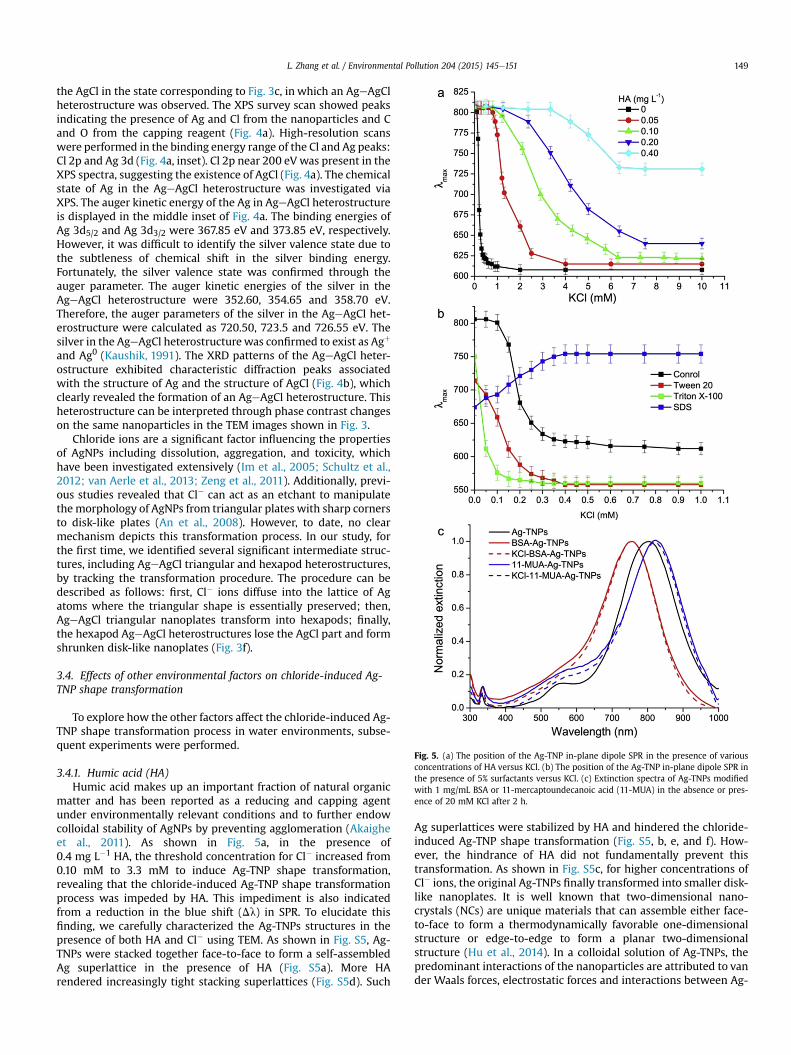
Fig. 5. (a) The position of the Ag-TNP in-plane dipole SPR in the presence of variousconcentrations of HA versus KCl. (b) The position of the Ag-TNP in-plane dipole SPR inthe presence of 5% surfactants versus KCl. (c) Extinction spectra of Ag-TNPs modifiedwith 1 mg/mL BSA or 11-mercaptoundecanoic acid (11-MUA) in the absence or pres-ence of 20 mM KCl after 2 h.
L. Zhang et al. / Environmental Pollution 204 (2015) 145e151 149
the AgCl in the state corresponding to Fig. 3c, in which an AgeAgClheterostructure was observed. The XPS survey scan showed peaksindicating the presence of Ag and Cl from the nanoparticles and Cand O from the capping reagent (Fig. 4a). High-resolution scanswere performed in the binding energy range of the Cl and Ag peaks:Cl 2p and Ag 3d (Fig. 4a, inset). Cl 2p near 200 eV was present in theXPS spectra, suggesting the existence of AgCl (Fig. 4a). The chemicalstate of Ag in the AgeAgCl heterostructure was investigated viaXPS. The auger kinetic energy of the Ag in AgeAgCl heterostructureis displayed in the middle inset of Fig. 4a. The binding energies ofAg 3d5/2 and Ag 3d3/2 were 367.85 eV and 373.85 eV, respectively.However, it was difficult to identify the silver valence state due tothe subtleness of chemical shift in the silver binding energy.Fortunately, the silver valence state was confirmed through theauger parameter. The auger kinetic energies of the silver in theAgeAgCl heterostructure were 352.60, 354.65 and 358.70 eV.Therefore, the auger parameters of the silver in the AgeAgCl het-erostructure were calculated as 720.50, 723.5 and 726.55 eV. Thesilver in the AgeAgCl heterostructurewas confirmed to exist as Agþ
and Ag0 (Kaushik, 1991). The XRD patterns of the AgeAgCl heter-ostructure exhibited characteristic diffraction peaks associatedwith the structure of Ag and the structure of AgCl (Fig. 4b), whichclearly revealed the formation of an AgeAgCl heterostructure. Thisheterostructure can be interpreted through phase contrast changeson the same nanoparticles in the TEM images shown in Fig. 3.
Chloride ions are a significant factor influencing the propertiesof AgNPs including dissolution, aggregation, and toxicity, whichhave been investigated extensively (Im et al., 2005; Schultz et al.,2012; van Aerle et al., 2013; Zeng et al., 2011). Additionally, previ-ous studies revealed that Cl� can act as an etchant to manipulatethe morphology of AgNPs from triangular plates with sharp cornersto disk-like plates (An et al., 2008). However, to date, no clearmechanism depicts this transformation process. In our study, forthe first time, we identified several significant intermediate struc-tures, including AgeAgCl triangular and hexapod heterostructures,by tracking the transformation procedure. The procedure can bedescribed as follows: first, Cl� ions diffuse into the lattice of Agatoms where the triangular shape is essentially preserved; then,AgeAgCl triangular nanoplates transform into hexapods; finally,the hexapod AgeAgCl heterostructures lose the AgCl part and formshrunken disk-like nanoplates (Fig. 3f).
3.4. Effects of other environmental factors on chloride-induced Ag-TNP shape transformation
To explore how the other factors affect the chloride-induced Ag-TNP shape transformation process in water environments, subse-quent experiments were performed.
3.4.1. Humic acid (HA)Humic acid makes up an important fraction of natural organic
matter and has been reported as a reducing and capping agentunder environmentally relevant conditions and to further endowcolloidal stability of AgNPs by preventing agglomeration (Akaigheet al., 2011). As shown in Fig. 5a, in the presence of0.4 mg L�1 HA, the threshold concentration for Cl� increased from0.10 mM to 3.3 mM to induce Ag-TNP shape transformation,revealing that the chloride-induced Ag-TNP shape transformationprocess was impeded by HA. This impediment is also indicatedfrom a reduction in the blue shift (Dl) in SPR. To elucidate thisfinding, we carefully characterized the Ag-TNPs structures in thepresence of both HA and Cl� using TEM. As shown in Fig. S5, Ag-TNPs were stacked together face-to-face to form a self-assembledAg superlattice in the presence of HA (Fig. S5a). More HArendered increasingly tight stacking superlattices (Fig. S5d). Such
Ag superlattices were stabilized by HA and hindered the chloride-induced Ag-TNP shape transformation (Fig. S5, b, e, and f). How-ever, the hindrance of HA did not fundamentally prevent thistransformation. As shown in Fig. S5c, for higher concentrations ofCl� ions, the original Ag-TNPs finally transformed into smaller disk-like nanoplates. It is well known that two-dimensional nano-crystals (NCs) are unique materials that can assemble either face-to-face to form a thermodynamically favorable one-dimensionalstructure or edge-to-edge to form a planar two-dimensionalstructure (Hu et al., 2014). In a colloidal solution of Ag-TNPs, thepredominant interactions of the nanoparticles are attributed to vander Waals forces, electrostatic forces and interactions between Ag-
L. Zhang et al. / Environmental Pollution 204 (2015) 145e151150
TNPs and capping agents. The addition of HA may change thecharges on the Ag-TNPs and the steric repulsion, thus promotingthe self-assembly via van der Waals forces.
3.4.2. SurfactantSurfactants are a diverse group of chemicals known for their
wide use in detergents and other cleaning products. After use, re-sidual surfactants are discharged into sewage systems or directlyinto surface waters, and most end up dispersed in various envi-ronmental compartments, such as soil, water and sediment. Toassess the effect of the surfactants on chloride-induced Ag-TNPshape transformation, we selected SDS, tween 20, and triton X-100as models. As shown in Fig. S6, the Ag-TNPs displayed a distinct in-plane dipole SPR blue shift (Dl) of 84, 53 and 127 nm in 5% solu-tions of tween 20, triton X-100 and SDS, respectively. Cl� ionsreversed the blue shift in SDS solution. Although the chloride-induced shape transformation process still occurred in the solu-tion of tween 20 and triton X-100. Further concentration-dependent experiments indicated that the shape transformationof the Ag-TNPs in the presence of non-ionic surfactants (tween 20and triton X-100) occurred more easily when induced by Cl-ions(Fig. 5b). These results demonstrate that surfactants in the waterenvironment impact the transformation of silver nanoparticles.
3.4.3. BiomacromoleculeIt was previously reported that bovine serum albumin (BSA), a
model protein, reduces the toxicity of AgNPs toward nitrosomonaseuropaea (Ostermeyer et al., 2013). In this study, we used BSA as arepresentative of biomacromolecule in environmental water. Fig. 5cshows the UVeViseNIR spectra of the as-prepared Ag-TNPs col-loids modified by BSA before (red, solid line) and after (red, dashedline) the addition of 20 mM KCl for 2 h. As shown, the extinctionspectra corresponding to the BSA-modified Ag-TNPs in 20 mM Cl�
ions are almost the same as the spectra recorded in the absence ofCl�. The BAS-modified Ag-TNPs can endure a higher concentrationof chlorine ions than the unmodified Ag-TNPs by at least 200-fold.It is clear that the presence of BSA restrains the chloride-inducedshape transformation of the Ag-TNPs. This result is likely due tothe presence of the sulfhydryl group in BSA, which may stabilizethe structure of the Ag nanoplates (Lee et al., 2010). To furtherexplore this mechanism, 11-mercaptoundecanoic acid (11-MUA)was chosen as a model to test the effect of the sulfhydryl group onthe chloride-induced Ag-TNP shape transformation process. Asshown in Fig. 5c, the extinction spectra remained the same before(blue, solid line) and after (blue, dashed line) the addition of KCl.This result demonstrated that the presence of sulfhydryl groupssuppresses the chloride-induced Ag-TNP shape transformation.
4. Conclusions
The natural water environment is a complex system. Otherenvironmental factors exert different influences on chloride-induced Ag-TNP shape transformation. Therefore, it is difficult toelucidate the environmental fate of Ag-TNPs. In this study, we showa unique shape transformation process of Ag-TNPs and demon-strate the effect of other environmental factors on the final fate ofAg-TNPs. These results are of value for expanding our knowledge ofthe intermediate products in the transformation process of a vari-ety of nanoparticles. Compared to previous studies involving Cl�,this process is tracked over the long-term using TEM, and the ef-fects of both the reaction time and concentration of Cl� are inves-tigated in detail. We found that Cl� reacts with Ag nanocrystals inthe presence of oxidants, and consequently, an AgeAgCl triangularheterostructure is formed. This AgeAgCl triangular heterostructurefurther evolves into a symmetrical hexapod structure over the
long-term reaction. Our studies also reveal that the hexapodstructures are unstable under irradiation in water environments.These results offer new information on the environmental fate ofAgNPs
Acknowledgments
This study was supported by MOST of China (Grant No.2014CB932002), Natural Science Foundation of China (Nos.11105150 and 51371164) and the Special Financial Grant from ChinaPostdoctoral Science Foundation (No. 2013T60613).
Appendix A. Supplementary data
Supplementary data related to this article can be found at http://dx.doi.org/10.1016/j.envpol.2015.04.018.
References
Akaighe, N., MacCuspie, R.I., Navarro, D.A., Aga, D.S., Banerjee, S., Sohn, M.,Sharma, V.K., 2011. Humic acid-induced silver nanoparticle formation underenvironmentally relevant conditions. Environ. Sci. Technol. 45, 3895e3901.
An, J., Tang, B., Zheng, X., Zhou, J., Dong, F., Xu, S., Wang, Y., Zhao, B., Xu, W., 2008.Sculpturing effect of chloride ions in shape transformation from triangular todiscal silver nanoplates. J. Phys. Chem. C 112, 15176e15182.
Burchardt, A.D., Carvalho, R.N., Valente, A., Nativo, P., Gilliland, D., Garcìa, C.P.,Passarella, R., Pedroni, V., Rossi, F., Lettieri, T., 2012. Effects of silver nano-particles in diatom Thalassiosira pseudonana and cyanobacterium synechococcussp. Environ. Sci. Technol. 46, 11336e11344.
Cathcart, N., Frank, A.J., Kitaev, V., 2009. Silver nanoparticles with planar twinneddefects: effect of halides for precise tuning of plasmon resonance maxima from400 to >900 nm. Chem. Commun. 7170e7172.
Chambers, B.A., Afrooz, A.R.M.N., Bae, S., Aich, N., Katz, L., Saleh, N.B., Kirisits, M.J.,2013. Effects of chloride and ionic strength on physical morphology, dissolution,and bacterial toxicity of silver nanoparticles. Environ. Sci. Technol. 48, 761e769.
Chen, S., Theodorou, I.G., Goode, A.E., Gow, A., Schwander, S., Zhang, J., Chung, K.F.,Tetley, T.D., Shaffer, M.S., Ryan, M.P., Porter, A.E., 2013. High-resolution analyt-ical electron microscopy reveals cell culture media-induced changes to thechemistry of silver nanowires. Environ. Sci. Technol. 47, 13813e13821.
El-Temsah, Y.S., Joner, E.J., 2012. Impact of Fe and Ag nanoparticles on seedgermination and differences in bioavailability during exposure in aqueoussuspension and soil. Environ. Toxicol. 27, 42e49.
Hou, X., Chen, S., Tang, J., Xiong, Y., Long, Y., 2014. Silver nanoplates-based colori-metric iodide recognition and sensing using sodium thiosulfate as a sensitizer.Anal. Chim. Acta 825, 57e62.
Hsu, M.-S., Cao, Y.-W., Wang, H.-W., Pan, Y.-S., Lee, B.-H., Huang, C.-L., 2010. Time-dependent surface plasmon resonance spectroscopy of silver nanoprisms in thepresence of halide ions. Chemphyschem 11, 1742e1748.
Hu, C., Lin, K., Wang, X., Liu, S., Yi, J., Tian, Y., Wu, B., Chen, G., Yang, H., Dai, Y., Li, H.,Zheng, N., 2014. Electrostatic self-assembling formation of Pd superlatticenanowires from surfactant-free ultrathin Pd nanosheets. J. Am. Chem. Soc. 136,12856e12859.
Im, S.H., Lee, Y.T., Wiley, B., Xia, Y., 2005. Large-scale synthesis of silver nanocubes:the role of HCl in promoting cube perfection and monodispersity. Angew. Chem.Int. 44, 2154e2157.
Jana, N.R., Gearheart, L., Murphy, C.J., 2001. Wet chemical synthesis of silvernanorods and nanowires of controllable aspect ratio. Chem. Commun. 617e618.
Jiang, X.C., Yu, A.B., 2008. Silver nanoplates: a highly sensitive material towardinorganic anions. Langmuir 24, 4300e4309.
Jin, R.C., Cao, Y.C., Hao, E.C., Metraux, G.S., Schatz, G.C., Mirkin, C.A., 2003. Con-trolling anisotropic nanoparticle growth through plasmon excitation. Nature425, 487e490.
Jin, R.C., Cao, Y.W., Mirkin, C.A., Kelly, K.L., Schatz, G.C., Zheng, J.G., 2001. Photoin-duced conversion of silver nanospheres to nanoprisms. Science 294,1901e1903.
Kaushik, V.K., 1991. XPS core level spectra and auger parameters for some silvercompounds. J. Electron Spectrosc. Relat. Phenom. 56, 273e277.
Kelly, K.L., Coronado, E., Zhao, L.L., Schatz, G.C., 2003. The optical properties of metalnanoparticles: the influence of size, shape, and dielectric environment. J. Phys.Chem. B 107, 668e677.
Kramer, J.R.B.,G., 1993. Environmental Chemistry of Silver. SETAC Press, NorthCarolina.
Kumari, M., Mukherjee, A., Chandrasekaran, N., 2009. Genotoxicity of silver nano-particles in Allium cepa. Sci. Total Environ. 407, 5243e5246.
Lee, B.-H., Hsu, M.-S., Hsu, Y.-C., Lo, C.-W., Huang, C.-L., 2010. A facile method toobtain highly stable silver nanoplate colloids with desired surface plasmonresonance wavelengths. J. Phys. Chem. C 114, 6222e6227.
Levard, C., Hotze, E.M., Colman, B.P., Dale, A.L., Truong, L., Yang, X.Y., Bone, A.J.,Brown, G.E., Tanguay, R.L., Di Giulio, R.T., Bernhardt, E.S., Meyer, J.N.,
L. Zhang et al. / Environmental Pollution 204 (2015) 145e151 151
Wiesner, M.R., Lowry, G.V., 2013a. Sulfidation of silver nanoparticles: naturalantidote to their toxicity. Environ. Sci. Technol. 47, 13440e13448.
Levard, C., Hotze, E.M., Lowry, G.V., Brown, G.E., 2012. Environmental trans-formations of silver nanoparticles: impact on stability and toxicity. Environ. Sci.Technol. 46, 6900e6914.
Levard, C., Mitra, S., Yang, T., Jew, A.D., Badireddy, A.R., Lowry, G.V., Brown, G.E.,2013b. Effect of chloride on the dissolution rate of silver nanoparticles andtoxicity to E. coli. Environ. Sci. Technol. 47, 5738e5745.
Li, X.A., Lenhart, J.J., Walker, H.W., 2010. Dissolution-accompanied aggregation ki-netics of silver nanoparticles. Langmuir 26, 16690e16698.
Linse, B., Linse, S., 2011. Monte Carlo simulations of protein amyloid formationreveal origin of sigmoidal aggregation kinetics. Mol. Biosyst. 7, 2296e2303.
Liu, B., Luo, W., Zhao, X., 2009. A facile synthesis of ordered ultralong silver nano-belts. Mater. Res. Bull. 44, 682e687.
Liu, J., Hurt, R.H., 2010. Ion release kinetics and particle persistence in aqueousnano-silver colloids. Environ. Sci. Technol. 44, 2169e2175.
Liu, Z.-h., Zhou, Y., Maszenan, A.M., Ng, W.J., Liu, Y., 2013. pH-dependent trans-formation of Ag nanoparticles in anaerobic processes. Environ. Sci. Technol. 47,12630e12631.
Ma, R., Levard, C., Judy, J.D., Unrine, J.M., Durenkamp, M., Martin, B., Jefferson, B.,Lowry, G.V., 2013. Fate of zinc oxide and silver nanoparticles in a pilot waste-water treatment plant and in processed biosolids. Environ. Sci. Technol. 48,104e112.
Ostermeyer, A.-K., Kostigen Mumuper, C., Semprini, L., Radniecki, T., 2013. Influenceof bovine serum albumin and alginate on silver nanoparticle dissolution andtoxicity to Nitrosomonas europaea. Environ. Sci. Technol. 47, 14403e14410.
Poynton, H.C., Lazorchak, J.M., Impellitteri, C.A., Blalock, B.J., Rogers, K., Allen, H.J.,Loguinov, A., Heckman, J.L., Govindasmawy, S., 2012. Toxicogenomic responsesof nanotoxicity in daphnia magna exposed to silver nitrate and coated silver
nanoparticles. Environ. Sci. Technol. 46, 6288e6296.Schultz, A.G., Ong, K.J., MacCormack, T., Ma, G., Veinot, J.G.C., Goss, G.G., 2012. Silver
nanoparticles inhibit sodium uptake in juvenile rainbow trout (Oncorhynchusmykiss). Environ. Sci. Technol. 46, 10295e10301.
Sun, Y.G., Mayers, B., Xia, Y.N., 2003. Transformation of silver nanospheres intonanobelts and triangular nanoplates through a thermal process. Nano Lett. 3,675e679.
Van Aerle, R., Lange, A., Moorhouse, A., Paszkiewicz, K., Ball, K., Johnston, B.D., de-Bastos, E., Booth, T., Tyler, C.R., Santos, E.M., 2013. Molecular mechanisms oftoxicity of silver nanoparticles in zebrafish embryos. Environ. Sci. Technol. 47,8005e8014.
Wirth, S.M., Lowry, G.V., Tilton, R.D., 2012. Natural organic matter alters biofilmtolerance to silver nanoparticles and dissolved Silver. Environ. Sci. Technol. 46,12687e12696.
Yang, X.-H., Ling, J., Peng, J., Cao, Q.-E., Ding, Z.-T., Bian, L.-C., 2013. A colorimetricmethod for highly sensitive and accurate detection of iodide by finding thecritical color in a color change process using silver triangular nanoplates. Anal.Chim. Acta 798, 74e81.
Yu, S.j., Chao, J.b., Sun, J., Yin, Y.g., Liu, J.f., Jiang, G.b., 2013. Quantification of theuptake of silver nanoparticles and ions to HepG2 cells. Environ. Sci. Technol. 47,3268e3274.
Zeng, J., Roberts, S., Xia, Y., 2010. Nanocrystal-based time-temperature indicators.Chem. - Eur. J. 16, 12559e12563.
Zeng, J., Tao, J., Su, D., Zhu, Y., Qin, D., Xia, Y., 2011. Selective sulfuration at the cornersites of a silver nanocrystal and its use in stabilization of the shape. Nano Lett.11, 3010e3015.
Zhang, Q., Ge, J., Pham, T., Goebl, J., Hu, Y., Lu, Z., Yin, Y., 2009. Reconstruction ofsilver nanoplates by UV irradiation: tailored optical properties and enhancedstability. Angew. Chem. Int. 48, 3516e3519.


