CHIMICA - unibo.itamsdottorato.unibo.it/7606/1/TESI_Sebastiano_Rupiani.pdf · CHIMICA Ciclo XXVIII...
132
Alma Mater Studiorum – Università di Bologna DOTTORATO DI RICERCA IN CHIMICA Ciclo XXVIII Settore Concorsuale di afferenza: 03/D1 Settore Scientifico disciplinare: CHIM/08 FIGHTING CANCER THROUGH DESIGNED AND NATURAL PRODUCTS: DISCOVERY OF NEW LDH-A INHIBITORS AND ROUTE TO THE TOTAL SYNTHESIS OF RAKICIDIN A Presentata da: Sebastiano Rupiani Coordinatore Dottorato Relatore Prof. Aldo Roda Prof. Marinella Roberti Esame finale anno 2016
Transcript of CHIMICA - unibo.itamsdottorato.unibo.it/7606/1/TESI_Sebastiano_Rupiani.pdf · CHIMICA Ciclo XXVIII...
AAllmmaa MMaatteerr SSttuuddiioorruumm –– UUnniivveerrssiittàà ddii BBoollooggnnaa
DOTTORATO DI RICERCA IN
CHIMICA
Ciclo XXVIII
Settore Concorsuale di afferenza: 03/D1 Settore Scientifico disciplinare: CHIM/08
FIGHTING CANCER THROUGH DESIGNED AND NATURAL PRODUCTS: DISCOVERY OF NEW LDH-A INHIBITORS AND ROUTE TO THE TOTAL
SYNTHESIS OF RAKICIDIN A
Presentata da: Sebastiano Rupiani
Coordinatore Dottorato Relatore
Prof. Aldo Roda Prof. Marinella Roberti
Esame finale anno 2016

CONTENTS
Abstract I
Abbreviations and acronyms III
1 Modern anti-cancer therapy: tools and strategies 1
1.1 An overview of classic and modern therapies 3
1.1.1 Cytotoxic therapy 3
1.1.2 Targeted therapy 6
1.2 An emerging hallmark: cancer cells reprogramming energy metabolism 8
1.3 Cancer hypoxia and cancer stem cells 11
1.4 Aims of this work 16
2 Identification of N-acylhydrazone derivatives as novel lactate
dehydrogenase A inhibitors 19
2.1 Introduction 19
2.2 Results and discussion 24
2.3 Conclusion 34
2.4 Experimental section 35
2.4.1 General Methods 35
2.4.2 Synthetic procedures 35
3 Synthesis of galloflavin analogs and their evaluation as lactate
dehydrogenase A inhibitors 51
3.1 Introduction 51
3.2 Results and discussion 59
3.3 Conclusion 70
3.4 Experimental section 71
3.4.1 General methods 71
3.4.2 Synthetic procedures 71
4 Route to the total synthesis of rakicidin A 83
4.1. Introduction 83
4.2. Results and Discussion 86
4.3. Conclusion 93
4.4. Experimental section 94
4.4.1. General Methods 94
4.4.2. Synthetic procedures 94
References 109
ACKNOWLEDGEMENTS 119


I
Abstract
The present work aimed to the discovery of new anticancer agents and to the
total synthesis of a natural compound with selectivity towards cancer hypoxia.
In this context, a first project involved the design and synthesis of N-
acylhydrazone based inhibitors of lactate dehydrogenase A (LDH-A). The
structures of the new molecules where designed by means of virtual screening
techniques and synthesized to obtain a library of analogs which were evaluated
on the isolated enzyme. Active compounds were also screened on cell cultures
of non-Hodgkins lymphoma and one of them proved to be a promising inhibitor,
suggesting that the N-acylhydrazone could be a suitable scaffold in the field of
LDH-A inhibitors.
The second project aimed to the synthesis of Galloflavin (GF) analogs and to
the study of the compound’s SAR. GF is an LDH-A inhibitor which was
previously identified and synthesized by our group. Its poor solubility and
stability prevented us from studying its SAR maintaining the core structure.
Therefore, the synthesis of three potential classes of structural analogs was
devised and carried out. One compound was found to reproduce GF’s
behaviour on the enzyme and in cell, therefore being a good starting point for
the study. A small library of analogs was synthesized and biological tests are
ongoing to acquire in-depth knowledge about the key pharmacophores of this
interesting inhibitor.
The third project was carried out at Aarhus University in the Chemical Biology
group lead by Prof. Thomas B. Poulsen. The work focused on the total
synthesis of Rakicidin A, a complex macrolide of natural origin which was
identified and isolated two decades ago from soil samples and since then is
known for its interesting properties in selectively inducing cell death in hypoxic
environments and being also active on cancer stem cells. The total synthesis
involved several steps including key enantioselective reactions to build the 5
stereocenters on the molecule. The project was carried out in collaboration with
other components of the group and was successfully completed in early 2015.

II

III
Abbreviations and acronyms
β-HOAsn: β-hydroxyasparagine
β-HOAsp: β-hydroxyaspartic acid
ALT1: Alanine transaminase 1
APD: (E)-4-amido-2,4-pentadienoate
BCR-ABL: Breakpoint cluster region – Abelson
BSA: Bovine serum albumin
CAM: Cerium ammonium molybdate
CaMKII γ: Calcium/calmodulin-dependent protein kinase type II gamma
CML: Chronic myelogenous leukemia
CSCs: Cancer stem cells
DCM: Dichloromethane
DCC: N,N'-Dicyclohexylcarbodiimide
DCU: Dicyclohexylurea
DIBAL: Diisobutylaluminum hydride
DMAP: Dimethylaminopyridine
DME: 1,2-Dimethoxyethane
DMF: N,N-dimethylformamide
DMP: Dess–Martin periodinane
DMSO: Dimethyl sulfoxide
DNA: Deoxyribonucleic acid
DSC: N,N′-disuccinimidyl carbonate
EA: Ellagic acid
EDCI: 1-Ethyl-3-(3-dimethylaminopropyl)carbodiimide
IV
EGFR: Epidermal growth factor receptor
EMT: Epithelial to mesenchymal transition
FCC: Flash column chromatography
GA: Gallic acid
GF: Galloflavin
HDAC: Histone deacetylase
HER2: Human epidermal growth factor receptor 2
HIF: Hypoxia-inducible factor
HPLC: High performance liquid chromatography
HWE: Horner-Wadsworth-Emmons
LDH: Lactate dehydrogenase
LiHMDS: Lithium bis(trimethylsilyl)amide
MET: Mesenchymal to epithelial transition
mTOR: Mammalian target of rapamycin
NAD+: Nicotinamide adenine dinucleotide (oxidized form)
NADH: Nicotinamide adenine dinucleotide (reduced form)
NADP+: Nicotinamide adenine dinucleotide phosphate (oxidized form)
NADPH: Nicotinamide adenine dinucleotide phosphate (reduced form)
NBS: N-bromosuccinimide
NCI: National cancer institute
NMR: Nuclear magnetic resonance
NOE: Nuclear Overhauser effect
PCC: Pyridinium chlorochromate
PDH: Pyruvate dehydrogenase
V
PDK: Pyruvate dehydrogenase kinase
PG: Protecting group
Ph: Philadelphia chromosome
pO2: Partial oxygen pressure
PyBOP: (benzotriazol-1-yl-oxytripyrrolidinophosphonium hexafluorophosphate)
RakA: Rakicidin A
RakB: Rakicidin B
RNA: Ribonucleic acid
SAR: Structure-activity relationship
SCs: Stem cells
THF: Tetrahydrofuran
TLC: Thin layer chromatography
TMEDA: Tetramethylethylenediamine
TOP1: Topoisomerase I
TOP2: Topoisomerase II
TyrK: Tyrosine kinase
UM6: Urolithin M6
VEGF: Vascular endothelial growth factor
VS: Virtual Screening

VI

1
Figure 1 Survival rates by type, US, 2004-2010, for some of the most common human cancers
CHAPTER 1
Modern anti-cancer therapy: tools and strategies
Cancer is currently the cause of almost 20% of deaths that occur every year in
high-income countries1.
During the past several decades more than 100 different types of cancer were
identified2 and nowadays we face a highly heterogeneous situation in terms of
prognosis and patient conditions, depending on the type and subtype of
neoplasm encountered. As shown in figure 1, the five-year relative survival rate
in the US in the interval 2004-2010 varied from a dreadful 7% in pancreas
cancer to an encouraging 99% in prostate cancer, with some of the other most
common types covering the whole range in between3.
The reasons behind this divergence are complex, and include the genetic and
epigenetic abnormalities that characterize differently each cancer type, causing
specific downstream pathways that can in some case be exploited for
therapeutic purposes4, as well as incidence and acquired resistance to chemo-
and radiotherapy, which can also vary considerably and are obviously to be
taken into account.
This scenario might suggest that researchers and physicians should hardly be
able to address cancer as a single disease, but it is nevertheless possible - to

2
some extent - to describe some crucial common features of cancer biology that
can contribute to set order in the vast complexity of cancer diagnosis and
treatment.
At the beginning of the XXI century, Hanahan and Weinberg established six key
features common to almost all types of cancer - since then defined as The
Hallmarks of Cancer2 - giving the scientific community some very precious tools
towards a better understanding of the neoplastic disease.
These hallmarks are: sustaining proliferative signaling, evading growth
suppressors, resisting cell death, enabling replicative immortality, inducing
angiogenesis, and activating invasion and metastasis.
The hallmarks of cancer outline a common profile of the neoplastic cell that can
be identified as an immortal cell with aggressive invasive power which is able to
create a local environment favorable to its own survival and, at the metastasis
state, can create colonies of spawned cells that attack distal tissues in different
organs.
However, despite this concept might suggest a view of cancer tissue as a
homogeneous malignant mass, recent advancements show that plasticity and
heterogeneity are in fact key features of tumors, and they need to be taken into
account in the quest for new treatments (see paragraph 1.3).
The therapeutic challenge is therefore remarkable, and it is nowadays clear that
a targeted therapy hitting specific molecular features that characterize different
cancer types is the most promising strategy, and most importantly the less
harmful for the patient4, since the principles on which classic chemotherapies
were based can no more be considered sustainable, due to their dose-limiting
toxicities and their sometimes life-threatening side effects.
In the next paragraphs, a selection of the most common classic and modern
approved therapies5 will be described together with their mechanisms of action,
as well as some recent cutting edge concepts inspiring new paths for the drug
discovery of anticancer compounds.

3
1.1 An overview of classic and modern therapies
Drugs approved for clinical use in cancer treatment can be divided in two main
areas: “classic” cytotoxic chemotherapy and targeted therapy6. Cytotoxic
chemotherapy is the traditional therapy based on compounds that interact with
some fundamental processes of cell cycle, therefore impairing the proliferation
of cancer and often inducing programmed cell death7. These compounds attack
non-selectively all types of cells, including healthy ones, and their efficacy is
mostly based on the fact that cancer cells divide with a much higher rate than
normal cells, making them more sensitive to compounds that arrest cell cycle
since at any time a higher portion of cancer cells are undergoing cell division.
The dose is chosen in an interval where the ratio of cancer cell death vs normal
cell death is optimal7. Side effects are obviously severe and they have always
constituted a major issue in cancer treatment since the life of the patient can
sometimes be seriously threatened by these compounds.
Targeted therapy is the modern approach to chemotherapy and it involves the
use of compounds that selectively target specific molecular abnormalities
occurring in certain types of cancer, therefore reducing considerably the risk of
side effects since effective targeted therapies are meant to only attack cancer
cells8. Of course this also reduces the range of applicability of the compound
and requires an intense study of the genetic variability of the different types of
cancer that can lead to peculiar processes which can be targeted to kill the
malignant cell.
1.1.1 Cytotoxic therapy
The commonly used cytotoxic chemotherapics approved for clinic exploit
several different mechanisms of action. They can be divided into: alkylating
agents, DNA crosslinkers, inhibitors of dihydrofolate reductase, nucleoside
analogs, antimicrotubular agents, DNA intercalators, topoisomerase inhibitors
and proteasome inhibitors6. Other less common compounds have different
mechanisms which will not be treated in this section.
Alkylating agents are among the very first compounds to be used as anticancer
drugs. They are mostly represented by the class of nitrogen mustards,

4
electrophilic compounds which alkylate guanine bases on the N-7 position,
causing permanent DNA damage which in turn causes p53 induced apoptosis9.
Cyclophosphamide is a famous member of this class, and it acts as a prodrug
requiring activation of the oxazaphosphorine ring by P450 enzymes to achieve
the active form in vivo10. The toxicity of cyclophosphamide is however quite
concerning, especially due to the fact that its first metabolite is the reactive
aldehyde acrolein, which is responsible for urotoxicity, neurotoxicity, and
nephrotoxicity11.
DNA crosslinkers such as cisplatin, carboplatin and oxaliplatin act through a
similar mechanism, binding to guanine after displacement of a ligand (chloride
in cisplatin) by a water molecule, making the platinum center more reactive12.
The same process takes place after the first bond is formed, generating a cross-
link between two strands of DNA, leading to programmed cell-death initiated by
proteins of the caspase family.
Inhibitors of dihydrofolate reductase interfere with the enzyme responsible of
the reduction of dihydrofolate to tetrahydrofolate, which is a key process in the
synthesis of thymine, a necessary nucleotide in DNA synthesis13. A famous
antifolate drug is methotrexate, a compound which binds competitively to the
binding site of natural folates with a 1000-fold higher affinity therefore blocking
the enzymatic activity and inducing cell death14. Methotrexate is also used in the
therapy against rheumatoid arthritis, where it acts with a different mechanism
based on a multitarget pattern which is still not completely disclosed15.
Nucleoside analogs are compounds that reproduce the structure of natural
nucleosides, which can be used as building blocks by the cell to synthesise
DNA. Minimal structural differences on a key site of the nucleoside lead the
DNA strand to be unusable, eventually causing apoptosis16. Gemcitabine is a
difluorinated analog of deoxycytidine which replaces cytidine in RNA during
DNA replication, it is a common compound in cytotoxic chemotherapy and it has
also shown to be active in the inhibition of ribonucleotide reductase, a process
which deprives the cell of the necessary supply of deoxyribonucleotides17.
Antimicrotubular agents are compounds that interact with the dynamic of
microtubules having the effect of inducing apoptosis. Microtubules are crucial
5
polymeric structures found in the cell, which are involved in several basic
functions, including the formation of mitotic spindles during chromosome
separation prior to cell division18. They are non-covalent polymers of a dimer of
alpha and beta tubulin, two isoforms of a class of structural globular proteins19.
They are continuously engaged in non-equilibrium dynamics and they are
characterized by a so called “dynamic instability”, meaning that their size vary
constantly during the life cycle of a cell with periods of shortening, growth and
paused states20. This dynamicity must be maintained to allow the cell to survive
and undergo mitosis, and for this reason it represents an interesting target for
compounds aiming at cytotoxicity. The two main classes of approved
antimicrotubular agents are Vinca alkaloids and Taxanes. The former, including
vincristine and vinblastine, are compounds extracted from the leaves of
Catharanthus roseus (Vinca rosea) and discovered in the 60's as potent
antitumor agents21; the latter, including paclitaxel and docetaxel, are alkaloids of
Taxus brevifolia and Taxus baccata, and were identified less than a decade
later than Vinca alkaloids21. These two classes of compounds bind tubulin in
two very distinct sites, defined as the Vinca binding domain and the Taxane
binding domain, and they were previously thought to act with two opposite
mechanisms. It is clear in fact that Vinca alkaloids stimulate in vitro
depolymerization of microtubules whereas Taxanes stabilize them22.
Nevertheless it has been demonstrated that at low, clinically relevant
concentration, Vinca alkaloids don’t destroy microtubules while still inducing
apoptosis23. As a consequence it is now widely accepted that both these
classes of molecules effectively interfere with cell mitosis by simply blocking
microtubule dynamics20, therefore acting with similar mechanisms despite the
different binding sites.
DNA intercalators are planar aromatic molecules, often constituted by polycyclic
structures, which show high affinity with DNA nitrogen bases and bind to DNA
double helixes by occupying the space between two nucleosides24. The
presence of the intercalator disrupts the optimal DNA geometry and interrupts
replication. Doxorubicin and daunorubicin, both commonly used chemoterapics,
are intercalators but also showed a range of additional mechanisms such as
topoisomerase II binding and free radical generation25.
6
Topoisomerase inhibitors interact with the crucial topoisomerase I (TOP1) and
topoisomerase II (TOP2) enzymes blocking their function of supercoiled DNA
unwinding agents which they carry out after transcription and replication26.
These enzymes are ubiquitous in mammalians and they are essential for cell
survival. Studies on TOP1 knockout mice showed that their absence causes
death during embryogenesis27. The inhibitors exert their action with the
stabilization of the covalent DNA cleavage complexes, therefore increasing their
steady-state concentration and causing accumulation of broken DNA strands,
leading to apoptosis28. Common TOP1 inhibitors are topotecan and irinotecan,
both synthetic analogs of natural compound camptothecin, whereas among the
TOP2 inhibitors we can find etoposide and teniposide, semisynthetic glycosides
of podophyllotoxin, an antimicotic extracted from Podophyllum peltatum (a.k.a.
mayapple or American mandrake)29.
Proteasome inhibitors are molecules that act through complex mechanisms,
interrupting the proteolytic action of the proteasome protein complexes30. In
cancer cells these complexes can destroy crucial tumor suppressors as p53,
therefore their inhibition can restore the normal function of pro-apoptotic factors
and induce cell death. The first approved proteasome inhibitor was bortezomib,
a boronic acid modified dipeptide31, and in 2012 a new compound (carfilzomib)
was approved for the treatment of multiple myeloma32.
1.1.2 Targeted therapy
Modern cancer treatment is rapidly expanding from the old cytotoxic based
therapies to more appropriate and focused targeted compounds. Advanced
molecular and chemical biology unveiled a number of aberrant molecular
pathways in cancer that have been exploited in the last few decades to obtain
potent drugs with high specificity and relatively lower toxicity. The focus in this
paragraph will be on tyrosine-kinase (TyrK) inhibitors, vascular endothelial
growth factor (VEGF) pathway inhibitors, epidermal growth factor receptor
(EGFR) inhibitors and mammalian target of rapamycin (mTOR) inhibitors.
TyrK inhibitors were the first compounds to open the era of targeted therapy,
with the milestone discovery of imatinib. This drug exploits the aberration of the

7
Philadelphia chromosome (Ph)33, a genetic mutation in chronic myelogenous
leukemia (CML) which generates the fusion protein BCR-ABL, a constitutively
active tyrosine kinase which can be found in all CML patients34. BCR-ABL itself
was found to be both sufficient and necessary to cause CML, therefore
representing an exceptionally appealing target for the treatment of this
neoplasm35–37. Since then imatinib has also been approved for the treatments of
other forms of cancer such as gastrointestinal stromal tumor and Ph-positive
acute lymphoblastic leukemia. The use of imatinib has more recently
encountered some issues arisen by the insurgence of mutation-induced
resistance38 and to the inability of the molecule to attack cancer stem cells
therefore leading to a risk of relapse after an apparent complete remission has
been achieved (see paragraph 1.4).
VEGF pathway inhibitors interact with a molecular mechanism involved in the
creation and sustainment of one of the hallmarks of cancer: angiogenesis.
VEGF is a pro-angiogenic growth factor responsible for the vascularization of
cancer cells and induced by hypoxic cells in need of increased blood supply39.
Bevacizumab is a recombinant humanized monoclonal antibody that prevents
the binding of VEGF to VEGF receptors therefore blocking its growth-
stimulating effect40. Despite the initial great hope behind this drug it has now
been recognized that its effect might be in reality less striking than expected
and nowadays bevacizumab is mostly used in combination with classic
chemotherapy41, especially for the treatment of advanced colorectal cancer42.
Its action is thought to be exerted through a “normalization” of blood vessels,
which facilitates the action of cytotoxic drugs by letting them further inside
neoplastic tissue, rather than a real block of blood supply and angiogenesis43.
EGFR inhibitors interfere with cell membrane receptors which are upregulated
in some cancers and stimulate cell proliferation. HER2 is a specific type of
EGFR, encoded by a gene which is known to be overexpressed in 20-30% of
breast cancers44 and it’s connected with severe disease and poor prognosis.
Trastuzumab is a monoclonal antibody EGFR inhibitor which binds to the HER2
receptor inducing cell cycle arrest during the G1 phase while furthermore
activating tumor suppressor p2745.

8
mTOR inhibitors are compounds interacting with a serine/threonine protein
kinase involved in several growth and proliferation signals46 which was first
identified while investigating the mechanism of action of rapamycin47, a natural
macrolide produced by Streptomyces hygroscopicus48 with known
immunosuppressant action and mainly used to prevent rejection in organ
transplantation. Temsirolimus is a synthetic analog of rapamycin and is
approved for the treatment of advanced renal-cell carcinoma49.
1.2 An emerging hallmark: cancer cells reprogramming energy
metabolism
In a later re-edition of their famous paper, Hanahan and Weinberg introduced
the idea of a new generation of “emerging hallmarks”, that arise from the
knowledge acquired thanks to the research carried out in the first decade of the
2000’s50. These emerging hallmarks were identified to be reprogramming
energy metabolism and evading immune destruction. Flanking these newly
identified capabilities, two “enabling characteristics” were described, as
conditions facilitating the acquisition of both core and emerging hallmarks:
genome instability and tumor-promoting inflammation.
We will here focus on the abnormal metabolic phenotype which can be
recognized in the majority of cancer types, is particularly appealing in a
therapeutic perspective and represents the core rationale of our research on
lactate dehydrogenase inhibitors. This unexpected feature was first discovered
in the first half of the XX century by Otto Warburg, who initially believed it to be
one, if not the most important, cause of cancer51. Genetic alterations and
cellular response to the neoplastic microenvironment (such as hypoxia in some
cases) contribute to induce a crucial switch in the way tumors carry out their
energetic metabolism and produce ATP: quiescent normal cells metabolize
pyruvate after glycolysis mainly through the oxygen-dependent cooperation of
Krebs cycle and oxidative phosphorylation, carried out in mitochondria,
oxidizing nutrients to CO2 and storing energy in a highly efficient manner52; on
the contrary, cancer cells reprogram their metabolic pathway towards the so
called aerobic glycolysis: regardless of the oxygen level, the cell relies largely
9
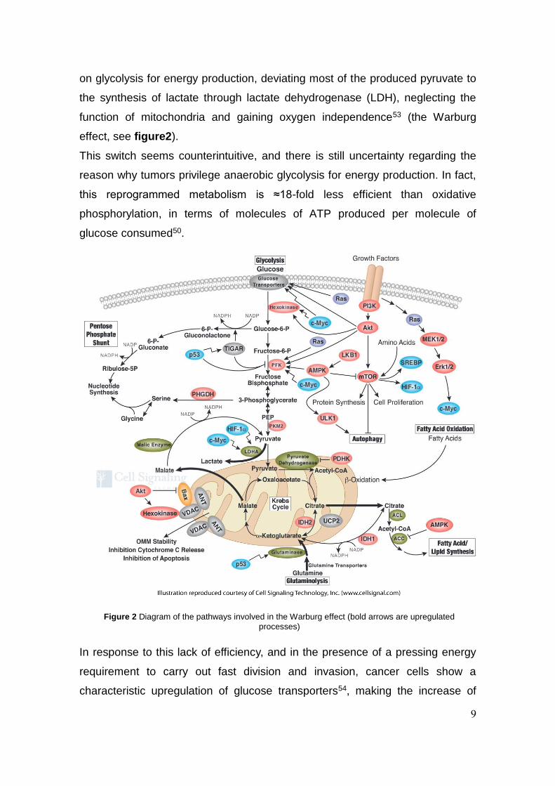
Figure 2 Diagram of the pathways involved in the Warburg effect (bold arrows are upregulated
processes)
on glycolysis for energy production, deviating most of the produced pyruvate to
the synthesis of lactate through lactate dehydrogenase (LDH), neglecting the
function of mitochondria and gaining oxygen independence53 (the Warburg
effect, see figure2).
This switch seems counterintuitive, and there is still uncertainty regarding the
reason why tumors privilege anaerobic glycolysis for energy production. In fact,
this reprogrammed metabolism is ≈18-fold less efficient than oxidative
phosphorylation, in terms of molecules of ATP produced per molecule of
glucose consumed50.
In response to this lack of efficiency, and in the presence of a pressing energy
requirement to carry out fast division and invasion, cancer cells show a
characteristic upregulation of glucose transporters54, making the increase of
10
glucose uptake a fundamental signature which has often been used for
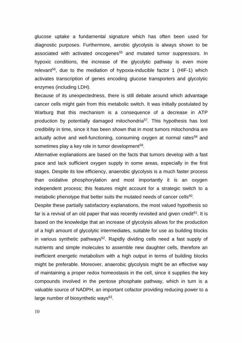
diagnostic purposes. Furthermore, aerobic glycolysis is always shown to be
associated with activated oncogenes55 and mutated tumor suppressors. In
hypoxic conditions, the increase of the glycolytic pathway is even more
relevant56, due to the mediation of hypoxia-inducible factor 1 (HIF-1) which
activates transcription of genes encoding glucose transporters and glycolytic
enzymes (including LDH).
Because of its unexpectedness, there is still debate around which advantage
cancer cells might gain from this metabolic switch. It was initially postulated by
Warburg that this mechanism is a consequence of a decrease in ATP
production by potentially damaged mitochondria57. This hypothesis has lost
credibility in time, since it has been shown that in most tumors mitochondria are
actually active and well-functioning, consuming oxygen at normal rates58 and
sometimes play a key role in tumor development59.
Alternative explanations are based on the facts that tumors develop with a fast
pace and lack sufficient oxygen supply in some areas, especially in the first
stages. Despite its low efficiency, anaerobic glycolysis is a much faster process
than oxidative phosphorylation and most importantly it is an oxygen
independent process; this features might account for a strategic switch to a
metabolic phenotype that better suits the mutated needs of cancer cells60.
Despite these partially satisfactory explanations, the most valued hypothesis so
far is a revival of an old paper that was recently revisited and given credit61. It is
based on the knowledge that an increase of glycolysis allows for the production
of a high amount of glycolytic intermediates, suitable for use as building blocks
in various synthetic pathways62. Rapidly dividing cells need a fast supply of
nutrients and simple molecules to assemble new daughter cells, therefore an
inefficient energetic metabolism with a high output in terms of building blocks
might be preferable. Moreover, anaerobic glycolysis might be an effective way
of maintaining a proper redox homeostasis in the cell, since it supplies the key
compounds involved in the pentose phosphate pathway, which in turn is a
valuable source of NADPH, an important cofactor providing reducing power to a
large number of biosynthetic ways63.
11
The established relationship between tumor cells and the Warburg phenotype
opened the path towards a number of valuable new targets for anticancer
therapy. Several enzymes are involved in glycolysis and some of them have
been recognized as potentially druggable with the aim of developing a new
family of compounds often referred to as glycolytic inhibitors64. Among the most
interesting targets over which drug development research is ongoing it is
possible to find enzymes such as hexokinase, phosphofructokinase,
glyceraldehyde-3-phosphate dehydrogenase, pyruvate kinase and lactate
dehydrogenase64. Part of the innovative research reported in this work is about
the design, synthesis and evaluation of new inhibitors of lactate dehydrogenase
(see chapters 2 and 3), based on the evidence that inhibition of such enzyme
can block tumor progression and, in some cases, induce programmed cell
death65.
1.3 Cancer hypoxia and cancer stem cells
Neoplastic tissues are highly heterogeneous63. This intrinsic characteristic
common to the vast majority of solid tumors is a consequence of several factors
related to the malignant phenotype with two of the most therapeutically relevant
ones being the proven existence of regions of hypoxia and the presence of local
aggregates of the so called cancer stem cells (CSCs).
Hypoxic tissues are characterized by a partial oxygen pressure (pO2) which falls
in a significantly lower range than healthy tissue. Depending on the topic
(diagnosis, chemotherapy resistance, radiotherapy resistance), hypoxia can be
defined with slightly different pO2 values but it is normally accepted that a tissue
with a pO2 lower than 10 mmHg is considered hypoxic (healthy tissues have
average pO2 values ranging from 40 to 50 mmHg)66.
The most common tumor types have median pO2 between 5 and 15 mmHg66,
with peak areas reaching levels between 0 and 5 mmHg therefore being close
to complete anoxia67.
The constantly low oxygen pressure in tumors is a result of the imbalance
between the O2 fast consumption and its inadequate supply, that is caused from
fast replication and insufficient or chaotic vascularization68. Two kinds of
hypoxia have been postulated: diffusion-limited (or chronic) hypoxia and

12
Figure 3 Diffusion-limited hypoxia in tumor angiogenesis.
perfusion-limited (or acute) hypoxia. Diffusion-limited hypoxia, discovered in the
50’s, is a consequence of vascularization not keeping pace with the fast
expanding tumor and forming tissue portions outside the range of influence of
local vessels69. In these conditions a gradient of pO2 is formed and it is
estimated that at a distance higher than 100 µm a chronic hypoxic environment
is established70 (figure 3).
Perfusion-limited hypoxia arises from the leaky and structural abnormal tumor
vasculature which can locally induce temporary closure or reduced flow with a
consequent transient hypoxia generated in the proximity of the involved
vessels71,72. Regional tumor oxygenation can vary intensively in short periods of
time with acute hypoxia being restored to normoxia often within a few hours63.
The two types of hypoxia have different biological consequences and
therapeutic implications. Chronic hypoxia, characterized by a pO2 gradient, can
often generate similar diffusion-induced gradients in the concentration of drugs
delivered from the blood stream73, causing peripheral hypoxic cells to be
exposed to only a negligible concentration of the active compound. Acute
hypoxia, leading to a non-distance-dependent decrease of pO2, is accompanied
by a higher risk of metastasis due to the presence of hypoxic cells in direct
contact with blood vessels74 and therefore more likely to enter the circulation.
Hypoxic cells have a selective survival advantage over the normoxic ones, due
to the contribution of at least three distinguished mechanisms: hypoxia-

13
mediated selection, inducing mutation of tumor suppressor proteins and
promoting the insurgence of aggressive phenotypes75; genomic instability,
which suppresses DNA repair pathways therefore selecting and promoting cells
with mutations76; changes in oxygen-sensitive pathways, which promote
changes in metabolism, angiogenesis and cell survival mechanisms77.
As a consequence of all the factors described above, hypoxia plays a key role
in resistance to chemo- and radiotherapy78 (an oxygen-dependent process), it
leads tissues to have an increased potential for invasive growth and metastasis
and it is always connected with aggressive tumors and poor prognosis79.
The second factor contributing to the heterogeneous phenotype of cancer is the
presence of cancer stem cells (CSCs) and their role in the life cycle of a tumor.
It has been demonstrated that tumor tissue is characterized by hierarchically
organized populations, recreating the common structure that can be found in
normal tissue80, with the presence of stem cells (SCs), progenitor cells and fully
differentiated cells. Classically, stem cells are defined as populations of cells
with three main abilities: self-renewal, creation of multiple lineages and
extensive proliferation81. CSCs are a type of cells that can be found in most
tumors in relatively small amounts, which show a set of properties which is
parallel to normal SCs and have therefore been associated with them under
many points of view. They are indeed characterized by a strong tumor-initiating
potential, they are immortal and self-renewing and they can spawn a progeny of
differentiated cells82. It is now commonly accepted to refer to these peculiar
subpopulations as stem cells even though it must be clear that it is a term that
arises from the parallel abilities that can be identified between normal stem cells
and CSCs and it doesn’t intend to equate their biological properties and
significance82.
There is uncertainty on whether CSCs arise from normal SCs after mutation or
through a “backwards” reacquisition of self-renewal ability by dedifferentiation of
committed progenitor cells83 and in fact both mechanisms might be relevant. On
the other hand, a clear connection between epithelial to mesenchymal transition
(EMT) and CSC formation has been established and it might be the key to
explain the origin and the role of CSCs in cancer84.
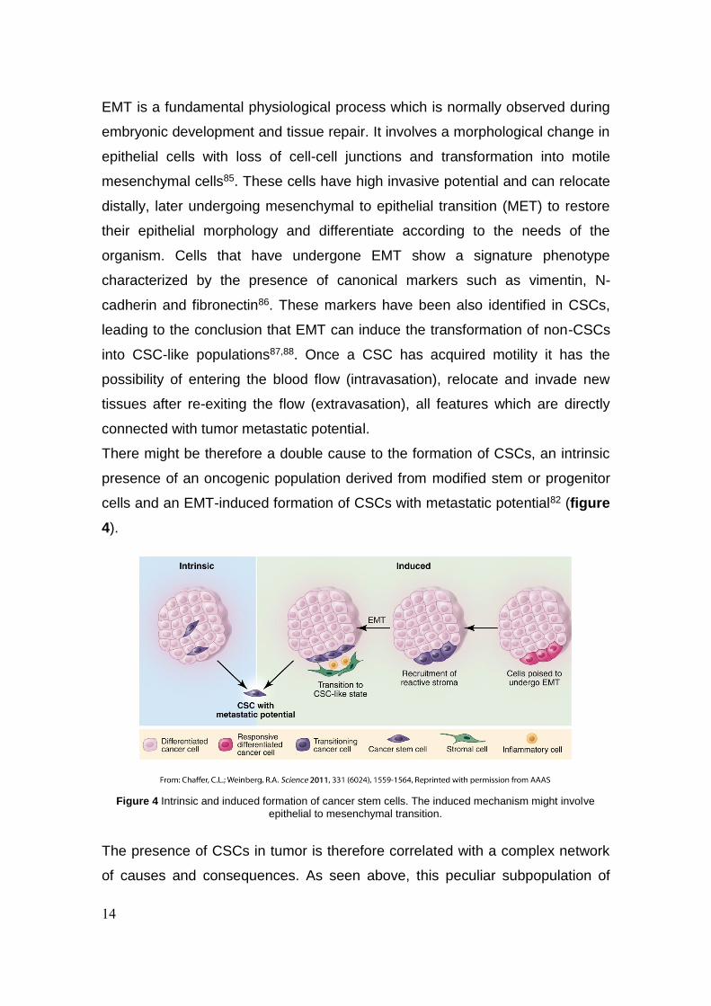
14
Figure 4 Intrinsic and induced formation of cancer stem cells. The induced mechanism might involve
epithelial to mesenchymal transition.
EMT is a fundamental physiological process which is normally observed during
embryonic development and tissue repair. It involves a morphological change in
epithelial cells with loss of cell-cell junctions and transformation into motile
mesenchymal cells85. These cells have high invasive potential and can relocate
distally, later undergoing mesenchymal to epithelial transition (MET) to restore
their epithelial morphology and differentiate according to the needs of the
organism. Cells that have undergone EMT show a signature phenotype
characterized by the presence of canonical markers such as vimentin, N-
cadherin and fibronectin86. These markers have been also identified in CSCs,
leading to the conclusion that EMT can induce the transformation of non-CSCs
into CSC-like populations87,88. Once a CSC has acquired motility it has the
possibility of entering the blood flow (intravasation), relocate and invade new
tissues after re-exiting the flow (extravasation), all features which are directly
connected with tumor metastatic potential.
There might be therefore a double cause to the formation of CSCs, an intrinsic
presence of an oncogenic population derived from modified stem or progenitor
cells and an EMT-induced formation of CSCs with metastatic potential82 (figure
4).
The presence of CSCs in tumor is therefore correlated with a complex network
of causes and consequences. As seen above, this peculiar subpopulation of
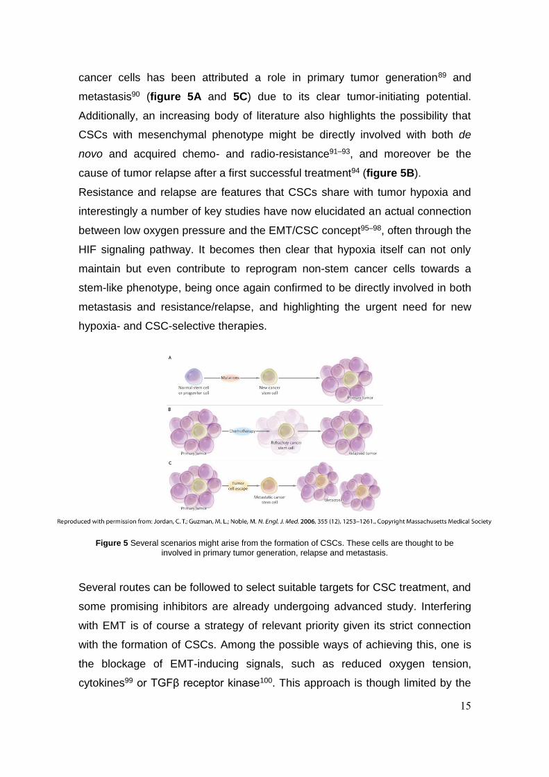
15
Figure 5 Several scenarios might arise from the formation of CSCs. These cells are thought to be
involved in primary tumor generation, relapse and metastasis.
cancer cells has been attributed a role in primary tumor generation89 and
metastasis90 (figure 5A and 5C) due to its clear tumor-initiating potential.
Additionally, an increasing body of literature also highlights the possibility that
CSCs with mesenchymal phenotype might be directly involved with both de
novo and acquired chemo- and radio-resistance91–93, and moreover be the
cause of tumor relapse after a first successful treatment94 (figure 5B).
Resistance and relapse are features that CSCs share with tumor hypoxia and
interestingly a number of key studies have now elucidated an actual connection
between low oxygen pressure and the EMT/CSC concept95–98, often through the
HIF signaling pathway. It becomes then clear that hypoxia itself can not only
maintain but even contribute to reprogram non-stem cancer cells towards a
stem-like phenotype, being once again confirmed to be directly involved in both
metastasis and resistance/relapse, and highlighting the urgent need for new
hypoxia- and CSC-selective therapies.
Several routes can be followed to select suitable targets for CSC treatment, and
some promising inhibitors are already undergoing advanced study. Interfering
with EMT is of course a strategy of relevant priority given its strict connection
with the formation of CSCs. Among the possible ways of achieving this, one is
the blockage of EMT-inducing signals, such as reduced oxygen tension,
cytokines99 or TGFβ receptor kinase100. This approach is though limited by the

16
large variety of EMT-inducing signals93. Alternatively, a promising path to follow
is targeting the mesenchymal phenotype in CSCs, interfering with proteins like
vimentin, N-cadherin and fibronectin101,102. Other options are the inhibition of
EMT-associated transcription factors103 and block of MET104, the latter more
directly connected with the prevention of metastasis and aimed to lock cells into
their high motility state preventing them from actually colonizing new tissue and
proliferate. HDAC inhibitors are interestingly believed to be an additional way of
counteracting EMT since histone deacetylation is crucial in this process and
since these compounds can also affect HIF-1 and nuclear factor-kB, inducing
differentiation of CSCs into normal tumor cells105.
Other successful approaches might be based on non EMT-related mechanisms.
Omacetaxine showed to be active in the inhibition of protein synthesis by
targeting ribosomes and inducing apoptosis of CSCs in tyrosine kinase resistant
CML106 and berbamine binds to the ATP site of CaMKII γ and by inhibiting its
phosphorylation triggers apoptosis of leukemia CSCs107.
HIF is clearly another appealing target due to its strict connection with hypoxic
environments: a high throughput screening identified new ligands able to bind to
the PAS-B domain of the HIF-2α subunit and prevent HIF-2 heterodimerization
and DNA-binding activity, though not affecting HIF-1 fucntion108, whereas a
cyclic CLLFVY peptide showed opposite selectivity, binding to HIF-1α and
reducing HIF-1-mediated hypoxia response signaling while showing no
interaction with HIF-2109.
The development of promising therapies to fight metastasis and cancer relapse
due to the presence of CSCs is therefore supported by a large spectrum of
potential tools that need to be properly explored, in order to find the ideal
Achilles’ heel (or, potentially, heels) of this reluctant subpopulations of cells.
1.4 Aims of this work
The totality of my PhD research has been revolving around the chemical
manipulation of synthetic building blocks in order to obtain biologically active
molecules able to stop, and to some extent kill, cancer cells.

17
The research on LDH-A inhibitors (see chapters 2 and 3) aimed to identify,
through means of virtual screening and analog generation, new classes of
compounds able to interfere with the energy reprogramming that characterizes
distinctively the neoplastic phenotype. In chapter 2 I will describe the discovery
of a promising N-acylhydrazone based compound which showed activity on the
isolated LDH-A enzyme and in cells. Chapter 3 is about the study of a
previously discovered LDH-A inhibitor (Galloflavin, or GF), specifically focusing
on the disclosure of its structure-activity relationship (SAR) through the
development of a structurally analog class of compounds which allowed us to
explore the function of the key pharmacophores without having to deal with the
challenging physicochemical properties of the original molecule.
On chapter 4 I will describe the results of the 6-months period I spent at Aarhus
University, working in the Chemical Biology lab under the supervision of Prof.
Thomas B. Poulsen, where I collaborated to the total synthesis of Rakicidin A, a
macrocyclic depsipeptide of natural origin which exhibits great hypoxia-selective
activity on cancer cells, and has the ability to also target CSCs. The synthetic
effort was successfully completed in 2015 thanks to the collaboration of many
members of the group and ongoing studies are now trying to disclose the
molecular target of the compound through modern chemical biology tools.

18
19
CHAPTER 2
Identification of N-acylhydrazone derivatives as novel lactate
dehydrogenase A inhibitors
2.1 Introduction
Reprogramming of cells energy metabolism is an emerging hallmark of cancer,
as described in paragraph 1.2. Cancer cells, compared to their healthy
counterparts, increase the rate of glucose uptake and mainly metabolize it to
lactate through the so called aerobic glycolysis, as opposed to the more energy-
efficient but oxygen-dependent mitochondrial oxidative phosphorylation
process. This metabolic shift termed Warburg is independent of in-cell oxygen
levels, and provides ATP and substrates for cell growth and division. Given the
dependence of cancer cells on anaerobic glycolysis, this peculiar metabolic
feature could be exploited for selective anticancer therapies.
Lactate dehydrogenase constitutes a key enzyme in glycolysis, catalyzing the
inter-conversion of pyruvate to lactate with simultaneous oxidation of NADH to
NAD+, which is essential to maintain the glycolytic flow. LDH is a tetrameric
enzyme built by the assembly of two types of subunits, LDH-A and LDH-B,
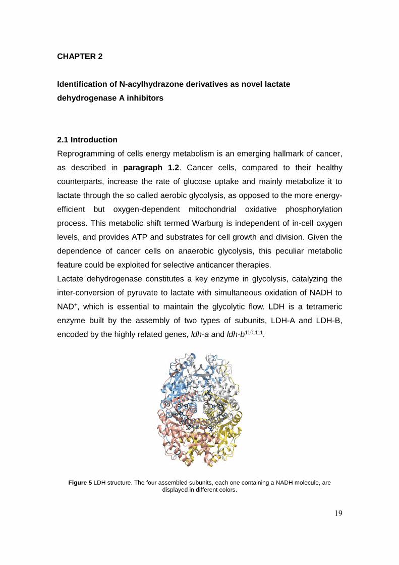
encoded by the highly related genes, ldh-a and ldh-b110,111.
Figure 5 LDH structure. The four assembled subunits, each one containing a NADH molecule, are
displayed in different colors.

20
Subunits A and B are also known as M (muscle) and H (heart) respectively112,
from the tissues where they can be commonly found in the body, and show a
high sequence similarity (~75%)113 in the binding site domains. Five different
combinations have been observed in the tetramer that forms after the assembly
of these subunits: the homotetramers 4H and 4M, and the three mixed
tetramers (3H1M, 2H2M, 1H3M)114. For clarity, 4H homotetramers (therefore
tetramers of LDH-A subunits) will be from now on referred to as simply LDH-A.
The relevance of the A isoform of the enzyme as a critical factor in
tumorigenesis is supported by a consistent amount of data, whereas conflicting
results have been reported concerning the implications of LDH-B115,116, and its
function in cancer cells has not yet been fully elucidated. LDH-A expression is
constantly up-regulated in tumors and it is widely accepted to correlate with
tumor size and poor prognosis55,117,118. In several tumor models, silencing LDH-
A expression by siRNA or shRNA was found to inhibit cell growth, migration and
in vivo tumorigenesis119,120. Furthermore, it has been demonstrated that when
LDH-A expression is silenced in noncancerous cultured cells, proliferation and
protein synthesis are not impaired121. These appealing traits depict LDH-A
inhibitors as potentially safe agents, able to impair cancer cell metabolism and
growth without causing damage to normal tissues.
Because of these features, the rush to the discovery of new LDH-A inhibitors
has recently started and the number of research papers published in the field is
constantly increasing (figure 6).
Figure 6 Papers published in peer reviewed journals concerning inhibition of LDH, source www.scopus.com
21
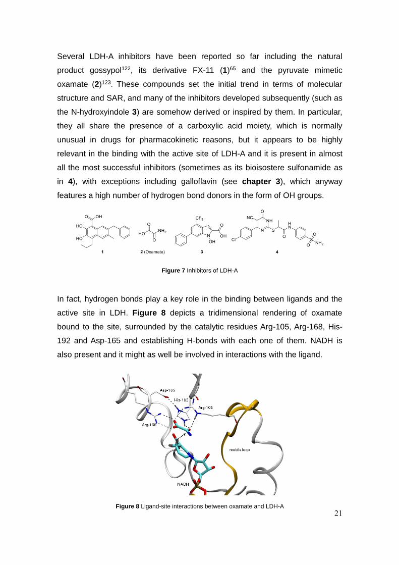
Several LDH-A inhibitors have been reported so far including the natural
product gossypol122, its derivative FX-11 (1)65 and the pyruvate mimetic
oxamate (2)123. These compounds set the initial trend in terms of molecular
structure and SAR, and many of the inhibitors developed subsequently (such as
the N-hydroxyindole 3) are somehow derived or inspired by them. In particular,
they all share the presence of a carboxylic acid moiety, which is normally
unusual in drugs for pharmacokinetic reasons, but it appears to be highly
relevant in the binding with the active site of LDH-A and it is present in almost
all the most successful inhibitors (sometimes as its bioisostere sulfonamide as
in 4), with exceptions including galloflavin (see chapter 3), which anyway
features a high number of hydrogen bond donors in the form of OH groups.
In fact, hydrogen bonds play a key role in the binding between ligands and the
active site in LDH. Figure 8 depicts a tridimensional rendering of oxamate
bound to the site, surrounded by the catalytic residues Arg-105, Arg-168, His-
192 and Asp-165 and establishing H-bonds with each one of them. NADH is
also present and it might as well be involved in interactions with the ligand.
Figure 7 Inhibitors of LDH-A
Figure 8 Ligand-site interactions between oxamate and LDH-A
22
These interactions are crucial and represent a direct consequence of the
physiological function of the enzyme, binding the endogenous substrates
(pyruvate and lactate) and carrying out redox transformations directly involving
proton transfer and carbonyl activation. It is therefore not surprising that many
of the inhibitors discovered so far carry key structural features mimicking this
behavior while binding to the active site.
A more recent class of LDH-A inhibitors is represented by the the N-
hydroxindole-derivatives124,125 developed by Minutolo and coworkers (e.g.
compound 3 in figure 7) which have been extensively studied, also in
combination with classic chemotherapics126 or in conjugation with glucose127.
Other compounds were developed through either a fragment based approach
by AstraZeneca128 and Ariad Pharmaceuticals129, or screening by Genentech130
and GSK131.
Despite a considerable share of the published molecules are active in the low
micromolar or high nanomolar range on the isolated enzyme, in many cases the
authors reported limited cellular activity or not suitable pharmacokinetic
properties.
In this context, the identification of new structures showing high inhibitory
activity both on the isolated enzyme and in the cell, associated with suitable
physicochemical properties, still represents an open challenge to researchers in
the field and no definitive answer has been found yet.
With the intention of entering this quest, our group undertook a first discovery
campaign that ended in the identification of galloflavin132, a promising inhibitor
which retains good cellular activity and which has been subsequently also
studied by competing groups133,134. However, the compound presents several
practical issues and is unsuitable for direct structural development. This aspect
is directly addressed in chapter 3, where the development of promising
galloflavin analogs is described.
In this chapter I report the discovery of a novel class of LDH-A inhibitors based
on an entirely new structure, selected through a virtual screening (VS)
campaign and subsequently investigated through the development of a small
library of analogs to disclose their structure-activity relationship (SAR). The

23
computational study and biological assays were carried out by collaborators of
Prof. Maurizio Recanatini and Prof. Giuseppina Di Stefano, who completed the
discovery team where our group supplied the synthetic contribute.

24
2.2 Results and discussion
This study aimed to discover new compounds with LDH-A inhibitory potential
without the structural bias of pre-existing ligands, in order to expand the
chemical space currently occupied by active scaffolds in the international
literature.
The process therefore started through a de novo virtual screening procedure,
which was carried out with a previously optimized protocol allowing for the
inclusion of the LDH active site loop flexibility and resulting in three different
protein conformations135. The ligands were extracted from the Asinex database
containing around 500000 unique structures, through a filter according to
physical and chemical descriptors based on a variation of Lipinski‘s rule of five,
selected to optimize pharmacokinetic properties, preventing poor absorption
and permeation and excluding poor chemical stability or toxicity. After an
iterative process of docking and selection, the most representative ligands were
selected considering their interactions with the catalytic residues, and 67
promising molecules were chosen for biological tests and purchased in
minimum quantity to obtain preliminary information.
The selected compounds were tested on purified human LDH-A and three of
them (5a-c, figure 9), featuring a common N-acylhydrazone scaffold, were
found to cause enzyme inhibition at micromolar level.
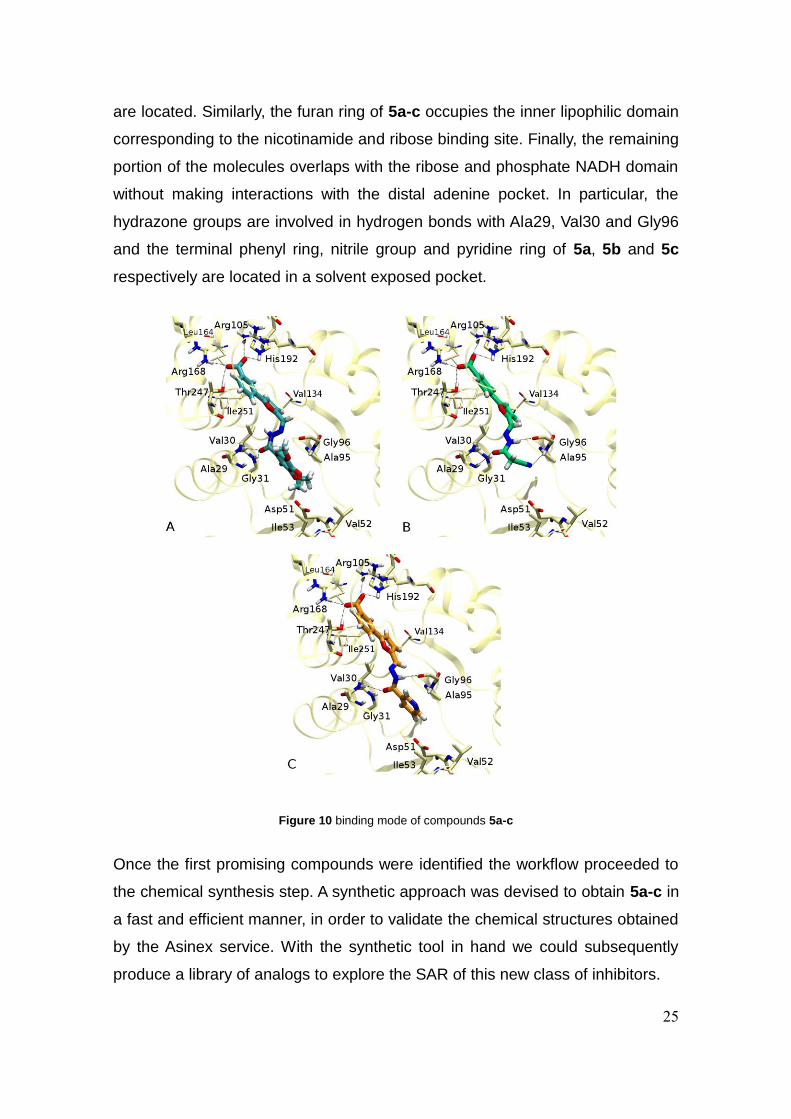
Figure 10 shows the binding mode of these three compounds as resulted from
the docking. As before mentioned, they enter the positively charged substrate
binding pocket through the negative carboxylate. Although 5b is characterized
by the carboxylic group in the para- position, this moiety is involved in
electrostatic interactions similarly to 5a and 5c, whereas its aromatic ring
reaches a deeper cavity where hydrophobic residues (e.g. Ile251 and Leu164)
Figure 9 Three new N-acylhydrazone based LDH-A inhibitors
25
are located. Similarly, the furan ring of 5a-c occupies the inner lipophilic domain
corresponding to the nicotinamide and ribose binding site. Finally, the remaining
portion of the molecules overlaps with the ribose and phosphate NADH domain
without making interactions with the distal adenine pocket. In particular, the
hydrazone groups are involved in hydrogen bonds with Ala29, Val30 and Gly96
and the terminal phenyl ring, nitrile group and pyridine ring of 5a, 5b and 5c
respectively are located in a solvent exposed pocket.
Once the first promising compounds were identified the workflow proceeded to
the chemical synthesis step. A synthetic approach was devised to obtain 5a-c in
a fast and efficient manner, in order to validate the chemical structures obtained
by the Asinex service. With the synthetic tool in hand we could subsequently
produce a library of analogs to explore the SAR of this new class of inhibitors.
Figure 10 binding mode of compounds 5a-c
26
The docking study suggested that the carboxylic moiety engaged interactions
similar as the ones that the endogenous substrate is involved in and therefore
that portion was kept unchanged in most variants, while we focused on
modifications concerning the central heterocycle and the other terminal part.
The common synthetic strategy to achieve the desired compounds 5a-p was
designed to bring diversity to the library through a fast and modular assembly of
building blocks.
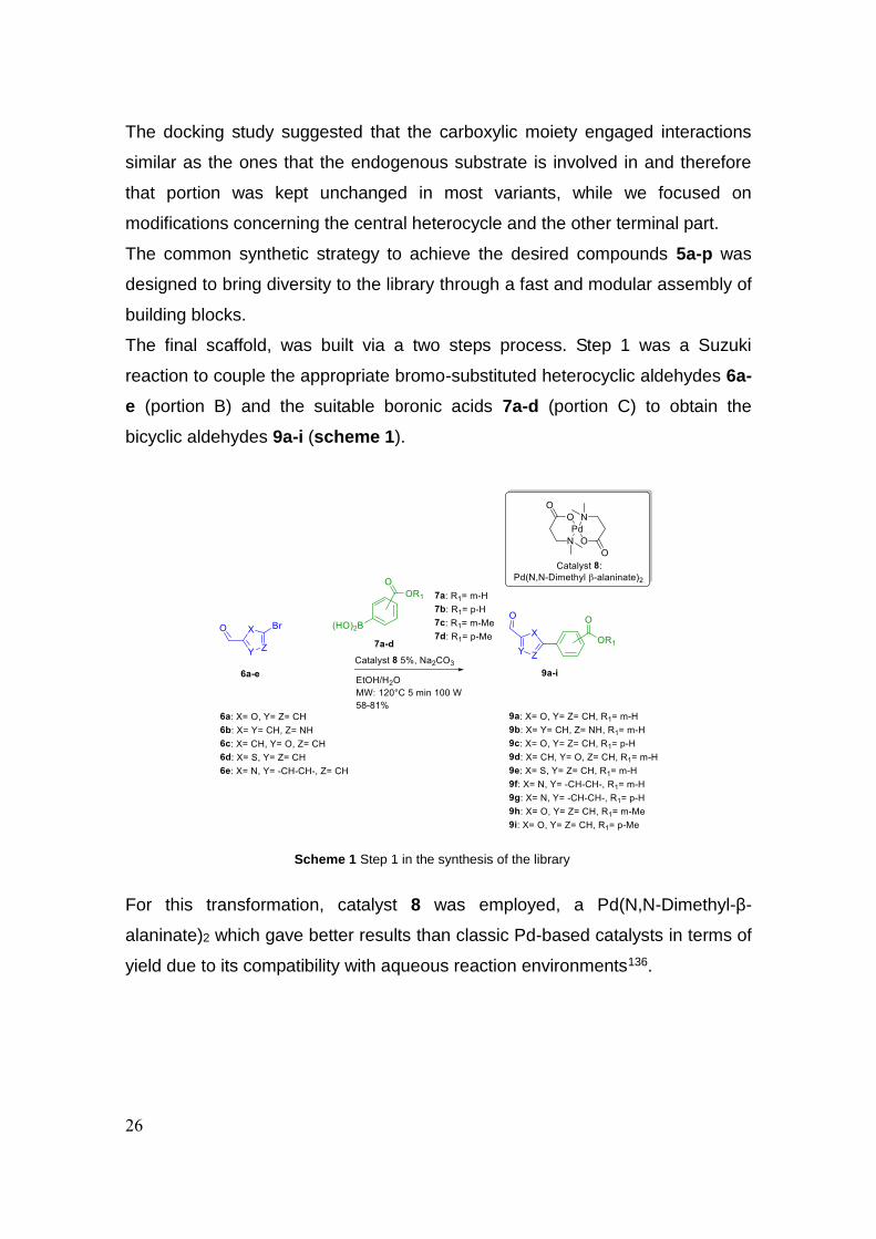
The final scaffold, was built via a two steps process. Step 1 was a Suzuki
reaction to couple the appropriate bromo-substituted heterocyclic aldehydes 6a-
e (portion B) and the suitable boronic acids 7a-d (portion C) to obtain the
bicyclic aldehydes 9a-i (scheme 1).
For this transformation, catalyst 8 was employed, a Pd(N,N-Dimethyl-β-
alaninate)2 which gave better results than classic Pd-based catalysts in terms of
yield due to its compatibility with aqueous reaction environments136.
Scheme 1 Step 1 in the synthesis of the library

27
The second step (scheme 2) consisted of a microwave-assisted condensation
of aldehydes 9a-i with hydrazides 10a-f to obtain the N-acylhydrazone moiety
and achieve the final compounds 5a-p, based on a common structure which
can ideally be subdivided into three portions (A, B, and C, as reported in
scheme 2).
Hydrazides 10c-f were not commercially available and were obtained by
reaction of dihydrated NH2NH2 with the corresponding ethyl or methyl esters,
using sealed vessel microwave heating. The synthesis of 10c-f, aldehyde 6b
and esters 11b-c are described in the experimental section.
After compounds 5a-c and their analogs 5d-p were obtained, they were
evaluated for their inhibitory activity on purified human LDH-A. The compounds
able to cause enzyme inhibition at micromolar level (5a-d, 5f-l) were also
investigated for their activity on lactic acid production and cell proliferation on
Raji cell line (table 1); these cells are derived from a Burkitt’s lymphoma and
characterized by overexpression of the MYC protein. This alteration, which
drives the neoplastic change leading to Burkitt’s lymphoma, directly alters cell
metabolism and causes increased LDH-A levels, rendering cells very
responsive to LDH-A inhibition137.
Scheme 2 Step 2 in the synthesis of the library
28
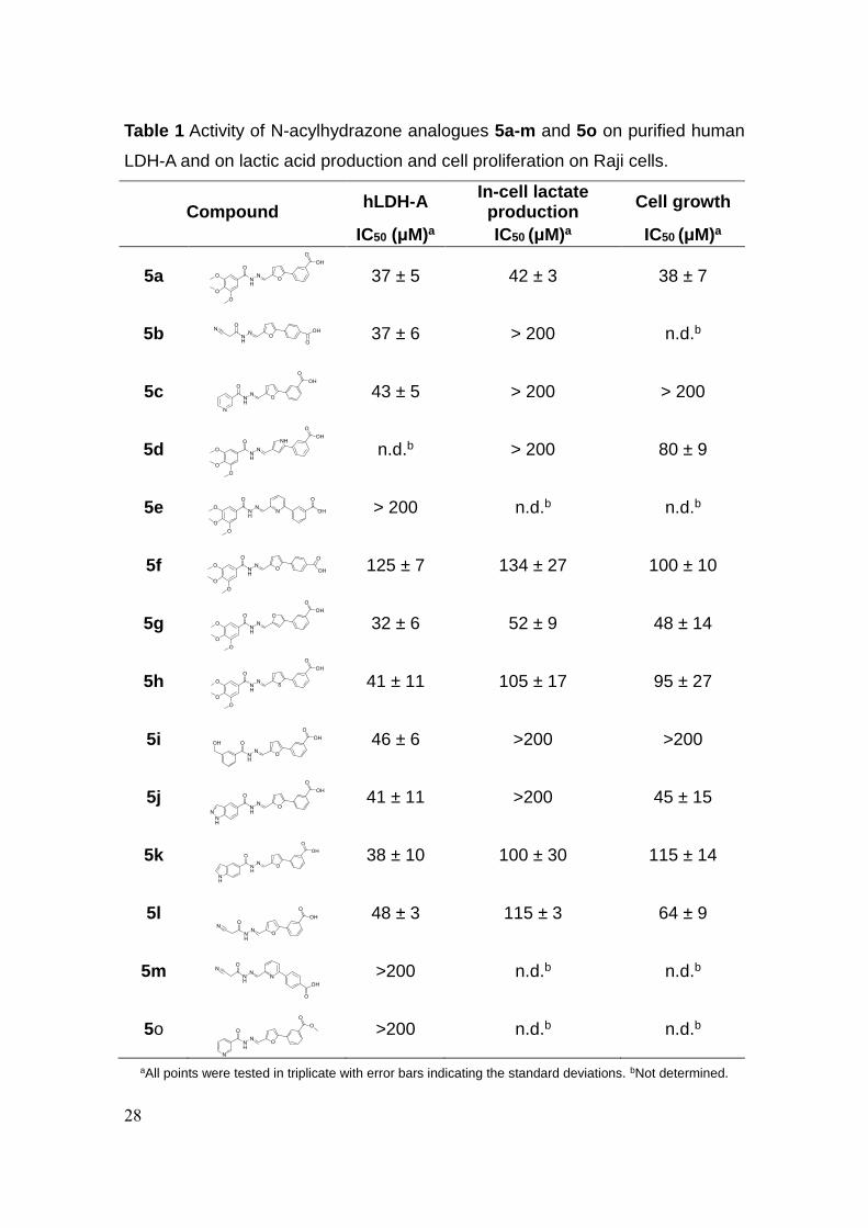
Table 1 Activity of N-acylhydrazone analogues 5a-m and 5o on purified human
LDH-A and on lactic acid production and cell proliferation on Raji cells.
Compound
hLDH-A In-cell lactate
production Cell growth
IC50 (μM)a IC50 (μM)a IC50 (μM)a
5a
37 ± 5 42 ± 3 38 ± 7
5b 37 ± 6 > 200 n.d.b
5c
43 ± 5 > 200 > 200
5d
n.d.b > 200 80 ± 9
5e
> 200 n.d.b n.d.b
5f
125 ± 7 134 ± 27 100 ± 10
5g
32 ± 6 52 ± 9 48 ± 14
5h
41 ± 11 105 ± 17 95 ± 27
5i
46 ± 6 >200 >200
5j
41 ± 11 >200 45 ± 15
5k
38 ± 10 100 ± 30 115 ± 14
5l
48 ± 3 115 ± 3 64 ± 9
5m
>200 n.d.b n.d.b
5o
>200 n.d.b n.d.b
aAll points were tested in triplicate with error bars indicating the standard deviations. bNot determined.
29
Compound 5a exerted a marked effect both on lactate production in cells and
on inhibiting purified LDH-A, thus showing a good capacity of cell penetration.
Moreover, 5a inhibited cell growth with an IC50 of 38 µM, whereas compound 5b
and 5c did not affect lactate production and cell growth.
On enzymatic assays, compounds 5i-k, in which the portion A is represented
respectively by 3-hydroxymethyl benzoyl, 5-1H indazolyl and 5-indolyl moieties,
maintained a comparable activity to the parent compound 5a. In terms of
binding mode, they preserved the interactions showed by compound 5a in the
substrate binding pocket, and reached the Asp51 in the cofactor cavity.
When portion B of 5a was replaced by 2,3 disubstituted furan or 2,5
disubstituted thiophene rings as in compounds 5g-h, once again no change in
the activity was observed, whereas the substitution with a 2,4 disubstituted
pyrrole ring present in 5d, made this compound too fluorescent to be analyzed
by fluorimetric method. The drop in potency after introduction of 2,6
disubstituted pyridine ring in compounds 5e and 5m suggested that 6-atom
rings may cause a different arrangement within the binding site.
Consistently with this hypothesis, the docking results showed that 5e and 5m
were oriented in a different way compared to the active compounds and fully
occupied the cofactor binding site without reaching the catalytic residues.
The modification of portion C gave conflicting results. Considering compound
5a, the shift of the carboxylic function from the m- to the p- position, as in
derivative 5f induced a decrease of activity. Differently, no relevant change in
the activity was observed between compound 5b showing a p-benzoic acid and
the analogue 5l bearing a m-benzoic acid. Finally, the esterification of
compounds 5a-b afforded insoluble derivatives 5n and 5p, while 5o, the methyl
ester of compound 5c, was inactive.
Despite some evident effects on the inhibitory activity, no clear SAR pattern
could be identified through the series described above. Considering the ability
of 5f-l to inhibit human LDH-A, we investigated their activity on lactic acid
production and cell proliferation on Raji cells. Compound 5g showed a cellular
activity comparable to the one of its parent 5a. On the contrary, the other
derivatives exhibited reduced activity in the inhibition of lactate production and
cell growth.
30
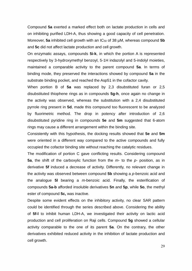
On the basis of these results, further studies were only performed on compound
5a. LDH-A enzymatic assays allowed to calculate the inhibition constants (Ki) vs
pyruvate (39 µM) and NADH (47 µM) (figure 11).
Further experiments were addressed at verifying the occurrence of biological
effects usually observed in cancer cells after LDH-A inhibition. These
experiments, which are summarized in figure 12, were performed on Raji cells
after 18h exposure to compound 5a at 40 μM. This dose was chosen on the
basis of the data reported on table 1 (50% inhibition on both lactate production
and cell growth). One of the main functions of LDH-A in cancer cells is to assure
the rapid reoxidation of NADH, needed to sustain the glycolytic process and
other biosynthetic pathways138. Figure 12A shows that, in agreement with
previous results obtained with other small molecule LDH-A inhibitors137,139,
treatment with compound 5a reduced NAD+ regeneration and caused a
statistically significant shifting of the redox balance in favor of the reduced form
of the dinucleotide. The extent of the observed NADH increase (+ 50%) fits well
with the effect caused by 40 μM compound 5a on lactate production in Raji cells
(50% inhibition).
Figure 11 Competition LDH-A assay of compound 5a versus pyruvate and NADH. The enzymatic reaction
was evaluated through the disappearance of NADH fluorescence, which is reported in the ordinate axis as ∆RFU/min

31
The effects caused by compound 5a on cell cycle phases distribution were
studied by propidium iodide staining of the treated cells and subsequent flow
cytometry analysis (figure 12B). This experiment showed that after an 18h
treatment, compound 5A caused a small but statistically significant decrease of
the cell fraction in S phase; on the contrary, cells in G2/M phase resulted
significantly increased. This latter result is in agreement with published data
obtained with oxamate140 and dichloroacetate141, a compound inhibiting
glycolysis and lactate production. A recent investigation concerning the
relationships between cell cycle progression and energy metabolism in cancer
cells showed that the ATP requirement for G1 and S phases is largely met by
accelerated glycolysis, while the energetic needs for G2/M phase are mainly
derived from mitochondrial oxidative phosphorylation. On the basis of the
published reports142, our flow cytometry data, indicating a reduction of the cell
population entering S phase and an increased fraction of G2/M cells, are
Figure 12 Experiments performed on Raji cell cultures treated with compound 5a. A: Evaluation of
NAD/NADH balance; B: Analysis of cell cycle phases by flow cytometry; C: Apoptosis evaluation; D: effect of compound 5a on cell viability of Raji cultures and normal lymphocytes

32
compatible with effects exerted at the glycolytic level and can be a further
evidence of the LDH-A inhibiting activity of compound 5a.
To assess whether the effect of compound 5a was not only limited to cell growth
arrest, we evaluated the expression of apoptosis markers (figure 12C). The
evaluated proteins (BAX and BCL2) are well studied regulators of the
mitochondrial apoptosis pathway143. Moreover, increased BAX levels were
already found in cancer cells treated with oxamate140 and other LDH
inhibitors144. As shown in figure 12C, an 18h treatment with compound 5a did
not substantially alter the level of BCL2 protein, but caused a 6.7-fold increase
in BAX expression, denoting the induction of cell death signaling. Interestingly,
contrary to oxamate, which was observed to trigger the mitochondrial apoptosis
pathway in cells after a prolonged period of exposition (48h)140, the effects
caused by compound 5a on Raji cultures appeared to occur at earlier time.
A further experiment with compound 5a was aimed at evaluating its potential in
combination tests with commonly used chemotherapeutic agents. Compound 5a
(40 μM) was tested on Raji cells in combination with four anticancer drugs
usually employed in the therapy of hematological neoplasms. For each drug, we
previously determined the lowest dose level causing statistically significant
effects on cell viability at 24h. This dose was subsequently tested in association
with compound 5a to calculate the combination index, according to the
procedure described in the experimental section. A result ranging from 0.8 to
1.2 is indicative of additive effects (table 2). As already observed in LDH
inhibition by oxamate or after LDH-A silencing, compound 5a showed the
potential of increasing the therapeutic efficacy of commonly used
chemotherapeutic agents.

33
Finally, we compared the effects on cell viability caused by compound 5a on
Raji cells and on normal lymphocytes, one of the cell populations more
susceptible to the adverse effects of anticancer chemotherapy. The results are
shown in figure 12C and they underline that no statistically significant effect
was found on normal cell viability even at the dose of 200 μM.
Although preliminary, all the obtained results were in support of the LDH
inhibitory effect of compound 5a; they also suggested a good tolerability of the
molecule on normal lymphocytes and its capability of improving the effects of
commonly administered chemotherapy.
Chemotherapeutic agent
Combination Indexa
Cisplatin 1.00 ± 0.02
Daunomycin 0.88 ± 0.15
Etoposide 0.86 ± 0.01
Sunitinib 1.08 ± 0.01
Table 2 Association compound 5a with
chemotherapeutic agents. aAssociation experiments were repeated twice. A result ranging from 0.8 to 1.2 is indicative of additive effects
34
2.3 Conclusion
In continuation of our research for innovative antitumor lead candidates, through
a VS campaign followed by SAR studies, we identified a new class of LDH-A
inhibitors in a series of N-acylhydrazone derivatives. The new molecules were
active at the micromolar range on purified LDH-A; notably 5a showed a marked
effect on lactate production in cells at the same concentration inhibiting purified
LDH-A. A more detailed characterization of its biological properties confirmed 5a
to be a suitable lead structure in the field of LDH-A inhibitors. Noteworthy, from
a medicinal chemistry point of view, the N-acylhydrazone scaffold is a privileged
structure, in which the biological relevance meets the synthetic accessibility,
allowing to rapidly obtain variously substituted analogues, making the follow-up
studies of the identified hits more efficient.

35
2.4 Experimental section
2.4.1 General Methods
Reaction progress was monitored by TLC on pre-coated silica gel plates
(Kieselgel 60 F254, Merck) and visualized by UV254 light. Flash column
chromatography was performed on silica gel (particle size 40-63 μM, Merck). If
required, solvents were distilled prior to use. All reagents were obtained from
commercial sources and used without further purification. When stated,
reactions were carried out under an inert atmosphere. Reactions involving
microwave irradiation were performed using a microwave synthesis system
(CEM Discover® SP, 2.45 GHz, maximum power 300 W), equipped with infrared
temperature measurement. Compounds were named relying on the naming
algorithm developed by CambridgeSoft Corporation and used in Chem-BioDraw
Ultra 15.0. 1H-NMR and 13C-NMR spectra were recorded on Varian Gemini at
400 MHz and 100 MHz respectively. Chemical shifts (δH) are reported relative to
TMS as internal standard.
2.4.2 Synthetic procedures
General procedure for the Suzuki coupling reaction to obtain 9a-g
To a solution of the appropriate bromo-substituted five or six membered
heterocyclic aldehydes 6a-e (1.0 mmol) in EtOH/H2O 5:3 (tot 12 mL) in a 35 mL
CEM microwave vessel, the correspondent carboxyphenyl boronic acids 7a-b
(1.2 mmol), Na2CO3 2M (2.0 mmol) and Pd(N,N-Dimethyl β-alaninate)2 (5
mol%) were added. The vessel was capped and placed in a microwave reactor
and the reaction carried out with the following method in dynamic mode: 120°C,
5 min, 100W, with high stirring. After completion the vessel was allowed to cool
to room temperature, HCl 2M was added until pH turned acidic, and the mixture
was extracted with EtOAc (3 X 10 mL). The organic phase was collected, dried
over anhydrous Na2SO4, and the solvent evaporated under vacuum. The crude
product was then purified via silica gel column chromatography (CH2Cl2/MeOH
elution gradient from a 100/0 ratio to a 90/10 ratio) to obtain the pure
compounds (yield 58-81%).
36
3-(5-formylfuran-2-yl)benzoic acid 9a
The product was prepared using the general procedure starting
from 6a and 7a (yield 70%). 1H NMR (DMSO-d6) δ 13.10 (bs, 1H), 9.66 (s, 1H),
8.24 (s, 1H), 8.06 (d, J = 8.4 Hz, 1H), 8.00 (d, J = 8.4 Hz, 1H), 7.88 (t, J = 8.4
Hz, 1H), 7.69 (d, J = 3.7 Hz, 1H), 7.45 (d, J = 3.7 Hz, 1H). 13C NMR (DMSO-d6)
δ 178.49, 167.11, 157.50, 152.30, 132.41, 130.61, 129.91, 129.55, 129.45,
125.75, 125.49, 109.87 ppm.
3-(4-formyl-1H-pyrrol-2-yl)benzoic acid 9b
The product was prepared using the general Suzuki coupling
procedure starting from 6b and 7a (pale yellow solid, yield 66%).
1H NMR (DMSO-d6) δ 13.76 (bs, 1H), 13.16 (s, 2H), 10.50 (s, 1H), 9.06 (s, 1H),
8.72 (d, J = 7.6 Hz, 1H), 8.60 (d, J = 7.6 Hz, 1H), 8.55 (s, 1H), 8.29 (d, J = 6.0
Hz, 1H), 7.74 (t, J = 7.6 Hz, 1H) ppm.
4-(5-formylfuran-2-yl)benzoic acid 9c
The product was prepared using the general Suzuki coupling
procedure starting from 6a and 7b (pale yellow solid, yield 75%). 1H NMR
(DMSO-d6) δ 13.13 (bs, 1H), 9.66 (s, 1H), 8.05 (d, J = 8.4 Hz, 2H), 8.00 (d, J =
8.4 Hz, 2H), 7.69 (d, J = 3.7 Hz, 1H), 7.45 (d, J = 3.7 Hz, 1H); 13C NMR (DMSO-
d6) δ 178.77, 167.15, 157.31, 152.06, 133.23, 130.56, 125.43, 124.77, 110.30
ppm.
3-(5-formylfuran-3-yl)benzoic acid 9d
The product was prepared using the general Suzuki coupling
procedure starting from 6c and 7a (pale yellow solid, yield 58%). 1H NMR
(DMSO-d6) δ 13.12 (bs, 1H), 9.66 (s, 1H), 8.74 (s, 1H), 8.22 (d, J = 13.9 Hz,
1H), 8.10 (s, 1H), 8.01 – 7.85 (m, 2H), 7.61 – 7.53 (m, 1H) ppm.
3-(5-formylthiophen-2-yl)benzoic acid 9e
The product was prepared using the general Suzuki coupling
procedure starting from 6d and 7a (pale yellow solid, yield 74%). 1H NMR
37
(DMSO-d6) δ 13.09 (bs, 1H), 9.94 (s, 1H), 8.26 (s, 1H), 8.07 (d, J = 3.7 Hz, 1H),
8.05 (d, J = 7.7 Hz, 1H), 7.99 (d, J = 7.7 Hz, 1H), 7.85 (d, J = 3.7 Hz, 1H), 7.63
(t, J = 7.7 Hz, 1H); 13C NMR (DMSO-d6) δ 184.58, 167.11, 151.64, 142.90,
139.59, 133.23, 132.37, 130.83, 130.50, 130.26, 126.97, 126.46 ppm.
3-(6-formylpyridin-2-yl)benzoic acid 9f
The product was prepared using the general Suzuki coupling
procedure starting from 6e and 7a (white solid, yield 74%). 1H NMR (DMSO-d6)
δ 13.18 (bs, 1H), 10.10 (s, 1H), 8.77 (s, 1H), 8.42 (d, J = 8.0 Hz, 1H), 8.36 (d, J
= 7.9 Hz, 1H), 8.17 (t, J = 8.0 Hz, 1H), 8.07 (d, J = 8.0 Hz, 1H), 7.94 (d, J = 7.9
Hz, 1H), 7.69 (t, J = 7.9 Hz, 1H); 13C NMR (DMSO-d6) δ 194.07, 167.57,
156.14, 152.71, 139.42, 138.26, 132.12, 131.37, 130.80, 129.71, 127.97,
125.34, 121.10 ppm.
4-(6-formylpyridin-2-yl)benzoic acid 9g
The product was prepared using the general Suzuki coupling
procedure starting from 6e and 7b (white solid, yield 81%).1H NMR (DMSO-d6)
δ 13.05 (bs, 1H), 10.06 (s, 1H), 8.35 (d, J = 7.8 Hz, 1H), 8.29 (d, J = 8.4 Hz,
2H), 8.16 (t, J = 7.8 Hz, 1H), 8.07 (d, J = 8.4 Hz, 2H), 8.02 (d, J = 7.8 Hz, 1H);
13C NMR (DMSO-d6) δ 194.03, 167.80, 155.99, 152.73, 141.73, 139.55,
133.24, 130.31, 129.66, 129.04, 127.33, 125.78, 121.37 ppm.
General procedure for the Suzuki coupling reaction to obtain 9h-i
To a solution of the appropriate bromo-substituted heterocyclic aldehydes 6a
(1.0 mmol) in EtOH/H20 5:3 (tot 12 mL) in a 35 mL CEM microwave vessel, the
correspondent boronic acids 7c-d (1.2 mmol), Na2CO3 2M (2.0 mmol) and
Pd(N,N-Dimethyl β-alaninate)2 (5 mol%) were added. The vessel was capped
and placed in a microwave reactor and the reaction carried out with the
following method in dynamic mode: 120°C, 10 min, 50W, with high stirring. After
completion the vessel was allowed to cool to room temperature and the mixture
was extracted with EtOAc (3 X 10 mL). The organic phase was collected, dried
over anhydrous Na2SO4, and the solvent evaporated under vacuum. The crude
product (containing a small portion of the ethyl ester as a transesterification
38
product) was then purified via silica gel column chromatography (petroleum
ether/EtOAc elution gradient from a 90/10 ratio to a 80/20 ratio) to obtain the
pure compounds (yield 73-77%).
Methyl 3-(5-formylfuran-2-yl)benzoate 9h
The product was prepared using the general Suzuki coupling
procedure starting from 6a and 7c (white solid, yield 77%). 1H NMR (CDCl3) δ
9.68 (s, 1H), 8.45 (s, 1H), 8.06 (t, J = 7.8 Hz, 1H), 8.02 (d, J = 7.8 Hz, 1H), 7.53
(t, J = 7.8 Hz, 1H), 7.34 (d, J = 3.7 Hz, 1H), 6.93 (d, J = 3.7 Hz, 1H), 3.96 (s,
3H) ppm.
Methyl 4-(5-formylfuran-2-yl)benzoate 9i
The product was prepared using the general procedure starting
from 6a and 7d (white solid, yield 73%). 1H NMR (CDCl3) δ 9.70 (s, 1H), 8.11
(d, J = 8.6 Hz, 2H), 7.89 (d, J = 8.6 Hz, 2H), 7.34 (d, J = 3.7 Hz, 1H), 6.96 (d, J
= 3.7 Hz, 1H), 3.94 (s, 3H); 13C NMR (DMSO-d6) δ 178.67, 166.06, 157.08,
152.69, 133.09, 130.42, 125.54, 125.40, 111.12, 52.56 ppm.
General procedure for the synthesis of hydrazides 10c-f
A solution of the appropriate ethyl or methyl esters 11a-d (1.0 mmol) in
methanol (5 mL) was prepared in a 10 mL CEM microwave vessel. Hydrazine
hydrate 50% (5.0 mmol) was added, the vessel was capped and placed in a
microwave reactor and the reaction carried out with the following method in
dynamic mode: 140°C, 60 min, 100W, with high stirring. After completion the
reaction mixture was transferred to a round bottom flask and the solvent
evaporated under reduced pressure. The crude product was transferred to an
Erlenmeyer flask and suspended in dichloromethane, heated at 50°C for 5
minutes, rapidly vacuum filtered and washed with the same solvent, to obtain
the pure product (yield 85-99%). When the product was found to be still not
pure, the purification procedure was repeated.
39
Nicotinic hydrazide 10c
The product was prepared using the general procedure for the
synthesis of hydrazides starting from ethyl ester 11a (white solid, yield 93%). 1H
NMR (DMSO-d6) δ 9.95 (s, 1H), 8.96 (d, J = 2.2 Hz, 1H), 8.69 (dd, J = 4.8, 1.6
Hz, 1H), 8.15 (ddd, J = 8.0, 2.2, 1.6 Hz, 1H), 7.49 (dd, J = 8.0, 4.8 Hz, 1H), 4.65
(bs, 2H) ppm.
3-(hydroxymethyl)benzohydrazide 10d
The product was prepared using the general procedure for the
synthesis of hydrazides starting from ethyl ester 11b (white solid, yield 99%). 1H
NMR (DMSO-d6) δ 9.73 (bs, 1H), 7.78 (s, 1H), 7.67 (d, J = 7.6 Hz, 1H), 7.45 (d,
J = 7.6 Hz, 1H), 7.38 (t, J = 7.6 Hz, 1H), 5.26 (t, J = 5.7 Hz, 1H), 4.54 (bs, 2H),
4.53 (d, J = 5.7 Hz, 2H) ppm.
1H-indazole-5-carbohydrazide 10e
The product was prepared using the general procedure for the
synthesis of hydrazides starting from ethyl ester 11c (white solid, 95% yield). 1H
NMR (DMSO-d6) δ 12.52 (s, 1H), 9.52 (s, 1H), 8.38 (s, 1H), 7.98 (s, 1H), 7.89
(d, J = 8.5 Hz, 1H), 7.40 (d, J = 8.5 Hz, 1H), 4.49 (bs, 2H) ppm.
1H-indole-5-carbohydrazide 10f
The product was prepared using the general procedure for the
synthesis of hydrazides starting from ethyl ester 11d (yield 85%). 1H NMR
(DMSO-d6) δ 11.28 (s, 1H), 9.54 (s, 1H), 8.08 (s, 1H), 7.59 (d, J = 8.4 Hz, 1H),
7.35-7.43 (m, 2H), 6.50 (s, 1H), 4.49 (bs, 2H) ppm.
Methyl 3-(hydroxymethyl)benzoate 11b
Mono-methyl isophthalate (2.0 mmol) was placed in a 50 mL round bottom flask
and dissolved in dry THF (10 mL) under nitrogen atmosphere. The reaction

40
flask was placed in an ice bath to reach 0°C and 1M borane tetrahydrofuran
complex solution (10.0 mmol) was added dropwise. After 15 minutes at 0°C the
ice bath was removed and the reaction was stirred at room temperature
overnight. After reaction completion ice was carefully added to the reaction, and
the mixture extracted three times with diethyl ether. The collected organic phase
was washed with brine, dried over anhydrous Na2SO4, and the solvent
evaporated in vacuum to obtain 11b (yield 97%) which needed no further
purification. 1H NMR (CDCl3) δ 8.04 (s, 1H), 7.97 (d, J = 7.8 Hz, 1H), 7.58 (d, J
= 7.8 Hz, 1H), 7.44 (t, J = 7.8 Hz, 1H), 4.76 (s, 2H), 3.92 (s, 3H) ppm.
Ethyl 1H-indazole-5-carboxylate 11c
1H-indazole-5-carboxylic acid (2.0 mmol) was placed in a 35 mL microwave
vessel and dissolved in ethanol (10 mL) under nitrogen atmosphere. The vessel
was placed in an ice bath to reach 0°C and thionyl chloride (10.0 mmol) was
added slowly while stirring. After 5 minutes the vessel was placed in a
microwave reactor and the reaction carried out with the following method in
dynamic mode: 130°C, 60 min, 80W. After completion the reaction mixture was
transferred to a round bottom flask, methanol was added to destroy remaining
thionyl chloride and the solvent evaporated under reduced pressure. The crude
product was dissolved in ethyl acetate and washed 3 times with 20% K2CO3.
The organic phase was dried over anhydrous Na2SO4, and the solvent
evaporated in vacuum to obtain 11c (yield 95%) which needed no further
purification. 1H NMR (CDCl3) δ 12.40 (s, 1H), 8.35 (s, 1H), 7.96 (s, 1H), 7.85 (d,
J = 8.9 Hz, 1H), 7.37 (d, J = 8.9 Hz, 1H), 4.23 (q, J = 7.2 Hz, 2H), 1.25 (t, J =
7.2 Hz, 3H) ppm.
41
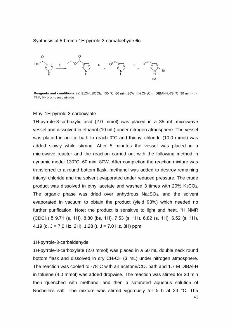
Synthesis of 5-bromo-1H-pyrrole-3-carbaldehyde 6c
Ethyl 1H-pyrrole-3-carboxylate
1H-pyrrole-3-carboxylic acid (2.0 mmol) was placed in a 35 mL microwave
vessel and dissolved in ethanol (10 mL) under nitrogen atmosphere. The vessel
was placed in an ice bath to reach 0°C and thionyl chloride (10.0 mmol) was
added slowly while stirring. After 5 minutes the vessel was placed in a
microwave reactor and the reaction carried out with the following method in
dynamic mode: 130°C, 60 min, 80W. After completion the reaction mixture was
transferred to a round bottom flask, methanol was added to destroy remaining
thionyl chloride and the solvent evaporated under reduced pressure. The crude
product was dissolved in ethyl acetate and washed 3 times with 20% K2CO3.
The organic phase was dried over anhydrous Na2SO4, and the solvent
evaporated in vacuum to obtain the product (yield 93%) which needed no
further purification. Note: the product is sensitive to light and heat. 1H NMR
(CDCl3) δ 9.71 (s, 1H), 8.80 (bs, 1H), 7.53 (s, 1H), 6.82 (s, 1H), 6.52 (s, 1H),
4.19 (q, J = 7.0 Hz, 2H), 1.28 (t, J = 7.0 Hz, 3H) ppm.
1H-pyrrole-3-carbaldehyde
1H-pyrrole-3-carboxylate (2.0 mmol) was placed in a 50 mL double neck round
bottom flask and dissolved in dry CH2Cl2 (3 mL) under nitrogen atmosphere.
The reaction was cooled to -78°C with an acetone/CO2 bath and 1.7 M DIBAl-H
in toluene (4.0 mmol) was added dropwise. The reaction was stirred for 30 min
then quenched with methanol and then a saturated aqueous solution of
Rochelle’s salt. The mixture was stirred vigorously for 5 h at 23 °C. The
42
aqueous layer was extracted with CH2Cl2 and the combined organic phase was
dried over anhydrous Na2SO4, and the solvent evaporated in vacuum to give
the crude alcohol. To a solution of crude alcohol in methanol (3 mL) was added
manganese (IV) oxide (20 mmol) and the reaction was stirred vigorously
overnight at room temperature. The mixture was then filtered through a pad of
Celite and the filtrate was concentrated in vacuum. The residue was then
purified via silica gel column chromatography (CH2Cl2/MeOH elution gradient
from a 100/0 ratio to a 98/2 ratio) to obtain the pure product (yield over the two
steps 13%). Note: the product is sensitive to light and heat. 1H NMR (CD3OD) δ
9.66 (s, 1H), 7.57 (s, 1H), 6.85 (s, 1H), 6.58 (s, 1H) ppm.
5-bromo-1H-pyrrole-3-carbaldehyde 6c
1H-pyrrole-3-carbaldehyde (1.3 mmol) was placed in a 50 mL double neck
round bottom flask and dissolved in dry THF (10 mL) under nitrogen
atmosphere. The reaction was cooled to -78°C with an acetone/CO2 bath and
stirred for 10 minutes. N-bromosuccinimide (1.3 mmol) was added, the reaction
was let reach -50°C and that temperature was kept for 10 hours with continuous
stirring. 2M NaOH was added to quench the reaction, followed by adding 3M
HCl until acidic pH was reached. The reaction was extracted 3 times with diethyl
ether, the organic phase was collected, dried over anhydrous Na2SO4, and the
solvent evaporated in vacuum. The residue was then purified via silica gel
column chromatography (petroleum ether/ethyl acetate elution gradient from a
100/0 ratio to a 70/30 ratio) to obtain the pure product (yield 45%). Note: the
product is sensitive to light and heat. 1H NMR (CDCl3) δ 9.68 (s, 1H), 8.72 (bs,
1H), 7.38 (s, 1H), 6.66 (s, 1H) ppm.
General procedure for the synthesis of N-acylhydrazones 5a-p
A solution of the appropriate aldehydes 9a-i (1.0 mmol) in ethanol (20 mL) was
prepared in a 35 mL CEM microwave vessel. The correspondent hydrazides
10a-f (1.0 mmol) were added, the vessel was capped and placed in a
microwave reactor and the reaction carried out with the following method in
dynamic mode: 120°C, 45 min, 130W. After completion the vessel was allowed
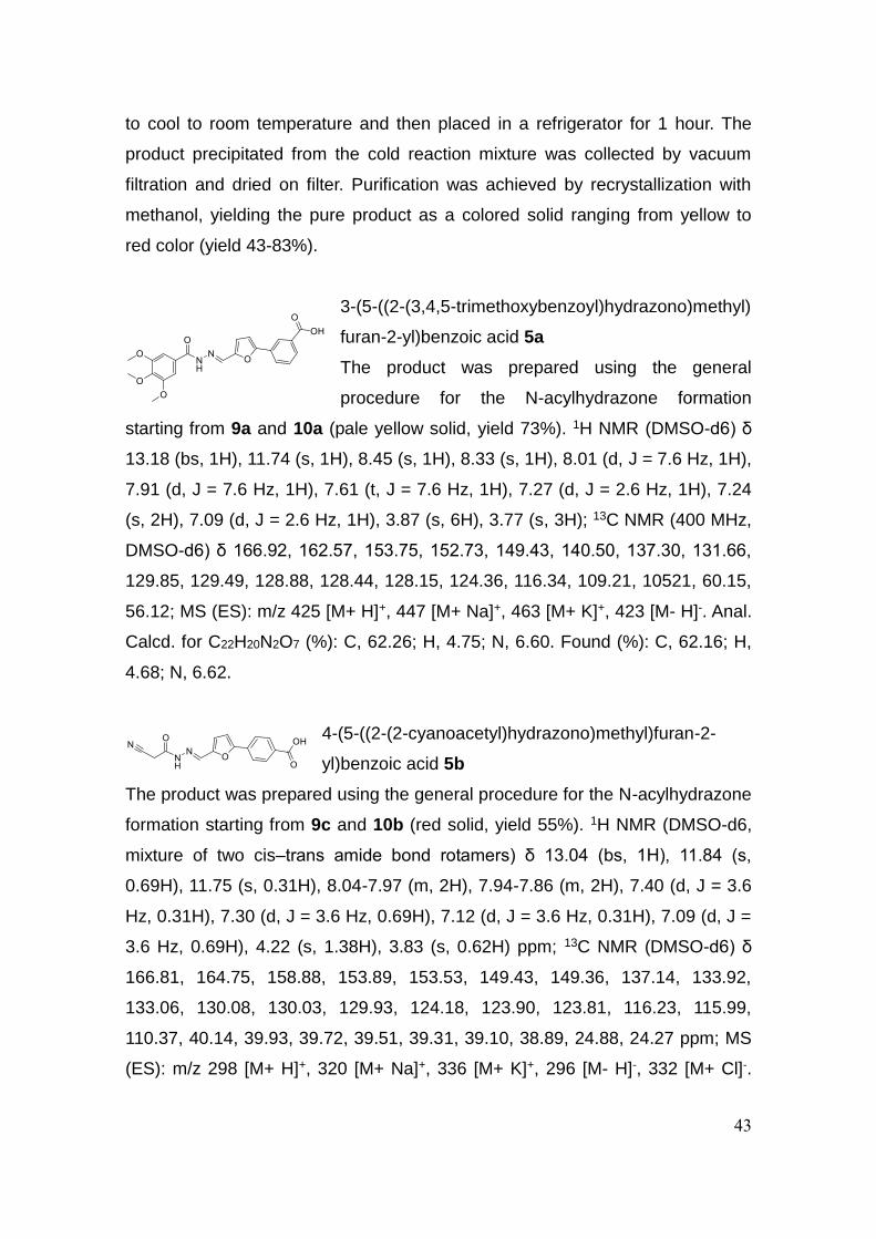
43
to cool to room temperature and then placed in a refrigerator for 1 hour. The
product precipitated from the cold reaction mixture was collected by vacuum
filtration and dried on filter. Purification was achieved by recrystallization with
methanol, yielding the pure product as a colored solid ranging from yellow to
red color (yield 43-83%).
3-(5-((2-(3,4,5-trimethoxybenzoyl)hydrazono)methyl)
furan-2-yl)benzoic acid 5a
The product was prepared using the general
procedure for the N-acylhydrazone formation
starting from 9a and 10a (pale yellow solid, yield 73%). 1H NMR (DMSO-d6) δ
13.18 (bs, 1H), 11.74 (s, 1H), 8.45 (s, 1H), 8.33 (s, 1H), 8.01 (d, J = 7.6 Hz, 1H),
7.91 (d, J = 7.6 Hz, 1H), 7.61 (t, J = 7.6 Hz, 1H), 7.27 (d, J = 2.6 Hz, 1H), 7.24
(s, 2H), 7.09 (d, J = 2.6 Hz, 1H), 3.87 (s, 6H), 3.77 (s, 3H); 13C NMR (400 MHz,
DMSO-d6) δ 166.92, 162.57, 153.75, 152.73, 149.43, 140.50, 137.30, 131.66,
129.85, 129.49, 128.88, 128.44, 128.15, 124.36, 116.34, 109.21, 10521, 60.15,
56.12; MS (ES): m/z 425 [M+ H]+, 447 [M+ Na]+, 463 [M+ K]+, 423 [M- H]-. Anal.
Calcd. for C22H20N2O7 (%): C, 62.26; H, 4.75; N, 6.60. Found (%): C, 62.16; H,
4.68; N, 6.62.
4-(5-((2-(2-cyanoacetyl)hydrazono)methyl)furan-2-
yl)benzoic acid 5b
The product was prepared using the general procedure for the N-acylhydrazone
formation starting from 9c and 10b (red solid, yield 55%). 1H NMR (DMSO-d6,
mixture of two cis–trans amide bond rotamers) δ 13.04 (bs, 1H), 11.84 (s,
0.69H), 11.75 (s, 0.31H), 8.04-7.97 (m, 2H), 7.94-7.86 (m, 2H), 7.40 (d, J = 3.6
Hz, 0.31H), 7.30 (d, J = 3.6 Hz, 0.69H), 7.12 (d, J = 3.6 Hz, 0.31H), 7.09 (d, J =
3.6 Hz, 0.69H), 4.22 (s, 1.38H), 3.83 (s, 0.62H) ppm; 13C NMR (DMSO-d6) δ
166.81, 164.75, 158.88, 153.89, 153.53, 149.43, 149.36, 137.14, 133.92,
133.06, 130.08, 130.03, 129.93, 124.18, 123.90, 123.81, 116.23, 115.99,
110.37, 40.14, 39.93, 39.72, 39.51, 39.31, 39.10, 38.89, 24.88, 24.27 ppm; MS
(ES): m/z 298 [M+ H]+, 320 [M+ Na]+, 336 [M+ K]+, 296 [M- H]-, 332 [M+ Cl]-.

44
Anal. Calcd. for C15H11N3O4 (%): C, 60.61; H, 3.73; N, 14.14. Found (%): C,
60.52; H, 3.60; N, 14.23.
3-(5-((2-nicotinoylhydrazono)methyl)furan-2-yl)benzoic
acid 5c
The product was prepared using the general procedure
for the N-acylhydrazone formation starting from 9a and 10c (pale yellow solid,
yield 60%). 1H NMR (DMSO-d6) δ 13.22 (bs, 1H), 12.03 (s, 1H), 9.07 (s, 1H),
8.78 (d, J = 4.2 Hz, 1H), 8.39 (s, 1H), 8.34 (s, 1H), 8.26 (d, J = 7.7 Hz, 1H), 8.05
(d, J = 7.7 Hz, 1H), 7.92 (d, J = 7.7 Hz, 1H), 7.66 – 7.54 (m, 2H), 7.28 (d, J =
3.3 Hz, 1H), 7.13 (d, J = 3.3 Hz, 1H) ppm; 13C NMR (DMSO-d6) δ 166.92,
161.67, 153.94, 152.36, 149.25, 148.54, 137.84, 135.45, 131.68, 129.81,
129.52, 129.13, 128.95, 128.23, 124.40, 123.68, 116.84, 109.25 ppm. Anal.
Calcd. for C18H13N3O4 (%): C, 64.48; H, 3.91; N, 12.53. Found (%): C, 64.55; H,
3.84; N, 12.67.
3-(4-((2-(3,4,5-
trimethoxybenzoyl)hydrazono)methyl)-1H-pyrrol-2-
yl)benzoic acid 5d
The product was prepared using the general
procedure for the N-acylhydrazone formation starting from 9b and 10a (gray
solid, yield 72%). 1H NMR (DMSO-d6) δ 13.03 (bs, 1H), 11.89 (s, 1H), 11.33 (s,
1H), 8.37 (s, 1H), 8.26 (s, 1H), 7.95 (d, J = 7.8 Hz, 1H), 7.78 (d, J = 7.8 Hz, 1H),
7.51 (t, J = 7.8 Hz, 1H), 7.35 (s, 1H), 7.22 (s, 2H), 6.94 (s, 1H), 3.86 (s, 6H),
3.72 (s, 3H) ppm; 13C NMR (DMSO-d6) δ 167.71, 162.38, 153.07, 144.76,
140.57, 132.86, 132.66, 131.90, 129.56, 129.48, 128.58, 127.42, 124.67,
123.85, 121.30, 110.01, 105.51, 103.96, 60.54, 56.53 ppm. Anal. Calcd. for
C22H21N3O6 (%): C, 62.41; H, 5.00; N, 9.92. Found (%): C, 62.29; H, 5.11; N,
9.80.
45
3-(6-((2-(3,4,5-
trimethoxybenzoyl)hydrazono)methyl)pyridin-2-
yl)benzoic acid 5e
The product was prepared using the general procedure for the N-acylhydrazone
formation starting from 9f and 10a (white solid, yield 66%). 1H NMR (DMSO-d6)
δ 13.12 (bs, 1H), 11.90 (s, 1H), 8.75 (s, 1H), 8.66 (s, 1H), 8.35 (d, J = 7.9 Hz,
1H), 8.09 (d, J = 7.9 Hz, 1H), 8.06 – 7.95 (m, 3H), 7.65 (t, J = 7.9 Hz, 1H), 7.28
(s, 2H), 3.88 (s, 6H), 3.74 (s, 3H) ppm; 13C NMR (DMSO-d6) δ 167.18, 154.92,
153.25, 152.76, 148.22, 139.38, 138.36, 138.31, 130.64, 130.00, 129.23,
128.20, 127.41, 120.88, 119.03, 110.70, 109.61, 105.32, 60.17, 56.16, 30.69
ppm. Anal. Calcd. for C23H21N3O6 (%): C, 63.44; H, 4.86; N, 9.65. Found (%): C,
63.32; H, 4.76; N, 9.60.
4-(5-((2-(3,4,5-
trimethoxybenzoyl)hydrazono)methyl)furan-2-
yl)benzoic acid 5f
The product was prepared using the general procedure for the N-acylhydrazone
formation starting from 9c and 10a (orange solid, yield 66%). 1H NMR (DMSO-
d6) δ 12.94 (bs, 1H), 11.74 (s, 1H), 8.45 (s, 1H), 8.03 (d, J = 8.1 Hz, 2H), 7.91
(d, J = 8.1 Hz, 2H), 7.33 (d, J = 2.3 Hz, 1H), 7.24 (s, 2H), 7.12 (d, J = 2.3 Hz,
1H), 3.87 (s, 6H), 3.74 (s, 3H) ppm; 13C NMR (DMSO-d6) δ 167.26, 162.99,
154.12, 153.15, 150.41, 140.99, 137.69, 133.59, 130.54, 130.33, 128.84,
124.27, 116.65, 110.95, 110.02, 105.66, 60.58, 56.56 ppm. Anal. Calcd. for
C22H20N2O7 (%): C, 62.26; H, 4.75; N, 6.60. Found (%): C, 62.31; H, 4.67; N,
6.73.
3-(5-((2-(3,4,5-
trimethoxybenzoyl)hydrazono)methyl)furan-3-
yl)benzoic acid 5g
The product was prepared using the general
procedure for the N-acylhydrazone formation starting from 9d and 10a (yellow
solid, yield 43%). 1H NMR (DMSO-d6) δ 13.98 (bs, 1H), 11.75 (s, 1H), 8.49 (s,
1H), 8.39 (s, 1H), 8.19 (s, 1H), 7.92 (d, J = 8.1 Hz, 1H), 7.87 (d, J = 8.1 Hz, 1H),

46
7.54 (t, J = 8.1 Hz, 1H), 7.49 (s, 1H), 7.24 (s, 2H), 3.87 (s, 6H), 3.73 (s, 3H)
ppm; 13C NMR (DMSO-d6) δ 167.34, 162.55, 152.76, 150.56, 141.96, 140.54,
137.19, 131.50, 129.79, 129.26, 128.38, 128.21, 127.15, 126.16, 111.84,
105.27, 60.19, 56.17 ppm. Anal. Calcd. for C22H20N2O7 (%): C, 62.26; H, 4.75;
N, 6.60. Found (%): C, 62.29; H, 4.80; N, 6.67.
3-(5-((2-(3,4,5-
trimethoxybenzoyl)hydrazono)methyl)thiophen-2-
yl)benzoic acid 5h
The product was prepared using the general
procedure for the N-acylhydrazone formation starting from 9e and 10a (pale
yellow solid, yield 68%). 1H NMR (DMSO-d6) δ 12.90 (bs, 1H), 11.74 (s, 1H),
8.68 (s, 1H), 8.22 (s, 1H), 7.99 (d, J = 7.5 Hz, 1H), 7.91 (d, J = 7.5 Hz, 1H), 7.66
(d, J = 3.2 Hz, 1H), 7.59 (t, J = 7.5 Hz, 1H), 7.51 (d, J = 3.2 Hz, 1H), 7.23 (s,
2H), 3.87 (s, 3H), 3.73 (s, 2H) ppm; 13C NMR (DMSO-d6) δ 166.89, 162.48,
152.74, 144.25, 142.68, 140.61, 138.97, 133.53, 132.16, 131.99, 129.72,
129.70, 128.96, 128.47, 125.96, 125.10, 105.34, 60.19, 56.19 ppm. Anal. Calcd.
for C22H20N2O6S (%): C, 59.99; H, 4.58; N, 6.36. Found (%): C, 60.04; H, 4.71;
N, 6.26.
3-(5-((2-(3-
(hydroxymethyl)benzoyl)hydrazono)methyl)furan-
2-yl)benzoic acid 5i
The product was prepared using the general procedure for the N-acylhydrazone
formation starting from 9a and 10d (yellow solid, yield 75%). 1H NMR (DMSO-
d6) δ 13.26 (bs, 1H), 11.91 (s, 1H), 8.67 (s, 1H), 8.42 (s, 1H), 8.35 (d, J = 11.8
Hz, 2H), 8.07 (dd, J = 15.0, 8.0 Hz, 2H), 7.62 (dd, J = 15.0, 8.0 Hz, 2H), 7.36 (d,
J = 2.9 Hz, 1H), 7.27 (t, J = 4.0 Hz, 1H), 7.09 (d, J = 2.9 Hz, 1H), 5.36 (t, J = 3.6
Hz, 1H), 4.59 (d, J = 3.6 Hz, 2H); 13C NMR (DMSO-d6) δ 172.56, 168.30,
163.63, 154.80, 149.65, 143.43, 137.73, 133.58, 130.18, 129.72, 129.37,
129.30, 128.68, 126.31, 126.04, 124.88, 116.66, 109.06, 62.98, 21.60 ppm.
47
Anal. Calcd. for C20H16N2O5 (%): C, 65.93; H, 4.43; N, 7.69. Found (%): C,
65.84; H, 4.34; N, 7.80.
3-(5-((2-(1H-indazole-5-
carbonyl)hydrazono)methyl)furan-2-yl)benzoic acid
5j
The product was prepared using the general procedure for the N-acylhydrazone
formation starting from 9a and 10e (dark yellow solid, yield 67%). 1H NMR
(DMSO-d6) δ 13.36 (bs, 1H), 13.26 (bs, 1H), 11.93 (s, 1H), 8.43 (s, 2H), 8.34 (s,
1H), 8.28 (s, 1H), 8.05 (d, J = 8.2 Hz, 1H), 7.91 (d, J = 8.0 Hz, 2H), 7.66 (d, J =
8.2 Hz, 1H), 7.61 (t, J = 8.2 Hz, 1H), 7.28 (d, J = 2.8 Hz, 1H), 7.08 (d, J = 2.8
Hz, 1H) ppm; 13C NMR (DMSO-d6) δ 166.94, 163.43, 153.61, 149.61, 141.10,
136.72, 134.89, 131.68, 129.89, 129.49, 128.83, 128.13, 125.64, 125.53,
124.32, 122.31, 121.22, 116.02, 110.20, 109.19 ppm. Anal. Calcd. for
C20H14N4O4 (%): C, 64.17; H, 3.77; N, 14.97. Found (%): C, 64.24; H, 3.72; N,
14.90.
3-(5-((2-(1H-indole-5-
carbonyl)hydrazono)methyl)furan-2-yl)benzoic acid
5k
The product was prepared using the general procedure for the N-acylhydrazone
formation starting from 9a and 10f (orange solid, yield 62%). 1H NMR (DMSO-
d6) δ 13.19 (bs, 1H), 11.79 (s, 1H), 11.41 (s, 1H), 8.44 (s, 1H), 8.34 (s, 1H),
8.22 (s, 1H), 8.05 (d, J = 7.7 Hz, 1H), 7.91 (d, J = 7.7 Hz, 1H), 7.70 (d, J = 8.1
Hz, 1H), 7.61 (t, J = 8.1 Hz, 1H), 7.51 (s, 1H), 7.49 – 7.46 (m, 1H), 7.26 (d, J =
4.1 Hz, 1H), 7.05 (d, J = 4.1 Hz, 1H), 6.59 (s, 1H) ppm; 13C NMR (DMSO-d6) δ
166.99, 164.16, 153.47, 149.79, 137.71, 136.16, 131.75, 129.92, 128.72,
128.19, 128.00, 127.03, 124.55, 124.29, 120.49, 120.33, 115.78, 110.95,
109.61, 109.06, 102.36, 102.14 ppm. Anal. Calcd. for C21H15N3O4 (%): C, 67.56;
H, 4.05; N, 11.25. Found (%): C, 67.61; H, 4.13; N, 11.16.
48
3-(5-((2-(2-cyanoacetyl)hydrazono)methyl)furan-2-
yl)benzoic acid 5l
The product was prepared using the general procedure
for the N-acylhydrazone formation starting from 9a and 10b (orange solid, yield
74%). 1H NMR (DMSO-d6, mixture of two cis–trans amide bond rotamers) δ
13.12 (bs, 1H), 11.81 (s, 0.63H), 11.72 (s, 0.37H), 8.27 (s, 1H), 8.01 (d, J = 7.9
Hz, 1H), 7.91 (s, 1H), 7.89 (d, J = 7.9 Hz, 1H), 7.58 (t, J = 7.9 Hz, 1H), 7.24 (d,
J = 3.3 Hz, 1H), 7.08 (d, J = 3.3 Hz, 0.37H), 7.05 (d, J = 3.3 Hz, 0.63H), 4.20 (s,
1.26H), 3.81 (s, 0.74H) ppm; 13C NMR (DMSO-d6) δ 166.92, 164.71, 153.71,
148.91, 137.29, 134.22, 131.67, 129.79, 129.53, 128.95, 128.12, 124.40,
116.23, 116.00, 109.18, 24.89, 24.28 ppm. Anal. Calcd. for C15H11N3O4 (%): C,
60.61; H, 3.73; N, 14.14. Found (%): C, 60.49; H, 3.82; N, 14.03.
4-(6-((2-(2-cyanoacetyl)hydrazono)methyl)pyridin-2-
yl)benzoic acid 5m
The product was prepared using the general
procedure for the N-acylhydrazone formation starting from 9g and 10b (white
solid, yield 83%). 1H NMR (DMSO-d6, mixture of two cis–trans amide bond
rotamers) δ 13.08 (bs, 1H), 12.10 (s, 0.77H), 11.99 (s, 0.23H), 8.26 (s, 1H), 8.23
(d, J = 7.9 Hz, 2H), 8.09 (m, 3H), 8.01 (d, J = 7.9 Hz, 2H), 4.30 (s, 1.54H), 3.89
(s, 0.46H) ppm; 13C NMR (DMSO-d6) δ 167.49, 165.64, 155.24, 153.22,
145.09, 142.30, 138.60, 131.72, 130.25, 127.14, 121.92, 119.72, 116.37,
109.99, 24.75 ppm. Anal. Calcd. for C16H12N4O3 (%): C, 62.33; H, 3.92; N,
18.17. Found (%): C, 62.44; H, 3.87; N, 18.20.
Methyl 3-(5-((2-(3,4,5-
trimethoxybenzoyl)hydrazono)methyl)furan-2-
yl)benzoate 5n
The product was prepared using the general
procedure for the N-acylhydrazone formation starting from 9h and 10a (pale
yellow solid, yield 68%). 1H NMR (DMSO-d6) δ 11.72 (s, 1H), 8.46 (s, 1H), 8.33
(s, 1H), 8.08 (d, J = 7.6 Hz, 1H), 7.94 (d, J = 7.6 Hz, 1H), 7.64 (t, J = 7.8 Hz,

49
1H), 7.30 (d, J = 3.3 Hz, 1H), 7.24 (s, 2H), 7.10 (d, J = 2.4 Hz, 1H), 3.92 (s, 3H),
3.87 (s, 6H), 3.74 (s, 3H) ppm; 13C NMR (DMSO-d6) δ 165.81, 159.99, 152.70,
149.49, 137.32, 131.28, 130.48, 129.97, 129.66, 128.69, 127.61, 123.96,
120.24, 116.34, 110.98, 109.41, 106.37, 105.21, 60.12, 56.90, 56.11, 55.61,
52.34 ppm. Anal. Calcd. for C23H22N2O7 (%): C, 63.01; H, 5.06; N, 6.39. Found
(%): C, 63.12; H, 5.00; N, 6.42.
Methyl 3-(5-((2-nicotinoylhydrazono)methyl)furan-2-
yl)benzoate 5o
The product was prepared using the general procedure
for the N-acylhydrazone formation starting from 9h and 10c (white solid, yield
48%). 1H NMR (DMSO-d6) δ 12.03 (s, 1H), 9.07 (s, 1H), 8.78 (d, J = 6.0 Hz,
1H), 8.40 (s, 1H), 8.33 (s, 1H), 8.26 (d, J = 7.8 Hz, 1H), 8.09 (d, J = 7.8 Hz, 1H),
7.94 (d, J = 8.2 Hz, 1H), 7.65 (t, J = 8.2 Hz, 1H), 7.61 – 7.55 (m, 1H), 7.31 (d, J
= 3.5 Hz, 1H), 7.14 (d, J = 3.5 Hz, 1H), 3.92 (s, 3H) ppm; 13C NMR (DMSO-d6)
δ 165.55, 164.23, 161.56, 153.82, 152.26, 149.29, 148.50, 137.89, 135.45,
130.50, 129.82, 129.04, 128.55, 128.48, 124.02, 123.72, 117.08, 109.57, 30.66
ppm. Anal. Calcd. for C19H15N3O4 (%): C, 65.32; H, 4.33; N, 12.03. Found (%):
C, 65.38; H, 4.45; N, 11.97.
Methyl 4-(5-((2-(2-
cyanoacetyl)hydrazono)methyl)furan-2-yl)benzoate
5p
The product was prepared using the general procedure for the N-acylhydrazone
formation starting from 9i and 10b (orange solid, yield 53%). 1H NMR (DMSO-
d6) δ 11.89 (s, 1H), 8.42 (s, 1H), 8.05 (d, J = 7.7 Hz, 2H), 7.95 (d, J = 7.7 Hz,
2H), 7.35 (d, J = 3.8 Hz, 1H), 7.15 (d, J = 3.8 Hz, 1H), 3.89 (s, 3H), 3.87 (s, 2H)
ppm; 13C NMR (DMSO-d6) δ 178.28, 165.73, 165.67, 156.58, 149.85, 133.43,
132.65, 129.98, 125.11, 124.83, 124.03, 111.93, 110.75, 109.54, 52.27, 30.63
ppm. Anal. Calcd. for C16H13N3O4 (%): C, 61.73; H, 4.21; N, 13.50. Found (%):
C, 61.65; H, 4.26; N, 13.56.

50
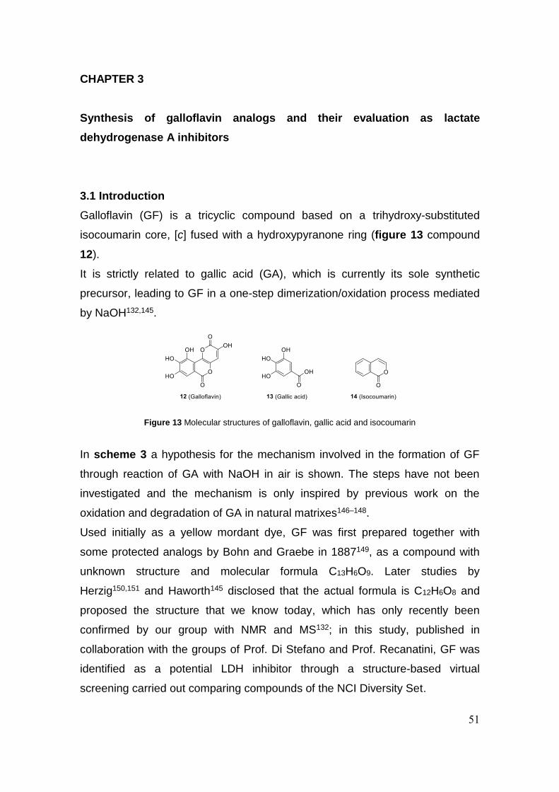
51
CHAPTER 3
Synthesis of galloflavin analogs and their evaluation as lactate
dehydrogenase A inhibitors
3.1 Introduction
Galloflavin (GF) is a tricyclic compound based on a trihydroxy-substituted
isocoumarin core, [c] fused with a hydroxypyranone ring (figure 13 compound
12).
It is strictly related to gallic acid (GA), which is currently its sole synthetic
precursor, leading to GF in a one-step dimerization/oxidation process mediated
by NaOH132,145.
In scheme 3 a hypothesis for the mechanism involved in the formation of GF
through reaction of GA with NaOH in air is shown. The steps have not been
investigated and the mechanism is only inspired by previous work on the
oxidation and degradation of GA in natural matrixes146–148.
Used initially as a yellow mordant dye, GF was first prepared together with
some protected analogs by Bohn and Graebe in 1887149, as a compound with
unknown structure and molecular formula C13H6O9. Later studies by
Herzig150,151 and Haworth145 disclosed that the actual formula is C12H6O8 and
proposed the structure that we know today, which has only recently been
confirmed by our group with NMR and MS132; in this study, published in
collaboration with the groups of Prof. Di Stefano and Prof. Recanatini, GF was
identified as a potential LDH inhibitor through a structure-based virtual
screening carried out comparing compounds of the NCI Diversity Set.
Figure 13 Molecular structures of galloflavin, gallic acid and isocoumarin

52
After an initial evaluation on the human LDH-A isolated enzyme to assess
inhibition of enzyme activity, GF was tested on different cancer cell lines. It was
found to inhibit both the enzymatic activity and lactate production in cancer cells
proving good cell permeability132.
Since then, GF has been considered as a reference compound among the
anticancer glycolytic inhibitors, and is now produced and sold by Sigma-Aldrich.
Several studies focusing on the discovery of new LDH inhibitors used the
comparison with GF as a prove for efficacy152,153. Other investigations focused
on its activity on different cancer cell lines and its mechanism of action on a
molecular level134,137,144,154–156. Notably, a 2D in-cell NMR metabolomics study
recently revealed that not only GF inhibits LDH, but it could exert a multi-target
effect including influence on PDK, ALT1 and PDH, thus leading the authors to
suggest that GF should be also defined as a “functional inhibitor” of PDK and
ALT1 and a “functional activator” of PDH157.
Scheme 3 Hypothesis of GF formation mechanism through dimerization/oxidation of gallic acid. Some
steps are simplified for clarity and proton transfers are arbitrary
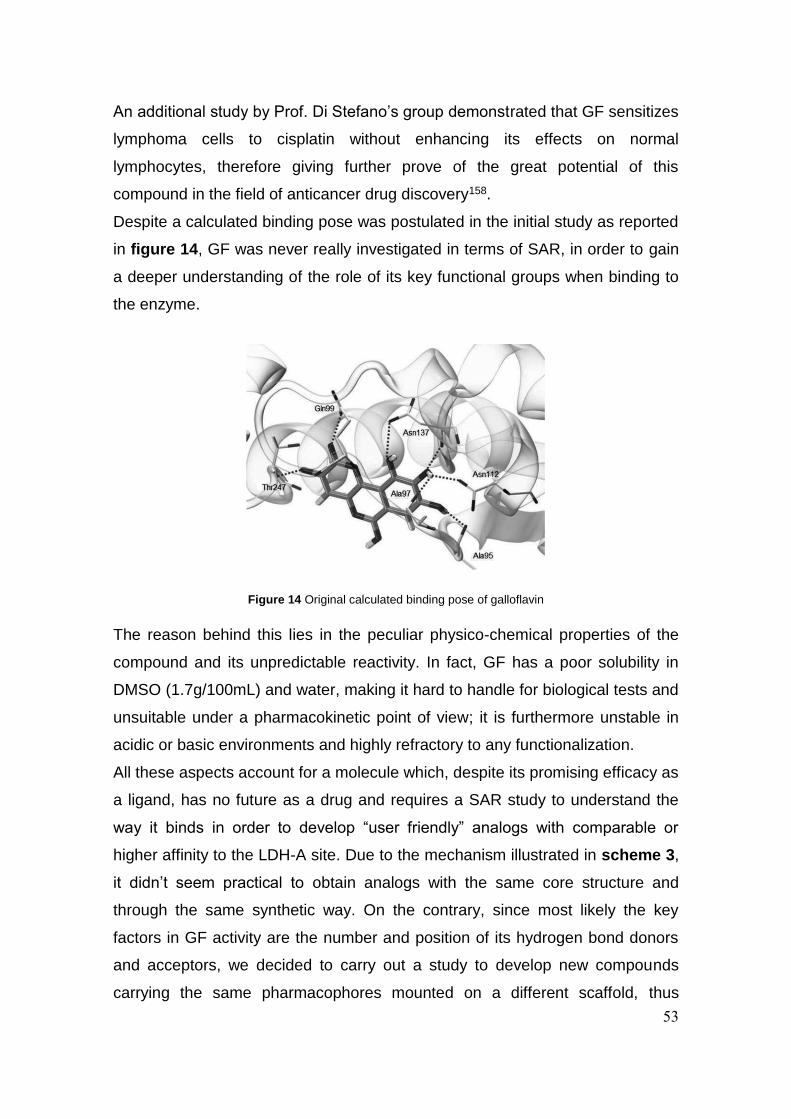
53
An additional study by Prof. Di Stefano’s group demonstrated that GF sensitizes
lymphoma cells to cisplatin without enhancing its effects on normal
lymphocytes, therefore giving further prove of the great potential of this
compound in the field of anticancer drug discovery158.
Despite a calculated binding pose was postulated in the initial study as reported
in figure 14, GF was never really investigated in terms of SAR, in order to gain
a deeper understanding of the role of its key functional groups when binding to
the enzyme.
The reason behind this lies in the peculiar physico-chemical properties of the
compound and its unpredictable reactivity. In fact, GF has a poor solubility in
DMSO (1.7g/100mL) and water, making it hard to handle for biological tests and
unsuitable under a pharmacokinetic point of view; it is furthermore unstable in
acidic or basic environments and highly refractory to any functionalization.
All these aspects account for a molecule which, despite its promising efficacy as
a ligand, has no future as a drug and requires a SAR study to understand the
way it binds in order to develop “user friendly” analogs with comparable or
higher affinity to the LDH-A site. Due to the mechanism illustrated in scheme 3,
it didn’t seem practical to obtain analogs with the same core structure and
through the same synthetic way. On the contrary, since most likely the key
factors in GF activity are the number and position of its hydrogen bond donors
and acceptors, we decided to carry out a study to develop new compounds
carrying the same pharmacophores mounted on a different scaffold, thus
Figure 14 Original calculated binding pose of galloflavin

54
creating a model system with synthetic accessibility and improved physico-
chemical properties which could help us explore the structure-activity
relationship of galloflavin without having to deal with its major drawbacks.
We therefore begun a process of structural simplification and rationalization of
galloflavin, analyzing its features and trying to reduce to the minimum its main
elements of synthetic and structural complexity.
One of the key factors driving the design of the new inhibitors was the presence
on GF of an unusual fusion of two 2-pyrone rings (red portion in figure 15); this
element was believed to bring unnecessary complexity and pose significant
synthetic challenges without being crucial for the compound’s activity. The
carbonyl oxygen of the lactone on the 2 position was originally calculated to be
involved in a H-bond in the 2012 paper132, but its actual contribution was still
uncertain so it was considered nonessential to the purposes of our research.
Specific attention was posed on the need to keep the same position and
distance for the 4 OH groups, to reproduce to the best the H-bond fingerprint of
GF. A tricyclic system was not intrinsically necessary but it proved to be useful
in mimicking the original structure, together with the lactone on the 6 position,
which emerges directly from the gallic acid building block, and also allows for
the introduction of the typical trihydroxy substitution pattern on the A ring.
Finally, an effort was done in making the synthesis fast and modular, in order to
establish a well-tested path to the future synthesis of analogs by simply
employing different commercial building blocks.
As a results of the considerations summarized above, a series of novel
compounds was identified, and a synthetic study was carried out to obtain the
Figure 15 Illustration of the main concepts driving the structural simplification of galloflavin
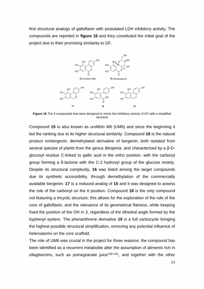
55
first structural analogs of galloflavin with postulated LDH inhibitory activity. The
compounds are reported in figure 16 and they constituted the initial goal of the
project due to their promising similarity to GF.
Compound 15 is also known as urolithin M6 (UM6) and since the beginning it
led the ranking due to its higher structural similarity. Compound 16 is the natural
product norbergenin, demethylated derivative of bergenin, both isolated from
several species of plants from the genus Bergenia, and characterized by a β-D-
glucosyl residue C-linked to gallic acid in the ortho position, with the carboxyl
group forming a δ-lactone with the C-2 hydroxyl group of the glucose moiety.
Despite its structural complexity, 16 was listed among the target compounds
due its synthetic accessibility, through demethylation of the commercially
available bergenin. 17 is a reduced analog of 15 and it was designed to assess
the role of the carbonyl on the 6 position. Compound 18 is the only compound
not featuring a tricyclic structure; this allows for the exploration of the role of the
core of galloflavin, and the relevance of its geometrical flatness, while keeping
fixed the position of the OH in 3, regardless of the dihedral angle formed by the
byphenyl system. The phenanthrene derivative 19 is a full carbocycle bringing
the highest possible structural simplification, removing any potential influence of
heteroatoms on the core scaffold.
The role of UM6 was crucial in the project for three reasons: the compound has
been identified as a recurrent metabolite after the assumption of aliments rich in
ellagitannins, such as pomegranate juice159–161, and together with the other
Figure 16 The 5 compounds that were designed to mimic the inhibitory activity of GF with a simplified structure

56
urolithins (figure 17) it has been attributed a number of beneficial properties
and biological activities162–165, but despite its relevance a reproducible synthetic
approach was never published; the origin of urolithins and their common
presence in gut microbiota suggested a promising safety profile; the structural
similarity with galloflavin is high and it represents a useful tool to explore the
substitution pattern on the rings affecting minimally the influence of the scaffold.
Other urolithins have been synthesized before and for some of them fast and
effective strategies are available in literature. Synthetic access is important for
this class of compounds since they are needed as standards for their analytical
determination.
Usual routes to the bezo[c]chromenone scaffold shared by all urolithins can be
summarized into three main strategies: 1) copper catalyzed Hurtley reaction
between an ortho- brominated benzylic acid derivative and resorcinol166; 2)
basic decarboxylative hydrolysis of ellagic acid (EA)167; 3) inverse electron
demand Diels–Alder between a coumarin-derived diene and the enamine
resulting from the condensation of dimethoxyacetaldehyde and pyrrolidine168.
Figure 17 Structures and classification of some of the most common urolithins
that are detectable in human microbiota after ingestion of ellagitannin-rich foods
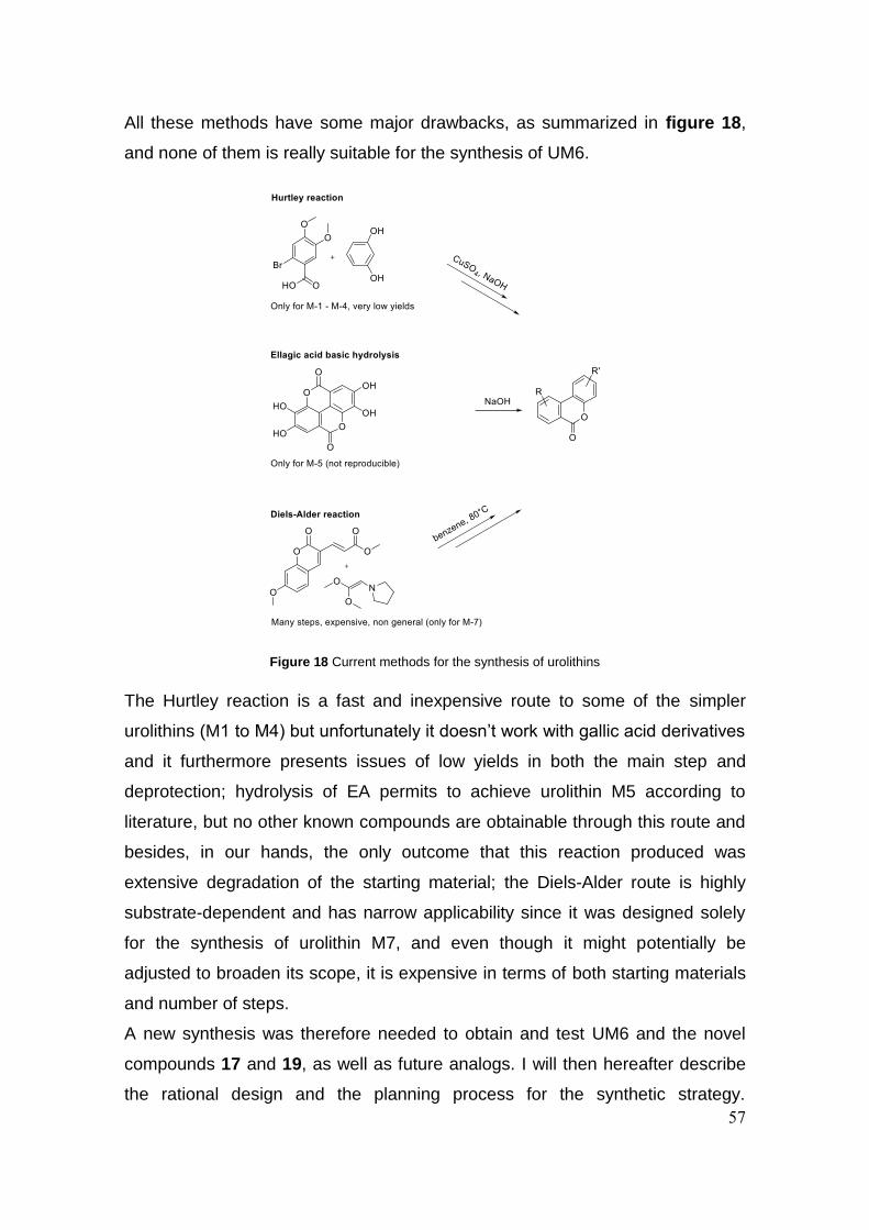
57
All these methods have some major drawbacks, as summarized in figure 18,
and none of them is really suitable for the synthesis of UM6.
The Hurtley reaction is a fast and inexpensive route to some of the simpler
urolithins (M1 to M4) but unfortunately it doesn’t work with gallic acid derivatives
and it furthermore presents issues of low yields in both the main step and
deprotection; hydrolysis of EA permits to achieve urolithin M5 according to
literature, but no other known compounds are obtainable through this route and
besides, in our hands, the only outcome that this reaction produced was
extensive degradation of the starting material; the Diels-Alder route is highly
substrate-dependent and has narrow applicability since it was designed solely
for the synthesis of urolithin M7, and even though it might potentially be
adjusted to broaden its scope, it is expensive in terms of both starting materials
and number of steps.
A new synthesis was therefore needed to obtain and test UM6 and the novel
compounds 17 and 19, as well as future analogs. I will then hereafter describe
the rational design and the planning process for the synthetic strategy.
Figure 18 Current methods for the synthesis of urolithins

58
Complementing the chemical synthesis, the collaboration of Prof. Di Stefano
and collaborators once again granted the obtainment of the necessary
biological data.

59
3.2 Results and discussion
The synthetic strategies devised in the early phase were based on two
disconnection patterns which lead to the same starting material through
different intermediates.
It seemed clear since the beginning that GA would be an ideal starting point
since the evident presence of its molecular outline inside the structure of UM6.
Route A (scheme 4) starts with the disconnection of the O-aryl bond in the
lactone ring which can be formed through a C-H oxygenation, the biphenyl
intermediate is then obtained through a Suzuki coupling between the
appropriate boronic acid and a halogenated GA analog, which in turn is formed
by simple halogenation of GA.
Route B is a logical inversion of the first two disconnections of route A, with the
aryl-aryl bond being formed through a Heck coupling and the ester moiety being
obtained by esterification between a resorcinol derivative and the halogenated
GA.
The presence of several OH moieties required the use of suitable protecting
groups, which were widely screened on both routes to optimize bulkiness,
stability of the PG and reactivity of the substrates in the key steps.
The forward synthesis planned for route A is reported in scheme 5 and it
features the use of a methyl ester and OH-protected GA derivative which is
brominated, coupled with the OH-protected para-arylboronic acid and in turn
hydrolyzed with a Brønsted base to give the free acid which is transformed into
Scheme 4 Two hypothesis of retrosynthesis for urolithin M6
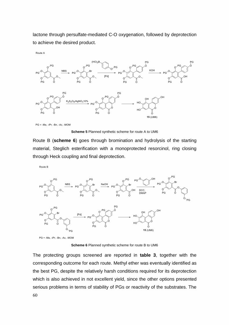
60
lactone through persulfate-mediated C-O oxygenation, followed by deprotection
to achieve the desired product.
Route B (scheme 6) goes through bromination and hydrolysis of the starting
material, Steglich esterification with a monoprotected resorcinol, ring closing
through Heck coupling and final deprotection.
The protecting groups screened are reported in table 3, together with the
corresponding outcome for each route. Methyl ether was eventually identified as
the best PG, despite the relatively harsh conditions required for its deprotection
which is also achieved in not excellent yield, since the other options presented
serious problems in terms of stability of PGs or reactivity of the substrates. The
Scheme 5 Planned synthetic scheme for route A to UM6
Scheme 6 Planned synthetic scheme for route B to UM6
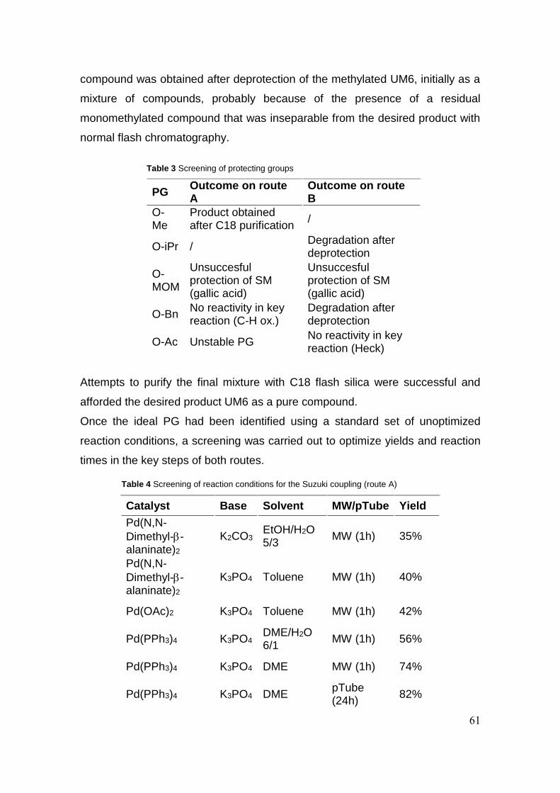
61
compound was obtained after deprotection of the methylated UM6, initially as a
mixture of compounds, probably because of the presence of a residual
monomethylated compound that was inseparable from the desired product with
normal flash chromatography.
PG Outcome on route A
Outcome on route B
O-Me
Product obtained after C18 purification
/
O-iPr / Degradation after deprotection
O-MOM
Unsuccesful protection of SM (gallic acid)
Unsuccesful protection of SM (gallic acid)
O-Bn No reactivity in key reaction (C-H ox.)
Degradation after deprotection
O-Ac Unstable PG No reactivity in key reaction (Heck)
Attempts to purify the final mixture with C18 flash silica were successful and
afforded the desired product UM6 as a pure compound.
Once the ideal PG had been identified using a standard set of unoptimized
reaction conditions, a screening was carried out to optimize yields and reaction
times in the key steps of both routes.
Catalyst Base Solvent MW/pTube Yield
Pd(N,N-
Dimethyl--alaninate)2
K2CO3 EtOH/H2O 5/3
MW (1h) 35%
Pd(N,N-
Dimethyl--alaninate)2
K3PO4 Toluene MW (1h) 40%
Pd(OAc)2 K3PO4 Toluene MW (1h) 42%
Pd(PPh3)4 K3PO4 DME/H2O 6/1
MW (1h) 56%
Pd(PPh3)4 K3PO4 DME MW (1h) 74%
Pd(PPh3)4 K3PO4 DME pTube (24h)
82%
Table 4 Screening of reaction conditions for the Suzuki coupling (route A)
Table 3 Screening of protecting groups
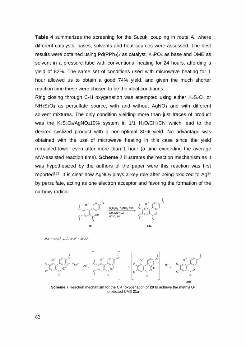
62
Table 4 summarizes the screening for the Suzuki coupling in route A, where
different catalysts, bases, solvents and heat sources were assessed. The best
results were obtained using Pd(PPh3)4 as catalyst, K3PO4 as base and DME as
solvent in a pressure tube with conventional heating for 24 hours, affording a
yield of 82%. The same set of conditions used with microwave heating for 1
hour allowed us to obtain a good 74% yield, and given the much shorter
reaction time these were chosen to be the ideal conditions.
Ring closing through C-H oxygenation was attempted using either K2S2O8 or
NH4S2O8 as persulfate source, with and without AgNO3 and with different
solvent mixtures. The only condition yielding more than just traces of product
was the K2S2O8/AgNO310% system in 1/1 H2O/CH3CN which lead to the
desired cyclized product with a non-optimal 30% yield. No advantage was
obtained with the use of microwave heating in this case since the yield
remained lower even after more than 1 hour (a time exceeding the average
MW-assisted reaction time). Scheme 7 illustrates the reaction mechanism as it
was hypothesized by the authors of the paper were this reaction was first
reported169. It is clear how AgNO3 plays a key role after being oxidized to Ag2+
by persulfate, acting as one electron acceptor and favoring the formation of the
carboxy radical.
Scheme 7 Reaction mechanism for the C-H oxygenation of 20 to achieve the methyl O-protected UM6 21a

63
The only step that needed optimization in route B was the lactone ring formation
through intramolecular Heck coupling. A rapid screening (table 5) revealed that
regardless of the changes made to the system the reactivity was intrinsically
lower than the Suzuki reaction in route A, never exceeding 35%.
After careful consideration of several aspects, the route that was chosen as the
most convenient was route A. In fact, despite the low yield in the C-H
oxygenation step, it was judged that the fast reaction time obtained in the
Suzuki step and the promising variability of future analogs brought by the use of
boronic acids as building blocks would widely justify the choice of this route as
by far the best one to obtain UM6 and its analogs.
The final optimized synthesis is reported in scheme 8 and in addition to the
steps already described it features an initial NBS-mediated bromination, the
hydrolysis of 25a by KOH and the final deprotection using BBr3 which leads to
Catalyst Base Solvent Yield
Pd(OAc)2/PPh3 1/4
NaOAc DMF 35%
Pd(OAc)2/PPh3 1/4
NaOAc DMA 9%
Pd(PPh3)2Cl2 NaOPiv DMA 14%
Table 5 Screening of reaction conditions for the
intramolecular Heck coupling (route B)
Scheme 8 Optimized synthesis of UM6

64
UM6 in an overall of 5 steps starting from commercially available compound
22a. Key to the development of the other structural analogs of GF (17, 18 and
19) was compound 25a. This molecule is central in the synthesis of UM6 and
can be obtained fast and in good yield, and it played the role of a nodal
commodity being incorporated into the synthesis of all the other compounds,
therefore building a divergent and versatile route to up to 4 different structures
all arising from one starting material.
Phenanthrene derivative 19 was initially planned to be obtained through
photocyclization of the corresponding stilbene170 (a standard approach to this
kind of structures), but following the optimization of UM6 synthesis 25a was
identified as a suitable precursor to an alternative bismuth-catalyzed synthesis
of phenanthrenes, leading to the desired arylvinyl intermediate in only 3
steps171.
The synthesis of 19 is reported in scheme 9. After obtaining 25a the compound
is converted to the corresponding aldehyde 26 and reacted in a Wittig fashion
with (methoxymethyl)triphenylphosphonium chloride to afford the corresponding
arylvinyl methyl ether 27. Bismuth-catalyzed cyclization-aromatization leads
almost instantaneously to the protected phenanthrene 28 which is finally
deprotected to obtain 19.
The open analog 18 can be obtained by hydrolysis of 25a leading to the
carboxilic acid 20 that is in turn deprotected by BBr3. The benzochromene
Scheme 9 Synthesis of the phenanthrene-based analog 19

65
derivative 17 can be obtained after NaBH4/BF3·Et2O-mediated reduction of
UM6. The synthesis of this compound has not yet been completed.
The complete divergent approach to the synthesis of the 4 novel analogs of GF
15, 17, 18 and 19 is reported in an overview scheme on page 62. It is possible
to appreciate the central role of 25a in the system, placed in a nodal position at
the intersection between 3 synthetic routes.
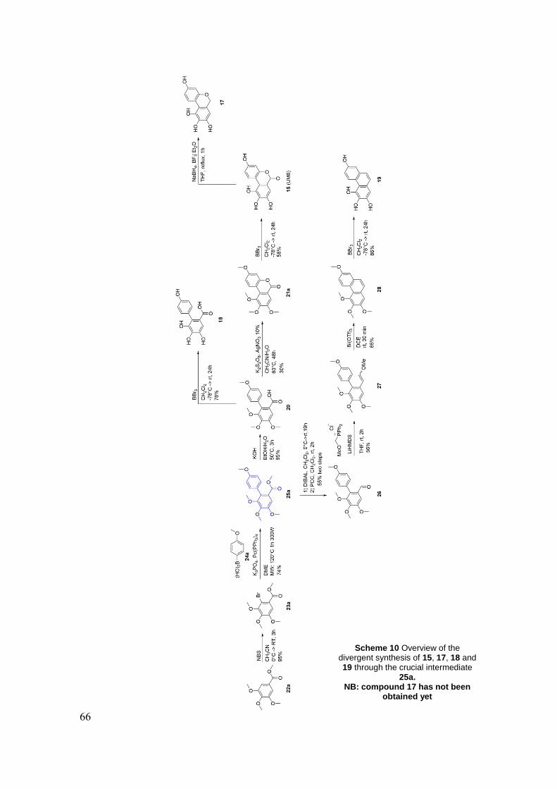
66
Scheme 10 Overview of the divergent synthesis of 15, 17, 18 and 19 through the crucial intermediate
25a. NB: compound 17 has not been
obtained yet
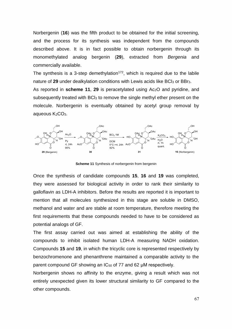
67
Norbergenin (16) was the fifth product to be obtained for the initial screening,
and the process for its synthesis was independent from the compounds
described above. It is in fact possible to obtain norbergenin through its
monomethylated analog bergenin (29), extracted from Bergenia and
commercially available.
The synthesis is a 3-step demethylation172, which is required due to the labile
nature of 29 under dealkylation conditions with Lewis acids like BCl3 or BBr3.
As reported in scheme 11, 29 is peracetylated using Ac2O and pyridine, and
subsequently treated with BCl3 to remove the single methyl ether present on the
molecule. Norbergenin is eventually obtained by acetyl group removal by
aqueous K2CO3.
Once the synthesis of candidate compounds 15, 16 and 19 was completed,
they were assessed for biological activity in order to rank their similarity to
galloflavin as LDH-A inhibitors. Before the results are reported it is important to
mention that all molecules synthesized in this stage are soluble in DMSO,
methanol and water and are stable at room temperature, therefore meeting the
first requirements that these compounds needed to have to be considered as
potential analogs of GF.
The first assay carried out was aimed at establishing the ability of the
compounds to inhibit isolated human LDH-A measuring NADH oxidation.
Compounds 15 and 19, in which the tricyclic core is represented respectively by
benzochromenone and phenanthrene maintained a comparable activity to the
parent compound GF showing an IC50 of 77 and 62 μM respectively.
Norbergenin shows no affinity to the enzyme, giving a result which was not
entirely unexpected given its lower structural similarity to GF compared to the
other compounds.
Scheme 11 Synthesis of norbergenin from bergenin

68
Considering the ability of 15 and 19 to inhibit human LDH-A, we investigated
their activity on lactic acid production and cell proliferation on Raji cells (see
table 6 on page 66). Both compounds exert a marked effect on lactate
production in cells at concentrations comparable to the ones inhibiting purified
LDH-A (IC50 of 37 and 72 μM respectively) thus showing a good capacity of cell
penetration. Moreover, 15 inhibited cell growth with an IC50 of 25 µM, whereas
compound 19 at concentration of 25 µM inhibited completely the cell
proliferation (100%), suggesting a mechanism of action related to different
targets than LDH-A.
In view of these encouraging results new analogs based on UM6 were designed
to explore the SAR of this class of compounds. In particular, we wanted to
evaluate the role of the number and position of the OH groups on the scaffold
and the influence of changes in the lipophilicity of the molecules.
To this purpose, through the previously established synthetic strategy three new
analogs were prepared, demonstrating the versatility and modularity of the
route. In figure 19 and scheme 12 the obtained compounds and synthetic
intermediates are shown.
Compound 33 is another member of the urolithins, urolithin M7 (UM7), the
synthesis of which is already reported in the literature168. 32 and 33 bear three
OH groups, one less than UM6, in different relative positions. Compound 34 is a
3-phenyl analog of 32 synthesized with the aim of exploring the lipophilicity on
that portion.
Figure 19 The new compounds obtained to explore the SAR of GF and its analog UM6
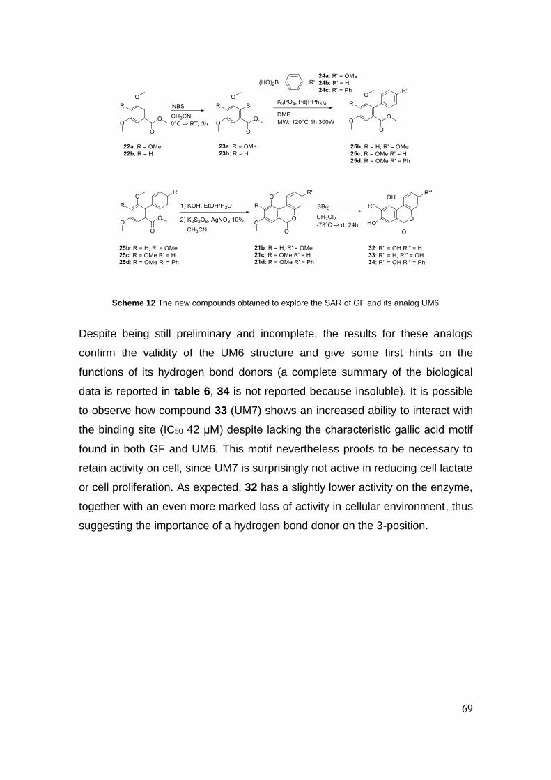
69
Despite being still preliminary and incomplete, the results for these analogs
confirm the validity of the UM6 structure and give some first hints on the
functions of its hydrogen bond donors (a complete summary of the biological
data is reported in table 6, 34 is not reported because insoluble). It is possible
to observe how compound 33 (UM7) shows an increased ability to interact with
the binding site (IC50 42 μM) despite lacking the characteristic gallic acid motif
found in both GF and UM6. This motif nevertheless proofs to be necessary to
retain activity on cell, since UM7 is surprisingly not active in reducing cell lactate
or cell proliferation. As expected, 32 has a slightly lower activity on the enzyme,
together with an even more marked loss of activity in cellular environment, thus
suggesting the importance of a hydrogen bond donor on the 3-position.
Scheme 12 The new compounds obtained to explore the SAR of GF and its analog UM6
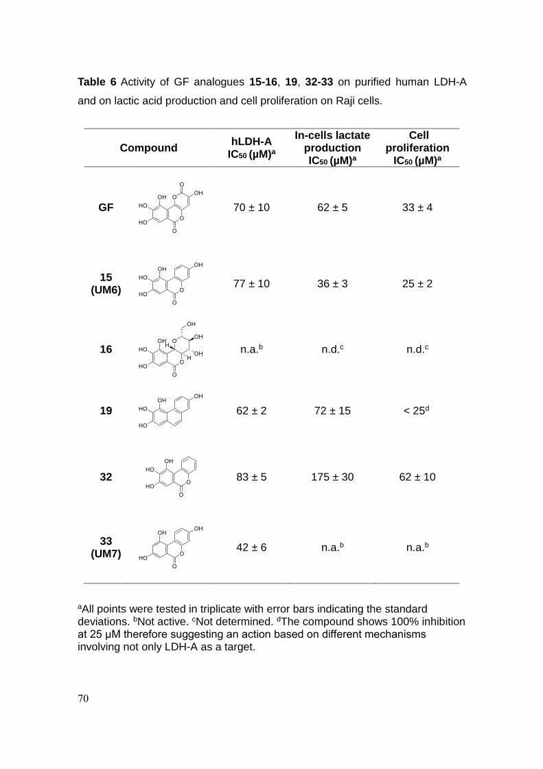
70
Table 6 Activity of GF analogues 15-16, 19, 32-33 on purified human LDH-A
and on lactic acid production and cell proliferation on Raji cells.
aAll points were tested in triplicate with error bars indicating the standard deviations. bNot active. cNot determined. dThe compound shows 100% inhibition at 25 μM therefore suggesting an action based on different mechanisms involving not only LDH-A as a target.
Compound hLDH-A
IC50 (µM)a
In-cells lactate production IC50 (µM)a
Cell proliferation
IC50 (µM)a
GF
70 ± 10 62 ± 5 33 ± 4
15 (UM6)
77 ± 10 36 ± 3 25 ± 2
16
n.a.b n.d.c n.d.c
19
62 ± 2 72 ± 15 < 25d
32
83 ± 5 175 ± 30
62 ± 10
33 (UM7)
42 ± 6 n.a.b n.a.b
71
3.3 Conclusion
With the aim of gaining a better insight to the SAR of galloflavin, a series of
analogs with high structural similarity to the original compound were designed
and synthesized. A new synthetic strategy to obtain urolithins has been devised,
together with the first reported synthesis of urolithin M6. The same strategy
gave access to an overall of four compounds in a versatile route diverging from
the same common precursor. Urolithin M6 reproduced in a satisfactory manner
the inhibitory activity of GF on human LDH-A and will serve as a starting point
for a further exploration. Preliminary information has been collected through the
synthesis of a small number of analogs and more compounds will be available
in the near future to better understand the influence of structural modifications
on the activity of these new inhibitors.

72
3.4 Experimental section
3.4.1 General methods
Reaction progress was monitored by TLC on pre-coated silica gel plates
(Kieselgel 60 F254, Merck) and visualized by UV254 light. Flash column
chromatography was performed on silica gel (particle size 40-63 μM, Merck) or
C18-reversed phase fully endcapped silica gel (particle size 40-63 μM, Sigma-
Aldrich). If required, solvents were distilled prior to use. All reagents were
obtained from commercial sources and used without further purification. When
stated, reactions were carried out under an inert atmosphere. Reactions
involving microwave irradiation were performed using a microwave synthesis
system (CEM Discover® SP, 2.45 GHz, maximum power 300 W), equipped with
infrared temperature measurement. Compounds were named relying on the
naming algorithm developed by CambridgeSoft Corporation and used in Chem-
BioDraw Ultra 15.0. 1H-NMR and 13C-NMR spectra were recorded on Varian
Gemini at 400 MHz and 100 MHz respectively. Chemical shifts (δH) are reported
relative to TMS as internal standard.
3.4.2 Synthetic procedures
Methyl 2-bromo-3,4,5-trimethoxybenzoate 23a
Methyl 3,4,5-trimethoxybenzoate 22a (10.200 g, 45.087 mmol, 1.0
eq.) was dissolved in CH3CN (400 mL). The mixture was cooled to
0°C and NBS (8.827 g, 49.596 mmol, 1.1 eq.) was added portionwise. The
reaction was let warm up to room temperature and stirred for 4 hours. After TLC
showed completion Na2S2O3 2M was added. After stirring for 5 minutes CH3CN
was evaporated in vacuum and the mixture extracted 3 times with EtOAc. The
combined organic phases were dried over anhydrous Na2SO4, and the solvent
evaporated in vacuum. The crude product was purified via silica gel column
chromatography (Petroleum ether/EtOAc 85/15 ratio) to obtain the pure product
as a colourless viscous liquid which solidifies after standing overnight (yield
95%). 1H NMR (400 MHz, CDCl3) δ 7.15 (s, 1H), 3.93 (s, 6H), 3.89 (s, 6H) ppm.
73
Methyl 4,4',5,6-tetramethoxy-[1,1'-biphenyl]-2-carboxylate 25a
Methyl 2-bromo-3,4,5-trimethoxybenzoate 23a (800 mg, 2.622
mmol, 1.0 eq.) was dissolved in 1,2-dimethoxyethane (90 ml, N.B. more
concentrated reactions might lead to extensive formation of side products and
deactivation of the catalyst) inside a pressure tube and 4-methoxyphenylboronic
acid 24a (598 mg, 3.933 mmol, 1.5 eq.), K2PO4 (1109 mg, 5.244 mmol, 2.0 eq.)
and tetrakis(triphenylphosphine)palladium (303 mg, 0.262 mmol, 0.1 eq.) were
added. The tube was sealed, the reaction was heated to 120°C and stirred for
24h. The mixture was transferred to a round bottom flask and the solvent
removed in vacuum. The crude product was purified via silica gel column
chromatography (Petroleum ether/EtOAc 90/10 ratio) to obtain the pure product
as a colourless viscous liquid (yield 74%). 1H NMR (400 MHz, CDCl3) δ 7.17 (s,
1H), 7.14 (d, J = 8.7 Hz, 2H), 6.90 (d, J = 8.7 Hz, 2H), 3.93 (s, 3H), 3.91 (s, 3H),
3.83 (s, 3H), 3.56 (s, 3H), 3.54 (s, 3H) ppm.
3,8,9,10-tetramethoxy-6H-benzo[c]chromen-6-one 21a
Methyl 4,4',5,6-tetramethoxy-[1,1'-biphenyl]-2-carboxylate 25a
(1590 mg, 4.784 mmol) was treated with 25 mL of a 1/1 mixture of
4M NaOH and EtOH at 60°C. After TLC showed absence of starting material
the reaction was treated with 37% HCl until acidic pH, brine was added and the
mixture was extracted 3 times with EtOAc. The combined organic phases were
dried over anhydrous Na2SO4, and the solvent evaporated in vacuum. The
crude product, used without further purification, was added 50 mL of a 1/1
H2O/CH3CN mixture. K2S2O8 (3880 mg, 14.353 mmol, 3.0 eq.) and AgNO3 (81
mg, 0.478 mmol, 0.1 eq.) were added and the reaction was stirred at 60°C for
48h. CH3CN was evaporated in vacuum and the mixture extracted 3 times with
EtOAc. The combined organic phases were dried over anhydrous Na2SO4, and
the solvent evaporated in vacuum. The crude product was purified via silica gel
column chromatography (Petroleum ether/EtOAc 85/15 ratio) to obtain the pure
product as an off white solid (yield 30%). 1H NMR (400 MHz, CDCl3) δ 8.74 (d, J
= 9.0 Hz, 1H), 7.72 (s, 1H), 6.90 (dd, J = 9.0, 2.7 Hz, 1H), 6.87 (d, J = 2.7 Hz,
1H), 4.04 (s, 3H), 3.99 (s, 3H), 3.96 (s, 3H), 3.88 (s, 3H) ppm.

74
3,8,9,10-tetrahydroxy-6H-benzo[c]chromen-6-one 15 (Urolithin
M6)
3,8,9,10-tetramethoxy-6H-benzo[c]chromen-6-one 21a (105 mg,
0.332 mmol, 1.0 eq.) was dissolved in dry DCM (10 mL) in a flame dried flask
and the mixture was cooled to -78°C. BBr3 1.0M in DCM (2.656 mL, 2.656
mmol, 8 eq. [2 eq. per methoxy group]) was slowly added dropwise. The
reaction was let warm up to room temperature and stirred for 24 hours. 5 mL of
MeOH were added and the solvent was removed in vacuum. The crude product
was purified via C18 silica gel reverse phase column chromatography
(H2O/MeOH/HCOOH elution gradient from a 70/30/0.1 ratio to a 30/70/0.1 ratio)
to obtain the product in a mixture with a minor dark impurity not detectable
using NMR, which was removed washing the product with small amounts of
MeOH, yielding the pure compound as a grey solid (yield 58%). 1H NMR (400
MHz, DMSO-d6) δ 10.19 (s, 1H), 9.96 (s, 1H), 9.91 (s, 1H), 9.49 (s, 1H), 8.80
(d, J = 8.9 Hz, 1H), 7.28 (s, 1H), 6.75 (d, J = 8.9 Hz, 1H), 6.67 (s, 1H) ppm; 13C
NMR (400 MHz, DMSO-d6) δ 160.74, 157.30, 150.68, 145.27, 142.52, 140.30,
128.02, 116.00, 112.01, 110.63, 110.23, 106.82, 102.57 ppm; MS (ES): m/z 261
[M + H]+, 283 [M + Na]+, Anal. Calcd. for C13H8O6 (%): C, 60.01; H, 3.10; O,
36.89. Found (%): C, 60.03; H, 3.12; O, 36.90.
Methyl 2-bromo-3,5-dimethoxybenzoate 23b
The compound was obtained as a colourless viscous liquid (yield
88%), with the same procedure used for 23a, using methyl-3,5-
dimethoxybenzoate 22b as starting material. 1H NMR (400 MHz, CDCl3) δ 6.79
(d, J = 2.8 Hz, 1H), 6.57 (d, J = 2.8 Hz, 1H), 3.92 (s, 3H), 3.87 (s, 3H), 3.81 (s,
3H) ppm.
Methyl 4,4',6-trimethoxy-[1,1'-biphenyl]-2-carboxylate 25b
The compound was obtained as an off white solid (yield 85%),
with the same procedure used for 25a, using methyl 2-bromo-3,5-
75
dimethoxybenzoate 23b as starting material. 1H NMR (400 MHz, CDCl3) δ 7.17
(d, J = 8.7 Hz, 2H), 6.92 (d, J = 8.7 Hz, 2H), 6.87 (d, J = 2.4 Hz, 1H), 6.65 (d, J
= 2.4 Hz, 1H), 3.87 (s, 3H), 3.83 (s, 3H), 3.78 (s, 3H), 3.57 (s, 3H) ppm.
3,8,10-trimethoxy-6H-benzo[c]chromen-6-one 21b
The compound was obtained as a white solid (yield 43%), with the
same procedure used for 21a, using methyl 4,4',6-trimethoxy-
[1,1'-biphenyl]-2-carboxylate 25b as starting material. 1H NMR (400 MHz,
CDCl3) δ 8.75 (dd, J = 7.7, 2.4 Hz, 1H), 7.48 (d, J = 2.5 Hz, 1H), 6.88 (d, J = 2.5
Hz, 1H), 6.87-6.84 (m, 2H), 4.02 (s, 3H), 3.92 (s, 3H), 3.87 (s, 3H) ppm.
3,8,10-trihydroxy-6H-benzo[c]chromen-6-one 33 (Urolithin M7)
The compound was obtained as a light yellow solid (yield 80%),
with the same procedure used for 15, using 3,8,10-trimethoxy-
6H-benzo[c]chromen-6-one 21b as starting material. 1H NMR (400 MHz,
DMSO-d6) δ 10.73 (s, 1H), 9.98 (s, 1H), 9.95 (s, 1H), 8.76 (d, J = 8.9 Hz, 1H),
7.14 (d, J = 1.6 Hz, 1H), 6.87 (d, J = 1.6 Hz, 1H), 6.77 (d, J = 8.9 Hz, 1H), 6.71
(s, 1H) ppm; 13C NMR (400 MHz, DMSO-d6) δ 160.78, 157.28, 156.99, 156.06,
150.23, 128.01, 121.64, 114.78, 112.36, 109.93, 109.63, 106.01, 102.65 ppm;
MS (ES): m/z 245 [M + H]+, 267 [M + Na]+, Anal. Calcd. for C13H8O5 (%): C,
63.94; H, 3.30; O, 32.76. Found (%): C, 63.92; H, 3.28; O, 32.76.
Methyl 4,5,6-trimethoxy-[1,1'-biphenyl]-2-carboxylate 25c
The compound was obtained as a colourless viscous liquid which
crystallizes after standing overnight (yield 82%), with the same
procedure used for 25a, using methyl 2-bromo-3,4,5-trimethoxybenzoate 23a
and phenylboronic acid 24b as starting materials. 1H NMR (400 MHz, CDCl3) δ
7.40 – 7.29 (m, 3H), 7.23 (d, J = 8.3 Hz, 2H), 7.22 (s, 1H), 3.95 (s, 3H), 3.92 (s,
3H), 3.56 (s, 3H), 3.52 (s, 3H) ppm.
8,9,10-trimethoxy-6H-benzo[c]chromen-6-one 21c
The compound was obtained as an off white solid (yield 60%), with

76
the same procedure used for 21a, using methyl 4,5,6-trimethoxy-[1,1'-biphenyl]-
2-carboxylate 25c as starting material. 1H NMR (400 MHz, CDCl3) δ 8.85 (d, J =
8.2 Hz, 1H), 7.77 (s, 1H), 7.44 (t, J = 7.7 Hz, 1H), 7.36 (d, J = 8.2 Hz, 1H), 7.32
(t, J = 7.7 Hz, 1H), 4.04 (s, 3H), 4.00 (s, 3H), 3.99 (s, 3H) ppm.
8,9,10-trihydroxy-6H-benzo[c]chromen-6-one 32
The compound was obtained as an off white solid (yield 87%), with
the same procedure used for 15, using 8,9,10-trimethoxy-6H-
benzo[c]chromen-6-one 21c as starting material. 1H NMR (400 MHz, DMSO-d6)
δ 10.40 (s, 1H), 10.03 (s, 1H), 9.74 (s, 1H), 9.01 (d, J = 7.7 Hz, 1H), 7.39 (dd, J
= 7.7, 6.8 Hz, 1H), 7.36 (s, 1H), 7.34 – 7.28 (m, 2H) ppm; 13C NMR (400 MHz,
DMSO-d6) δ 160.42, 149.28, 146.38, 143.78, 140.25, 127.83, 126.87, 124.02,
118.59, 116.46, 114.78, 112.11, 107.16 ppm; MS (ES): m/z 245 [M + H]+, 267
[M + Na]+, Anal. Calcd. for C13H8O5 (%):C, 63.94; H, 3.30; O, 32.76. Found
(%):C, 63.96; H, 3.31; O, 32.77.
Methyl 4,5,6-trimethoxy-[1,1':4',1''-terphenyl]-2-carboxylate
25d
The compound was obtained as a yellow viscous liquid (yield
70%), with the same procedure used for 25a, using methyl 2-
bromo-3,4,5-trimethoxybenzoate 23a and 4-biphenylboronic 24c acid as
starting materials. 1H NMR (400 MHz, CDCl3) δ 7.68 (d, J = 7.5 Hz, 2H), 7.64
(d, J = 8.1 Hz, 2H), 7.46 (t, J = 7.5 Hz, 2H), 7.36 (t, J = 7.5 Hz, 1H), 7.32 (d, J =
8.1 Hz, 2H), 7.25 (s, 1H), 3.98 (s, 3H), 3.95 (s, 3H), 3.62 (s, 3H), 3.57 (s, 3H)
ppm.
8,9,10-trimethoxy-3-phenyl-6H-benzo[c]chromen-6-one 21d
The compound was obtained as a light yellow solid (yield
33%), with the same procedure used for 21a, using methyl
4,5,6-trimethoxy-[1,1':4',1''-terphenyl]-2-carboxylate 25d as starting material. 1H
NMR (400 MHz, CDCl3) δ 8.90 (d, J = 8.0 Hz, 1H), 7.78 (s, 1H), 7.66 (d, J = 7.6

77
Hz, 2H), 7.62 (d, J = 8.0 Hz, 1H), 7.44 (t, J = 7.6 Hz, 2H), 7.36 (d, J = 8.0 Hz,
1H), 7.33 (t, J = 8.0 Hz, 1H), 4.06 (s, 3H), 4.02 (s, 3H), 4.01 (s, 3H) ppm.
8,9,10-trihydroxy-3-phenyl-6H-benzo[c]chromen-6-one 34
The compound was obtained as a beige solid (yield 62%), with
the same procedure used for 15, using 8,9,10-trimethoxy-3-
phenyl-6H-benzo[c]chromen-6-one 21d as starting material. 1H NMR (CD3OD)
δ 9.15 (d, J = 8.5 Hz, 1H), 7.71 (d, J =7.5, 2H), 7.59 (dd, J = 8.5, 1.9 Hz, 1H),
7.56 (d, J = 1.9 Hz, 1H), 7.47 (t, J = 7.5 Hz, 2H), 7.44 (s, 1H), 7.37 (t, J = 7.5
Hz, 1H) ppm; 13C NMR (400 MHz, DMSO-d6) δ 160.50, 149.84, 146.49, 143.79,
140.28, 139.35, 138.60, 129.02, 127.96, 127.92, 127.38, 126.63, 122.22,
119.50, 117.73, 114.68, 114.02, 112.03, 107.27 ppm; MS (ES): m/z 321 [M +
H]+, 343 [M + Na]+, Anal. Calcd. for C19H12O5 (%):C, 71.25; H, 3.78; O, 24.98.
Found (%):C, 71.27; H, 3.80; O, 24.99.
(4,4',5,6-tetramethoxy-[1,1'-biphenyl]-2-yl)methanol 25r
Methyl 4,4',5,6-tetramethoxy-[1,1'-biphenyl]-2-carboxylate 25a
(650 mg, 1.956 mmol, 1.0 eq.) was dissolved in dry CH2Cl2 (8
mL) and the mixture was cooled to 0°C. DIBAL 1.0 M in hexane (8.2 mL, 8.214
mmol, 4,2 eq.) was added dropwise. The reaction was then warmed to room
temperature and stirred for 3 hours. H2O and CH2Cl2 were added and the
mixture was filtered to remove the precipitate. The phases were separated and
the aqueous phase was extracted 2 times with CH2Cl2. The combined organic
phases were dried over anhydrous Na2SO4, and the solvent evaporated in
vacuum. The crude product was purified via silica gel column chromatography
(Petroleum ether/EtOAc 80/20 ratio) to obtain the pure product as a light brown
viscous liquid (yield 73%). 1H NMR (400 MHz, CDCl3) δ 7.18 (d, J = 8.5 Hz,
2H), 6.96 (d, J = 8.5 Hz, 2H), 6.89 (s, 1H), 4.42 (d, J = 5.7 Hz, 2H), 3.92 (s, 3H),
3.90 (s, 3H), 3.85 (s, 3H), 3.60 (s, 3H) ppm.
4,4',5,6-tetramethoxy-[1,1'-biphenyl]-2-carbaldehyde 26
(4,4',5,6-tetramethoxy-[1,1'-biphenyl]-2-yl)methanol 25r (359 mg,
78
1.180 mmol, 1.0 eq.) was dissolved CH2Cl2 (10 mL), PCC (381 mg, 1.769
mmol, 1.5 eq.) was added and the reaction was stirred at room temperature for
4 hours. The reaction was filtered on celite and the solvent evaporated to yield
the product as a pale yellow deliquescent solid which was used without further
purification (yield 75%). 1H NMR (400 MHz, CDCl3) δ 9.68 (s, 1H), 7.35 (s, 1H),
7.25 (d, J = 8.5 Hz, 2H), 6.98 (d, J = 8.5 Hz, 2H), 4.00 (s, 3H), 3.95 (s, 3H), 3.87
(s, 3H), 3.59 (s, 3H) ppm.
2,3,4,4'-tetramethoxy-6-(2-methoxyvinyl)-1,1'-biphenyl 27
(Methoxymethyl)triphenylphosphonium chloride (352 mg, 1.027
mmol, 1.5 eq.) was dissolved in dry THF (3 mL) and the mixture
was cooled to 0°C under inert atmosphere. LiHMDS 1.0 M in THF (1.370 mL,
1.370 mmol, 2 eq.) was slowly added and the reaction was stirred for 10
minutes. 4,4',5,6-tetramethoxy-[1,1'-biphenyl]-2-carbaldehyde 26 (207 mg,
0.685 mmol, 1.0 eq.) was dissolved in dry THF (4 mL) in a separate flask and
the solution was added slowly to the reaction mixture. The reaction was warmed
to room temperature and stirred for 1 hour. H2O was added and the mixture was
extracted 3 times with EtOAc. The combined organic phases were dried over
anhydrous Na2SO4, and the solvent evaporated in vacuum. The crude product
was purified via silica gel column chromatography (Petroleum ether/EtOAc
90/10 ratio) to obtain the pure product as a colourless viscous liquid (yield 56%,
60/40 mixture of E/Z isomers) 1H NMR (400 MHz, CDCl3) δ 7.51 (s, 0.40X1H, Z-
isomer), 7.16 (d, J = 8.3 Hz, 2H), 6.92 (d, J = 8.3 Hz, 2H), 6.79 (d, J = 12.9 Hz,
0.60X1H, E-isomer), 6.68 (s, 0.60X1H, E-isomer), 5.95 (d, J = 7.2 Hz, 0.40X1H,
Z-isomer), 5.50 (d, J = 12.9 Hz, 0.60X1H, E-isomer), 4.91 (d, J = 7.2 Hz,
0.40X1H, Z-isomer), 3.89 (s, 3H), 3.87 (s, 3H), 3.84 (s, 3H), 3.73 (s, 0.40X3H,
Z-isomer), 3.56 (s, 0.60X3H, E-isomer), 3.54 (s, 0.40X3H, Z-isomer), 3.46 (s,
0.60X3H, E-isomer) ppm.
2,3,4,7-tetramethoxyphenanthrene 28
2,3,4,4'-tetramethoxy-6-(2-methoxyvinyl)-1,1'-biphenyl 27 (127
mg, 0.384 mmol, 1.0 eq.) was dissolved in 1,2-dichloroethane (3

79
mL) under nitrogen atmosphere. Bismuth(III) trifluoromethanesulfonate (17 mg,
0.019 mmol, 0.05 eq.) was added and the reaction was stirred at room
temperature for 30 minutes. After TLC showed full conversion the solvent was
evaporated. The crude was dissolved in CH2Cl2 and filtered on a sintered glass
funnel equipped with a 1.5 cm thick layer of silica, washing several times to
assure complete elution. The pure compound was obtained as a white solid
(yield 65%). 1H NMR (400 MHz, CDCl3) δ 9.42 (d, J = 9.2 Hz, 1H), 7.59 (s, 2H),
7.29 – 7.22 (m, 2H), 7.08 (s, 1H), 4.04 (s, 3H), 4.02 (s, 3H), 4.01 (s, 3H), 3.96
(s, 3H) ppm.
Phenanthrene-2,3,4,7-tetraol 19
The compound was obtained as a gray solid (yield 86%), with
the same procedure used for 15, using 2,3,4,7-
tetramethoxyphenanthrene 28 as starting material. 1H NMR (400 MHz, DMSO-
d6) δ 9.66 (s, 1H), 9.43 (d, J = 9.2 Hz, 1H), 9.42 (s, 1H), 8.94 (s, 1H), 8.87 (s,
1H), 7.40 (d, J = 8.8 Hz, 1H), 7.33 (d, J = 8.8 Hz, 1H), 7.09 (d, J = 2.5 Hz, 1H),
7.02 (dd, J = 9.2, 2.5 Hz, 1H), 6.79 (s, 1H) ppm; 13C NMR (400 MHz, DMSO-d6)
δ 153.99, 144.84, 143.25, 132.77, 132.73, 128.63, 126.86, 125.30, 123.83,
123.52, 115.64, 113.60, 110.92, 103.81 ppm; MS (ES): m/z 243 [M + H]+, 265
[M + Na]+, Anal. Calcd. for C14H10O4 (%): C, 69.42; H, 4.16; O, 26.42. Found
(%): C, 69.43; H, 4.18; O, 26.42.
Norbergenin 16
Bergenin 29 (500 mg, 1.523 mmol, 1.0 eq.) was dissolved in
pyridine (2 mL) and Ac2O (2 mL) and the reaction was stirred at
room temperature for 24 hours. Water was added and the
mixture was extracted 3 times with EtOAc. The combined organic phases were
dried over anhydrous Na2SO4, and the solvent evaporated in vacuum and dried
in high vacuum. The crude (600 mg, 1.226 mmol, 1.0 eq.) was dissolved in
CH2Cl2 (15 mL) and then BCl3 1.0 M in CH2Cl2 (7.354 mL, 7.354 mmol, 5.0 eq. )
was added at 0°C and stirred for 1 h. After stirring at room temperature for 24 h,
the reaction mixture was poured into ice-water. The organic solvent was
removed in vacuum, and then the water layer was added to 10% K2CO3

80
solution, and stirred for 1 h. The reaction mixture was acidified with 2.0 M HCl,
and then the water layer absorbed with DIAION HP-20 resin. The fraction eluted
with MeOH was concentrated in vacuum, and the residue was purified by
recrystallization to afford the product as a white solid (yield 78% over the 3
steps). 1H NMR (400 MHz, CD3OD) δ 7.09 (s, 1H), 4.94 (d, J = 10.5 Hz, 1H),
4.04 (t, J = 9.9 Hz, 2H), 3.81 (t, J = 9.0 Hz, 1H), 3.76 – 3.61 (m, 2H), 3.43 (t, J =
9.0 Hz, 1H) ppm; 13C NMR (400 MHz, CD3OD) δ 166.39, 147.25, 143.64,
141.17, 117.34, 114.28, 110.98, 82.97, 81.40, 75.64, 74.35, 71.95, 62.71 ppm;
MS (ES): m/z 315 [M + H]+, 337 [M + Na]+, Anal. Calcd. for C13H14O9 (%): C,
49.69; H, 4.49; O, 45.82. Found (%): C, 49.70; H, 4.51; O, 45.83.
4,4',5,6-tetrahydroxy-[1,1'-biphenyl]-2-carboxylic acid 18
The compound was obtained as a gray solid (yield 86%), with
the same procedure used for 15, using methyl 4,4',5,6-
tetramethoxy-[1,1'-biphenyl]-2-carboxylic acid 20 as starting material. 1H NMR
(400 MHz, DMSO-d6) δ 11.76 (s, 1H), 9.34 (s, 1H), 9.14 (s, 1H), 8.91 (s, 1H),
8.02 (s, 1H), 6.91 (d, J = 8.4 Hz, 1H), 6.77 (s, 1H), 6.66 (d, J = 8.4 Hz, 1H) ppm;
13C NMR (400 MHz, DMSO-d6) δ 169.21, 155.57, 149.73, 143.84, 143.69,
135.80, 130.91, 128.21, 122.84, 121.65, 115.65, 114.09, 108.24 ppm; MS (ES):
m/z 263 [M + H]+, 285 [M + Na]+, Anal. Calcd. for C13H10O6 (%): C, 59.55; H,
3.84; O, 36.61. Found (%): C, 59.54; H, 3.86; O, 36.62.

81

82
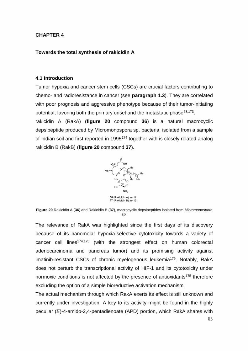
83
CHAPTER 4
Towards the total synthesis of rakicidin A
4.1 Introduction
Tumor hypoxia and cancer stem cells (CSCs) are crucial factors contributing to
chemo- and radioresistance in cancer (see paragraph 1.3). They are correlated
with poor prognosis and aggressive phenotype because of their tumor-initiating
potential, favoring both the primary onset and the metastatic phase68,173.
rakicidin A (RakA) (figure 20 compound 36) is a natural macrocyclic
depsipeptide produced by Micromonospora sp. bacteria, isolated from a sample
of Indian soil and first reported in 1995174 together with is closely related analog
rakicidin B (RakB) (figure 20 compound 37).
The relevance of RakA was highlighted since the first days of its discovery
because of its nanomolar hypoxia-selective cytotoxicity towards a variety of
cancer cell lines174,175 (with the strongest effect on human colorectal
adenocarcinoma and pancreas tumor) and its promising activity against
imatinib-resistant CSCs of chronic myelogenous leukemia176. Notably, RakA
does not perturb the transcriptional activity of HIF-1 and its cytotoxicity under
normoxic conditions is not affected by the presence of antioxidants175 therefore
excluding the option of a simple bioreductive activation mechanism.
The actual mechanism through which RakA exerts its effect is still unknown and
currently under investigation. A key to its activity might be found in the highly
peculiar (E)-4-amido-2,4-pentadienoate (APD) portion, which RakA shares with
Figure 20 Rakicidin A (36) and Rakicidin B (37), macrocyclic depsipeptides isolated from Micromonospora sp.
84
a few other known natural compounds, such as vinylamycin and microtermolide
A177,178. Using a model macrocyclic system, it has been recently demonstrated
that this functionality can react as an electrophilic site in a 1,6 Michael fashion
with thiol nucleophiles, including cysteine residues incased in BSA-derived
tryptic peptides179. Further data will be described later on in this chapter to
demonstrate how, most likely, the molecule needs this electrophilic portion to
retain its activity, but despite this the biological molecular target (or targets) is
still unknown.
Surrounding the APD portion, the complex architecture characterizing RakA is
completed by a β-hydroxy fatty acid side chain and two non-proteinogenic
amino acids: sarcosine and β-hydroxyasparagine (β-HOAsn). Unexpectedly, the
fatty acid portion is also crucial for the high activity and selectivity of RakA,
since its analog RakB shows a significantly lower cytotoxicity towards cancer
cells174, despite differing by one single carbon atom in the length of the alkyl
chain (figure 20).
The configuration of the 5 stereogenic centers has been uncertain until 2014,
when Chen and co-workers claimed the synthesis of a compound that matched
the spectroscopic data reported for the natural product, assigning the
stereochemistry as (2S,3S,14S,15S,16R)180. Shortly after this paper was
published, Igarashi and co-workers paradoxically concluded the
stereochemistry to be (2R,3R,14S,15S,16R) by direct degradation of the natural
product and comparison with authentic standards181. The latter was later on
found to arise from a practical mistake and was corrected and aligned with the
(2S,3S,14S,15S,16R) configuration, therefore completing a characterization of
the compound which can be agreed upon beyond any reasonable doubt.
At the onset of the project for the total synthesis of RakA in T.B. Poulsen’s lab,
the stereochemistry was still unknown and the synthetic effort was originally
directed towards several candidate isomers, with the most promising being the
one that later one turned out to be ent-RakA (i.e. 2R,3R,14R,15R,16S). This
configuration arose from a combination of NOE and computational
conformational analysis of a previously developed model system of the
macrocyclic ring182 as well as surveys of the literature. After the clarification of

85
the stereochemistry the strategy was adjusted and directed towards the total
synthesis of the natural isomer.
In the coral effort that was carried out to obtain this complex compound, I was
involved in the optimization and adjustment of reaction conditions to obtain
some of the precursors of the natural product as well precursors of ent-RakA
and other non-natural epimers, that might play a future role in the investigation
of the compound’s mechanism of action. In the next paragraph I will mostly
describe my personal contribution, and I will conclude by shortly reporting the
overall successful strategy that was completed and published shortly after the
end of my visiting period.

86
4.2 Results and discussion
With β-HOAsn being one of the four components of the molecular structure of
RakA, we wanted to obtain access the four possible diasteroisomers of the
compound. The precursor to β-HOAsn is β-hydroxyaspartic acid (β-HOAsp),
which exists in the threo and erythro form. The threo form is commercial and is
sold as a racemic mixture which can be resolved via a known procedure183. The
erythro form had to be prepared and its synthesis184 is reported in scheme 13.
Diethyl tartrate in the suitable optically pure diasteroisomer was treated with
SOCl2 to obtain the intermediate cyclic sulfite 38 which was not isolated and
was treated in-situ with NaN3 to yield the corresponding organic azide 39 with
the correct configuration after inversion of the stereocenter. The compound was
in turn reduced to the corresponding amine 40 and subsequently hydrolyzed to
obtain the desired erythro (R,S)-β-HOAsp.
The transformation of HOAsp to HOAsn was planned to belong to a later stage
of the synthesis, after the incorporation of all the fragments and the formation of
the macrocycle, so the next fragment prepared was the β-hydroxy fatty acid
side chain. With no known stereochemistry at the time one of the first
configurations tested was the (R,R,R). On scheme 14 the synthesis of
precursor aldehyde 46 is shown.
Scheme 13 Synthesis of erythro (R,S)-β-HOAsp 41 (the opposite enantiomer can be obtained with the
same procedure starting from (+)-Diethyl L-tartrate)
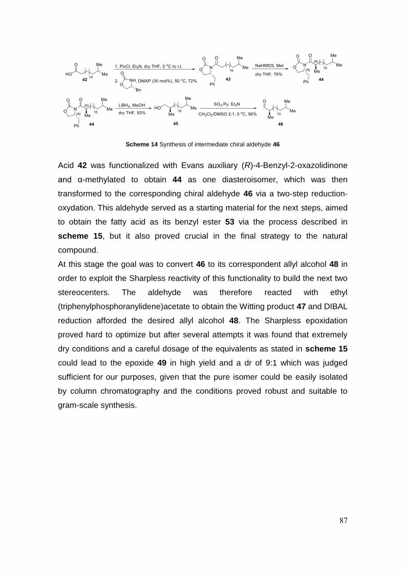
87
Acid 42 was functionalized with Evans auxiliary (R)-4-Benzyl-2-oxazolidinone
and α-methylated to obtain 44 as one diasteroisomer, which was then
transformed to the corresponding chiral aldehyde 46 via a two-step reduction-
oxydation. This aldehyde served as a starting material for the next steps, aimed
to obtain the fatty acid as its benzyl ester 53 via the process described in
scheme 15, but it also proved crucial in the final strategy to the natural
compound.
At this stage the goal was to convert 46 to its correspondent allyl alcohol 48 in
order to exploit the Sharpless reactivity of this functionality to build the next two
stereocenters. The aldehyde was therefore reacted with ethyl
(triphenylphosphoranylidene)acetate to obtain the Witting product 47 and DIBAL
reduction afforded the desired allyl alcohol 48. The Sharpless epoxidation
proved hard to optimize but after several attempts it was found that extremely
dry conditions and a careful dosage of the equivalents as stated in scheme 15
could lead to the epoxide 49 in high yield and a dr of 9:1 which was judged
sufficient for our purposes, given that the pure isomer could be easily isolated
by column chromatography and the conditions proved robust and suitable to
gram-scale synthesis.
Scheme 14 Synthesis of intermediate chiral aldehyde 46
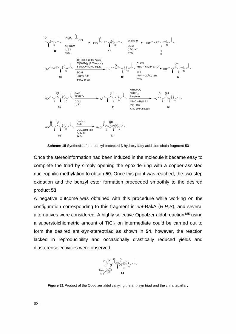
88
Once the stereoinformation had been induced in the molecule it became easy to
complete the triad by simply opening the epoxide ring with a copper-assisted
nucleophilic methylation to obtain 50. Once this point was reached, the two-step
oxidation and the benzyl ester formation proceeded smoothly to the desired
product 53.
A negative outcome was obtained with this procedure while working on the
configuration corresponding to this fragment in ent-RakA (R,R,S), and several
alternatives were considered. A highly selective Oppolzer aldol reaction185 using
a superstoichiometric amount of TiCl4 on intermediate could be carried out to
form the desired anti-syn-stereotriad as shown in 54, however, the reaction
lacked in reproducibility and occasionally drastically reduced yields and
diastereoselectivities were observed.
Scheme 15 Synthesis of the benzyl protected β-hydroxy fatty acid side chain fragment 53
Figure 21 Product of the Oppolzer aldol carrying the anti-syn triad and the chiral auxiliary
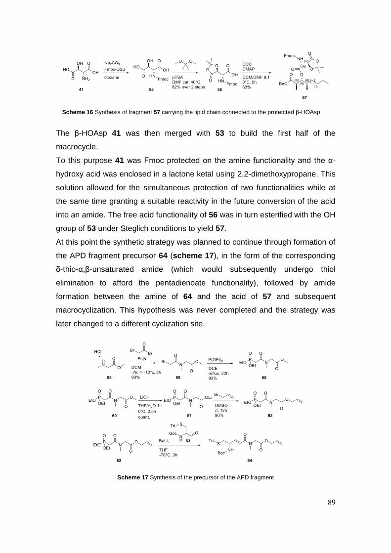
89
The β-HOAsp 41 was then merged with 53 to build the first half of the
macrocycle.
To this purpose 41 was Fmoc protected on the amine functionality and the α-
hydroxy acid was enclosed in a lactone ketal using 2,2-dimethoxypropane. This
solution allowed for the simultaneous protection of two functionalities while at
the same time granting a suitable reactivity in the future conversion of the acid
into an amide. The free acid functionality of 56 was in turn esterified with the OH
group of 53 under Steglich conditions to yield 57.
At this point the synthetic strategy was planned to continue through formation of
the APD fragment precursor 64 (scheme 17), in the form of the corresponding
δ-thio-α,β-unsaturated amide (which would subsequently undergo thiol
elimination to afford the pentadienoate functionality), followed by amide
formation between the amine of 64 and the acid of 57 and subsequent
macrocyclization. This hypothesis was never completed and the strategy was
later changed to a different cyclization site.
Scheme 16 Synthesis of fragment 57 carrying the lipid chain connected to the protetcted β-HOAsp
Scheme 17 Synthesis of the precursor of the APD fragment

90
Compound 64 was obtained starting from sarcosine methylester, acylated with
bromoacetyl bromide to obtain 59. The compound was transformed into the
corresponding phosphonate and in turn converted to the allyl ester 62. This
compound was merged with aldehyde 63186 in a Horner-Wadsworth-Emmons
(HWE) reaction to obtain the desired α,β-unsaturated amide.
After evaluating many alternative options for the aldol reaction, the analog of 57
carrying the natural product configuration (67, scheme 18) was obtained
through a proline-catalyzed cross-aldol reaction187 between propanal and 46
which proceeded with high anti-selectivity yielding the desired anti-aldol-Felkin
motif, thus constituting an efficient and reliable method for accessing hydroxy
ester 66 after Pinnick oxidation and benzylation of the carboxylic acid. The
typical isolated yields from the cross aldol reaction ranged from 30-40%
following chromatographic isolation but a large part of unreacted starting
material was recovered and re-reacted to increase the final yield.
After several attempts, the option of carrying out the cyclization through amide
formation between the APD unit precursor and the fatty acid was judged
unfeasible, and the project required a thorough reprogramming of the synthetic
strategy towards a more effective cyclization site.
The total synthesis was successfully concluded thanks to the collaboration of
other members of the group188 and the synthetic scheme for the last steps is
reported as published in figure 22 (compound numbers in this figure refer to the
article numeration system). The new strategy focused on a HWE-mediated
cyclization thus building the APD portion as the last step.
Scheme 18 Synthesis of the lipid chain/β-HOAsp fragment with the natural product configuration

91
The cyclization precursor was obtained by two efficient EDCI-mediated
couplings resulting in the desired phosphonate in high yield. The
macrocyclization sequence commenced with initial DMP-oxidation of the
primary alcohol followed by exposure of the resulting aldehyde-phosphonate to
TMEDA-Zn(OTf)2189 and trimethylamine, thus affording HWE reactivity.
Deprotection of the lactone-ketal to the hydroxy acid could be carried out
efficiently using aq. AcOH in THF and this crude material was directly subjected
to a PyBOP-mediated coupling with ammonium-bicarbonate resulting in the
corresponding hydroxy-amide. Dehydration to the sensitive APD-functionality
could be achieved using a one-pot reaction employing N,N′-disuccinimidyl
carbonate (DSC)190 at slightly elevated temperature. Final purification by HPLC
and subsequent lyophilization delivered pure rakicidin A as a white powder.
Figure 22 Construction of the linear precursor, cyclization and DSC-mediated dehyration to complete the
total synthesis of RakA (compound numbers are reported as in the final publication)

92
Synthetic rakicidin A matched all spectroscopic data reported for the natural
product.
A biological evaluation of the compound was then conducted and the assay on
the synthetic compound matched the promising activity of the natural isolate:
RakA showed nanomolar inhibitory activity on cell growth of PANC-1 cells
(GI50=36±1.4 nM) with a high Selectivity Index for inhibition under hypoxic vs.
normoxic conditions (SI=9.8±1.0). It was furthermore demonstrated that RakA is
not a proteasome inhibitor (as it may be suggested by structural similarity with
the proteasome inhibitors of the syrbactin family191), by comparison with
bortezomib activity under hypoxic conditions and by direct evaluation of the
inhibition of the proteolytic activities of the proteasome. The actual mechanism
of action of the compound and its biological target are still undisclosed and are
currently under active investigation.

93
4.3 Conclusion
A study towards the total synthesis of rakicidin A was conducted, leading to the
optimization of reaction conditions for the several steps leading to crucial
intermediates. Different strategies were undertaken to obtain a number of
stereochemical configurations apt to the obtainment of natural rakA and other
synthetic isomers. The project focused on the obtainment of three crucial
building blocks such as the fatty acid side chain (carrying a challenging triad of
stereogenic centers), the β-HOAsn fragment and the precursor to the APD
fragment. Specific attention was put on the asymmetric methods allowing for the
construction of the 5 stereocenters of the molecule in a reproducible and
reliable way. After a full total synthesis had been completed, rakA was
confirmed as a correct reproduction of the natural compound and further studies
are being conducted to disclose its activity on a biomolecular level.
94
4.4 Experimental section
4.4.1 General methods
All reactions with air and moisture sensitive compounds were conducted in
flame-dried glassware under an atmosphere of argon. Dichloromethane, MeCN,
THF and toluene were dried over aluminum oxide via an MBraun SPS-800
solvent purification system. Benzene and Et2O were dried over activated
molecular sieves (4 Å). DMF, DME and MeOH were purchased as dried. If not
otherwise stated reagents were used as received from commercial suppliers.
TLC analysis was carried out on silica coated aluminum foil plates (Merck
Kieselgel 60 F254). The TLC plates were visualized by UV irradiation and/or by
staining with either CAM stain or KMnO4 stain. Flash column chromatography
(FCC) was carried out using silica gel (230-400 mesh particle size, 60 Å pore
size) as stationary phase. Optical rotation was measured on an ADP 440+
spectrometer. Mass spectra (HRMS) were recorded on a Bruker Daltonics
MicrOTOF time-of-flight spectrometer with positive electrospray ionization, or
negative ionization when stated. Nuclear magnetic resonance (NMR) spectra
were recorded on a Varian Mercury 400 MHz spectrometer or a Bruker BioSpin
GmbH 400 MHz spectrometer, running at 400 and 100 MHz for 1H and 13C,
respectively. 1H and 13C NMR spectra of Rakicidin A were recorded on a Bruker
950 MHz spectrometer. Chemical shifts (δ) are reported in ppm relative to the
residual solvent signals. Multiplicities are indicated using the following
abbreviations: s = singlet, d = doublet, t = triplet, q = quartet, m = multiplet, br =
broad. LC-MS and HPLC analysis and purification were performed using a
Gilson HPLC system and a PerkinElmer Flexar SQ 300 MS detector.
4.4.2 Synthetic procedures
Diethyl (2R,3S)-2-azido-3-hydroxysuccinate 39
To a stirred solution of (-)-diethyl D-tartrate (2.3 g, 11.2 mmol, 1.0
eq.) in 12 mL CCl4 was added SOCl2 (4.0 g, 33.6 mmol, 3.0 eq.). The
reaction was heated to reflux and stirred for 6 h. The mixture was cooled to
room temperature and concentrated to dryness under reduced pressure. The
residue was dissolved in DMF and sodium azide (1.46 g, 22.4 mmol, 2.0 eq.)
95
was added to the solution. The reaction was stirred at room temperature for 4 h
and quenched with water. The mixture was extracted with ethyl acetate and the
combined organic layer was washed with brine and then dried with Na2SO4. The
solution was concentrated under reduced pressure and the residue was purified
by column chromatography (33%EtOAc/67% Hexane) to give 39 as a colorless
oil (yield 65%). αD25.6 = -6.8 (c 1.0, CHCl3). 1H NMR (400 MHz, CDCl3) δ 4.63
(dd, J = 5.5, 2.7 Hz, 1H), 4.33 – 4.20 (m, 4H), 3.34 (d, J = 5.5 Hz, 1H), 1.30 (t, J
= 2.9 Hz, 6H). 13C NMR (100 MHz, CDCl3) 170.87, 167.06, 76.82, 72.16, 64.52,
62.83, 62.48, 14.15. HRMS (ESI): Calc. C8H14N3O5+: 232.0928; found:
232.0930 [M + H]+.
Diethyl (2R,3S)-2-amino-3-hydroxysuccinate 40
To a stirred solution of azide 39 (342 mg, 1.48 mmol) in ethyl acetate
was added palladium in carbon (10%, 30 mg). The reaction
atmosphere was evacuated and replaced by H2 three times, and then stirred
vigorously under 1 atm hydrogen gas for 2 h. The mixture was concentrated
under reduced pressure and the residue was purified by column
chromatography (10% MeOH/90% CH2Cl2) to give 40 (yield 60%) as a colorless
oil. αD26.9 = 16.8 (c 1.0, CHCl3). 1H NMR (400 MHz, CDCl3) δ 4.48 (d, J = 3.1
Hz, 1H), 4.30 – 4.11 (m, 4H), 3.89 (d, J = 3.1 Hz, 1H), 2.53 – 2.17 (br s, 2H),
1.26 (q, J = 7.2 Hz, 6H). 13C NMR (100 MHz, CDCl3) δ 172.11, 171.90, 72.92,
62.05, 61.54, 57.65, 14.20, 14.18. HRMS (ESI): Calc. C8H16NO5+: 206.1023;
found: 206.1027 [M + H]+.
(2R,3S)-Erythro-β-hydroxyaspartic acid 41
A stirred solution of 40 (300 mg, 1.46 mmol, 1.0 eq.) in ethanol was
cooled to 0°C with an ice bath and LiOH·H2O (245 mg, 5.84 mmol, 4.0 eq.) was
added. The bath was removed and mixture was stirred at RT for 2h. The pH
was adjusted to neutral and the compound was extracted 3 times with EtOAc.
The solvent was concentrated under reduced pressure and the product was
used without further purification (quant.). 1H NMR (400 MHz, dmso-d6) δ 3.86
(s, 1H), 3.41 (s, 1H). HRMS (ESI): Calc. C4H6NO5-: 148.0251; found: 148.0248
[M - H]-.
96
(R)-4-Benzyl-3-(14-methylpentadecanoyl)oxazolidin-2-one 43
A stirring solution of 14-methylpentadecanoic acid (670 mg, 2.61
mmol, 1 equiv.) in anhydrous THF (26 mL) was cooled to 0°C
under Ar and NEt3 (1.09 mL, 7.84 mmol, 3 equiv.) was added. After 5 minutes
PivCl (0.35 mL, 2.87 mmol, 1.1 equiv.) was added dropwise. The mixture was
stirred for 20 minutes at 0°C and for 30 minutes at ambient temperature (TLC
analysis). Then, DMAP (95.8 mg, 0.784 mmol, 0.3 equiv.) was added followed
by (R)-4-benzyl-2-oxazolidinone (556 mg, 3.14 mmol, 1.2 equiv.) and the
mixture was stirred at 50°C for 5 hours. The reaction was quenched at ambient
temperature with brine and extracted with ether (3 x 20 mL). The organic phase
was dried (Na2SO4), filtered, concentrated and the crude product was purified
by FCC (pentane/EtOAc 95:5 to 90:10) to furnish 43 (782 mg, 72%) as a white
solid. αD28 = -62 (c 1.0, EtOH). 1H NMR (400 MHz, CDCl3) δ 7.36-7.19 (m, 5H),
4.71-4.62 (m, 1H), 4.22-4.13 (m, 2H), 3.29 (dd, J = 13.4 and 3.3 Hz, 1H), 3.02-
2.83 (m, 2H), 2.76 (dd, J = 13.4 and 9.6 Hz, 1H), 1.74-1.63 (m, 2H), 1.51 (n, J =
6.6 Hz, 1H), 1.42-1.11 (m, 20H), 0.86 (d, J = 6.6 Hz, 6H). 13C NMR (100 MHz,
CDCl3) δ 173.6, 153.6, 135.5, 129.6, 129.1, 127.5, 66.3, 55.3, 39.2, 38.1, 35.7,
30.1, 29.9, 29.8, 29.6, 29.3, 28.1, 27.6, 24.4, 22.8. HRMS (ESI): Calc.
C26H41O3NNa+: 438.2979; found: 438.2982 [M + Na]+.
(R)-4-Benzyl-3-((R)-2,14-dimethylpentadecanoyl)oxazolidin-2-
one 44
(R)-4-Benzyl-3-(14-methylpentadecanoyl)oxazolidin-2-one 43
(6.54 g, 15.7 mmol, 1 equiv.) was dissolved in anhydrous THF (95 mL) and
cooled to -78 °C, before NaHMDS (1.0 M in THF, 1.2 equiv., 18.9 mL, 18.9
mmol) was added slowly. The reaction mixture was then stirred at -78 °C for 1
hour before MeI (5 equiv., 4.9 mL, 78.5 mmol) was added slowly. The reaction
mixture was stirred for 3 hours at -78 °C, before it was quenched with sat.
NH4Cl (8 mL). The aqueous phase was extracted with CH2Cl2 and the organic
phase was then washed with KHSO4 (5%, aq.), sat. Na2S2O3 (aq.) and brine,
before it was dried (Na2SO4), filtered and concentrated. The crude product was

97
purified by dry column vacuum chromatography (0-15% EtOAc in pentane, 1%
increments) to furnish 44 (5.10 g, 76%) as a colourless oil. αD28 = -69.1 (c 1.0,
CHCl3). 1H NMR (400 MHz, CDCl3) δ 7.36-7.19 (m, 5H), 4.67 (ddt, J = 10.1, 6.7
and 3.2 Hz, 1H), 4.23-4.13 (m, 2H), 3.70 (h, J = 6.8 Hz, 1H), 3.26 (dd, J = 13.3
and 3.3 Hz, 1H), 2.76 (dd, J = 13.3 and 9.6 Hz, 1H), 1.78-1.67 (m, 1H), 1.56-
1.46 (m, 1H), 1.44-1.35 (m, 1H), 1.32-1.18 (m, 18H), 1.22 (d, J = 6.9 Hz, 3H),
1.18-1.10 (m, 2H), 0.86 (dd, J = 6.6 and 0.7 Hz, 6H). 13C NMR (100 MHz,
CDCl3) δ 177.4, 153.1, 135.4, 129.5, 129.0, 127.4, 66.1, 55.4, 39.1, 38.0, 37.8,
33.5, 30.0, 29.8, 29.7, 29.6, 28.1, 27.5, 27.4, 22.8, 17.5. HRMS (ESI): Calc.
C27H44O3N+: 430.3316; found: 430.3319 [M + H]+.
(R)-2,14-Dimethylpentadecan-1-ol 45
(R)-4-Benzyl-3-((R)-2,14-dimethylpentadecanoyl)oxazolidin-2-one
44 (1.03 g, 2.39 mmol, 1 equiv.) was dissolved in anhydrous THF (60 mL) and
cooled to 0°C, before anhydrous MeOH (0.15 mL) and LiBH4 (2.0 M in THF, 2.0
equiv., 2.4 mL, 4.8 mmol) were added. The reaction mixture was then stirred at
0°C for 2 hours and an additional 5 hours at ambient temperature. The reaction
mixture was quenched by slow addition of NaOH 2N (10 mL) and stirred until
the solution was clear (5-10 minutes). The solution was extracted with Et2O (3 x
50 mL) and the combined organic phases were washed with H2O and brine,
dried (Na2SO4), filtered and concentrated. The crude product was purified by
flash column chromatography (pentane/Et2O 9:1) to afford alcohol 45 (0.57 g,
93%) as a colourless liquid. αD28 = +7.2 (c 1.0, CHCl3). 1H NMR (400 MHz,
CDCl3) δ 3.50 (dd, J = 10.5 and 5.7 Hz, 1H), 3.41 (dd, J = 10.5 and 6.6 Hz, 1H),
1.66-1.55 (m, 1H), 1.50 (m, 1H), 1.43-1.06 (m, 22H), 0.91 (d, J = 6.7 Hz, 3H),
0.86 (d, J = 6.6 Hz, 6H). 13C NMR (100 MHz, CDCl3) δ 68.6, 39.2, 35.9, 33.3,
30.1, 29.9, 29.8, 28.1, 27.6, 27.1, 22.8, 16.7.
(R)-2,14-Dimethylpentadecanal 46
The alcohol 45 (7.8 mmol, 2.0 g, 1 equiv.) was azeotroped with
anhydrous benzene and then dissolved in CH2Cl2/DMSO (2:1, 106 mL) and
cooled to 0°C. Triethylamine (5 equiv., 39 mmol, 5.4 mL) and SO3·pyridine (4
equiv., 31.2 mmol, 4.97 g) were added sequentially and the reaction mixture

98
was stirred for 60 minutes at 0°C before it was quenched with brine. The layers
were separated and the aqueous phase was extracted with CH2Cl2 (3 x 20 mL).
The organic phase was dried (Na2SO4), filtered and concentrated. The crude
product was purified by flash column chromatography (pentane/CH2Cl2 6:1) to
give the desired aldehyde 46 (1.9 g, 96%) as a colourless liquid. αD28 = -19.7 (c
1.0, CHCl3). 1H NMR (400 MHz, CDCl3) δ 9.61 (d, J = 2.0 Hz, 1H), 2.37-2.27
(m, 1H), 1.75-1.64 (m, 1H), 1.50 (m, 1H), 1.32-1.12 (m, 21H), 1.08 (d, J = 7.0
Hz, 3H), 0.85 (d, J = 6.6 Hz, 6H). 13C NMR (100 MHz, CDCl3) δ 205.6, 46.5,
39.2, 30.7, 30.1, 29.9, 29.8, 29.7, 29.6, 28.1, 27.6, 27.1, 22.8, 13.5 HRMS
(ESI): Calc. C17H34ONa+: 277.2507; found 277.2502 [M + Na]+.
Ethyl (R,E)-4,16-dimethylheptadec-2-enoate 47
Ethyl (triphenylphosphoranylidene)acetate (8.96
g, 25.73 mmol, 4.8 equiv.) was dissolved in 16 mL dry CH2Cl2. Aldehyde 46
(1.74 g, 5.36 mmol, 1 equiv.) was dissolved in 9 mL dry CH2Cl2 and added to
the solution containing the Wittig reagent. The mixture was stirred for 3 hours at
room temperature. The reaction was then diluted with H2O after completion and
extracted with CH2Cl2 three times. The extract was dried with Na2SO4 and
concentrated in vacuum. The crude residue was purified by FCC (yield 95%)
(AcOEt/pentane 1:99). 1H NMR (400 MHz, CDCl3) δ 6.86 (dd, J = 15.7, 7.9 Hz,
1H), 5.76 (d, J = 15.7 Hz, 1H), 4.18 (q, J = 7.1 Hz, 2H), 2.28 (dt, J = 13.7, 6.8
Hz, 1H), 1.57 – 1.45 (m, 2H), 1.30 – 1.24 (m, 21H), 1.14 (t, J = 7.1 Hz, 3H), 1.03
(d, J = 6.7 Hz, 3H), 0.86 (d, J = 6.6 Hz, 6H). HRMS (ESI): Calc. C21H40O2Na+:
347.2921; found 347.2917 [M + Na]+.
(R,E)-4,16-dimethylheptadec-2-en-1-ol 48
To a solution of ethyl (R,E)-4,16-
dimethylheptadec-2-enoate 47 (904 mg, 3.2 mmol, 1.0 equiv.) in dry CH2Cl2 (10
mL) was added diisobutylaluminum hydride (9.6 mL of a 1.0 M solution in
hexane, 9.6 mmol, 3.0 equiv.) at 0 °C. After stirring for 2 hours at room
temperature, the reaction was quenched with methanol. After an aqueous
Rochelle Salt solution and diethyl ether were added, the mixture was warmed to
99
room temperature and stirred vigorously until the resulting white slurry was
completely dissolved. After the phases were separated, the aqueous layer was
extracted with diethyl ether. The combined organic extracts were washed with
water and brine, dried over Na2SO4, and concentrated in vacuum. The residue
was purified by FCC on silica gel (pentane/EtOAc = 8/1) to give 48 as a
colorless oil (yield 96%) 1H NMR (400 MHz, CDCl3) δ 5.56-5.59 (m, 2H), 4.09
(d, J = 3.2 Hz, 2H), 2.37-2.27 (m, 1H), 1.75-1.64 (m, 1H), 1.50 (m, 1H), 1.32-
1.12 (m, 21H), 1.08 (d, J = 7.0 Hz, 3H), 0.85 (d, J = 6.6 Hz, 6H). HRMS (ESI):
Calc. : C19H38ONa+: 305.2815; found 305.2811 [M + Na]+.
((2R,3R)-3-((R)-14-methylpentadecan-2-yl)oxiran-
2-yl)methanol 49
tert-Butyl hydroperoxide (4 M in decane, 0.138 mL, 2.0 equiv.) was diluted with
dry CH2Cl2 (0.75 mL) and dried over microwave activated 4 Å ground molecular
sieves. A Schlenk flask was charged with titanium tetraisopropoxide (4 μL,
0.0138 mmol, 0.05 equiv.) and dry CH2Cl2 (2 mL) and dried over microwave
activated 4 Å ground molecular sieves. (R,E)-4,16-dimethylheptadec-2-en-1-ol
48 (82 mg, 0.275 mmol, 1.00 equiv.) was dissolved in dry CH2Cl2 (1 mL) and
dried over microwave activated 4 Å ground molecular sieves. The titanium
tetraisopropoxide was cooled to -20°C and stirred for another 30 min. Then D-(-
)-diethyltartrate (3.4 mg, 0.0165 mmol, 0.06 equiv.) was added. After 30 min the
solution of the allyl alcohol was added slowly. Directly afterwards the tert-butyl
hydroperoxide solution was added and the reaction mixture stirred overnight at -
20°C. The reaction mixture was quenched at -20°C. by addition of a solution
containing iron(II)sulfate heptahydrate (3.3 g), citric acid (1.1 g), and water (10
mL). The mixture was stirred vigorously and allowed to warm to room
temperature for 30 min, followed by extraction with diethyl ether and drying over
Na2SO4. Purification by FCC (pentane/diethyl ether) to obtain 49 as colorless
oil. (Yield 86%, dr 9:1 determined by 1H-NMR). 1H NMR (400 MHz, CDCl3) δ
3.92 (dd, J = 12.5, 2.4 Hz, 1H), 3.62 (dd, J = 12.5, 4.3 Hz, 1H), 2.93 (dt, J = 4.3,
2.4 Hz, 1H), 2.77 (dd, J = 7.3, 2.4 Hz, 1H), 1.70-1.60 (m, 1H), 1.51 (dt, J = 13.3,
6.6 Hz, 2H), 1.26 (m, 21H), 0.92 (d, J = 6.6 Hz, 3H), 0.86 (d, J = 6.6 Hz, 6H).
HRMS (ESI): Calc. : C19H38O2Na+: 321.2764; found 321.2760 [M + Na]+.
100
(2S,3R,4R)-2,4,16-trimethylheptadecane-1,3-diol
50
Copper(I)cyanide (223 mg, 2.49 mmol, 3.00 equiv.) was suspended in dry THF
(10 mL) and cooled to -70°C. A solution of methyl lithium (1.6 M in diethyl ether,
5 mL, 6 equiv.) was slowly added. The solution was stirred for 20 min at -70°C.
Then ((2R,3R)-3-((R)-14-methylpentadecan-2-yl)oxiran-2-yl)methanol 49 (261
mg, 0.829 mmol, 1.00 equiv.) dissolved in dry THF (5 mL) was added slowly.
The solution was stirred for 1 h at -70°C, then it was warmed to -20°C and
stirred at that temperature overnight. The reaction was allowed to reach 0°C.,
quenched with saturated ammonium chloride solution, and stirred vigorously for
10 min. Then aqueous ammonia solution (about 25%, 5mL) was added and the
mixture was stirred until the aqueous phase turned into a light. The phases
were separated and the aqueous phase was extracted with ethyl acetate. The
combined organic phases were dried over Na2SO4 and concentrated in vacuum.
Purification by FCC (pentane/diethyl ether 1:1) to obtain the title compound as a
colorless oil (yield 82%). 1H NMR (400 MHz, CDCl3) δ 3.77 (d, J = 10.4 Hz, 1H),
3.70 – 3.59 (dd, J= 10.5, 8.3 Hz, 1H), 2.75 – 2.70 (br s, 1H), 2.25 – 2.20 (br s,
1H), 1.97 – 1.79 (m, 1H), 1.70 – 1.60 (m, 1H), 1.55 – 1.45 (m, 2H), 1.35 – 1.21
(m, 21H), 0.95 (d, J = 6.9 Hz, 3H), 0.89 (d, J = 7.0 Hz, 3H), 0.86 (d, J = 6.6 Hz,
6H). HRMS (ESI): Calc. : C20H42O2Na+: 337.3077; found 337.3074 [M + Na]+.
(2R,3R,4R)-3-hydroxy-2,4,16-
trimethylheptadecanoic acid 52
PhI(OAc)2 (483 mg, 1.5 mmol, 1.5 equiv.) and TEMPO (31 mg, 0.2 mmol, 0.2
equiv.) were added sequentially to a stirred solution of the diol 50 (329 mg, 1.0
mmol, 1 equiv.) in CH2Cl2 (6 mL) at room temperature. After completion, the
mixture was treated with sat. Na2S2O3. The separated organic phase was
washed with sat. NaHCO3 solution and brine, dried over Na2SO4 and
concentrated in vacuum. The crude aldehyde was used in next step without
purification. The aldehyde (260 mg, 0.83 mmol, 1 equiv.) was dissolved in t-
BuOH (5.4 mL) and 2-Me-2-butene (10 equiv., 8.32 mmol, 0.88 mL) was added
101
followed by H2O (1.4 mL). The mixture was then cooled to 0°C and NaClO2 (4
equiv., 3.33 mmol, 301 mg) was added followed by NaH2PO4 (5 equiv., 4.16
mmol, 499 mg). The mixture was left stirring at 0°C for 17 hours. After
completion, Et2O (30 mL) was added followed by citric acid 0.5 M (15 mL). The
layers were separated and the aqueous phase was back-extracted with Et2O (2
x 10 mL). The combined organic phases were dried over Na2SO4 and
concentrated in vacuum. The crude product was purified by FCC
(pentane/EtOAc 9:1 then CH2Cl2/MeOH/AcOH 90:10:0.1) and obtained as a
colorless oil. (yield 73% over 2 steps) 1H NMR (400 MHz, CDCl3) δ 3.60-3.50
(br s, 1H), 2.79-2.56 (br s, 1H), 1.67-1.47 (m, 2H), 1.45-1.07 (m, 25H), 0.93-
0.80 (m, 9H). 13C NMR (100 MHz, CDCl3) δ 181.7, 76.0, 39.2, 34.9, 34.1, 33.3,
30.1, 30.0, 29.9, 29.8, 28.1, 27.6, 27.4, 22.8, 14.4, 12.7. HRMS (ESI): Calc. :
C20H39O3-: 327.2905; found 327.2915 [M – H]-.
Benzyl (2R,3R,4R)-3-hydroxy-2,4,16-
trimethylheptadecanoate 53
The β-hydroxy acid 52 (0.83 mmol, 1 equiv.) was dissolved in anhydrous
DMF/CH2Cl2 (2:1, 9.4 mL) and K2CO3 (3.0 equiv., 2.49 mmol, 344 mg) was
added followed by BnBr (2.5 equiv., 2.08 mmol, 247 μL). The reaction was
stirred for 48 hours at ambient temperature and poured into H2O (15 mL). The
aqueous phase was extracted with Et2O (30 mL) and the organic phase was
washed with HCl 1 M (10 mL) and brine (10 mL), dried (Na2SO4), filtered and
concentrated. The crude product was purified by flash column chromatography
(pentane/Et2O 10:1) to afford the desired benzyl ester 53 (yield 82%) as a
colourless oil. 1H NMR (400 MHz, CDCl3) δ 7.40-7.30 (m, 5H), 5.15 (s, 2H),
3.45-3.35 (m, 1H), 2.81 – 2.71 (br s, 1H), 2.53 (d, J = 7.7 Hz, 1H), 1.60-1.47 (m,
2H), 1.44-1.10 (m, 25H), 0.86 (d, J = 6.6 Hz, 9H).13C NMR (100 MHz, CDCl3) δ
176.5, 135.9, 128.7, 128.4, 128.3, 76.2, 66.5, 43.3, 39.2, 35.2, 34.1, 30.1, 30.0,
29.9, 29.8, 28.1, 27.6, 27.4, 22.8, 14.6, 13.0. HRMS (ESI): Calc. C27H46O3+:
419.3520; found: 419.3528 [M + H]+.
102
(R)-2-((((9H-fluoren-9-yl)methoxy)carbonyl)amino)-2-((S)-2,2-
dimethyl-5-oxo-1,3-dioxolan-4-yl)acetic acid 56
To a stirring solution of (R,S)-HOAsp 41 (460 mg, 3.09 mmol) in
dioxane (9 mL) was added 10% Na2CO3 (aq., 8.1 mL) followed by a solution of
FmocOSu (1.2 g, 3.55 mmol) in dioxane (9 mL) dropwise. The mixture was left
stirring for 24 hours at ambient temperature. After completion, H2O (20 mL) was
added and the aqueous phase was washed with Et2O (10 mL). The organic
phase was extracted with sat. NaHCO3 (10 mL) and the combined aqueous
layers were then acidified to pH 1 with HCl 1 N. The aqueous phase was
extracted with EtOAc (3 x 20 mL) and the combined organic phases were dried
(Na2SO4), filtered and concentrated under reduced pressure. The remaining oil
was precipitated from CH2Cl2/pentane and concentrated again to give the N-
protected HOAsp (1.11 g, quantitative) as a white solid, which was of high
enough purity to be advanced to the next reaction without further purification.
αD24 = -7 (c 1.0, CHCl3). 1H NMR (400 MHz, DMSO-d6) δ 7.90 (d, J = 7.5 Hz,
2H), 7.72 (t, J = 7.4 Hz, 2H), 7.42 (t, J = 7.5 Hz, 2H), 7.33 (q, J = 6.8 Hz, 2H),
7.24 (d, J = 9.7 Hz, 1H), 5.93 (d, J = 8.2 Hz, 1H), 4.72 (d, J = 6.6 Hz, 1H), 4.38
(s, 1H), 4.28 – 4.13 (m, 3H).
To a stirring solution of the above N-Fmoc-HOAsp (200 mg, 0.53 mmol) in 2,2-
dimethoxypropane (5.42 mL, 42.4 mmol) was added DMF (0.77 mL) and
pTSA·H2O (30 mg, 0.15 mmol). The mixture was stirred at 40°C for 24 hours.
The solvents were evaporated under reduced pressure and the residue was re-
dissolved in EtOAc (20 mL) and washed with H2O (10 mL) and brine (10 mL).
The combined organic phases were dried (Na2SO4), filtered and concentrated
under reduced pressure. The crude compound was purified by flash column
chromatography (EtOAc/MeOH 9:1) to furnish the ketal-protected product 56 as
a colourless oil. The compound was precipitated from EtOAc/pentane and
filtered to give a powdered white solid (178 mg, 82% over two steps). αD23 = -
14.4 (c 1.0, CHCl3). 1H NMR (400 MHz, CDCl3) δ 7.77 (d, J = 7.4 Hz, 2H), 7.67
– 7.53 (m, 2H), 7.41 (t, J = 7.3 Hz, 2H), 7.32 (t, J = 7.3 Hz, 2H), 5.54 (d, J = 8.2
Hz, 1H), 5.07 (d, J = 7.0 Hz, 1H), 4.86 (s, 1H), 4.63 – 4.51 (m, 1H), 4.46 (d, J =
103
6.0 Hz, 2H), 1.55 (d, J = 5.1 Hz, 6H). HRMS (ESI): Calc. C22H20NO7-: 410.1245;
found: 410.1240 [M - H]-.
Benzyl (2R,3R,4R)-3-((R)-2-((((9H-fluoren-9-
yl)methoxy)carbonyl)amino)-2-((S)-2,2-dimethyl-5-oxo-1,3-
dioxolan-4-yl)acetoxy)-2,4,16-trimethylheptadecanoate 57
The starting alcohol 53 (270 mg, 0.645 mmol, 1 equiv.) was dissolved in a
mixture of anhydrous CH2Cl2/DMF (9:1, 6.5 mL) and acid 56 (2.5 equiv., 1.63
mmol, 669 mg) was added under an Ar atmosphere. The mixture was cooled to
0°C and DCC (3 equiv., 1.95 mmol, 402 mg) and DMAP (0.3 equiv., 0.188
mmol, 23 mg) were added sequentially. The reaction was stirred at 0°C for 5
hours before it was diluted with ether (25 mL). The generated white solid (DCU)
was filtered off, and the mixture was poured into a separating funnel containing
H2O and ether. The layers were separated and the aqueous phase was
backextracted with ether (2 x 45 mL). The combined organic phases were
washed with brine (90 mL), dried (Na2SO4), filtered, and concentrated. The
crude was purified by flash column chromatography (pentane/Et2O 9:1 to 1:1)
to afford the desired product 57 (327 mg, 63%) as a colourless oil. 1H NMR
(400 MHz, CDCl3) δ 7.76 (d, J = 7.6 Hz, 2H), 7.58 (dd, J = 7.5 and 4.0 Hz, 2H),
7.42-7.29 (m, 9H), 5.46 (d, J = 10.1 Hz, 1H), 5.26-5.14 (m, 3H), 5.05-4.92 (m,
2H), 4.38-4.30 (m, 2H), 4.20 (t, J = 7.2 Hz, 1H), 2.82 (p, J = 7.1 Hz, 1H), 1.84-
1.74 (m, 1H), 1.65 (s, 3H), 1.57-1.47 (m, 4H), 1.41-1.06 (m, 25H), 0.88-0.86 (m,
9H). 13C NMR (100 MHz, CDCl3) δ 173.2, 169.6, 168.4, 155.9, 143.9, 143.7,
141.4, 135.7, 128.8, 128.6, 128.4, 127.9, 127.2, 125.2, 120.1, 111.4, 80.5, 74.9,
67.8, 66.7, 53.7, 47.1, 42.0, 39.2, 34.5, 33.4, 30.1, 29.9, 29.8, 29.7, 28.1, 27.6,
27.1, 26.8, 26.0, 22.8, 14.2, 14.0. HRMS (ESI): C49H69N2O9+: 829.4998, found:
829.4985 [M + NH4]+.
Methyl N-(2-bromoacetyl)-N-methylglycinate 59
Sarcosine methyl ester hydrochloride (2.00 g, 14.3 mmol, 1
equiv.) was dissolved in anhydrous CH2Cl2 (85 mL) under argon. Anhydrous
triethylamine (43.0 mmol, 6.0 mL, 3.0 equiv.) was added, and the mixture was
cooled to -78 °C. A solution of bromoacetyl bromide (1.9 mL. 21.5 mmol, 1.5
104
equiv.) in anhydrous CH2Cl2 (85 mL) was added dropwise over 5 minutes via an
addition funnel. The reaction was stirred at -78°C for 30 min and was then
allowed to warm to -12°C over 1.5 hours. The reaction mixture was washed with
1 M HCl (aq., 100 mL) and brine (100 mL). The water phases were back-
extracted with CH2Cl2 (3 x 50 mL) and the combined organic phases were dried
(Na2SO4), filtered and concentrated in vacuo to give a brown oil that was further
purified by flash column chromatography (pentane/EtOAc 6:4) to furnish the
product as a clear oil (yield 93%). 1H NMR (400 MHz, CDCl3) δ 4.02 (s, 2H),
3.83 (s, 2H), 3.63 (s, 3H), 3.07 (s, 3H). 13C NMR (100 MHz, CDCl3) δ 169.1,
167.2, 52.1, 49.6, 37.2, 26.0. HRMS calc.: C6H10BrNO3Na+: 245.9742; found:
245.9738 [M + Na]+.
Methyl N-(2-(diethoxyphosphoryl)acetyl)-N-methylglycinate 60
Methyl N-(2-bromoacetyl)-N-methylglycinate 59 (2.70 g, 12.1
mmol, 1 equiv.) was dissolved in anhydrous 1,2-dichloroethane (12.8 mL) under
argon while stirred. P(OEt)3 (3.1 mL, 18.1 mmol, 1.5 equiv.) was added in one
portion and the reaction was heated to reflux. After 22 hours the reaction was
allowed to cool to ambient temperature and concentrated in vacuo. Flash
column chromatography (EtOAc/MeOH 98:2 to 90:10) furnished the desired
phosphonate (yield 93%) as a clear yellow oil. 1H NMR (400 MHz, CDCl3) δ
4.22-4.08 (m, 6H), 3.73 (s, 3H), 3.19 (s, 3H), 3.11 (d, J = 22.2 Hz, 1H), 1.34 (t, J
= 7.0, Hz, 6H). 13C NMR (100 MHz, CDCl3) δ 169.3, 165.6, 62.7, 52.35, 49.6,
37.8, 33.8, 16.3. HRMS calc.: C10H20NPO5Na+: 304.0926; found: 304.0926 [M +
Na]+.
Lithium N-(2-(diethoxyphosphoryl)acetyl)-N-methylglycinate 61
The methyl ester 60 (1.34 g, 4.76 mmol, 1 equiv.) was dissolved
in THF/H2O (1:1, 48 mL) and cooled to 0°C while stirred. LiOH·H2O (0.21 g, 5.0
mmol, 1.05 equiv.) was added in one portion. After 2.5 hours the reaction was
concentrated in vacuo to yield the desired lithium salt (quantitative) as a white
solid. 1H NMR (400 MHz, D2O) δ 4.23-4.09 (m, 4H), 4.01 (3.91) (s, 2H), 3.11
(2.92) (s, 3H), 1.31 (td, J = 7.1 and 3.6 Hz, 6H). 13C NMR (100 MHz, CDCl3) δ
105
175.8 (175.4), 167.2 (166.8), 64.1, 54.7 (52.0), 37.7 (35.2), 15.5. HRMS calc.:
C9H18NPO5Na+: 290.0769; found: 290.0764 [M + Na]+.
Allyl N-(2-(diethoxyphosphoryl)acetyl)-N-methylglycinate 62
In a flame dried flask equipped with a magnetic stirring bar,
salt 61 (26 mg, 0.093 mmol, 1 equiv.) was dissolved in anhydrous CH2Cl2 (1
mL), a solution of HCl in dioxane (4 N, 1.2 equiv., 0.112 mmol, 28 μL) was
added and the mixture was stirred under Ar for 20 minutes. The volatiles were
evaporated under reduced pressure and the residue was re-dissolved in DMSO
(1 mL). K2CO3 (51 mg, 0.372 mmol., 4 equiv.) and allyl bromide (24 μL, 0.279
mmol, 3 equiv.) were added. The mixture was stirred at rt for 12 h and then
partitioned between EtOAc and brine. The organic phase was separated and
the aqueous phase was extracted with EtOAc. The combined organic phase
was dried over Na2SO4 and concentrated. Flash chromatography (petroleum
ether/EtOAc, 15/1) gave 62 (yield 90%) as a colorless oil. 1H NMR (400 MHz,
CDCl3) δ 6.04 – 5.75 (m, 1H), 5.40 – 5.15 (m, 2H), 4.63 (dt, J = 5.8, 1.3 Hz, 2H),
4.20-4.10 (m, 4H), 3.22 (s, 3H), 3.11 (s, 2H), 1.31 (td, J = 7.1 and 3.6 Hz, 6H).
HRMS calc.: C12H22NO6PNa+: 330.1077; found: 330.1073 [M + Na]+.
(2S,3S,4R)-3-Hydroxy-2,4,16-trimethylheptadecanal
65
Chiral aldehyde 46 (670 mg, 2.63 mmol, 1 equiv.) was dissolved in anhydrous
DMF (1.5 mL) under an Ar atmosphere, cooled to 4°C, and L-proline (0.2 equiv.,
0.53 mmol, 61 mg) was added. Then a solution of propanal (2 equiv., 5.27
mmol, 0.38 mL) in anhydrous DMF (1.5 mL) was added over a period of 40
hours using a syringe pump. After the end of the addition the mixture was
stirred at the same temperature for an additional 24 hours. The reaction was
quenched with water (10 mL) and extracted with ether (3 x 20 mL). The organic
phase was dried (Na2SO4), filtered and concentrated. The crude product was
purified by flash column chromatography (pentane/Et2O 10:1) to furnish the
desired anti-product 65 (yield 33%) as a colourless liquid, along with syn-
product (3%) and starting aldehyde (57%). The diastereoselectivity of the
106
reaction was calculated as anti:syn 10:1, based on the weight of the
diastereoisomers and 1H NMR analysis. 1H NMR (400 MHz, CDCl3) δ 9.78 (d,
J = 1.8 Hz, 1H), 3.77-3.69 (m, 1H), 2.54 (p, J = 7.7 Hz, 1H), 2.11 (d, J = 4.8 Hz,
1H), 1.66-1.47 (m, 2H), 1.44-1.11 (m, 22H), 1.08 (d, J = 7.3 Hz, 3H), 0.91-0.81
(m, 9H).
Benzyl (2S,3S,4R)-3-hydroxy-2,4,16-
trimethylheptadecanoate 66
Aldehyde 66 was oxidized to the corresponding carboxylic acid according to the
procedure used for 52. The acid was benzylated according to the procedure
used for 53. (yield 68% for two steps) colourless oil. αD27 = +9.4 (c 1.0, CHCl3).
1H NMR (400 MHz, CDCl3) δ 7.40-7.30 (m, 5H), 5.16 (s, 2H), 3.63-3.59 (m, 1H),
2.69 (p, J = 7.3 Hz, 1H), 2.40 (d, J = 6.4 Hz, 1H), 1.60-1.47 (m, 2H), 1.44-1.10
(m, 25H), 0.86 (d, J = 6.6 Hz, 9H).13C NMR (100 MHz, CDCl3) δ 176.5, 135.9,
128.7, 128.4, 128.3, 76.2, 66.5, 43.3, 39.2, 35.2, 34.1, 30.1, 30.0, 29.9, 29.8,
28.1, 27.6, 27.4, 22.8, 14.6, 13.0. HRMS (ESI): Calc. C27H47O3+: 419.3520;
found: 419.3528 [M + H]+.
Benzyl (2S,3S,4R)-3-((S)-2-((((9H-fluoren-9-
yl)methoxy)carbonyl)amino)-2-((S)-2,2-dimethyl-5-oxo-1,3-
dioxolan-4-yl)acetoxy)-2,4,16-rimethylheptadecanoate 67
67 was obtained with the same procedure used for 57.
αD26 = -4.1 (c 1.0, CH2Cl2). 1H NMR (400 MHz, CDCl3) δ 7.76
(d, J = 7.6 Hz, 2H), 7.58 (dd, J = 7.5 and 4.0 Hz, 2H), 7.42-7.29 (m, 9H), 5.42
(d, J = 10.1 Hz, 1H), 5.16-5.04 (m, 3H), 5.01-4.88 (m, 2H), 4.42-4.34 (m, 2H),
4.26 (t, J = 7.2 Hz, 1H), 2.92 (p, J = 7.1 Hz, 1H), 1.84-1.74 (m, 1H), 1.65 (s,
3H), 1.57-1.47 (m, 4H), 1.41-1.06 (m, 25H), 0.88-0.86 (m, 9H). 13C NMR (100
MHz, CDCl3) δ 173.2, 169.6, 168.4, 155.9, 143.9, 143.7, 141.4, 135.7, 128.8,
128.6, 128.4, 127.9, 127.2, 125.2, 120.1, 111.4, 80.5, 74.9, 67.8, 66.7, 53.7,
47.1, 42.0, 39.2, 34.5, 33.4, 30.1, 29.9, 29.8, 29.7, 28.1, 27.6, 27.1, 26.8, 26.0,
22.8, 14.2, 14.0. HRMS (ESI): C49H69N2O9+: 829.4998, found: 829.4985 [M +
NH4]+.

107

108
109
References
(1) World Cancer Report 2014 http://www.iarc.fr/en/publications/books/wcr/wcr-order.php
(accessed Dec 10, 2015).
(2) Hanahan, D.; Weinberg, R. A. Cell 2000, 100 (1), 57–70.
(3) Cancer Facts & Figures 2015 - American Cancer Society
http://www.cancer.org/acs/groups/content/@editorial/documents/document/acspc-
044552.pdf (accessed Dec 10, 2015).
(4) Stegmeier, F.; Warmuth, M.; Sellers, W. R.; Dorsch, M. Clin. Pharmacol. Ther. 2010, 87
(5), 543–552.
(5) When mentioning approved drugs the standard reference in this work is the FDA
approved compounds as of december 2015.
(6) British National Formulary (BNF) 70 - Joint Formulary Committee.
(7) Corrie, P. G. Medicine (Baltimore). 2008, 36 (1), 24–28.
(8) Allen, T. M. Nat. Rev. Cancer 2002, 2 (10), 750–763.
(9) Brookes, P.; Lawley, P. D. Biochem. J. 1961, 80 (3), 496–503.
(10) Huttunen, K. M.; Raunio, H.; Rautio, J. Pharmacol. Rev. 2011, 63 (3), 750–771.
(11) Giraud, B.; Hebert, G.; Deroussent, A.; Veal, G. J.; Vassal, G.; Paci, A. Expert Opin.
Drug Metab. Toxicol. 2010.
(12) Wang, D.; Lippard, S. J. Nat. Rev. Drug Discov. 2005, 4 (4), 307–320.
(13) Goodsell, D. S. Oncologist 1999, 4 (4), 340–341.
(14) Rajagopalan, P. T. R.; Zhang, Z.; McCourt, L.; Dwyer, M.; Benkovic, S. J.; Hammes, G.
G. Proc. Natl. Acad. Sci. U. S. A. 2002, 99 (21), 13481–13486.
(15) Wessels, J. A. M.; Huizinga, T. W. J.; Guchelaar, H.-J. Rheumatology (Oxford). 2008, 47
(3), 249–255.
(16) Noble, S.; Goa, K. L. Drugs 1997, 54 (3), 447–472.
(17) Cerqueira, N. M. F. S. A.; Fernandes, P. A.; Ramos, M. J. Chemistry 2007, 13 (30),
8507–8515.
(18) Walczak, C. E.; Heald, R. Int. Rev. Cytol. 2008, 265, 111–158.
(19) Desai, A.; Mitchison, T. J. Annu. Rev. Cell Dev. Biol. 1997, 13, 83–117.
(20) Jordan, M. A.; Wilson, L. Nat. Rev. Cancer 2004, 4 (4), 253–265.
(21) Jordan, M. A. Curr. Med. Chem. Agents 2002, 2 (1), 1–17.
(22) Bensch, K. G.; Malawista, S. E. J. Cell Biol. 1969, 40 (1), 95–107.
(23) Jordan, M. A.; Thrower, D.; Wilson, L. Cancer Res. 1991, 51 (8), 2212–2222.
(24) Momparler, R. L.; Karon, M.; Siegel, S. E.; Avila, F. Cancer Res. 1976, 36 (8), 2891–
2895.
(25) Tacar, O.; Sriamornsak, P.; Dass, C. R. J. Pharm. Pharmacol. 2013, 65 (2), 157–170.
(26) Wang, J. C. Nat. Rev. Mol. Cell Biol. 2002, 3 (6), 430–440.
(27) Morham, S. G.; Kluckman, K. D.; Voulomanos, N.; Smithies, O. Mol. Cell. Biol. 1996, 16
(12), 6804–6809.
110
(28) Pommier, Y. Nat. Rev. Cancer 2006, 6 (10), 789–802.
(29) Hande, K. . Eur. J. Cancer 1998, 34 (10), 1514–1521.
(30) Gelman, J. S.; Sironi, J.; Berezniuk, I.; Dasgupta, S.; Castro, L. M.; Gozzo, F. C.; Ferro,
E. S.; Fricker, L. D. PLoS One 2013, 8 (1), e53263.
(31) Bonvini, P.; Zorzi, E.; Basso, G.; Rosolen, A. Leukemia 2007, 21 (4), 838–842.
(32) Katsnelson, A. Nat. Biotechnol. 2012, 30 (11), 1011–1012.
(33) Nowell, P. C.; Hungerford, D. A. Science 1960, 132 (3438), 1497.
(34) Druker, B. J.; Talpaz, M.; Resta, D. J.; Peng, B.; Buchdunger, E.; Ford, J. M.; Lydon, N.
B.; Kantarjian, H.; Capdeville, R.; Ohno-Jones, S.; Sawyers, C. L. N. Engl. J. Med. 2001,
344 (14), 1031–1037.
(35) Kelliher, M. A.; McLaughlin, J.; Witte, O. N.; Rosenberg, N. Proc. Natl. Acad. Sci. U. S.
A. 1990, 87 (17), 6649–6653.
(36) Heisterkamp, N.; Jenster, G.; ten Hoeve, J.; Zovich, D.; Pattengale, P. K.; Groffen, J.
Nature 1990, 344 (6263), 251–253.
(37) Lugo, T. G.; Pendergast, A. M.; Muller, A. J.; Witte, O. N. Science 1990, 247 (4946),
1079–1082.
(38) Shah, N. P.; Nicoll, J. M.; Nagar, B.; Gorre, M. E.; Paquette, R. L.; Kuriyan, J.; Sawyers,
C. L. Cancer Cell 2002, 2 (2), 117–125.
(39) Folkman, J. APMIS 2004, 112 (7-8), 496–507.
(40) Shih, T.; Lindley, C. Clin. Ther. 2006, 28 (11), 1779–1802.
(41) Hayden, E. C. Nature 2009, 458 (7239), 686–687.
(42) Hurwitz, H.; Fehrenbacher, L.; Novotny, W.; Cartwright, T.; Hainsworth, J.; Heim, W.;
Berlin, J.; Baron, A.; Griffing, S.; Holmgren, E.; Ferrara, N.; Fyfe, G.; Rogers, B.; Ross,
R.; Kabbinavar, F. N. Engl. J. Med. 2004, 350 (23), 2335–2342.
(43) Los, M.; Roodhart, J. M. L.; Voest, E. E. Oncologist 2007, 12 (4), 443–450.
(44) Burstein, H. J. N. Engl. J. Med. 2005, 353 (16), 1652–1654.
(45) Hudis, C. A. N. Engl. J. Med. 2007, 357 (1), 39–51.
(46) Rubio-Viqueira, B.; Hidalgo, M. Curr. Opin. Investig. Drugs 2006, 7 (6), 501–512.
(47) Strimpakos, A. S.; Karapanagiotou, E. M.; Saif, M. W.; Syrigos, K. N. Cancer Treat. Rev.
2009, 35 (2), 148–159.
(48) Vézina, C.; Kudelski, A.; Sehgal, S. N. J. Antibiot. (Tokyo). 1975, 28 (10), 721–726.
(49) Hudes, G.; Carducci, M.; Tomczak, P.; Dutcher, J.; Figlin, R.; Kapoor, A.; Staroslawska,
E.; Sosman, J.; McDermott, D.; Bodrogi, I.; Kovacevic, Z.; Lesovoy, V.; Schmidt-Wolf, I.
G. H.; Barbarash, O.; Gokmen, E.; O’Toole, T.; Lustgarten, S.; Moore, L.; Motzer, R. J.
N. Engl. J. Med. 2007, 356 (22), 2271–2281.
(50) Hanahan, D.; Weinberg, R. A. Cell 2011, 144 (5), 646–674.
(51) Warburg, O. Science 1956, 123 (3191), 309–314.
(52) Nelson, D. L.; Cox, M. M. Lehninger Principles of Biochemistry, 5th ed.; W. H. Freeman,
2008.
111
(53) Koppenol, W. H.; Bounds, P. L.; Dang, C. V. Nat. Rev. Cancer 2011, 11 (5), 325–337.
(54) Hsu, P. P.; Sabatini, D. M. Cell 2008, 134 (5), 703–707.
(55) DeBerardinis, R. J.; Lum, J. J.; Hatzivassiliou, G.; Thompson, C. B. Cell Metab. 2008, 7
(1), 11–20.
(56) Semenza, G. L. Curr. Opin. Genet. Dev. 2010, 20 (1), 51–56.
(57) Warburg, O. Science 1956, 124 (3215), 269–270.
(58) Frezza, C.; Gottlieb, E. Semin. Cancer Biol. 2009, 19 (1), 4–11.
(59) Funes, J. M.; Quintero, M.; Henderson, S.; Martinez, D.; Qureshi, U.; Westwood, C.;
Clements, M. O.; Bourboulia, D.; Pedley, R. B.; Moncada, S.; Boshoff, C. Proc. Natl.
Acad. Sci. 2007, 104 (15), 6223–6228.
(60) Gillies, R. J.; Robey, I.; Gatenby, R. A. J. Nucl. Med. 2008, 49 (Suppl_2), 24S – 42S.
(61) Potter, V. R. Fed. Proc. 1958, 17 (2), 691–697.
(62) Vander Heiden, M. G.; Cantley, L. C.; Thompson, C. B. Science 2009, 324 (5930),
1029–1033.
(63) Tannock, I.; Hill, R.; Bristow, R.; Harrington, L. Basic Science of Oncology, 5th ed.;
McGraw-Hill Education / Medical, 2013.
(64) Pelicano, H.; Martin, D. S.; Xu, R.-H.; Huang, P. Oncogene 2006, 25 (34), 4633–4646.
(65) Le, A.; Cooper, C. R.; Gouw, A. M.; Dinavahi, R.; Maitra, A.; Deck, L. M.; Royer, R. E.;
Vander Jagt, D. L.; Semenza, G. L.; Dang, C. V. Proc. Natl. Acad. Sci. U. S. A. 2010,
107 (5), 2037–2042.
(66) Brown, J. M.; Wilson, W. R. Nat. Rev. Cancer 2004, 4 (6), 437–447.
(67) Vaupel, P.; Schlenger, K.; Knoop, C.; Höckel, M. Cancer Res. 1991, 51 (12), 3316–
3322.
(68) Vaupel, P.; Mayer, A. Cancer Metastasis Rev. 2007, 26 (2), 225–239.
(69) Thomlinson, R. H.; Gray, L. H. Br. J. Cancer 1955, 9 (4), 539–549.
(70) Carmeliet, P.; Jain, R. K. Nature 2000, 407 (6801), 249–257.
(71) Dewhirst, M. W.; Cao, Y.; Moeller, B. Nat. Rev. Cancer 2008, 8 (6), 425–437.
(72) Pries, A. R.; Höpfner, M.; le Noble, F.; Dewhirst, M. W.; Secomb, T. W. Nat. Rev. Cancer
2010, 10 (8), 587–593.
(73) Primeau, A. J.; Rendon, A.; Hedley, D.; Lilge, L.; Tannock, I. F. Clin. Cancer Res. 2005,
11 (24 Pt 1), 8782–8788.
(74) Chaudary, N.; Hill, R. P. Clin. Cancer Res. 2007, 13 (7), 1947–1949.
(75) Graeber, T. G.; Osmanian, C.; Jacks, T.; Housman, D. E.; Koch, C. J.; Lowe, S. W.;
Giaccia, A. J. Nature 1996, 379 (6560), 88–91.
(76) Bristow, R. G.; Hill, R. P. Nat. Rev. Cancer 2008, 8 (3), 180–192.
(77) Mujcic, H.; Hill, R. P.; Koritzinsky, M.; Wouters, B. G. Curr. Mol. Med. 2014, 14 (5), 565–
579.
(78) Harris, A. L. Nat. Rev. Cancer 2002, 2 (1), 38–47.
(79) Vaupel, P.; Mayer, A.; Höckel, M. Methods Enzymol. 2004, 381, 335–354.

112
(80) Al-Hajj, M.; Wicha, M. S.; Benito-Hernandez, A.; Morrison, S. J.; Clarke, M. F. Proc. Natl.
Acad. Sci. U. S. A. 2003, 100 (7), 3983–3988.
(81) Jordan, C. T.; Guzman, M. L.; Noble, M. N. Engl. J. Med. 2006, 355 (12), 1253–1261.
(82) Chaffer, C. L.; Weinberg, R. A. Science 2011, 331 (6024), 1559–1564.
(83) Krivtsov, A. V; Twomey, D.; Feng, Z.; Stubbs, M. C.; Wang, Y.; Faber, J.; Levine, J. E.;
Wang, J.; Hahn, W. C.; Gilliland, D. G.; Golub, T. R.; Armstrong, S. A. Nature 2006, 442
(7104), 818–822.
(84) Mani, S. A.; Guo, W.; Liao, M.-J.; Eaton, E. N.; Ayyanan, A.; Zhou, A. Y.; Brooks, M.;
Reinhard, F.; Zhang, C. C.; Shipitsin, M.; Campbell, L. L.; Polyak, K.; Brisken, C.; Yang,
J.; Weinberg, R. A. Cell 2008, 133 (4), 704–715.
(85) Singh, A.; Settleman, J. Oncogene 2010, 29 (34), 4741–4751.
(86) Yu, M.; Bardia, A.; Wittner, B. S.; Stott, S. L.; Smas, M. E.; Ting, D. T.; Isakoff, S. J.;
Ciciliano, J. C.; Wells, M. N.; Shah, A. M.; Concannon, K. F.; Donaldson, M. C.; Sequist,
L. V; Brachtel, E.; Sgroi, D.; Baselga, J.; Ramaswamy, S.; Toner, M.; Haber, D. A.;
Maheswaran, S. Science 2013, 339 (6119), 580–584.
(87) Morel, A.-P.; Lièvre, M.; Thomas, C.; Hinkal, G.; Ansieau, S.; Puisieux, A. PLoS One
2008, 3 (8), e2888.
(88) Bonnomet, A.; Syne, L.; Brysse, A.; Feyereisen, E.; Thompson, E. W.; Noël, A.; Foidart,
J.-M.; Birembaut, P.; Polette, M.; Gilles, C. Oncogene 2012, 31 (33), 3741–3753.
(89) Singh, S. K.; Hawkins, C.; Clarke, I. D.; Squire, J. A.; Bayani, J.; Hide, T.; Henkelman, R.
M.; Cusimano, M. D.; Dirks, P. B. Nature 2004, 432 (7015), 396–401.
(90) Thiery, J. P.; Acloque, H.; Huang, R. Y. J.; Nieto, M. A. Cell 2009, 139 (5), 871–890.
(91) Thomson, S.; Petti, F.; Sujka-Kwok, I.; Epstein, D.; Haley, J. D. Clin. Exp. Metastasis
2008, 25 (8), 843–854.
(92) Creighton, C. J.; Li, X.; Landis, M.; Dixon, J. M.; Neumeister, V. M.; Sjolund, A.; Rimm,
D. L.; Wong, H.; Rodriguez, A.; Herschkowitz, J. I.; Fan, C.; Zhang, X.; He, X.; Pavlick,
A.; Gutierrez, M. C.; Renshaw, L.; Larionov, A. A.; Faratian, D.; Hilsenbeck, S. G.;
Perou, C. M.; Lewis, M. T.; Rosen, J. M.; Chang, J. C. Proc. Natl. Acad. Sci. U. S. A.
2009, 106 (33), 13820–13825.
(93) Davis, F. M.; Stewart, T. a.; Thompson, E. W.; Monteith, G. R. Trends Pharmacol. Sci.
2014, 35 (9), 479–488.
(94) Visvader, J. E.; Lindeman, G. J. Cell Stem Cell 2012, 10 (6), 717–728.
(95) Heddleston, J. M.; Li, Z.; McLendon, R. E.; Hjelmeland, A. B.; Rich, J. N. Cell Cycle
2009, 8 (20), 3274–3284.
(96) Conley, S. J.; Gheordunescu, E.; Kakarala, P.; Newman, B.; Korkaya, H.; Heath, A. N.;
Clouthier, S. G.; Wicha, M. S. Proc. Natl. Acad. Sci. U. S. A. 2012, 109 (8), 2784–2789.
(97) Salnikov, A. V; Liu, L.; Platen, M.; Gladkich, J.; Salnikova, O.; Ryschich, E.; Mattern, J.;
Moldenhauer, G.; Werner, J.; Schemmer, P.; Büchler, M. W.; Herr, I. PLoS One 2012, 7
(9), e46391.

113
(98) Bao, B.; Azmi, A. S.; Ali, S.; Ahmad, A.; Li, Y.; Banerjee, S.; Kong, D.; Sarkar, F. H.
Biochim. Biophys. Acta - Rev. Cancer 2012, 1826 (2), 272–296.
(99) Polyak, K.; Weinberg, R. A. Nat. Rev. Cancer 2009, 9 (4), 265–273.
(100) Halder, S. K.; Beauchamp, R. D.; Datta, P. K. Neoplasia 2005, 7 (5), 509–521.
(101) Bednarz-Knoll, N.; Alix-Panabières, C.; Pantel, K. Cancer Metastasis Rev. 2012, 31 (3-
4), 673–687.
(102) Gupta, P. B.; Onder, T. T.; Jiang, G.; Tao, K.; Kuperwasser, C.; Weinberg, R. A. Cell
2009, 138 (4), 645–659.
(103) Yang, J.; Mani, S. A.; Donaher, J. L.; Ramaswamy, S.; Itzykson, R. A.; Come, C.;
Savagner, P.; Gitelman, I.; Richardson, A.; Weinberg, R. A. Cell 2004, 117 (7), 927–939.
(104) Chaffer, C. L.; Brennan, J. P.; Slavin, J. L.; Blick, T.; Thompson, E. W.; Williams, E. D.
Cancer Res. 2006, 66 (23), 11271–11278.
(105) Singh, A.; Settleman, J. Oncogene 2010, 29 (34), 4741–4751.
(106) Allan, E. K.; Holyoake, T. L.; Craig, A. R.; Jørgensen, H. G. Leukemia 2011, 25 (6), 985–
994.
(107) Gu, Y.; Chen, T.; Meng, Z.; Gan, Y.; Xu, X.; Lou, G.; Li, H.; Gan, X.; Zhou, H.; Tang, J.;
Xu, G.; Huang, L.; Zhang, X.; Fang, Y.; Wang, K.; Zheng, S.; Huang, W.; Xu, R. Blood
2012, 120 (24), 4829–4839.
(108) Scheuermann, T. H.; Li, Q.; Ma, H.-W.; Key, J.; Zhang, L.; Chen, R.; Garcia, J. A.;
Naidoo, J.; Longgood, J.; Frantz, D. E.; Tambar, U. K.; Gardner, K. H.; Bruick, R. K. Nat.
Chem. Biol. 2013, 9 (4), 271–276.
(109) Miranda, E.; Nordgren, I. K.; Male, A. L.; Lawrence, C. E.; Hoakwie, F.; Cuda, F.; Court,
W.; Fox, K. R.; Townsend, P. A.; Packham, G. K.; Eccles, S. A.; Tavassoli, A. J. Am.
Chem. Soc. 2013, 135 (28), 10418–10425.
(110) Read, J. A.; Winter, V. J.; Eszes, C. M.; Sessions, R. B.; Brady, R. L. Proteins 2001, 43
(2), 175–185.
(111) Adams, M. J.; Buehner, M.; Chandrasekhar, K.; Ford, G. C.; Hackert, M. L.; Liljas, A.;
Rossmann, M. G.; Smiley, I. E.; Allison, W. S.; Everse, J.; Kaplan, N. O.; Taylor, S. S.
Proc. Natl. Acad. Sci. U. S. A. 1973, 70 (7), 1968–1972.
(112) Eventoff, W.; Rossmann, M. G.; Taylor, S. S.; Torff, H. J.; Meyer, H.; Keil, W.; Kiltz, H. H.
Proc. Natl. Acad. Sci. U. S. A. 1977, 74 (7), 2677–2681.
(113) Granchi, C.; Bertini, S.; Macchia, M.; Minutolo, F. Curr. Med. Chem. 2010, 17 (7), 672–
697.
(114) Markert, C. L.; Shaklee, J. B.; Whitt, G. S. Science 1975, 189 (4197), 102–114.
(115) McCleland, M. L.; Adler, A. S.; Shang, Y.; Hunsaker, T.; Truong, T.; Peterson, D.;
Torres, E.; Li, L.; Haley, B.; Stephan, J.-P.; Belvin, M.; Hatzivassiliou, G.; Blackwood, E.
M.; Corson, L.; Evangelista, M.; Zha, J.; Firestein, R. Cancer Res. 2012, 72 (22), 5812–
5823.
(116) Brown, N. J.; Higham, S. E.; Perunovic, B.; Arafa, M.; Balasubramanian, S.; Rehman, I.
114
PLoS One 2013, 8 (2), e57697.
(117) Levine, A. J.; Puzio-Kuter, A. M. Science 2010, 330 (6009), 1340–1344.
(118) Buchakjian, M. R.; Kornbluth, S. Nat. Rev. Mol. Cell Biol. 2010, 11 (10), 715–727.
(119) Wang, Z.-Y.; Loo, T. Y.; Shen, J.-G.; Wang, N.; Wang, D.-M.; Yang, D.-P.; Mo, S.-L.;
Guan, X.-Y.; Chen, J.-P. Breast Cancer Res. Treat. 2012, 131 (3), 791–800.
(120) Rong, Y.; Wu, W.; Ni, X.; Kuang, T.; Jin, D.; Wang, D.; Lou, W. Tumour Biol. 2013, 34
(3), 1523–1530.
(121) Jeong, D.; Cho, I. T.; Kim, T. S.; Bae, G. W.; Kim, I.-H.; Kim, I. Y. Mol. Cell. Biochem.
2006, 284 (1-2), 1–8.
(122) Gomez, M. S.; Piper, R. C.; Hunsaker, L. A.; Royer, R. E.; Deck, L. M.; Makler, M. T.;
Vander Jagt, D. L. Mol. Biochem. Parasitol. 1997, 90 (1), 235–246.
(123) Thornburg, J. M.; Nelson, K. K.; Clem, B. F.; Lane, A. N.; Arumugam, S.; Simmons, A.;
Eaton, J. W.; Telang, S.; Chesney, J. Breast Cancer Res. 2008, 10 (5), R84.
(124) Granchi, C.; Roy, S.; Giacomelli, C.; Macchia, M.; Tuccinardi, T.; Martinelli, A.; Lanza,
M.; Betti, L.; Giannaccini, G.; Lucacchini, A.; Funel, N.; León, L. G.; Giovannetti, E.;
Peters, G. J.; Palchaudhuri, R.; Calvaresi, E. C.; Hergenrother, P. J.; Minutolo, F. J.
Med. Chem. 2011, 54 (6), 1599–1612.
(125) Granchi, C.; Roy, S.; De Simone, A.; Salvetti, I.; Tuccinardi, T.; Martinelli, A.; Macchia,
M.; Lanza, M.; Betti, L.; Giannaccini, G.; Lucacchini, A.; Giovannetti, E.; Sciarrillo, R.;
Peters, G. J.; Minutolo, F. Eur. J. Med. Chem. 2011, 46 (11), 5398–5407.
(126) Maftouh, M.; Avan, a; Sciarrillo, R.; Granchi, C.; Leon, L. G.; Rani, R.; Funel, N.; Smid,
K.; Honeywell, R.; Boggi, U.; Minutolo, F.; Peters, G. J.; Giovannetti, E. Br. J. Cancer
2014, 110 (1), 172–182.
(127) Calvaresi, E. C.; Granchi, C.; Tuccinardi, T.; Di Bussolo, V.; Huigens, R. W.; Lee, H. Y.;
Palchaudhuri, R.; Macchia, M.; Martinelli, A.; Minutolo, F.; Hergenrother, P. J.
Chembiochem 2013, 14 (17), 2263–2267.
(128) Ward, R. A.; Brassington, C.; Breeze, A. L.; Caputo, A.; Critchlow, S.; Davies, G.;
Goodwin, L.; Hassall, G.; Greenwood, R.; Holdgate, G. A.; Mrosek, M.; Norman, R. A.;
Pearson, S.; Tart, J.; Tucker, J. A.; Vogtherr, M.; Whittaker, D.; Wingfield, J.; Winter, J.;
Hudson, K. J. Med. Chem. 2012, 55 (7), 3285–3306.
(129) Kohlmann, A.; Zech, S. G.; Li, F.; Zhou, T.; Squillace, R. M.; Commodore, L.; Greenfield,
M. T.; Lu, X.; Miller, D. P.; Huang, W.-S.; Qi, J.; Thomas, R. M.; Wang, Y.; Zhang, S.;
Dodd, R.; Liu, S.; Xu, R.; Xu, Y.; Miret, J. J.; Rivera, V.; Clackson, T.; Shakespeare, W.
C.; Zhu, X.; Dalgarno, D. C. J. Med. Chem. 2013, 56 (3), 1023–1040.
(130) Labadie, S.; Dragovich, P. S.; Chen, J.; Fauber, B. P.; Boggs, J.; Corson, L. B.; Ding, C.
Z.; Eigenbrot, C.; Ge, H.; Ho, Q.; Lai, K. W.; Ma, S.; Malek, S.; Peterson, D.; Purkey, H.
E.; Robarge, K.; Salphati, L.; Sideris, S.; Ultsch, M.; VanderPorten, E.; Wei, B.; Xu, Q.;
Yen, I.; Yue, Q.; Zhang, H.; Zhang, X.; Zhou, A. Bioorg. Med. Chem. Lett. 2015, 25 (1),
75–82.
115
(131) Billiard, J.; Dennison, J. B.; Briand, J.; Annan, R. S.; Chai, D.; Colón, M.; Dodson, C. S.;
Gilbert, S. a; Greshock, J.; Jing, J.; Lu, H.; McSurdy-Freed, J. E.; Orband-Miller, L. a;
Mills, G. B.; Quinn, C. J.; Schneck, J. L.; Scott, G. F.; Shaw, A. N.; Waitt, G. M.;
Wooster, R. F.; Duffy, K. J. Cancer Metab. 2013, 1 (1), 19.
(132) Manerba, M.; Vettraino, M.; Fiume, L.; Di Stefano, G.; Sartini, A.; Giacomini, E.;
Buonfiglio, R.; Roberti, M.; Recanatini, M. ChemMedChem 2012, 7 (2), 311–317.
(133) Bader, A.; Tuccinardi, T.; Granchi, C.; Martinelli, A.; Macchia, M.; Minutolo, F.; De
Tommasi, N.; Braca, A. Phytochemistry 2015, 116, 262–268.
(134) Han, X.; Sheng, X.; Jones, H. M.; Jackson, A. L.; Kilgore, J.; Stine, J. E.; Schointuch, M.
N.; Zhou, C.; Bae-Jump, V. L. J. Hematol. Oncol. 2015, 8, 2.
(135) Buonfiglio, R.; Ferraro, M.; Falchi, F.; Cavalli, A.; Masetti, M.; Recanatini, M. J. Chem.
Inf. Model. 2013, 53 (11), 2792–2797.
(136) Cui, X.; Qin, T.; Wang, J.-R.; Liu, L.; Guo, Q.-X. Synthesis (Stuttg). 2007, 2007 (3), 393–
399.
(137) Vettraino, M.; Manerba, M.; Govoni, M.; Di Stefano, G. Anticancer. Drugs 2013, 24 (8),
862–870.
(138) Fiume, L.; Manerba, M.; Vettraino, M.; Di Stefano, G. Future Med. Chem. 2014, 6 (4),
429–445.
(139) Le, A.; Cooper, C. R.; Gouw, A. M.; Dinavahi, R.; Maitra, A.; Deck, L. M.; Royer, R. E.;
Vander Jagt, D. L.; Semenza, G. L.; Dang, C. V. Proc. Natl. Acad. Sci. U. S. A. 2010,
107 (5), 2037–2042.
(140) Zhai, X.; Yang, Y.; Wan, J.; Zhu, R.; Wu, Y. Oncol. Rep. 2013, 30 (6), 2983–2991.
(141) Madhok, B. M.; Yeluri, S.; Perry, S. L.; Hughes, T. A.; Jayne, D. G. Br. J. Cancer 2010,
102 (12), 1746–1752.
(142) Bao, Y.; Mukai, K.; Hishiki, T.; Kubo, A.; Ohmura, M.; Sugiura, Y.; Matsuura, T.;
Nagahata, Y.; Hayakawa, N.; Yamamoto, T.; Fukuda, R.; Saya, H.; Suematsu, M.;
Minamishima, Y. A. Mol. Cancer Res. 2013, 11 (9), 973–985.
(143) Chalah, A.; Khosravi-Far, R. Adv. Exp. Med. Biol. 2008, 615, 25–45.
(144) Farabegoli, F.; Vettraino, M.; Manerba, M.; Fiume, L.; Roberti, M.; Di Stefano, G. Eur. J.
Pharm. Sci. 2012, 47 (4), 729–738.
(145) Haworth, R. D.; McLachlan, J. M. J. Chem. Soc. 1952, 1583.
(146) Kamel, M. Y.; Saleh, N. A.; Ghazy, A. M. Phytochemistry 1977, 16 (5), 521–524.
(147) Tulyathan, V.; Boulton, R. B.; Singleton, V. L. J. Agric. … 1989, 37, 844–849.
(148) Grimshaw, J.; Haworth, R. D.; Pindred, H. K. J. Chem. Soc. 1955, 833.
(149) Bohn, R.; Graebe, C. Berichte der Dtsch. Chem. Gesellschaft 1887, 20 (2), 2327–2331.
(150) Herzig, J. Monatshefte für Chemie 1910, 31 (7), 799–818.
(151) Herzig, J.; Wachsler, R. Monatshefte für Chemie 1914, 35 (1), 77–84.
(152) Calvaresi, E. C.; Granchi, C.; Tuccinardi, T.; Di Bussolo, V.; Huigens, R. W.; Lee, H. Y.;
Palchaudhuri, R.; Macchia, M.; Martinelli, A.; Minutolo, F.; Hergenrother, P. J.
116
Chembiochem 2013, 14 (17), 2263–2267.
(153) Bader, A.; Tuccinardi, T.; Granchi, C.; Martinelli, A.; Macchia, M.; Minutolo, F.; De
Tommasi, N.; Braca, A. Phytochemistry 2015, 116, 262–268.
(154) Fiume, L.; Vettraino, M.; Carnicelli, D.; Arfilli, V.; Di Stefano, G.; Brigotti, M. Biochem.
Biophys. Res. Commun. 2013, 430 (2), 466–469.
(155) Mohammed Abdul, K. S.; Jovanović, S.; Du, Q.; Sukhodub, A.; Jovanović, A. Biochim.
Biophys. Acta 2015, 1852 (5), 709–719.
(156) Kottmann, R.; Sime, P.; Phipps, R. Lactic dehydrogenase (LDH) inhibitors as treatment
for fibrosis and fibrotic-related disorders. US 20150335674, 2015.
(157) Wen, H.; An, Y. J.; Xu, W. J.; Kang, K. W.; Park, S. Angew. Chem. Int. Ed. Engl. 2015,
54 (18), 5374–5377.
(158) Manerba, M.; Di Ianni, L.; Fiume, L.; Roberti, M.; Recanatini, M.; Di Stefano, G. Eur. J.
Pharm. Sci. 2015, 74, 95–102.
(159) González-Barrio, R.; Truchado, P.; Ito, H.; Espín, J. C.; Tomás-Barberán, F. a. J. Agric.
Food Chem. 2011, 59 (4), 1152–1162.
(160) García-Villalba, R.; Beltrán, D.; Espín, J. C.; Selma, M. V.; Tomás-Barberán, F. a. J.
Agric. Food Chem. 2013, 61 (37), 8797–8806.
(161) García-Villalba, R.; Carlos Espín, J.; Tomás-Barberán, F. A. J. Chromatogr. A 2015.
(162) Seeram, N. P.; Aronson, W. J.; Zhang, Y.; Henning, S. M.; Moro, A.; Lee, R. P.;
Sartippour, M.; Harris, D. M.; Rettig, M.; Suchard, M. a.; Pantuck, A. J.; Belldegrun, A.;
Heber, D. J. Agric. Food Chem. 2007, 55 (19), 7732–7737.
(163) Heber, D. Cancer Lett. 2008, 269 (2), 262–268.
(164) González-Sarrías, A.; Espín, J.-C.; Tomás-Barberán, F. A.; García-Conesa, M.-T. Mol.
Nutr. Food Res. 2009, 53 (6), 686–698.
(165) Larrosa, M.; González-Sarrías, A.; Yáñez-Gascón, M. J.; Selma, M. V; Azorín-Ortuño,
M.; Toti, S.; Tomás-Barberán, F.; Dolara, P.; Espín, J. C. J. Nutr. Biochem. 2010, 21 (8),
717–725.
(166) Ito, H.; Iguchi, A.; Hatano, T. J. Agric. Food Chem. 2008, 56 (2), 393–400.
(167) Cozza, G.; Gianoncelli, A.; Bonvini, P.; Zorzi, E.; Pasquale, R.; Rosolen, A.; Pinna, L. a.;
Meggio, F.; Zagotto, G.; Moro, S. ChemMedChem 2011, 6, 2273–2286.
(168) Pottie, I.; Nandaluru, P.; Bodwell, G. Synlett 2011, 2011 (15), 2245–2247.
(169) Wang, Y.; Gulevich, A. V.; Gevorgyan, V. Chem. - A Eur. J. 2013, 19 (47), 15836–
15840.
(170) Liu, L.; Yang, B.; Katz, T. J.; Poindexter, M. K. J. Org. Chem. 1991, 56, 3769–3775.
(171) Murai, M.; Hosokawa, N.; Roy, D.; Takai, K. Org. Lett. 2014, 16 (16), 4134–4137.
(172) Nazir, N.; Koul, S.; Qurishi, M. a.; Taneja, S. C.; Ahmad, S. F.; Bani, S.; Qazi, G. N. J.
Ethnopharmacol. 2007, 112, 401–405.
(173) Gort, E. H.; van Haaften, G.; Verlaan, I.; Groot, A. J.; Plasterk, R. H. A.; Shvarts, A.;
Suijkerbuijk, K. P. M.; van Laar, T.; van der Wall, E.; Raman, V.; van Diest, P. J.;
117
Tijsterman, M.; Vooijs, M. Oncogene 2008, 27 (11), 1501–1510.
(174) Mcbrien, K. K.; Berry, K. L.; Lowe, S. E.; Neddermann, K. M.; Bursuker, I.; Huang, S.;
Klohr, S. E.; Leet, J. E. J. Antibiot. (Tokyo). 1995, 48 (12), 1446–1452.
(175) Yamazaki, Y.; Kunimoto, S.; Ikeda, D. Biol. Pharm. Bull. 2007, 30 (2), 261–265.
(176) Takeuchi, M.; Ashihara, E.; Yamazaki, Y.; Kimura, S.; Nakagawa, Y.; Tanaka, R.; Yao,
H.; Nagao, R.; Hayashi, Y.; Hirai, H.; Maekawa, T. Cancer Sci. 2011, 102 (3), 591–596.
(177) Igarashi, M.; Shida, T.; Sasaki, Y.; Kinoshita, N.; Naganawa, H.; Hamada, M.; Takeuchi,
T. J. Antibiot. (Tokyo). 1999, 52 (10), 873–879.
(178) Carr, G.; Poulsen, M.; Klassen, J. L.; Hou, Y.; Wyche, T. P.; Bugni, T. S.; Currie, C. R.;
Clardy, J. Org. Lett. 2012, 14 (11), 2822–2825.
(179) Clement, L. L.; Tsakos, M.; Schaffert, E. S.; Scavenius, C.; Enghild, J. J.; Poulsen, T. B.
Chem. Commun. (Camb). 2015, 51 (62), 12427–12430.
(180) Sang, F.; Li, D.; Sun, X.; Cao, X.; Wang, L.; Sun, J.; Sun, B.; Wu, L.; Yang, G.; Chu, X.;
Wang, J.; Dong, C.; Geng, Y.; Jiang, H.; Long, H.; Chen, S.; Wang, G.; Zhang, S.;
Zhang, Q.; Chen, Y. J. Am. Chem. Soc. 2014, 136, 15787–15791.
(181) Oku, N.; Matoba, S.; Yamazaki, Y. M.; Shimasaki, R.; Miyanaga, S.; Igarashi, Y. J. Nat.
Prod. 2014, No. Figure 1, 1–5.
(182) Poulsen, T. B. Chem. Commun. (Camb). 2011, 47 (48), 12837–12839.
(183) Bionda, N.; Cudic, M.; Barisic, L.; Stawikowski, M.; Stawikowska, R.; Binetti, D.; Cudic,
P. Amino Acids 2012, 42 (1), 285–293.
(184) Shangguan, N.; Joullié, M. Tetrahedron Lett. 2009, 50 (49), 6748–6750.
(185) Oppolzer, W.; Starkemann, C.; Rodriguez, I.; Bernardinelli, G. Tetrahedron Lett. 1991,
32 (1), 61–64.
(186) Dose, C.; Seitz, O. Org. Lett. 2005, 7 (20), 4365–4368.
(187) Northrup, A. B.; MacMillan, D. W. C. J. Am. Chem. Soc. 2002, 124 (24), 6798–6799.
(188) Tsakos, M.; Clement, L. L.; Schaffert, E. S.; Olsen, F. N.; Rupiani, S.; Djurhuus, R.; Yu,
W.; Jacobsen, K. M.; Villadsen, N. L.; Poulsen, T. B. Angew. Chem. Int. Ed. Engl. 2016,
55 (3), 1030–1035.
(189) Schauer, D.; Helquist, P. Synthesis (Stuttg). 2006, 2006 (21), 3654–3660.
(190) Ogura, H.; Sato, O.; Takeda, K. Tetrahedron Lett. 1981, 22 (48), 4817–4818.
(191) Groll, M.; Schellenberg, B.; Bachmann, A. S.; Archer, C. R.; Huber, R.; Powell, T. K.;
Lindow, S.; Kaiser, M.; Dudler, R. Nature 2008, 452 (7188), 755–758.

118

119
ACKNOWLEDGEMENTS
At the end of this three year journey I want to thank many people for making it
possible and for supporting me both scientifically and personally.
I thank my supervisor, Prof. Marinella Roberti and the other group leaders at
UniBo I had the pleasure to collaborate with, Prof. Maurizio Recanatini, Prof.
Luigi Fiume and Prof. Giuseppina di Stefano.
I thank Prof. Thomas B. Poulsen and all the members of his group of
exceptionally talented scientists at Aarhus University.
I thank Dr. Michail Tsakos, a friend and a source of wisdom and inspiration, and
the major contributor to the positive outcome of the Rakicidin A project.
I thank the past and current members of our group I worked with during the past
three years, Dr. Cristina Ianni, Dr. Elisa Giacomini, Laura Guidotti and Diego
Messana.
I thank the other precious collaborators that contributed to make this thesis
possible, Dr. Federico Falchi, Dr. Matteo Masetti, Dr. Rosa Buonfiglio, Dr.
Marcella Manerba and Dr. Marina Vettraino.
I thank all the good friends that I met during my eight years in Bologna, you
know who you are and what you mean for me.
I thank my girlfriend Line, my parents Marta and Gianfranco and my brother
Enrico for always supporting me and my choices, regardless of how peculiar
they are.
I thank Italy for giving me birth and making me who I am and Denmark for giving
me a new home and a new future.
FINANCIAL SUPPORT:
I thank the Italian Ministry for Education University and Research for funding our
work with the PRIN2012 grant and the Danish Government for awarding me a
scholarship within the Cultural Agreements programme supporting me during
my six month stay in 2014.

120