Child Protection Evidence Systematic review on Head and ...
48
Child Protection Evidence Systematic review on Head and Spinal Injuries Published: August 2019 The Royal College of Paediatrics and Child Health (RCPCH) is a registered charity in England and Wales (1057744) and in Scotland (SC038299) Original reviews and content © Cardiff University, funded by NSPCC Updates and new material by RCPCH August 2019 While the format of each review has been revised to fit the style of the College and amalgamated into a comprehensive document, the content remains unchanged until reviewed and new evidence is identified and added to the evidence-base. Updated content will be indicated on individual review pages.
Transcript of Child Protection Evidence Systematic review on Head and ...

Child Protection Evidence Systematic review on
Head and Spinal Injuries
Published: August 2019
The Royal College of Paediatrics and Child Health (RCPCH) is a registered charity in England and Wales (1057744) and in Scotland (SC038299) Original reviews and content © Cardiff University, funded by NSPCC Updates and new material by RCPCH August 2019
While the format of each review has been revised to fit the style of the College and amalgamated into a comprehensive document, the content remains unchanged until reviewed and new evidence is identified and added to the evidence-base. Updated content will be indicated on individual review pages.
Child Protection Evidence – Systematic review on Head and Spinal injuries RCPCH
2
Table of contents Summary ................................................................................................................................................... 4
Background ............................................................................................................................................... 7
Methodology ............................................................................................................................................ 7
Findings of clinical question 1 What neuroradiological investigations are indicated to identify abusive head trauma in children? .......................................................................................................... 8
1.1 Magnetic resonance imaging ............................................................................................................. 9
1.2 Advanced MRI techniques ............................................................................................................... 10
1.3 High resolution ultrasound scans ..................................................................................................... 12
1.4 Key evidence statements ................................................................................................................. 12
1.5 Research implications ...................................................................................................................... 12
1.6 Limitations of review findings .......................................................................................................... 13
Findings of clinical question 2 What are the distinguishing clinical features of abusive head trauma in children with intracranial injury? ........................................................................................ 13
2.1 Apnoea ............................................................................................................................................. 13
2.2 Retinal findings ................................................................................................................................. 14
2.3 Rib fracture ...................................................................................................................................... 14
2.4 Long bone fracture ........................................................................................................................... 14
2.5 Seizure .............................................................................................................................................. 15
2.6 Bruising to the head and/or neck .................................................................................................... 15
2.7 Skull fracture .................................................................................................................................... 15
2.8 Key evidence statements ................................................................................................................. 16
2.9 Research implications ...................................................................................................................... 16
2.10 Limitations of review findings .......................................................................................................... 17
Findings of clinical question 3 What neuroradiological features distinguish abusive from non-abusive head trauma? ............................................................................................................................ 17
3.1 Extra-axial haemorrhages ................................................................................................................ 17
3.2 Pattern of subdural haemorrhages .................................................................................................. 18
3.3 Cerebral lesions ................................................................................................................................ 19
3.4 Key evidence statements ................................................................................................................. 21
3.5 Limitations of review findings .......................................................................................................... 21
Findings of clinical question 4 Can you date inflicted intracranial injuries in children neuroradiologically? ............................................................................................................................... 21
4.1 Key evidence statements ................................................................................................................. 22
4.2 Limitations of review findings .......................................................................................................... 22
4.3 Research implications ...................................................................................................................... 23

Child Protection Evidence – Systematic review on Head and Spinal injuries RCPCH
3
Findings of clinical question 5 What are the clinical and radiological characteristics of spinal injuries in AHT? ....................................................................................................................................... 23
5.1. Key evidence statements ................................................................................................................. 25
5.2. Limitations of review findings .......................................................................................................... 26
Other useful resources .......................................................................................................................... 26
Clinical question 1 ...................................................................................................................................... 26
Clinical question 2 ...................................................................................................................................... 26
Clinical question 3 ...................................................................................................................................... 27
Clinical question 4 ...................................................................................................................................... 28
Clinical question 5 ...................................................................................................................................... 28
Related publications ............................................................................................................................. 29
References ............................................................................................................................................... 31
Appendix 1 – Methodology ................................................................................................................... 39
Inclusion criteria ......................................................................................................................................... 40
Neurological injuries ................................................................................................................................... 40
Spinal injuries in AHT .................................................................................................................................. 42
Ranking of abuse ........................................................................................................................................ 42
Search strategy ........................................................................................................................................... 44

Child Protection Evidence – Systematic review on Head and Spinal injuries RCPCH
4
Summary This systematic review evaluates the scientific literature on abusive and non-abusive head and
spinal injury published up until June 2018 and reflects the findings of eligible studies. The review
aims to answer five clinical questions:
1. What neuroradiological investigations are indicated to identify abusive head trauma (AHT) in
children?
2. What are the distinguishing clinical features of abusive head trauma in children?
3. What neuroradiological features distinguish abusive from non-abusive head trauma (nAHT)?
4. Can you date inflicted intracranial injuries in children neuroradiologically?
5. What are the clinical and radiological characteristics of spinal injury in abusive head trauma?
Fifteen new high-quality studies published between 2014 and June 2018 met the inclusion
criteria and have been included in this systematic review with new evidence added to each
clinical question.1-15 Three studies included useful information regarding the identification of
occult abusive head trauma (AHT)4,15 and a study that explored whether children with
‘acute/chronic subdural haemorrhage (SDH)’ represent repeated trauma or rebleeding16 are
summarised in the useful reference section.
Six new studies addressed clinical question one, regarding the neuroradiological investigations
that are indicated to identify AHT. The advent of more studies using MRI has expanded the ability
to identify and interpret parenchymal lesions. Three studies provided data that compared
computerised tomography (CT) and Magnetic resonance imaging (MRI) findings.5,7 ,9 Two studies
explored the findings of advanced MRI techniques that can characterise the nature and extent
of parenchymal lesions in comparison to standard MRI in children with AHT13,17 and one study
assessed the utility of high-resolution coronal susceptibility-weighted imaging (SWI) in
depicting bridging vein thrombosis and the rupture of bridging veins.14
The second question, ‘what are the distinguishing clinical features of abusive head trauma in
children’ only included data from three new studies.3,7,10 Eight studies were added that described
the neuroradiological features that distinguish abusive from non-abusive head trauma,1,2,5,6,9,10,18,17
and enabled an update of the meta-analyses.
Limited new evidence was available when assessing the dating of intracranial injuries from AHT
neuroradiologically, one systematic review was added which assessed the dating of subdural
hematomas found on CT and MRI scans.12

Child Protection Evidence – Systematic review on Head and Spinal injuries RCPCH
5
The clinical and radiological characteristics of spinal injuries in AHT were investigated in four new
studies.1,2,8,19
Key evidence findings:
Question 1 ‘What neuroradiological investigations are indicated to identify abusive head trauma
in children?
Widely accepted clinical guidelines used as part of best practice promote a computerised
tomography scan (CT) as the preferred first line imaging technique in acutely ill children with
suspected AHT in all children less than one year of age when physical abuse is suspected.
• Evidence shows that if the initial CT is abnormal, magnetic resonance imaging (MRI) has the
capacity to identify further intracranial lesions, particularly parenchymal lesions.
• Studies describe a number of children with AHT who have a normal initial CT scan, but
abnormalities were identified on MRI.
• Advanced MRI techniques have the ability to further delineate the extent and regions of
parenchymal damage in terms of abnormal; parenchymal diffusion, cerebral blood flow,
haemorrhage. These features can help to inform the full extent of brain injury and the
prognosis.
• Cranial ultrasound is not an effective diagnostic investigation, whilst it can identify some
features, it will miss many others. High resolution ultrasound scans (USS) may have some
advantage as a secondary investigation in experienced hands to monitor or follow the
development of a lesion already identified on CT or MRI.
Question 2: What are the distinguishing clinical features of abusive head trauma in children?
• Certain features (retinal haemorrhage, apnoea) correlate strongly with AHT rather than non-
abusive head trauma (nAHT) in children less than three years of age.
• Other features such as seizures, rib and long-bone fractures show a positive association with
AHT that failed to reach statistical significance (once missing data had been accounted for).
• Skull fractures and bruising to the head and neck were more strongly associated with nAHT
but this association failed to reach statistical significance.
Question 3: What neuroradiological features distinguish abusive from non-abusive head
trauma?
• Subdural haemorrhages (SDH) are statistically significantly associated with AHT,
subarachnoid haemorrhages are equally prevalent in AHT and nAHT and extradural
haemorrhages are statistically significantly associated with nAHT.

Child Protection Evidence – Systematic review on Head and Spinal injuries RCPCH
6
• Subdural haemorrhages in AHT are significantly more likely to be multiple, occur in the
interhemispheric fissure, over the convexities, in the posterior fossa and be bilateral than
SDHs in nAHT.
• Multiple SDH identified on CT scans of different attenuations and those of low attenuation
are more commonly seen in AHT than nAHT. Those of mixed attenuation (different
attenuation seen in the same SDH) have been reported in both AHT and nAHT.
• Cerebral oedema, hypoxic ischaemia, diffuse axonal injury and closed head injury were
statistically significantly associated with AHT as compared with nAHT.
Question 4: Can you date inflicted intracranial injuries in children neuroradiologically?
• The time scale of the different appearances of subdural haemorrhages as they resolve, vary
and overlap mean that CT or MRI findings cannot be used to accurately date SDH.
Question 5: What are the clinical and radiological characteristics of spinal injury in abusive
head trauma?
• There is a significant association between spinal injury found on MRI and abusive head
trauma, particularly in the cervical region. The prevalence of spinal injury in AHT ranges from
13%-78%.
• There is growing evidence of an association between ligamentous injury and soft tissue injury
to the cervical spine and AHT.
• Spinal subdural haemorrhages reported in AHT were associated with intracranial SDH (there
is debate as to whether relates to redistribution of intracranial SDH).
• These findings would support consideration of a guideline to include spinal MRI in the
assessment of children with AHT to include STIR sequences.

Child Protection Evidence – Systematic review on Head and Spinal injuries RCPCH
7
Background Abusive head trauma (AHT) is the term the American Academy of Paediatrics recommends that
paediatricians use when describing an inflicted injury to the head and its contents.20 AHT remains
the most common form of fatal child abuse and multiple studies show that it predominantly
affects infants. This review has used the term AHT to include cases of inflicted head injury where
there is intra cranial injury. Cases of skull fracture alone have not been included, as this is
addressed in the skeletal fractures section of Child Protection Evidence.
It is recognised that a number of children with AHT may have this diagnosis missed when they
first come into contact with child health practitioners.21 Current controversies exist around
clinicians’ ability to confidently diagnose AHT; we hope that this review explains the strength of
evidence that one can rely on in this regard.
This systematic review evaluates the scientific literature on abusive and non-abusive
neurological and spinal injuries in children published up until June 2018 and reflects the findings
of eligible studies. The review aims to answer the five clinical questions:
1. What neuroradiological investigations are indicated to identify abusive central
neurological system injury in children?
2. What are the distinguishing clinical features of abusive head trauma in children?
3. What neuroradiological features distinguish abusive from non-abusive head trauma?
4. Can you date inflicted intracranial injuries in children neuroradiologically?
5. What are the clinical and radiological characteristics of spinal injuries in abusive head
trauma?
Methodology A comprehensive literature search was performed using a number of databases for all original
articles and conference abstracts published since 1970. Supplementary search techniques were
used to identify further relevant references. See Appendix 1 for full methodology including
search strategy and inclusion criteria (please note that due to improving quality of publications
over the time scale of this programme of work, the study types included and ranking of abuse
criteria may have changed from the original criteria for certain questions). The first review was
completed in 2010 and has been updated regularly. This report includes the most recent update
2014-2018.

Child Protection Evidence – Systematic review on Head and Spinal injuries RCPCH
8
Potentially relevant studies underwent full text screening and critical appraisal. To ensure
consistency, a ranking system was used to indicate the level of confidence that abuse had taken
place (Appendix 1).
Findings of clinical question 1 What neuroradiological investigations are indicated to identify abusive head trauma in children? Abusive head trauma is associated with high morbidity and mortality in children.22-24 AHT
includes a variety of features such as extra-axial haemorrhages with or without parenchymal
lesions (e.g. cerebral oedema, lacerations, hypoxic ischaemic injury or cerebral contusion).25 The
identification of these injuries influences both clinical management and subsequent child
protection procedures.
Neuroimaging is essential to identify these injuries; however, concerns have been expressed
about the radiation dosage associated with computerised tomography (CT) scanning and the
need for anaesthesia or sedation for MRI. The Royal College of Radiology guidelines26 reassure
that a head CT is said to be equivalent to 18 months of background radiation, that radiologists
adhere to the ALARA principal (As low as is Reasonably Achievable) and that radiation doses
from CT are reducing over time as the imaging techniques are upgraded and the process
becomes quicker. Imaging decisions need to weigh the risk of missing significant injury, radiation
dosage and the need for sedation/general anaesthesia for magnetic resonance imaging (MRI) in
young children.27
Computerised tomography (CT)
In an acutely ill child, CT is the preferred imaging technique, due to its widespread availability
and ability to identify and localise acute extra-axial bleeding. Current published clinical
guidelines in the UK recommend a CT scan as the first investigation and should be undertaken
in all children less than one year old with suspected physical abuse.26 NICE guidance
recommends a CT scan in children with a head injury when non accidental injury is suspected.28
This systematic review aims to address the value of neuroimaging modalities in addition to the
initial CT scan.
Child Protection Evidence – Systematic review on Head and Spinal injuries RCPCH
9
1.1 Magnetic resonance imaging
When investigating cases of suspected AHT, does magnetic resonance imaging add information in children with abnormal computerised tomography scans?
Eleven studies including children aged 0-4 years addressed this issue.5,7,9,29-36 In children with an
abnormal brain CT, at least 20.5% (95% CI: 15.3 – 26.9) would have additional abnormalities
detected by MRI.30-33 One of the studies31 had a lower rate of additional findings which may be
due to the ‘heightened scrutiny by the imaging readers’ of CT images.
A study of 0-21 year olds (65.7% less than five years of age) with traumatic brain injury (TBI),
compared the agreement of findings between early CT and MRI scans within two weeks (median
delay one day), (sagittal T1 Axial T1/T2/fluid, attenuated inversion recovery, DWI/gradient echo
and coronal T2).5 There were 37 patients with AHT in the study population and 68 accidental
cases. The level of agreement between CT and MRI in AHT cases was worse than in nAHT cases
(extra parenchymal findings Kappa score 0.23 vs 0.71: intra parenchymal Kappa 0.27 vs 0.52). In
AHT cases MRI identified more lesions than CT.5
Additional findings seen on MRI
Eight studies confirmed that further subdural haemorrhages (SDHs) not seen on the initial CT
were seen on MRI.5,29,30,32-36 These SDHs were found in occipital, posterior fossa, subtemporal,
subfrontal, convexity and interhemispheric locations.29,30,32-36 Two studies identified the same
numbers of subdural haemorrhages on CT and MRI.7,31 MRI gave additional information about the
signal intensity of the SDHs.29-31,33
Additional SAHs could be seen on MRI that had been missed on the initial CT.29,32,35 However in
4/16 children MRI missed SAHs seen on CT.36
Seven studies confirmed that additional parenchymal lesions can be seen on MRI that are not
seen on CT.5,9,29-33 Buttram et al5 identified parenchymal lesions in 16/37 children on MRI
compared to 4/37 on CT scanning (p=0.03). 5 Palifka showed that only half (9/18) cases of AHT
with parenchymal lacerations seen on MRI were suspected on non-contrast CT.9 Cerebral
contusions were identified more clearly on MRI than on CT.36 MRI demonstrated cranial shearing
injury that was not apparent on CT.32,33,36 MRI identified additional features that included diffuse
axonal injury and ischemia.31

Child Protection Evidence – Systematic review on Head and Spinal injuries RCPCH
10
When investigating cases of suspected AHT, does magnetic resonance imaging add information in children with normal initial computerised tomography scans?
Six studies identified lesions on MRI in children with normal initial CT scan.4,5,19 ,29,34,36 Of eight
children with normal CT undergoing MRI, subdural collections, cortical contusion and shearing
injury were shown on MRI.29,34,36 Buttram et al reported three cases of AHT with normal CT where
lesions were identified on MRI.5 Jacob et al reported restricted parenchymal diffusion in one of
seven children with normal CT who also had MRI.19 (Of note Jacob et al describe three of eight
children with normal cranial CT who had evidence of cervical -spine injury on MRI and injuries
consistent with physical abuse, of these one had restricted parenchymal diffusion on MRI and
two had normal MRI).
In Boehnke et al’s study of 714 children less than two years old investigated for suspected
physical abuse but with normal neurological status at presentation, there were 100 children who
underwent both CT and MRI of whom five (5%) had head trauma diagnosed on MRI that was not
evident on CT.4 MRI findings included ischemia, subdural hematoma or intraparenchymal
haemorrhage.
1.2 Advanced MRI techniques
When investigating cases of suspected AHT, do diffusion weighted/advanced MRI techniques add information to standard magnetic resonance imaging?
There were eight studies which included children aged 0–36 months that addressed this
issue.7,14,17,37-41
Diffusion weighted imaging (DWI)
Diffusion weighted imaging (DWI) demonstrated additional findings that were not apparent on
conventional magnetic resonance imaging (MRI),38-41 such as more extensive brain injury.41 DWI
with apparent diffusion coefficient (ADC) mapping allowed better delineation of the extent of
white matter injury.41 The severity of injury on DWI correlated with prognosis.41
DWI identified restricted diffusion of cortical and subcortical areas.38-40 Diffuse cortical infarction
and early subacute phase hypoxic ischaemic encephalopathy (HIE) shown on DWI, were found
to correlate with later poor prognosis.38-40
Susceptibility-weighted MR
Susceptibility-weighted MR imaging demonstrated brain micro-haemorrhages and intra-
parenchymal brain micro-haemorrhages in cases of AHT that were significantly more common
Child Protection Evidence – Systematic review on Head and Spinal injuries RCPCH
11
amongst children with poorer outcome than those with good outcome, predictive accuracy of
poor outcome was 92.5%.37
Low resolution axial susceptibility weighted imaging (SWI) identified 11/17 of the children as
having possible thrombosis of the bridging veins. However, only 4/11 (36%) had findings
consistent with bridging vein thrombosis on high resolution coronal SWI. Where axial images
showed the "Tadpole sign", this was not found to be a predictor of bridging vein thrombosis on
coronal SWI (odds ratio = 0.3 [0.02, 5.01], p=0.538). Coronal SWI identified irregularities in the
walls of the bridging veins that was statistically significantly associated with SDH. Disruption of
the normal anatomy of bridging veins further supports the traumatic nature of AHT.14
Diffusion Tensor imaging (DTI)
One study supported the role of MRI with diffusion tensor imaging (DTI) to identify white matter
micro-structural abnormalities in children with AHT.7 Data from 17 children (between three
months and three years of age) found reduced axial diffusivity (AD) (consistent with axonal
injury) in white matter regions in the AHT children compared with the 34 age matched controls,
namely children undergoing MRI for other clinical reasons e.g. possible seizure activity or
meningitis. The AHT ‘severe outcome’ group had significantly higher incidences of diffuse
oedema on CT group than the ‘mild/moderate outcome’ group (p=0.04) but no significant
differences were found in the incidence of subdural haemorrhage on CT or MRI between these
two outcome groups. The AHT ‘severe outcome’ group had lower axial diffusivity compared with
the AHT ‘mild/moderate outcome’ group or compared to control infants.7 AHT cases had
significantly reduced AD in regions associated with neuro cognition and executive functioning
including auditory and visual systems when compared with the control cohort. The study
advocated DTI as a possible technique with therapeutic and prognostic implications.
Arterial Spin Labelling (ASL)
A study of Arterial Spin Labelling (ASL) perfusion imaging measured cerebral blood flow in 12
children less than two years of age with SDH (six with AHT and six with nAHT), and 21 controls
who had a normal conventional MRI undertaken in the course of investigation of structural
abnormalities or medical conditions.17 This small study identified a higher proportion of cases
with brain perfusion abnormalities on MRI in five (83%) of six with AHT compared to one (17%) in
the nAHT group. In the AHT group two had hyperperfusion, two had hypoperfusion and one had
both hypo- and hyperperfusion, the nAHT case had hypoperfusion. The four patients with
hypoperfused lesions had a poor outcome, two died and one experienced hemiparesis, all in the
AHT group. The study suggests that these findings may be associated with axonal injury in AHT
and that ASL may be used to monitor the clinical conditions of patients, to asses prognosis.

Child Protection Evidence – Systematic review on Head and Spinal injuries RCPCH
12
1.3 High resolution ultrasound scans
What is the value of high-resolution ultrasound scans?
There are no large-scale studies of the utilisation of ultrasound scanning of the head in children
with suspected AHT. Three case series studies including 21 children aged between 0-12 months
with AHT addressed this issue.42-44
Ultrasound scans (USS) found SDHs and staging (ageing the lesion) was possible in 15/20 SDHs
identified and also echogenic cortical oedema in five patients; this was visible on CT or MRI.42
Ultrasound scans also found SDHs and contusional tears43 and cerebral oedema.44
However, USS missed two posterior cranial fossa SDHs and three basal cistern subarachnoid
haemorrhages (SAHs) that were visible on CT or MRI.42 Ultrasound scans also missed SAH,44 skull
fractures and characterisation and ageing of SDHs.43
1.4 Key evidence statements Clinical guidelines promote a computerised tomography scan (CT) as the preferred imaging
technique in acutely ill children with suspected AHT and in all children less than one year of age
when physical abuse is suspected.
• If the initial CT is abnormal magnetic resonance imaging (MRI) has the capacity to identify
further intracranial lesions, particularly parenchymal lesions.
• Studies describe a number of children with AHT who have a normal initial CT scan, but
abnormalities identified on MRI.
• Advanced MRI techniques have the ability to further delineate the extent and regions of
parenchymal damage in terms of abnormal; parenchymal diffusion, cerebral blood flow,
haemorrhage. These features can help to inform the full extent of brain injury and inform
prognosis.
• Cranial ultrasound is not an effective diagnostic investigation whilst it can identify some
features, it will miss many others. High resolution ultrasound scans (USS) may have some
advantage as a secondary investigation in experienced hands to monitor or follow the
development of a lesion already identified on CT or MRI.
1.5 Research implications • Further research to identify the value of magnetic resonance imaging as a first line
investigation, to inform prognosis and management of children with AHT would be of value.

Child Protection Evidence – Systematic review on Head and Spinal injuries RCPCH
13
• Further studies or a systematic review to determine the positive diagnostic yield of head CT
in all children less than one year of age with suspected abuse and whether there are specific
clinical indicators to inform the validity of this recommendation would be of value.
1.6 Limitations of review findings • Limited data on the value of magnetic resonance imaging (MRI)/diffusion weighted imaging
(DWI) in children with a normal initial computerised tomography (CT) scan.
• Lack of prospective studies evaluating MRI/DWI at standard time intervals after initial CT
scan.
Findings of clinical question 2 What are the distinguishing clinical features of abusive head trauma in children with intracranial injury? Abusive head trauma may present in a variety of ways, from overt neurological symptoms to
mild irritability or co-existent physical injuries.21,45 Symptoms and signs of AHT can be subtle
especially in babies and it is well documented that children with AHT may be missed on initial
presentation.21,46 We systematically reviewed the literature to identify key clinical indicators of
AHT versus nAHT.
There were 18 studies included that addressed the demographic features of children with
AHT.1,29-43,44 ,47 Children were aged less than three years in 14 studies 1,29,30,32-39,41,43,47 and 0-16 years
in three studies.31,40,42 Children with AHT were younger than those with nAHT.33,36,37,43 Overall boys
sustained more intracranial injuries than girls but there was no statistically significant difference
between AHT and nAHT.29-31,35-38,42,43,47
The influence of ethnicity and socio-economic group were reported in three studies. Ethnicity
showed no significant difference between groups.29,42,47 AHT was found to be more common in
the lower socio-economic groups, in two out of the three studies addressing this issue.29,38,42 No
study addressed the diagnosis of AHT in disabled children.
We analysed the data to determine the odds ratio and positive predictive value for abusive head
trauma for each of the following features.
2.1 Apnoea Three studies addressed the association between apnoea and AHT, apnoea was variably defined
between studies and was not always recorded.25,48,49 In a child less than three years of age with
Child Protection Evidence – Systematic review on Head and Spinal injuries RCPCH
14
intracranial injury and apnoea, a meta-analysis showed that the positive predictive value for
abusive head trauma was 93% (95% confidence interval (CI) 73%-99%) and odds ratio of 17.1 (95%CI
5 – 58, p<0.001).25,48,49
2.2 Retinal findings In the original Systematic review, 13 studies addressed the association between retinal findings
and AHT.33,48-59 Not all children in the comparison groups (nAHT) were examined for retinal
haemorrhage and a conservative approach was taken to account for this within a meta-analysis60
that showed that in children less than three years old with intracranial injury and retinal findings,
the positive predictive value for AHT was 71% (95% CI 48%-87%) and odds ratio 3.5 (95% CI 1.1 -11.3,
p=0.03).33,48-59 This meta-analysis was based on the presence or absence of retinal findings
without details of the retinal findings.
Further studies included in the most recent update documented the prevalence of RH in AHT vs
nAHT, Bhardwaj confirmed that 2% (2/86) of nAHT cases had RH in comparison to 78% (14/18) of
AHT cases.3 Baerg reported retinal findings in 48% (25/52) AHT but in none of 21 cases of nAHT.1
2.3 Rib fracture Eight studies addressed the association between rib fractures and AHT, full skeletal surveys were
not performed in all cases of suspected AHT and even less so in cases of nAHT, whilst rib
fractures were infrequently recorded but predominant in AHT.33,49,51,53,54,57,58,61
In a child with intracranial injury and rib fracture, the meta-analysis60 showed that the positive
predictive value for AHT was 73% (95% confidence interval (CI) 50%-88%) and odds ratio of 3.0
(95%CI 0.7 – 12.8).33,49,51,53,54,57,58,61 A combination of rib fractures and intracranial injury is associated
with AHT, however the odds ratio did not reach statistical significance (this may be due to the
small numbers of rib fractures recorded and the imputation strategy used to account for the fact
that not all cases of nAHT had skeletal survey).
2.4 Long bone fracture Eight studies addressed the association between long bone fractures and AHT. In a child with
intracranial injury and long bone fracture the positive predictive value for AHT was 59% (95%
confidence interval (CI) 48%-69%) and odds ratio 1.7 (95%CI 0.8 – 3.6).33,49,51,53,54,57,58,61 A combination
of long bone fractures (including metaphyseal fractures) and intracranial injury is associated with
AHT, however the odds ratio did not reach statistical significance (this may be due to the small
numbers of long bone fractures recorded and the imputation strategy used to account for the
fact that not all cases of nAHT had skeletal survey).
Child Protection Evidence – Systematic review on Head and Spinal injuries RCPCH
15
2.5 Seizure Eight studies addressed the association between seizures and AHT, seizures were infrequently
recorded and only had a weak association with abusive head trauma (AHT).48-54,58 In a child less
than three years of age with intracranial injury and seizure the meta-analysis60 showed that the
positive predictive value for AHT was 66% (95% confidence interval (CI) 45% – 82%) and odds ratio
2.9 (95%CI 0.7 – 11.7).48-54,58 Seizures and intracranial injury were associated with AHT, however
the odds ratio did not reach statistical significance (this may be due to the small number of cases
with seizures recorded).
The character and number of seizures was not recorded in all studies. It may be important to
look at this in more detail.
2.6 Bruising to the head and/or neck Three studies addressed the association between head or neck bruising and AHT.30,48,62 Bruising
was the least recorded item across the studies, giving very weak statistical influence.60 In a child
aged less than three years with intracranial injury and bruising to the head and/or neck, the
meta-analysis showed that the positive predictive value for abusive head trauma was 37% (95%
confidence interval (CI) 3% – 91%) and odds ratio 0.8 (95%CI 0.07 – 9.4) showing that head and
neck bruising was not a significant indicator of AHT over nAHT.60
2.7 Skull fracture Skull fracture is associated with non-abusive head trauma.10,11,33,49-51,53,54,56,58,61,62 In the meta-
analysis60 a child aged less than three years with intracranial injury and skull fracture the positive
predictive value for AHT was 44% (95% confidence interval (CI) 22%-68%) and odds ratio for AHT
was 0.8 (95%CI 0.3-2.3) showing that a skull fracture was not a significant indicator of AHT over
nAHT.33,49-51,53,54,56,58,61 The most recently added studies confirmed these findings: Roach et al
identified skull fractures in 53% (378/716) nAHT and 23% (121/533) AHT in children under two years
old with traumatic brain injury giving an odds ratio for AHT of 0.26 (95% CI 0.2-0.37).18 Pontarelli
et al showed that skull fractures were more common after falls in infants less than 12 months of
age with intra cranial injury (ICI) than in AHT 42% (11/26) vs. 36% (7/19) OR 0.8 (0.2-2.7).10
Clinical Decision Rules
Recursive partitioning, a statistical method for performing multivariable analysis, was used on a
large dataset of children aged less than three years old that identified variables that may assist
in “screening out” children where abusive head trauma is unlikely.63
Individual patient data from six comparative studies of children less than three years old with
intracranial injury were analysed to determine the association between AHT and combinations

Child Protection Evidence – Systematic review on Head and Spinal injuries RCPCH
16
of apnoea; retinal haemorrhage; rib, skull, and long-bone fractures; seizures; and head and/or
neck bruising. An aggregate analysis of data from these studies provides estimates of predictive
values and odds ratios for AHT from 64 different combinations of features.64
Results from a further systematic review
In addition to figures quoted from the meta-analysis undertaken from the initial systematic
review and meta-analysis published in 2009, Piteau et al published a similar review in 2013, their
definition of head trauma included children with skull fracture as well as cases of intracranial
injury.65 A similar list of included studies to those listed above met their inclusion criteria and
search (1950-2010). Analysing high quality studies, the authors identified ‘retinal hemorrhage(s),
skull fracture(s) plus intracranial injury, metaphyseal fracture(s), long bone fracture(s), rib
fracture(s), seizure(s), apnoea, were significantly associated with AHT. Isolated skull fracture(s)
were significantly associated with nAHT and head and neck bruising, any bruising, and vomiting
were not significantly associated with either type of trauma. The authors undertook a sensitivity
analysis for skeletal fractures and retinal findings to account for the potential lack of skeletal
survey or ophthalmology examination in nAHT cases. While retinal hemorrhages remained
significantly associated with AHT, long bone and rib fractures did not, and there were no studies
examining metaphyseal fractures to include in the analysis.65
With the exception of a strong association between skull fractures and ICI with AHT, these
findings are similar to those described above. It is likely that the strong association between skull
fracture and ICI is influenced by the wider population included in the review which would include
more children within the nAHT group (children with skull fracture alone).
2.8 Key evidence statements • Certain features (retinal haemorrhage, apnoea) appear to correlate strongly with AHT rather
than nAHT in children less than three years of age.
• Other features such as seizures, rib and long-bone fractures show a positive association with
AHT that failed to reach statistical significance.
• Skull fractures and bruising to the head and neck were more strongly associated with nAHT
but this association failed to reach statistical significance.
2.9 Research implications • Few studies have been conducted over the last 10 years that address the clinical features
associated with AHT. Large-scale studies evaluating multiple variables and how they combine
to inform a diagnosis of AHT would enhance this field.

Child Protection Evidence – Systematic review on Head and Spinal injuries RCPCH
17
• Further studies addressing the history offered in AHT versus nAHT may assist in distinguishing
these two conditions.
2.10 Limitations of review findings • Neither apnoea nor seizure type/duration were consistently defined, limiting the value of this
item.
• The AHT population varied between studies (some ascertaining all children with subdural
haemorrhage and others all children with brain injury), which may influence results.
• Not all children with nAHT had full radiological or ophthalmology examination that potentially
biased results, albeit this was taken into consideration within the meta analytic analysis.
Findings of clinical question 3 What neuroradiological features distinguish abusive from non-abusive head trauma? Good-quality studies are included in this meta-analysis which make an important contribution
to our understanding of the neuroradiological findings that help when determining the likelihood
of AHT.
Neuroimaging is undertaken in infants where AHT is suspected. The neuroimaging must be
interpreted carefully, in the context of the historical and clinical features, giving due
consideration to the differential causes of intracranial injury in infancy e.g. accidental trauma,
birth-related injury, bleeding disorders, encephalitis, meningitis, congenital abnormality or
metabolic conditions such as glutaric aciduria. This systematic review evaluates the strength of
the scientific evidence behind the neuroradiological features that are associated with AHT.
A total of 33 studies were included.1,3,5,6,9,10,17,18,22,30,40,45,48-50,57-59,61-63,66-77 All studies included an initial
head CT, many with additional MRI, with the exception of two studies which relied solely upon
MRI.17,,45 Where possible data were added to the meta analyses cited below.
3.1 Extra-axial haemorrhages There are 20 included studies that described details of extra-axial haemorrhages.2,3,5,6,18,22,48-
50,59,61-63,66,67,69,70,72,73,77
Subdural haemorrhage (SDH)
The prevalence of SDH in children with AHT and nAHT was reported in 18 studies.1,3,5,6,18,22,48-50,59,61-
63,69,70,72,73,75 All studies confirmed that SDH was statistically significantly associated with AHT.
Child Protection Evidence – Systematic review on Head and Spinal injuries RCPCH
18
Meta-analysis using a random effects model gave an overall odds ratio (OR) of 8.75 (95%
confidence interval 7.37 – 10.39, p<0.0001; I2 = 0%;) for SDH in AHT.
Subarachnoid haemorrhage (SAH)
There were 13 studies3,18,48-50,62,63,66,67,69,72,75,77 that described SAH which was equally prevalent in
AHT and nAHT. The overall OR for AHT was 1.39 (95% CI 0.83 – 2.33, p=0.21; I2 = 78%;).
Extradural Haemorrhage (EDH)
Extradural haemorrhage was reported in 14 studies5,18,22,48-50,61-63,67,69,73,75,77 and confirmed a
statistically significant association between EDH and nAHT. The overall OR for EDH and AHT was
0.16 (95% CI 0.11 – 0.22, p<0.0001; I2= 0%).
3.2 Pattern of subdural haemorrhages
Interhemispheric haemorrhages
Meta-analysis of seven studies30,63,67,69-71,77 showed that interhemispheric haemorrhages were
significantly associated with abusive head trauma (AHT), with an odds ratio (OR) of 8.03 (95% CI
5.58 – 11.56, p<0.00001;I2 = 0%;).
Multiple subdural haemorrhages
There were only two studies30,67 that looked at children with multiple extra-axial haemorrhages,
both demonstrating a strong association with AHT and an overall OR of 6.01 (95% CI 2.52 – 14.35,
p<0.0001;I2=0%;).
Subdural haemorrhage (SDH) over the convexities
Meta-analysis of three studies30,67,77 gave an overall OR for convexity SDH and AHT of 4.93 (95%
CI 1.25 – 19.42, p=0.02; I2=75%).
Infra-tentorial/posterior fossa haemorrhages
Infra-tentorial/posterior fossa haemorrhages were associated with AHT; meta-analysis of three
studies30,67,70 gave an overall OR for AHT of 2.55 (95% CI 1.06 – 6.13, p=0.047; I2=0%).
Bilateral haemorrhages
A meta-analysis of five studies3,58,63,67,71 showed that bilateral subdurals are significantly
associated with AHT (OR 4.92, 95% CI 1.68 – 14.46, p=0.004; I2=80%).

Child Protection Evidence – Systematic review on Head and Spinal injuries RCPCH
19
Attenuation of extra-axial haemorrhages on the initial computerised tomography (CT) scan
Despite the fact that different studies used different terminology, five studies concluded that
multiple SDH of different attenuations were reported on initial CT, predominantly in
AHT.30,62,68,71,77 Low attenuation haemorrhages were more commonly seen in AHT than in
nAHT.62,67,68,70,71,77
Two studies reported SDH of mixed attenuation (different attenuation in the same SDH). Tung
et al74 stated they were seen significantly more often in AHT than in nAHT and Vinchon76 noted
that they were equally prevalent in both conditions.
Complex subdural haemorrhages
In a study comparing neuroimaging findings in children less than four years old with AHT versus
nAHT,6 SDHs were classified as complex if bilateral, differing internal densities on CT or signal
intensities on MR or a neo-membrane web and simple if unilateral of homogenous signal
intensity.
AHT was associated with complex subdural haematoma (81% (13/16) vs 29% (10/35), p=0.0007).6
Simple subdural hematomas were absent in all 16 cases of AHT but present in 20% (7/35) cases
of nAHT (p=0.08).
3.3 Cerebral lesions The advent of more studies reporting MRI findings is expanding knowledge regarding cerebral
lesions (lesions within the brain itself).
Cerebral oedema
A meta-analysis of eight studies3,30,49,67,68,71,73,77 showed that cerebral oedema was significantly
associated with AHT OR 2.56, (95% CI: 1.42.-4.61,p=0.02; I2=60%).
Intra-Parenchymal haemorrhage
Nine studies 45,48,49,62,63,66,67,69,77 described intra-parenchymal injury; however, there was no
statistically significant association with AHT: OR 1.3, (95% CI 0.57-2.97, = I275%; p=0.53).
Diffuse axonal injury
Nine studies5,6,18,30,45,63,67,70,75 addressed shear injury or diffuse axonal injury, enabling a meta-
analysis to be performed for the first time giving OR for AHT of 2.18, (95% CI 1.22-3.91,
I2=24%;p=0.008).

Child Protection Evidence – Systematic review on Head and Spinal injuries RCPCH
20
Parenchymal Laceration
A recently included study of 165 children with head injury who had non contrast CT and MRI
found that parenchymal brain lacerations were identified by MRI in 18 (13.1%) of the 137 cases of
AHT, while none (0%) were detected in the 28 patients with moderate to severe accidental injury
(mean GCS=5.9), representing a statistically significant difference (P=0.045) in the risk of brain
laceration between the groups. Parenchymal brain lacerations had a specificity and positive
predictive values of 100% for AHT.9 Associated parenchymal injuries included contusions (4/18),
shear (7/18) and ischaemic injury (7/18). Most lacerations were in the subcortical white matter
and present in various locations with frontal lobe lacerations predominating. Only half (9/18) had
lacerations suspected on non-contrast CT.9
Hypoxic ischaemic injury
Ten studies addressed hypoxic ischemic injury (HII).5,6,40,45,49,63,73,75,78,79 Ichord et al stated that HII
was predominantly bilateral and generalised in 9/22 cases of AHT, compared to 1/30 cases of
nAHT.45 The overall OR for HII in association with AHT from the ten studies was 4.06 (95% CI 2.60-
6.32; p=0.00001; I2=22%).
Perfusion Abnormalities
Wong et al used advanced MR techniques (Arterial spin-labeling perfusion imaging) in a small
group of children (six AHT and six nAHT) and showed that 5/6 AHT cases had perfusion
abnormality scores compared with 1/6 with nAHT. Of the five AHT cases two had hyperperfused
lesions, two had hypoperfused lesions and one case had both hyper- and hypoperfused lesions.
The three AHT cases with hypoperfused lesions had poor outcomes.17
Closed head injury
The prevalence of closed head injury, i.e. intracranial injury in the absence of skull fracture was
described in 16 studies.3,10,18,45,49,58,61-63,66,68,69,71,73,75,77 The meta-analysis showed a very significant
association with AHT, OR 4.34 (95% CI 3.22-5.84;p<0.00001; I2=50%).
Other Systematic Review
Piteau et al identified very similar results within their systematic review for SDH, EDH, SAH,
Cerebral oedema and ischaemia, however they did not identify a statistically significant
association between diffuse axonal injury and AHT.65 The findings in our systematic review for
DAI have only reached significance following the addition of the most recently published studies.
Child Protection Evidence – Systematic review on Head and Spinal injuries RCPCH
21
3.4 Key evidence statements • Subdural haemorrhages are statistically significantly associated with AHT, subarachnoid
haemorrhages are equally prevalent in AHT and nAHT and extradural haemorrhages are
statistically significantly associated with nAHT.
• Subdural haemorrhages in AHT are significantly more likely to be ‘complex’, multiple, occur in
the interhemispheric fissure, over the convexities, in the posterior fossa and be bilateral than
SDHs in nAHT.
• Multiple SDH identified on CT scans of different attenuations and those of low attenuation
are more commonly seen in AHT than nAHT. Those of mixed attenuation (different
attenuation seen in the same SDH) have been reported in both AHT and nAHT.
• Cerebral oedema, hypoxic ischaemia, diffuse axonal injury and closed head injury were
statistically significantly associated with AHT as compared with nAHT.
3.5 Limitations of review findings This systematic review is valuable as it includes several similar studies that all draw upon
populations of children less than three years of age.25 The limitations include variation in
composition of AHT groups, inclusion criteria and imaging techniques used, together with small
study numbers and datasets that support some of the meta-analyses.
Findings of clinical question 4 Can you date inflicted intracranial injuries in children neuroradiologically? Four studies addressed this issue, concluding that as of yet, the age of intra cranial injury could
not be accurately assessed on MRI or CT.12,47,76,80 One cross sectional study demonstrates that
there is a considerable variation among radiologists regarding the age determination of subdural
hematomas. The study surveyed 172 radiologist’s confidence and ability to age subdural
haemorrhages in four cases of AHT from CT and four from MRI images. In this study, 51 of 172
radiologists surveyed replied with regard to their confidence in dating subdural haemorrhages
in abusive head trauma.80 The percentage that reported that it was possible to estimate the age
of the four CT cases varied from 58-83% – in 2/4 of cases the known age of the SDH fell within
the range given by the participants. In the four cases with MRI, the level of confidence reporting
the age was 63-90% – in 2/4 cases the estimated age was correct.
A systematic review (SR) of dating SDH on CT or MRI in an all age population included 25 studies,
of which 10 addressed children with SDH. The SR examined changes in signal intensity (MRI) or

Child Protection Evidence – Systematic review on Head and Spinal injuries RCPCH
22
density (CT) of SDH by time interval between trauma and neuroimaging based upon the
hypothesis that SDH follows a progression from hyperdense through isodense to hypodense as
the SDH resolves. The review looked at the timing post trauma that each appearance of SDH was
recorded.12 The SR was rigorously conducted but limited to a small number of studies of
moderate quality, high levels of heterogeneity and lack of clarity as to how the time intervals
between trauma and neuroimaging were calculated. However, a specific analysis was
undertaken for the child related data, giving the following findings that have relevance to the
assessment of children with AHT with an SDH.
A pooled analysis of 339 cases included 148 children (all except three cases were less than three
years old) and showed that in children hypodense, isodense, hyperdense or mixed density were
seen after a median time interval of two days and there was no significant difference between
these time intervals and these did not differ between AHT and nAHT. By contrast in adults the
results were very different and cannot therefore be extrapolated to children; hypo, iso and
hyperdense appeared significantly later than in children and in adults hyperdense and mixed
density appeared on CT (or different intensities on MRI) significantly earlier that isodense and
hypodense.
Two studies previously included in our review47,76 were included in the systematic review
described above. Vinchon et al found consistent time related modifications of MRI signal in the
sediment within mixed density SDHs on T1-weighted and FLAIR sequences.76 They set out a
schematic of temporal evolution of CT and MRI findings and propose a method to develop a
time scale for dating traumatic events but advocate that a large study is needed.76
Bradford et al outlined the serial changes over time of SDH appearances and parenchymal
hypodensities on CT and MRI from the time of known injury in 43 cases of AHT.47 Whilst the
authors claim that their data provides a framework on which AHT intracranial injuries can be
broadly timed, they urge caution against using imaging findings alone to do so.47
These findings confirm that the time intervals of the different appearances of SDH are broad and
overlapping in children and cannot be relied upon to age SDH and secondly findings in adults
differ significantly to that in children and thus adult findings cannot be extrapolated to children.12
4.1 Key evidence statements • CT or MRI findings cannot be used to accurately date SDH.
4.2 Limitations of review findings The systematic review included above12 (identified that studies were of variable quality, few set
out to directly address the question of aging but gave time scales for appearance changes of

Child Protection Evidence – Systematic review on Head and Spinal injuries RCPCH
23
SDH on neuroimaging, the actual date of trauma was difficult to capture, there were few studies
that described changes on MRI.
4.3 Research implications • With rapidly advancing neuroimaging techniques, there is a need for larger scale studies of
clinical cases to determine the accuracy of dating by radiologists (however there are
significant methodological and ethical issues surrounding serial imaging that would be
required, studies must rely upon the pooling of images that are undertaken for clinical
reasons).
Spinal Injury in Abusive Head Trauma The first systematic review in this series explored the scientific literature about spinal injury in
child abuse.81 Scientific literature was limited to a series of case reports and small case studies,
since that time the evidence base has expanded and attention has turned to the recognition of
spinal injury co-existing with AHT.
This review aims to characterise spinal injury in children with AHT and its associated
radiological features.
Findings of clinical question 5: What are the clinical and radiological characteristics of spinal injuries in AHT? The initial systematic review of spinal injury in physical abuse81 was limited to individual case
studies and small case series amongst which we identified a handful of cases of AHT with co-
existing spinal injury, most of which were to the cervical spine.82-86 Whilst spinal fractures are
relatively uncommon in physical abuse, in 2013 Barber et al identified AHT in 10/14 children with
spinal fractures on skeletal survey.87
Over the past 10 years larger studies using MRI, in particular using STIR sequence (Short-TI
Inversion Recovery) to null the signal from fat have identified and described the association
between spinal injuries and AHT.1,2,19,88-92
Spinal subdural haemorrhage
Koumellis analysed head and spinal MRI (T1-T2 sagittal imaging and axial T1 and T2 images where
appropriate) on 18 children with AHT and identified occult spinal subdural collections in 8/18
(44%).92 All eight cases were associated with subdural haematomas in the supratentorial and
infratentorial compartment. In six cases the spinal subdurals were large extending from the

Child Protection Evidence – Systematic review on Head and Spinal injuries RCPCH
24
sacrum to the cervical spine in two cases, up to the thoracic spine in three cases, and up to the
upper lumbar spine in one. Two were small, one tracking along the posterior aspect of the
cervical spine, and a similar one in the thoracolumbar region.
Choudhary conducted a case-control study of 67 AHT cases versus 70 non-AHT cases.88 Of 67
cases of AHT 31 had spinal subdural haemorrhage (SDH). All cases had intracranial supratentorial
and posterior fossa, subdural haematoma including 15 small intracranial SDH and 16 moderate
SDH. The majority of the spinal SDH was along the posterior dura although some were
circumferential, and some were exclusively anterior. Only 1/70 nAHT had spinal subdural – this
child had substantial posterior fossa injuries including displaced comminuted occipital bone
fracture and cerebellar contusions. Of the 70 accidental cases, 22 had small intracranial subdural
haemorrhages, one of whom had the spinal subdural haemorrhage.
Spinal cord and ligamentous injuries
Two case-control studies of children with AHT versus nAHT addressed the correlation between
intra-cranial and spinal injury on MRI.78,79 Kadom identified cervical spinal injury (most of which
were ligamentous in 27/74 children included in the study) but found no significant difference
between cervical spine injuries and AHT (confirmed or suspected) and nAHT.79 Choudhary
identified that cervical spine ligamentous injuries were significantly more common on MRI
(including STIR sequence) in AHT and found a strong association between cervical ligamentous
injury and cerebral ischaemia in children with AHT.89
Knox et al identified that of 29 children, less than two years old with spinal injuries, 11 (38%) were
injured as a result of physical abuse (in the nAHT cases 11 were MVCs, four pedestrian motor
vehicle incidents and three were falls).8 All 11 AHT cases were younger than two years, 8/11 had
c-spine injuries and 7/8 cases had associated intracranial injury. Injuries were most common at
the atlanto-occipital junction or atlanto-axial spine in 7/8 cases. MRI demonstrated that
ligamentous injury was the most common injury type (8/11, 73%).8
In their prospective study (with children less than three years old) Baerg et al compared the
pattern of cervical spine injury between 52 of the children with AHT (witnessed or admitted:
17/52 were shaken) and 21 accidental head trauma (Motor vehicle related 10, falls 11). MRI (T1,T2
and STIR sequence) identified a C-spine injury in 7/52 (13%) AHT cases and 3/21 (14%) accidental
cases.1 C-spine injury in AHT cases included atlanto-occipital dislocation (1), cord injury and
epidural haematoma (2), cord haematoma (1), vertebral artery shear (1), ligamentous injury (2)
and in the three accidental cases; atlanto-occipital dislocation (1), cord injury and epidural
haematoma (1) and cord haematoma (1). The stated mechanism of those with AHT and c-spine
injury was shaking and for the accident group was MVA/pedestrian injury, no c-spine injury was
seen after accidental falls. The seven children with a c-spine injury were significantly more likely
to have been shaken (p=0.01), have a lower GCS at presentation (p=0.01), brain infarction at 48
Child Protection Evidence – Systematic review on Head and Spinal injuries RCPCH
25
hrs on MRI (p=0.04), and profound neurodisability (vegetative state) from hypoxic-ischemic
brain injury (p=0.01), when compared to the 45 with AHT without a c-spine injury.1
A cohort study by the same group of authors explored the same group of AHT cases (with one
further case added). The study included 53 children less than three years old with AHT. The
authors identified a prevalence of c spinal injury of 15.1% (8/53). They compared the eight cases
of AHT who had c-spine injury1 (ligamentous injury (2), vertebral artery shear injury (1),
atlantooccipital dissociation (1), cord injury with cord epidural hematoma (2) and an isolated cord
epidural hematoma (2)) with the 45 cases that did not. There was a significantly higher incidence
of lower Glasgow coma score (GCS) (p=0.01), retinal haemorrhages (p=0.02), shaking mechanism
(p=0.04), brain infarcts (p=0.01), hypoxic/ischemic injury (p=0.01), stroke (p<0.01) and multiple
findings (p=0.01) on brain neuroimaging in those with cervical-spine injury when compared to
those without cervical spinal injury.2 The authors recommended DWI MRI of the cervical spine
when investigating children with suspected AHT.
Jacob et al. analysed 85 children less than five years of age (82 cases had a diagnosis of AHT)
who were admitted to hospital with physical abuse. Cervical MRI (sagittal STIR images, sagittal
T1Wis axial T2W1s and axial T2 gradient-echo) performed as well as cranial neuroimaging in these
children.19 C-spine injury was identified in 61/85 (69%) of cases, 60/61 had ligamentous injury, the
most common were cervical interspinous (65%), upper thoracic interspinous (46%) and nuchal
(39%) with no transverse, anterior or posterior longitudinal ligament injury. Abnormal capsular
fluid (atlanto occipital atlanto axial) was present in 32%, cervical/thoracic subdural haemorrhage
in 18% (always associated with intracranial SDH), epidural fluid 10%, cord haemorrhage in 5%.
Cervical spinal injury on MRI was significantly associated with parenchymal restricted diffusion
on cranial MR. Three of eight children with normal cranial CT had evidence of c-spine injury and
injuries consistent with physical abuse (of these one had restricted parenchymal diffusion on
MRI and two had normal MRI).19
5.1. Key evidence statements • There is a significant association between spinal injury found on MRI and abusive head
trauma, particularly in the cervical region. The prevalence of spinal injury in AHT ranges from
13%-78%.
• There is growing evidence of an association between ligamentous injury and soft tissue injury
to the cervical spine and AHT.
• Spinal subdural haemorrhages reported in AHT were associated with intracranial SDH (there
is debate as to whether relates to redistribution of intracranial SDH).
• These findings would support consideration of a guideline to include spinal MRI in the
assessment of children with AHT to include STIR sequences.
Child Protection Evidence – Systematic review on Head and Spinal injuries RCPCH
26
5.2. Limitations of review findings • With increasing use of MRI, more injuries are being detected, however the imaging sequences
selected by authors vary from one study to another.
Other useful resources The review identified a number of interesting findings that were outside of the inclusion criteria.
These are as follows:
Clinical question 1: What neuroradiological investigations are indicated to identify abusive head trauma in children? • Proton and phosphorus magnetic resonance spectroscopy (MRS) may have a role in
identifying metabolic abnormalities93,94 The use of apparent diffusion coefficient on MRI may
be of value in predicting poor long term neurodevelopmental outcome.95
• Standards for radiological investigations of suspected non-accidental injury Royal College of
Radiology – November 2018.96
• Two new studies4,15 together with two older studies97,98 describe the prevalence of occult head
injury in children with suspected physical abuse and contribute to the decision as to whether
CT scan is indicated in all children less than one year of age with suspected physical abuse.
Clinical question 2: What are the distinguishing clinical features of abusive head trauma in children? • Differential diagnoses for the clinical features seen in AHT (e.g. subdural haemorrhage) need
to be considered (e.g. glutaric aciduria, coagulopathy, metabolic disorders etc).99-101
• The postulated association between immunisation and neuropathology was explored among
5,545 investigations; 37 underwent post-mortem and there was no association between
vaccines administered and lesions found.102
• The importance of identifying children with minor abusive injuries (sentinel injuries) to
prevent further severe injury, including AHT, has been highlighted.21,103 There is increasing
interest in the identification of a clinical decision rule or screening investigations which may
indicate which children have sustained intracranial injury/AHT.104

Child Protection Evidence – Systematic review on Head and Spinal injuries RCPCH
27
• There is ongoing debate as to the significance of pre-existing macrocephaly amongst infants
who present with intracranial injury.105
• There is a welcome expansion of the literature relating to long term outcomes in children who
sustain AHT.106
• Work continues to identify the biomechanics underlying shaking as a cause of intracranial
injury.107,108 In addition studies of the biomechanics of falls are an important contribution to
our understanding of AHT.109,110
• Publications relating to perpetrator admissions may provide insight into the mechanism of
injury in AHT.111
Clinical question 3: What neuroradiological features distinguish abusive from non-abusive head trauma? • Missed physical abuse: of 38 cases of AHT, five had a history of missed diagnosis of AHT, three
of which died. There were two cases of missed fractures and one case of missed abuse by
shaking. These children presented with fatal abuse.112
• Asymptomatic intra-cranial haemorrhage has been described in newborns undergoing MRI
imaging. Supratentorial subdural haemorrhage was noted in 46/101 infants, a further 20
infants had infratentorial SDH. All had resolved by three months and most children with SDH’s
had normal developmental examinations at 24 months.113
• From the basis that there is controversy as to whether acute (high density) SDH associated
with chronic SDH (low density) results from trauma or re bleed, one study examined the
differences in clinical presentation between young children diagnosed with AHT with acute
SDH (n=291) and acute/chronic SDH (n=92). The study concluded that the clinical presentation
of these two groups of children did not differ suggesting that the acute SDH in children with
acute and chronic SDH is a result of new trauma rather than a rebleed. Within the study there
were eight children with asymptomatic macrocephaly and acute/chronic SDH, the acute and
chronic SDH was co-located. The authors suggest that in these circumstances the acute SDH
may occur spontaneously or after minor trauma.16

Child Protection Evidence – Systematic review on Head and Spinal injuries RCPCH
28
Clinical question 4: Can you date inflicted intracranial injuries in children neuroradiologically? There were no recommended resources for clinical question 4.
Clinical question 5: What are the clinical and radiological characteristics of spinal injury in abusive head trauma? • Study of accidental spinal injury confirming that upper cervical injury is commoner in younger
children with thoraco-lumbar injuries becoming more common with increasing age.114
• One post-mortem study demonstrated that 70% of children with abusive head trauma had
pathological cervical cord injuries.115
• In another, a three and a half-year-old child was found dead with complete fracture
dislocation through L2-L3 intervertebral disc with completely disrupted anterior longitudinal
ligament, haematomas in para-spinal muscles, and extradural haematoma around caudal
spinal cord. This was associated with traumatic transection of the abdominal aorta.116
• Study describing the correlation between radiology and histopathology of vertebral fractures
in fatal cases of child abuse.117
• Infants who may present with features suggestive of physical abuse including spinal injury
should have differential diagnoses (e.g. Menkes disease) considered.118
• Optimal radiological techniques are essential for the identification of spinal injury in children;
currently MRI would appear to be the imaging technique of choice to identify relevant soft
tissue injuries.79,119-121
• Physiological anomaly a coronal cleft may be present in the vertebral body in infants in the
first year of life. They are predominantly in the lumber-spine but may be mistaken for a
compression fracture.122
• Biomechanical studies have explored the failure properties of cervical spine ligaments123 and
the pattern of histological injury following shaking in animal studies.108

Child Protection Evidence – Systematic review on Head and Spinal injuries RCPCH
29
Related publications Publications arising from neurological injuries review (these were the first systematic reviews published prior to the more recent updates)
Kemp AM, Rajaram S, Mann M, Tempest V, Farewell D, Gawne-Cain ML, Jaspan T, Maguire S,
Welsh Child Protection Systematic Review Group. What neuroimaging should be performed in
children in whom inflicted brain injury (iBI) is suspected? A systematic review. Clinical Radiology.
2009;64(5):473-483
Maguire SM, Pickerd N, Farewell D, Mann MK, Tempest V, Kemp AM. Which clinical features
distinguish inflicted from non-inflicted brain injury? A systematic review. Archives of Disease in
Childhood. 2009;94(11):860-867
Kemp AM, Jaspan T, Griffiths J, Stoodley N, Mann MK, Tempest V, Maguire SA. Neuroimaging:
what neuroradiological features distinguish abusive from non-abusive head trauma? A
systematic review. Archives of Disease in Childhood. 2011;96(12):1103-1112
Kemp AM. Abusive head trauma: recognition and the essential investigation. Archives of Disease
in Childhood - Education and Practice. 2011;96(6):202-208
Kemp A, Joshi A, Mann M, Tempest V, Liu A, Holden S, Maguire S. What are the clinical and
radiological characteristics of spinal injuries from physical abuse: a systematic review. Archives
of Disease in Childhood. 2010;95(5):355 -360 [Pubmed]

Child Protection Evidence – Systematic review on Head and Spinal injuries RCPCH
30
Part primary study deriving from neurological injuries review
Maguire SA, Kemp AM, Lumb RC, Farewell DM. Estimating the probability of abusive head
trauma: A pooled analysis. Pediatrics. 2011;128(3):e550-e564
Comment: Reading R. Estimating the probability of abusive head trauma: a pooled analysis.
Child: Care, Health and Development. 2011;37(6):897-898

Child Protection Evidence – Systematic review on Head and Spinal injuries RCPCH
31
References 1. Baerg J., Thirumoorthi A., Hazboun R., et al. Cervical spine injuries in young children:
pattern and outcomes in accidental versus inflicted trauma. Journal of Surgical Research 2017; 219: 366-373. https://www.journalofsurgicalresearch.com/article/S0022-4804(17)30435-3/pdf
2. Baerg J., Thirumoorthi A., Vannix R., et al. Cervical spine imaging for young children with inflicted trauma: Expanding the injury pattern. Journal of Pediatric Surgery 2017; 52(5): 816-821. https://www.jpedsurg.org/article/S0022-3468(17)30082-9/abstract
3. Bhardwaj G., Jacobs M.B., Martin F.J., et al. Photographic assessment of retinal hemorrhages in infant head injury: the Childhood Hemorrhagic Retinopathy Study. Journal of Aapos: American Association for Pediatric Ophthalmology & Strabismus 2017; 21(1): 28-33.e22. https://www.sciencedirect.com/science/article/pii/S1091853117300216?via%3Dihub
4. Boehnke M., Mirsky D., Stence N., et al. Occult head injury is common in children with concern for physical abuse. Pediatric Radiology 2018; 13: 13. https://link.springer.com/article/10.1007/s00247-018-4128-6
5. Buttram S.D., Garcia-Filion P., Miller J., et al. Computed tomography vs magnetic resonance imaging for identifying acute lesions in pediatric traumatic brain injury. Hospital Pediatrics 2015; 5(2): 79-84. http://hosppeds.aappublications.org/content/5/2/79.long
6. Gencturk M., Tore H.G., Nascene D.R., et al. Various Cranial and Orbital Imaging Findings in Pediatric Abusive and Non-abusive Head trauma, and Relation to Outcomes. Clinical Neuroradiology 2018; 23: 23. https://link.springer.com/article/10.1007/s00062-018-0663-7
7. Imagawa K.K., Hamilton A., Ceschin R., et al. Characterization of microstructural injury: a novel approach in infant abusive head trauma-initial experience. Journal of Neurotrauma 2014; 31(19): 1632-1638. https://www.ncbi.nlm.nih.gov/pmc/articles/PMC4171035/pdf/neu.2013.3228.pdf
8. Knox J., Schneider J., Wimberly R.L., et al. Characteristics of spinal injuries secondary to nonaccidental trauma. Journal of Pediatric Orthopaedics 2014; 34(4): 376-381. https://www.ncbi.nlm.nih.gov/pubmed/24172665
9. Palifka L.A., Frasier L.D., Metzger R.R., et al. Parenchymal Brain Laceration as a Predictor of Abusive Head Trauma. Ajnr: American Journal of Neuroradiology 2016; 37(1): 163-168. http://www.ajnr.org/content/ajnr/37/1/163.full.pdf
10. Pontarelli E.M., Jensen A.R., Komlofske K.M., et al. Infant head injury in falls and nonaccidental trauma: does injury pattern correlate with mechanism? Pediatric Emergency Care 2014; 30(10): 677-679. https://www.ncbi.nlm.nih.gov/pubmed/25272072
11. Acker S.N., Partrick D.A., Ross J.T., et al. Head injury and unclear mechanism of injury: initial hematocrit less than 30 is predictive of abusive head trauma in young children. Journal of Pediatric Surgery 2014; 49(2): 338-340. https://www.jpedsurg.org/article/S0022-3468(13)00790-2/fulltext
12. Sieswerda-Hoogendoorn T., Postema F.A., Verbaan D., et al. Age determination of subdural hematomas with CT and MRI: a systematic review. European Journal of Radiology 2014; 83(7): 1257-1268. http://www.ejradiology.com/article/S0720-048X(14)00153-3/fulltext
13. Kwong T.Y., Hayes D.K. Adverse family experiences and flourishing amongst children ages 6-17 years: 2011/12 National Survey of Children's Health. Child Abuse & Neglect 2017; 70: 240-246. https://www.ncbi.nlm.nih.gov/pubmed/28633059
14. Zuccoli G., Khan A.S., Panigrahy A., et al. In Vivo Demonstration of Traumatic Rupture of the Bridging Veins in Abusive Head Trauma. Pediatric Neurology 2017; 72: 31-35. https://www.ncbi.nlm.nih.gov/pubmed/28465135
15. Shaikh H., Wrotniak B.H., Mazur P.M., et al. Occult Head Injury in Children Less Than 2 Years With Suspected Child Abuse in the Emergency Department. Pediatric Emergency Care 2017; 30: 30. https://www.ncbi.nlm.nih.gov/pubmed/28562465

Child Protection Evidence – Systematic review on Head and Spinal injuries RCPCH
32
16. Feldman K.W., Sugar N.F., Browd S.R. Initial clinical presentation of children with acute and chronic versus acute subdural hemorrhage resulting from abusive head trauma. Journal of Neurosurgery. Pediatrics. 2015; 16(2): 177-185. https://www.ncbi.nlm.nih.gov/pubmed/25932780
17. Wong A.M., Yeh C.H., Liu H.L., et al. Arterial spin-labeling perfusion imaging of children with subdural hemorrhage: Perfusion abnormalities in abusive head trauma. Journal of Neuroradiology. Journal de Neuroradiologie 2017; 44(4): 281-287. https://www.sciencedirect.com/science/article/pii/S0150986116301353
18. Roach J.P., Acker S.N., Bensard D.D., et al. Head injury pattern in children can help differentiate accidental from non-accidental trauma. Pediatric Surgery International 2014; 30(11): 1103-1106. https://link.springer.com/article/10.1007%2Fs00383-014-3598-3
19. Jacob R., Cox M., Koral K., et al. MR imaging of the cervical spine in nonaccidental trauma: A tertiary institution experience. American Journal of Neuroradiology 2016; 37(10): 1944-1950. http://www.ajnr.org/content/ajnr/37/10/1944.full.pdf
20. Christian C.W., Block R. Abusive Head Trauma in Infants and Children. Pediatrics 2009; 123(5): 1409-1411. http://pediatrics.aappublications.org/content/pediatrics/123/5/1409.full.pdf
21. Jenny C., Hymel K.P., Ritzen A., et al. Analysis of missed cases of abusive head trauma. JAMA 1999; 281(7): 621-626. http://www.ncbi.nlm.nih.gov/pubmed/10029123
22. Duhaime A.C., Alario A.J., Lewander W.J., et al. Head injury in very young children: mechanisms, injury types, and ophthalmologic findings in 100 hospitalized patients younger than 2 years of age. Pediatrics 1992; 90(2 Pt 1): 179-185. http://pediatrics.aappublications.org/content/90/2/179.long
23. Holloway M., Bye A.M., Moran K. Non-accidental head injury in children. Med J Aust 1994; 160(12): 786-789. http://www.ncbi.nlm.nih.gov/pubmed/8208195
24. King W.J., MacKay M., Sirnick A. Shaken baby syndrome in Canada: clinical characteristics and outcomes of hospital cases. Cmaj 2003; 168(2): 155-159. https://www.ncbi.nlm.nih.gov/pmc/articles/PMC140423/pdf/20030121s00018p155.pdf
25. Kemp A.M., Jaspan T., Griffiths J., et al. Neuroimaging: what neuroradiological features distinguish abusive from non-abusive head trauma? A systematic review. Arch Dis Child 2011; 96(12): 1103-1112. http://adc.bmj.com/content/archdischild/96/12/1103.full.pdf
26. The radiological investigation og suspected physical child abuse in children London: The Royal College of Radiologists; 2018.
27. Brody A.S., Frush D.P., Huda W., et al. Radiation risk to children from computed tomography. Pediatrics 2007; 120(3): 677-682. http://pediatrics.aappublications.org/content/pediatrics/120/3/677.full.pdf
28. Excellence N.I.f.H.a.C. (2014). Selection of children for CT head scan. 29. Chabrol B., Decarie J.C., Fortin G. The role of cranial MRI in identifying patients suffering
from child abuse and presenting with unexplained neurological findings. Child Abuse Negl 1999; 23(3): 217-228. http://www.ncbi.nlm.nih.gov/pubmed/10219941
30. Datta S., Stoodley N., Jayawant S., et al. Neuroradiological aspects of subdural haemorrhages. Arch Dis Child 2005; 90(9): 947-951. https://www.ncbi.nlm.nih.gov/pmc/articles/PMC1720557/pdf/v090p00947.pdf
31. Foerster B.R., Petrou M., Lin D., et al. Neuroimaging evaluation of non-accidental head trauma with correlation to clinical outcomes: a review of 57 cases. J Pediatr 2009; 154(4): 573-577. https://www.ncbi.nlm.nih.gov/pmc/articles/PMC2762656/pdf/nihms143603.pdf
32. Ghahreman A., Bhasin V., Chaseling R., et al. Nonaccidental head injuries in children: a Sydney experience. J Neurosurg 2005; 103(3 Suppl): 213-218. http://www.ncbi.nlm.nih.gov/pubmed/16238073
33. Hoskote A., Richards P., Anslow P., et al. Subdural haematoma and non-accidental head injury in children. Childs Nerv Syst 2002; 18(6-7): 311-317. https://link.springer.com/article/10.1007%2Fs00381-002-0616-x
34. Morad Y., Avni I., Benton S.A., et al. Normal computerized tomography of brain in children with shaken baby syndrome. J aapos 2004; 8(5): 445-450. http://www.ncbi.nlm.nih.gov/pubmed/15492737
35. Morad Y., Avni I., Capra L., et al. Shaken baby syndrome without intracranial hemorrhage on initial computed tomography. J aapos 2004; 8(6): 521-527. http://www.ncbi.nlm.nih.gov/pubmed/15616498

Child Protection Evidence – Systematic review on Head and Spinal injuries RCPCH
33
36. Sato Y., Yuh W.T., Smith W.L., et al. Head injury in child abuse: evaluation with MR imaging. Radiology 1989; 173(3): 653-657. http://www.ncbi.nlm.nih.gov/pubmed/2813768
37. Colbert C.A., Holshouser B.A., Aaen G.S., et al. Value of cerebral microhemorrhages detected with susceptibility-weighted MR Imaging for prediction of long-term outcome in children with nonaccidental trauma. Radiology 2010; 256(3): 898-905. https://www.ncbi.nlm.nih.gov/pubmed/20720073
38. Dan B., Damry N., Fonteyne C., et al. Repeated diffusion-weighted magnetic resonance imaging in infantile non-haemorrhagic, non-accidental brain injury. Dev Med Child Neurol 2008; 50(1): 78-80. http://www.ncbi.nlm.nih.gov/pubmed/18297757
39. Küker W., Schöning M., Krägeloh-Mann I., et al. Schütteltrauma. Monatsschrift Kinderheilkunde 2006; 154(7): 659-668. https://link.springer.com/article/10.1007%2Fs00112-004-1043-8
40. McKinney A.M., Thompson L.R., Truwit C.L., et al. Unilateral hypoxic-ischemic injury in young children from abusive head trauma, lacking craniocervical vascular dissection or cord injury. Pediatr Radiol 2008; 38(2): 164-174. https://link.springer.com/article/10.1007%2Fs00247-007-0673-0
41. Suh D.Y., Davis P.C., Hopkins K.L., et al. Nonaccidental pediatric head injury: diffusion-weighted imaging findings. Neurosurgery 2001; 49(2): 309-318; discussion 318-320. http://www.ncbi.nlm.nih.gov/pubmed/11504106
42. Chen C.Y., Huang C.C., Zimmerman R.A., et al. High-resolution cranial ultrasound in the shaken-baby syndrome. Neuroradiology 2001; 43(8): 653-661. http://www.ncbi.nlm.nih.gov/pubmed/11548174
43. Jaspan T., Narborough G., Punt J.A., et al. Cerebral contusional tears as a marker of child abuse--detection by cranial sonography. Pediatr Radiol 1992; 22(4): 237-245. http://www.ncbi.nlm.nih.gov/pubmed/1523042
44. Zepp F., Bruhl K., Zimmer B., et al. Battered child syndrome: cerebral ultrasound and CT findings after vigorous shaking. Neuropediatrics 1992; 23(4): 188-191. https://www.thieme-connect.com/DOI/DOI?10.1055/s-2008-1071338
45. Ichord R.N., Naim M., Pollock A.N., et al. Hypoxic-ischemic injury complicates inflicted and accidental traumatic brain injury in young children: the role of diffusion-weighted imaging. J Neurotrauma 2007; 24(1): 106-118. https://www.ncbi.nlm.nih.gov/pubmed/17263674
46. Puls H.T., Anderst J.D., Bettenhausen J.L., et al. Potential Opportunities for Prevention or Earlier Diagnosis of Child Physical Abuse in the Inpatient Setting. Hosp Pediatr 2018. https://www.ncbi.nlm.nih.gov/pubmed/29371238
47. Bradford R., Choudhary A.K., Dias M.S. Serial neuroimaging in infants with abusive head trauma: timing abusive injuries. J Neurosurg Pediatr 2013; 12(2): 110-119. http://www.ncbi.nlm.nih.gov/pubmed/23799250
48. Hettler J., Greenes D.S. Can the initial history predict whether a child with a head injury has been abused? Pediatrics 2003; 111(3): 602-607. http://pediatrics.aappublications.org/content/111/3/602.long
49. Keenan H.T., Runyan D.K., Marshall S.W., et al. A Population-Based Comparison of Clinical and Outcome Characteristics of Young Children With Serious Inflicted and Noninflicted Traumatic Brain Injury. Pediatrics 2004; 114(3): 633-639. https://www.ncbi.nlm.nih.gov/pmc/articles/PMC2366031/pdf/nihms45797.pdf
50. Bechtel K., Stoessel K., Leventhal J.M., et al. Characteristics that distinguish accidental from abusive injury in hospitalized young children with head trauma. Pediatrics 2004; 114(1): 165-168. https://pediatrics.aappublications.org/content/114/1/165
51. Ewing-Cobbs L., Kramer L., Prasad M., et al. Neuroimaging, physical, and developmental findings after inflicted and noninflicted traumatic brain injury in young children. Pediatrics 1998; 102(2 Pt 1): 300-307. http://pediatrics.aappublications.org/content/102/2/300.long
52. Fung E.L., Sung R.Y., Nelson E.A., et al. Unexplained subdural hematoma in young children: is it always child abuse? Pediatr Int 2002; 44(1): 37-42. http://onlinelibrary.wiley.com/doi/10.1046/j.1442-200X.2002.01500.x/abstract
53. Hobbs C., Childs A.M., Wynne J., et al. Subdural haematoma and effusion in infancy: an epidemiological study. Arch Dis Child 2005; 90(9): 952-955. https://www.ncbi.nlm.nih.gov/pmc/articles/PMC1720567/pdf/v090p00952.pdf

Child Protection Evidence – Systematic review on Head and Spinal injuries RCPCH
34
54. Kemp A.M., Stoodley N., Cobley C., et al. Apnoea and brain swelling in non-accidental head injury. Arch Dis Child 2003; 88(6): 472-476; discussion 472-476. https://www.ncbi.nlm.nih.gov/pmc/articles/PMC1763133/pdf/v088p00472.pdf
55. Pierre-Kahn V., Roche O., Dureau P., et al. Ophthalmologic findings in suspected child abuse victims with subdural hematomas. Ophthalmology 2003; 110(9): 1718-1723. http://www.ncbi.nlm.nih.gov/pubmed/13129868
56. Ruppel R.A., Kochanek P.M., Adelson P.D., et al. Excitatory amino acid concentrations in ventricular cerebrospinal fluid after severe traumatic brain injury in infants and children: the role of child abuse. J Pediatr 2001; 138(1): 18-25. http://www.ncbi.nlm.nih.gov/pubmed/11148507
57. Shugerman R.P., Paez A., Grossman D.C., et al. Epidural hemorrhage: is it abuse? Pediatrics 1996; 97(5): 664-668. http://pediatrics.aappublications.org/content/97/5/664.long
58. Tzioumi D., Oates R.K. Subdural hematomas in children under 2 years. Accidental or inflicted? A 10-year experience. Child Abuse Negl 1998; 22(11): 1105-1112. http://www.ncbi.nlm.nih.gov/pubmed/9827315
59. Vinchon M., Defoort-Dhellemmes S., Desurmont M., et al. Accidental and nonaccidental head injuries in infants: a prospective study. J Neurosurg 2005; 102(4 Suppl): 380-384. http://www.ncbi.nlm.nih.gov/pubmed/15926388
60. Maguire S., Pickerd N., Farewell D., et al. Which clinical features distinguish inflicted from non-inflicted brain injury? A systematic review. Arch Dis Child 2009; 94(11): 860-867. http://adc.bmj.com/content/archdischild/94/11/860.full.pdf
61. Ettaro L., Berger R.P., Songer T. Abusive head trauma in young children: characteristics and medical charges in a hospitalized population. Child Abuse Negl 2004; 28(10): 1099-1111. http://www.ncbi.nlm.nih.gov/pubmed/15519438
62. Adamo M.A., Drazin D., Smith C., et al. Comparison of accidental and nonaccidental traumatic brain injuries in infants and toddlers: demographics, neurosurgical interventions, and outcomes. J Neurosurg Pediatr 2009; 4(5): 414-419. http://www.ncbi.nlm.nih.gov/pubmed/19877772
63. Hymel K.P., Willson D.F., Boos S.C., et al. Derivation of a clinical prediction rule for pediatric abusive head trauma. Pediatr Crit Care Med 2013; 14(2): 210-220. https://www.ncbi.nlm.nih.gov/pubmed/23314183
64. Maguire S.A., Kemp A.M., Lumb R.C., et al. Estimating the probability of abusive head trauma: a pooled analysis. Pediatrics 2011; 128(3): e550-564. https://pediatrics.aappublications.org/content/128/3/e550
65. Piteau S., Ward M., Barrowman N., et al. Clinical and radiographic characteristics associated with abusive and nonabusive head trauma: a systematic review. Pediatrics 2012; 130(2): 315-323. https://www.ncbi.nlm.nih.gov/pubmed/22778309
66. Diaz-Olavarrieta C., Garcia-Pina C.A., Loredo-Abdala A., et al. Abusive head trauma at a tertiary care children's hospital in Mexico City. A preliminary study. Child Abuse Negl 2011; 35(11): 915-923. http://www.ncbi.nlm.nih.gov/pubmed/22104189
67. Ewing-Cobbs L., Prasad M., Kramer L., et al. Acute neuroradiologic findings in young children with inflicted or noninflicted traumatic brain injury. Childs Nerv Syst 2000; 16(1): 25-33; discussion 34. https://link.springer.com/article/10.1007%2Fs003810050006
68. Feldman K.W., Bethel R., Shugerman R.P., et al. The cause of infant and toddler subdural hemorrhage: a prospective study. Pediatrics 2001; 108(3): 636-646. http://pediatrics.aappublications.org/content/108/3/636.long
69. Goldstein B., Kelly M.M., Bruton D., et al. Inflicted versus accidental head injury in critically injured children. Crit Care Med 1993; 21(9): 1328-1332. http://www.ncbi.nlm.nih.gov/pubmed/8370297
70. Hymel K.P., Rumack C.M., Hay T.C., et al. Comparison of intracranial computed tomographic (CT) findings in pediatric abusive and accidental head trauma. Pediatr Radiol 1997; 27(9): 743-747. https://link.springer.com/article/10.1007%2Fs002470050215
71. Kelly P., Hayes I. Infantile subdural haematoma in Auckland, New Zealand: 1988-1998. N Z Med J 2004; 117(1201): U1047. http://www.ncbi.nlm.nih.gov/pubmed/15476007
72. Reece R.M., Sege R. Childhood head injuries: accidental or inflicted? Arch Pediatr Adolesc Med 2000; 154(1): 11-15. https://www.scirp.org/reference/ReferencesPapers.aspx?ReferenceID=425651

Child Protection Evidence – Systematic review on Head and Spinal injuries RCPCH
35
73. Thalayasingam M., Veerakumarasivam A., Kulanthayan S., et al. Clinical clues for head injuries amongst Malaysian infants: accidental or non-accidental? Injury 2012; 43(12): 2083-2087. http://www.ncbi.nlm.nih.gov/pubmed/22424957
74. Tung G.A., Kumar M., Richardson R.C., et al. Comparison of accidental and nonaccidental traumatic head injury in children on noncontrast computed tomography. Pediatrics 2006; 118(2): 626-633. http://pediatrics.aappublications.org/content/118/2/626.long
75. Vinchon M., de Foort-Dhellemmes S., Desurmont M., et al. Confessed abuse versus witnessed accidents in infants: comparison of clinical, radiological, and ophthalmological data in corroborated cases. Childs Nerv Syst 2010; 26(5): 637-645. https://link.springer.com/article/10.1007%2Fs00381-009-1048-7
76. Vinchon M., Noule N., Tchofo P.J., et al. Imaging of head injuries in infants: temporal correlates and forensic implications for the diagnosis of child abuse. J Neurosurg 2004; 101(1 Suppl): 44-52. http://www.ncbi.nlm.nih.gov/pubmed/16206971
77. Wells R.G., Vetter C., Laud P. Intracranial hemorrhage in children younger than 3 years: prediction of intent. Arch Pediatr Adolesc Med 2002; 156(3): 252-257. https://www.ncbi.nlm.nih.gov/pubmed/11876669
78. Choudhary A.K., Ishak R., Zacharia T.T., et al. Imaging of spinal injury in abusive head trauma: a retrospective study. Pediatr Radiol 2014; 44(9): 1130-1140. https://link.springer.com/article/10.1007%2Fs00247-014-2959-3
79. Kadom N., Khademian Z., Vezina G., et al. Usefulness of MRI detection of cervical spine and brain injuries in the evaluation of abusive head trauma. Pediatr Radiol 2014; 44(7): 839-848. https://link.springer.com/article/10.1007%2Fs00247-014-2874-7
80. Postema F.A., Sieswerda-Hoogendoorn T., Majoie C.B., et al. Age determination of subdural hematomas: survey among radiologists. Emergency Radiology 2014; 21(4): 349-358. https://link.springer.com/article/10.1007%2Fs10140-014-1196-2
81. Kemp A.M., Joshi A.H., Mann M., et al. What are the clinical and radiological characteristics of spinal injuries from physical abuse: a systematic review. Arch Dis Child 2010; 95(5): 355-360. https://adc.bmj.com/content/95/5/355
82. Agner C., Weig S.G. Arterial dissection and stroke following child abuse: case report and review of the literature. Childs Nerv Syst 2005; 21(5): 416-420. https://link.springer.com/article/10.1007%2Fs00381-004-1056-6
83. Feldman K.W., Avellino A.M., Sugar N.F., et al. Cervical spinal cord injury in abused children. Pediatr Emerg Care 2008; 24(4): 222-227. https://www.ncbi.nlm.nih.gov/pubmed/18418259
84. Ghatan S., Ellenbogen R.G. Pediatric spine and spinal cord injury after inflicted trauma. Neurosurg Clin N Am 2002; 13(2): 227-233. http://www.ncbi.nlm.nih.gov/pubmed/12391706
85. Gruber T.J., Rozzelle C.J. Thoracolumbar spine subdural hematoma as a result of nonaccidental trauma in a 4-month-old infant. J Neurosurg Pediatr 2008; 2(2): 139-142. http://www.ncbi.nlm.nih.gov/pubmed/18671621
86. Oral R., Rahhal R., Elshershari H., et al. Intentional avulsion fracture of the second cervical vertebra in a hypotonic child. Pediatr Emerg Care 2006; 22(5): 352-354. https://www.ncbi.nlm.nih.gov/pubmed/16714964
87. Barber I., Perez-Rossello J.M., Wilson C.R., et al. Prevalence and relevance of pediatric spinal fractures in suspected child abuse. Pediatr Radiol 2013; 43(11): 1507-1515. https://link.springer.com/article/10.1007%2Fs00247-013-2726-x
88. Choudhary A.K., Bradford R.K., Dias M.S., et al. Spinal subdural hemorrhage in abusive head trauma: a retrospective study. Radiology 2012; 262(1): 216-223. http://www.ncbi.nlm.nih.gov/pubmed/22069156
89. Choudhary A.K., Ishak R., Zacharia T.T., et al. Imaging of spinal injury in abusive head trauma: a retrospective study.[Erratum appears in Pediatr Radiol. 2015 Apr;45(5):784; PMID: 25794487]. Pediatric Radiology 2014; 44(9): 1130-1140. https://link.springer.com/article/10.1007%2Fs00247-014-2959-3
90. Kadom N., Khademian Z., Vezina G., et al. Usefulness of MRI detection of cervical spine and brain injuries in the evaluation of abusive head trauma. Pediatric Radiology 2014; 44(7): 839-848. https://link.springer.com/article/10.1007%2Fs00247-014-2874-7
Child Protection Evidence – Systematic review on Head and Spinal injuries RCPCH
36
91. Knox J., Johnson Z., Riccio A. Non-accidental spinal trauma in very young children: A nationwide perspective over a seven year period. European Spine Journal 2016; 25 (11): 3831-3832. https://online.boneandjoint.org.uk/doi/full/10.1007/s11832-016-0740-x
92. Koumellis P., McConachie N.S., Jaspan T. Spinal subdural haematomas in children with non-accidental head injury. Arch Dis Child 2009; 94(3): 216-219. http://adc.bmj.com/content/archdischild/94/3/216.full.pdf
93. Haseler L.J., Arcinue E., Danielsen E.R., et al. Evidence from proton magnetic resonance spectroscopy for a metabolic cascade of neuronal damage in shaken baby syndrome. Pediatrics 1997; 99(1): 4-14. http://pediatrics.aappublications.org/content/99/1/4.long
94. Makoroff K.L., Cecil K.M., Care M., et al. Elevated lactate as an early marker of brain injury in inflicted traumatic brain injury. Pediatr Radiol 2005; 35(7): 668-676. https://link.springer.com/article/10.1007%2Fs00247-005-1441-7
95. Tanoue K., Aida N., Matsui K. Apparent diffusion coefficient values predict outcomes of abusive head trauma. Acta Paediatr 2013; 102(8): 805-808. http://onlinelibrary.wiley.com/doi/10.1111/apa.12281/abstract
96. Standards for radiological investigations of suspected non-accidental injury. Available from https://www.rcr.ac.uk/publication/radiological-investigation-suspected-physical-abuse-children (last accessed
97. Laskey A.L., Holsti M., Runyan D.K., et al. Occult head trauma in young suspected victims of physical abuse. J Pediatr 2004; 144(6): 719-722. https://www.ncbi.nlm.nih.gov/pubmed/15192615
98. Rubin D.M., Christian C.W., Bilaniuk L.T., et al. Occult head injury in high-risk abused children. Pediatrics 2003; 111(6 Pt 1): 1382-1386. https://www.ncbi.nlm.nih.gov/pubmed/12777556
99. Gago L.C., Wegner R.K., Capone A., Jr., et al. Intraretinal hemorrhages and chronic subdural effusions: glutaric aciduria type 1 can be mistaken for shaken baby syndrome. Retina 2003; 23(5): 724-726. http://www.ncbi.nlm.nih.gov/pubmed/14574269
100. Hendrickson D.J., Knisely A.S., Coulter K., et al. An inborn error of bile salt transport with features mimicking abusive head trauma. Child Abuse Negl 2010; 34(7): 472-476. http://www.ncbi.nlm.nih.gov/pubmed/20627390
101. Rutty G.N., Smith C.M., Malia R.G. Late-form hemorrhagic disease of the newborn: a fatal case report with illustration of investigations that may assist in avoiding the mistaken diagnosis of child abuse. Am J Forensic Med Pathol 1999; 20(1): 48-51. http://www.ncbi.nlm.nih.gov/pubmed/10208337
102. Rorke-Adams L.B., Evans G., Weibel R.E., et al. Neuropathology of vaccination in infants and children. Vaccine 2011; 29(47): 8754-8759. http://www.ncbi.nlm.nih.gov/pubmed/21854821
103. Sheets L.K., Leach M.E., Koszewski I.J., et al. Sentinel injuries in infants evaluated for child physical abuse. Pediatrics 2013; 131(4): 701-707. http://pediatrics.aappublications.org/content/pediatrics/131/4/701.full.pdf
104. Kochanek P.M., Berger R.P., Fink E.L., et al. The potential for bio-mediators and biomarkers in pediatric traumatic brain injury and neurocritical care. Front Neurol 2013; 4: 40. https://www.ncbi.nlm.nih.gov/pmc/articles/PMC3636482/pdf/fneur-04-00040.pdf
105. McKeag H., Christian C.W., Rubin D., et al. Subdural hemorrhage in pediatric patients with enlargement of the subarachnoid spaces. J Neurosurg Pediatr 2013; 11(4): 438-444. http://www.ncbi.nlm.nih.gov/pubmed/23394356
106. Greiner M.V., Lawrence A.P., Horn P., et al. Early clinical indicators of developmental outcome in abusive head trauma. Childs Nerv Syst 2012; 28(6): 889-896. https://link.springer.com/article/10.1007%2Fs00381-012-1714-z
107. Finnie J.W., Blumbergs P.C., Manavis J., et al. Neuropathological changes in a lamb model of non-accidental head injury (the shaken baby syndrome). J Clin Neurosci 2012; 19(8): 1159-1164. http://www.ncbi.nlm.nih.gov/pubmed/22705132
108. Finnie J.W., Blumbergs P.C., Manavis J., et al. Pattern of cerebrospinal immediate early gene c-fos expression in an ovine model of non-accidental head injury. J Clin Neurosci 2013; 20(12): 1759-1761. http://www.ncbi.nlm.nih.gov/pubmed/24035422
109. Haney S.B., Starling S.P., Heisler K.W., et al. Characteristics of falls and risk of injury in children younger than 2 years. Pediatr Emerg Care 2010; 26(12): 914-918. https://www.ncbi.nlm.nih.gov/pubmed/21088634

Child Protection Evidence – Systematic review on Head and Spinal injuries RCPCH
37
110. Thompson A., Bertocci G., Pierce M.C. Assessment of injury potential in pediatric bed fall experiments using an anthropomorphic test device. Accid Anal Prev 2013; 50: 16-24. http://www.ncbi.nlm.nih.gov/pubmed/23131474
111. Adamsbaum C., Grabar S., Mejean N., et al. Abusive head trauma: judicial admissions highlight violent and repetitive shaking. Pediatrics 2010; 126(3): 546-555. http://pediatrics.aappublications.org/content/pediatrics/126/3/546.full.pdf
112. Oral R., Yagmur F., Nashelsky M., et al. Fatal abusive head trauma cases: consequence of medical staff missing milder forms of physical abuse. Pediatr Emerg Care 2008; 24(12): 816-821. https://www.ncbi.nlm.nih.gov/pubmed/19050665
113. Rooks V.J., Eaton J.P., Ruess L., et al. Prevalence and evolution of intracranial hemorrhage in asymptomatic term infants. AJNR Am J Neuroradiol 2008; 29(6): 1082-1089. http://www.ajnr.org/content/ajnr/29/6/1082.full.pdf
114. Bilston L.E., Brown J. Pediatric spinal injury type and severity are age and mechanism dependent. Spine (Phila Pa 1976) 2007; 32(21): 2339-2347. https://www.ncbi.nlm.nih.gov/pubmed/17906576
115. Brennan L.K., Rubin D., Christian C.W., et al. Neck injuries in young pediatric homicide victims. J Neurosurg Pediatr 2009; 3(3): 232-239. http://www.ncbi.nlm.nih.gov/pubmed/19338471
116. Lieberman I., Chiasson D., Podichetty V.K. Aortic disruption associated with L2-L3 fracture-dislocation in a case of child abuse: a case report. J Bone Joint Surg Am 2010; 92(7): 1670-1674. https://www.ncbi.nlm.nih.gov/pubmed/20595576
117. Kleinman P.K., Marks S.C. Vertebral body fractures in child abuse. Radiologic-histopathologic correlates. Invest Radiol 1992; 27(9): 715-722. http://www.ncbi.nlm.nih.gov/pubmed/1399455
118. Hill S.C., Dwyer A.J., Kaler S.G. Cervical spine anomalies in Menkes disease: a radiologic finding potentially confused with child abuse. Pediatr Radiol 2012; 42(11): 1301-1304. https://www.ncbi.nlm.nih.gov/pmc/articles/PMC3482292/pdf/nihms391709.pdf
119. Henry M., Riesenburger R.I., Kryzanski J., et al. A retrospective comparison of CT and MRI in detecting pediatric cervical spine injury. Childs Nerv Syst 2013; 29(8): 1333-1338. https://link.springer.com/article/10.1007%2Fs00381-013-2092-x
120. Leroux J., Vivier P.H., Ould Slimane M., et al. Early diagnosis of thoracolumbar spine fractures in children. A prospective study. Orthop Traumatol Surg Res 2013; 99(1): 60-65. http://www.ncbi.nlm.nih.gov/pubmed/23276683
121. Ulbrich E.J., Carrino J.A., Sturzenegger M., et al. Imaging of acute cervical spine trauma: when to obtain which modality. Semin Musculoskelet Radiol 2013; 17(4): 380-388. https://www.thieme-connect.com/DOI/DOI?10.1055/s-0033-1356467
122. Doberentz E., Madea B., Muller A.M. Coronal clefts in infants - rare differential diagnosis of traumatic injuries of vertebral bodies in battered children. Leg Med (Tokyo) 2014; 16(6): 333-336. http://www.ncbi.nlm.nih.gov/pubmed/25082734
123. Trajkovski A., Omerovic S., Hribernik M., et al. Failure properties and damage of cervical spine ligaments, experiments and modeling. J Biomech Eng 2014; 136(3): 031002. http://biomechanical.asmedigitalcollection.asme.org/article.aspx?articleid=1812591
124. Centre for Reviews and Dissemination U.o.Y. CRD's Guidance for Undertaking Reviews in Health Care. 2009. https://www.york.ac.uk/crd/guidance/
125. Fraser J.A., Flemington T., Doan T.N.D., et al. Prevention and recognition of abusive head trauma: training for healthcare professionals in Vietnam. Acta Paediatrica 2017; 106(10): 1608-1616. https://onlinelibrary.wiley.com/doi/abs/10.1111/apa.13977
126. Polgar A T.S. Chapter 22. Critical evaluation of published research in Introduction to research in the health sciences. 3rd Edition ed. Melbourne: Churchill Livingstone; 1995.
127. A schema for evaluating evidence on public health interventions (version 4). Available from https://www.ncbi.nlm.nih.gov/pmc/articles/PMC1731014/pdf/v009p00383.pdf (last accessed
128. Weaver N., Williams J.L., Weightman A.L., et al. Taking STOX: developing a cross disciplinary methodology for systematic reviews of research on the built environment and the health of the public. J Epidemiol Community Health 2002; 56(1): 48-55. https://www.ncbi.nlm.nih.gov/pmc/articles/PMC1732000/pdf/v056p00048.pdf
129. Health Evidence Bulletins Wales: A systematic approach to identifying the evidence. Available from (last accessed 2017).

Child Protection Evidence – Systematic review on Head and Spinal injuries RCPCH
38
130. Kemp A.M., Rajaram S., Mann M., et al. What neuroimaging should be performed in children in whom inflicted brain injury (iBI) is suspected? A systematic review. Clin Radiol 2009; 64(5): 473-483. https://www.ncbi.nlm.nih.gov/pubmed/19348842
131. van Belle G. Statistical Rules of Thumb. Second Edition ed. Hoboken, NJ, USA.: John Wiley & Sons, Inc; 2008.
132. Matthews D.E., Farewell V.T. Using and Understanding Medical Statistics, Fourth Edition. ed. Basel, Switzerland. : Karger; 2007.
Child Protection Evidence – Systematic review on Head and Spinal injuries RCPCH
39
Appendix 1 – Methodology We performed an all-language literature search of original articles, their references and
conference abstracts published since 1970. Studies published post-1970 were considered for
inclusion, since radiological techniques prior to this would not be relevant to current practice.
The initial search strategy was developed across OVID Medline databases using keywords and
Medical Subject Headings (MeSH headings) and was modified appropriately to search the
remaining bibliographic databases. The search sensitivity was augmented by the use of a range
of supplementary ‘snowballing’ techniques including consultation with subject experts and
relevant organisations, and hand searching selected websites, non-indexed journals and the
references of all full-text articles.
Prior to the 2018 update, identified articles, once scanned for duplicates and relevancy, were
transferred to a purpose-built Microsoft Access database to coordinate the review and collate
critical appraisal data. Where applicable, authors were contacted for primary data and additional
information. Translations were obtained when necessary. Relevant studies were scanned for
eligibility by the lead researcher and those that met our inclusion criteria were reviewed. For the
2018 update studies were managed using Endnote and only data included in English language
papers or with an English language abstract were accessed for relevancy. No contact was made
with authors in this update.
Identified articles, once scanned for duplicates and relevancy, were transferred to a purpose-
built Microsoft Access database to coordinate the review and collate critical appraisal data.
Where applicable, authors were contacted for primary data and additional information.
Translations were obtained, when necessary. Relevant studies were scanned for eligibility by the
lead researcher; those that met our inclusion criteria were reviewed.
Standardised data extraction and critical appraisal forms were based on criteria defined by the
National Health Service’s Centre for Reviews and Dissemination (CRD).124 We also used a
selection of systematic review advisory articles to develop our critical appraisal forms.125-129
Articles were independently reviewed by two reviewers. A third review was undertaken to
resolve disagreement between the initial reviewers when determining either the evidence type
of the article or whether the study met the inclusion criteria.
Our panel of reviewers included paediatricians, paediatric neurologists, neuro-radiologists,
ophthalmologists, pathologists, designated and named doctors and specialist nurses in child
protection. All reviewers underwent standardised critical appraisal training, based on the CRD
critical appraisal standards, and this was supported by a dedicated electronic critical appraisal
module.
Child Protection Evidence – Systematic review on Head and Spinal injuries RCPCH
40
Question 5 aims to characterise abusive spinal injury and its associated radiological features.
Since both musculoskeletal and spinal cord injuries are included, studies before 2013 were
identified and reviewed during our fractures and neurological injury systematic reviews.
Therefore, prior to 2013 we have used both the fractures and neurological injury methodology
and tools.
In October 2013 we combined terms from both the fractures and neurological injuries search
strategies in order to create a separate search strategy specific to spinal injuries.
Definition of intracranial injury
We defined the term “intracranial injury” as:
• children with brain injury diagnosed on computerised tomography (CT) scan/magnetic
resonance imaging (MRI) with any combination of the following:
o intracranial haemorrhage
o extra-axial haemorrhage (subdural, subarachnoid or extradural haemorrhage)
o intra-cerebral injury:
- intra-parenchymal haemorrhage
- diffuse axonal injury
- hypoxic ischaemic injury
- cerebral contusion cerebral oedema
We defined the term “abusive head trauma” (AHT) as:
• intracranial injury caused by inflicted injury
We defined the term “non-abusive head trauma” (nAHT) as:
• intracranial injury as a consequence of accident (e.g. witnessed falls, motor vehicle collision,
etc.)
Inclusion criteria We included comparative studies of abusive head trauma (AHT) and non-abusive head trauma
(nAHT) with consecutive case ascertainment. We included AHT where the rank of abuse was 1 or
2. We looked for confirmation of non-abusive causes (trauma or medical). We excluded from the
final analysis studies which included a category where the aetiology was ‘indeterminate’. To
minimise selection bias and circularity, we did not include any studies where the decision of
abuse had relied solely on clinical features.
Neurological injuries Clinical question 1: What neuroradiological investigations are indicated to identify abusive
central neurological system injury in children?
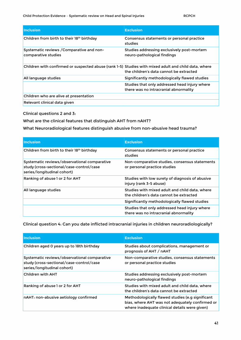
Child Protection Evidence – Systematic review on Head and Spinal injuries RCPCH
41
Inclusion Exclusion
Children from birth to their 18th birthday Consensus statements or personal practice studies
Systematic reviews /Comparative and non-comparative studies
Studies addressing exclusively post-mortem neuro-pathological findings
Children with confirmed or suspected abuse (rank 1-5) Studies with mixed adult and child data, where the children’s data cannot be extracted
All language studies Significantly methodologically flawed studies
Studies that only addressed head injury where there was no intracranial abnormality
Children who are alive at presentation
Relevant clinical data given
Clinical questions 2 and 3:
What are the clinical features that distinguish AHT from nAHT?
What Neuroradiological features distinguish abusive from non-abusive head trauma?
Inclusion Exclusion
Children from birth to their 18th birthday Consensus statements or personal practice studies
Systematic reviews/observational comparative study (cross-sectional/case-control/case series/longitudinal cohort)
Non-comparative studies, consensus statements or personal practice studies
Ranking of abuse 1 or 2 for AHT Studies with low surety of diagnosis of abusive injury (rank 3-5 abuse)
All language studies Studies with mixed adult and child data, where the children’s data cannot be extracted
Significantly methodologically flawed studies
Studies that only addressed head injury where there was no intracranial abnormality
Clinical question 4: Can you date inflicted intracranial injuries in children neuroradiologically?
Inclusion Exclusion
Children aged 0 years up to 18th birthday Studies about complications, management or prognosis of AHT / nAHT
Systematic reviews/observational comparative study (cross-sectional/case-control/case series/longitudinal cohort)
Non-comparative studies, consensus statements or personal practice studies
Children with AHT Studies addressing exclusively post-mortem neuro-pathological findings
Ranking of abuse 1 or 2 for AHT Studies with mixed adult and child data, where the children’s data cannot be extracted
nAHT: non-abusive aetiology confirmed Methodologically flawed studies (e.g significant bias, where AHT was not adequately confirmed or where inadequate clinical details were given)
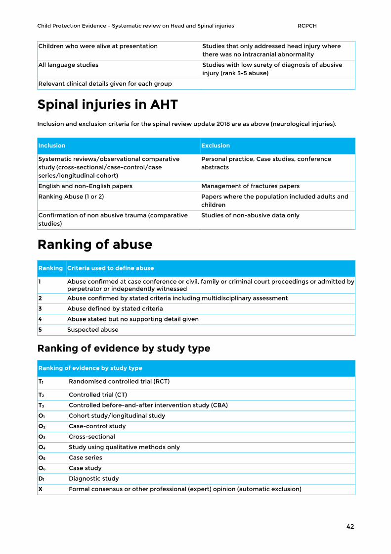
Child Protection Evidence – Systematic review on Head and Spinal injuries RCPCH
42
Children who were alive at presentation Studies that only addressed head injury where there was no intracranial abnormality
All language studies Studies with low surety of diagnosis of abusive injury (rank 3-5 abuse)
Relevant clinical details given for each group
Spinal injuries in AHT Inclusion and exclusion criteria for the spinal review update 2018 are as above (neurological injuries).
Inclusion Exclusion
Systematic reviews/observational comparative study (cross-sectional/case-control/case series/longitudinal cohort)
Personal practice, Case studies, conference abstracts
English and non-English papers Management of fractures papers
Ranking Abuse (1 or 2) Papers where the population included adults and children
Confirmation of non abusive trauma (comparative studies)
Studies of non-abusive data only
Ranking of abuse
Ranking Criteria used to define abuse
1 Abuse confirmed at case conference or civil, family or criminal court proceedings or admitted by perpetrator or independently witnessed
2 Abuse confirmed by stated criteria including multidisciplinary assessment
3 Abuse defined by stated criteria
4 Abuse stated but no supporting detail given
5 Suspected abuse
Ranking of evidence by study type
Ranking of evidence by study type
T1 Randomised controlled trial (RCT)
T2 Controlled trial (CT)
T3 Controlled before-and-after intervention study (CBA)
O1 Cohort study/longitudinal study
O2 Case-control study
O3 Cross-sectional
O4 Study using qualitative methods only
O5 Case series
O6 Case study
D1 Diagnostic study
X Formal consensus or other professional (expert) opinion (automatic exclusion)

Child Protection Evidence – Systematic review on Head and Spinal injuries RCPCH
43
Statistics
Used to answer our question: 1. What neuroradiological investigations are indicated to identify
abusive central neurological system injury in children?
The aim of the review published in 2009 was to analyse the relative value of an MRI examination
additional to the initial CT examination, and to estimate the proportion of cases in which an
additional MRI would provide supplementary information to an initial CT examination.130
Since not all children underwent both examinations, there remained the possibility that, had MRI
examinations been performed (when they were not), additional information would have been
provided. In these cases, a conservative assumption was used. It was assumed that such MRI
examinations would have revealed the same information as the CT investigation. The proportion
of cases in which an MRI examination provided additional information was computed.
A narrative description of the findings of studies added to the review thereafter is provided, as
many individual studies described the findings of advanced MRI techniques.
Used to answer our question: 2. What are the distinguishing clinical features of abusive
intracranial injury in children?
We analysed the following clinical features:
• Apnoea
• Retinal haemorrhages
• Rib fractures
• Long bone fractures
• Bruising to the head and/or neck
• Seizures
• Skull fractures
The analysis was limited by the items that authors chose to report. Further, even when a feature
was commented upon by an author, not all children in each group were examined for the feature
in question. In the systematic review published in 201060 we used an extremely conservative
imputation strategy to allow for this131 we chose to assume that any missing data (e.g.
fundoscopy) in the abusive head trauma (AHT) group would have been negative, had the child
been examined. If a data item was missing from the non-abusive head trauma (nAHT) group we
assumed it would have been positive, had the child been examined (e.g. rib fractures). Only in
the case of skull fractures, a feature whose presence we suspect to be associated with nAHT,
was the opposite imputation performed. We believe that this strategy may underestimate the

Child Protection Evidence – Systematic review on Head and Spinal injuries RCPCH
44
discriminating power of an individual feature, but that this counteracts any circularity in the data
collection.
These imputations being given, we then conducted a multilevel logistic regression analysis132,
allowing not only the prevalence of abuse to vary between studies, but also the odds ratios (OR)
for the features in question. By allowing the OR to differ between studies, we again aim to
minimise the risk of circularity, where an individual study may have overly relied upon a particular
feature in order to arrive at the diagnosis of abuse. For each feature, we report the estimated OR
for the feature in discriminating between AHT and nAHT (with a 95% confidence interval (CI)),
and a positive predictive value ((PPV) the estimated probability of abuse given the presence of
this feature in a child with brain injury (with a 95% CI)).
Thereafter, we have provided a narrative description of data published in studies identified
within the updated literature searches.
Question 3: What neuroradiological features distinguish abusive from non-abusive head trauma?
Included studies had the advantage that cases all had neuroimaging and we were able to
conduct a meta-analysis of the data grouped according to primary items of interest. We used
Odds ratios as a measure of effect with 95% confidence intervals. Data were pooled using a
random effects model to allow for inter and intra study variance and to give a more conservative
estimate of effect. RevMan was used to present data in Forest Plots. We have been able to
update these meta analyses with sequential updates of the systematic reviews.
Data for questions 4 and 5 are presented in a narrative synthesis.
Search strategy The below tables present the search terms used in the 2018 Medline database search for
neurological and spinal injuries, truncation and wildcard characters were adapted to the
different databases where necessary.
Neurological injuries
1. CHILD/ 2. (paediatric or pediatric or neonate*).af. 3. (child: or infant: or toddler: or babies or baby).af. 4. or/1-3 5. ((non-accidental or nonaccidental) adj3 (trauma
or injur:)).af. 6. ((non-abusive or nonabusive) adj3 (injur: or
trauma)).af. 7. (non-accidental: and injur:).af. 8. soft tissue injur:.af. 9. physical abuse.af.
55. spinal subdural.tw. 56. cervical.af. 57. exp Spinal Cord Injuries/ 58. Spinal Injuries/ 59. spinal cord trauma.af. 60. Spinal Cord Compression/ 61. Cervical Vertebrae/ 62. hyperflexion injur*.af. 63. hyperextension injur*.af. 64. or/23-6365. Fractur*.af. 66. exp Fractures, Comminuted/ 67. exp Fractures, Bone/
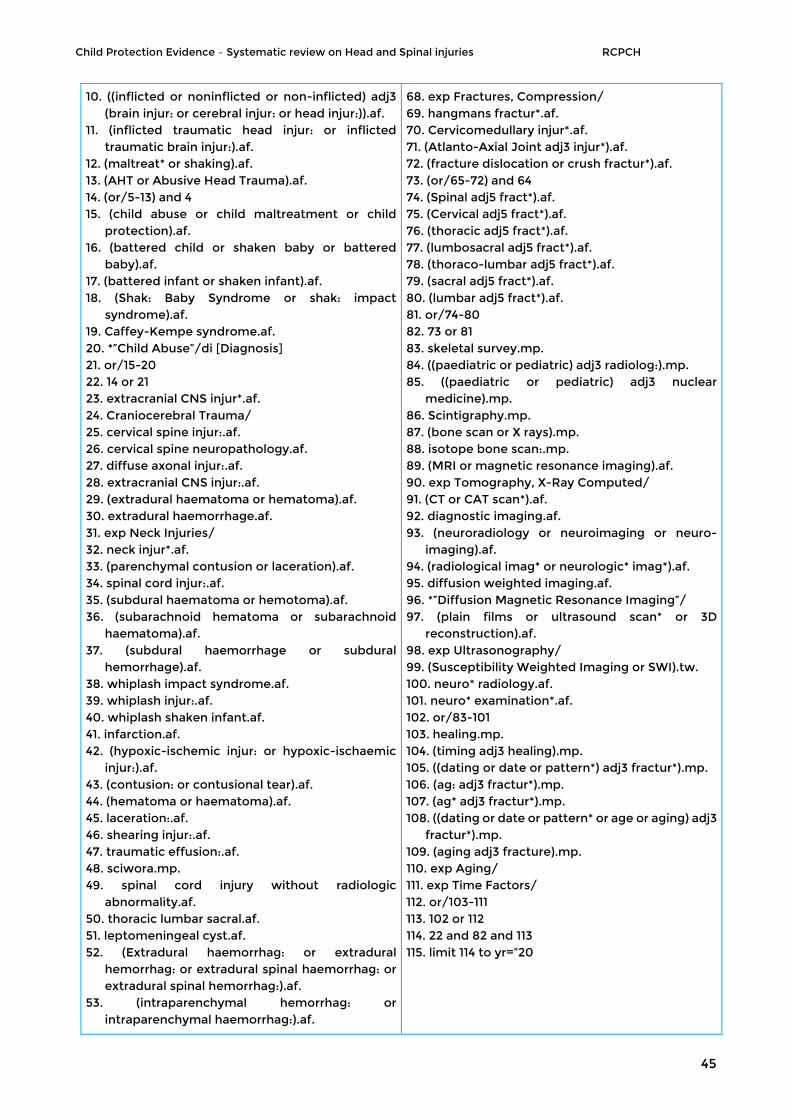
Child Protection Evidence – Systematic review on Head and Spinal injuries RCPCH
45
10. ((inflicted or noninflicted or non-inflicted) adj3 (brain injur: or cerebral injur: or head injur:)).af.
11. (inflicted traumatic head injur: or inflicted traumatic brain injur:).af.
12. (maltreat* or shaking).af. 13. (AHT or Abusive Head Trauma).af. 14. (or/5-13) and 4 15. (child abuse or child maltreatment or child
protection).af. 16. (battered child or shaken baby or battered
baby).af. 17. (battered infant or shaken infant).af. 18. (Shak: Baby Syndrome or shak: impact
syndrome).af. 19. Caffey-Kempe syndrome.af. 20. *”Child Abuse”/di [Diagnosis] 21. or/15-20 22. 14 or 21 23. extracranial CNS injur*.af. 24. Craniocerebral Trauma/ 25. cervical spine injur:.af. 26. cervical spine neuropathology.af. 27. diffuse axonal injur:.af. 28. extracranial CNS injur:.af. 29. (extradural haematoma or hematoma).af. 30. extradural haemorrhage.af. 31. exp Neck Injuries/ 32. neck injur*.af. 33. (parenchymal contusion or laceration).af. 34. spinal cord injur:.af. 35. (subdural haematoma or hemotoma).af. 36. (subarachnoid hematoma or subarachnoid
haematoma).af. 37. (subdural haemorrhage or subdural
hemorrhage).af. 38. whiplash impact syndrome.af. 39. whiplash injur:.af. 40. whiplash shaken infant.af. 41. infarction.af. 42. (hypoxic-ischemic injur: or hypoxic-ischaemic
injur:).af. 43. (contusion: or contusional tear).af. 44. (hematoma or haematoma).af. 45. laceration:.af. 46. shearing injur:.af. 47. traumatic effusion:.af. 48. sciwora.mp. 49. spinal cord injury without radiologic
abnormality.af. 50. thoracic lumbar sacral.af. 51. leptomeningeal cyst.af. 52. (Extradural haemorrhag: or extradural
hemorrhag: or extradural spinal haemorrhag: or extradural spinal hemorrhag:).af.
53. (intraparenchymal hemorrhag: or intraparenchymal haemorrhag:).af.
68. exp Fractures, Compression/ 69. hangmans fractur*.af. 70. Cervicomedullary injur*.af. 71. (Atlanto-Axial Joint adj3 injur*).af. 72. (fracture dislocation or crush fractur*).af. 73. (or/65-72) and 64 74. (Spinal adj5 fract*).af. 75. (Cervical adj5 fract*).af. 76. (thoracic adj5 fract*).af. 77. (lumbosacral adj5 fract*).af. 78. (thoraco-lumbar adj5 fract*).af. 79. (sacral adj5 fract*).af. 80. (lumbar adj5 fract*).af. 81. or/74-80 82. 73 or 81 83. skeletal survey.mp. 84. ((paediatric or pediatric) adj3 radiolog:).mp. 85. ((paediatric or pediatric) adj3 nuclear
medicine).mp. 86. Scintigraphy.mp. 87. (bone scan or X rays).mp. 88. isotope bone scan:.mp. 89. (MRI or magnetic resonance imaging).af. 90. exp Tomography, X-Ray Computed/ 91. (CT or CAT scan*).af. 92. diagnostic imaging.af. 93. (neuroradiology or neuroimaging or neuro-
imaging).af. 94. (radiological imag* or neurologic* imag*).af. 95. diffusion weighted imaging.af. 96. *”Diffusion Magnetic Resonance Imaging”/ 97. (plain films or ultrasound scan* or 3D
reconstruction).af. 98. exp Ultrasonography/ 99. (Susceptibility Weighted Imaging or SWI).tw. 100. neuro* radiology.af. 101. neuro* examination*.af. 102. or/83-101 103. healing.mp. 104. (timing adj3 healing).mp. 105. ((dating or date or pattern*) adj3 fractur*).mp. 106. (ag: adj3 fractur*).mp. 107. (ag* adj3 fractur*).mp. 108. ((dating or date or pattern* or age or aging) adj3
fractur*).mp. 109. (aging adj3 fracture).mp. 110. exp Aging/ 111. exp Time Factors/ 112. or/103-111 113. 102 or 112 114. 22 and 82 and 113 115. limit 114 to yr=”20

Child Protection Evidence – Systematic review on Head and Spinal injuries RCPCH
46
Spinal Injuries
54. diffuse axonal injur*.tw.
1. CHILD/ 2. (paediatric or pediatric or neonate*).af. 3. (child: or infant: or toddler: or babies or baby).af. 4. or/1-3 5. ((non-accidental or nonaccidental) adj3 (trauma
or injur:)).af. 6. ((non-abusive or nonabusive) adj3 (injur: or
trauma)).af. 7. (non-accidental: and injur:).af. 8. soft tissue injur:.af. 9. physical abuse.af. 10. ((inflicted or noninflicted or non-inflicted) adj3
(brain injur: or cerebral injur: or head injur:)).af. 11. (inflicted traumatic head injur: or inflicted
traumatic brain injur:).af. 12. (maltreat* or shaking).af. 13. (AHT or Abusive Head Trauma).af. 14. (or/5-13) and 4 15. (child abuse or child maltreatment or child
protection).af. 16. (battered child or shaken baby or battered
baby).af. 17. (battered infant or shaken infant).af. 18. (Shak: Baby Syndrome or shak: impact
syndrome).af. 19. Caffey-Kempe syndrome.af. 20. *”Child Abuse”/di [Diagnosis] 21. or/15-20 22. 14 or 21 23. extracranial CNS injur*.af. 24. Craniocerebral Trauma/ 25. cervical spine injur:.af. 26. cervical spine neuropathology.af. 27. diffuse axonal injur:.af. 28. extracranial CNS injur:.af. 29. (extradural haematoma or hematoma).af. 30. extradural haemorrhage.af. 31. exp Neck Injuries/ 32. neck injur*.af. 33. (parenchymal contusion or laceration).af. 34. spinal cord injur:.af. 35. (subdural haematoma or hemotoma).af. 36. (subarachnoid hematoma or subarachnoid
haematoma).af. 37. (subdural haemorrhage or subdural
hemorrhage).af. 38. whiplash impact syndrome.af. 39. whiplash injur:.af. 40. whiplash shaken infant.af. 41. infarction.af. 42. (hypoxic-ischemic injur: or hypoxic-ischaemic
injur:).af.
55. spinal subdural.tw. 56. cervical.af. 57. exp Spinal Cord Injuries/ 58. Spinal Injuries/ 59. spinal cord trauma.af. 60. Spinal Cord Compression/ 61. Cervical Vertebrae/ 62. hyperflexion injur*.af. 63. hyperextension injur*.af. 64. or/23-6365. Fractur*.af. 66. exp Fractures, Comminuted/ 67. exp Fractures, Bone/ 68. exp Fractures, Compression/ 69. hangmans fractur*.af. 70. Cervicomedullary injur*.af. 71. (Atlanto-Axial Joint adj3 injur*).af. 72. (fracture dislocation or crush fractur*).af. 73. (or/65-72) and 64 74. (Spinal adj5 fract*).af. 75. (Cervical adj5 fract*).af. 76. (thoracic adj5 fract*).af. 77. (lumbosacral adj5 fract*).af. 78. (thoraco-lumbar adj5 fract*).af. 79. (sacral adj5 fract*).af. 80. (lumbar adj5 fract*).af. 81. or/74-80 82. 73 or 81 83. skeletal survey.mp. 84. ((paediatric or pediatric) adj3 radiolog:).mp. 85. ((paediatric or pediatric) adj3 nuclear
medicine).mp. 86. Scintigraphy.mp. 87. (bone scan or X rays).mp. 88. isotope bone scan:.mp. 89. (MRI or magnetic resonance imaging).af. 90. exp Tomography, X-Ray Computed/ 91. (CT or CAT scan*).af. 92. diagnostic imaging.af. 93. (neuroradiology or neuroimaging or neuro-
imaging).af. 94. (radiological imag* or neurologic* imag*).af. 95. diffusion weighted imaging.af. 96. *”Diffusion Magnetic Resonance Imaging”/ 97. (plain films or ultrasound scan* or 3D
reconstruction).af. 98. exp Ultrasonography/ 99. (Susceptibility Weighted Imaging or SWI).tw. 100. neuro* radiology.af. 101. neuro* examination*.af. 102. or/83-101 103. healing.mp. 104. (timing adj3 healing).mp.
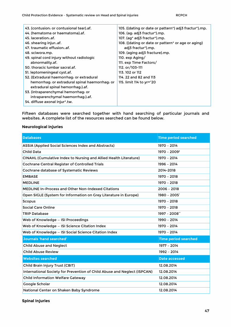
Child Protection Evidence – Systematic review on Head and Spinal injuries RCPCH
47
Fifteen databases were searched together with hand searching of particular journals and websites. A complete list of the resources searched can be found below.
Neurological injuries
Databases Time period searched
ASSIA (Applied Social Sciences Index and Abstracts) 1970 – 2014
Child Data 1970 – 2009†
CINAHL (Cumulative Index to Nursing and Allied Health Literature) 1970 – 2014
Cochrane Central Register of Controlled Trials 1996 – 2014
Cochrane database of Systematic Reviews 2014-2018
EMBASE 1970 – 2018
MEDLINE 1970 – 2018
MEDLINE In-Process and Other Non-Indexed Citations 2006 – 2018
Open SIGLE (System for Information on Grey Literature in Europe) 1980 – 2005*
Scopus 1970 – 2018
Social Care Online 1970 – 2018
TRIP Database 1997 – 2008**
Web of Knowledge — ISI Proceedings 1990 – 2014
Web of Knowledge — ISI Science Citation Index 1970 – 2014
Web of Knowledge — ISI Social Science Citation Index 1970 – 2014
Journals ‘hand searched’ Time period searched
Child Abuse and Neglect 1977 – 2014
Child Abuse Review 1992 – 2014
Websites searched Date accessed
Child Brain Injury Trust (CBIT) 12.08.2014
International Society for Prevention of Child Abuse and Neglect (ISPCAN) 12.08.2014
Child Information Welfare Gateway 12.08.2014
Google Scholar 12.08.2014
National Center on Shaken Baby Syndrome 12.08.2014
Spinal Injuries
43. (contusion: or contusional tear).af. 44. (hematoma or haematoma).af. 45. laceration:.af. 46. shearing injur:.af. 47. traumatic effusion:.af. 48. sciwora.mp. 49. spinal cord injury without radiologic
abnormality.af. 50. thoracic lumbar sacral.af. 51. leptomeningeal cyst.af. 52. (Extradural haemorrhag: or extradural
hemorrhag: or extradural spinal haemorrhag: or extradural spinal hemorrhag:).af.
53. (intraparenchymal hemorrhag: or intraparenchymal haemorrhag:).af.
54. diffuse axonal injur*.tw.
105. ((dating or date or pattern*) adj3 fractur*).mp. 106. (ag: adj3 fractur*).mp. 107. (ag* adj3 fractur*).mp. 108. ((dating or date or pattern* or age or aging)
adj3 fractur*).mp. 109. (aging adj3 fracture).mp. 110. exp Aging/ 111. exp Time Factors/ 112. or/103-111 113. 102 or 112 114. 22 and 82 and 113 115. limit 114 to yr=”20
Child Protection Evidence – Systematic review on Head and Spinal injuries RCPCH
48
Databases Time period searched
ASSIA (Applied Social Sciences Index and Abstracts) 2010-2014
Child Data N/A
CINAHL (Cumulative Index to Nursing and Allied Health Literature) 2010-2014
Cochrane Central Register of Controlled Trials 2010-2014
Cochrane database of Systematic Reviews 2014-2018
EMBASE 2010-2018
MEDLINE 2010-2018
MEDLINE In-Process and Other Non-Indexed Citations 2010-2018
Open SIGLE (System for Information on Grey Literature in Europe) N/A
Pubmed e-publications 2018
Scopus 2010-2018
Social Care online (previously Caredata) 2010-2018
Trip Plus N/A
Web of Knowledge — ISI Proceedings 2010-2014
Web of Knowledge — ISI Science Citation Index 2010-2014
Web of Knowledge — ISI Social Science Citation Index 2010-2014
Journals ‘hand searched’ Time period searched
Child Abuse and Neglect 2010-2014
Child Abuse Review 2010-2014
Websites searched Date accessed
The Alberta Research Centre for Health Evidence (ARCHE) 12 Nov 2014
Child Welfare Information Gateway (formerly National Clearinghouse on Child Abuse and Neglect)
12 Nov 2014
Google Scholar Nov
Pre-review screening and critical appraisal Papers found in the database and hand searches underwent three rounds of screening before
they were included in this update. The first round was a title screen where papers that obviously
did not meet the inclusion criteria were excluded. The second was an abstract screen where
papers that did not meet the inclusion criteria based on the information provided in the abstract
were excluded. These first two stages were carried out by a systematic reviewer at the RCPCH
and a clinical expert. Finally, a full text screen with a critical appraisal was carried out by members
of the clinical expert sub-committee. Critical appraisal forms were completed for each of the
papers reviewed at this stage. Examples of the pre-review screening and critical appraisal forms
used in previous reviews are available on request ([email protected]).


![WikiJournal Preprint/Systematic review...Systematic reviews can be used in many areas, such as evidence-based medicine and evidence-based policy and practice [17]. A systematic review](https://static.fdocuments.net/doc/165x107/5f109a937e708231d449ebdc/wikijournal-preprintsystematic-review-systematic-reviews-can-be-used-in-many.jpg)