Chest pain in a patient with congenital heart disease
3
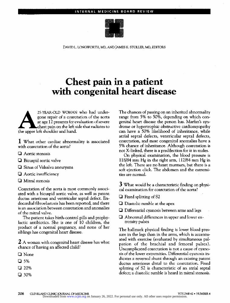
INTERNAL MEDICINE BOARD REVIEW DAVID L. LONGWORTH, MD, AND JAMES K. STOLLER, MD, EDITORS Chest pain in a patient with congenital heart disease A 25-YEAR-OLD WOMAN who had under- gone repair of a coarctation of the aorta at age 12 presents for evaluation of severe chest pain on the left side that radiates to the upper left shoulder and hand. 1 What other cardiac abnormality is associated with coarctation of the aorta? • Aortic stenosis • Bicuspid aortic valve • Sinus of Valsalva aneurysms • Aortic insufficiency • Mitral stenosis Coarctation of the aorta is most commonly associ- ated with a bicuspid aortic valve, as well as patent ductus arteriosus and ventricular septal defect. En- docardial fibroelastosis has been reported, and there is an association between coarctation and anomalies of the mitral valve. The patient takes birth-control pills and prophy- lactic antibiotics. She is one of 10 children, the product of a normal pregnancy, and none of her siblings has congenital heart disease. 2 A woman with congenital heart disease has what chance of having an affected child? • None • 5% • 20% • 30% The chances of passing on an inherited abnormality range from 5% to 50%, depending on which con- genital heart disease the person has. Marian's syn- drome or hypertrophic obstructive cardiomyopathy can have a 50% likelihood of inheritance, while atrial septal defects, ventricular septal defects, coarctation, and most congenital anomalies have a 5% chance of inheritance. Although coarctation is not X-linked, there is a predilection for it in males. On physical examination, the blood pressure is 118/84 mm Hg in the right arm, 112/84 mm Hg in the left. There are no heart murmurs, but there is a soft ejection click. The abdomen and the extremi- ties are normal. 3 What would be a characteristic finding on physi- cal examination for coarctation of the aorta? • Fixed splitting of S2 • Diastolic rumble at the apex • Differential cyanosis between arms and legs • Abnormal differences in upper and lower ex- tremity pulses The hallmark physical finding is lower blood pres- sure in the legs than in the arms, which is accentu- ated with exercise (evaluated by simultaneous pal- pation of the brachial and femoral pulses). Uncomplicated coarctation is not a cause of cyano- sis of the lower extremities. Differential cyanosis in- dicates a reversed shunt through an existing patent ductus arteriosus distal to the coarctation. Fixed splitting of S2 is characteristic of an atrial septal defect; a diastolic rumble is heard in mitral stenosis. 206 CLEVELAND CLINIC JOURNAL OF MEDICINE VOLUME 62 • NUMBER 4 on January 26, 2022. For personal use only. All other uses require permission. www.ccjm.org Downloaded from
Transcript of Chest pain in a patient with congenital heart disease
I N T E R N A L M E D I C I N E B O A R D R E V I E W
DAVID L. LONGWORTH, MD, AND JAMES K. STOLLER, MD, EDITORS
Chest pain in a patient with congenital heart disease
A2 5 - Y E A R - O L D W O M A N who had under-gone repair of a coarctation of the aorta at age 12 presents for evaluation of severe chest pain on the left side that radiates to
the upper left shoulder and hand.
1 What other cardiac abnormality is associated with coarctation of the aorta?
• Aortic stenosis • Bicuspid aortic valve
• Sinus of Valsalva aneurysms • Aortic insufficiency
• Mitral stenosis
Coarctation of the aorta is most commonly associ-ated with a bicuspid aortic valve, as well as patent ductus arteriosus and ventricular septal defect. En-docardial fibroelastosis has been reported, and there is an association between coarctation and anomalies of the mitral valve.
The patient takes birth-control pills and prophy-lactic antibiotics. She is one of 10 children, the product of a normal pregnancy, and none of her siblings has congenital heart disease.
2 A woman with congenital heart disease has what chance of having an affected child?
• None
• 5% • 20%
• 30%
The chances of passing on an inherited abnormality range from 5% to 50%, depending on which con-genital heart disease the person has. Marian's syn-drome or hypertrophic obstructive cardiomyopathy can have a 50% likelihood of inheritance, while atrial septal defects, ventricular septal defects, coarctation, and most congenital anomalies have a 5% chance of inheritance. Although coarctation is not X-linked, there is a predilection for it in males.
On physical examination, the blood pressure is 118/84 mm Hg in the right arm, 112/84 mm Hg in the left. There are no heart murmurs, but there is a soft ejection click. The abdomen and the extremi-ties are normal.
3 What would be a characteristic finding on physi-cal examination for coarctation of the aorta?
• Fixed splitting of S2
• Diastolic rumble at the apex
• Differential cyanosis between arms and legs
• Abnormal differences in upper and lower ex-tremity pulses
The hallmark physical finding is lower blood pres-sure in the legs than in the arms, which is accentu-ated with exercise (evaluated by simultaneous pal-pation of the brachial and femoral pulses). Uncomplicated coarctation is not a cause of cyano-sis of the lower extremities. Differential cyanosis in-dicates a reversed shunt through an existing patent ductus arteriosus distal to the coarctation. Fixed splitting of S2 is characteristic of an atrial septal defect; a diastolic rumble is heard in mitral stenosis.
206 CLEVELAND CLINIC JOURNAL OF MEDICINE VOLUME 62 • NUMBER 4 on January 26, 2022. For personal use only. All other uses require permission.www.ccjm.orgDownloaded from

I N T E R N A L M E D I C I N E B O A R D R E V I E W
A chest roentgenogram and magnetic resonance imaging (MRI) scan were obtained and are shown in Figures 1 and 2, respectively.
4 The chest roentgenogram and MRI scan are most consistent with which diagnosis?
• Recurrent aortic coarctation
• Poststenotic dilatation of the aorta
• Normal postoperative state
• Pseudoaneurysm of the thoracic aorta
During follow-up of patients who have undergone coarctation repair, the coarctation can recur (67% in some series, not all requiring repeat surgery) or an aneurysm can form. The abnormality on this pa-tient's chest roentgenogram is more than "post-stenotic dilation." The MRI suggests pseudoaneu-rysm of the aorta in the area of the patch annuloplasty. This is not uncommon; in one study of 152 patients who underwent patch annuloplasty re-pair, 35% had significant dilation and 19.5% re-quired a repeat operation.
5 What is the most common noncardiac anomaly in patients with coarctation of the aorta?
• Asplenia
• Circle of Willis aneurysm
• Renal artery stenosis
• Pulmonary hypertension
Circle of Willis aneurysms are found in 10% of pa-tients with coarctation. It may be clinically silent or manifest as abrupt rupture.
F I G U R E 1. Chest roentgenogram in a 25-year-old woman with chest pain who had undergone repair of a coarctation of the aorta 13 years previously.
D I S C U S S I O N
This case illustrates the need to be aware of problems in adult patients with repaired congenital heart defects. Coarctation of the aorta accounts for 15% of adult congenital heart abnormalities. There is a male predominance, and one third have a bicus-pid aortic valve. Symptoms include headache, epis-taxis, forceful carotid impulses, and lower extremity claudication. Two thirds of patients with untreated coarctation develop congestive heart failure as adults. A cerebrovascular accident can occur in 20% of cases. F I G U R E 2 . Magnetic resonance imaging scan in the same
patient.
JULY • AUGUST 1995 CLEVELAND CLINIC JOURNAL OF MEDICINE 2 0 7
19: Du« to insufficient »nnot*tion 19 cout<j um to »isdiasnosis, >0» Explicit Ho«Je. ¥t 6H8/L: 2<
on January 26, 2022. For personal use only. All other uses require permission.www.ccjm.orgDownloaded from

CLEVELAND CLINIC JOURNALOF
E D I C T
u
•MING URES
Perioperative management of common medical conditions
Coronary heart disease in African Americans: primary and secondary prevention
I N T E R N A L M E D I C I N E B O A R D R E V I E W
Clinically, the hallmark is a discrepancy in pulses, the pressure in the arms being greater than the legs, a difference often accentuated after exercise.
Electrocardiographic findings are nonspecific un-til the third or fourth decade, when left ventricular hypertrophy develops in untreated patients. Roentgenography of the chest can be diagnostic. An indentation followed by a poststenotic dilation of the aorta is known as the "three sign." An important finding is rib notching, because the more severe the coarctation, the more developed are the collaterals.
Intervention should be undertaken before the pa-tient reaches school age to decrease mortality. Bal-loon aortoplasty is an option, but it carries the risk of late aneurysm, which may be lessened with place-ment of a metallic stent. Surgical repair requires preoperative assessment of adequate collateral cir-culation to permit safe aortic cross-clamping with-out the risk of spinal-cord ischemia. Postoperative prophylaxis against subacute bacterial endocarditis should continue for life.
ANITA M. ARNOLD, DO Milwaukee Cardiology Associates
S U G G E S T E D R E A D I N G
Aebert H, Laas ], Bednarski P, Koch U , Prokop M, Borst HG. High incidence of aneurysm formation following patch plasty repair of coarctation. Eur J Cardiothorac Surg 1993; 7 :200-204. Kappetein PA, Guit GL, Bogers A], et al. Noninvasive long-term follow-up after coarctation repair. Ann Thorac Surg 1993; 5 5 : 1 1 5 3 -1159.
Liberthson RR. Congenital heart disease diagnosis and management in children and adults. Boston: Little, Brown, and Co., 1989.
An update on prostate cancer
2 0 8 CLEVELAND CLINIC JOURNAL OF MEDICINE VOLUME 62 • NUMBER 4
on January 26, 2022. For personal use only. All other uses require permission.www.ccjm.orgDownloaded from