Chenopodium ambrosioides as a bone graft … ARTICLE Open Access Chenopodium ambrosioides as a bone...
10
RESEARCH ARTICLE Open Access Chenopodium ambrosioides as a bone graft substitute in rabbits radius fracture Vicente F. Pinheiro Neto 1† , Rachel M. Ribeiro 2† , Camila S. Morais 3 , Matheus B. Campos 2 , Denilson A. Vieira 2 , Porfírio C. Guerra 3 , Ana L. Abreu-Silva 3 , José R. Silva Junior 3 , Flavia Raquel F. Nascimento 4 , Marilene O. R. Borges 2* and Antonio C.R. Borges 2 Abstract Background: Bone defects caused by trauma, infection or tumor resection are common in orthopedic clinic and depending on the extent of the fracture; the vast majority require treatment with bone substitutes. Among the bone grafts employees, the autograft is defined as the gold standard, but with some limitations, principally related to morbidity at the site of its removal, with the need to search for other biomaterials as adjuvant in bone regeneration. Therefore, the objective of the present study was to evaluate the use of Chenopodium ambrosioides as a bone graft substitute for the osseointegration of fractures in rabbits, compared to other bone grafts already employed in the surgical routine as Ricinus communis (castor oil) polyurethane and autogenous bone marrow. Methods: Forty-eight rabbits were divided into four groups. After anesthesia, a radius fracture was created and the animals were treated with the following grafts: C. ambrosioides graft, autogenous bone marrow, castor oil and saline (control). After 30, 60 and 90 days, the animals were submitted to radiographical and histological analyses and bone alkaline phosphatase, osteocalcin, biomechanical tension, and collagen were measured. We also realized a phytochemical screening and in vitro antioxidant activity including 1,1-Diphenyl-2-picrylhydrazyl (DPPH) radical. Results: The data showed that the growth of the bone callus was more expressive and biomechanical assessment showed better tensile strength in C. ambrosioides graft. The experimental results also revealed that there was significantly greater activity of the bone alkaline phosphatase and osteocalcin during early fracture healing, similarly to the group receiving autogenous bone marrow. Histologically, immature bone was observed in C. ambrosioides graft at 30 days, whereas the formation of cartilaginous tissue predominated in the other groups. A higher amount of type I collagen was observed in C. ambrosioides graft throughout treatment. It was detected strong presence of flavonoids and appreciable antioxidant activity. Conclusions: The results indicate that C. ambrosioides graft and autogenous bone marrow have similar ability to enhance bone regeneration, higher than the castor oil graft, suggesting that the medicinal plant can provide therapeutic benefits for bone tissue. Keywords: Chenopodium ambrosioides, Plant biomaterials, Heterograft, Bone regeneration, Fracture healing * Correspondence: [email protected] † Equal contributors 2 Departamento de Ciências Fisiológicas, Laboratório de Farmacologia, Universidade Federal do Maranhão, Av. dos Portugueses, 1966 - Bacanga, São Luis, MA 65080-805, Brazil Full list of author information is available at the end of the article © The Author(s). 2017 Open Access This article is distributed under the terms of the Creative Commons Attribution 4.0 International License (http://creativecommons.org/licenses/by/4.0/), which permits unrestricted use, distribution, and reproduction in any medium, provided you give appropriate credit to the original author(s) and the source, provide a link to the Creative Commons license, and indicate if changes were made. The Creative Commons Public Domain Dedication waiver (http://creativecommons.org/publicdomain/zero/1.0/) applies to the data made available in this article, unless otherwise stated. Pinheiro Neto et al. BMC Complementary and Alternative Medicine (2017) 17:350 DOI 10.1186/s12906-017-1862-5
Transcript of Chenopodium ambrosioides as a bone graft … ARTICLE Open Access Chenopodium ambrosioides as a bone...

RESEARCH ARTICLE Open Access
Chenopodium ambrosioides as a bone graftsubstitute in rabbits radius fractureVicente F. Pinheiro Neto1†, Rachel M. Ribeiro2†, Camila S. Morais3, Matheus B. Campos2, Denilson A. Vieira2,Porfírio C. Guerra3, Ana L. Abreu-Silva3, José R. Silva Junior3, Flavia Raquel F. Nascimento4,Marilene O. R. Borges2* and Antonio C.R. Borges2
Abstract
Background: Bone defects caused by trauma, infection or tumor resection are common in orthopedic clinic anddepending on the extent of the fracture; the vast majority require treatment with bone substitutes. Among thebone grafts employees, the autograft is defined as the gold standard, but with some limitations, principally relatedto morbidity at the site of its removal, with the need to search for other biomaterials as adjuvant in boneregeneration. Therefore, the objective of the present study was to evaluate the use of Chenopodium ambrosioides asa bone graft substitute for the osseointegration of fractures in rabbits, compared to other bone grafts alreadyemployed in the surgical routine as Ricinus communis (castor oil) polyurethane and autogenous bone marrow.
Methods: Forty-eight rabbits were divided into four groups. After anesthesia, a radius fracture was created and theanimals were treated with the following grafts: C. ambrosioides graft, autogenous bone marrow, castor oil and saline(control). After 30, 60 and 90 days, the animals were submitted to radiographical and histological analyses andbone alkaline phosphatase, osteocalcin, biomechanical tension, and collagen were measured. We also realized aphytochemical screening and in vitro antioxidant activity including 1,1-Diphenyl-2-picrylhydrazyl (DPPH) radical.
Results: The data showed that the growth of the bone callus was more expressive and biomechanical assessmentshowed better tensile strength in C. ambrosioides graft. The experimental results also revealed that there wassignificantly greater activity of the bone alkaline phosphatase and osteocalcin during early fracture healing, similarlyto the group receiving autogenous bone marrow. Histologically, immature bone was observed in C. ambrosioidesgraft at 30 days, whereas the formation of cartilaginous tissue predominated in the other groups. A higher amountof type I collagen was observed in C. ambrosioides graft throughout treatment. It was detected strong presence offlavonoids and appreciable antioxidant activity.
Conclusions: The results indicate that C. ambrosioides graft and autogenous bone marrow have similar ability toenhance bone regeneration, higher than the castor oil graft, suggesting that the medicinal plant can providetherapeutic benefits for bone tissue.
Keywords: Chenopodium ambrosioides, Plant biomaterials, Heterograft, Bone regeneration, Fracture healing
* Correspondence: [email protected]†Equal contributors2Departamento de Ciências Fisiológicas, Laboratório de Farmacologia,Universidade Federal do Maranhão, Av. dos Portugueses, 1966 - Bacanga,São Luis, MA 65080-805, BrazilFull list of author information is available at the end of the article
© The Author(s). 2017 Open Access This article is distributed under the terms of the Creative Commons Attribution 4.0International License (http://creativecommons.org/licenses/by/4.0/), which permits unrestricted use, distribution, andreproduction in any medium, provided you give appropriate credit to the original author(s) and the source, provide a link tothe Creative Commons license, and indicate if changes were made. The Creative Commons Public Domain Dedication waiver(http://creativecommons.org/publicdomain/zero/1.0/) applies to the data made available in this article, unless otherwise stated.
Pinheiro Neto et al. BMC Complementary and Alternative Medicine (2017) 17:350 DOI 10.1186/s12906-017-1862-5

BackgroundBone grafting consists of the transplantation of biocompat-ible material at the site where significant loss of bonesubstance had occurred, in order to reconstruct bonedefects generally caused by trauma, infection or tumorresection [1, 2].Autologous bone or autograft represents up to this
date, the gold standard and most effective method forbone regeneration as it promotes bone formation overits surface by direct bone bonding (osteoconduction)and induces local stem cells to differentiate into bonecells (osteoinduction) without any associated immuneresponse. However, often results in a high donor sitemorbidity and its availability is limited [3–5].The emergence of new bone graft options has generated
significant uncertainties regarding the determination of themost adequate product for surgical procedures that usesynthetic materials. Natural bone substitutes have beeninvestigated including tissues obtained from the samespecies (allografts) or from different species (hetero-grafts). These graft materials should be biocompatibleand non-toxic and should not stimulate inflammatoryprocesses [6, 7].Use of medicinal plants for graft production is a promis-
ing alternative since they are biocompatible, easily appliedand stored, and have been shown to favor bone growth [8].Chenopodium ambrosioides L. (syn. Dysphania ambro-sioides (L.) Mosyakin & Clemants), Chenopodiaceae, popu-larly known as “mastruz” or “erva-de-santa-maria”, is usedby the population in Brazil and Latin America [9] as teas,infusions or syrups for the treatment of inflammatoryconditions [10] and or contusions and fractures [11, 12]. Inaddition, Chenopodium ambrosioides occupies the 17thposition on the National Register of Plants of Interest tothe National Health System (Relação Nacional de Plantasde Interesse do Sistema Único de Saúde - RENISUS), a listcompiled by the Brazilian Government which consists of 71plant species used in folk medicine for the alternativetreatment of health conditions [13]. Experimental studieshave shown that C. ambrosioides exerts immunostimula-tory [14, 15], antimicrobial [16], anti-inflammatory andantinociceptive activities [17, 18].Studies from our group evaluating the effect of a poult-
ice prepared from the leaves of C. ambrosioides on bonehealing in rabbits have shown that this medicinal plantaccelerates bone regeneration as demonstrated by radio-logical and histological analysis, highlighting the import-ance of medicinal plants as biomaterials [8]. Therefore,the objective of the present study was to evaluate the useof C. ambrosioides as a bone graft substitute for theosseointegration of fractures in rabbits, compared to otherbone grafts already employed in the surgical routine asRicinus communis (castor oil) polyurethane and autogen-ous bone marrow.
MethodsAnimalsForty-eight adult male New Zealand rabbits (Oryctolaguscuniculus), weighing 3.0 ± 0.5 kg, obtained from the AnimalHouse of the State University of Maranhão (UniversidadeEstadual do Maranhão - UEMA) were used. The animalswere kept at a temperature of 24 ± 1 °C and received rationand water ad libitum. The animals were acclimated for10 days and handled under the same conditions. For theexperiment, the rabbits were divided randomly into fourgroups: Control group, C. ambrosioides graft group,Autogenous bone marrow group and Ricinus communisgraft (castor oil) group. All surgery was performed underketamine hydrochloride and xylazine hydrochlorideanesthesia, and all efforts were made to minimize suffering.
PlantChenopodium ambrosioides leaves were collected at thetime of use, in March 2009 from the “Berta Lanjes deMorretes” Medicinal Plant Garden, Federal University ofMaranhão (São Luís, MA, Brazil). A voucher specimenwas cataloged, identified and deposited under the regis-tration number 0998.
Preparation of gel from the lyophilized aqueous extractof C. ambrosioidesThe aqueous extract was obtained by macerating freshleaves (1000 g) in distilled water (1:4, v/v) at roomtemperature, under mechanical stirring for 24 h. Next, theaqueous extract was concentrated under reduced pressureand lyophilized (Fauber - LB1500, Terroni Equipamentos,São Carlos-SP), yielding 90 g and subsequently used in thepreparation of C. ambrosioides gel for use as a graft [19]in experimental protocols. As a basis gel was used carbo-pol gel. Each tube of 20 g of gel obtained exhibited lyophi-lized aqueous extract of C. ambrosioides 5%, pH 6, darkgreen color and odor characteristic of the plant species.
Phytochemical analysisThe methanol extract was subjected to phytochemicalanalysis for constituent identification using the phyto-chemical methods, which were previously described [20].In general, tests for the presence or absence of phyto-chemical compounds were involved the addition of anappropriate chemical agent to the preparation in a testtube. The presence or absence of saponins, flavonoids,tannins, alkaloids was subsequently detected.
Determination of DPPH·scavenging assayThe antioxidant activity of the extracts was measured onthe basis of the scavenging activity of the stable 1, 1- diphe-nyl 2-picrylhyorazyl (DPPH) free radical according to themethod described by Brand-Williams et al. [21] with slightmodifications [21]. DPPH solution (1 ml/0.1 mM) in
Pinheiro Neto et al. BMC Complementary and Alternative Medicine (2017) 17:350 Page 2 of 10

methanol was mixed with 1 ml of plant extract solution ofvarying concentrations (1,5, 3, 6, 12, 24, 48 and 100 μg/ml).The reaction was carried out in triplicate and the decreasein absorbance (A) was measured at 517 nm using UV-visspectrophotometer. Gallic acid was used as a positivecontrol. DPPH free radical scavenging ability (%) wascalculated by using the formula:
A517 nm of control=A517 nm of sample� �
=A517 nm of control� X 100Results were expressed as IC50.
Anesthesia and creation of bone defectsThe animals were anesthetized by intramuscular injectionof 5% ketamine hydrochloride (30 mg/kg) and 2% xylazinehydrochloride (4 mg/kg). An incision was made in the skinand subcutaneous tissue in the middle third of the leftradius. A complete, simple, transverse diaphyseal fracture(1 cm) was then created at this site with an oscillating bonesaw. In C. ambrosioides and Ricinus communis grafts,sufficient amounts of the bone grafts were applied to fill thebone defect. In Autogenous bone marrow, 1.0 ml of a bonemarrow aspirate was collected from the iliac crest with a bi-opsy needle and injected directly into the fracture focus.The tissues were closed with simple sutures. During thepostoperative period, dressings were applied daily for 10consecutive days and the sutures were removed after thisperiod. The animals were euthanized with lethal doses ofthe anesthetics used in the surgical procedure.
Radiographical analysisTo monitor fracture consolidation and the formation of abone callus, radiographs were obtained with a 100-mA X-rayapparatus (Siemens®, Hamburg-Germany) operating at 40 kVand 0.5 mA/s, using a 18 × 24 cm film. The radiographs wereobtained in the craniocaudal and mediolateral positions 30,60 and 90 days after surgery. For this analysis, the intensity ofthe periosteal reaction, closure of the fracture line, bonebridge, and bone callus formation were evaluated [22].
Biochemical markers of bone formationBlood samples were collected from the animals 30, 60 and90 days after surgery for serum measurement of thefollowing bone formation markers: osteocalcin weremeasured by electrochemiluminescent immunoassay byElecsys 1010/2010 Modular analytics E170 analyser withthe Roche Elecsys 1010/2010 (Roche Diagnostics,Mannheim, Germany), and thermostable bone alkalinephosphatase (BAP) measured with the Labtest® kit in asemi-automated apparatus - Bioplus® (Bioplus Produtospara Laboratório Ltda., Barueri, São Paulo).
Biomechanical evaluationTension testing of the fractured segments was performedafter 60 and 90 days to monitor the evolution of bone
callus strength. A computerized universal testing machine(model TT2420, TIRA Maschinenbau, Germany) wasused, applying a standard load in the velocity of 5 mm/min. The maximum load (force, N) and maximumdeformation (stretching) were obtained from the result ofthe tensile force sustained by the specimens and isexpressed as maximum force [23].
Histological analysisHistological analysis was performed 30, 60 and 90 daysafter surgery and application of the lyophilized aqueous C.ambrosioides, Ricinus communis and Autogenous bonemarrow grafts to monitor the evolution of bone callus for-mation. The formation of new fibrous, cartilaginous andbone tissues during the healing process was analyzed.Cross-sections were removed from the fractured bonesegments and fixed in 10% buffered formalin for 24 h [24].After decalcification in 10% nitric acid, the fragments wereprocessed and stained with hematoxylin-eosin and Siriusred [25, 26]. The histological sections were submitted todescriptive qualitative analysis to determine the pattern ofbone regeneration.
Statistical analysisThe results were expressed as the mean ± standard errorof the mean (SEM) and were submitted to analysis ofvariance (ANOVA), followed by the Newman-Keulspost-test, using the GraphPad Prism 5.0 program. Ap < 0.05 was considered significant. The histologicalresults were reported qualitatively.
ResultsPhytochemical screening and DPPH radical scavengingactivity of C. ambrosioidesPhytochemical analysis of the aqueous extract of C.ambrosioides revealed the strong presence of flavonoids,alkaloids and saponins as shown in Table 1.
Table 1 Phytochemical screening of the aqueous extract ofChenopodium ambrosioides L.
Class of compounds Aqueous extract
Flavonoids +++
Alkaloids ++
Tannins +
Phenolic compound ++
Catechins +++
Saponins +++
Anthocyanin +++
Steroids +
Terpenes -
+: Presence of constituents; −: Traces of constituents; 0: Absenceof constituents
Pinheiro Neto et al. BMC Complementary and Alternative Medicine (2017) 17:350 Page 3 of 10

Free radical scavenging activities of extract were assessedby the DPPH assay. The results show that aqueous extractof C. ambrosioides presented high DPPH elimination activitywith a with an IC50 value of 39,10 ± 2,90 μg/ml. IC50 valueof the positive control Gallic acid was 1,5 ± 0,35 μg/mL.
Radiographic analysis of the C. ambrosioides, autogenousbone marrow and Ricinus communis graftsGrafted material was observed in the fracture focus in allgroups at 30 days, as well as the presence of a fracture lineand a mild periosteal reaction. The onset of a periostealreaction was only seen in C. ambrosioides graft andAutogenous bone marrow (Fig. 1).At 60 days, a fibrous bone callus started to form in all
groups, but the fracture line was absent only in C.ambroioides graft (Fig. 2).After 90 days, consolidated fractures were already
observed in C. ambroioides graft and Autogenous bonemarrow, but a cortical fracture line was still present inthe latter. In contrast, a mature bone callus with anexuberant periosteal reaction was observed in theControl group and Ricinus communis graft (Fig. 3).
Biochemical markers of bone formation of the C.ambrosioides, autogenous bone marrow and Ricinuscommunis graftsAfter application of the grafts, only C. ambrosioides graftpresented an increase in BAP at 30 (54.06 ± 2.80% and43.50 ± 1.70%, respectively) and 60 days (49.07 ± 5.51%and 25.64 ± 6.40%, respectively) when compared to theControl group (Table 2). A significant difference inosteocalcin was observed between C. ambrosioides graft
and the Control group at 30 days (9.0 ± 1.3 and4.05 ± 1.0 ng/ml, respectively) (Table 2).
Biomechanical assessment of the C. ambrosioides,autogenous bone marrow and Ricinus communis graftsAt 60 days, applying a load velocity of 5 mm/min, speci-mens of C. ambrosioides graft and autogenous bonemarrow withstood the same maximum force, with theobservation of a slightly higher tensile strength in 60.98and 55.89 N, respectively (Fig. 4).At 90 days, specimens of C. ambrosioides graft with-
stood approximately double the maximum force com-pared to Autogenous bone marrow (272.32 and 149.87 N,respectively) when the same velocity was applied (Fig. 5).
Histological analysis of the C. ambrosioides, autogenousbone marrow and Ricinus communis graftsThirty days after application of the grafts (Fig. 6), a predom-inance of immature bone and discrete presence of cartilagewere only observed in C. ambrosioides graft animals. Onthe other hand, abundant cartilaginous tissue (cartilaginouscollar) was seen in Control animals which were also sub-mitted to complete diaphyseal fracture of the radius, butwere not treated with the grafts. A large number of chon-drocytes and the onset of bone remodeling were observedin Ricinus communis graft, whereas immature bone (non-lamellar bone) and the massive presence of cartilaginoustissue were noted in Autogenous bone marrow. At 60 days,all groups exhibited similar tissue architecture, includingthe presence of endochondral ossification and osteoclasts,and mature bone was observed at 90 days.Analysis of collagen at 30 days (Fig. 7) showed the forma-
tion of a mature osteon and greater collagen organization
Fig. 1 Effect of a graft of lyophilized aqueous Chenopodium ambrosioides L. extract on bone formation. Radiographs showing bone callusevolution after 30 day. Control group and Ricinus communis graft bone defect exhibiting the absence of a fibrous bone callus. C. ambroioidesgraft and Autogenous bone marrow bone defect exhibiting the beginning of formation of a fibrous bone callus
Pinheiro Neto et al. BMC Complementary and Alternative Medicine (2017) 17:350 Page 4 of 10

in C. ambrosioides graft when compared to the Controlgroup. A forming osteon predominated in Autogenousbone marrow and non-lamellar bone in Ricinus communisgraft. Both groups exhibited less organized collagen whencompared to C. ambrosioides.
DiscussionWe evaluated the bone repair process in rabbit radiowith significant loss of osseous tissue and use of natural
osseous graft as a gel of lyophilized aqueous extract ofChenopodium ambrosioides. The findings indicate thatthe gel of the plant promotes early bone formation bystimulating osteoblast production increasing tissue re-sistance, and favoring better collagen deposition, beingbiocompatible this way rabbits.In the process of bone repair occurs equilibrium be-
tween deposition and tissue absorption, the assessmentof specific biomarkers of new bone tissue formation is
Fig. 2 Effect of a graft of lyophilized aqueous Chenopodium ambrosioides L. extract on bone formation. Radiographs showing bone callusevolution after 60 day. Control group: Bone defect exhibiting the formation of a fibrous bone callus. C. ambroioides graft: Formation of a fibrousbone callus throughout the fracture line. Autogenous bone marrow: Bone defect exhibiting the beginning of formation of a fibrous bone callus.Ricinus communis graft: Bone defect exhibiting the formation of a fibrous bone callus and retraction of the fracture line
Fig. 3 Effect of a graft of lyophilized aqueous Chenopodium ambrosioides L. extract on bone formation. Radiographs showing bone callusevolution after 90 day. Groups: Control group, C. ambrosioides graft, Autogenous bone marrow and Ricinus communis graft. Control group:Formation of a prominent fibrous bone callus. C. ambrosioides graft: Formation of a mature bone callus throughout the fracture line. Autogenousbone marrow: Formation of a mature bone callus. Ricinus communis graft: Formation of a prominent fibrous bone callus
Pinheiro Neto et al. BMC Complementary and Alternative Medicine (2017) 17:350 Page 5 of 10

required. Bone alkaline phosphatase, the bone-specificisoform of alkaline phosphatase, is a specific marker ofbone formation, is only produced by osteoblasts and isessential for bone mineralization [27]. Elevated levels ofbone alkaline phosphatase are observed during theprocess of fracture consolidation [28]. In this respect,Peng et al. [29], studying the effect of a topical pasteconsisting of the extracts of different herbs on bone re-pair in rabbits, observed an increase in bone alkalinephosphatase activities [29]. In the present study, an in-crease in the serum levels of this enzyme was seen in an-imals receiving the C. ambrosioides graft 30 and 60 daysafter fracture creation (Table 2), revealing that this medi-cinal plant can stimulate bone alkaline phosphatase ac-tivity during early healing of the fracture in a timedependent manner, suggesting osteogenesis.
Another specific marker of bone formation is osteocalcin,which accounts for 80% of the gamma-carboxyglutamylcontent of mature bone. Osteocalcin is important for thediagnosis and investigation of bone alterations and servesas an indicator of the formation and maturation of bonetissue [28, 30]. In the present study, no significant differ-ence in osteocalcin was observed between C. ambrosioidesgraft and the other groups after 30, 60 and 90 days of bonecallus formation (Table 2). In contrast, animals that re-ceived Autogenous bone marrow showed elevated serumlevels of osteocalcin at 60 days compared with the controlgroup, suggesting this stage more active bone metabolism.Radiographic analysis at 30 days revealed the forma-
tion of bone callus in the fibrous C. ambrosioides graftdemonstrating early bone regeneration (Fig. 1), similarlyto Autogenous bone marrow. Previous results reported
Table 2 Comparison of bone alkaline phosphatase and osteocalcin after the application of Chenopodium ambrosioides L.
Group Bone Alkaline Phosphatase (%) Osteocalcin (ng/ml)
30 days 60 days 90 days 30 days 60 days 90 days
Control 43.5 ± 1.7 25.6 ± 6.4 45.4 ± 2.4 4.0 ± 1.0 10.1 ± 0.8 8.6 ± 1.0
C. ambrosioides graft 54.1 ± 2.8* 49.1 ± 5.5* 41.1 ± 4.8 9.0 ± 1.3* 13.8 ± 1.7 9.4 ± 1.9
Autogenous bone marrow 44.4 ± 2.7 32.4 ± 4.4 38.1 ± 6.9 7.2 ± 1.0 18.0 ± 1.9*,a 14.3 ± 1.8
Ricinus communis graft 45.9 ± 3.7 29.8 ± 0.6 37.4 ± 5.1 5.9 ± 2.1 8. 5 ± 2.2 10.0 ± 1.7
Results are reported as the mean ± SEM. *p < 0.05, significantly different compared to Control group. aSignificant difference compared to the Ricinus communisgrafts (ANOVA, Tukey test)
Fig. 4 Effect of a graft of lyophilized aqueous Chenopodium ambrosioides L. extract on bone formation. Biomechanical analysis at 60 days. Theresults are reported as maximum force (Fmax). Groups: Control group, C. ambrosioides graft, Autogenous bone marrow e Ricinus communis graft
Pinheiro Neto et al. BMC Complementary and Alternative Medicine (2017) 17:350 Page 6 of 10
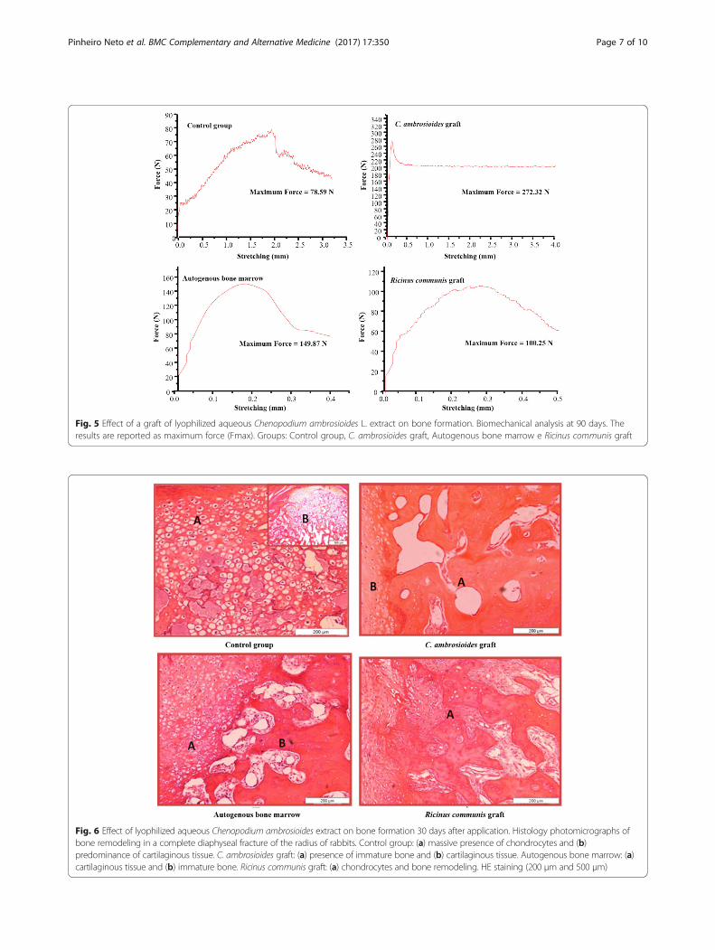
Fig. 5 Effect of a graft of lyophilized aqueous Chenopodium ambrosioides L. extract on bone formation. Biomechanical analysis at 90 days. Theresults are reported as maximum force (Fmax). Groups: Control group, C. ambrosioides graft, Autogenous bone marrow e Ricinus communis graft
Fig. 6 Effect of lyophilized aqueous Chenopodium ambrosioides extract on bone formation 30 days after application. Histology photomicrographs ofbone remodeling in a complete diaphyseal fracture of the radius of rabbits. Control group: (a) massive presence of chondrocytes and (b)predominance of cartilaginous tissue. C. ambrosioides graft: (a) presence of immature bone and (b) cartilaginous tissue. Autogenous bone marrow: (a)cartilaginous tissue and (b) immature bone. Ricinus communis graft: (a) chondrocytes and bone remodeling. HE staining (200 μm and 500 μm)
Pinheiro Neto et al. BMC Complementary and Alternative Medicine (2017) 17:350 Page 7 of 10

in literature corroborate our results by Lima et al. [31]who used bone morphogenetic proteins for theconsolidation of radius fractures in rabbits [31], and byFreitas et al. [32] who observed bone mineralizationafter application of a cortical bone heterograft in thesame species [32].In addition, the results show a fracture line absent only
in C. ambrosioides graft animals after 60 days, indicatingearly bone formation (Fig. 2), in contrast to other groupswhere there was still prominent bone defect. These find-ings contrast with those reported by Guerra et al. [33],in the study of bone perforations in dogs, still detectedthe presence of a fracture line at 60 days, but in a stageof consolidation [33]. The formation of the mature bonecallus was observed in all groups at 90 days (Fig. 3),agreeing with the findings of Pinheiro Neto et al. [8],which evaluated the action of C. ambrosioides poulticeof leaves in osseous tissue of rabbits [8].The results of the tensile strength test showed a better
performance in C. ambrosioides graft at 60 days (Fig. 4) and90 days (Fig. 5), compared to other groups suggesting moremature consolidation and increased resistance of bone tis-sue and confirming the radiographic results. Biomechanicalassays should only be performed after 60 days in view ofthe evolution of bone callus formation, as reported by Penget al. [29] using a paste of herbs for fracture treatment inrabbits [30] and by Kupczik et al. [34] using ciprofloxacinfor bone consolidation of rat femoral fractures [34].
In continuity, significant histological alterations wereonly observed after 30 days, when C. ambrosioides graftanimals exhibited lamellar bone (Fig. 6) and better colla-gen organization (Fig. 7) compared to the other groups.The tissue architecture was similar in all groups at 60 and90 days, with the observation of the presence of endo-chondral ossification, osteoclasts and mature bone. Wongand Rabie, 2006, 2007, 2010, when analyzed different plantspecies on bone formation also observed similar results[35–37]. Furthermore, Pereira Junior et al., [38] alsoreport a comparative study between polyurethanescontaining castor oil (soft segment) and cancellous boneautograft in the treatment of segmental bone defectsinduced in rabbits [38]. As well by Laureano Filho et al.,[39] analyzed the effect of Ricinus communis polymer onbone regeneration in rabbit calvaria [39].Flavonoids are the most ubiquitous groups of plant sec-
ondary metabolites [40], comprising flavones, isoflavonesand neoflavonoides and have, as one of the main func-tions, protection against oxidative stress in cells [41, 42].This class has direct effects on bone metabolism, such asdecreased bone resorption by osteoclasts, promotion ofdifferentiation and pro-osteoblastic cell mineralization,and an increase in alkaline phosphatase activity [43]. Thus,the presence of flavonoids in C. ambrosioides graft may becontributing to its effect on bone neoformation.DPPH radical scavenging is considered a good in vitro
model widely used to assess antioxidant efficacy within a
Fig. 7 Effect of lyophilized aqueous Chenopodium ambrosioides extract on bone formation 30 days after application. Histology photomicrographsof collagen organization of bone remodeling in a complete diaphyseal fracture of the radius of rabbits. Control group: (a) non-organized collagenand (b) beginning of osteon formation. C. ambrosioides graft: mature osteon. Autogenous bone marrow: predominance of non-lamellar bonetissue. Ricinus communis graft: (a) predominance of a forming osteon and (b) non-organized collagen. Sirius red staining under polarizedlight (200 μm)
Pinheiro Neto et al. BMC Complementary and Alternative Medicine (2017) 17:350 Page 8 of 10

very short time [44]. In its radical form, DPPH· has dis-appears on reduction by an antioxidant compound or aradical species to become a stable diamagnetic moleculeresulting the colour change from purple to yellow, whichcould be taken as an indication of the hydrogen donat-ing ability of the tested sample [45, 46].The results of this study revealed the removal capacity
of DPPH of C. ambrosioides, but lower than that ofgallic acid. On the other hand, however, its value is ap-preciable and antioxidant activity can be attributed. Thisfinding is in agreement with other studies reported inthe literature for antioxidant activity of C. ambroisoides[47, 48]. Removal of free radicals is positively associatedwith increased differentiation and proliferation of osteo-blasts [49], which may explain the effects of C. ambroi-soides graft on early bone healing.
ConclusionIn conclusion, the findings of this study suggest that theC. ambrosioides graft proved to be biocompatible bone byinducing earlier bone neoformation. Further studies arebeing developed in our laboratory in order to evaluate theclinical efficacy of this gel formulation as bone graft.
AcknowledgementsThis study is part of the thesis of Vicente Férrer Pinheiro Neto presented tothe Postgraduate Program in Biotechnology (RENORBIO)/UniversidadeFederal do Maranhão (UFMA) in cooperation with Instituto Federal doMaranhão (IFMA) and Universidade Estadual do Maranhão (UEMA).
FundingThis work was supported by CAPES (Comissão de Aperfeiçoamento dePessoal de Nível Superior) and Fundação de Amparo à Pesquisa do Estadodo Maranhão, Brasil (FAPEMA).
Availability of data and materialsThe materials are available from the authors.
Authors’ contributionsVFPN, ACRB, and FRFN designed the study. VFPN, RMR, DAV and MBCperformed the experiments. ALAS analysis and interpretation of histologicalstudy. PCG, CSM and JRSJ contributed data analysis. VFPN and RMR wrotethe manuscript. MORB revised the manuscript. All authors read andapproved the final manuscript.
Ethics approval and consent to paticipateThis study was approved by the Ethics Committee and AnimalExperimentation of State University of Maranhão, based on the Ethic ofAnimal Experimentation of Brazilian Society of Science in Laboratory Animals(Approval Number: CEEA-0027/2014).
Consent to participateNot applicable.
Competing interestsThe authors declare that they have no competing interests.
Publisher’s NoteSpringer Nature remains neutral with regard to jurisdictional claims inpublished maps and institutional affiliations.
Author details1Departamento de Medicina, Laboratório de Cirurgia Experimental, CentroUniversitário do Maranhão, São Luís, Maranhão, Brazil. 2Departamento deCiências Fisiológicas, Laboratório de Farmacologia, Universidade Federal doMaranhão, Av. dos Portugueses, 1966 - Bacanga, São Luis, MA 65080-805,Brazil. 3Departamento de Medicina Veterinária, Universidade Estadual doMaranhão, São Luis, Maranhão, Brazil. 4Departamento de Patologia,Laboratório de Imunofisiologia, Universidade Federal do Maranhão, São Luís,Maranhão, Brazil.
Received: 3 March 2017 Accepted: 27 June 2017
References1. Müller SS, Curcelli EC, Sardenberg T, Zuccon A, De Crudis Junior JL, Padovani
CR. Análise clínica e biomecânica do efeito do diclofenaco sódico naconsolidação da fratura da tíbia no rato. Acta Ortop. Bras. 2004;12:197–204.
2. García-Gareta E, Coathup MJ, Blunn GW. Osteoinduction of bone graftingmaterials for bone repair and regeneration. Bone. 2015;8:112–21.
3. Zhang J, Liu W, Schnitzler V, Tancret F, Bouler JM. Calcium phosphatecements for bone substitution: chemistry, handling and mechanicalproperties. Acta Biomater. 2014;10:1035–49.
4. Goulet JA, Senunas LE, DeSilva GL, Greenield MLVH. Autogenous iliac crestbone graft. Clin Orthop Relat Res. 1997;339:76–81.
5. Moore WR, Graves SE, Bain GI. Synthetic bone graft substitutes. ANZ J Surg.2001;71:354–61.
6. Blokhuis TJ, Arts JJC. Bioactive and osteoinductive bone graft substitutes:definitions, facts and myths. Injury. 2011;42:S26–9. doi:10.1016/j.injury.2011.06.010.
7. Zizzari VL, Zara S, Tetè G, Vinci R, Gherlone E, Cataldi A. Biologic and clinicalaspects of integration of different bone substitutes in oral surgery: aliterature review. Oral Surg Oral Med Oral Pathol Oral Radiol. 2016;122(4):392–402. doi:10.1016/j.oooo.2016.04.010.
8. Pinheiro Neto VF, Ribeiro RM, Morais CS, Vieira DA, Guerra PC, Abreu-SilvaAL, et al. Chenopodium ambroisioides in the repair of fractures in rabbits. IntJ Pharmacol. 2015;11:732–7. doi:10.3923/ijp.2015.732.737.
9. Lorenzi H, Matos FJA. Plantas medicinais no Brasil: nativas e exóticas. EditoraInstituto Plantarum. São Paulo: Nova Odessa; 2012.
10. Moraes EM. Perfil Botânico, Químico, Farmacológico e Toxicológico dasPlantas Medicinais Utilizadas no Estado do Maranhão. Monografia. São Luís:UFMA; 1996.
11. Pinheiro Neto VF, Araújo BMA, Candanedo P, Borges ACR. Efeito docataplasma das folhas de mastruz (Chenopodium ambrosioides) na reparaçãode tecidos moles e ósseo em rádio de coelho. J Bras Fitomed. 2015;3:62–6.
12. Baptistel AC, Coutinho JMCP, Lins Neto EMF, Monteiro JM. Plantasmedicinais utilizadas na Comunidade Santo Antônio, Currais, Sul do Piauí:um enfoque etnobotânico. Rev Bras Plantas Med. 2014;16:406–25.
13. Brasil. Ministério da Saúde, Secretaria de Ciências, Tecnologia e InsumosEstratégicos. Portaria Interministerial n° 2960. Programa de Nacional dePlantas Medicinais e Fitoterápicos. Diário Oficial da União, 10 dez, 2008.Available: http://bvsms.saude.gov.br/bvs/saudelegis/gm/2008/pri2960_09_12_2008.html. Accessed 13 Oct 2015.
14. Rios CE, Abreu AG, Braga Filho JA, Nascimento JR, Guerra RN, Amaral FM,Maciel MC, Nascimento FR. Chenopodium ambrosioides L. improvesphagocytic activity and decreases bacterial growth and the systemicinflammatory response in sepsis induced by cecal ligation and puncture.Front Microbiol. 2017 Feb 1;8:1–7. doi:10.3389/fmicb.2017.00148.
15. Cruz GVB, Pereira PVS, Patrício FJ, Costa GC, Sousa SM, Frazão JB, et al.Increase of cellular recruitment, phagocytosis ability and nitric oxideproduction induced by hydroalcoholic extract from Chenopodiumambrosioides leaves. J Ethnopharmacol. 2007;111:148–54.
16. Mabona U, Viljoen A, Shikanga E, Marston A, Van Vuuren S. Antimicrobialactivity of southern African medicinal plants with dermatological relevance:from an ethnopharmacological screening approach to combination studiesand the isolation of a bioactive compound. J Ethnopharmacol. 2013;148:45–55.
17. Ibironke GF, Ajiboye KI. Studies on the anti–inflammatory and analgesicproperties of Chenopodium ambrosioides leaf extract in rats. Int J Pharmacol.2007;3:111–5. doi:10.3923/ijp.2007.111.115.
18. TrivellatoGrassi L, Malheiros A, Meyre-Silva C, Buss ZS, Monguilhott ED,Fröde TS, et al. From popular use to pharmacological validation: a study of
Pinheiro Neto et al. BMC Complementary and Alternative Medicine (2017) 17:350 Page 9 of 10

the anti-inflammatory, anti-nociceptive and healings effects of Chenopodiumambrosioides extract. J Ethnopharmacol. 2013;145:127–38.
19. Pinheiro Neto VF, Borges ACR, Borges MOR, BMA A, Abreu-Silva AL, SMF F,et al. Processo para obtenção de liofilizado de extrato aquoso das partesaéreas de Chenopodium ambrosioides, formulação de composiçãofarmacêutica e seu uso como enxerto ósseo (BRPI1101651). Revista daPropriedade Industrial. 2013;2215:134.
20. MATOS FJA. Introdução à fitoquímica experimental. Fortaleza: Edições UFC; 2009.21. Brand-Williams W, Cuvelier ME, Berset C. Use of free radical method to
evaluate antioxidant activity. Lebensm Wiss Technol. 1995;28:25–30.22. Miranda ES, Cardoso FTS, Medeiros Filho JF, Barreto MDR, Teixeira RMM,
Wanderley AL, et al. Estudo experimental comparativo no uso de enxertoósseo orgânico e inorgânico no reparo de fraturas cirúrgicas em rádios decoelho. Acta Ortop Bras. 2005;13:245–8.
23. Gomes CS, Campos ACL, Torres OJM, Vasconcelos PRL, Moreira ATR, TenórioSB, et al. Efeito do extrato de Passiflora edulis na cicatrização da paredeabdominal de ratos: estudo morfológico e tensiométrico. Acta Cir Bras.2006;21:9–16.
24. Tolosa EMC. Manual de técnicas para histologia normal e patológica. 2ed.São Paulo: Manole; 2003.
25. Franco KL, Borges APB, Vilória MIV, Fernandes ES, Fehlberg AF. Puresynthetic hydroxyapatite, collagen associated synthetic hydroxyapatite andliposome associated synthetic hydroxyapatite as a bone substitute fordefects in bone healing of dogs: transmitted light microscopyosteointegration aspects. Arq Bras Med Vet. 2001;53:1–7.
26. Montes GS. Structural biology of the fibers of the collagenous and elasticsystems. Cell Biol Int. 1996;20:15–27.
27. Breur GJ, Allen MJ, Carlson SJ, Richardson DC. Markers of bone metabolismin dog breeds of different size. Res Vet Sci. 2004;76:53–5.
28. Alien MJ. Biochemical markers of bone metabolism in animals: uses andlimitations. Vet Clin Pathol. 2003;32:101–13.
29. Peng LH, Ko CH, Siu SW, Koon CM, Yue GL, Cheng WH, Lau TW, et al. Invitro & in vivo assessment of an herbal formula used tropically for bonefracture treatment. J Ethnopharmacol. 2010;131:282–9.
30. Ramalho RA, Peres WA, Campos LF, Accioly E. Vitamina K e saúde óssea. RevBras Nutr Clin. 2006;21:224–8.
31. Lima AFM, Rahal SC, Volpi RS, Mamprim MJ, Vulcano LC, Correia MA.Aspectos radiográficos e densitométricos na consolidação de fraturastratadas com proteínas morfogenéticas ósseas em rádios de coelhos. Braz JVet Res Anim Sci. 2004;41:416–22.
32. Freitas SH, Dória RGS, Mendonça FS, Neto JE, Camargo LM. Aspectoradiológico de heteroenxerto ósseo cortical fragmentado na reparação defalhas ósseas em coelhos. Rev Bras Ciênc Vet. 2008;15:107–10.
33. Guerra PC, Vulcano LC, Rocha NS. Radiographic, densitometric andhistological evaluation of the use of bone perforation on fracturesconsolidation of distal third of radius from dogs. Braz J Vet Res Anim Sc.2006;43:186–95.
34. Kupczik F, Vialle LRG, Nobre LO, Vieira LA, Fernandes AEO. Influência daciprofloxacina na consolidação óssea de fraturas de fêmur em ratos. ActaOrtop. Bras. 2009;17:228–31.
35. Wong RWK, Rabie ABM. Effect of Gusuibu graft on bone formation. J OralMaxillofac Surg. 2006;64:770–7.
36. Wong RWK, Rabie ABM. Effect of Savia miltiorrhiza extract on boneformation. J Biomed Mater Res A. 2008;85:506–12.
37. Wong RWK, Rabie ABM. Effect of Buguzhi (Psoralea corylifolia fruit) extracton bone formation. Phytother Res 2010;24:S155-S160.
38. Pereira-Júnior OCM, Rahal SC, Iamaguti P, Felisbino SL, Pavan PT, VulcanoLC. Comparison between polyurethanes containing castor oil (soft segment)and cancellous bone autograft in the treatment of segmental bone defectinduced in rabbits. J Biomater Appl. 2007;21:283–97.
39. Laureano Filho JR, Andrade ESS, Barbosa JRA, Camargo IB, Garcia RR. Effectsof demineralized bone matrix and a Ricinus communis polymer on boneregeneration: a histological study in rabbit calvaria. J Oral Sci. 2009;51:451–6.
40. Prasain JK. Wang CC, Barnes S: flavonoids and isoflavones (photoestrogens):absorption, metabolism, and bioactivity. Free Radical Bio Med. 2004;37:1324–50. doi:10.1016/j.freeradbiomed.2004.07.026.
41. Winkel-Shirley B. Flavonoid biosynthesis: a colorful model for genetics,biochemistry, cell biology and biotechnology. Plant Physiol. 2001;126(2):485–93.
42. Field TS, Lee DW, Holbrook NM. Why leaves turn red in autumn. The role ofanthocyanins in senescing leaves of red-osier dogwood. Plant Physiol. 2001;127(2):566–74.
43. Pang JL, Ricupero DA, Huang S, et al. Differential activity of kaempferol andquercetin in attenuating tumor necrosis factor receptor family signaling inbone cells. Biochem Pharmacol. 2006;71:818–26.
44. Rao AS, Reddy SG, Babu PP, Reddy AR. The antioxidant and antiproliferativeactivities of methanolic extracts from Njavara rice bran. BMC ComplementAltern Med. 2010;10:1–9. doi:10.1186/1472-6882-10-4.
45. Marxen K, Vanselow KH, Lippemeier S, Hintze R, Ruser A, Hansen UP.Determination of DPPH radical oxidation caused by methanolic extracts ofsome microalgal species by linear regression analysis of spectrophotometricmeasurements. Sensors. 2007;7:2080–95.
46. Lee YR, Woo KS, Kim KJ, Son JR, Jeong HS. Antioxidant activities of ethanolextracts from germinated specialty rough Rice. Food Sci Biotechnol. 2007;16(5):765–70.
47. Kumar R, Mishra AK, Dubey NK, Tripathi YB. Evaluation of Chenopodiumambrosioides oil as a potential source of antifungal, antiaflatoxigenic andantioxidant activity. Int J Food Microbiol. 2007;115:159–64.
48. Ghareeb MA, Saad AM, Abdou AM, Refahy LAG, Ahmed WS. A newKaempferol glycoside with antioxidant activity from Chenopodiumambrosioides growing in Egypt. Orient J Che. 2016;32:3053–61.
49. Dai Z, Li Y, Quarles LD, Song T, et al. Resveratrol enhances proliferation andosteoblastic differentiation in human mesenchymal stem cells viaER-dependent ERK1/2 activation. Phytomedicine. 2007;14(12):806–14.
• We accept pre-submission inquiries
• Our selector tool helps you to find the most relevant journal
• We provide round the clock customer support
• Convenient online submission
• Thorough peer review
• Inclusion in PubMed and all major indexing services
• Maximum visibility for your research
Submit your manuscript atwww.biomedcentral.com/submit
Submit your next manuscript to BioMed Central and we will help you at every step:
Pinheiro Neto et al. BMC Complementary and Alternative Medicine (2017) 17:350 Page 10 of 10