Chemotherapy-resistant osteosarcoma is highly …...Chemotherapy-resistant osteosarcoma is highly...
12
ORIGINAL ARTICLE Chemotherapy-resistant osteosarcoma is highly susceptible to IL-15-activated allogeneic and autologous NK cells Emilie P. Buddingh • Marco W. Schilham • S. Eriaty N. Ruslan • Dagmar Berghuis • Karoly Szuhai • Jolien Suurmond • Antonie H. M. Taminiau • Hans Gelderblom • R. Maarten Egeler • Massimo Serra • Pancras C. W. Hogendoorn • Arjan C. Lankester Received: 10 October 2010 / Accepted: 22 December 2010 / Published online: 15 January 2011 Ó The Author(s) 2011. This article is published with open access at Springerlink.com Abstract High-grade osteosarcoma occurs predomi- nantly in adolescents and young adults and has an overall survival rate of about 60%, despite chemotherapy and surgery. Therefore, novel treatment modalities are needed to prevent or treat recurrent disease. Natural killer (NK) cells are lymphocytes with cytotoxic activity toward virus- infected or malignant cells. We explored the feasibility of autologous and allogeneic NK cell–mediated therapies for chemotherapy-resistant and chemotherapy-sensitive high-grade osteosarcoma. The expression by osteosarcoma cells of ligands for activating NK cell receptors was studied in vitro and in vivo, and their contribution to NK cell– mediated cytolysis was studied by specific antibody blockade. Chromium release cytotoxicity assays revealed chemotherapy-sensitive and chemotherapy-resistant osteo- sarcoma cell lines and osteosarcoma primary cultures to be sensitive to NK cell–mediated cytolysis. Cytolytic activity was strongly enhanced by IL-15 activation and was dependent on DNAM-1 and NKG2D pathways. Autolo- gous and allogeneic activated NK cells lysed osteosarcoma primary cultures equally well. Osteosarcoma patient– derived NK cells were functionally and phenotypically unimpaired. In conclusion, osteosarcoma cells, including chemoresistant variants, are highly susceptible to lysis by IL-15-induced NK cells from both allogeneic and autolo- gous origin. Our data support the exploitation of NK cells or NK cell–activating agents in patients with high-grade osteosarcoma. Keywords Osteosarcoma Á Immunotherapy Á NK cells Á Bone sarcoma Introduction High-grade osteosarcoma is the most common primary malignant bone sarcoma, occurring mainly in adolescents and young adults [1]. Despite multi-agent chemotherapy and surgery, overall survival is still poor at about sixty percent [2–4]. Therefore, novel treatment modalities are urgently needed to either prevent or treat chemotherapy refractory and recurrent disease. Immunomodulatory agents such as interferon (IFN)-a and muramyl-tri-peptide Electronic supplementary material The online version of this article (doi:10.1007/s00262-010-0965-3) contains supplementary material, which is available to authorized users. E. P. Buddingh Á M. W. Schilham Á S. E. N. Ruslan Á D. Berghuis Á J. Suurmond Á R. M. Egeler Á A. C. Lankester (&) Department of Pediatrics, Leiden University Medical Center, PO Box 9600, 2300 RC Leiden, The Netherlands e-mail: [email protected] D. Berghuis Á P. C. W. Hogendoorn Department of Pathology, Leiden University Medical Center, PO Box 9600, 2300 RC Leiden, The Netherlands K. Szuhai Department of Molecular Cell Biology, Leiden University Medical Center, PO Box 9600, 2300 RC Leiden, The Netherlands A. H. M. Taminiau Department of Orthopedic Surgery, Leiden University Medical Center, PO Box 9600, 2300 RC Leiden, The Netherlands H. Gelderblom Department of Clinical Oncology, Leiden University Medical Center, PO Box 9600, 2300 RC Leiden, The Netherlands M. Serra Laboratorio di Oncologia Sperimentale, Istituto Ortopedico Rizzoli, Via di Barbiano 1/10, 40136 Bologna, Italy 123 Cancer Immunol Immunother (2011) 60:575–586 DOI 10.1007/s00262-010-0965-3
Transcript of Chemotherapy-resistant osteosarcoma is highly …...Chemotherapy-resistant osteosarcoma is highly...

ORIGINAL ARTICLE
Chemotherapy-resistant osteosarcoma is highly susceptibleto IL-15-activated allogeneic and autologous NK cells
Emilie P. Buddingh • Marco W. Schilham • S. Eriaty N. Ruslan • Dagmar Berghuis •
Karoly Szuhai • Jolien Suurmond • Antonie H. M. Taminiau • Hans Gelderblom •
R. Maarten Egeler • Massimo Serra • Pancras C. W. Hogendoorn • Arjan C. Lankester
Received: 10 October 2010 / Accepted: 22 December 2010 / Published online: 15 January 2011
� The Author(s) 2011. This article is published with open access at Springerlink.com
Abstract High-grade osteosarcoma occurs predomi-
nantly in adolescents and young adults and has an overall
survival rate of about 60%, despite chemotherapy and
surgery. Therefore, novel treatment modalities are needed
to prevent or treat recurrent disease. Natural killer (NK)
cells are lymphocytes with cytotoxic activity toward virus-
infected or malignant cells. We explored the feasibility of
autologous and allogeneic NK cell–mediated therapies
for chemotherapy-resistant and chemotherapy-sensitive
high-grade osteosarcoma. The expression by osteosarcoma
cells of ligands for activating NK cell receptors was studied
in vitro and in vivo, and their contribution to NK cell–
mediated cytolysis was studied by specific antibody
blockade. Chromium release cytotoxicity assays revealed
chemotherapy-sensitive and chemotherapy-resistant osteo-
sarcoma cell lines and osteosarcoma primary cultures to be
sensitive to NK cell–mediated cytolysis. Cytolytic activity
was strongly enhanced by IL-15 activation and was
dependent on DNAM-1 and NKG2D pathways. Autolo-
gous and allogeneic activated NK cells lysed osteosarcoma
primary cultures equally well. Osteosarcoma patient–
derived NK cells were functionally and phenotypically
unimpaired. In conclusion, osteosarcoma cells, including
chemoresistant variants, are highly susceptible to lysis by
IL-15-induced NK cells from both allogeneic and autolo-
gous origin. Our data support the exploitation of NK cells
or NK cell–activating agents in patients with high-grade
osteosarcoma.
Keywords Osteosarcoma � Immunotherapy � NK cells �Bone sarcoma
Introduction
High-grade osteosarcoma is the most common primary
malignant bone sarcoma, occurring mainly in adolescents
and young adults [1]. Despite multi-agent chemotherapy
and surgery, overall survival is still poor at about sixty
percent [2–4]. Therefore, novel treatment modalities are
urgently needed to either prevent or treat chemotherapy
refractory and recurrent disease. Immunomodulatory
agents such as interferon (IFN)-a and muramyl-tri-peptide
Electronic supplementary material The online version of thisarticle (doi:10.1007/s00262-010-0965-3) contains supplementarymaterial, which is available to authorized users.
E. P. Buddingh � M. W. Schilham � S. E. N. Ruslan �D. Berghuis � J. Suurmond � R. M. Egeler �A. C. Lankester (&)
Department of Pediatrics, Leiden University Medical Center,
PO Box 9600, 2300 RC Leiden, The Netherlands
e-mail: [email protected]
D. Berghuis � P. C. W. Hogendoorn
Department of Pathology, Leiden University Medical Center,
PO Box 9600, 2300 RC Leiden, The Netherlands
K. Szuhai
Department of Molecular Cell Biology, Leiden University
Medical Center, PO Box 9600, 2300 RC Leiden,
The Netherlands
A. H. M. Taminiau
Department of Orthopedic Surgery, Leiden University Medical
Center, PO Box 9600, 2300 RC Leiden, The Netherlands
H. Gelderblom
Department of Clinical Oncology, Leiden University Medical
Center, PO Box 9600, 2300 RC Leiden, The Netherlands
M. Serra
Laboratorio di Oncologia Sperimentale, Istituto Ortopedico
Rizzoli, Via di Barbiano 1/10, 40136 Bologna, Italy
123
Cancer Immunol Immunother (2011) 60:575–586
DOI 10.1007/s00262-010-0965-3
(MTP) have been added to standard chemotherapy regi-
mens in recent clinical trials [5–7]. Immunotherapy with
stimulatory cytokines such as IL-2 and IL-15 or the
adoptive transfer of ex vivo cytokine-activated cytotoxic
lymphocytes such as natural killer (NK) cells could be
another adjunct to current therapy [8–10].
NK cells lack a clonally rearranged antigen-specific
receptor. Instead, cytolytic activity toward virus-infected or
malignant cells is dependent on the balance between
inhibitory and activating signals. NK cell–activating sig-
nals are provided when the activating receptors Natural
Killer Group 2, member D (NKG2D), DNAX accessory
molecule-1 (DNAM-1), and the natural cytotoxicity
receptors (NCRs) NKp30, NKp44, and NKp46 bind their
respective ligands [11–13]. Although NK cell recognition
of tumor cells has been reported to be partially mediated
through NCRs, the responsible ligands are unknown [14].
The DNAM-1 ligands poliovirus receptor (PVR, CD155)
and Nectin-2 (CD112) are highly expressed by many
tumors, including sarcomas [13, 15, 16]. Ligands for
NKG2D are the stress-inducible major histocompatibility
class I polypeptide-related sequence (MIC) A and B and
the UL-16-binding proteins (ULBPs) 1–4. Inhibitory
ligands are the classical and non-classical human leukocyte
antigen (HLA) class I molecules expressed on all normal
cells [17]. These ligands bind to inhibitory killer immu-
noglobulin receptors (KIRs) and the C-type lectin hetero-
dimer CD94/NKG2A on NK cells, respectively. In addition
to high expression of NKG2D and DNAM-1 ligands, many
tumors show loss of HLA class I, possibly rendering them
susceptible to NK cell–mediated lysis [18, 19].
In the current study, we demonstrated the sensitivity of
chemotherapy-resistant and chemotherapy-sensitive osteo-
sarcoma cells to lysis by IL-15-activated NK cells and
identified the molecular mechanisms involved. NK cells of
osteosarcoma patients were not functionally impaired and
were able to lyse autologous tumor cells, supporting the use
of NK cell–activating agents in the treatment of osteosar-
coma patients.
Materials and methods
Patient material
A tissue array was constructed from formalin-fixed, paraffin-
embedded (FFPE) tissue retrospectively collected from 88
osteosarcoma patients treated at the LUMC as previously
described [20] (Suppl. Table 1). Peripheral blood mononu-
clear cells (PBMCs) were collected from healthy controls
and 22 pre-treatment osteosarcoma patients after written
informed consent was obtained, as approved by the Institu-
tional Review Board on Medical Ethics. Osteosarcoma
tissue samples were used for research in accordance with
national ethical guidelines (Code for Proper Secondary Use
of Human Tissue, Dutch Federation of Medical Scientific
Societies). All patient material was handled in a coded
fashion. Clinical and pathological details of all patients can
be found in Suppl. Table 2.
Cell lines and primary cultures
The osteosarcoma cell lines HOS, 143B/HOS, IOR-OS14,
SJSA-1, OHS, ZK-58, U2-OS, and SAOS-2 were charac-
terized and maintained as described earlier [21]. The EBV
B-LCL cell line 107 (EBV) and the erythroleukemic cell line
K562 were maintained in RPMI 1640 medium (Invitrogen,
Carlsbad, CA) supplemented with 10% fetal calf serum
(FCS, Invitrogen) and 1% penicillin/streptomycin (PS,
Invitrogen). The chemotherapy-resistant variant cell lines of
U2-OS and SAOS-2 were established as described previ-
ously [22–24] and were maintained in Iscove’s modified
Dulbecco’s medium (IMDM, Invitrogen) supplemented
with 10% FCS and PS. The doxorubicin (DX)-resistant
variants U2-OS-DX580 and SAOS-2-DX580 were cultured
in the presence of 580 lg/ml DX. The methotrexate (MTX)-
resistant variants U2-OS-MTX300 and SAOS-2-MTX1 lg
were cultured in the presence of 300 and 1,000 ng/ml MTX,
respectively. The cisplatin (cis-diamminedichloroplatinum,
CDDP)-resistant variants U2-OS-CDDP4 lg and SAOS-
2CDDP6 lg were cultured in the presence of 4 and 6 lg/ml
CDDP, respectively.
Fresh osteosarcoma samples L2808, L2599, L2792,
L2635, and L2531 were cultured as described previously by
our group for related tumors [25]. Clinical and histopa-
thological details can be found in Suppl. Table 3. L2531,
L2792, and L2599 were derived from patients with poor
histological response to pre-operative chemotherapy in the
primary tumor. L2808, a pulmonary metastatic sample, was
derived from a patient with good histological response in
the primary tumor but who relapsed nonetheless. L2635
originated from a patient with good histological response
to pre-operative chemotherapy who is currently in persis-
tent first complete remission (follow-up since diagnosis
20 months). Collected tissue pieces were dissociated
mechanically and cultured in RPMI 1640 medium supple-
mented with 20% FCS and PS. When subconfluence was
reached, cells were harvested using 0.05% Trypsin/EDTA
(Invitrogen) and passaged. Chromium release assays and
flow cytometric analyses were performed no later than at
passage 3.
Isolation and culture of NK cells
PBMCs were isolated using a Ficoll density gradient sep-
aration followed by NK cell enrichment using the MACS
576 Cancer Immunol Immunother (2011) 60:575–586
123

NK enrichment kit and LS columns (Miltenyi Biotec,
Bergisch Gladbach, Germany) according to the manufac-
turer’s protocol. Purity of NK cells was assessed by flow
cytometry and was typically around 95% (less than 1% T
cells). NK cells were cultured in AIM-V medium (Invit-
rogen), supplemented with 10% pooled human AB serum
(Sanquin, Rotterdam, the Netherlands), PS and glutamine
(Glutamax I, Invitrogen). Activated NK cells were cultured
with 10 ng/ml recombinant human interleukin-15 (IL-15)
for 3 days or 2 weeks (Peprotech, Rocky Hill, NJ).
Flow cytometry
Surface staining of cells for flow cytometry was performed
as described elsewhere [18]. Intracellular flow cytometry
staining was done using permeabilization and fixation kits
as per manufacturer’s instructions (00-5123; 00-5223;
00-8333, eBioscience, San Diego, CA). An overview of
antibodies can be found in Suppl. Table 4. Flow cytometry
of PBMC of osteosarcoma patients and healthy controls
was performed on a BD LSRII and analyzed using FACS
Diva Software 5.0 (both from Becton-Dickinson, San
Diego, CA). Mean fluorescence intensity (MFI) of cell
subsets was calculated by subtracting the MFI of a negative
population from the MFI of the population of interest
within one individual to correct for interindividual vari-
ability of background staining. Flow cytometry of cell lines
and purified NK cells was performed on a FACScalibur and
analyzed using Cellquest software (both Becton-Dickin-
son). MFI ratio was calculated by MFI of the specific
staining relative to the MFI of the appropriate isotype
control staining.
Immunohistochemistry
Immunohistochemistry was performed on FFPE tissue
array sections as previously described [20]. Testis was used
as a positive control for the activating NK ligands and
tonsil for the inhibitory ligands. Sections were blocked
using 10% swine or goat serum in PBS and subsequently
incubated with primary antibody diluted in 0.5% bovine
serum albumin (BSA) in PBS overnight. As a negative
control, 0.5% BSA/PBS without primary antibody was
used. All primary antibodies are listed in Suppl. Table 4.
Anti-rabbit/rat/mouse PowerVision Poly-HRP (Leica Bio-
systems, Newcastle Upon Tyne, United Kingdom) was
used as a secondary antibody, except for the MICA stain-
ing, in which case the Universal LSAB? Kit (DAKO,
Glostrup, Denmark) was used. DAB? (DAKO) was used
as a chromogen. Sections were counterstained using
Mayer’s hematoxylin. Tissue array images were acquired
using the MIRAX slide scanner (3DHISTECH, Budapest,
Hungary) and analyzed using the MIRAX viewer version
1.14 (3DHISTECH). Slides were scored by two observers
(EPB and PCWH) in a modified semi-quantitative scoring
system as proposed by Ruiter et al. [26]. The intensity of
staining was scored as 0, 1, 2, or 3 indicating absent, weak,
clear, or strong expression, respectively. Percentages of
positive cells were scored as 0 for 0%, 1 for 1–30%, 2 for
31–70%, and 3 for 71–100%.
Chromium release assays
Cytotoxicity was determined in standard 4-h Chromium
release assays. For experiments using PBMCs of OS
patients and controls, PBMCs were thawed from storage in
liquid nitrogen and allowed to recover for 16 h in RPMI
1640 supplemented with 10% FCS and PS. The E:T ratios
in these experiments were corrected for the percentage of
NK cells of PBMCs as determined by flow cytometry. For
all other experiments, purified unstimulated or IL-15-acti-
vated NK cells were used as effector cells. Target cells
(cell lines or primary cultures) were incubated with
100 lCi sodium-51-chromate (PerkinElmer, Wellesley,
MA) for 1 h. Effector cells (PBMCs, unstimulated purified
NK cells, or activated NK cells) were incubated for 4 h
with 2,500 target cells at eight effector:target (E:T) ratios
in triplicate. Maximum and spontaneous release was
determined by incubating targets in 2 N HCl or medium,
respectively. Supernatants were harvested and measured in
a gamma-counter (Wallac, PerkinElmer). Specific lysis
was determined as: (experimental release-spontaneous
release)/(maximum release-spontaneous release) 9 100%.
In all NK cytotoxicity experiments, K562 and EBV were
used as positive and negative controls, respectively. For
blocking experiments, NK cells were pre-incubated with
blocking anti-NKG2D (R&D systems, clone 149810) and/
or blocking anti-DNAM-1 (BD Pharmingen, clone DX11)
at a concentration of 20 lg/ml. To disrupt perforin/gran-
zyme–mediated cytolysis, NK cells were pre-incubated for
2 h at 37�C with or without 1 lM Concanamycin A
(Sigma-Aldrich, Zwijndrecht, the Netherlands) prior to
adding the NK cells to the target cells. To block
Fas-induced apoptosis, target cells were pre-incubated with
2 lg/ml neutralizing anti-Fas antibody (Clone ZB4,
Millipore, Temecula, CA).
Statistical analysis
Statistical analyses were performed using GraphPad Prism
5.0 (LaJolla, CA). Data with non-normal distribution or
small sample size were analyzed using non-parametric
methods (Mann–Whitney U, Kruskal–Wallis, Friedman,
and Dunn’s tests), and data with normal distribution were
analyzed using parametric methods (t tests, one-way
analysis of variance (ANOVA), and Bonferroni’s tests).
Cancer Immunol Immunother (2011) 60:575–586 577
123

Survival analyses were performed using Kaplan–Meier
curves and compared using the logrank method.
Results
Osteosarcoma cells are highly susceptible
to IL-15-activated allogeneic NK cells
We tested eight osteosarcoma cell lines for susceptibility to
cytolytic activity of freshly isolated (‘unstimulated’) and
IL-15-cultured (‘activated’) healthy donor-derived NK
cells. All cell lines were lysed by unstimulated allogeneic
NK cells at levels comparable to the positive control cell
line K562 (Fig. 1a and b). Cytolysis of all osteosarcoma
cell lines was strongly enhanced when IL-15-cultured
allogeneic NK cells were used.
Osteosarcoma cells express inhibitory and activating
NK cell ligands
Osteosarcoma cells expressed activating NK cell ligands
and HLA class I, both in vivo and in vitro (Table 1 and
Fig. 2). All osteosarcoma cell lines expressed HLA class I,
at least 3/5 NKG2D ligands and both DNAM-1 ligands.
Expression of ligands in vivo was determined on the tissue
array containing 144 samples of 88 patients. In chemo-
therapy-naive tumor material MICA, DNAM-1 ligands and
HLA class I were also expressed, albeit at different levels
(Fig. 2a). In tumor cells persisting after chemotherapy,
levels of MICA, HLA class I, and b-2 microglobulin
expression were unaltered but the expression levels of the
DNAM-1 ligands CD112 and CD155 were significantly
decreased (Fig. 2b). There was a trend for patients with
high (score [ 4) versus low (score B 4) expression of
MICA in pre-treatment diagnostic biopsies to have better
overall survival (n = 53, P-value logrank test = 0.07).
Expression level of HLA class I in diagnostic biopsies as
determined by staining with antibodies recognizing b-2
microglobulin, HLA-A, and HLA-B/C did not correlate
with tumor progression.
NK cells lyse osteosarcoma cells
in a DNAM-1- and NKG2D-dependent manner
NK cell–mediated cytolysis of osteosarcoma cells was
dependent on NKG2D and DNAM-1 pathways, as was
demonstrated by blocking DNAM-1, NKG2D, or both
receptors (Fig. 3). In resting NK cells, the DNAM-1
pathway appeared to predominate the cytolytic potential,
whereas the contribution of the NKG2D pathway was more
prominent in the cytolytic activity of IL-15-cultured NK
cells. In case of IL-15-cultured NK cells, blockade of both
pathways was required for the optimal inhibition of NK
cytolytic activity. Levels of expression of HLA class I did
not correlate with magnitude of lysis by unstimulated or
IL-15-activated NK cells. Similarly, levels of expression of
ligands for the activating receptors NKG2D or DNAM-1
did not correlate with degree of lysis by NK cells (data not
shown).
ba
Fig. 1 Osteosarcoma cells were sensitive to lysis by freshly isolated
NK cells (solid lines) and NK cells cultured in IL-15 for 2 weeks
(dashed lines). a examples of percentage of specific lysis are shown
for the osteosarcoma cell lines SAOS-2 (filled circle), 143B/HOS
(open circle), IOR/OS-14 (filled square), and ZK-58 (open square).
Cell lines were incubated with increasing numbers of NK cells (E:T;
effector to target ratio). Error bars represent standard error of the
mean lysis of a representative experiment performed in triplicate.
b mean percentage of specific lysis by unstimulated (white bars) and
IL-15-activated (black bars) NK cells of the osteosarcoma cell lines
143B/HOS, SJSA-1, OHS, U2-OS, SAOS-2, IOR/OS-14, HOS, and
ZK-58 at an effector to target ratio of 10:1. Error bars represent
standard error of the mean of independent experiments using different
healthy donor NK cells. Numbers in the bars represent number of
experiments. K562 and an EBV-transformed B-LCL (‘‘EBV’’) were
used as positive and negative controls, respectively. Mann–Whitney
U test was done comparing IL-15-activated NK cells with unstim-
ulated NK cells for each cell line; P-value \0.05 noted as
*; \0.01 as **; \0.001 as ***
578 Cancer Immunol Immunother (2011) 60:575–586
123

Ta
ble
1E
xp
ress
ion
of
inh
ibit
ory
and
acti
vat
ing
NK
lig
and
sb
yo
steo
sarc
om
a
HO
S1
43
B/
HO
S
SJS
A-1
IOR
/OS
-14
OH
SZ
K-5
8U
2-O
SS
AO
S-2
L2
53
1L
25
99
L2
63
5L
27
92
L2
80
8
U2
-OS
DX
58
0M
TX
30
0C
DD
P4
SA
OS
-2D
X5
80
MT
X1
CD
DP
6
MH
Ccl
ass
I?
??
??
??
?±
??
??
??
??
??
??
??
??
??
??
??
??
??
?
CD
48
(2B
4li
gan
d)
--
--
--
--
--
--
--
--
--
-
MIC
A±
?±
±-
±?
??
??
??
??
??
??
--
??
±-
MIC
B-
?-
±-
--
--
--
--
--
--
--
UL
BP
-1±
?±
±±
--
--
--
±±
±-
--
--
UL
BP
-2±
??
??
??
??
??
?±
??
??
??
??
-±
±±
±
UL
BP
-3-
±-
±?
±?
±?
±±
-±
--
--
±±
CD
11
2(N
ecti
n-2
)?
??
??
±±
??
??
??
??
?±
?±
±±
±±
??
CD
15
5(P
VR
)±
??
??
?±
??
??
??
??
??
?±
±?
?±
?
CD
54
(IC
AM
-1)
??
??
??
??
-±
--
--
--
--
-?
?±
±?
CD
58
(LF
A-3
)?
??
??
??
??
??
??
??
??
??
??
??
??
±?
?±
?
CD
95
(Fas
)±
±±
±±
±?
±?
?±
-±
-±
?-
±?
Eig
ht
ost
eosa
rco
ma
cell
lin
es(H
OS
,1
43
B/H
OS
,S
JSA
-1,
OH
S,
ZK
-58
,U
2-O
S,
and
SA
OS
-2),
six
chem
oth
erap
y-r
esis
tan
tv
aria
nt
cell
lin
es(d
ox
oru
bic
in(D
X)-
,m
eth
otr
exat
e(M
TX
)-,
and
cisp
lati
nu
m(C
DD
P)-
resi
stan
tv
aria
nts
of
U2
-OS
and
SA
OS
-2),
and
fiv
esh
ort
-ter
mcu
ltu
res
(L2
80
8,
L2
59
9,
L2
79
2,
L2
63
5,
and
L2
53
1;
all
no
late
rth
anp
assa
ge
3)
wer
eev
alu
ated
for
the
exp
ress
ion
of
NK
cell
lig
and
sb
yfl
ow
cyto
met
ry.
Ex
pre
ssio
no
fth
ein
hib
ito
ryli
gan
ds
CD
48
and
MH
Ccl
ass
Ian
dex
pre
ssio
no
fli
gan
ds
for
the
acti
vat
ing
rece
pto
rsN
KG
2D
(MIC
A,
MIC
B,
UL
BP
-1,
UL
BP
-2,
and
UL
BP
-3)
and
DN
AM
-1(C
D1
12
and
CD
15
5).
Ex
pre
ssio
no
fth
ead
hes
ion
mo
lecu
les
CD
54
and
CD
58
and
of
the
dea
thre
cep
tor
CD
95
(Fas
).(-
)m
ean
flu
ore
scen
ce
inte
nsi
ty(M
FI)
rati
oo
fsp
ecifi
cst
ain
ing
ver
sus
iso
typ
eco
ntr
ol\
2;
(±)
MF
Ira
tio
bet
wee
n2
and
5;
(?)
MF
Ira
tio
bet
wee
n5
and
10
;(?
?)
MF
Ira
tio
[1
0
Cancer Immunol Immunother (2011) 60:575–586 579
123
100 µM
MICA CD112 CD155
MICA CD112 CD155 HLA class I
a
b
c
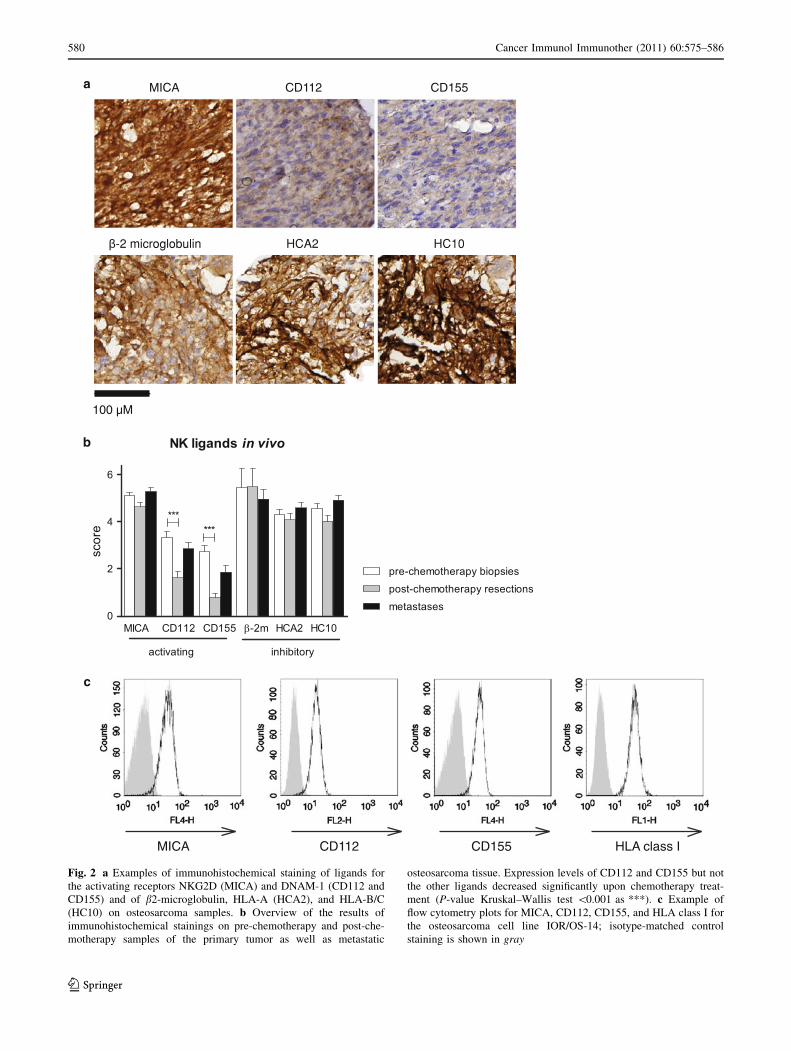
Fig. 2 a Examples of immunohistochemical staining of ligands for
the activating receptors NKG2D (MICA) and DNAM-1 (CD112 and
CD155) and of b2-microglobulin, HLA-A (HCA2), and HLA-B/C
(HC10) on osteosarcoma samples. b Overview of the results of
immunohistochemical stainings on pre-chemotherapy and post-che-
motherapy samples of the primary tumor as well as metastatic
osteosarcoma tissue. Expression levels of CD112 and CD155 but not
the other ligands decreased significantly upon chemotherapy treat-
ment (P-value Kruskal–Wallis test \0.001 as ***). c Example of
flow cytometry plots for MICA, CD112, CD155, and HLA class I for
the osteosarcoma cell line IOR/OS-14; isotype-matched control
staining is shown in gray
580 Cancer Immunol Immunother (2011) 60:575–586
123

Chemotherapy-resistant osteosarcoma cells remain
sensitive to lysis by IL-15-activated NK cells
To study whether chemotherapy-resistant cell lines have
become resistant to NK cell–mediated lysis as well, the
sensitivity of a panel of chemotherapy-resistant variants of
the osteosarcoma cell lines SAOS-2 and U2-OS (selected
in vitro to be resistant to DX, CDDP or MTX) to lysis by
NK cells was tested (Fig. 4a). Although some SAOS
variants, e.g., CDDP, were less sensitive to lysis by resting
NK cells, activation of NK cells with IL-15 greatly
enhanced the lysis of all U2-OS and SAOS-2 chemother-
apy-resistant variant cell lines (Fig 4a). Expression levels
of NKG2D and DNAM-1 ligands were similar in chemo-
therapy-resistant variants and parental cell lines, as was the
dependency on NKG2D and DNAM-1 signaling in cyto-
toxicity assays (Table 1 and Suppl. Fig. 1). Expression
levels of HLA class I and of the adhesion molecules
ICAM-1 and LFA-3 were unaltered in the chemotherapy-
resistant variants, but expression of CD95 (death receptor
Fas) was lost in the SAOS-2 CDDP- and DX-resistant
variants (Table 1 and Fig. 4b). Since the loss of CD95
could provide an explanation for reduced susceptibility to
NK cell–induced lysis, we performed blocking experiments
in which both CD95 and the granule exocytosis pathway
were blocked with a blocking antibody and Concanamycin
A, respectively. These experiments were performed using
IL-15-activated NK cells at an effector to target ratio of
40–1. Blocking the GrB but not the CD95 cytolytic path-
way almost completely abrogated NK cytolytic potential,
demonstrating the predominance of the granzymeB path-
way in NK cell–mediated lysis of parental as well as
CDDP-resistant variants of osteosarcoma cells (Fig. 4c).
Similar results were obtained when the U2-OS parental cell
line was used (data not shown).
Peripheral blood NK cell phenotype is unaltered
and cytolytic potential is unimpaired in newly
diagnosed osteosarcoma patients
Since peripheral NK cells in patients with other tumor
types show altered phenotype and function, we analyzed
a
b
c
Fig. 3 a cytolysis of U2-OS by unstimulated (solid lines) and IL-15-
activated (dashed lines) NK cells was almost completely abrogated
when the NK cells were pre-incubated with both anti (a)-DNAM-1-
and a-NKG2D-blocking antibodies (filled square vs. open square).
Unstimulated NK cells were most dependent on DNAM-1 (opencircle) signaling, whereas activated NK cells were most dependent on
NKG2D (filled circle). Error bars represent standard error of the
mean lysis of experiment performed in triplicate. Similar results were
obtained for SAOS-2, HOS, and ZK-58 using unstimulated (b) and
IL-15-activated NK cells (c). Bars represent mean lysis in at least
three independent experiments using healthy donor NK cells; errorbars represent standard error of the mean. Friedman test, Dunn’s post-
test compared to non-blocked; P-value \0.05 noted as *; \0.01 as
**; \0.001 as ***
Cancer Immunol Immunother (2011) 60:575–586 581
123
PBMCs of 22 newly diagnosed osteosarcoma patients and
23 age-matched healthy controls by flow cytometry for NK
cell number and phenotype (Suppl. Fig. 2a). NK cell
number and phenotype were comparable between patients
and controls (Fig. 5a and Suppl. Fig. 2b and c). Following
3 days of culture in IL-15, there was a larger increase in
NKG2D and granzyme B expression levels on both
CD56dim and bright NK cells of osteosarcoma patients
than of healthy controls (Fig. 5a and Suppl. Fig. 2c). We
assessed the functionality of NK cells of osteosarcoma
patients at diagnosis in cytotoxicity assays using unstimu-
lated and 3 days IL-15-activated PBMCs as effector cells.
Resting NK cells from osteosarcoma patients and healthy
donors lysed the allogeneic target HOS equally well
(Fig. 5b), but IL-15-cultured NK cells of patients lysed
HOS significantly better than healthy donor NK cells
(ANOVA, Bonferroni’s post-test P-value \0.0001). Per-
centage specific lysis of HOS correlated with the level of
NKG2D expression on CD56 dim NK cells (Fig. 5c;
Pearson correlation efficient r2 0.45, P-value \0.0001),
and similar results were obtained for the correlation with
granzyme B expression (not shown). To test whether the
functional integrity of NK cells from osteosarcoma patients
was also preserved toward autologous tumor cells, we took
advantage of the fact that we were able to derive short-term
cultured cells from fresh biopsies. Autologous, patient-
derived NK cells lysed short-term cultured tumor cells to a
similar degree as allogeneic NK cells from healthy controls
(Fig. 5d). In all cases, culture of both autologous and
allogeneic NK cells in IL-15 resulted in greatly enhanced
tumor cell killing.
Discussion
There is increasing interest in the potential for NK cells to be
used in the treatment of pediatric solid tumors [27]. Previous
studies have shown that osteosarcoma cell lines may be
sensitive to cytokine-activated NK cell–mediated cytotox-
icity [28–36]. However, little is known about the mecha-
nisms involved or the extent to which short-term cultured or
chemotherapy-resistant osteosarcoma cells are susceptible
to NK cell–mediated lysis. In addition, there is some evi-
dence for NK cell–mediated antiosteosarcoma activity in
vivo. Post-operative osteomyelitis-associated inhibition of
tumor growth was dependent on the activation of monocytes
and NK cells in a murine osteosarcoma model [37]. In
human osteosarcoma, treatment with interleukin (IL)-2 in a
small cohort of patients resulted in NK cell activation, which
was correlated with better clinical outcome [38]. Together,
these studies suggest that exploitation of NK cell activity
may be a suitable therapeutic tool in the adjuvant treatment
of osteosarcoma. In the present study, we demonstrate that
a
c
CD95
b
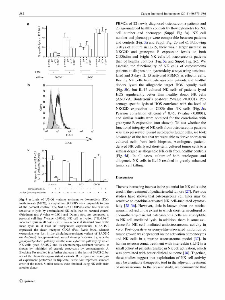
Fig. 4 a Lysis of U2-OS variants resistant to doxorubicin (DX),
methotrexate (MTX), or cisplatinum (CDDP) was comparable to lysis
of the parental control. The SAOS-2 CDDP-resistant line was less
sensitive to lysis by unstimulated NK cells than its parental control
(Friedman test P-value = 0.001 and Dunn’s post-test compared to
parental cell line P-value \0.001). NK cell activation (‘‘IL-15?’’)
increased lysis in all cases. Error bars represent standard error of the
mean lysis in at least six independent experiments. b SAOS-2
expressed the death receptor CD95 (Fas; black line), whereas
expression was lost in the cisplatinum-resistant variant of SAOS-2
(dashed line). Isotype-matched control staining is shown in gray. c the
granzyme/perforin pathway was the main cytotoxic pathway by which
NK cells lysed SAOS-2 and its chemotherapy-resistant variants, as
shown by inhibition of granule exocytosis by concanamycin A.
Blocking Fas resulted in a further decrease in the lysis of SAOS-2, but
not of the chemotherapy-resistant variants. Bars represent mean lysis
of experiment performed in triplicate; error bars represent standard
error of the mean. Similar results were obtained using NK cells from
another donor
582 Cancer Immunol Immunother (2011) 60:575–586
123

osteosarcoma cells are highly susceptible to NK cell–med-
iated cytolysis (Fig. 1). Osteosarcoma cells expressed acti-
vating NKG2D and DNAM-1 ligands in vivo as well as in
vitro, and lysis was dependent on the interaction between
these ligands on osteosarcoma cells and their receptors on
NK cells (Figs. 2 and 3). Despite expression of the poten-
tially inhibitory KIR ligand HLA class I by osteosarcoma
cells, all cell lines and short-term cultures were highly
sensitive to lysis by IL-15-activated NK cells (Table 1 and
Figs. 1 and 5d). Together, these data suggest that the balance
between expression of activating and inhibitory ligands in
osteosarcoma is shifted toward activation.
To investigate whether NK cell–based immunotherapy
is also feasible for patients with chemotherapy-resistant
disease, we tested the susceptibility of in vitro selected
chemotherapy-resistant osteosarcoma cells to NK cell–
mediated lysis. Methotrexate (MTX)-, doxorubicin (DX)-,
or cisplatin (CDDP)-resistant variants of the cell lines
SAOS-2 and U2-OS remained sensitive to lysis by IL-15-
activated NK cells (Fig. 4a). NK cells kill their targets by
the release of perforin and granzyme containing granules
and by the ligation of death receptors such as CD95 (Fas)
[39, 40]. Expression of CD95 was lost in the CDDP- and
DX-resistant SAOS-2 variants (Table 1 and Fig. 4), but
dual blocking studies demonstrated only a minor role of
Fas ligation in the lysis of osteosarcoma by cytokine-acti-
vated NK cells. Expression of Fas is frequently lost in
osteosarcoma pulmonary metastases, but our data show
that this will probably not hinder NK cell–based immu-
notherapeutic approaches [41–43].
ba c
d
Fig. 5 a NKG2D level was similar in unstimulated NK cells of
newly diagnosed osteosarcoma patients and healthy controls. Fol-
lowing culture for 3 days in IL15, there was a larger increase in the
expression level of NKG2D in NK cells of patients. b Unstimulated
PBMCs of 12 of 22 patients and 16 of 23 healthy controls and IL-15-
activated PBMCs of 19 of 22 patients and 17 of 23 controls were
available for functional testing. NK cells of newly diagnosed
osteosarcoma patients and of healthy controls lysed HOS at similar
levels. Following IL-15 activation, NK cells of osteosarcoma patients
showed a larger increase in cytotoxic activity than NK cells of healthy
donors. c cytotoxicity of IL-15-activated NK cells correlated with
level of NKG2D expression on the CD56dim subset (Pearson
correlation coefficient). d primary osteosarcoma cell cultures were
tested for sensitivity to lysis by autologous (open circle) and
allogeneic (closed circle) NK cells. NK cells were unstimulated
(solid lines) or 3 days IL-15 activated (dashed lines). Autologous IL-
15-activated NK cells were available for all patients except L2792.
One-way analysis of variance (ANOVA) P-value \0.0001 for a, b,and c. Bonferroni’s multiple comparison post-test; P-value \0.05
noted as *; \0.01 as **; \0.001 as ***
Cancer Immunol Immunother (2011) 60:575–586 583
123

Studies on the feasibility of immunotherapeutic strate-
gies in bone tumors are often hampered by the technical
difficulty to isolate viable fresh tumor cells for functional
testing. To circumvent this problem, we used short-term
cultured cells. Susceptibility to NK cell–mediated lysis was
determined no later than at passage three. Four out of five
cultures originated from patients with chemotherapy-
resistant disease (L2531, L2792, L2599, and L2808). Still,
all were highly sensitive to lysis by cytokine-activated
autologous and allogeneic NK cells (Fig. 5d). Importantly,
our experiments on in vitro selected chemotherapy-resis-
tant cells and on short-term cell cultures generated from
patients with chemotherapy-resistant disease in vivo show
that IL-15-activated NK cells are capable of lysis of oste-
osarcoma cells resistant to chemotherapeutic agents com-
monly used in high-grade osteosarcoma treatment.
In many tumor types, including Ewing sarcoma, host
immune cells have decreased functionality when compared
with healthy donor cells [13, 44, 45]. In these cases, using
allogeneic immune cells instead of autologous cells is an
attractive option to increase efficacy. However, it also
increases the risk of serious complications such as graft-
versus-host-disease. Our data show that NK cells of oste-
osarcoma patients are as potent as NK cells of healthy
controls in lysing osteosarcoma cells. Remarkably, upon
activation with IL-15, patient-derived NK cells even
showed a larger increase in the expression of NKG2D and
granzymeB than healthy donor–derived NK cells, which
correlated with an increased lysis of the osteosarcoma cell
line HOS (Fig. 5a and Suppl. Fig. 2c). This, and the lysis
of autologous tumor cells by ex vivo IL-15-activated NK
cells, indicates that immunotherapeutic strategies employ-
ing activated autologous NK cells could be as successful as
allogeneic NK cells in the treatment of osteosarcoma. In
preclinical validation studies, we obtained evidence that
IL15- and IL2-stimulated NK cells have similar cytolytic
activity against various tumor cell lines [46].
In conclusion, chemotherapy-resistant and chemother-
apy-sensitive osteosarcoma cells were lysed at high levels
by NK cells, particularly when NK cells were cytokine-
activated. Lysis of osteosarcoma cells was dependent on
DNAM-1 and NKG2D, ligands of which were expressed
by osteosarcoma cells both in vivo and in vitro. In contrast
to what has been reported in patients with other tumor
types, there was no intrinsic functional NK cell defect that
could hamper antitumor activity. Our study shows a
potential benefit of either activating NK cells in vivo by the
administration of cytokines or adoptive transfer of ex vivo
activated autologous or allogeneic NK cells in the treat-
ment of high-grade osteosarcoma.
Acknowledgments This study is supported by a grant from the
European Commission (EuroBoNeT, grant No 018814). EPB is
funded by a grant from the Netherlands Organization for Health
Research and Development (ZonMw grant 920-03-399). The authors
wish to thank Monique van Ostaijen-ten Dam for help in the cyto-
toxicity assays, Maarten van Tol for fruitful discussions and provision
of healthy donor PBMCs, Danielle de Jong for establishment and
characterization of short-term tumor cell cultures, and Nicolette
Leyerzapf, Judith Kroep, and Jakob Anninga for obtaining informed
consent of osteosarcoma patients.
Open Access This article is distributed under the terms of the
Creative Commons Attribution Noncommercial License which per-
mits any noncommercial use, distribution, and reproduction in any
medium, provided the original author(s) and source are credited.
References
1. Klein MJ, Parisien MV, Schneider-Stock R (2002) Osteogenic
Tumours. In: Fletcher CDM, Unni KK, Mertens F (eds) World
health classification of tumours. Pathology and genetics of
tumours of soft tissue and bone. IARC Press, Lyon, pp 259–285
2. Bielack SS, Kempf-Bielack B, Delling G, Exner GU, Flege S,
Helmke K, Kotz R, Salzer-Kuntschik M, Werner M, Winkelmann
W, Zoubek A, Jurgens H, Winkler K (2002) Prognostic factors in
high-grade osteosarcoma of the extremities or trunk: an analysis
of 1,702 patients treated on neoadjuvant cooperative osteosar-
coma study group protocols. J Clin Oncol 20:776–790
3. Buddingh EP, Anninga JK, Versteegh MI, Taminiau AH, Egeler
RM, van Rijswijk CS, Hogendoorn PCW, Lankester AC, Geld-
erblom H (2010) Prognostic factors in pulmonary metastasized
high-grade osteosarcoma. Pediatr Blood Cancer 54:216–221
4. Lewis IJ, Nooij MA, Whelan J, Sydes MR, Grimer R, Hogen-
doorn PCW, Memon MA, Weeden S, Uscinska BM, van Gla-
bbeke M, Kirkpatrick A, Hauben EI, Craft AW, Taminiau AHM
(2007) Improvement in histologic response but not survival in
osteosarcoma patients treated with intensified chemotherapy: a
randomized phase III trial of the European Osteosarcoma Inter-
group. J Natl Cancer Inst 99:112–128
5. Marina N, Bielack S, Whelan J, Smeland S, Krailo M, Sydes MR,
Butterfass-Bahloul T, Calaminus G, Bernstein M (2009) Inter-
national collaboration is feasible in trials for rare conditions: the
EURAMOS experience. Cancer Treat Res 152:339–353
6. Meyers PA, Schwartz CL, Krailo M, Kleinerman ES, Betcher D,
Bernstein ML, Conrad E, Ferguson W, Gebhardt M, Goorin AM,
Harris MB, Healey J, Huvos A, Link M, Montebello J, Nadel H,
Nieder M, Sato J, Siegal G, Weiner M, Wells R, Wold L, Womer R,
Grier H (2005) Osteosarcoma: a randomized, prospective trial of
the addition of ifosfamide and/or muramyl tripeptide to cisplatin,
doxorubicin, and high-dose methotrexate. J Clin Oncol
23:2004–2011
7. Meyers PA, Schwartz CL, Krailo MD, Healey JH, Bernstein ML,
Betcher D, Ferguson WS, Gebhardt MC, Goorin AM, Harris M,
Kleinerman E, Link MP, Nadel H, Nieder M, Siegal GP, Weiner
MA, Wells RJ, Womer RB, Grier HE (2008) Osteosarcoma: the
addition of muramyl tripeptide to chemotherapy improves overall
survival—a report from the Children’s Oncology Group. J Clin
Oncol 26:633–638
8. Terme M, Ullrich E, Delahaye NF, Chaput N, Zitvogel L (2008)
Natural killer cell-directed therapies: moving from unexpected
results to successful strategies. Nat Immunol 9:486–494
9. Miller JS, Soignier Y, Panoskaltsis-Mortari A, McNearney SA,
Yun GH, Fautsch SK, McKenna D, Le C, Defor TE, Burns LJ,
Orchard PJ, Blazar BR, Wagner JE, Slungaard A, Weisdorf DJ,
584 Cancer Immunol Immunother (2011) 60:575–586
123

Okazaki IJ, McGlave PB (2005) Successful adoptive transfer and
in vivo expansion of human haploidentical NK cells in patients
with cancer. Blood 105:3051–3057
10. Ljunggren HG, Malmberg KJ (2007) Prospects for the use of NK
cells in immunotherapy of human cancer. Nat Rev Immunol
7:329–339
11. Moretta A, Bottino C, Vitale M, Pende D, Cantoni C, Mingari
MC, Biassoni R, Moretta L (2001) Activating receptors and
coreceptors involved in human natural killer cell-mediated
cytolysis. Annu Rev Immunol 19:197–223
12. Mistry AR, O’Callaghan CA (2007) Regulation of ligands for the
activating receptor NKG2D. Immunology 121:439–447
13. Verhoeven DH, de Hooge AS, Mooiman EC, Santos SJ, ten Dam
MM, Gelderblom H, Melief CJ, Hogendoorn PCW, Egeler RM,
van Tol MJ, Schilham MW, Lankester AC (2008) NK cells
recognize and lyse Ewing sarcoma cells through NKG2D and
DNAM-1 receptor dependent pathways. Mol Immunol 45:
3917–3925
14. Lakshmikanth T, Burke S, Ali TH, Kimpfler S, Ursini F, Ruggeri
L, Capanni M, Umansky V, Paschen A, Sucker A, Pende D, Groh
V, Biassoni R, Hoglund P, Kato M, Shibuya K, Schadendorf D,
Anichini A, Ferrone S, Velardi A, Karre K, Shibuya A, Carbone
E, Colucci F (2009) NCRs and DNAM-1 mediate NK cell rec-
ognition and lysis of human and mouse melanoma cell lines in
vitro and in vivo. J Clin Invest 119:1251–1263
15. Bottino C, Castriconi R, Pende D, Rivera P, Nanni M,
Carnemolla B, Cantoni C, Grassi J, Marcenaro S, Reymond N,
Vitale M, Moretta L, Lopez M, Moretta A (2003) Identification of
PVR (CD155) and nectin-2 (CD112) as cell surface ligands for
the human DNAM-1 (CD226) activating molecule. J Exp Med
198:557–567
16. El-Sherbiny YM, Meade JL, Holmes TD, McGonagle D, Mackie
SL, Morgan AW, Cook G, Feyler S, Richards SJ, Davies FE,
Morgan GJ, Cook GP (2007) The requirement for DNAM-1,
NKG2D, and NKp46 in the natural killer cell-mediated killing of
myeloma cells. Cancer Res 67:8444–8449
17. Karre K (2008) Natural killer cell recognition of missing self. Nat
Immunol 9:477–480
18. Berghuis D, de Hooge ASK, Santos SJ, Horst D, Wertz EJ, van
Eggermond MC, van den Elsen PJ, Taminiau AHM, Ottaviano L,
Schaefer KL, Dirksen U, Hooijberg E, Mulder A, Melief CJM,
Egeler RM, Schilham MW, Jordanova ES, Hogendoorn PCW,
Lankester AC (2009) Reduced human leukocyte antigen
expression in advanced-stage Ewing sarcoma: implications for
immune recognition. J Pathol 218:222–231
19. Chang CC, Ferrone S (2007) Immune selective pressure and HLA
class I antigen defects in malignant lesions. Cancer Immunol
Immunother 56:227–236
20. Mohseny AB, Szuhai K, Romeo S, Buddingh EP, Briaire-de
Bruijn I, de Jong D, van Pel M, Cleton-Jansen AM, Hogendoorn
PCW (2009) Osteosarcoma originates from mesenchymal stem
cells in consequence of aneuploidization and genomic loss of
Cdkn2. J Pathol 219:294–305
21. Ottaviano L, Schaefer KL, Gajewski M, Huckenbeck W, Baldus
S, Rogel U, Mackintosh C, de Alava E, Myklebost O, Kresse SH,
Meza-Zepeda LA, Serra M, Cleton-Jansen AM, Hogendoorn
PCW, Buerger H, Aigner T, Gabbert HE, Poremba C (2010)
Molecular characterization of commonly used cell lines for bone
tumor research: a trans-European EuroBoNet effort. Genes
Chromosomes Cancer 49:40–51
22. Pasello M, Michelacci F, Scionti I, Hattinger CM, Zuntini M,
Caccuri AM, Scotlandi K, Picci P, Serra M (2008) Overcoming
glutathione S-transferase P1-related cisplatin resistance in oste-
osarcoma. Cancer Res 68:6661–6668
23. Serra M, Reverter-Branch MauriciD, Benini S, Shen JN, Chano
T, Hattinger CM, Manara MC, Pasello M, Scotlandi K, Picci P
(2004) Analysis of dihydrofolate reductase and reduced folate
carrier gene status in relation to methotrexate resistance in oste-
osarcoma cells. Ann Oncol 15:151–160
24. Serra M, Scotlandi K, Manara MC, Maurici D, Lollini PL,
Degiovanni C, Toffoli G, Baldini N (1993) Establishment and
characterization of multidrug-resistant human osteosarcoma cell-
lines. Anticancer Res 13:323–329
25. Szuhai K, Ijszenga M, Tanke HJ, Rosenberg C, Hogendoorn
PCW (2006) Molecular cytogenetic characterization of four
previously established and two newly established Ewing sarcoma
cell lines. Cancer Genet Cytogenet 166:173–179
26. Ruiter DJ, Ferrier CM, van Muijen GNP, Henzen-Logmans SC,
Kennedy S, Kramer MD, Nielsen BS, Schmitt M (1998) Quality
control of immunohistochemical evaluation of tumour-associated
plasminogen activators and related components. Eur J Cancer
34:1334–1340
27. Cho D, Shook DR, Shimasaki N, Chang YH, Fujisaki H, Campana
D (2010) Cytotoxicity of activated natural killer cells against
pediatric solid tumors. Clin Cancer Res 16:3901–3909
28. Nakashima Y, Deie M, Yanada S, Sharman P, Ochi M (2005)
Magnetically labeled human natural killer cells, accumulated in
vitro by an external magnetic force, are effective against HOS
osteosarcoma cells. Int J Oncol 27:965–971
29. Liebau C, Merk H, Schmidt S, Roesel C, Karreman C, Prisack JB,
Bojar H, Baltzer AWA (2002) Interleukin-12 and interleukin-18
change ICAM-1 expression, and enhance natural killer cell
mediated cytolysis of human osteosarcoma cells. Cytokines Cell
Mol Ther 7:135–142
30. Honorati MC, Neri S, Cattini L, Facchini A (2003) IL-17
enhances the susceptibility of U-2OS osteosarcoma cells to NK
cell lysis. Clin Exp Immunol 133:344–349
31. Kubista B, Trieb K, Blahovec H, Kotz R, Micksche M (2002)
Hyperthermia increases the susceptibility of chondro- and oste-
osarcoma cells to natural killer cell-mediated lysis. Anticancer
Res 22:789–792
32. Mariani E, Tarozzi A, Meneghetti A, Tadolini M, Facchini A
(1996) Lytic activity of IL-2 and IL-12 stimulated NK cells
against HOS osteosarcoma cell line. Boll Soc Ital Biol Sper
72:21–27
33. Mariani E, Tarozzi A, Meneghetti A, Cattini L, Facchini A
(1997) Human osteosarcoma cell susceptibility to natural killer
cell lysis depends on CD54 and increases after TNF alpha incu-
bation. FEBS Lett 406:83–88
34. Mariani E, Tarozzi A, Meneghetti A, Cattini L, Facchini A
(1998) TNF-alpha but not IL-1 and IL-6 modifies the suscepti-
bility of human osteosarcoma cells to NK lysis. Int J Oncol
13:349–353
35. Mariani E, Meneghetti A, Tarozzi A, Cattini L, Facchini A
(2000) Interleukin-12 induces efficient lysis of natural killer-
sensitive and natural killer-resistant human osteosarcoma cells:
The synergistic effect of interleukin-2. Scand J Immunol
51:618–625
36. Meneghetti A, Mariani E, Santi S, Riccio M, Cattini L, Paoletti S,
Facchini A (1999) NK binding capacity and lytic activity depend
on the expression of ICAM-1 on target bone tumours. Int J Oncol
15:909–914
37. Sottnik JL, U’ren LW, Thamm DH, Withrow SJ, Dow SW (2010)
Chronic bacterial osteomyelitis suppression of tumor growth
requires innate immune responses. Cancer Immunol Immunother
59:367–378
38. Luksch R, Perotti D, Cefalo G, Passerini CG, Massimino M,
Spreafico F, Casanova M, Ferrari A, Terenziani M, Polastri D,
Gambirasio F, Podda M, Bozzi F, Ravagnani F, Parmiani G,
Bellani FF (2003) Immunomodulation in a treatment program
including pre- and post-operative interleukin-2 and chemotherapy
for childhood osteosarcoma. Tumori 89:263–268
Cancer Immunol Immunother (2011) 60:575–586 585
123
39. Chowdhury D, Lieberman J (2008) Death by a thousand cuts:
granzyme pathways of programmed cell death. Annu Rev
Immunol 26:389–420
40. Screpanti V, Wallin RPA, Ljunggren HG, Grandien A (2001) A
central role for death receptor-mediated apoptosis in the rejection
of tumors by NK cells. J Immunol 167:2068–2073
41. Gordon N, Koshkina NV, Jia SF, Khanna C, Mendoza A, Worth
LL, Kleinerman ES (2007) Corruption of the Fas pathway delays
the pulmonary clearance of murine osteosarcoma cells, enhances
their metastatic potential, and reduces the effect of aerosol
gemcitabine. Clin Cancer Res 13:4503–4510
42. Gordon N, Arndt CAS, Hawkins DS, Dohero DK, Inwards CY,
Munsell MF, Stewart J, Koshkina NV, Kleinerman ES (2005)
Fas expression in lung metastasis from osteosarcoma patients.
J Pediatr Hematol Oncol 27:611–615
43. Koshkina NV, Khanna C, Mendoza A, Guan H, DeLauter L,
Kleinerman ES (2007) Fas-negative osteosarcoma tumor cells are
selected during metastasis to the lungs: the role of the fas path-
way in the metastatic process of osteosarcoma. Mol Cancer Res
5:991–999
44. Konjevic G, Mirjacic MK, Vuletic A, Jovic V, Jurisic V, Babovic
N, Spuzic I (2007) Low expression of CD161 and NKG2D
activating NK receptor is associated with impaired NK cell
cytotoxicity in metastatic melanoma patients. Clin Exp Metas-
tasis 24:1–11
45. Garcia-Iglesias T, del Toro-Arreola A, barran-Somoza B, del
Toro-Arreola S, Sanchez-Hernandez PE, Ramirez-Duenas MG,
Balderas-Pena LM, Bravo-Cuellar A, Ortiz-Lazareno PC, neri-
Navarro A (2009) Low NKp30, NKp46 and NKG2D expression
and reduced cytotoxic activity on NK cells in cervical cancer and
precursor lesions. BMC Cancer 9:186
46. van Ostaijen-ten Dam M, Verhoeven D, Kraal K, Bongaerts R,
van Bergen J, Ball LM, Lankester A, van Tol M, Zwaginga J
(2010) Preclinical validation of an NK cell preparation: pheno-
type and function of NK cell products isolated via CliniMacs and
subsequent ex vivo activation with IL-2 or IL-15. Bone Marrow
Transplant 45:S302–S303
586 Cancer Immunol Immunother (2011) 60:575–586
123