Chemistry and Molecular Biology of Transmissible ... · Chemistry and Molecular Biology of...
21
-
Upload
doankhuong -
Category
Documents
-
view
221 -
download
0
Transcript of Chemistry and Molecular Biology of Transmissible ... · Chemistry and Molecular Biology of...
REVIEW
Chemistry and Molecular Biology of Transmissible Spongiform Encephalopathies” *
Frank Edenhofer, Stefan Weiss, Ernst-Ludwig Winnacker,* and Michael Famulok*
Prion diseases are currently in the spotlight. Among them, the Creutz- feldt - Jakob disease in humans, scrapie in sheep, and bovine spongiform en- cephalopathy, or mad cow disease, are most commonly known. The term “spongiform” refers to the characteris- tic appearance of the lesions found in affected brains. It is likely that prion dis- eases originate from a causative agent that replicates independently of nucleic
acids. Current research assumes that a structural isoform of prion protein, the scrapie form PrPS“, is the responsible pathogen. The three-dimensional struc- ture, but not the amino acid sequence of the isoform differs from that of the nor- mal cellular isoform, PrP”. According to a widely accepted hypothesis, the nor- mal isoform of the protein is converted by an autocatalytic process into the scrapie form upon contact with the lat-
ter. This hypothesis has not yet been proven. However, considerable progress has been made in the last few years, which might provide answers to many open questions about prion diseases, the subject of this review.
Keywords: Creutzfeldt- Jakob disease - gene technology * prion protein - protein-only hypothesis - protein struc- tures
1. Introduction
The “tin pest” is a remarkable phenomenon, which shows some analogies to an even more remarkable disease, the trans- missible spongiform encephalopathy (TSE) . TSEs are thought to be transmitted through prions, which are infectious proteina- ceous particles included in fibrillous plaques in the brains of affected species. This infectious agent is probably a structural isoform of the normal cellular form, which is expressed by the host organism. This pathogen enters the host organism upon infection, finds its way to the brain, forces the cellular isoform to change its structural conformation, and converts more and more of the physiological molecules into the deadly version. The phenomenon “tin pest” appears to proceed with striking simi- larity: From the molten state tin solidifies as the “normal” metallic fi-tin in which every Sn atom is surrounded by six other Sn atoms to form a distorted octahedron. At temperatures be- low 13°C the metallic fi-tin can transform into a nonmetallic isoform, the cubic cc-tin, a grey powder. Normally, this transi- tion occurs at an infinitely slow rate. However, if the /&tin is “infected” by microscopically small particles of a-tin, the latter can act as crystal seeds for the conversion of the metallic into the powdery isoform. This damaging transformation, which in case
[‘I Prof. Dr. E.-L Winnacker, Priv.-Doz. Dr. M. Famulok, Dip].-Chem. F. Edenhofer, Dr. S. Weiss Institut fur Biochemie der Universitat Feodor-Lynen-Strasse 25, D-81377 Munchen (Germany) Fax: Int. code +(89)7401-7448
[**I The technical terms used in this publication are explained in Appendix 1
of very valuable tinware is particularly vexatious-think only of the famous Viennese “Kapuzinergruft”-spreads like an infec- tious disease. The name “tin pest” thus illustrates this phe- nomenon quite vividly.[’]
The a-tin particles of the “tin pest” phenomenon thus re- semble the prions, the pathogen of the spongiform encephalo- pathies. These diseases include scrapie (in sheep), bovine spongiform encephalopathy (BSE), transmissible mink en- cephalopathy (TME), and the human forms Creutzfeldt-Jakob disease (CJD), fatal familial insomnia (FFI), Kuru, and the Gerstmann-Straussler-Scheinker syndrome (GSS), which all give rise to amyloid depositions in the brains of affected species from which the prions can be A large amount, if not all, of the infectious protein particles consists of PrPSc (ab- breviation for prion protein scrapie) . The amino acid sequence and charge distribution of PrPSc is identical to that of the nonin- fectious isoform of this protein, the cellular prion protein PrPc.i41 A significant amount of the infectious particles consists of PrP27-30, which is generated from the precursor PrPSc by amino-terminal proteolysis. The infectious PrPSc or PrP 27 -30 is assumed to transform the cellular PrP” by a yet unknown mechanism into PrPSc. In this way, PrPSc propagates and finally induces the disease. The difference between PrP‘ and PrPSc might be reflected in different tertiary structures; PrPSc possibly acts as a “seed” for the conversion of the regular cellu- lar form, PrP“, into the scrapie isoform, PrPS’, by some un- known autocatalytic process. Prions are thus unique pathogens of an infectious disease, because their propagation functions without the information contained in a nucleic acid but rather
Angeu. Chern. Inr. Ed. Engl. 1997, 36, 1674-1694 0 WILEY-VCH Verlag GmbH, D-69451 Weinhelm, 1997 0570-0833/97/3616-16?5 $17.50+.50/0 1675
E.-L. Winnacker, M. Famulok et al. REVlEW
seems to be solely determined by the amino acid sequence or tertiary structure of these proteins.
In this review we summarize the current state of prion re- search. The latest hypotheses concerning the replication of the pathogens of transmissible spongiform encephalopathies are presented and critically analyzed.
2. Pathology of Prion Diseases
2.1. Pathological Aberrations
The bovine spongiform encephalopathy (BSE) is the most commonly known form of transmissible spongiform encephalo- pathy (TSE) because of its epidemic occurrence in Britain. As a matter of fact, this kind of neurodegenerative disorder can occur in almost every mammalian species (Table 1) .[51 One of the most remarkable characteristics of TSEs is the unusually long incuba- tion period. For example, the time between infection and the first appearance of symptoms can be as long as 15 years in humans. The disease was therefore related to the “slow virus diseases” in the sixties. According to the current state of re- search, however, it seems unlikely that the spongiform en- cephalopathies are caused by viral infections (see Section 2.2.1). Because the immune system does not respond to the infection,[61 the infected organism is defenceless against the pathological
Table 1. Transmissible spongiform encephalopathies
Name Species Origin
Scrapie BSE [a] FSE[b] TME[c] CWD[d] CJD[e] GSS [fl FFI [ d Kuru
sheep, goat, mouse cattle cat mink mule, deer, elk human human human human
infection infection infection infection infection sporadic, genetic, infection (iatrogenic) genetic genetic infection
[a] Bovine spongiform encephalopathy. [b] Feline spongiform encephalopathy. [c] Transmissable mink encephalopathy. [d] Chronic wasting disease. [el Creutz- feldt-Jakoh disease. [fl Gerstmann-Straussler-Scheinker syndrome. [g] Fatal fa- milial insomnia.
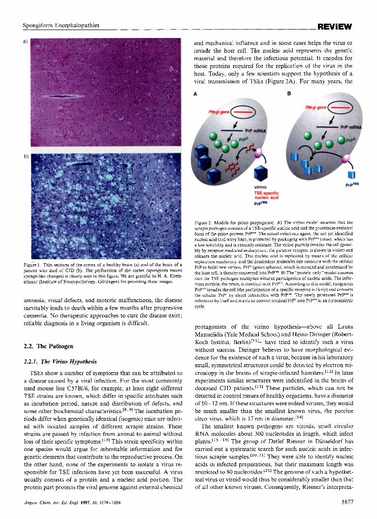
changes. TSEs are neurodegenerative diseases. Consequently they display their destructive potential in the brain of the affect- ed species. Neuropathologists diagnose TSE if the three follow- ing pathological changes are seen : 1) a spongiform change of the cortex (Figure I), which perforates the brain, 2) a pathological proliferation of glia cells (gliosis), and 3) a loss of neuronal cells[’’ linked to the deposition of an insoluble isoform of the prion protein “PrPsc”. This pathological morphology in the brain of a CJD patient, for example, is accompanied by drastic disorders of body functions. In the EEG periodic anomalies are seen. After the appearance of the first symptoms such as
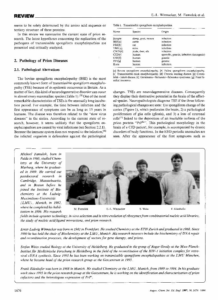
Michael Famulok, born in Fulda in 1960, studied Chem- istry at the University of Marburg, where he graduat- ed in 1989. He carried out postdoctoral research in Cambridge, Massachusetts, and in Boston before he joined the Institute of Bio- chemistry at the Ludwig- Maximilians- Universitat ( L M U ) . Munich, in 1992, where he completed his habil- itation in 1996. His research fields include aptamer technology, in vitro selection and in vitro evolution of ribozymes from combinatorial nucleic acid libraries, the study of nucleic acidlligand interactions, and prion research.
M. Famulok E.-L. Winnacker S . Weiss F. Edenhofer
Ernst-Ludwig Winnacker was born in 1941 in Frankfurt. He studied Chemistry at the ETH Zurich andgraduated in 1968. Since 1980 he has held the chair of Biochemistry at the L M U , Munich. His research interests include the biochemistry of D N A repair and recombination processes, the development of vectors for gene therapy, and prions.
Stefan Weiss studied Biology at the University of Heidelberg. He graduated in the group of Roger Goody at the Max-Planck- Institut fur Medizinische Forschung in Heidelberg in the field of the reconstitution of the HIV-1 initiation complex for retro- viral cDNA synthesis. Since 1993 he has been working on transmissible spongiform encephalopathies at the L M U Munchen, where he became head of the prion research group at the Genzentrum in 1995.
Frank Edenhofer was born in 1968 in Munich. He studied Chemistry at the L M U , Munich, from 1989 to 1994. In his graduate work since 1995 in the prion research group at the Genzentrum, he is working on the identification and characterization ofprion cofactors and the heterologous expression of P r F .
1676 Angew. Chem. Int. Ed. Engl. 1997, 36, 1614-1694
Spongiform Encephalopathies REVIEW
Figure 1. Thin sections of the cortex of a healthy brain (a) and of the brain of a patient who died of CJD (b). The perforation of the cortex (spongiosis means sponge-like changes) is clearly seen in this figure. We are grateful to H. A. Kretz- schmar (Institute of Neuropathology, Gottingen) for providing these images.
amnesia, visual defects, and motoric malfunctions, the disease inevitably leads to death within a few months after progressive dementia. No therapeutic approaches to cure the disease exist; reliable diagnosis in a living organism is difficult.
2.2. The Pathogen
2.2.1. The Virino Hypothesis
TSEs show a number of symptoms that can be attributed to a disease caused by a viral infection. For the most commonly used mouse line C57B1/6, for example, at least eight different TSE strains are known, which differ in specific attributes such as incubation period, nature and distribution of defects, and some other biochemical characteristics.[*, 91 The incubation pe- riods differ when genetically identical (isogenic) mice are infect- ed with isolated samples of different scrapie strains. These strains are passed by infection from animal to animal without loss of their specific This strain specificity within one species would argue for inheritable information and for genetic elements that contribute to the reproductive process. On the other hand, none of the experiments to isolate a virus re- sponsible for TSE infections have yet been successful. A virus usually consists of a protein and a nucleic acid portion. The protein part protects the viral genome against external chemical
and mechanical influence and in some cases helps the virus to invade the host cell. The nucleic acid represents the genetic material and therefore the infectious potential. I t encodes for those proteins required for the replication of the virus in the host. Today, only a few scientists support the hypothesis of a viral transmission of TSEs (Figure 2A). For many years, the
Figure 2. Models for prion propagation. A) The virino model assumes that the scrapie pathogen consists of a TSE-specific nucleic acid and the proteinase-resistant form of the prion protein Pry"'. The actual infectious agent, the not yet identified nucleic acid (red wavy line), is protected by packaging with PrP"' (blue), which has a low solubility and is extremly resistant. The virino particle invades the cell (possi- bly by receptor-mediated endocytosis; the putative receptor is shown in violet) and releases the nucleic acid. This nucleic acid is replicated by means of the cellular replication machinery, and the descendant molecules can associate with the cellular PrP to build new virinos. PrP' (green spheres), which is encoded and synthesized by the host cell, is thereby converted into PrP"' B) The "protein-only" model assumes that the TSE pathogen multiplies without participation of nucleic acids. The infec- tious particle, the prion, is identical with PrP"'. According to this model, exogenous Pryes invades the cell (the participation of a specific receptor is likely) and converts the cellular PIP' by direct interaction with PrP"*. The newly generated P ry ' is infectious by itself and is able to convert residual PrPC into PrP"'in an autocatalytic cycle.
protagonists of the virino hypothesis-above all Laura Manuelidis (Yale Medical School) and Heino Diringer (Robert- Koch Institut, Berlin)r"l-have tried to identify such a virus without success. Diringer believes to have morphological evi- dence for the existence of such a virus, because in his laboratory small, symmetrical structures could be detected by electron mi- croscopy in the brains of scrapie-infected hamsters.['21 In later experiments similar structures were indentified in the brains of deceased CJD patients.['31 These particles, which can not be detected in control tissues of healthy organisms, have a diameter of 10-12 nm. If these structures were indeed viruses, they would be much smaller than the smallest known virus. the porcine circo virus, which is 17 nm in diameter.['41
The smallest known pathogens are viroids, small circular RNA molecules about 300 nucleotides in length, which infect plant^.['^-'^] The group of Detlef Riesner in Diisseldorf has carried out a systematic search for such nucleic acids in infec- tious scrapie samples.[". They were able to identify nucleic acids in infected preparations, but their maximum length was restricted to 80 nucleotides.[''] The genome of such a hypothet- ical virus or viroid would thus be considerably smaller than that of all other known viruses. Consequently, Riesner's interpreta-
Angew. Chem. Int. Ed. Engl. 1997,36, 1674-1694 1677
E.-L. Winnacker, M. Famulok et al. REVIEW
tion of these results is that the existence of a scrapie-specific virus can be regarded as highly unlikely.[’. ’ ’ 7 231
2.2.2. The “Protein-Only” Hypothesis
The fact that no TSE virus could be detected so far is, of course, no proof that such a virus does not exist. How difficult the identification of pathogens of viral diseases can be was demonstrated, for example, by the search for a hepatitis C pathogen. It took ten years of intensive research to identify it as a virus.1241 But there is other clear evidence that nucleic acids are unlikely to be involved in the replication of TSE pathogens. Tikvah Alper et al. recognized in the sixties already that scrapie samples are resistant to nucleases and UV irradiation.[”] Usual- ly, nucleic acids are inactivated under such conditions. Even more remarkable than these results was the fact that the scrapie pathogens lost their infectivity when they were treated with protein-denaturing agents like 8h.1 urea or phenol. This unusual feature of the scrapie pathogen led to the first-very cautious- speculation about the existence of infectious proteins as indicat- ed by the title of Alper et al.’s paper:[261 “Does the scrapie agent replicate without nucleic acid?’. At that time this unorthodox hypothesis could not make headway against the conventional knowledge about infectious diseases. How might an infectious agent replicate in the host without nucleic acids? Can a protein replicate itself? The classical dogma of molecular biology-the flow of genetic information from nucleic acid to protein-seems to be violated.
In 1967 J. S. Griffith was the first to suggest a possible mech- anism for a self-replicating protein, which caused scrapie.[”l Stanley Prusiner of UCSF supported and extended this hypoth- esis by a series of experiments, which clearly showed a correla- tion between a protein and the observed infectivity. For ex- ample, Prusiner’s group demonstrated for the first time that the infectivity of the samples increased when a specific protein con- tained in the samples was enriched. The concentration of this protein was found to be proportional to the titer of the infectiv- ity in the anima1.128~291 On the basis of these results Prusiner developed the “protein-only” hypothesis. He dubbed this new kind of pathogen as “prion”, which is a short form for proteina- ceous infectious particle.[301 The purification and thorough bio- chemical examination of this remarkable protein from the brains of affected animals was achieved by Prusiner’s group after several years of intensive research. The continuing charac- terization of the pathogen resulted in the surprising discovery that the pathogenic protein possesses a celiular homologue. To- gether with the group of Charles Weissmann at the ETH Zurich they were able to clone and sequence the host gene (Prn-p) which encodes for PrPc.[31* 321 This cellular prion protein, designated as PrP“, is expressed in every normal mammal-mainly in the brain-without causing any harm to the organism. Today we know that the prion protein plays a dual biochemical role. On one hand it exists as the normal cellular form PrP” (c for cellu- lar); on the other hand, it can exist as a pathogenic and possibly infectious isoform PrPSc (sometimes prprer: Sc for Scrapie, res for proteinase K resistant). The PrPSc leads to death of the af- fected organism, whereas the biological significance of the PrP” form is still unknown (see Section 5.3.2). A very strong argu- ment for the protein-only hypothesis was provided by Weiss-
mann and his co-workers, who generated transgenic mice whose Prn-p gene was destroyed so that they no longer expressed PrP (see Section 5.3). These so-called “PrP-knockout mice” were resistant to scrapie infections.[331 Without any doubt, the long- accepted hypothesis that the amino acid sequence of a protein is the only determinant for the biologically relevant 3D struc-
is contradicted by the protein-only hypothesis. Although the arguments for the protein-only hypothesis are
quite compelling, a final experimental proof of the infectivity of the prion protein is still missing. The advocates of the protein- only hypothesis interpret this lack of proof as a simple prepara- tive problem, because in purified samples only one of lo5 PrPres molecules is infectious ;Iz2] enrichment of infectious portions is thus correspondingly difficult. Critics of the protein-only hy- pothesis claim that the formation of proteinase K resistant Prp’es is just a concomitant phenomenon, a pathological product of the infection by a not yet identified virus. This argu- ment can be supported by experiment as well. Lasmirzas et al. recentIy reported that mice infected with BSE samples indeed showed symptoms similar to TSE, but in 55% of the cases they could not detect proteinase K resistant PrPres in the brains of the test ar1irna1s.r~~~ These and other results show that the prion protein undoubtedly plays a central role for the pathogenesis of TSE, but until today it has not been proven that it represents the only infectious agent.
3. PrP‘ and PrPSC: Differences and Common Features
The prion protein seems to be of considerable importance for the appearance of spongiform encephalopathies. In mammals as well as in some avian species the protein is expressed mainly in the brain. The prion protein is assumed to play an important biological role as is the case for many other highly conserved proteins. Various recent evidence indicates that it is important for the normal physiological function of the synapses[361 and for the long-term stability of Purkinje neurons (huge dendritic gan- glial cells in the mid-layer of the cerebellum as well as for the regulation of circadian rhythms and sleep patternsr3*] (see Section 5.3.2. for more details).
The following statements about the composition and struc- ture of the prion protein refer to the Syrian golden hamster, unless otherwise stated. This species is particularly suited for experimental research into TSEs, because the incubation period is only 70 days and correlates exactly with the dosage of the infectivity of the samples. According to present knowledge the characteristics of hamster PrP can be transferred with only mi- nor modifications to PrP of other mammalian species.
3.1. Posttranslational Modifications and Infectivity
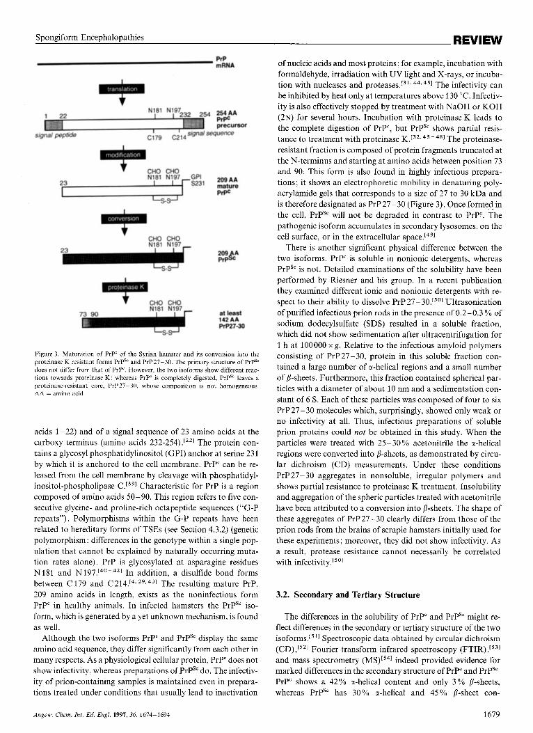
The PrP coding sequence is located entirely within an exon of the unique Pm-p gene.[321 Therefore, different splicing variants of PrP mRNA can be excluded. Translation of PrP mRNA leads to a PrP precursor protein of 254 amino acids in length (Fig- ure 3 ) . Posttranslational modification then leads to the removal of an amino-terminal signal peptide of 22 amino acids (amino
1678 Angew. Chem. Int. Ed. Engl. 1997, 36, 1614-1694
REVIEW Spongiform Encephalopathies
PIP mRNA
4 254AA PrPC precursor
signal peptide c;79 C;14signa/ sequence
' CHO CHO
23 r S 2 3 1 g:ik N181 N197 GPI
CHO CHO ^^ N181 N197-
71 on
* CHO CHO
209 A Pr&
at least
PrP27-30 142 AA
- S - W
Figure 3. Maturation of PrP' of the Syrian hamster and its conversion into the proteinase K resistant forms PrPS" and PrP27-30. The primary structure of PrPS' does not differ from that of PrP'. However, the two isoforms show different reac- tions towards proteinase K: whereas PrP' is completely digested, PrPS' leaves a proteinase-resistant core, PrP27-30, whose composition is not homogeneous. AA = amino acid.
acids 1-22) and of a signal sequence of 23 amino acids at the carboxy terminus (amino acids 232-254) .["I The protein con- tains a glycosyl phosphatidylinositol (GPI) anchor at serine 231 by which it is anchored to the cell membrane. PrP" can be re- leased from the cell membrane by cleavage with phosphatidyl- inositol-phospholipase C.[391 Characteristic for PrP is a region composed of amino acids 50-90. This region refers to five con- secutive glycine- and proline-rich octapeptide sequences ("G-P repeats"). Polymorphisms within the G-P repeats have been related to hereditary forms of TSEs (see Section 4.3.2) (genetic polymorphism: differences in the genotype within a single pop- ulation that cannot be explained by naturally occurring muta- tion rates alone). PrP is glycosylated at asparagine residues N181 and N197.[40-421 In addition, a disulfide bond forms between C 179 and C214.[4.293431 The resulting mature PrP, 209 amino acids in length, exists as the noninfectious form PrP" in healthy animals. In infected hamsters the PrPS" iso- form, which is generated by a yet unknown mechanism, is found as well.
Although the two isoforms PrP" and PrPS" display the same amino acid sequence, they differ significantly from each other in many respects. As a physiological cellular protein, PrP" does not show infectivity, whereas preparations of PrPSc do. The infectiv- ity of prion-containing samples is maintained even in prepara- tions treated under conditions that usually lead to inactivation
of nucleic acids and most proteins; for example, incubation with formaldehyde, irradiation with UV light and X-rays, or incuba- tion with nucleases and pro tease^.'^ '* 44, 451 The infectivity can be inhibited by heat only at temperatures above 130 "C. Infectiv- ity is also effectively stopped by treatment with NaOH or KOH (2N) for several hours. Incubation with proteinase K leads to the complete digestion of PrP", but PrPSc shows partial resis- tance to treatment with proteinase K.[32,45 -481 Th e proteinase- resistant fraction is composed of protein fragments truncated at the N-terminus and starting at amino acids between position 73 and 90. This form is also found in highly infectious prepara- tions; it shows an electrophoretic mobility in denaturing poly- acrylamide gels that corresponds to a size of 27 to 30 kDa and is therefore designated as PrP27-30 (Figure 3). Once formed in the cell, PrPS' will not be degraded in contrast to PrP". The pathogenic isoform accumulates in secondary lysosomes, on the cell surface, or in the extracellular space.[49]
There is another significant physical difference between the two isoforms. PrPc is soluble in nonionic detergents, whereas PrPSc is not. Detailed examinations of the solubility have been performed by Riesner and his group. In a recent publication they examined different ionic and nonionic detergents with re- spect to their ability to dissolve PrP 27 -30.[s01 Ultrasonication of purified infectious prion rods in the presence of 0.2-0.3 % of sodium dodecylsulfate (SDS) resulted in a soluble fraction, which did not show sedimentation after ultracentrifugation for 1 h at I00000 x g . Relative to the infectious amyloid polymers consisting of PrP27-30, protein in this soluble fraction con- tained a large number of a-helical regions and a small number of 0-sheets. Furthermore, this fraction contained spherical par- ticles with a diameter of about 10 nm and a sedimentation con- stant of 6 s. Each of these particles was composed of four to six PrP 27- 30 molecules which, surprisingly, showed only weak or no infectivity at all. Thus, infectious preparations of soluble prion proteins could not be obtained in this study. When the particles were treated with 25- 30 YO acetonitrile the a-helical regions were converted into 0-sheets, as demonstrated by circu- lar dichroism (CD) measurements. Under these conditions PrP 27- 30 aggregates in nonsoluble, irregular polymers and shows partial resistance to proteinase K treatment. Insolubility and aggregation of the spheric particles treated with acetonitrile have been attributed to a conversion into 0-sheets. The shape of these aggregates of PrP27-30 clearly differs from those of the prion rods from the brains of scrapie hamsters initially used for these experiments; moreover, they did not show infectivity. As a result, protease resistance cannot necessarily be correlated with infectivity.[50]
3.2. Secondary and Tertiary Structure
The differences in the solubility of PrP" and PrPSc might re- flect differences in the secondary or tertiary structure of the two isof~rms.[~'] Spectroscopic data obtained by circular dichroism (CD) ,[521 Fourier transform infrared spectroscopy (FTIR) ,[531
and mass spectrometry (MS)[541 indeed provided evidence for marked differences in the secondary structure of PrP" and PrPSc PrP" shows a 42% a-helical content and only 3 % 0-sheets, whereas PrPSc has 30% a-helical and 45% P-sheet con-
Anprw. Chrm. In[. Ed. Engl. 1997, 36, 1674-1694 1679
REVIEW E.-L. Winnacker, M. Famulok et al.
tent.[51. 521 These studies indicated that during conversion about half of the a-helical portions in PrP" are converted into 8-sheets of PrPsc.[52. 551 The mechanism by which the PrP' is unfolded and then changed into PrPS', however, is unknown. It is possible that the change in protein structure associated with this process requires a high activation energy.[5 'I
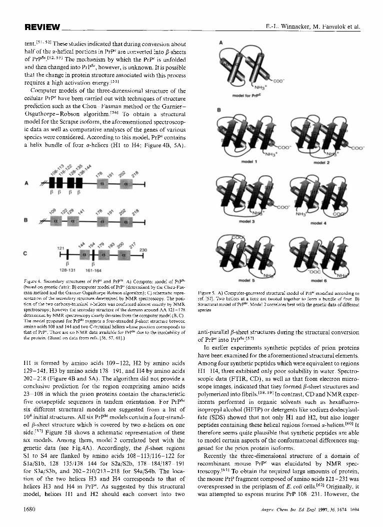
Computer models of the three-dimensional structure of the cellular PrP" have been carried out with techniques of structure prediction such as the Chou-Fasman method or the Garnier- Osguthorpe- Robson a1g0rithrn.I~~~ To obtain a structural model for the Scrapie isoform, the aforementioned spectroscop- ic data as well as comparative analyses of the genes of various species were considered. According to this model, PrP' contains a helix bundle of four a-helices (H1 to H4; Figure 4B, SA).
A
B
C
128-131 161-164
Figure 4. Secondary structures of PrP' and PrPS' A) Computer model of PrPS' (based on genetic data); B) computer model of PrP' (determined by the Chou-Fas- man method and the Gamier-Osguthorpe-Robson algorithm); C) schematlc repre- sentation of the secondary structure determined by NMR spectroscopy. The posi- tion of the two carboxy-terminal 2-helices was confirmed almost exactly by NMR spectroscopy; however the seconday structure of the domain around AA 121 -178 determined by NMR spectroscopy clearly deviates from the computer model (B, C) The model proposed for PrPS' suggests a four-stranded &sheet structure between amino acids 108 and 144 and two C-terminal helices whose position corresponds to that of PrP'. There are no NMR data available for P r p due to the insolubility of the protein. (Based on data from refs. [56, 57, 611.)
H1 is formed by amino acids 109-122, H2 by amino acids 129-141, H3 by amino acids 178-191, and H4 by amino acids 202-218 (Figure 4B and 5A). The algorithm did not provide a conclusive prediction for the region comprising amino acids 23- 108 in which the prion proteins contain the characteristic five octapeptide sequences in tandem orientation. For PrPSc six different structural models are suggested from a list of lo6 initial structures. All six PrPSc models contain a four-strand- ed 8-sheet structure which is covered by two a-helices on one side.[571 Figure SB shows a schematic representation of these six models. Among them, model 2 correlated best with the genetic data (see Fig.4A). Accordingly, the 8-sheet regions S1 to S4 are flanked by amino acids 108-113/116-122 for SlaIS1 b, 128- 1351138- 144 for S2a/S2b, 178-1841187-191 for S3a/S3b, and 202-2101213-218 for S4aIS4b. The Ioca- tion of the two helices H3 and H4 corresponds to that of helices H3 and H4 in PrP". As suggested by this structural model, helices H1 and H2 should each convert into two
Figure 5 . A) Computer-generated structural model of PrP' modlfied according to ref. [57]. Two helices at a time are twisted together to form a bundle of four. B) Structural model of PrPS'. Model 2 correlates best with the genetx data of different species.
anti-parallel a-sheet structures during the structural conversion of PrP" into PrPsc.[571
In earlier experiments synthetic peptides of prion proteins have been examined for the aforementioned structural elements. Among four synthetic peptides which were equivalent to regions H1 -H4, three exhibited only poor solubility in water. Spectro- scopic data (FTIR, CD), as well as that from electron micro- scope images, indicated that they formed /?-sheet structures and polymerized into fibrils."** 591 In contrast, CD and NMR exper- iments performed in organic solvents such as hexafluoro- isopropyl alcohol (HFIP) or detergents like sodium dodecylsul- fate (SDS) showed that not only H1 and H2, but also longer peptides containing these helical regions formed a-helices.[601 It therefore seems quite plausible that synthetic peptides are able to model certain aspects of the conformational differences sug- gested for the prion protein isoforms.
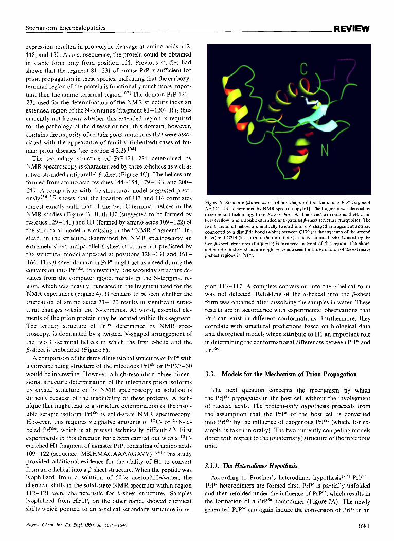
Recently the three-dimensional structure of a domain of recombinant mouse PrP" was elucidated by NMR spec- troscopy.'6'1 To obtain the required large amounts of protein, the mouse PrP fragment composed of amino acids 121 -231 was overexpressed in the periplasm of E. coli cells.[621 Originally, it was attempted to express murine PrP 108-231. However, the
1680 Angeu.. Chem. Int. Ed. Engl. 1991.36, 1674-1694
Spongiform Encephalopathies REVIEW
expression resulted in proteolytic cleavage at amino acids 112, 118, and 120. As a consequence, the protein could be obtained in stable form only from position 121. Previous studies had shown that the segment 81-231 of mouse PrP is sufficient for prion propagation in these species, indicating that the carboxy- terminal region of the protein is functionally much more impor- tant then the amino terminal region.[63' The domain PrP 121 - 231 used for the determination of the NMR structure lacks an extended region of the N-terminus (fragment 81 - 120). It is thus currently not known whether this extended region is required for the pathology of the disease or not; this domain, however, contains the majority of certain point mutations that were asso- ciated with the appearance of familial (inherited) cases of hu- man prion diseases (see Section 4.3.2).'641
The secondary structure of PrP 121 -231 determined by NMR spectroscopy is characterized by three sr-helices as well as a two-stranded antiparallel 8-sheet (Figure 4C). The helices are formed from amino acid residues 144-154, 179-193, and 200- 217. A comparison with the structural model suggested previ- ously[56.571 shows that the location of H3 and H4 correlates almost exactly with that of the two C-terminal helices in the NMR studies (Figure 4). Both H2 (suggested to be formed by residues 129-141) and Hl (formed by amino acids 109-122) of the structural model are missing in the "NMR fragment". In- stead, in the structure determined by NMR spectroscopy an extremely short antiparallel 8-sheet structure not predicted by the structural model appeared at positions 128-131 and 161 - 164. This 8-sheet domain in PrP' might act as a seed during the conversion into PrPS'. Interestingly, the seconday structure de- viates from the computer model mainly in the N-terminal re- gion, which was heavily truncated in the fragment used for the NMR experiment (Figure 4). It remains to be seen whether the truncation of amino acids 23-120 results in significant struc- tural changes within the N-terminus. At worst, essential ele- ments of the prion protein may be located within this segment. The tertiary structure of PrP', determined by NMR spec- troscopy, is dominated by a twisted, V-shaped arrangement of the two C-terminal helices in which the first a-helix and the 8-sheet is embedded (Figure 6).
A comparison of the three-dimensional structure of PrP" with a corresponding structure of the infectious PrPS' or PrP27-30 would be interesting. However, a high-resolution, three-dimen- sional structure determination of the infectious prion isoforms by crystal structure or by NMR spectroscopy in solution is difficult because of the insolubility of these proteins. A tech- nique that might lead to a structure determination of the insol- uble scrapie isoform PrPS' is solid-state NMR spectroscopy. However, this requires weighable amounts of 13C- or "N-la- beled PrPSc, which is at present technically difficult.[65] First experiments in this direction have been carried out with a I3C- enriched H1 fragment of hamster PrP, consisting of amino acids 109- 122 (sequence: MKHMAGAAAAGAVV).[661 This study provided additional evidence for the ability of H1 to convert from an a-helical into a 8-sheet structure. When the peptide was lyophilized from a solution of 50 O h acetonitrile/water, the chemical shifts in the solid-state NMR spectrum within region 11 2- 121 were characteristic for 8-sheet structures. Samples lyophilized from HFIP, on the other hand, showed chemical shifts which pointed to an a-helical secondary structure in re-
Figure6. Structure (shown as a "ribbon diagram") of the mouse PrP' fragment AA 121-231, determined by NMR spectroscopy[6l]. The fragment was derived by recombinant technology from Escherichio coli. The structure contains three a-he- lices (yellow) and a double-stranded anti-parallel p-sheet structure (turquoise). The two C-terminal helices are mutually twisted into a V-shaped arrangement and are connected by a disulfide bond (white) between C179 (at the first turn of the second helix) and C214 (last turn of the third helix). The N-terminaf helix flanked by the two p-sheet structures (turquoise) is arranged in front of this region. The short, antiparallel &sheet structure might serve as a seed for the formation of the extensive &sheet regions in P r P .
gion 11 3- 11 7. A complete conversion into the x-helical form was not detected. Refolding of the a-helical into the 8-sheet form was obtained after dissolving the samples in water. These results are in accordance with experimental observations that PrP can exist in different conformations. Furthermore, they correlate with structural predictions based on biological data and theoretical models which attribute to H1 an important role in determining the conformational differences between PrP' and PrPSc.
3.3. Models for the Mechanism of Prion Propagation
The next question concerns the mechanism by which the PrPSF propagates in the host cell without the involvement of nucleic acids. The protein-only hypothesis proceeds from the assumption that the PrP' of the host cell is converted into PrPS' by the influence of exogenous PrPSc (which, for ex- ample, is taken in orally). The two currently competing models differ with respect to the (quaternary) structure of the infectious unit.
3.3.1. The Heterodimer Hypothesis
According to Prusiner's heterodimer hypothesis1221 P r p - PrP" heterodimers are formed first. PrP" is partially unfolded and then refolded under the influence of PrPSc, which results in the formation of a PrPSc homodimer (Figure 7A). The newly generated PrPSc can again induce the conversion of PrP" in an
Angew. Chem. Inr. Ed. Engl. 1997, 36, 1674-1694 1681
E.-L. Winnacker, M. Famulok et al. REVIEW
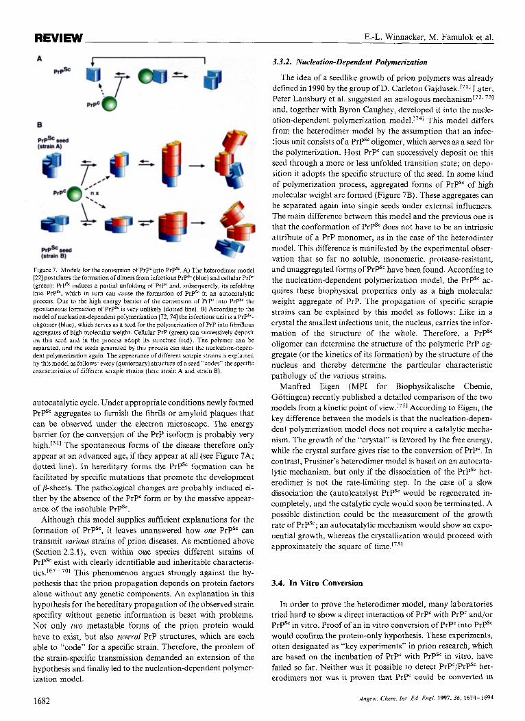
Figure 7. Models for the conversion of PrP' into PrPS'. A) The heterodimer model [22] postulates the formation of dimers from infectious PrPS' (blue) and cellular PrP' (green): PrPS' induces a partial unfolding of PrP' and, subsequently, its refolding into P r p , which in turn can cause the formation of PrPS' in an autocatalytic process. Due to the high energy barrier of the conversion of PrP' into PrPS' the spontaneous formation of PrPS' is very unlikely (dotted line). B) According to the model of nucleation-dependent polymerization 172,741 the infectious unit is a PrPS'- oligomer (blue), which serves as a seed for the polymerization of PrP into fibrillous aggregates of high molecular weight. Cellular PrP (green) can successively deposit on this seed and in the process adopt its structure (red). The polymer can be separated, and the seeds generated by this process can start the nucleation-depen- dent polymerization again. The appearance of different scrapie strains is explained by this model as follows: every (quaternary) structure of a seed "codes" the specific characteristics of different scrapie strains (here strain A and strain B).
autocatalytic cycle. Under appropriate conditions newly formed PrPSc aggregates to furnish the fibrils or amyloid plaques that can be observed under the electron microscope. The energy barrier for the conversion of the PrP isoform is probably very high.15 The spontaneous forms of the disease therefore only appear at an advanced age, if they appear at all (see Figure 7A; dotted line). In hereditary forms the PrPSc formation can be facilitated by specific mutations that promote the development of B-sheets. The pathological changes are probably induced ei- ther by the absence of the PrP' form or by the massive appear- ance of the insoluble PrPSc.
Although this model supplies sufficient explanations for the formation of PrPSc, it leaves unanswered how one PrPSc can transmit various strains of prion diseases. As mentioned above (Section 2.2.1), even within one species different strains of PrPSc exist with clearly identifiable and inheritable characteris- t i c~ . '~ ' - 701 This phenomenon argues strongly against the hy- pothesis that the prion propagation depends on protein factors alone without any genetic components. An explanation in this hypothesis for the hereditary propagation of the observed strain specifity without genetic information is beset with problems. Not only two metastable forms of the prion protein would have to exist, but also several PrP structures, which are each able to "code" for a specific strain. Therefore, the problem of the strain-specific transmission demanded an extension of the hypothesis and finally led to the nucleation-dependent polymer- ization model.
3.3.2. Nucleation-Dependent Polymerization
The idea of a seedlike growth of prion polymers was already defined in 1990 by the group of D. Carleton G a j d ~ s e k . ~ ~ ~ ] Later, Peter Lansbury et al. suggested an analogous mechanism[72. 731
and, together with Byron Caughey, developed it into the nucle- ation-dependent polymerization This model differs from the heterodimer model by the assumption that an infec- tious unit consists of a PrPSc oligomer, which serves as a seed for the polymerization. Host PrP" can successively deposit on this seed through a more or less unfolded transition state; on depo- sition it adopts the specific structure of the seed, In some kind of polymerization process, aggregated forms of PrPS' of high molecular weight are formed (Figure 7B). These aggregates can be separated again into single seeds under external influences. The main difference between this model and the previous one is that the conformation of PrPS' does not have to be an intrinsic attribute of a PrP monomer, as in the case of the heterodimer model. This difference is manifested by the experimental obser- vation that so far no soluble, monomeric, protease-resistant, and unaggregated forms of PrPS' have been found. According to the nucleation-dependent polymerization model, the PrPS' ac- quires these biophysical properties only as a high molecular weight aggregate of PrP. The propagation of specific scrapie strains can be explained by this model as follows: Like in a crystal the smallest infectious unit, the nucleus, carries the infor- mation of the structure of the whole. Therefore, a PrPSc oligomer can determine the structure of the polymeric PrP ag- gregate (or the kinetics of its formation) by the structure of the nucleus and thereby determine the particular characteristic pathology of the various strains.
Manfred Eigen (MPI for Biophysikalische Chemie, Gottingen) recently published a detailed comparison of the two models from a kinetic point of view.[751 According to Eigen, the key difference between the models is that the nucleation-depen- dent polymerization model does not require a catalytic mecha- nism. The growth of the "crystal" is favored by the free energy, while the crystal surface gives rise to the conversion of PrP". In contrast, Prusiner's heterodimer model is based on an autocata- lytic mechanism, but only if the dissociation of the PrPSc het- erodimer is not the rate-limiting step. In the case of a slow dissociation the (auto)catalyst PrPS' would be regenerated in- completely, and the catalytic cycle would soon be terminated. A possible distinction could be the measurement of the growth rate of PrPSc; an autocatalytic mechanism would show an expo- nential growth, whereas the crystallization would proceed with approximately the square of time.[751
3.4. In Vitro Conversion
In order to prove the heterodimer model, many laboratories tried hard to show a direct interaction of PrP" with PrP' and/or PrPSc in vitro. Proof of an in vitro conversion of PrPc into PrP' would confirm the protein-only hypothesis. These experiments, often designated as "key experiments" in prion research, which are based on the incubation of PrP' with PrPS' in vitro, have failed so far. Neither was it possible to detect PrPc/PrPsc het- erodimers nor was it proven that PrP" could be converted in
1682 Angew. Chem. Int. Ed. Engl. 1997, 36, 1614-1694
REVIEW Spongiform Encephalopathies
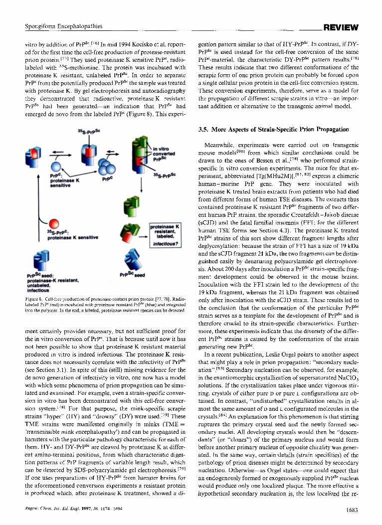
vitro by addition of PrPs‘.”6J In mid 1994 Kocisko et al. report- ed for the first time the cell-free production of protease-resistant prion protein.[771 They used proteinase K sensitive PrP”, radio- labeled with 35S-methionine. The protein was incubated with proteinase K resistant, unlabeled PrPSc. In order to separate PrPC from the potentially produced PrPS‘ the sample was treated with proteinase K. By gel electrophoresis and autoradiography they demonstrated that radioactive, proteinase K resistant PrPSc had been generated-an indication that PrPSc had emerged de novo from the labeled PrP‘ (Figure 8). This experi-
Figure 8. Cell-free production of proteinase-reistant prion protein [77,78]. Radio- labeled PrP‘ (red) is incubated with proteinase-resistan1 PrPSE (blue) and integrated into the polymer. In the end. a labeled, proteinase-resistant species can be detected.
ment certainly provides necessary, but not sufficient proof for the in vitro conversion of PrP‘. That is because until now it has not been possible to show that proteinase K resistant material produced in vitro is indeed infectious. The proteinase K resis- tance does not necessarily correlate with the infectivity of PrPSc (see Section 3.1). In spite of this (still) missing evidence for the de novo generation of infectivity in vitro, one now has a model with which some phenomena of prion propagation can be simu- lated and examined. For example, even a strain-specific conver- sion in vitro has been demonstrated with this cell-free conver- sion system.[78] For that purpose, the mink-specific scrapie strains “hyper” (HY) and “drowsy” (DY) were used.[79] These TME strains were manifested originally in minks (TME =
‘transmissible mink encephalopathy’) and can be propagated in hamsters with the particular pathology characteristic for each of them. HY- and DY-PrPS‘ are cleaved by proteinase K at differ- ent amino-terminal positions, from which characteristic diges- tion patterns of PrP fragments of variable length result, which can be detected by SDS-polyacrylamide gel e l ec t roph~res i s .~~~~ If one uses preparations of HY-PrP” from hamster brains for the aforementioned conversion experiments a resistant protein is produced which, after proteinase K treatment, showed a di-
gestion pattern similar to that of HY-PrPSC. In contrast, if DY- PrPSc is used instead for the cell-free conversion of the same PrP”-material, the characteristic DY-PrPS‘ pattern results.[781 These results indicate that two different conformations of the scrapie form of one prion protein can probably be forced upon a single cellular prion protein in the cell-free conversion system. These conversion experiments, therefore, serve as a model for the propagation of different scrapie strains in vitro-an impor- tant addition or alternative to the transgenic animal model.
3.5. More Aspects of Strain-Specific Prion Propagation
Meanwhile, experiments were carried out on transgenic mouse models[80] from which similar conclusions could be drawn to the ones of Bessen et al.,[781 who performed strain- specific in vitro conversion experiments. The mice for that ex- periment, abbreviated [T~(MHU~M)] ,~”* ’’] express a chimeric human-murine PrP gene. They were inoculated with proteinase K treated brain extracts from patients who had died from different forms of human TSE diseases. The extracts thus contained proteinase K resistant PrPS“ fragments of two differ- ent human PrP strains, the sporadic Creutzfeldt -.lakob disease (sCJD) and the fatal familial insomnia (FFI; for the different human TSE forms see Section 4.3). The proteinase K treated PrPSc strains of this sort show different fragment lengths after deglycosylation: because the strain of FFI has a size of 19 kDa and the sCJD fragment 21 kDa, the two fragments can be distin- guished easily by denaturing polyacrylamide gel electrophore- sis. About 200 days after inoculation a PrPS‘ strain-specific frag- ment development could be observed in the mouse brains. Inoculation with the FFI strain led to the development of the 19 kDa fragment, whereas the 21 kDa fragment was obtained only after inoculation with the sCJD strain. These results led to the conclusion that the conformation of the particular PrPSc strain serves as a template for the development of PrPSc and is therefore crucial to its strain-specific characteristics. Further- more, these experiments indicate that the diversity of the differ- ent PrPSc strains is caused by the conformation of the strain generating new PrPSc.
In a recent publication, Leslie Orgel points to another aspect that might play a role in prion propagation: “secondary nucle- a t i~n” . [*~] Secondary nucleation can be observed, for example, in the enantiomorphic crystallization of supersaturated NaClO, solutions. If the crystallization takes place under vigorous stir- ring, crystals of either pure D or pure L configurations are ob- tained. In contrast, “undisturbed” crystallization results in al- most the same amount O f D and L configurated molecules in the crystals.[s41 An explanation for this phenomenon is that stirring ruptures the primary crystal seed and the newly formed sec- ondary nuclei. All developing crystals would then be “descen- dants” (or “clones”) of the primary nucleus and would form before another primary nucleus of opposite chirality was gener- ated. In the same way, certain details (strain specifities) of the pathology of prion diseases might be determined by secondary nucleation. Otherwise-as Orgel states-one could expect that an endogenously formed or exogenously supplied PrPSc nucleus would produce only one localized plaque. The more effective a hypothetical secondary nucleation is, the less localized the re-
Angeu. Chem. In[ . E d Engl. 1997,36, 1674-1694 1683
REVIEW E.-L. Winnacker, M. Famulok et al.
sulting plaques would be. The number of developing nuclei would therefore determine the seriousness of the disease-as in the case of cancer where the severity of the disease depends on the tendency of metastasation of the primary tumor. Of course, secondary nucleation in the case of prion propagation has not yet be shown and will probably be very difficult to prove.
As discussed in greater detail later, other possibilities can explain the strain specifity of prion propagation, for example, the existence of a third component such as a strain-specific co- factor or protein x,@’] which is directly or indirectly involved in the propagation of PrPSc (see Section 5.2).
4. Epidemiology and Transmission
After close analysis of the pathogen the question arises, how are prion diseases transmitted. How could BSE in England reach epidemic proportions? What about the transmission from one species to the other? The risk of transmission of BSE to humans has, of course, important public, economic, and sani- tary implications.
4.1. The Scrapie- Kuru Connection: a Historical Summary
Spongiform encephalopathies have been known since the 18th century. The oldest written record gives a description of certain peculiar behavior in sheep: at an early stage in the dis- ease, disorientation and itching was observed in the affected animals: they scraped themselves sore at pales and trees; this is why the illness was called “scrapie” in the English speaking- world. In German-speaking countries the disease was named “Traberkrankheit”, which points to the fact that affected ani- mals progressively lose control over their body in an advanced phase, move in an uncoordinated manner (“traben”), cannot support themselves on their hind legs, and finally perish in com- plete paralysis. Ways of transmission of the disease and the cause of the illness remained obscure for centuries, mainly be- cause scrapie never developed into an economic problem. The rate of infection always remained quite low-an epidemic out- break of the disease has never been registered to date. Nowa- days it has almost completely vanished in Germany, whereas only a few cases are reported in Great Britain at regular inter- vals.
Scientific interest arose first in this century, when William Hadlow speculated about a possible connection between the human Kuru disease and ~ c r a p i e . ~ ~ ~ ] Earlier, independent of scrapie, a series of unusually slowly degenerative diseases of the central nervous system in humans such as the Creutzfeldt- Jakob disease (CJD), Gerstmann -Straussler-Scheinker syn- drome (GSS), and Kuru had been described. Kuru broke out in epidemic dimensions in the Fore, a tribe in Papua-New Guinea, and was probably transmitted by religous cannibalistic rites. D. Carleton Gajdusek succeeded in an experimental trans- mission of this disease to chimpanzees by injection of brain material of patients who had died of Kuru.1861 In a similar way, CJD could be transmitted to chimpanzees. The remarkably long incubation period and the pathological pattern of this neurode- generation by spongiosis were identical in all forms of this dis-
ease. Important information about the transmissibility of scrapie resulted from experiments by J. Cuillk and P. L. Chelle, who demonstrated in the thirties that scrapie can be transmitted to healthy sheep and goats.[871 Hamsters and mice were also prone to this disease; both species are used today on a large scale for animal tests to investigate prion diseases. Nevertheless, as we will discuss later (Section 5. I), the unrestricted transmission of TSE from one species to another is not possible.
4.2. BSE-from Bonemeal to Epidemic
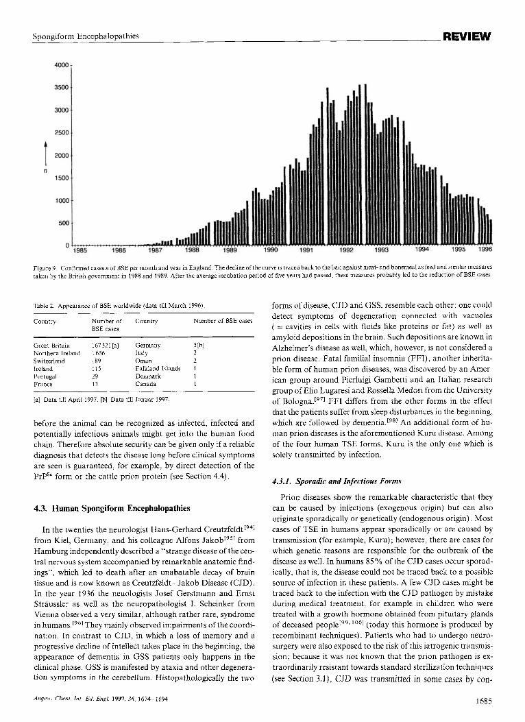
Because of the limited occurrence, TSE diseases in the first instance were a subject of academic interest. This changed sud- denly when the first case of BSE in England was histologically confirmed in 1986.[881 Fibrils isolated from the brains of BSE- infected cattle were shown to contain a scrapie-associated protein.[891 This report was immediately followed by others. The BSE epidemic in England had its climax between 1992 and 1993 with up to 3500 new cases per month; now it has dwindled to about 500 new cases per month (Figure 9). By April 1997 a total of more than 167 300 (source: Ministry of Agriculture, Fisheries and Food [MAFF], UK) BSE cases had definitely been con- firmed in England.[go1 The epidemic in cattle began when in the eighties the method of meat- and bonemeal production was changed in response to economic pressures. This feed, used mainly in cattle breeding, is produced in part from sheep cadav- ers. In the beginning of the eighties British companies modified the sterilization temperature, lowering it from 130 “C to 110 “C. Furthermore, extractions with organic solvents were no longer performed.[’ll Whether the epidemic was induced by bonemeal contaminated with scrapie (that is, the disease was transmitted from sheep to cattle) or with BSE pathogens from spontaneous- ly affected cattle themselves (and therefore not transmitted across species) can no longer be determined with certainty. In any case the spreading of the infectious agent through feed has been confirmed as the trigger for the BSE epidemi~.[”.’~1 The accumulated number of BSE cases in Switzerland, which was the main importer of British bonemeal until the feed ban (July 18, 1988) on the continent, also supports this argument. BSE was detected and confirmed worldwide in 12 countries (Table 2). Besides infection through feed, even newer indica- tions suggest that other ways of transmission, mainly from mother to calf, might be possible.[931
A current study of the course of the BSE epidemic in Great Britain from the very beginning of the first cases to a potential epidemiological development in the future came to the conclu- sion that by the end of 1994 new infections by contaminated feed were almost zero, and all new cases result from horizontal (ma- ternal) transmission.[901 Absolute numbers of new infections by this form of transmission were so small that the epidemic would vanish by the year 2001 even without slaughtering programs. In September 1996 the British government used this study as an argument to interrupt their cattle slaughtering project, which they had initiated in order to bring the epidemic under control. However, this argument neglects another important result of the study-the incubation period. It takes about five years from infection to the outbreak of the first symptoms. Since most of the cattle are killed at an age of about two years, that is, long
1684 Angew Chem. inr. Ed. Engl. 1997, 36, i614-1594
Spongiform Encephalopathies REVIEW
Figure 9. Confirmed cases n of BSE per month and year in England. The decline of the curve is traced back to the ban against meat- and bonemeal as feed and similar measures taken by the British government in 1988 and 1989. After the average incubation period of five years had passed, these measures probably led to the reduction of BSE cases.
Table 2. Appearance of BSE worldwide (data till March 1996).
Country Number of Country Number of BSE cases BSE cases
Great Britain 167321 [a] Germany 5 Ibl Northern Ireland 1656 Italy 2 Switzerland 189 Oman 2 Ireland 115 Falkland Islands 1 Portugal 29 Denmark 1 France 13 Canada 1
[a] Data till April 1997. [b] Data till Januar 1997.
before the animal can be recognized as infected, infected and potentially infectious animals might get into the human food chain. Therefore absolute security can be given only if a reliable diagnosis that detects the disease long before clinical symptoms are seen is guaranteed, for example, by direct detection of the PrPSc form or the cattle prion protein (see Section 4.4).
4.3. Human Spongiform Encephalopathies
In the twenties the neurologist Hans-Gerhard C r e ~ t z f e l d t [ ~ ~ ] from Kiel, Germany, and his colleague Alfons J a k ~ b [ ~ ~ ] from Hamburg independently described a “strange disease of the cen- tral nervous system accompanied by remarkable anatomic find- ings”, which led to death after an unabatable decay of brain tissue and is now known as Creutzfeldt-Jakob Disease (CJD). In the year 1936 the neuologists Josef Gerstmann and Ernst Straussler as well as the neuropathologist I. Scheinker from Vienna observed a very similar, although rather rare, syndrome in humans.r961 They mainly observed impairments of the coordi- nation. In contrast to CJD, in which a loss of memory and a progressive decline of intellect takes place in the beginning, the appearance of dementia in GSS patients only happens in the clinical phase. GSS is manifested by ataxia and other degenera- tion symptoms in the cerebellum. Histopathologically the two
forms of disease, CJD and GSS, resemble each other: one could detect symptoms of degeneration connected with vacuoles ( = cavities in cells with fluids like proteins or fat) as well as amyloid depositions in the brain. Such depositions are known in Alzheimer’s disease as well, which, however, is not considered a prion disease. Fatal familial insomnia (FFI), another inherita- ble form of human prion diseases, was discovered by an Amer- ican group around Pierluigi Gambetti and an Italian research group of Elio Lugaresi and Rossella Medori from the University of Bologna.[971 FFI differs from the other forms in the effect that the patients suffer from sleep disturbances in the beginning, which are followed by dementia.[98] An additional form of hu- man prion diseases is the aforementioned Kuru disease. Among of the four human TSE forms, Kuru is the only one which is solely transmitted by infection.
4.3.1. Sporadic and Infectious Forms
Prion diseases show the remarkable characteristic that they can be caused by infections (exogenous origin) but can also originate sporadically or genetically (endogenous origin). Most cases of TSE in humans appear sporadically or are caused by transmission (for example, Kuru) ; however, there are cases for which genetic reasons are responsible for the outbreak of the disease as well. In humans 85% of the CJD cases occur sporad- ically, that is, the disease could not be traced back to a possible source of infection in these patients. A few CJD cases might be traced back to the infection with the CJD pathogen by mistake during medical treatment, for example in children who were treated with a growth hormone obtained from pituitary glands of deceased people[99. ‘‘‘1 (today this hormone is produced by recombinant techniques). Patients who had to undergo neuro- surgery were also exposed to the risk of this iatrogenic transmis- sion; because it was not known that the prion pathogen is ex- traordinarily resistant towards standard sterilization techniques (see Section 3.1), CJD was transmitted in some cases by con-
A n p i r . Clrem. I n t Ed. Engl. 1997. 36, 1674-1694 1685
E.-L. Winnacker, M. Famulok et al. REVIEW
taminated instruments used in neurosurgery. Besides the iatro- genic and sporadic cases about 10% of the familial cases have a genetic dispostion, such as GSS and fatal familial insomnia (FFI).
4.3.2. Mutations in the Pm-p Gene Locus
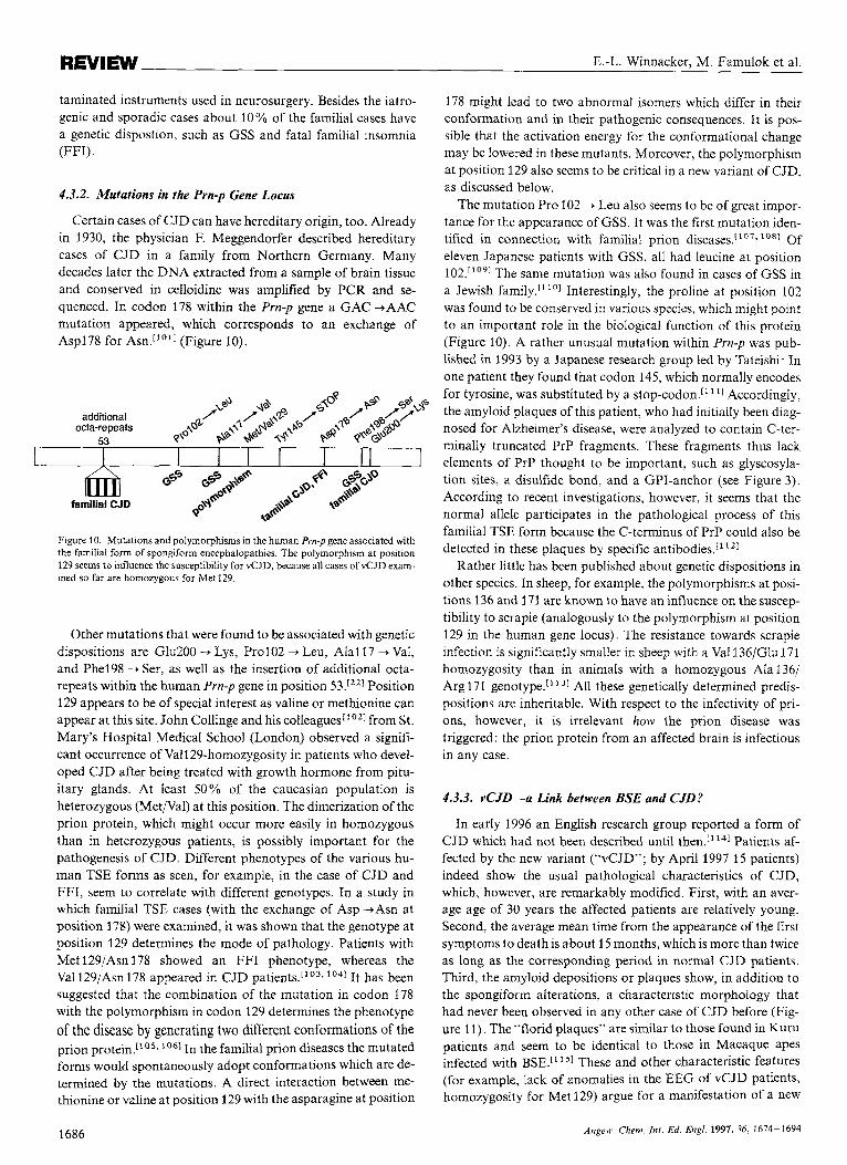
Certain cases of CJD can have hereditary origin, too. Already in 1930, the physician F. Meggendorfer described hereditary cases of CJD in a family from Northern Germany. Many decades later the DNA extracted from a sample of brain tissue and conserved in celloidine was amplified by PCR and se- quenced. In codon 178 within the Prn-p gene a GAC -1AAC mutation appeared, which corresponds to an exchange of Asp178 for Asn.r'o'J (Figure 10).
Figure 10. Mutations and polymorphisms in the human Pm-p gene associated with the familial form of spongiform encephalopathies. The polymorphism at position 129 seems to influence the susceptibility For vCJD. because all cases of vCJD exam- ined so far are homozygous for Met 129.
Other mutations that were found to be associated with genetic dispositions are Glu200 -+ Lys, Pro102 + Leu, Ala117 -1 Val, and Phe198 --t Ser, as well as the insertion of additional octa- repeats within the human Prn-p gene in position 53.l2'1 Position 129 appears to be of special interest as valine or methionine can appear at this site. John Collinge and his colleagues['02~ from St. Mary's Hospital Medical School (London) observed a signifi- cant occurrence of Vall29-homozygosity in patients who devel- oped CJD after being treated with growth hormone from pitu- itary glands. At least 50% of the Caucasian population is heterozygous (MetiVal) at this position. The dimerization of the prion protein, which might occur more easily in homozygous than in heterozygous patients, is possibly important for the pathogenesis of CJD. Different phenotypes of the various hu- man TSE forms as seen, for example, in the case of CJD and FFI, seem to correlate with different genotypes. In a study in which familial TSE cases (with the exchange of Asp +Asn at position 178) were examined, it was shown that the genotype at position 129 determines the mode of pathology. Patients with Met 129/Asn 178 showed an FFI phenotype, whereas the Val 129/Asn 178 appeared in CJD lo4] It has been suggested that the combination of the mutation in codon 178 with the polymorphism in codon 129 determines the phenotype of the disease by generating two different conformations of the prion protein.["'. In the familial prion diseases the mutated forms would spontaneously adopt conformations which are de- termined by the mutations. A direct interaction between me- thionine or valine at position 129 with the asparagine at position
178 might lead to two abnormal isomers which differ in their conformation and in their pathogenic consequences. It is pos- sible that the activation energy for the conformational change may be lowered in these mutants. Moreover, the polymorphism at position 129 also seems to be critical in a new variant of CJD, as discussed below.
The mutation Pro 102 -1 Leu also seems to be of great impor- tance for the appearance of GSS. It was the first mutation iden- tified in connection with familial prion diseases.['o7* lo81 Of eleven Japanese patients with GSS, all had leucine at position 102.11091 The same mutation was also found in cases of GSS in a Jewish family." ''1 Interestingly, the proline at position 102 was found to be conserved in various species, which might point to an important role in the biological function of this protein (Figure 10). A rather unusual mutation within Pm-p was pub- lished in 1993 by a Japanese research group led by Tateishi: In one patient they found that codon 145, which normally encodes for tyrosine, was substituted by a stop-codon." Accordingly, the amyloid plaques of this patient, who had initially been diag- nosed for Alzheimer's disease, were analyzed to contain C-ter- minally truncated PrP fragments. These fragments thus lack elements of PrP thought to be important, such as glyscosyla- tion sites, a disulfide bond, and a GPI-anchor (see Figure 3). According to recent investigations, however, it seems that the normal allele participates in the pathological process of this familial TSE form because the C-terminus of PrP could also be detected in these plaques by specific antibodies.['
Rather little has been published about genetic dispositions in other species. In sheep, for example, the polymorphisms at posi- tions 136 and 171 are known to have an influence on the suscep- tibility to scrapie (analogously to the polymorphism at position 129 in the human gene locus). The resistance towards scrapie infection is significantly smaller in sheep with a Val 136/Glu 171 homozygosity than in animals with a homozygous Ala 136/ Arg 171 genotype." 31 All these genetically determined predis- positions are inheritable. With respect to the infectivity of pri- ons, however, it is irrelevant how the prion disease was triggered: the prion protein from an affected brain is infectious in any case.
4.3.3. vCJD-a Link between BSE and CJD?
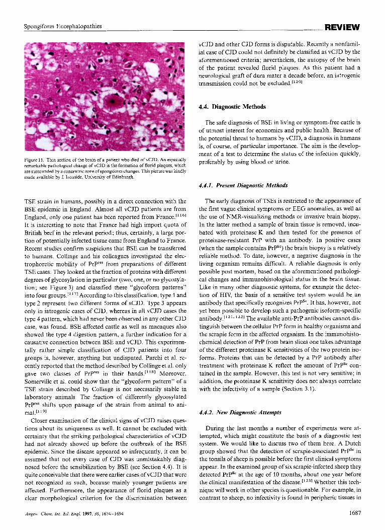
In early 1996 an English research group reported a form of CJD which had not been described until then." 14] Patients af- fected by the new variant ("vCJD"; by April 1997 15 patients) indeed show the usual pathological characteristics of CJD, which, however, are remarkably modified. First, with an aver- age age of 30 years the affected patients are relatively young. Second, the average mean time from the appearance of the first symptoms to death is about 15 months, which is more than twice as long as the corresponding period in normal CJD patients. Third, the amyloid depositions or plaques show, in addition to the spongiform alterations, a characteristic morphology that had never been observed in any other case of CJD before (Fig- ure 11). The "florid plaques" are similar to those found in Kuru patients and seem to be identical to those in Macaque apes infected with BSE." ' 51 These and other characteristic features (for example, lack of anomalies in the EEG of vCJD patients, homozygosity for Met 129) argue for a manifestation of a new
1686 Angew. Chem. Inr. Ed. Engl. 1997. 36, 1674-1694
Spongiform Encephalopathies
Figure 11. Thin section of the brain of a patient who died of vCJD. An especially remarkable pathological change of vCJD is the formation of florid plaques, which are surrounded by a concentric zone of spongiform changes. This picture was kindly made available by J. Ironside, University of Edinburgh.
TSE strain in humans, possibly in a direct connection with the BSE epidemic in England. Almost all vCJD patients are from England, only one patient has been reported from France.["61 It is interesting to note that France had high import quota of British beef in the relevant period; thus, certainly, a large por- tion of potentially infected tissue came from England to France. Recent studies confirm suspicions that BSE can be transferred to humans. Collinge and his colleagues investigated the elec- trophoretic mobility of PrPres from preparations of different TSE cases. They looked at the fraction of proteins with different degrees of glycosylation in particular (two, one, or no glycosyla- tion; see Figure 3 ) and classified these "glycoform patterns" into four groups.['I7' According to this classification, type 1 and type 2 represent two different forms of sCJD. Type 3 appears only in iatrogenic cases of CJD, whereas in all vCJD cases the type 4 pattern, which had never been observed in any other CJD case, was found. BSE-affected cattle as well as macaques also showed the type 4 digestion pattern, a further indication for a causative connection between BSE and vCJD. This experimen- tally rather simple classification of CJD patients into four groups is, however, anything but undisputed. Parchi et al. re- cently reported that the method described by Collinge et al. only gave two classes of Pryes in their Moreover, Somerville et al. could show that the "glycoform pattern" of a TSE strain described by Collinge is not necessarily stable in laboratory animals. The fraction of differently glycosylated Prp'es shifts upon passage of the strain from animal to ani- mal.[' 191
Closer examination of the clinical signs of vCJD raises ques- tions about its uniqueness as well. It cannot be excluded with certainty that the striking pathological characteristics of vCJD had not already showed up before the outbreak of the BSE epidemic. Since the disease appeared so infrequently, it can be assumed that not every case of CJD was unmistakably diag- nosed before the sensibilization by BSE (see Section 4.4). It is quite conceivable that there were earlier cases of vCJD that were not recognized as such, because mainly younger patients are affected. Furthermore, the appearance of florid plaques as a clear morphological criterion for the discrimination between
vCJD and other CJD forms is disputable. Recently a nonfamil- ial case of CJD could not definitely be classified as vCJD by the aforementioned criteria; nevertheless, the autopsy of the brain of the patient revealed florid plaques. As this patient had a neurological graft of dura mater a decade before, an iatrogenic transmission could not be excluded.['201
4.4. Diagnostic Methods
The safe diagnosis of BSE in living or symptom-free cattle is of utmost interest for economics and public health. Because of the potential threat to humans by vCJD, a diagnosis in humans is, of course, of particular importance. The aim is the develop- ment of a test to determine the status of the infection quickly, preferably by using blood or urine.
4.4.1. Present Diagnostic Methods
The early diagnosis of TSEs is restricted to the appearance of the first vague clinical symptoms or EEG anomalies, as well as the use of NMR-visualizing methods or invasive brain biopsy. In the latter method a sample of brain tissue is removed, incu- bated with proteinase K and then tested for the presence of proteinase-resistant PrP with an antibody. In positive cases (when the sample contains PrPSc) the brain biopsy is a relatively reliable method. To date, however, a negative diagnosis in the living organism remains difficult. A reliable diagnosis is only possible post mortem, based on the aforementioned pathologi- cal changes and immunohistological status in the brain tissue. Like in many other diagnostic systems, for example the detec- tion of HIV, the basis of a sensitive test system would be an antibody that specifically recognizes PrPSc. It has, however, not yet been possible to develop such a pathogenic isoform-specific antibody."". l Z z 1 The available anti-PrP antibodies cannot dis- tinguish between the cellular PrP form in healthy organisms and the scrapie form in the affected organism. In the immunohisto- chemical detection of PrP from brain slices one takes advantage of the different proteinase K sensitivities of the two protein iso- forms. Proteins that can be detected by a PrP antibody after treatment with proteinase K reflect the amount of PrPSc con- tained in the sample. However, this test is not very sensitive; in addition, the proteinase K sensitivity does not always correlate with the infectivity of a sample (Section 3.1).
4.4.2. New Diagnostic Attempts
During the last months a number of experiments were at- tempted, which might constitute the basis of a diagnostic test system. We would like to discuss two of them here. A Dutch group showed that the detection of scrapie-associated PrPS' in the tonsils of sheep is possible before the first clinical symptoms appear. In the examined group of six scrapie-infected sheep they detected PrPSc at the age of 10 months, about one year before the clinical manifestation of the disease.['z31 Whether this tech- nique will work in other species is questionable. For example, in contrast to sheep, no infectivity is found in peripheric tissues in
Angew Chem. In!. Ed Engl. 1997,36, 1674-1694 1687
E.-L. Winnacker, M. Famulok et al. REVIEW
cattle; that means that by biopsy of tonsils a detection of PrPSc would not be possible in cattle. In contrast, PrPSc was detected post mortem in the tonsils of a human vCJD case, as reported recently, although in only one case.['241
The group of Michael Harrington does not use PrPSc itself as a diagnostic basis, but a marker protein, which is contained in the cerebrospinal fluid of CJD patients. In 1986 he and his colleagues discovered by two-dimensional gel electrophoresis that two proteins (designated p 130 and p 131) are found in CJD patients, which might serve as diagnostic markers. Recent- ly these proteins were indentified as degradation products of 14-3-3 proteins, and antibodies directed against them were sucessfully used in an immunoassay for CJD diagnosis.['261 Cerebrospinal fluid is taken from the spinal cord of the patients and tested for the presence of the 14-3-3 protein by SDS-gel electrophoresis and immunoblot. The required antibody can be purchased, and the test can be carried out in every biochemistry lab within a few hours. Nevertheless, this test system has a few disadvantages: the appearance of the 14-3-3 proteins is an epiphenonemon, that is, besides CJD other diseases like herpes simplex encephalopathy lead to an increased occurrence of 14-3- 3 protein in the cerebrospinal fluid. The specifity in the exam- ined group of 71 patients was therefore only 88%. The results of the application of the test in cattle were barely conclusive: only a few animals have been tested to date, and moreover, these were not infected with BSE, but with TME. This experiment revealed higher specifity concomitant with decreased sensitivi- ty-only six of nine infected cattle were diagnosed correctly. Finally, hitherto existing data indicate that the test works only at a relatively late stage of the disease, probably not before the first clinical symptoms appear. An effective diagnostic system should respond as soon as possible after infection, so that infect- ed cattle can, for instance, be withdrawn from the market before the epidemic can spread further.
5. Transgenic Models
In many fields of biochemisty the establishment of transgenic animal models is widely accepted for the study of molecular- biological and pathophysiological problems. Also in the case of the research into prion diseases, transgenic mouse lines led to important breakthroughs and results. One of the first animal models for prion dieseases was introduced in 1990 by Prusiner's laboratory. A mouse line was produced, into whose Pm-p gene a point mutation corresponding to one of the human GSS point mutations (Pro 102 + Leu; Figure 10) was introduced. As a consequence of this changed mouse genotype, it spontaneously developed neurological malfunctions, spongiform changes, and gliosis of astrocytes in the brain['271-the three classical charac- teristics of transmissible encephalopathies. It had thus been pos- sible for the first time to produce a genetically determined prion disease in mice. This neurodegeneration, induced by mutation, is hard to distinguish from experimental murine scrapie. One variation, however, is seen in these samples, which have been prepared from brains of the affected transgenic mice: they are only weakly infectious, if at Nevertheless the transgenic "GSS mouse" not only was an important support for the
protein-only hypothesis, it simultaneously opened up the exciting possibility of transfering experimental TSE studies from other species to mice. With the help of such transgenic mouse lines it is possible, for example, to examine species barriers.
5.1. Investigations of the Species Barrier
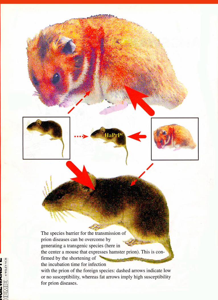
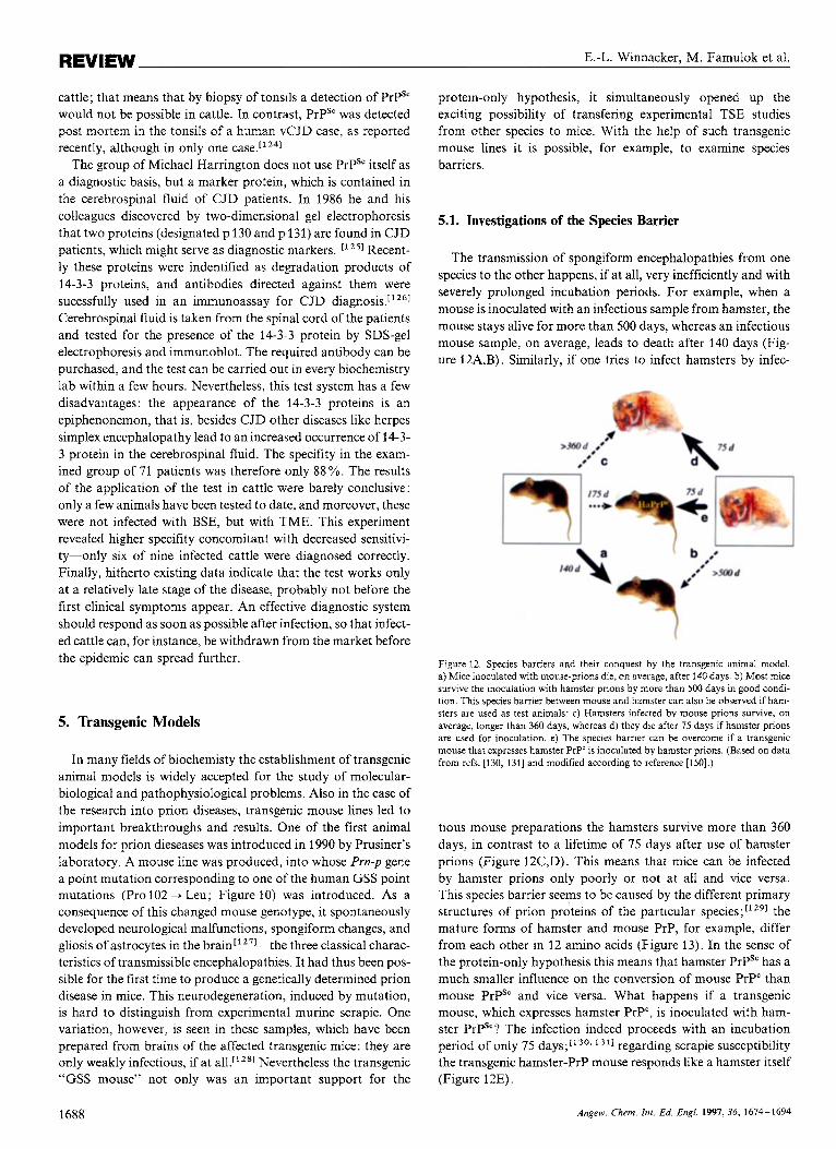
The transmission of spongiform encephalopathies from one species to the other happens, if at all, very inefficiently and with severely prolonged incubation periods. For example, when a mouse is inoculated with an infectious sample from hamster, the mouse stays alive for more than 500 days, whereas an infectious mouse sample, on average, leads to death after 140 days (Fig- ure 12A,B). Similarly, if one tries to infect hamsters by infec-
Figure 12. Species barriers and their conquest by the transgenic animal model. a) Mice inoculated with mouse-prions die, on average, after 140 days. b) Most mice survive the inoculation with hamster prions by more than 500 days in good condi- tion. This species barrier between mouse and hamster can also be observed if ham- sters are used as test animals: c) Hamsters infected by mouse prions survive, on average, longer than 360 days, whereas d) they die after 75 days if hamster prions are used for inoculation. e) The species barrier can be overcome if a transgenic mouse that expresses hamster PrP' is inoculated by hamster prions. (Based on data from refs. [130, 1311 and modified according to reference [150].)
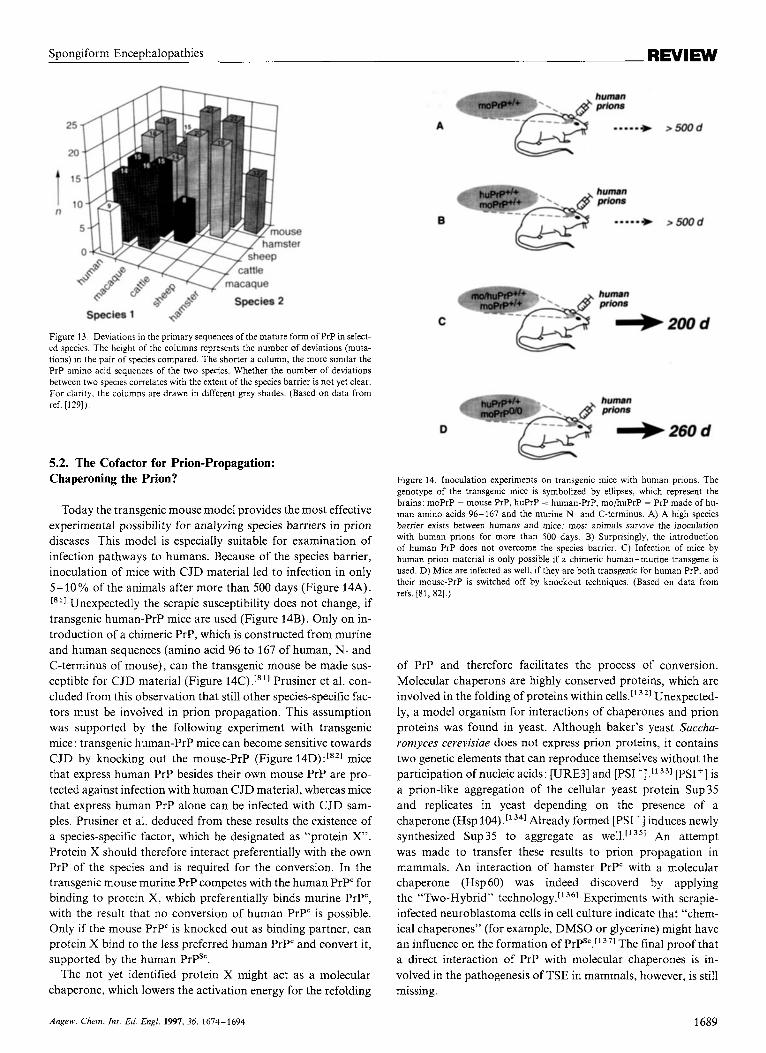
tious mouse preparations the hamsters survive more than 360 days, in contrast to a lifetime of 75 days after use of hamster prions (Figure 12C,D). This means that mice can be infected by hamster prions only poorly or not at all and vice versa. This species barrier seems to be caused by the different primary structures of prion proteins of the particular species;['291 the mature forms of hamster and mouse PrP, for example, differ from each other in 12 amino acids (Figure 13). In the sense of the protein-only hypothesis this means that hamster PrPSC has a much smaller influence on the conversion of mouse PrP" than mouse PrPSc and vice versa. What happens if a transgenic mouse, which expresses hamster PrP", is inoculated with ham- ster PrPS'? The infection indeed proceeds with an incubation period of only 75 days;['30. 311 regarding scrapie susceptibility the transgenic hamster-PrP mouse responds like a hamster itself (Figure 12E).
1688 Angew. Chem. Int. Ed. Engl. 1997,36, 1674-1694
Spongiform Encephalopathies REVIEW
Figure 13. Deviations in the primary sequences of the mature form of PrP in select- ed species. The height of the columns represents the number of deviations (muta- tions) in the pair of species compared. The shorter a column, the more similar the PrP amino acid sequences of the two species. Whether the number of deviations between two species correlates with the extent of the species barrier is not yet clear. For clarity. the columns are drawn in different grey shades. (Based on data from ref. [129])
5.2. The Cofactor for Prion-Propagation: Chaperoning the Prion?
Today the transgenic mouse model provides the most effective experimental possibility for analyzing species barriers in prion diseases. This model is especially suitable for examination of infection pathways to humans. Because of the species barrier, inoculation of mice with CJD material led to infection in only 5 - 10 % of the animals after more than 500 days (Figure 14A). I’ll Unexpectedly the scrapie susceptibility does not change, if transgenic human-PrP mice are used (Figure 14B). Only on in- troduction of a chimeric PrP, which is constructed from murine and human sequences (amino acid 96 to 167 of human, N- and C-terminus of mouse), can the transgenic mouse be made sus- ceptible for CJD material (Figure 14C).[”] Prusiner et al. con- cluded from this observation that still other species-specific fac- tors must be involved in prion propagation. This assumption was supported by the following experiment with transgenic mice: transgenic human-PrP mice can become sensitive towards CJD by knocking out the mouse-PrP (Figure 14D):[”] mice that express human PrP besides their own mouse PrP are pro- tected against infection with human CJD material, whereas mice that express human PrP alone can be infected with CJD sam- ples. Prusiner et al. deduced from these results the existence of a species-specific factor, which he designated as “protein X”. Protein X should therefore interact preferentially with the own PrP of the species and is required for the conversion. In the transgenic mouse murine PrP competes with the human PrP“ for binding to protein X, which preferentially binds murine PrP“, with the result that no conversion of human PrP” is possible. Only if the mouse PrP‘ is knocked out as binding partner, can protein X bind to the less preferred human PrP’ and convert it, supported by the human PrPS‘.
The not yet identified protein X might act as a molecular chaperone, which lowers the activation energy for the refolding
A
B
C
D
Figure 14. Inoculation experiments on transgenic mice with human prions. The genotype of the transgenic mice is symbolized by ellipses, which represent the brains: moPrP = mouse PrP, huPrP == human-PrP, mo/huPrP = PrP made of hu- man amino acids 96- 167 and the murine N- and C-terminus. A) A high species barrier exists between humans and mice: most animals survive the inoculation with human prions for more than SO0 days. B) Surprisingly. the introduction of human PrP does not overcome the species barrier. C) Infection of mice by human prion material is only possible if a chimeric human-murine transgene is used. D) Mice are infected as well, if they are both transgenic for human PrP, and their mouse-PrP is switched off by knockout techniques. (Based on data from refs. [Sl, 821.)
of PrP and therefore facilitates the process of conversion. Molecular chaperons are highly conserved proteins, which are involved in the folding of proteins within cells.[’321 Unexpected- ly, a model organism for interactions of chaperones and prion proteins was found in yeast. Although baker’s yeast Saccha- romyces cerevisiae does not express prion proteins, it contains two genetic elements that can reproduce themselves without the participation of nucleic acids: [URE3] and [PSI+].[133] [PSI+] is a prion-like aggregation of the cellular yeast protein Sup35 and replicates in yeast depending on the presence of a chaperone (Hsp 104) Already formed [PSI+] induces newly synthesized Sup 35 to aggregate as well.“351 An attempt was made to transfer these results to prion propagation in mammals. An interaction of hamster PrP” with a molecular chaperone (Hsp 60) was indeed discoverd by applying the “Two-Hybrid’’ technology.[’361 Experiments with scrapie- infected neuroblastoma cells in cell culture indicate that “chem- ical chaperones” (for example, DMSO or glycerine) might have an influence on the formation of PrPsc.r1371 The final proof that a direct interaction of PrP with molecular chaperones is in- volved in the pathogenesis of TSE in mammals, however, is still missing.
Angew Chem. Inr Ed. Engl. 1997, 36, 1614-1694 1689
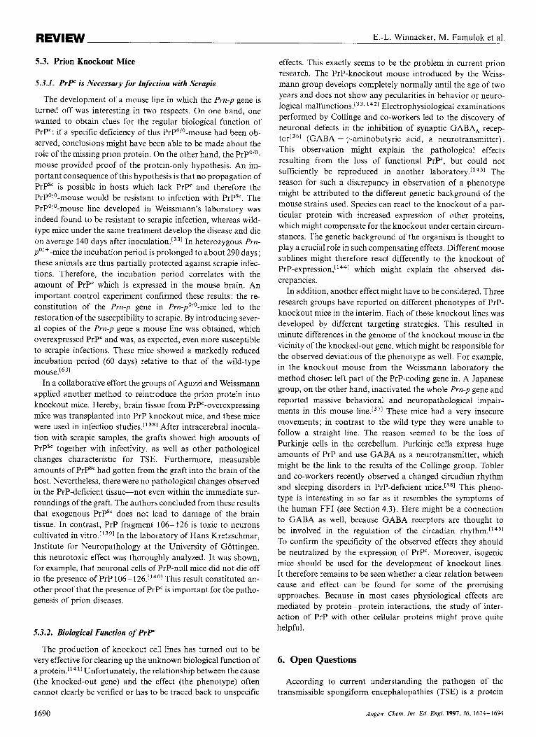
REVIEW E.-L. Winnacker, M. Famulok et al.
5.3. Prion Knockout Mice
5.3.1. P r P is Necessary for Infection with Scrapie
The development of a mouse line in which the Prn-p gene is turned off was interesting in two respects. On one hand, one wanted to obtain clues for the regular biological function of PrP': if a specific deficiency of this PrP'''-mouse had been ob- served, conclusions might have been able to be made about the role of the missing prion protein. On the other hand, the PrPO'O- mouse provided proof of the protein-only hypothesis. An im- portant consequence of this hypothesis is that no propagation of PrPSc is possible in hosts which lack PrP' and therefore the PrP'/'-mouse would be resistant to infection with PrPSc. The PrP'/'-mouse line developed in Weissmann's laboratory was indeed found to be resistant to scrapie infection, whereas wild- type mice under the same treatment develop the disease and die on average 140 days after inoculation.[331 In heterozygous Prn- poi+-mice the incubation period is prolonged to about 290 days; these animals are thus partially protected against scrapie infec- tions. Therefore, the incubation period correlates with the amount of PrP' which is expressed in the mouse brain. An important control experiment confirmed these results: the re- constitution of the Prn-p gene in Prn-p'/'-mice led to the restoration of the susceptibility to scrapie. By introducing sever- al copies of the Prn-p gene a mouse line was obtained, which overexpressed PrP" and was, as expected, even more susceptible to scrapie infections. These mice showed a markedly reduced incubation period (60 days) relative to that of the wild-type
In a collaborative effort the groups of Aguzzi and Weissmann applied another method to reintroduce the prion protein into knockout mice. Hereby, brain tissue from PrP'-overexpressing mice was transplanted into PrP knockout mice, and these mice were used in infection studies." 381 After intracerebral inocula- tion with scrapie samples, the grafts showed high amounts of PrPS' together with infectivity, as well as other pathological changes characteristic for TSE. Furthermore, measurable amounts of PrPS' had gotten from the graft into the brain of the host. Nevertheless, there were no pathological changes observed in the PrP-deficient tissue-not even within the immediate sur- roundings of the graft. The authors concluded from these results that exogenous PrPSc does not lead to damage of the brain tissue. In contrast, PrP fragment 106-126 is toxic to neurons cultivated in ~ i t r o . ~ ' ~ ~ ] In the laboratory of Hans Kretzschmar, Institute for Neuropathology at the University of Gottingen, this neurotoxic effect was thoroughly analyzed. It was shown, for example, that neuronal cells of PrP-null mice did not die off in the presence of PrP 106-126.[1401 This result constituted an- other proof that the presence of PrP' is important for the patho- genesis of prion diseases.
5.3.2. Biological Function of P r P
The production of knockout ceIl lines has turned out to be very effective for clearing up the unknown biological function of a protein.['411 Unfortunately, the relationship between the cause (the knocked-out gene) and the effect (the phenotype) often cannot clearly be verified or has to be traced back to unspecific
effects. This exactly seems to be the problem in current prion research. The PrP-knockout mouse introduced by the Weiss- mann group develops completely normally until the age of two years and does not show any pecularities in behavior or neuro- logical malfunctions.[33. 1421 Electrophysiological examinations performed by Collinge and co-workers led to the discovery of neuronal defects in the inhibition of synaptic GABA, recep- torL3@ (GABA = y-aminobutyric acid, a neurotransmitter). This observation might explain the pathological effects resulting from the loss of functional P r y , but could not sufficiently be reproduced in another The reason for such a discrepancy in observation of a phenotype might be attributed to the different genetic background of the mouse strains used. Species can react to the knockout of a par- ticular protein with increased expression of other proteins, which might compensate for the knockout under certain circum- stances. The genetic background of the organism is thought to play a crucial role in such compensating effects. Different mouse sublines might therefore react differently to the knockout of PrP-expre~sion,"~~] which might explain the observed dis- crepancies.
In addition, another effect might have to be considered. Three research groups have reported on different phenotypes of PrP- knockout mice in the interim. Each of these knockout lines was developed by different targeting strategies. This resulted in minute differences in the genome of the knockout mouse in the vicinity of the knocked-out gene, which might be responsible for the observed deviations of the phenotype as well. For example, in the knockout mouse from the Weissmann laboratory the method chosen left part of the PrP-coding gene in. A Japanese group, on the other hand, inactivated the whole Pm-p gene and reported massive behavioral and neuropathological impair- ments in this mouse line.13'] These mice had a very insecure movements; in contrast to the wild-type they were unable to follow a straight line. The reason seemed to be the loss of Purkinje cells in the cerebellum. Purkinje cells express huge amounts of PrP and use GABA as a neurotransmitter, which might be the link to the results of the Collinge group. Tobler and co-workers recently observed a changed circadian rhythm and sleeping disorders in PrP-deficient mice.[381 This pheno- type is interesting in so far as it resembles the symptoms of the human FFI (see Section 4.3). Here might be a connection to GABA as well, because GABA receptors are thought to be involved in the regulation of the circadian To confirm the specificity of the observed effects they should be neutralized by the expression of PrP'. Moreover, isogenic mice should be used for the development of knockout lines. It therefore remains to be seen whether a clear relation between cause and effect can be found for some of the promising approaches. Because in most cases physiological effects are mediated by protein-protein interactions, the study of inter- action of PrP with other cellular proteins might prove quite helpful.
6. Open Questions
According to current understanding the pathogen of the transmissible spongiform encephalopathies (TSE) is a protein
1690 Angew. Chem. Int . Ed. EngI. 1991, 36, 1674-1694
Spongiform Encephalopathies REVIEW
that does not require a nucleic acid for infectivity and propaga- tion. Proof of the protein-only hypothesis by the de novo gener- ation of infectivity in vitro after incubation of PrP' with PrP'" is still missing. The conversion of host PrP" into the pathogenic PrP'" seems to be the central event in the propagation of TSE. It is possible that other not yet identified cofactors take part in this process, for example a "prion receptor" or other proteins or molecules, which interact with the prion protein. It is therefore important to identify possible prion-protein interactions and to examine whether they influence the conversion of the cellular into the scrapie isoform.
Both isoforms of the prion protein differ only in their confor- mation as determined by C D and I R spectroscopy. Whether differences in the three-dimensional structure of different prion strains exist within one species is not clear but has to be postu- lated, because of the present state of knowledge, to explain certain strain-specific characteristics of prion propagation. Therefore. structural studies are in the center of research efforts to shed light into the replication mechanism of prions. The structure of a mouse PrP' fragment identified by N M R shows the expected extended r-helical regions besides two smaller an- tiparallel a-sheets, which might serve as a seed for the develop- ment of []-sheet structures in PrP". Whether the structure of the amino-terminally truncated fragment really contains all the im- portant domains required for the prion replication remains to be seen. Structural elucidations of PrPSc have also still not been achieved because of the difficulties connected with the low soh- bility of the protein.
The cell-free in vitro conversion of proteinase K sensitive PrP' into proteinase K resistant protein material was an impor- tant step in prion research, especially with respect to the current models for prion propagation. Although the conversion is strain-specific, it proceeds relatively ineffectively and can cur- rently be achieved only on use of a surplus of PrP". Whether the in vitro converted material is not only proteinase K resistant but also infectious cannot be answered yet. This question is of highest relevance for the verification of a hypothesis for prion propagation.
PrP-knockout mice are not susceptible to TSE and otherwise d o not show any noticeable phenotype. The prion protein itself seems to be of no immediate benefit for the organism; the obser- vations of the three different knockout cell lines, however, are contradictory. Maybe PrP' participates in synaptic processes. The identification of PrP-protein interactions might suggest functional relationships in this context. The transmission of TSE is affected by species barriers, which are manifested, for example, in extended incubation periods. The reasons for the barriers are thought to be the slight differences in amino acid sequences, which result in structural differences of prion proteins of individual species. The central question whether BSE can be transferred from cattle to humans cannot be examined directly. Macaques, however, can be infected by infectious BSE material accompanied by the development of not previously observed histological characteristics. These novel pathological changes seem to be identical with those of a new variant of Creutzfeld- Jakob disease (vCJD) in humans, which ap- peared in England just recently. A causal relation between the BSE epidemic and this new form of TSE in humans is quite probable.
7. Concluding Remarks
"It struck me recently that one should really consider the se- quence o fa protein molecule, about to fold into aprecise geometric form, as a line of melody written in canon form andso designed by nature to fold back upon itself, creating harmonic chords ofinter- action consistent with biolgical function. One might carry the analogy further by suggesting that the kinds of chords formed in a protein with scrambled disulphide bridge, . . . are dissonant, but that, by giving an opportunity for rearrangement. . .they modulate to give the pleasing harmonics of the native molecule. Whether or not some conclusion can be drawn about the greater thermody- namic stability of Mozart's over Schonberg 's music is something I will leave to the philosophers of the audience."
C. Anfinsen in Neis Perspecfires in tliologj (Ed.: M. Sela), Elstvier, New York. pp. 42-50.
In Anfinsen's visionary world of harmony between sequence and structure, dissonant overtones entered not only because of the results of the prion research. Experiments which were car- ried out with the denatured enzyme "Rubisco" (Ribulose bis- phosphate carboxylase oxygenase) showed that a spontaneous renaturation into the correct structure merely by dilution with the renaturating buffer is not possible. The correct refolding can be achieved only in the presence of the chaperonin G ~ o E L . " ~ ~ ] Citrate synthetase from the mitochondria of pig hearts tends to self-aggregate if the enzyme is denaturated in guanidinium hy- drochloride solution and is then diluted again."471 These and other examples show that not all proteins spontaneously fold into the thermodynamically most stable conformation, because, for example, they might be caught in a kinetic trap during the course of folding. Such proteins d o indeed fold in Anfinsen's sense, but only within a protective
If the protein-only hypothesis is correct, the situation seems to be different for the prion protein. It can exist in a t least two, maybe even more, metastable structural states.['491 The prion protein is the prototype of a structure-labile protein. By a relatively slight trigger, such as a single mutation in the Prn-p gene or contact with the smallest amounts of the incorrectly folded isoform, this lability is manifested in the refolding of the normal isoform. The change of one structure into the other has dramatic consequences for the organism. The understand- ing of "prions" and their characteristics is proceeding very rapidly-this review describes the most important progress achieved until April 1997 (a deadline we imposed on ourselves, otherwise we would not yet have completed the article be- cause of the breathtaking development of this research field). Nevertheless, we are far from understanding the function and characteristics of this partly frightening and partly fascinating protein.
We thank H . Schatzl for critical reading of the manuscript. We are grateful to R. Glockshuber and K. Wuthrich for providing us with a picture of the N M R structure of PrP, to J. Ironside and H . Kretzschmar for the images of histological thin sections, to H . Kretzschmar and G. Ourisson for helpful discussions, and to the Deutsche Forschungsgemeinschaft and the Bundesministerium fur Bildung, Forschung, Wissenschaft und Technologie ( B M B F ) for jinancial support.
Angrii Clrem. In1 Ed Engl 1997. 36. 1674- 1694 1691
REVIEW E.-L. Winnacker, M. Famulok et al.
Appendix 1. Glossary of the most important abbreviations and medical terms
amyloid AA BSE CD cerebrospinal fluid
chaperones CJD EEG FFI GABA glia cells
gliosis GPI GSS Octa-repeat
polymorphism
prion PrP PrP27-30
PrP' PrPS', PrP"' Purkinje cells
spongiosis TSE
__
pathological deposition of proteins amino acid(s) bovine spongiform encephalopathy circular dichroism lymphoid liquid present in the medulla; secreted by the brain proteins that are involved in folding of cellular proteins Creutzfeldt -Jakob disease electroencephalogram fatal familial insomnia y-aminobutyric acid cells of the connective tissue of the central nervous sys- tem pathological proliferation of glia cells glycosyl phosphatidylinositol Gerstmann-Striussler-Scheinker syndrome repeating motif of eight glycine/proline-rich AA, characeteristic for PrP differences in the genotype within a single population that cannot be explained by naturally occurring muta- tion rates alone abbreviation for proteinaceous infectious particle prion protein fraction of proteins truncated at the N-terminal that have an electrophoretic mobility of 27-30 kDa cellular isoform of PrP pathogenic isofonns of PrP huge dendritic ganglia1 cells in the mid-layer of the cerebellum cortex spongelike alteration in tissues transmissible sponBform encephalopathy
Received: February 11, 1997 [AZIOIE] German version: Angew. Cheni. 1997, 109, 1748-1769
Translated by Claudia Famulok, Munich
A. F. Hollemann, E. Wiberg, Lehrbuch der Anorgunischen Chemre, 81st-90th ed., DeGruyter, Berlin, 1976. S. B. Prusiner, Science 1982, 216, 136-144. S. B. Prusiner, M. P. McKinley, K. A. Bowman, D. C. Bolton, P. E. Bend- heim. D. F. Groth, G. G. Glenner, Cell 1983, 35, 349-358. J. Hope, L. J. D. Morton, C. F. Farquhar, G. Multhaup, K. Beyreuther, R. H. Kimberlin, EMBO J 1986,5, 2591-2597. D. Westaway, G. A. Carlson, S. B. Prusiner, Tr. Microhiol. 1995,3. 141-147. L. J. Berg, Proc. Natl. Acud. Sci. USA 1994, 91, 429-432. G. W. Roberts, J. Clinton In Prion Diseuse: The Specwum of Purhology und Diagnostic Considerorions (Eds.: S. B. Prusiner, J. Collinge, J. Powell, B. An- derton), Ellis Horwood, New York, 1992. C. I. Lasmizas, J. P. Deslys, R. Demaimay, K. T. Adjou, J. J. Hauw, D. Dor- mont, J Gen. Virol. 1996, 77, 1601 - 1609. M E. Bruce, Br. Med. Bull. 1993, 49, 822-838. This phenomenon is known from viral pathogens: the pathology changes because of mutations in the viral genome, but the principal infectivity re- mains. H. Diringer in Spongijorme Encephuloputhirn hei Tieren unter hesondprer Beriicksichtigungder BSE, Band 75 (Ed.: W. Kohler), Deutsche Akademie der Naturforscher Leopoldina, Halle, 1996, pp. 26-28. Diringer also commented on his hypothesis in a round table discussion at the Deutsche Akademie der Naturforscher Leopoldina, moderated by Prof. Rudolf Rott. M. Ozel, H. Diringer, Luncet 1994, 343, 894-895. M. Ozel, Y.-G. Xi, E. Baldauf, H. Diringer, M. Pocchiari, Luncet 1994. 344, 923-924. 1. Tischer, H. Gelderhlom, W. Vettermann. M. A. Koch, Nature 1982, 295. 64 - 66. T. 0. Diener, R. A. Owens, R. W. Hammond, Intervirology 1993, 35. 186- 195. T. 0. Diener, FASEB J 1991,5, 2808-2813. D. Riesner, M. Colpan, T. C. Goodman, L. Nagel, J. Schumacher. G. Steger. H. Hofmann, J Biomol. Struct. Dyn. 1983, 1, 669-688. J. Smarda, Actu Virol. Engl. Ed. 1987. 31, 506-524. T. 0. Diener, Tr. Microbiol. 1993, I , 289-294.
K. Kellings, N. Meyer. C. Mirenda, S. B. Prusiner, D. Riesner, J Gen. Virol. 1992, 73, 1025-1029. K . Kellings, S. B. Prusiner, D. Riesner, Philos. Tram. R. Soc. London B 1994, 343,425 -430. S. B. Prusiner, Science 1991,252, 1515-1522. N. Meyer, V. Rosenbaum, B. Schmidt, K. Gilles, C. Mirenda, D. Groth. S. B. Prusiner. D. Riesner. J. Gen. Virol. 1991. 72, 37-49. R. Mestel. Science 1996, 273. 184-189 T. Alper, D. A. Haig. M. C. Clarke, Biochen?. Bioplrys. Res. Commun. 1966. 22. 278-284. T. Alper, W. A. Cramp. D. A. Haig, M. C. Clarke. Nature 1967,214,764-766. J. S . Griffith, Nuture 1967, 215, 1043-1044. D. C. Bolton. M. P. McKinley, S B. Prusiner, Science 1982,218,1309-1311. E. Turk. D. B. Teplow. L. E. Hood, S. B. Prusiner, Eur. J Biocheni. 1988.176, 21 -30. "Prion" is a slight modification of the abbreviation "Proin". B. Oesch. D. Westaway, M. Walchli, M P. McKinley, S B H. Kent, R. Aebersold. R. A. Barry, P. Tempst. D. B. Teplow, L. E. Hood, S. B. Prusiner, C. Weissmann. Cell 1985, 40, 735-746. K. Basler, B Oesch, M. Scott, D. Westaway, M. Wilchli, D. F. Groth, M. P McKinley, S. B. Prusiner, C. Weissmann, Cell 1986, 46, 417-428. H. Buehler. A. Aguzzi. A. Sailer, R.-A. Greiner, P. Autenried. M. Aguet. C. Weissmann. Cell 1993, 73, 1339- 1347. C. B. Anfinsen. Science 1973, 181. 223-230. C. I. Lasmezas. J:P. Deslys. 0. Robain, A. Jaegly. V. Beringue, J.-M. Peyrin, 1.-G. Fournier, JLJ. H a w . J. Rossier. D. Dormont. Science 1997, 275. 402- 405. J. Collinge. M. A. Whittington. K. C. L. Sidle, C. J. Smith. M. S. Palmer, A. R. Clarke. J. G. R. Jeffreys. Nature 1994. 370. 295-297. S. Sakaguchi, S. Katamine, N. Nishida, R. Moriuchi, K. Shigematsu, T. Sugimoto. A. Nakatani, Y Kataoka. T. Houtani, S. Shirahe, H. Okada, S. Hasegawa. T. Miyamoto. T. Noda. Nuture 1996, 380, 528-531. I. Tobler. S. E. Gaus. T. Deboer, P. Achermann, M. Fischer, T. Rulicke, M. Moser, B. Oesch, P. A. McBride, J. C. Manson, Nuturp 1996, 380, 639 - 642. N . Stahl. D. R. Borchelt, K. Hsiao, S. B. Prusiner. Cell 1987,51. 229-240. D. C. Bolton, R. K. Meyer, S. B. Prusiner, J Virol. 1985. 53, 596-606. L. Manuelidis, S. Valley, E. E. Manuelidis, Proc. Nutl. Acad Sci. USA 1985, 92,4263-4267. T. Endo. D. Groth. S . B. Prusiner, A. Kobdta, Biochemistry 1989. 28. 8380- 8388. J. Safar. W. Wang, M. P. Padgett, M. Ceroni, P. Piccardo. D. Zopf, D. C. Gajdusek, C. J. J. Gibbs. Proc. Nutl. Acad. Sci. U S A . 1990.87,6373-6377.
1441 R A. Gibbons, G. D. Hunter, Nature 1967,215. 1041-1043 [451 R. K. Meyer, M. P. McKinley, K. A. Bowman, M. B. Braunfeld, R. A. Barry,
S. B Prusiner. Proc. Null. Acud. Sci. USA 1986,83. 2310-2314. [46] B. Caughey, G. J. Raymond. J Bid . Cliem. 1991,266. 18217-18223. I471 B. Chesebro. R. Race. K Wehrly, J. Nishio, M. Bloom. D. Lechner, S.
Bergstrom. K. Rohbins, L. Mayer, J. M. Keith, C. Garon, A. Haase, Nuture 1985,315, 331-333.
[48] C. Locht, B. Chesebro, R. Race, J. M. Keith, Proc. Null. Acud. Sci. USA 1986,
[49] B. Caughey. Br. Med. Bull. 1993, 49, 860-872. [50] D. Riesner, K. Kellings. K. Post, H. Wille, H. Serban, D. Groth, M. A.
Baldwin. S B. Prusiner, J Virol. 1996, 70, 1714-1722 [Sl] F. E. Cohen, K.-M. Pan, Z. Huang, M. Baldwin, R J. Fletterick, S. B. Prusin-
er. Science 1994, 264, 530-531. [S21 K.-M. Pan, M. Baldwin. J. Nguyen, M. Gasset, A. Serban. D. Groth, 1.
Mehlhorn. 2. Huang, R. J. Fletterick, F. E. Cohen. S. B. Prusiner, Proc. Nut[. Acod. Sci USA 1993, 90, 10962-10966
[53] B. W. Caughey, A. Dong, K. S. Bhat. D. Ernst, S. F. Hayes, W S. Caughey, Biochemi.strj. 1991, 31, 7672-7680.
[54] N. Stahl, M. A. Baldwin, D. B. Teplow, L. Hood, B. W. Gibson. A. L Burlingame, S. B. Prusiner. 5iochemi.rrry 1993, 32, 1991 -2002
I551 1. Safar. P. P. Roller, D. C . Gajdusek, C. 1. Gibbs. Jr., J B i d Chem. 1993.268, 20 276-20284.
[56] Z. Huang, J.-M. Gabriel. M. A. Baldwin, R. J. Fletterick. S. B. Prusiner, F. E.
[57] Z. Huang, S B. Prusiner, F. E. Cohen, Fold. Design 1995, 1, 13-19. [58] M. Gasset. M. A. Baldwin, D. Lloyd. J. M. Gabriel, D. M. Holtzmann, F. E.
Cohen, R. J. Fletterick, S. B. Prusiner. Proc. Nut!. Acud. Sci. USA 1992, 89, 10940- 10944.
I591 J. T. Nguyen. M. A. Baldwin, F. E. Cohen, S. B. Prusiner, Biochemistry 1995, 34.4186-4192.
[60] H. Zhang, K. Kaneko. J. T. Nguyen, T. L. Livshits. M. A. Baldwin, F. E. Cohen. T. L. James, S. 8. Prusiner, J M d Biol. 1995, 250. 514-526.
[61] R. Riek. S. Hornemann. G. Wider, M. Billeter, R. Glockshuher, K. Wuthrich. Nuture 1996,382, 180- 182.
(621 S. Hornemann. R. Glockshuber, J Mol. Brol 1996,262.614-619. [63] M. Fischer. T. Rulicke, A. Raeher, A. Sailer, M. Moser, B. Oesch, S. Brand-
ner. A. Aguzzi. C Weissmann, EMBO J 1996, 15. 1255-1264
83. 6372-6376.
Cohen, Proc Nutl. Acud. Sci. USA 1994, 91, 7139-7143.
1692 Angen. Chem. Int. Ed. Engl. 1997, 36, 1614-1694
Spongiform Encephalopathies
[64] S. Prusiner, Arch. Neurol. 1993. 50, 1129-1153. [65] A possible way to produce labeled PrPS' would be. for instance, the in vitro
conversion of "C- and "N-labeled PrP'. By in vitro or cell-free conversion, PrPC IS converted into PrPSC by addition of exogenous PrPSC.
[66] J. Heller, A. C . Kolhert, R. Larsen, M. Ernst. T Bekker, M. Baldwin, S. B. Prusiner. A Pines, D E. Wemmer, Protein Sci. 1996, 5 , 1655-1661
[67] M. E Bruce. A. G. Dickinson, J. Gen. Virol. 1987, 68, 79-89. [68] R. H. Kimherlin. C. A. Walker. H. Fraser, J. Gen. Virol. 1989. 70, 2017-
[69] R I Carp, S . M. Callahan, J. Gun. V t r d 1991, 72, 293-298. [70] R. Hecker. A. Tarahoulos. M. Scott, K. M. Pan. S. L. Yang. M. Torchia, K.
Jendroska. S J DeArmond, S. B. Prusiner, Genes Der. 1992. 6, 1213-1228. [71] P Brown, P P Liherski. A. Wolff. D. C. Gajdusek, J In/ecf. Dis. 1990, 161,
467-472. [72] J. H. Come, P E Fraser, P T. J Lanshury, Proc Nut/. Acud. Sci. USA 1993,
90. 5959 - 5963 [73] T .T Ashhurn. P T Lanshury, J Am. Chem. SOL.. 1993, fl5, 11012-11013. [74] P. T. Lanshury, Jr., B. Caughey. Cheni. Biol. 1995, 2. 1-5. [75] M Eigen. Eiup/?w Ch& 1996. 63, Al-A18. [76] A. J. Raeher. D. R. Borchelt. M. Scott. S. B. Prusiner, J Virol 1992, 66,
[77] D A. Kocisko. J H. Come. S. A. Priola, B. Chesehro, G. J. Raymond. P. T.
[78] R. A. Bessen. D. A. Kocisko, G. J. Raymond, S. Nandan, P. T. Lansbury.
[79] R A. Bessen. R. F. Marsh, J . Virol. 1994, 68, 7859-7868. [SO] G C Telling. P. Parchi, S. J DeArmond, P. Cortelli. P. Montagna, R. Gahi-
Lon. J. Masrrianni. E. Lugaresi, P. Gamhetti, S. B. Prusiner, Science 1996, 274. 1079-2082
[St] G. C. Telling. M. Scott, K. K. Hsiao, D. Foster, S.-L. Yang. M. Torchia, K C . L Sidle. J. Collinge, S. J. DeArmond. S. B Prusiner, Proc. Nut/. Acad. Sci C:SA 1994. 91. 9936-9940.
[82] G. C. Telling, M Scott, J. Mastrianni, R. Gahizon, M. Torchia. F E. Cohen, S. J DeArmond. S. B. Prusiner, Cell 1995,83, 79-90.
[83] L E. Orgel, C h m . Biol. 1996, 3. 413-414. I841 D. K. Kondepud!, R. J. Kaufman, N. Singh, Science 1990,250,915-976. [85] W. J. Hadlow. Luncet 1957, 2, 289-290. [86] D. C. Gajdusek. C. J. J. Gihhs, M Alpers. Science 1967, f55, 212-214. [87] J. Cuille. P. L. Chelle, C. R. Hebd. Seonces Atad. Sci. Ser. D 1939, 208,
I058 - 1060. [88] First reports on a putative case of scrapie in cattle go back to the 19th
century[l51]. [89] J. Hope, L. J. Reekie, N. Hunter. G. Multhaup, K. Beyreuther, H. White,
A C. Scott. M. J. Stack. M. Dawson. G. A. Wells, Nature 1988,336,390-392. [90] R. M. Anderson, C. A. Donnelly, N. M. Ferguson, M. E. 1. Woolhouse, C. J.
Watt. H. J. Udy. S. MaWhinney, S. P. Dunstan, T. R. E. Southwood, J. W. Wilesmith. J. B M. Ryan, L. J. Hoinville, J. E. Hillerton, A. R. Austin, G A H. Wells. Nature 1996, 382, 779-788.
[91] J. W. Wilesmith. J. B. M. Ryan, M. J. Atkinson, Ver. Rec. 1991, 128,199-203. [92] J. W. Wilesmith. J B. M. Ryan, W. D. Hueston, L. J. Hoinville, Vet. Rec 1992,
[93] J. Bradhury. Luncef 1996, 348, 393. [94j H:G. Creutrfeldt. Z. Ges. N ~ u F o ~ . Psychiatr. 1920, LVII, 1-18. [95] A Jakob. Dtsch. 2. Nrrvenheilkd. 1921, 70, 132-146. [96] J. Gerstmann, E. Striussler, I . Scheinker, 2. Ges. Neural. Psychiatr. 1936,154,
136 - 762. [97] R. Medori. H -J. Tritschler. A. LeBlanc, F. Villare. V. Manetto, H Y Chen,
R. Xue. S Leal, P. Montagna, P. Cortelli, P. Tinuper, P. Avoni, M. Mochi, A. Baruzzi. J. 1. Hauw. J. Ott. E. Lugaresi, L. Autilio-Gambetti, P Gamhetti, Neii Eng. J Med 1992. 326. 444-449.
[98] S . B Prusiner. Sci. Am. 1995, 272, 48-57. [99] C . Masson. i Delalande, J. P. Deslys, D. Henin, C. Fallet-Banco, D. Dor-
[loo] A. Jaegly, F. Boussin, J P. Deslys. D. Dormont, Cenomrcs 1995,27,382-383. [lot] H. A. Kretzschmar, M. Neumann, D. Stavrou, Acta Neuroputhol. 1995, 89,
[lo21 J. Collinge, M S Palmer, A. J. Dryden, Lancet 1991, 337, 1441 -1442. [I031 L. G. Goldfarb. R. 8. Petersen, M. Tahaton, P. Brown, A. C. LeBlanc, P.
Montagna. P. Cortelli, J. Julien, C. Vital, W W. Pendelhury, M. Haltia, P. R. Wills. J. J Hauw. P. E. McKeever, L. Monari, B. Schrank, G. D. Swergold, L Autilio-Gamhetti. D. C. Gajdusek, E. Lugaresi, P. Gamhetti, Science 1992, 258. 806-808
[lo41 L. G. Goldfarb. P. Brown, M. Haltia. F. Cathala. W. R. McComhie, J. Ko- vanen. L. Cervendkovd, L. Goldin, A. Nieto, M. S. Godec, D. M. Asher, D. C. Gajdusek. Ann. Neurol. 1992. 31, 274-281.
[lo51 P. Gamhetti. R. Petersen. L. Monari, M. Tahaton, L. Autilio-Gamhetti, Br. Mpd. Bull. 1993, 49, 980-994.
I1061 L. Monari. S G. Chen. P. Brown, P. Parchi, R. B. Petersen. J. Mikol, F. Gray, P. Cortelli, P. Montagna, B. Ghetti, L. G. Goldfarb, D. C. Gajdusek, E. Lugarest. P. Gambetti, L Autilio-Gamhetti, Proc. Nurl. Acud S o . USA 1994, Y I . 2839-2842
2025.
6155-6163
Lanshury. Jr.. B Caughey, Nature 1994, 370,471 -474.
B. Caughey. .Vuture 1995, 375. 698-700.
130.90-94.
mont. D. Ley\. Neurology 1994. 44, 179-180.
96-98.
REVIEW
[lo71 K. Hsiao, H. F. Baker, T. J. Crow, M. Poulter, F. Owen. J. D. Terwilliger, D. Westaway, J. Ott, S. B. Prusmer, Nature 1989. 338. 342-345.
[lo81 D. Goldgaber, L. G. Goldfarb, P. Brown, D . M. Asher, W. T. Brown, S. Lin. J. W. Teener, S. M. Feinstone, R. Ruhenstein, R. J. Kascsak. J. W. Boelaard. D. C. Gajdusek, Erp. Neurol. 1989, 106, 204-206.
11091 K Doh-ura, J. Tateishi. H. Sasaki, T. Kitamoto. Y. Sakaki. Biochem. Biophys. Res. Commun. 1989. 163.914-979.
[I101 Y. Goldhammer. R. Gahizon, Z. Meiner, M. Sadeh, ivriirologj, 1993. 43. 2718-2719
[1111 T. Kitamoto, R. Iizuka. J. Tateishi, Biochem. Biophys. Re\. Comnrun. 1993, 192. 525-531.
[112] B. Ghetti. P. Piccardo, M. G. Spillantini, Y. Ichimiyd, M. Porro, F. Perini, T. Kitamoto, J. Tateishi. C. Seiler, B. Frangione. 0. Bugiani. G. Giaccone, F. Prelli, M. Goedert, S. R. Dlouhy, F. Tagliavini, Proc. Nuti Acud Sci. USA. 1996, 93. 744-748.
[I 131 P. Belt. 1. Muiiemdn, B. Schreuder, J. BosdeRuijter. A. Gielkens, M. Smits. J. Gen. Virol. 1995, 76, 509-517.
[114] R. G Will, J. W Ironside. M. Zeidler, S N. Cousens. K. Estiheiro, A. Alper- ovitch, S. Poser, M. Pocchiari. A. Hofman, P. G. Smith. Luncet 1996. 347, 921 -925: this publication had ramifications beyond its scientific significance (the article was the most-cited scientific publication in 1996; source: Institute for Scientific Information, Hot Papers Database, Novemher/Decem- her 1994- Novemher/Decemher 1996)-it also had political effects: in spring 1996 the British government was forced to admit for the first time that a transmission of BSE to humans cannot be excluded. At European level, the commission of the European Union introduced anexport ban for British beef.
[l IS] C. 1. Lasmezas, J.-P. Deslys. R. Demalmay. K. T. Adlou. F. Lamoury, D. Dormont, 0. Rohain. J. Ironside, J:J. Hauw, Nuture 1996. 3x1. 743-744.
[116] G. Chazot. E. Broussolle, C . 1. Lapras, T. Blittler, A. Aguzzi, N. Kopp, Luncet 1996. 347. 1181
[117] J. Collinge, K. C . L. Sidle, J. Meads, J. Ironside, A F. Hill. Nuture 1996, 383. 685-690.
[I 181 P. Parchi, S. Capellan, S. G. Chenn, R. B. Petersen, P. Gainbetti, N Kopp. P. Brown. T Kitamoto. J. Tateishi. A. Giese. H. Kretzschmar, Nufure 1997, 386.232-233.
I1191 R. A. Somerville, A. Chong, 0. U. Mulqueen, C R. Birkett, S. C. E. R. Wood, J. Hope, Nature 1997, 386, 564.
(1201 N. Kopp. N. Streichenherger. J. Deslys, J. Laplanche, G. Chazot. Lancet 1996, 348. 1239-1240.
[121] S. B. Prusiner, D. Groth, H. Serban, R. Koehler, D. Foster, M. Torchia, D. R. Burton, S. L. Yang, S. J. DeArmond, Proc. Natl. Acud. Sci. USA 1993, 90, 10608 - 10612
[122] R. A. Williamson, D. Peretz, N. Smorodinsky, R. Bastidas, H. Serhan, 1. Mehlhorn. S. J. DeAmond, S. B. Prusiner. D. R. Burton, Proc. Natl. Acad. Sci. USA. 1996, 93, 7279-7282.
[123] B. E. C. Schreuder. L. J. M. van Keulen, M. E. W. Vromans, J. P. M. Langeveld, M. A. Smits, Nature 1996. 381, 563.
[124] A. F. Hill, M. Zeidler, J. Ironside, J. Collinge, Lancet 1997, 349, 99-100. [125] M. G. Harrington, C. R. Merril, D. M. Asher, D. C. Gajdusek. New Engl. J
Med. 1986,315,219-283. 11261 G. Hsich. K. Kenney, C. J. J. Gihhs, K. H. Lee. M. G. Harrington, New Engl.
J Med. 1996,335, 924-930. [127] K. K. Hsiao, M. Scott, D. Foster, D. F. Groth, S. J. DeArmond. S B. Prusin-
er, Science 1990,250. 1587- 1590. [128] K. K. Hsiao, D. Groth, M. Scott, S. L. Yang. H. Serhan, D. Rapp, D Foster,
M. Torchia, S J. DeArmond, S. B. Prusiner. Proc. Natl. Auid. Sci. USA 1994. 91.9126-9130.
[129] H. M. Schitzl. M. Da Costa, L. Taylor, F. E. Cohen. S. B. Prusiner, J Mol. Biol. 1995, 245, 362-374.
[130] M. Scott, D. Foster. C. Mirenda, D. Serban, F. Coutal. M. Wilchli, M. Torchia, D . Gr0th.G. Carlson. S. J. DeArmond, D. Westaway. S. B. Prusiner. Cell 1989, 59. 847-857.
[131] S. B. Prusiner, M. Scott, D. Foster, K.-M. Pan, D. Groth. C. Mirenda, M. Torchia, S:L. Yang, D. Serhan, G. A. Carlson. P. C. Hoppe, D. Westaway, S. J. DeArmond, Cell 1990,63,673-686.
[I321 F. U. Hartl. J. Martin, Curr. Opin. Struct. Biol. 1995, 5 , 92 -102. [133] R. B. Wickner. D. C. Masison. H. K. Edskes, Yeust 1995, fl. 1671 -
[134] Y. 0. Chernoff, S. L. Lindquist, B.-I. Ono, S. G. Inge-Vechtomov, S. W. Lieh-
[135] M. M. Patino. J.-J. Liu, J. R. Glover, S. Lindquist. Sciencc, 1996, 273, 622-
[136] F. Edenhofer, R. Rieger. M. Famulok, W. Wendler. S. Weisc. E -L. Winnack-
[137] J. Tatzelt, S. B. Prusiner, W. J. Welch, EMEO J 1996, 15. 6363-6373. [138] S. Brandner, S . Isenmann, A Raeher, M. Fischer, A. Sailer. Y. Kohayashi, S.
Marino, C. Weissmann, A Aguzzi. Nature 1996, 379, 339-343. 11391 G. Forloni. N. Angeretti, R. Chiesa, E. Monzani, M. Salmona. 0. Bugiani, F.
Tagliavini, Nuture 1993, 362. 543- 546. 11401 D. R. Brown, J. Herms, H. A. Kretzschmar. NetrroReporr 1994, 5. 2057-
2060.
1685.
man, Science 1995,268, 880-884.
626.
er. J. Virol. 1996, 70. 4724-4728.
Angew. Chm7. Int. Ed. Engl. 1997. 36. 1674-1694 1693
E.-L. Winnacker, M. Famulok et al. REVIEW
[I411 E. P. Brandon, R. L. Idzerda, G. S . McKnight, Curr. Biol. 1995.5.625-634. 11421 H. Buehler, M. Fischer, Y Lang, H. Bluethmann, H.-P. Lipp, S . J. DeAr-
mond, S. B. Prusiner, M. Aguet, C. Weissmann, Nuture 1992,356. 577-582. 11431 P.-M. Lledo, P. Tremblay, S . J. DeArmond, S . B. Prusiner, R. A. Nicoll, Proc.
Null. Acud. Sci. USA 1996, 93, 2403-2407. 11441 D. R. Borchelt, S. S. Sisodia, Chem. Biol. 1996, 3, 619-621. [145] J. P. Estibeiro, Trends Neurosci. 1996, 19, 257-258. [1461 P. Goloubinoff, J. P. Christeller, A. A. Gatenby, G. C. Lorimer, Narure 1989,
371. 578-586.
11471 J. Buchner, M. Schmidt, M. Fuchs. R. Jaenicke, R. Rudolph, F. X. Schmid.
[148] R. John Ellis also pictorially calls this container an “Anfinsen cage”[152]. [149] This statement is valid at least in the limited sense of Prusiner’s heterodimer
[lSO] C. Weissmann, Nuture 1991, 349, 569-571. [151] M. Sarradet, Rev. Vet. 1883. 3, 310-312. [152] R. J. Ellis, Fold. Design 1995, 1, R9-Rl5.
T. Kiefiaber, Biochemistry 1991, 30, 1586- 1591.
model.
1694 Angew. Chem. Int. Ed. Engl. 1997.36, 1674-1694