Protein Synthesis: DNA Transcription into mRNA, RNA Processing and mRNA Translation into Protein.
1
W I S S E N T E C H N I K L E I D E N S C H A F T
u www.tugraz.at
CHE.167 Genetics

2
CHE.167 Genetics
Topics Contents
Introduction Historical facts
„Classical“ Genetics Mendel
DNA Composition, higher-level structure, configurations, cellularorganisation, chromatin, chromosome structure, genome
Extrachromosomal elements, mobile DNA (Is, Tn), repetitive DNA
Replication Basic concept, uni-bi, rolling circle, telomeres
Segregation - Partitioning
Cell cycle Meiosis, Mitosis
Gene expression Transcription: promoters, termination, mRNA processing, mRNA stability; translation initiation
Regulation of gene expression Regulation of transcription in prokaryotes/eukaryotes, operons, regulatory proteins, regulation on translationallevel, regulation on DNA level
Changing genetic information Mutation, recombination, genetic engineering, genetransfer (parasexual mechanisms)
Genetic analysis Crossing analyses, molecular analyses
OVERVIEW

3Presentations: in TUG online (link to FTP server)
Essential books:
Benjamin LewinGenes XIPearson Education Inc. 2008
Benjamin LewinEssential GenesIntern. EditionPearson,2006
Klug, Cummings, Spencer, PalladinoConcepts of Genetics, 9th EditionPearson Int. Edition, 2009
Additional literature:
Rolf KnippersMolekulare Genetik, 9. AuflageThieme Verlag, 2006
D.L. Hartl, E.W. JonesGenetics: Analysis of Genes and Genomes. 6th EditionJones and Bartlett, 2005
Klug, Cummings, SpencerGenetik, 8th EditionPearson Studium, 2007
T.A. BrownGenomes 3, Garland Science (3rd Edition, 2006)Genome und Gene, 3. Auflage, Spektrum, 2007
CHE.167 Genetics

4
Cell factory
EnzymesGenes
Metabolism andBiochemical Reactions
NutrientsSubstrates
Product(s)
Command Unit
Machines
Process
Organelles
CHE.167 Genetics
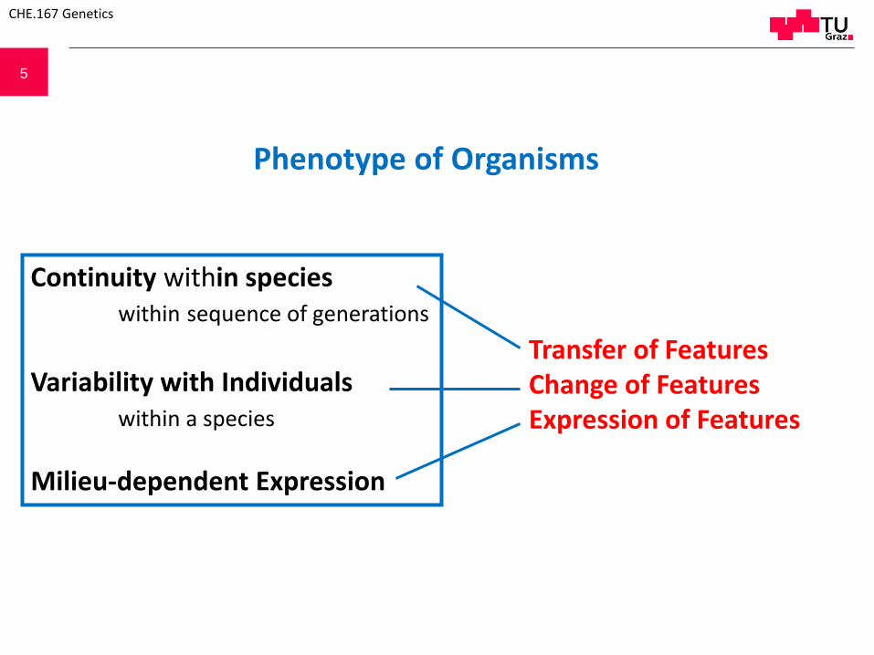
5
Phenotype of Organisms
Continuity within specieswithin sequence of generations
Variability with Individualswithin a species
Milieu-dependent Expression
Transfer of FeaturesChange of FeaturesExpression of Features
CHE.167 Genetics

6
Genetics:
Deals with Inherited Features
Includes Environment-determined Features
CHE.167 Genetics

7
What is genetic information?How can genetic information be maintained?How is genetic information transferred to progeny?How can genetic information be altered?How is genetic information transmitted into features?
How can one manipulate organisms at the gene level?
Basic Science Applied Research
Tools
CHE.167 Genetics
Main Questions

8 Gregor Mendel~ 1860 Genes determine
featuresof living organisms
Taken from: A.J.F Griffiths, J.H. Miller, D.T. Suzuki, R.C. Lewontin, W.M. Gelbart; An Introduction to Genetic Analysis, 5th Edition; W.H. Freeman and Company, NY

9
Mendel´s seven traits:
Clearly defined traits that behave equally stable over generations Traits are independent from environmental factors
Traits Dominant allele Recessive allele
Seed shape round, roundish wrinkled, angular
Endosperm colour pale yellow, light yellow, orange more or less intensively green
Seed coat colour grey, greyish brown or leather brown with or without violet dotting; violet leaf banner, purple blossom leaves and reddish stems at the leaf axes
white with white blossoms
Pod shape arched , inflated Constricted and more or less wrinkled
Pod colour light to dark green vibrant yellow
Flower and pod position axial terminal
Stem length long short
CHE.167 Genetics

10
CHE.167 Genetics
Taken from: Daniel L. Hartl & Elizabeth W. Jones, Genetics: Analysis of Genes and Genomes, 6th Edition, Jones and Bartlett Publishers

11
Trait is independent of the sexual origin.
Targeted fertilizations
CHE.167 Genetics
Taken from: A.J.F Griffiths, J.H. Miller, D.T. Suzuki, R.C. Lewontin, W.M. Gelbart; An Introduction to Genetic Analysis, 5th Edition; W.H. Freeman and Company, NY
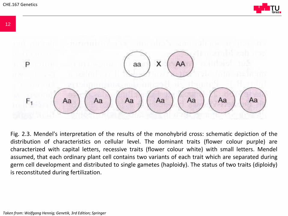
12
CHE.167 Genetics
Fig. 2.3. Mendel's interpretation of the results of the monohybrid cross: schematic depiction of thedistribution of characteristics on cellular level. The dominant traits (flower colour purple) arecharacterized with capital letters, recessive traits (flower colour white) with small letters. Mendelassumed, that each ordinary plant cell contains two variants of each trait which are separated duringgerm cell development and distributed to single gametes (haploidy). The status of two traits (diploidy)is reconstituted during fertilization.
Taken from: Wolfgang Hennig; Genetik, 3rd Edition; Springer
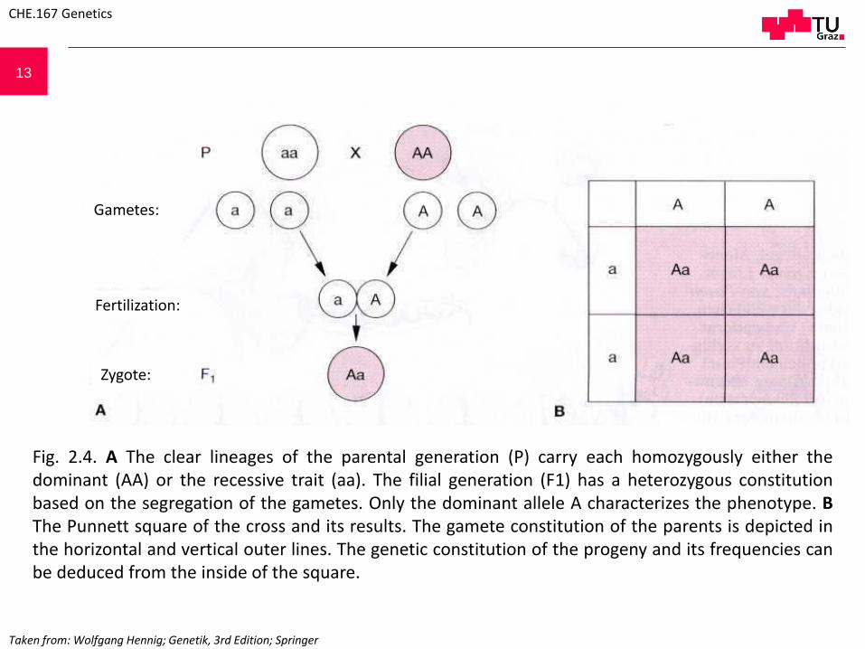
13
Fig. 2.4. A The clear lineages of the parental generation (P) carry each homozygously either thedominant (AA) or the recessive trait (aa). The filial generation (F1) has a heterozygous constitutionbased on the segregation of the gametes. Only the dominant allele A characterizes the phenotype. BThe Punnett square of the cross and its results. The gamete constitution of the parents is depicted inthe horizontal and vertical outer lines. The genetic constitution of the progeny and its frequencies canbe deduced from the inside of the square.
CHE.167 Genetics
Gametes:
Fertilization:
Zygote:
Taken from: Wolfgang Hennig; Genetik, 3rd Edition; Springer

14
Segregation of heterozygous traits
CHE.167 Genetics
Fig. 2.5 2nd Mendelian Law. Cross of theF1 individuals resulting from the formercross by self-fertilization. The progeny(F2) segregate in a 3:1 ratio and 25% ofthe individuals show the recessive traitof the parental generation (whiteflowers). Those individuals keep theirrecessive phenotype when crossedwith other individuals of the recessivephenotype. Thus, they are homozygousfor the recessive trait. Whereas, in casethey are further crossed with adominant phenotype (purple flowers)by self-fertilization, 2/3 of thefollowing F3 progeny is segregated intothe recessive/dominant phenotype in a1:3 ratio. The remaining 1/3 of theindividuals with a dominant phenotypekeep their phenotype unaltered even inthe following generations. The geneticconstitution of the F2 individuals isthus 25% homozygous for the recessivetrait (white:aa), 25% homozygous forthe dominant trait (purple:AA) and 50%heterozygous (Aa) (see Fig. 2.6).
F1 cross
purple
purple purple
purple purple
white
white white
Segregation (3:1)
Segregation (3:1)
Taken from: Wolfgang Hennig; Genetik, 3rd Edition; Springer
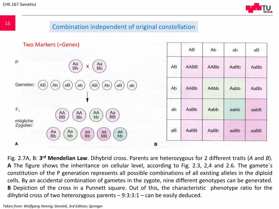
15
Two Markers (=Genes)
Combination independent of original constellation
Taken from: Wolfgang Hennig; Genetik, 3rd Edition; Springer
CHE.167 Genetics
Fig. 2.7A, B: 3rd Mendelian Law. Dihybrid cross. Parents are heterozygous for 2 different traits (A and B).A The figure shows the inheritance on cellular level, according to Fig. 2.3, 2,4 and 2.6. The gamete´sconstitution of the P generation represents all possible combinations of all existing alleles in the diploidcells. By an accidental combination of gametes in the zygote, nine different genotypes can be generated.B Depiction of the cross in a Punnett square. Out of this, the characteristic phenotype ratio for thedihybrid cross of two heterozygous parents – 9:3:3:1 – can be easily deduced.

16
Taken from: D. P. Snustad, M.J. Simmons; Principles of Genetics, 5th Edition; John Wiley & Sons, Inc.
CHE.167 Genetics
17
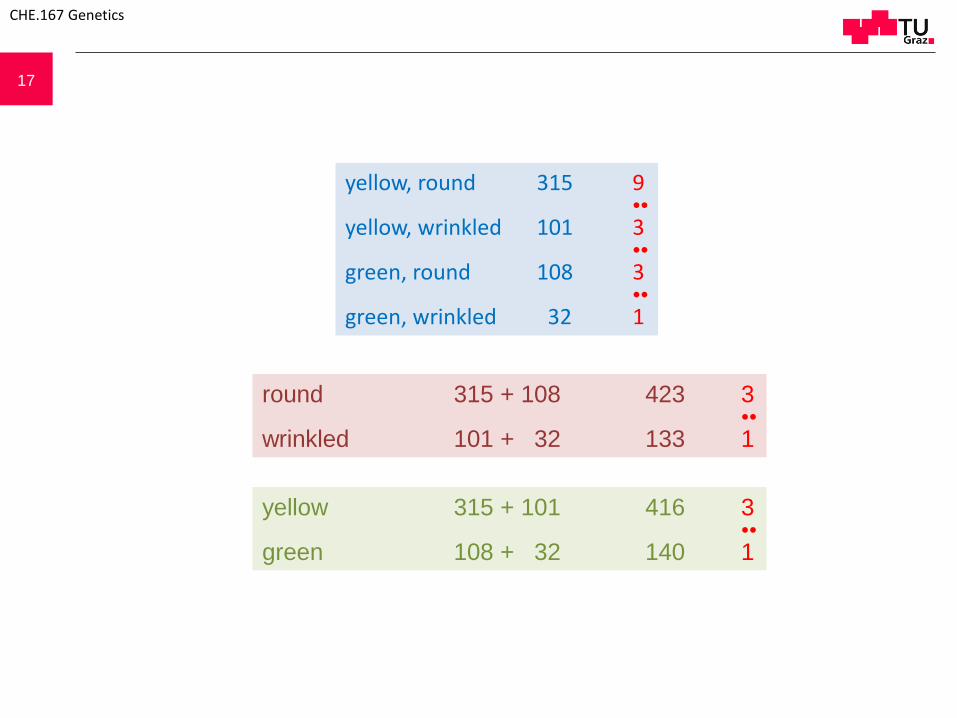
yellow, round 315 9●●
yellow, wrinkled 101 3●●
green, round 108 3●●
green, wrinkled 32 1
round 315 + 108 423 3●●
wrinkled 101 + 32 133 1
yellow 315 + 101 416 3●●
green 108 + 32 140 1
CHE.167 Genetics

18
Important experimental basis: statistical treatment of data
F2 generation of a monohybrid cross
CHE.167 Genetics
Taken from: Daniel L. Hartl & Elizabeth W. Jones, Genetics: Analysis of Genes and Genomes, 6th Edition, Jones and Bartlett Publishers

19
Question:Is the genetic information Nucleic Acid or protein??
1928: Griffith – Experiments with Diplococcus pneumoniae
S-form: lethalR-form: no effect
Extract of S-form + viable R-Form: lethalviable S-form present
1944: Avery, MacLeod, McCartney: Nucleic acid of desoxyribosetype is responsible for transformation
CHE.167 Genetics
20
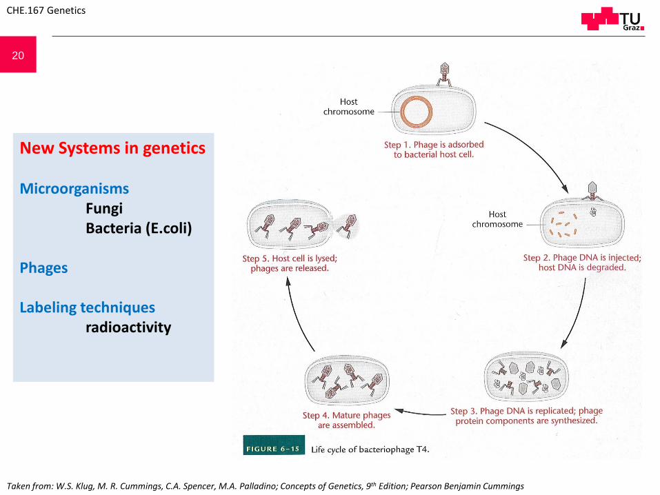
New Systems in genetics
MicroorganismsFungiBacteria (E.coli)
Phages
Labeling techniquesradioactivity
Taken from: W.S. Klug, M. R. Cummings, C.A. Spencer, M.A. Palladino; Concepts of Genetics, 9th Edition; Pearson Benjamin Cummings
CHE.167 Genetics

21
> 80% P32 inside the cell> 80% S35 outside the cell
Hershey – Chase („blender“ ) Experiment (1952)
T4 phage with32P labeledDNA
T4 phage with35S labeledprotein
32P-DNA entersthe host cell andcauses theformation ofphage progeny
35S-protein sticks to thesurface of the host cellsand can be removed byshearing in a blender. The infection process takesplace normally.
Quelle???
CHE.167 Genetics

22
https://prezi.com/cws9dhcj34m-/griffith-hershey-and-chase/
Hershey – Chase („blender“ ) Experiment (1952)
> 80% P32 inside the cell> 80% S35 outside the cell
CHE.167 Genetics
23
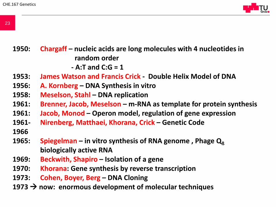
1950: Chargaff – nucleic acids are long molecules with 4 nucleotides inrandom order
- A:T and C:G = 11953: James Watson and Francis Crick - Double Helix Model of DNA1956: A. Kornberg – DNA Synthesis in vitro1958: Meselson, Stahl – DNA replication 1961: Brenner, Jacob, Meselson – m-RNA as template for protein synthesis1961: Jacob, Monod – Operon model, regulation of gene expression1961- Nirenberg, Matthaei, Khorana, Crick – Genetic Code 19661965: Spiegelman – in vitro synthesis of RNA genome , Phage Qß
biologically active RNA1969: Beckwith, Shapiro – Isolation of a gene1970: Khorana: Gene synthesis by reverse transcription1973: Cohen, Boyer, Berg – DNA Cloning1973 now: enormous development of molecular techniques
CHE.167 Genetics

24
4.10.

25
DNA is the moleculefor storage of
geneticinformation
Quelle???
CHE.167 Genetics
26
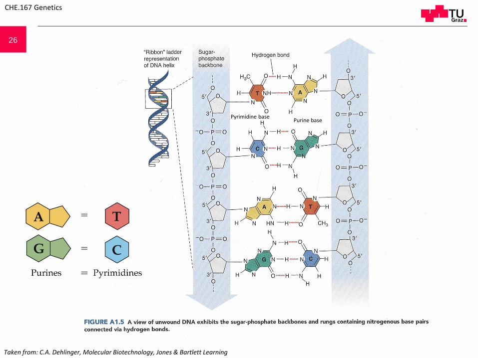
Pyrimidine basePurine base
Taken from: C.A. Dehlinger, Molecular Biotechnology, Jones & Bartlett Learning
CHE.167 Genetics

27
A The Nucleotide is the basic building block, consisting of a deoxyribose molecule (or ribose molecule for RNA), a heterocyclic organic base, which is connected by a N-glycosidicbond to the C1 atom of the (deoxy)ribose and phosphate group linked to the C5 atom of the (deoxy)ribose. If the phosphate group is missing, the molecule is called Nucleoside. BThe organic bases are either the purines adenine (A) or guanine (G) or the pyrimidines cytosine (C), thymine (T) or in case of RNA uracil (U) instead of thymine. C Numbering of the bases and the ribose atoms. D Nucleotides linked via 3´-5´-phosphate-diesterbonds between the sugar units build the macromolecules of the nucleic acids. They can be distinguished chemically only by the sequence of the organic bases.
DNA and RNA components:
N-glycosidic bond
Phosphate
Nucleoside
Nucleotide
Adenine Guanine
Cytosine Thymine Uracil
5´end
3´end
CHE.167 Genetics

28
Watson Crick base pairing
Taken from: W.S. Klug, M. R. Cummings, C.A. Spencer, M.A. Palladino; Concepts of Genetics, 9th Edition; Pearson Benjamin Cummings
CHE.167 Genetics
29
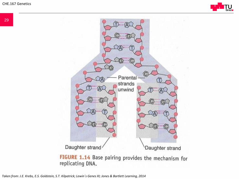
Taken from: J.E. Krebs, E.S. Goldstein, S.T. Kilpatrick; Lewin´s Genes XI; Jones & Bartlett Learning, 2014
CHE.167 Genetics

30
Absorption rises with increasing temperature.
Rel
ativ
e ab
sorp
tio
no
fU
V li
ght
at 2
60
nm
Temperature [°C]
CHE.167 Genetics
31
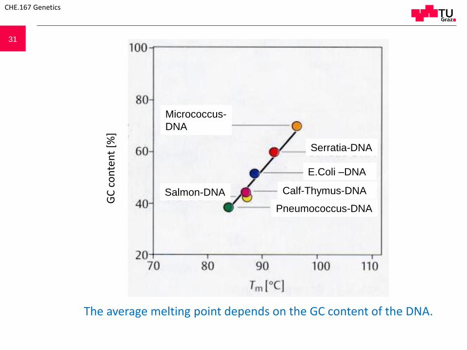
The average melting point depends on the GC content of the DNA.
GC
co
nte
nt
[%]
CHE.167 Genetics
Salmon-DNA Calf-Thymus-DNA
Micrococcus-
DNA
Serratia-DNA
E.Coli –DNA
Pneumococcus-DNA

32
A - conformation B - conformation Z - conformation
A A - and B – DNA are right-handed helices (twisted clockwise), whereas Z – DNA is a left-handed helix(twisted counter-clockwise ). Additionally, Z –DNA is less twisted and has a reduced diameter, causingchanges in the „major“ and „minor groove“. B Double helix top view. The axis of B – DNA double helix (B)is central (inner circle), base pairs are in the centre and the sugar-phosphate backbone twists round thecentral area (outer circle). The axis of the A – and Z – conformations (A, Z) are located asymmetrically andcause structural changes of the grooves. (A: Weaver & Hendrick, 1992; B: Watson et al. 1987)
CHE.167 Genetics

33
B A
CHE.167 Genetics
Dehydrogenation causes a change from B form DNA into the rigid A form. In both cases, thedouble helix is right-handed but shows structural differences. Concerning the B form, basepairs are located vertical to the central axis whereas in the A form base pairs are inclined atan angle of ~ 70° and displaced from the central axis to the major groove. This creates anopen space inside of the molecule and the occurrence of a deep, but narrow major groove.

34
Taken from: http://de.slideshare.net/MUBOSScz/10-polysacch-heteroglycosidesnucleicacids
CHE.167 Genetics

35
C3´-endo sugar conformation
C2´-endo sugar conformationB – DNA compared to Z – DNA. The phosphatesugar backbone in the Z – DNA follows a zig-zag path around the helix.
CHE.167 Genetics
36
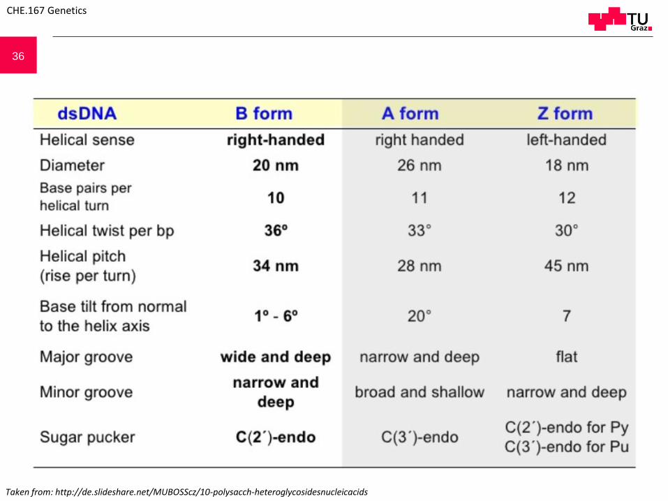
Taken from: http://de.slideshare.net/MUBOSScz/10-polysacch-heteroglycosidesnucleicacids
CHE.167 Genetics

37Fixed bent structure caused by specific primary sequence structure
Kinetoplast DNA of Trypanosoma. Blocks of adenine residues succeed each other in distance of a helix turn (10-11 base pairs).
CHE.167 Genetics

38
5‘
5‘3‘
3‘
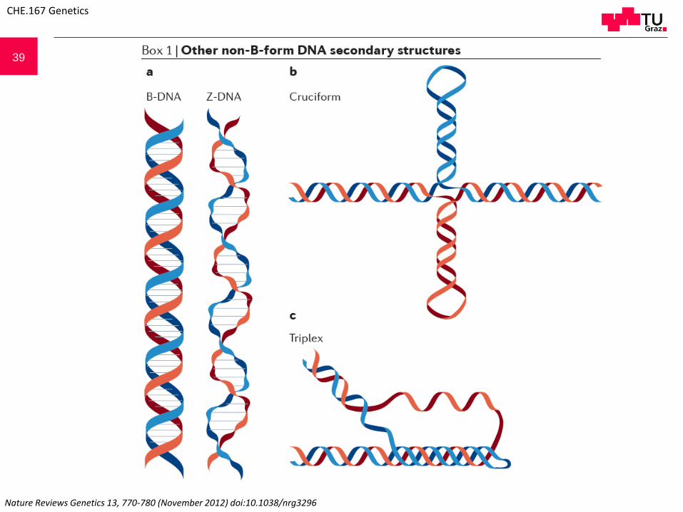
Cruciform DNA
Left: lac operon DNA (E.coli); right: cruciform DNA model
CHE.167 Genetics
39
Nature Reviews Genetics 13, 770-780 (November 2012) doi:10.1038/nrg3296
CHE.167 Genetics

40 Triple Helix DNA
http://atlasgeneticsoncology.org/Educ/Images/H-DNAE.jpg
Model of an intramolecular triplex helix.Partial separation of a polypurine strand from apolypyrimidine strand: the polypyrimidinestrand folds back and is taken up by the majorgroove where Hoogsteen pairings with thepurine are formed. Low pH values promote therearrangement as protonated cytosine is ableto form a Hoogsteen base pairing.
CHE.167 Genetics

41 Z-DNAIn contrast to standard B-form DNA (B-DNA), Z-DNA is a left-handed helix128 (see the figure, part a). Z-DNAmotifs (that is, sequences that form Z-DNA in vitro) are tracts of alternating purines and pyrimidines, whichoccur about once every 3,000 bp in metazoans129. Negative supercoiling stabilizes the formation of Z-DNA underphysiological salt conditions130, and it is hypothesized that Z-DNA relieves transcription-induced torsionalstress131. Z-DNA motifs are tightly associated with transcriptional start sites in eukaryotic genomes132, and thesemotifs can also cause genome instability, although the type of damage they cause varies from prokaryotes(dinucleotide insertions and deletions) to eukaryotes (double-strand breaks resulting in larger deletions)120, 121,
133, 134.Cruciform structuresNegative supercoiling can also cause B-DNA to adopt a four-armed, cruciform secondary structure thatresembles a Holliday junction135 (see the figure, part b). These structures require ≥6-nucleotide inverted repeats(cruciform motif) to form, and such motifs are located near replication origins, breakpoint junctions andpromoters in diverse organisms136, 137. In metazoans, cruciform motifs are enriched near sites of grosschromosomal rearrangements138, and deletions and translocations occur more frequently in vivo at sites ofcruciform motifs than in B-DNA139, 140, 141. However, cruciforms might also serve positive roles (for example,stabilizing the human Y chromosome (reviewed in Ref. 134)).Triplex DNAThree-stranded triplex DNA occurs when single-stranded DNA forms Hoogsteen hydrogen bonds in the majorgroove of purine-rich double-stranded B-DNA142 (see the figure, part c). Triplexes in which the third strand isantiparallel to the DNA duplex can form at physiological pH, and these structures are stabilized by negativesupercoiling142. Sequences capable of forming triplexes are common in eukaryotes but much rarer inprokaryotes143. In mammals, triplex-forming motifs are enriched in the introns of a variety of essential genes,including those involved in development and signalling144. Additionally, triplexes are hypothesized to causegenomic instability by causing double-strand breaks that result in translocations145. However, the formation of atriplex structure in a trinucleotide repeat sequence (for example, (CAG)n) can prevent the expansion of therepeat138, 139; repeat expansion is related to human genetic disorders146, 147.
Nature Reviews Genetics 13, 770-780 (November 2012) doi:10.1038/nrg3296
CHE.167 Genetics

42
Circular and linear DNA. The complementary strains of the linear, but not of the circular, DNA can be separated by melting. The linkage number of the relaxed circular DNA corresponds to the number of helical twists. Lk = Tw = 12
Circular relaxed DNA
Linear DNA
Quelle???
CHE.167 Genetics

43
Taken from: Daniel L. Hartl & Elizabeth W. Jones, Genetics: Analysis of Genes and Genomes, 6th Edition, Jones and Bartlett Publishers
CHE.167 Genetics

44
Lk: Linkage numberTw: TwistWr: Writhe
Topology of underwound DNA. Linkage number values with and withoutunderwinding
left right
CHE.167 Genetics

45
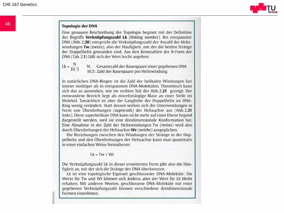
DNA Topology:A more detailed description of topology starts with the definition of the term „linking number“ (Lk). Concerning relaxed DNA, the linkage number is equivalent to the number of helical twists (Tw). Based on the following formula, „Lk“ can be determined easily:
Lk = N / 10,5
N: Total number of base pairs of a given DNA10,5: number of base pairs per helical twist
In natural DNA rings the number of helical twists is mostly lower than in relaxed DNA molecules. Theoretically, the consequence can be as shown in Fig. 2.21: the unwound region is located as a single strand bubble somewhere in the molecule. Effectively, the pitch of the double helix in the DNA ring remains almost unchanged. Instead, the underwindings cause supercoils of the helical axis. Superhelical DNA has a three-dimensional conformation. Thus, a decrease in the number of helical twists is compensated by supercoils of the helical axis (writhe).
This relationship can be quantitatively drafted in a very simple way: Lk = Tw + Wr
Thus, Lk indicates the frequency of DNA strand cross overs. Lk represents a topological property of closed DNA molecules: the values for Tw and Wr can vary, but Lk remains the same. In other words, closed DNA molecules with a given linking number can have various three-dimensional forms.
46
CHE.167 Genetics

47
The function of topoisomerase II (gyrase). A Subunit A of topoisomerase II cuts the DNA double strand and separates the cutting sites from each other. After the intact double helix has passed the open DNA site, the severed strand is re-joined. B Consequences on DNA level. A negative supercoiling can be created by topoisomerase II (steps I to III).
Quelle???
CHE.167 Genetics

48
Taken from: L. Stryer; Biochemistry, 3rd Edition; W.H. Freeman and Company / New York
CHE.167 Genetics

49
Taken from: B. Lewin, Essential Genes, Pearson Education International
CHE.167 Genetics
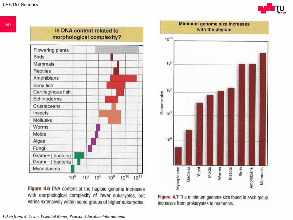
50
Taken from: B. Lewin, Essential Genes, Pearson Education International
CHE.167 Genetics

51
Taken from: B. Lewin, Essential Genes, Pearson Education International
CHE.167 Genetics

52
Length(µm)
Basepairs(bp)
Number ofGenes
Simian Virus 40(SV40, animal virus)
1,8 5243 6
Bacteriophage M13(double stranded, replicative form)
2,2 6407 10
Bacteriophage Lambda 16,5 48502 ca. 50
Bacteriophage T4 ca. 60 ca. 166000 > 100
Escherichia coli ca. 1300 ca. 4720000 > 3000
Some bacterial and viral genomes
CHE.167 Genetics

53
Taken from: W.S. Klug, M. R. Cummings, C.A. Spencer, M.A. Palladino; Concepts of Genetics, 9th Edition; Pearson Benjamin Cummings
CHE.167 Genetics

54
Taken from: R. Knippers; Molekulare Genetik; Thieme
Merodiploid cell (type I-/F´I+). lacI gene is drawn too big in proportion to the rest of the chromosome. In fact it´s just 0,15% of the E.coli chromosome. Wild type lacI gene produces an active repressor (green dots), which is free in the cell and can thus dock to the chromosomal lac operator as well as to the plasmid lac operator.
CHE.167 Genetics
55
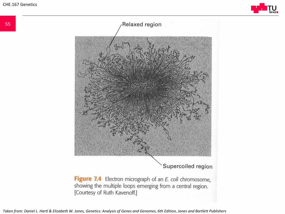
Taken from: Daniel L. Hartl & Elizabeth W. Jones, Genetics: Analysis of Genes and Genomes, 6th Edition, Jones and Bartlett Publishers
CHE.167 Genetics

56
Taken from: Daniel L. Hartl & Elizabeth W. Jones, Genetics: Analysis of Genes and Genomes, 6th Edition, Jones and Bartlett Publishers
CHE.167 Genetics

57
Taken from: G.M. Cooper, R.E. Hausman; THE CELL A Molecular Approach, 4th Edition; ASM Press
CHE.167 Genetics

58
http://faculty.ccbcmd.edu/~gkaiser/SoftChalk%20BIOL%20230/Prokaryotic%20Cell%20Anatomy/proeu/proeu/proeu_print.html
CHE.167 Genetics

59
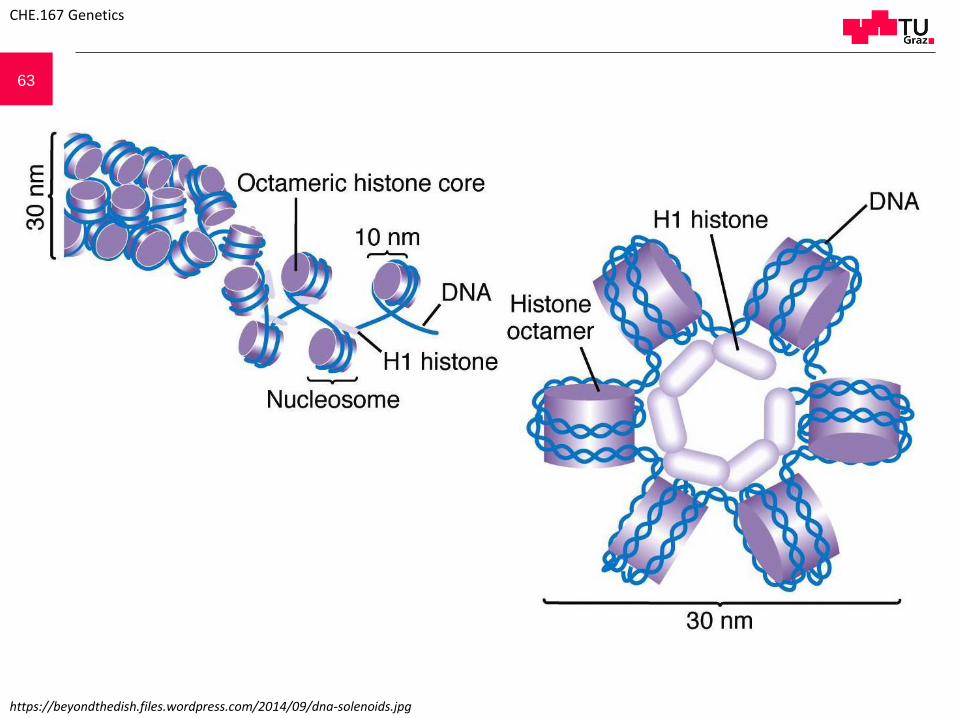
Structure of a nucleosome. A A single nucleosome with DNA: The DNA double helix winds twice aroundthe protein core. The 8 different histone molecules are indicated (see Table 9.1). The histone core buildsa symmetrical structure (an octamer made of 2 tetramers). The linkage number of DNA in thenucleosome is decreased causing a negative supercoiling. B Hypothetical model of nucleosomal structureduring transcription. RNA polymerase cannot pass an intact nucleosome but demands a (at least partial)disintegration of the nucleosome. One of the models proposed assumes that the nucleosomedisintegrates for a short time during transcription into two tetramers. A (Kornberg and Klug, 1981; B Prioret al. 1983)
CHE.167 Genetics

60
Type Amino acids M r Lys/Arg ratio Remarks
H1 215 21000 20,0 Variable
H2A 129 14500 1,25 Lysin rich, variability limited
H2B 125 13700 2,5 Lysin rich, variability limited
H3 135 15300 0,72 Arginine rich, very conserved
H4 102 11200 0,79 Arginine rich, very conserved
Quelle???
CHE.167 Genetics

61
Quelle???
Nucleosomes in oocyte chromatin of Pleurodeles waltlii (from Scheer 1987)
CHE.167 Genetics

62
Model of a chromatid after partial unfolding of the nucleosome chain to the 300 Å fibril. Histone H1 molecules, connecting consecutive nucleosomes, are not shown. (according to Klug from Darnell et al. 1990)
CHE.167 Genetics
63
https://beyondthedish.files.wordpress.com/2014/09/dna-solenoids.jpg
CHE.167 Genetics

64
with H1
without H1
CHE.167 Genetics

65
Taken from: Daniel L. Hartl & Elizabeth W. Jones, Genetics: Analysis of Genes and Genomes, 6th Edition, Jones and Bartlett Publishers
CHE.167 Genetics

66
CHE.167 Genetics
Structure of metaphase chromosomes. A Submetacentric human chromosome from a cell line (COLO-320). The chromatid coils in the electron micrograph are easily visible. B Electron micrograph of a submetacentric chromosome from a mouse cell line (L929). Due to a special pre-treatment the coils in the centromere region are particularly clear. C The helical/spiral chromatid structure of human metaphase chromosomes (COLO-320) can be seen in a light microscope. (A and C: Rattner & Lin 1987a; B Rattner & Lin 1987b)

67
CHE.167 Genetics
Inter bands
Giant Chromosomes
Amplified chromosomes present at specific stages of organisms
(e.g. larvae of Insects