
Charge density wave transport in irradiated orthorhombic TaS3

325
R E S E A R C H P A P E R S
Acta Cryst. (1997). B53, 325-336
Charge Density in Orthorhombic NiSO4.7H20 at Room Temperature and 25 K
H . PTASIEWICZ-BAK, a t I. OLOVSSON a* AND G . J. M C I N T Y R E h
Hnorganic Chemistry, AngstrOm Laboratory, University of Uppsala, Box 538, S 751 21 Uppsala, Sweden, and hlnstitut Laue-Langevin, BP 156, 38042 Grenoble CEDEX 9, France. E-mail: [email protected]
(Received 9 May 1996; accepted l l November 1996)
Abstract
The charge-density distribution in the title compound nickel sulfate heptahydrate has been determined by mul- tipole refinement against single crystal X-ray intensity data. For the refinement at 25 K hydrogen positions and displacement parameters were fixed to values determined from neutron data. The charge density based on the deformation functions of all atoms in the structure is compared with the individual densities calculated from the deformation functions of only nickel or the separate water molecules. In this way the effects of simple super- position of the individual densities have been studied. The individual deformation density around nickel is in good qualitative agreement with that expected for an approximately octahedral Ni(H20) 2+ complex in a weak ligand field. However, the maximum densities are not found precisely in the planes defined by nickel and the six water ligands, which illustrates that it is necessary to consider the crystal field due to the whole crystalline environment. The individual densities of the water molecules show clear polarization of the lone- pair densities according to the coordination of the water molecules: tetrahedral coordination leads to two resolved lone-pair peaks, whereas planar trigonal coordination leads to just one single peak. Crystal data: NiSO4.7H20, M,-= 280.87, P212121, Z = 4. At T= 25 K: a -- 6.706 (3), b = 11.796 (6), c = 11.949 (6) ]k, V = 945 (1)/~3, Dx - 1.977 Mg m -3. At 295 K: a = 6.751 (4), b = 11.746 (7), c = 12.003 (8) ]k, V = 952 (1)/~3, Dx = 1.959 Mg m -3. X-ray study: F(000) = 584, A(MoKc0 = 0.71069/~,, 11. = 2 .255mm -I. At 25 K: R(F) = 0.014 for 10 185 reflections. At 295 K: R(F) = 0.015 for 5723 reflections. Neutron study at 25 K: A = 1.215 A, # = 0.261 mm -1, R(F) = 0.034 for 1381 reflections.
1. Introduction
The present work is part of a series of studies on the charge and spin densities in some hydrates of 3d transition-metal salts. The charge densities of
5" Permanent address: Institute of Nuclear Chemistry and Technology, Dorodna 16, 03-195 Warsaw, Poland.
tetragonal NiSO4.6H20 and monoclinic COSO4.6H20 at room temperature and 25 K have been published previously (McIntyre, Ptasiewicz-Bak & Olovsson, 1990; Ptasiewicz-Bak, Olovsson & McIntyre, 1993; Kellersohn, Delaplane, Olovsson & McIntyre, 1993). The influence of the environment on the charge density of the water molecules as well as the details of the charge density of the metal ions have been studied in model deformation density maps. In the earlier studies it has been demonstrated that the true bonding deformation features are to a large extent obscured in maps based on the complete structure by superposition of the densities of adjacent atoms/molecules. The use of partial model deformation maps, based on individual atoms or molecules, appears to be an effective way to overcome these superposition effects and the resulting maps appear physically attractive (cf. Olovsson, Ptasiewicz-Bak & McIntyre, 1993). For example, the partial maps show clear polarization of the lone-pair densities of the water O atoms according to the coordination, tetrahedral or planar trigonal, of the water molecules. A significant difference was also noted between the deformation densities around Ni at room temperature and 25 K, which was tentatively attributed to changes in the relative occupations of the uppermost partially occupied energy levels of Ni. These observations are drawn from very small perturbations of the total predominantly spherical charge-density distributions. It is therefore very important to repeat these analyses in other similar structures.
Orthorhombic NiSO4.7H20, the crystal structure of which was determined in fair detail by Beevers & Schwartz (1935), is a good example for comparison (note that the diagrams of Figs. 4 and 5 of this refer- ence, describing the bond structures in NiSO4.7H20 and tetragonal NiSO4.6H20, are interchanged). It consists of tetrahedral SO4 and octahedral Ni(H20)6 groups, as in NiSOa.6H20, but with one extra water molecule, and the water molecules exhibit both tetrahedral and trigonal coordination. All six water molecules around nickel are crystallographically unique, which should provide a good internal comparison of the polarization according to coordination.
© 1997 International Union of Crystallography Printed in Great Britain - all rights reserved
Acta C~stallographica Section B ISSN 0108-7681 © 1997

326 CHARGE DENSITY IN NiSO4.7H20
Crystal data Chemical formula Chemical formula weight Cell setting Space group a (A) b (A) c (h) V (m 3 ) Z Dr (Mg m -3) Radiation typq ° Wavelength (A) No. of reflections for cell parameters 0 range (o) # (mm - l ) Temperature (K) Crystal form Crystal size (mm) Crystal colour
Data collection
Table 1. Experimental details
295 K X-ray 25 K neutron 25 K X-ray
NiSO4.7H20 NiSO4.7H20 NiSO4.7H20 280.87 280.87 280.87 Orthorhombic Orthorhombic Orthorhombic P2~2j2~ P2~2~2~ P2~2~2~ 6.751 (4) 6.706 (3)* 6.706 (3) 11.746 (7) 11.796 (6) 11.796 (6) 12.003 (8) I 1.949 (6) 11.949 (6) 952 (1) 945 (1) 945 (1) 4 4 4 1.959 1.977 1.977 Mo Kc~ Neutron Mo Ko~ 0.71069 1.215 0.71069 20 - 20 20-40 - 20-40 2.255 0.261 2.255 295 25 25 Rectangular prism Rectangular prism Rectangular prism 0.48 x 0.40 x 0.30 7.0 x 2.3 x 1.6 0.24 x 0.21 x 0.21 Green Green Green
Diffractometer Stoe four-circle Huber-Aracor four circle
Data collection method w-20 scans w-20 scans Absorption correction Gaussian (Coppens, Leiserowitz & Gaussian integration using # =
Rabinovitch, 1965) 0.261 m m - i Tmin 0.43 0.53 Tmax 0.54 0.68
No. of measured reflections 7733 1849 No. of independent reflections 6240 1381 No. of observed reflections 5723 1849 Criterion for observed reflections F, 2, >5o'(Fo 2) None Rint 0.020 0.042 0max (o) 45 53.3 Range of h, k, I 0 ~ h ---* 13 - 5 ---* h ~ 8
- 2 3 ~ k ---* 23 - 6 ---, k ---* 15 - 16 ---* I --, 23 - 15 ---~ I ---, 15
No. of standard reflections 5 1 Frequency of standard reflections 240 min Every 33 reflections Intensity decay (%) None None
Ref inement Refinement on F 2 F 2 R(F) 0.0137 0.034 wR( F 2) 0.0327 0.046 S 2.162 1.68 No. of reflections used in refinement 5723 1381 No. of parameters used 297 245 H-atom treatment H atoms: see text, O--H 0.97 ,~ Refined
Weighting scheme w = l/[o-2(k) 2) + k2Fo 41 w = 1/[o2(Fo 2) + k2F,, 4 ] (A/o')max 0.(X)8 0.25 (extinction parameter) Z~max (e ,&-3) 0.25 - Z~min (e ~ - 3 ) -0 .25 - Extinction method Gaussian type I isotropic (Becker & Lorentzian type I isotropic (Becker &
Coppens, 1974, 1975) Coppens, 1974, 1975) Extinction coefficient 21 610 y = 0.80 Source of atomic scattering factors International Tables for X-ray Sears (1992)
Crystallography (1974, Vol. IV) for Ni, S and O, and Stewart, Davidson & Simpson (1965) for H
* From the 25 K X-ray data.
Huber-Aracor 4(X) mm diameter four circle w-20 scans Gaussian (Coppens, Leiserowitz & Rabinovitch, 1965) 0.57 0.67 10 988 10415 10 185 Fo2> 3a(F,~) 0.019 50 0 ---~ h --~ 15 0 ---* k ---* 26 - 2 5 ~ l ---* 26 6 240 min None
0.0149 0.0338 1.458 10 185 297 H atoms placed at positions found by neutron diffraction w = l/Icr2(F~) + k2F,, 4] 0.001 0.15 -0 .15 Gaussian type I isotropic (Becker & Coppens, 1974, 1975) 5552 International Tables fi)r X-ray Crystallography (1974, Vol. IV) for Ni, S and O, and Stewart, Davidson & Simpson (1965) for H
2. Experimental
Slow evaporation at 295 K of a concentrated solution of nickel sulfate produced deep-green semi-translucent rectangular crystals which exhibited the forms {011 } and {111}.
2.1. X-ray data
Intensity data were collected at room temperature and at 25 K; most experimental details are given in Table 1. To avoid dehydration the crystal used in the room-temperature collection was mounted inside a thin-

H. PTASIEWICZ-BAK, I. OLOVSSON A N D G. J. MclNTYRE 327
walled glass capillary. Graphite (002) monochromatized Mo Ko~ radiation was used in both measurements. All equivalent reflections to sin0/A = 0.364/~ -l and an almost complete set of reflections for all +h, +k'and +l to sin 0/A = 0 . 9 9 5 / ~ - 1 were measured at room tempera-
295 K X-ray ture; at 25 K all equivalent reflections were measured to Ni sin 0/A = 0.364/~-1 and a set for +h, +k, +l to sin 0/A = s 1.077/~-1. Average instability constants were 0.0065 and Ol
02 0.0058, respectively. Background corrections following o3 Lehmann & Larsen (1974) and Lorentz and polarization o4 corrections were applied. 05
0 6 O7
2.2. Neutron data o8
The neutron diffraction data were measured at the R2 o9 o~0 reactor in Studsvik, Sweden, in a beam of wavelength o lJ 1.215/~, obtained by reflection from a Cu(220) double 25 K ncutron
Ni monochromator, see Table 1. The long axis of the crystal s was mounted parallel to the diffractometer ~ axis. All ol unique reflections with h, k and l > 0 to sin0/A = o2
o3 0.66/~ -l were measured, as well as most equivalent o4
reflections with h and k > 0 and l < 0 in the range o5 0.53 < sin 0/A < 0.66/~ -I" 10-15 min per reflection, o6
' 07 Background corrections following Lehmann & Larsen o8 (1974) and Lorentz corrections were applied. 09
Collection of data at room temperature from this 010 01~ crystal was then attempted, but was soon terminated H51 due to the gradual dehydration of the crystal in the dry H52
H61 atmosphere in the reactor experimental hall. H62
All data reduction programs and the full-matrix U7~ program UPALS used for structure refinement and fitting H72 H81 of the charge deformation have been described by H82 Lundgren (1982). Hgl
I-t92
3. Refinements
The atomic positions of the heavy atoms from the previous X-ray study (Beevers & Schwartz, 1935) were used as starting values in the conventional refinement of our room-temperature X-ray data and the hydrogen- bonding scheme proposed earlier was confirmed. The coordinates derived were used as starting values in all later refinements at room temperature and 25 K. In all refinements the quantity minimized was Ew(F~ - F~.) 2, using weights according to w -I - cr2(F~,) + k2F~,,, where cr 2 was derived from Poisson counting statistics and k was fixed at 0.01; the constant k was determined empirically from weighting analyses.
Table 2. Fractional atomic coordinates and equivalent isotropic displacement parameters (,~2 )
HI01 H 102 H i l l Hl12 25 K X-ray Ni S Ol 02 03 0 4 05 O6 O7 O8 0 9 010 ( ) l l
Ucq = ( I / 3 ) E i ~ j U O a ~ aj* ai.a).
0.04106 (2) 0.49O94 (2) 0.42704 (17) 0.48169 (14) 0.69542 (13) 0.36068 (14) O.00529 (8) 0.18986 (16)
-(5.22480 (14) 0.07373 (10)
--0.10690 (9) 0.29961 (15)
- 0.06940 (15)
0.04120 (12) 0.4927 (4) 0.4282 (2) 0 .4gl3 (2) 0.6986 (2) 0.3615 (2) 0.0043 (2) 0.1898 (2)
-O.2295 (25 0.0711 (2)
-0 .1045 (2) 0.3051 (2)
-0 .0711 (2) 0.1073 (5)
-0 .1258 (5) 0.3017 (4) 0.2476 (4)
-0 .3073 (55 - 0 . 2 5 3 6 (5)
0.1954 (5) 0.0143 (5)
-0 .0222 (5) -0 .2197 (45
0.3349 (5) 0.4032 (5)
--0.0544 (5) - 0 . 0 0 1 0 (5)
0.04114g (8) 0.493171 (14) 0.428(X) (5) O.48151 (5) 0.69950 (5) 0.36109 (5) 0.(X)398 (5) 0. 18979 (6)
-0 .22951 (5) 0.07134 (5)
-0 .10487 (5) 0.30489 (6)
-0 .07086 (5)
y Z Oeq
0.42089 (1) 0.10442 (1) 0.01753 t5) 0.72630 (2) 0.18354 (2) 0.01873 (9) 0.68381 (12) 0.07465 (9) 0.0371 (5) 0.85142 (7) 0.18668 (10) 0.0309 (4) 0.68889 (9) 0.20501 (10) 0.0322 (4) 0.67853 (8) 0.27191 (8) 0.0274 (3) 0.26070 (6) 0.16624 ( I 0) 0.0270 (4) 0.46923 (5) 0.24842 (5) 0.0233 (4) 0.46753 (6) 0.16931 (8) 0.0296 (4) 0.58217 (9) 0.04556 (8) 0.0331 (4) 0.37229 (7) -0 .03912 (10) 0.0261 (3) 0.36550 (7) 0.03700 (10) 0.0342 (5) 0.48952 (13) 0.43590 (6) 0.0336 (5)
0.41850 (7) 0.10334 (6) 0.0047 (2) 0.7226 (2) 0.1809 (2) 0.0046 (7) 0.6792 ( I ) 0.0720 ( 1 ) 0.0069 (3) 0.8485 (1) 0.1819 (I) 0.0071 (3) 0.6867 ( I ) 0.2040 ( 1 ) 0.(X)68 (3) 0.6773 ( 1 ) 0.2709 ( I ) 0.(X)67 (3) 0.2587 ( I ) 0.1652 ( 1 ) 0.tY070 (4) 0.4680 (1) 0.2492 (1) 0.0067 (~) 0.4646 ( 1 ) 0.1675 ( I ) 0.0079 (4) 0.5785 ( 1 ) 0.0435 ( 1 ) 0.0081 (4) 0.3701 (1) - 0 .0418 (I) (I.0069 (4) 0.3635 ( I ) 0.0390 ( 1 ) 0.0082 (4) 0.4925 ( 1 ) 0.4333 ( 1 ) 0.0079 (4) 0.2327 (2) 0.2178 (2) 0.0209 (8) 0.2354 (3) 0.1933 (2) 0.0206 (8) 0.418(5 (2) 0.2701 (2) 0.0196 (7) 0.5449 (2) 0.2492 (2) 0.0202 (8) 0.4215 (3) 0.2208 (2) 0.0230 (8) 0.5451 (2) 0.1803 (2) 0.0205 (8) 0.6219 (2) 0.0531 (2) 0.0216 (8) 0.6037 (3) -0.(5263 (3) 0.(5252 (8) 0.3634 (2) - 0 .1087 (2) 0.0217 (7) 0.4185 (3) -0 .0593 (2) 0.0199 (7) 0.2840 (3) 0.0291 (35 0.0217 (7) 0.4090 (3) (5.0001 (2) 0.0215 (7) 0.4274 (3) 0.4815 (2) 0.0245 (8) 0.4746 (3) 0.3650 (2) 0.0248 (8)
0.418464 (5) 0.103336 (4) 0.00330 (2) 0.723098 (9) 0.180883 (9) 0.00365 (3) 0.67914 (3) 0.07186 (3) 0.(X)68 (I) 0.84873 (3) 0.18183 (3) 0.0061 (1) 0.68665 (3) 0.20373 (3) 0.(X)65 (1) 0.67726 (3) 0.27108 (3) 0.0061 (I) 0.25867 (3) 0.16514 (3) 0.(X)65 (1) 0.46803 (3) 0.24910 (3) 0.0061 (1) 0.46441 (3) 0.16713 (3) 0.(X)69 (I) 0.57943 (3) 0.94371 (3) 0.(X)71 (1) 0.36998 (3) -0 .04192 (3) 0.0062 (I) 0.36346 (3) 0.03906 (3) 0.0079 (15 0.49267 (4) 0.43325 (3) 0.0077 (1)
3.1. Neutron data Refinement details are given in Table 1. Final atomic
coordinates and equivalent isotropic displacement parameters are given in Table 2(b).
3.2. X-ray data Two sets of refinements were carried out for both
the room temperature and 25 K data: (i) a conventional refinement with the assumption of spherical atomic
scattering factors and (ii) deformation refinements in which the experimental electron distribution was fitted to multipole deformation functions. Most refinement details are given in Table 1.
For the room-temperature data the hydrogen positional and isotropic displacement parameters in the deformation refinements (ii) were fixed to the values derived from the conventional X-ray refinement, but with the positions shifted out to 0.97 ]k from the

328 CHARGE DENSITY IN NiSO4.7H20
appropriate water O atom. The positional parameters thus derived, together with the standard deviations obtained in the conventional refinement, are given in Table 2(a) and were used for distance and angle calculations involving H atoms at room temperature. The crystal used for the room-temperature data collection was rather large and mounted in a thin-walled glass capillary; due to the resulting lower accuracy only the 5723 reflections with Fo 2 > 5cr(Fo 2) were used in the final conventional and deformation refinements. If reflections with Fo 2 > 3tr(F 2) were used instead, the R values increased significantly.
For the 25 K data the positional parameters for hydrogen were fixed to the neutron values. The refined coordinates for atoms other than H agreed very well with the neutron coordinates, but the displacement parameters were systematically higher in the neutron case. The average ratio/3(X-ray)/fl(neutron) for atoms other than H was 0.872, with very little individual variation. In the final X-ray refinements the anisotropic displacement parameters for hydrogen were therefore fixed to the neutron values multiplied by 0.872. Only the 10 185 reflections with Fo 2 > 3cr(Fo 2) were used in the final refinements.
3.2.1. Conven t iona l re f inement . The following parameters were refined: scale factor, mosaic width, positional and anisotropic displacement parameters of all atoms other than the H atoms; for the room-temperature data the positional and isotropic displacement parameters for hydrogen were refined, for the 25 K data the treatment of hydrogen has been described above.
3.2.2. Deformat ion refinement. To minimize the effects of experimental noise and the phase uncertainty in this non-centrosymmetric structure, the experimental electron deformation density was modelled by multipole refinement by least-squares, using deformation functions proposed by Hirshfeld (1971) with modifications by Harel & Hirshfeld (1975) and Hirshfeld (1977). The charge density was modelled by an expansion of up
to 35 terms centred on each atom. Details of the procedure were given in a previous paper (Mclntyre, Ptasiewicz-Bak & Olovsson, 1990).
A series of deformation refinements were performed where initially imposed symmetry constraints were suc- cessively relaxed and the level of multipoles n increased. Strong correlation effects prohibited simultaneous refine- ment of the 3' parameters, which determine the breadth of the radial functions, together with the other deformation functions. Therefore, their values were chosen on the basis of an analysis of the results of refinements with fixed 7 parameters in the range 3' = 2.0-7.0 for Ni and 7 = 2.0-5.0 for S, O and H atoms. Gaussian radial functions were used, since for exponential radial functions the refinements diverged. Modifying the values of 7 within reasonable limits had very little effect on the total deformation density maps, but 7 values outside these limits changed the maps drastically and were not chemically reasonable.
In the final sets at both temperatures the following parameters were refined (7 fixed to 4.0 for all atoms). (i) Ni and S: x, y, z, flij; n < 4; 18 deformation parameters per atom. Twofold symmetry imposed. (ii) O atoms in the sulfate ion: x, y, z, flij; n < 2; 10 deformation parameters per atom. No symmetry restrictions and all oxygens treated independently. (iii) O atoms in the water molecules: x, y, z, 13ij; n < 4; 14 deformation parameters per atom; mm2 symmetry imposed, all oxygens treated independently. (iv) H: n < 2; 4 deformation parameters. Cylindrical symmetry imposed, all hydrogens assumed to be equivalent. The positional and displacement param- eters were fixed as described above. The arbitrariness in the refined sums of the odd-order multipole terms - a potential problem in all non-centrosymmetric structures (Terpstra, Craven & Stewart, 1993) - was avoided by the chemically reasonable assumption that the bonding deformations of all H atoms are equivalent.
In total, 178 deformation parameters were refined. In addition, the scale factor and an isotropic extinction
v
bO. II
Fig. 1. Stereoscopic picture of the crystal structure of NiSO4.7H20. The a axis is directed towards the reader.

H. PTASIEWICZ-BAK, I. OLOVSSON AND G. J. MclNTYRE 329
parameter were refined as in the conventional refine- ments.
If no symmetry restrictions were imposed on nickel the fit did not improve and the features in the maps were approximately the same. If the symmetry restrictions on the water oxygens were removed the features in the lone-pair regions were similar but not quite symmet- rical; however, two maxima were still present when the restrictions were removed.
Local axes for the multipole functions were assumed. The possible dependence of the results on this assumption was checked by inspecting the goodness-of- fit and deformation density maps in three independent refinements at both temperatures. The local axes for the multipole functions for nickel were then assumed to be: to oxygen 05 in the first refinement, to the nearest Ni atom in the second and to the nearest S atom in the third. The goodness-of-fit as well as the density features in the maps were found to be quite independent of the local coordination system assumed.
The agreement factors from the different refinements are given in Table 1. The positional and equivalent isotropic displacement parameters from the final model refinement are given in Table 2.*
4. Results and discussion
The crystal structure is illustrated in Fig. 1. Selected distances and angles are listed in Table 3. All seven water molecules are crystallographically independent. The nickel ion is surrounded by six water molecules in a close-to-octahedral arrangement (Fig. 2); Ni---O dis- tances range from 2.034 to 2.090 A,, O--Ni---O angles from 87.0 to 94.5 ° (from the X-ray study at 25 K). The average Ni---O distance, 2.053 A,, is in close agreement with the average value, 2.051 A,, in NiSO4.6H20 at 25 K (Ptasiewicz-Bak, Olovsson & Mclntyre, 1993). The geometry of the coordination of these water molecules to nickel is shown in Fig. 3: the coordination to water(5) (i.e. water with O5), water(6) and (9) is close to tetra- hedral (in one of the lone-pair directions), to water(10) almost trigonal planar, and in-between to water(7) and (8). Most of these water molecules donate hydrogen bonds to sulfate oxygens. The non-coordinated water (11) is hydrogen-bonded to one sulfate oxygen and to three of the water molecules. All hydrogen bonds are of medium strength; O---O distances range from 2.67 to 2.89 A,, O---H.. .O angles (neutron) from 159 to 179 °. The average O- -H distance in the water molecules from the neutron study at 25 K is 0.974 (not corrected for riding motion), the average H--O---H angle is 107.2 °. In NiSOa.6H20 at 25 K the average O- -H distance was
* Lists of atomic coordinates, anisotropic displacement parameters and structure factors have been deposited with the IUCr (Reference: AB0357). Copies may be obtained through The Managing Editor, International Union of Crystallography, 5 Abbey Square, Chester CHI 2HU, England.
observed to be 0.975/~,. These values are close to the average distance rg = 0.974 A, in the vibrational ground state of the free water molecule (Kuchitsu & Bartell, 1962).
The geometry of the sulfate ion is almost iaeally tetrahedral. The average S---O distance in the sulfate ion from the X-ray study at 25 K is 1.480 A, and the average O--S---O angle 109.9 °. At 295 K the corre-
Hl12 ii Hl01iii H91
09 ~ HlI2 i ~ ) Hll l n
- ~ 1 1 - -~..H111' /
~ Ni H81
H 5 1 ~
H72
H61 W/f/ n ~ H62 ii
HlI2 ~ ~
~ O l l
HIll "~'
Fig. 2. The approximately octahedral coordination of the six water molecules around nickel. The seventh water molecule, not coor- dinated to Ni, is also shown. The thermal ellipsoids correspond
I _ 21_. to 50% probability. Symmetry codes: (i) 5 - x , 1 - y , z 1 1 ' - " 1 1 (ii) 2 x, 1 -- y, z -- 5 ; ( 1 1 1 ) X - - ~, ~ - - y, -- z.
7
J
¢ ,
plane ~[*] o~[*] (05, H51, H52) 37.1 0.0 (06, H61, H62) 45.9 2.8 (07, H71, H72) 16.3 4.6 (08, H81, H82) 22.5 4.6 (09, H91, H92) 44.7 3.7 (O10, H101, H102) 11.6 3.4
Fig. 3. The geometry of the coordination of the water molecules to nickel.

330 C H A R G E D E N S I T Y IN N i S O 4 . 7 H 2 0
T a b l e 3. Selected bond lengths (ft) and angles (°) for NiSO 4. 71-120
Standard deviations are given in parentheses; atomic positions, except for hydrogen, from the X-ray deformation refinements. Hydrogen positions and standard deviations: at 25 K from neutron data results, at 295 K, see text.
Bond (a) Covalent bonds
at 295 K at 25 K
Ni--O5 2.037 (1) 2.040 (1) Ni - -O6 2.078 (1) 2.090 (1) Ni - -O7 2.032 (1) 2.042 (1) Ni- -O8 2.034 (2) 2.038 (1) Ni - -O9 2.072 (1) 2.073 (1) Ni--O10 2.031 (1) 2.034 (1)
S--O1 1.464 (1) 1.469 (1) S- -O2 1.471 (l) 1.484 (1) S- -O3 1.471 (1) 1.474 (1) S - - 0 4 1.488 (1) 1.496 (1)
(b) Water molecules O5--H51 0.97 (2) 0.985 (3) O5--H52 0.97 (2) 0.973 (3) O6--H61 0.97 (1) 0.987 (3) O6--H62 0.97 (1) 0.986 (3) O7--H71 0.97 (2) 0.969 (3) O7--H72 0.97 (2) 0.979 (3) O8--H81 0.97 (2) 0.978 (3) O8--H82 0.97 (2) 0.964 (3) O9--H91 0.97 (2) 0.975 (3) O9--H92 0.97 (2) 0.982 (3) O10--H101 0.97 (2) 0.966 (3) O10--H102 0.97 (1) 0.970 (3) O11 - - H I 11 0.97 (2) 0.968 (3) O l l - - H l I 2 0.97 (2) 0.964 (3)
at 295 K at 25 K
O 5 - - N i - - O 6 90.38 (3) 90.87 (2) O 5 - - N i - - O 7 90.27 (3) 90.09 (1) O 5 - - N i - - O 8 178.79 (4) 178.45 (1) O 5 - - N i - - O 9 89.50 (4) 89.45 (2) O 5 - - N i - - O 1 0 87.20 (3) 87.03 (1) O 6 - - N i - - O 7 91.98 (4) 92.21 (2) O 6 - - N i - - O 8 88.97 (3) 89.04 (1) O 6 - - N i - - O 9 179.85 (3) 179.59 (1) O 6 - - N i - - O 1 0 90.21 (4) 89.38 (2) O 7 - - N i - - O 8 88.73 (3) 88.37 (1) O 7 - - N i - - O 9 88.12 (4) 88.04 (2) O 7 - - N i - - O 1 0 176.67 (3) 176.73 (1) O 8 - - N i - - O 9 91.15 (4) 90.65 (2) O 8 - - N i - - O l 0 93.83 (3) 94.52 (1) O 9 - - N i - - O 1 0 89.69 (4) 90.38 (2)
Ol - - S - - O 2 110.54 (7) 110.10 (2) O 1 - - S - - O 3 109.33 (7) 109.91 (2) O 1 - - S - - 0 4 109.50 (6) 109.58 (2) O 2 - - S - - O 3 109.49 (6) 109.83 (2)
H51 - - O 5 - - H 5 2 111.5 (9) 108.7 (2)
H61- -O6- -H62 103.5 (8) 104.4 (2)
H71- -O7- -H72 108.1 (9) 108.2 (2)
H81- -O8- -H82 107.4 (9) 106.5 (3)
H91- -O9- -H92 109.8 (9) 108.6 (2)
H 1 0 1 - - O l 0 - - H l 0 2 114.3 (9) 109.7 (3)
H111 --O11 --H112 103.8 (8) 105.9 (3)
(c) Hydrogen bonds 0 5 . . . 0 3 05. • .04 06- • .02 06 . - . 04 [O11.. .06 07. -02 0 7 . . 0 3 0 8 . . O 1 0 8 . . 0 2 0 9 . . 0 4 09. -O11 [O10.. .09 O10.- .09 O10-..O11 O11.- .O1 O11.. -06 [09...O11 [O10...O11
(d) Hydrogen bonds 05.- .03 O5-. .04 0 6 . . . 0 2 0 6 . . . 0 4 [O11.- .06 07.- .02
at 295 K 2.680 (2) H51. . .03 1.71 (2) 2.755 (2) H52. . .04 1.79 (2) 2.727 (2) H61. . .02 1.77 (2) 2.730 (2) H62. . .04 1.76 (2) 2.861 (2) H112.. .06 1.92 (2) 2.803 (2) H71-. .02 1.83 (2) 2.690 (2) H72. . .03 1.72 (2) 2.690 (2) H81...O1 1.72 (2) 2.961 (2) H82. . .02 2.05 (1) 2.875 (2) H91. . .04 1.92 (2) 2.739 (2) H92...O11 1.77 (2) 2.864 (2) H101.. .09 1.91 (2) 2.864 (2) H101--.09 1.91 (2) 2.773 (2) H102...O11 1.80 (1) 2.801 (2) H111..-O1 1.87 (2) 2.861 (2) H112.. .06 1.92 (2) 2.739 (2) H92.- .Oll 1.77 (2) 2.773 (2) H102...O11 1.80 (1)
at 25 K 2.670 (l) H51. . .03 1.688 (3) 2.738 (1) H52.-.04 1.772 (3) 2.742 (1) n61 . . . 02 1.764 (3) 2.735 (1) H62. . .04 1.756 (3) 2.825 (1) n112 . . .06 1.887 (3) 2.824 (l) H71-. .02 1.859 (3)
O5--H51. • .03 O5--H52. • .04 O6--H61. • .02 O6--H62. • .04 O l l - - H l 1 2 . . . O 6 O 7 - - H 7 1 . . 0 2 O 7 - - H 7 2 . . 0 3 O 8 - - H 8 1 . . O 1 O 8 - - H 8 2 . . 0 2 O 9 - - H 9 1 . . 0 4 O9- -H92 . .O11 O10--HI01. • .09 OI0--HI01. • .09 O10--H102.. .O11 O l l - - H l l l . . . O l O l l - - H l 1 2 . . . O 6 O9--H92. • .O11 O10--H102. • .O11
O5--H51. • .03 O5--H52. • .04 O6--H61. • .02 O6--H62. • .04 O l l - - H l 1 2 . . . O 6 O7--H71. • .02
175 (2) 170 (2) 168 (2) 172 (2) 162 (1)] 175 (1) 174 (1) 173 (2) 154 (2) 169 (2) 171 (2) 165 (2)] 165 (2) 176 (2) 160 (2) 162 (1) 171 (2)] 176 (2)]
173.9 (3) 171.4 (2) 170.6 (2) 171.1 (2) 163.6 (2)] 173.4 (2)

H. PTASIEWICZ-BAK, I. OLOVSSON AND G. J. MclNTYRE 331
(d) Hydrogen bonds at 25 K (cont.) 07 . - -03 2.700 (1) 0 8 . . . O I 2.686 (1) 0 8 - . . 0 2 2.888 (1) 0 9 . - - 0 4 2.824 (1) 09--.O11 2.728 (1) [O10. . .09 2.820 (1) O10 . . . 09 2.820 (1) O 1 0 . . . O l l 2.767 (1) O11...O1 2.787 (1) O11.--06 2.825 (1) [09.--O11 2.728 (1) [O10...O11 2.767 (1)
Table 3 (cont.)
H72.. -03 1.721 (3) O 7 - - H72 . . . 03 178.9 (2) H81. . .OI 1.714 (3) O8- -H81 . . .O1 172.3 (2) H82 . . . 02 1.952 (3) O8- -H82- - -02 163.0 (2) H91 . . -04 1.860 (3) O 9 - - H 9 1 . . . 0 4 169.4 (2) H92.-.O11 1.754 (3) O9- -H92 . . .O11 170.7 (2) H101 . . .09 1.867 (3) O 1 0 - - H 1 0 1 . . . 0 9 168.3 (2)] H101 . . .09 1.867 (3) O 1 0 - - H I 0 1 - . . 0 9 168.3 (2) H102- • -O11 1.801 (3) O10--H102. • .O11 173.6 (2) H I l I . . . O 1 1.861 (3) O l l - - H I I I - . . O I 159.1 (2) HI 12 . . .06 1.887 (3) Ol I - - H I 12 . . .06 163.6 (2) H91...O11 1.754 (3) O9--H92. • .O11 170.7 (2)] HI02- - .O11 1.801 (3) O10--H102. . .O11 173.6 (2)]
sponding X-ray values are 1.474/~ and 109.7 °. This foreshortening of the S---O distance at room temperature compared with 25 K, 0.006/~,, is most probably an artifact of larger anisotropic displacements of the O atoms at room temperature. Correction for riding motion gives an average value of 1.484/~ at 25 K and 1.487/~, at room temperature. The corresponding average S---O distances in NiSOa.6H20, 1.481 A at 25 K and 1.490 A at room temperature, are in close agreement with the present values.
4.1. Deformation charge densities
Some general arguments for the use of deformation model maps instead of X - N maps were given in a previous paper (Mclntyre, Ptasiewicz-Bak & Olovsson, 1990). This approach also makes it possible to calculate partial deformation maps to eliminate effects due to superposition of the deformation functions of neighbour- ing atoms (cf. Olovsson, Ptasiewicz-Bak & Mclntyre, 1993). In the maps the three atoms defining each plane are written without parentheses. For orientation purposes other atoms close to these planes and the relevant H atoms are indicated within parentheses. In an attempt to distinguish the characteristic features of the nickel ion and the water molecules, the maps calculated from only the deformation functions of each of these constituents are also plotted separately. All maps shown are static multipole deformation maps; the corresponding dynamic maps are very similar. Only a few of all the maps calculated are illustrated here.
4.1.1. The nickel ion. The total and partial deforma- tion densities at 25 K and room temperature in the plane defined by Ni, 05 and 0 6 are shown in Figs. 4(a) and (a'). For unknown reasons the superposition effects are less pronounced in the present case than in NiSO4.6H20. These maps may be taken as typical representatives of the characteristic features in the other planes defined by nickel and the ligands in the coordination octahedron. In these other planes only partial deformation densities are shown in Figs. 4(b) and (c).
Qualitatively, the features in the maps - deficiency in the ligand directions compared with the diagonal
Table 4. The geometry (4, ° ) o f the nearest nickel neighbours of the nickel ion in the NiSO4.7H20
structure at 295 and 25 K
295 K 25 K
N i - - N i ~ 5.814 (2) 5 757 (2) N i - -Ni ~ 5.814 (2) 5.757 (2) N i - -Ni iii 6.751 (3) 6.706 (3) N i - - N i ~v 6.751 (3) 6.706 (3) N i - - N i v 6.887 (3) 6.873 (3) N i - -Ni vi 6.887 (3) 6.873 (3) N i - - N i vii 6.856 (3) 6.883 (3) N i - -Ni viii 6.856 (3) 6.883 (3) N i - - N i ix 7.411 (3) 7.392 (3) N i - - N i x 7.411 (3) 7.392 (3)
Ni i i i - -Ni- -Ni v 114.18 (2) 114.05 (1) Ni i i i - -Ni- -Ni vii 85.36 (1) 85.40 (1) NiV--Ni - -Ni vii 104.25 (3) 103.62 (3)
1 1 1 I Symmetry codes: (i) x - ~, ~ - y, - z ; (ii) i + x, ~ - y, - z ; (iii) x - 1, y,z; (iv) I+x,y,z; (v) ½ - x , l - y , z - ½ ; (vi) ½ - x , l - Y , ½+z;
• " 1 1 . " ' " 1 1 . " 1 1 . (vn) - x , ~ + y , ~ - z , ( v m ) - - x , y - - ~ , ~ - z , 0 x ) - ~ - x , l - y , z - ~ , (x)-½-x, 1 -y , ½+z.
directions - are those expected for a Ni 2÷ ion in a weak, approximately octahedral crystal field: a ~d~ electron configuration where the diagonally directed d, orbitals (d~, dr: and d~:) are all completely full, whereas the two dr orbitals share two electrons. The distances and angles in the coordination octahedron at 25 and 295 K are not significantly different (Table 3). The maximum difference is observed for N i - - O 6 : 2 . 0 9 0 (1)/~, at 25 K and 2.078 (1) .& at 295 K. The differences in the electron densities around nickel at 25 and 295 K are obviously not due to changes in the nearest environment.
A very extensive search in other planes around nickel have shown that the maximum features are actually not in the planes just mentioned, but in certain planes delined by nickel and the nearest nickel neighbours in the geometry shown in Fig. 6 and Table 4. This is illustrated by the partial maps in three planes through nickel in Figs. 5(a)-(c). The maximum electron density is observed along the Ni - -Ni "ii direction at 25 K and along the Ni - -Ni v direction at 295 K, as shown in Fig. 5(a). There is a small but significant change in the

332 C H A R G E D E N S I T Y IN N i S O a . 7 H 2 0
25 K
I ..::-~~'4is~) '~y_\ I -(-'..~.-'~..-... :.:::!--:..ca62) • . . . ~ . : - ~ o 8 ) ,-:,:~.~::f~
Ni : ' ~ " ~ ((~.~,.'.(::O6
..... . . . . . . . . .¢'".. ,
(a)
295 K
. : : ..." .'{:-.
. . . .
~T~
• 0 5
(a')
• 0 5
" 0 6
0 5 , i i °°-°°. ..;:.. :./;?-:.': ~-~ "::-..
~~- ' ! : ' :"
' 0 1 0
(b)
0 5 "
. O 1 0
O10'
. . . . . . . . . - : : : . .
0 9 "
(c)
09"
O 1 0 •
Fig. 4. Deformation densities at 25 and 295 K. (a) and (a') in the plane defined by Ni, 05 and 06; (b) by Ni, 05 and O10; (c) by Ni, 09 and O10. (a) All deformation functions included (total deformation maps). (a'), (b) and (c) only deformation functions centred on Ni included (partial deformation maps). Contours in Figs. 5-8 are drawn at intervals of 0.05 e,~-3. Negative contours are dashed; the zero contour is omitted.

distances: Ni - -Ni vii is longer at 25 K, whereas Ni - -Ni v is longer at 295 K. The distances Ni - -Ni v and Ni - -Ni vii are 6.873 (3) and 6.883 ( 3 ) ~ at 25 K and 6.887(3) and 6.856 (3)/~ at 295 K, respectively (Table 4)..Other N i m N i pairs do not show similar inversion in the distances with temperature. It seems quite possible that small differences in the structural arrangement between 25 and 295 K may cause such a change in direction of the maximum of the electron density. However, we
25 K
do not want to draw any definite conclusions from the present study as the relative experimental uncertainty must also be considered: The room-temperature maps are less reliable than the 25 K maps, because the thermal parameters are larger in the room-temperature study (and significant anharmonic contributions are likely), a larger crystal was used and no neutron data were available.
A corresponding extensive search of the features in different planes around nickel in other compounds
(a)
Ni TM
Ni" Ir
,,Niat
I
,.Ni~i -
N i , , ~
Ni" ¼
(b)
(c)
295 K
H. PTASIEWICZ-BAK, I. OLOVSSON AND G. J. MclNTYRE 333
Ni ~
Ni ~
i
-Ni •
N i "~ ÷
Fig. 5. Partial deformation maps in three different planes through nickel, which do not involve the water ligands in the coor- dination octahedron, at 25 and 295 K. (a) Plane perpendicular to the Ni--Ni iii line (Ni v and Ni vii are close to this plane); (b) plane perpendicular to the Ni--Ni vii line; (c) plane pe.rpendicular to the Ni--Ni v line. Ni 'l', Ni v and Ni vii denote the equivalent positions described in Table 4 and illustrated in Fig. 6.
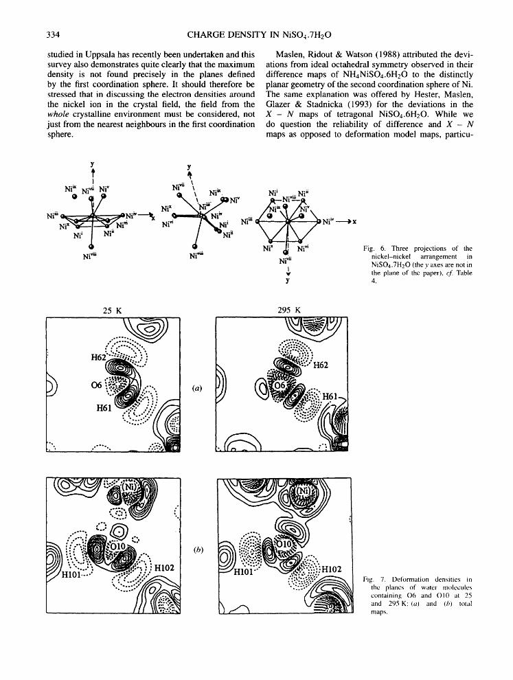
334 CHARGE DENSITY IN NiSO4.7H20
studied in Uppsala has recently been undertaken and this survey also demonstrates quite clearly that the maximum density is not found precisely in the planes defined by the first coordination sphere. It should therefore be stressed that in discussing the electron densities around the nickel ion in the crystal field, the field from the whole crystalline environment must be considered, not just from the nearest neighbours in the first coordination sphere.
Maslen, Ridout & Watson (1988) attributed the devi- ations from ideal octahedral symmetry observed in their difference maps of NHnNiSOn.6H20 to the distinctly planar geometry of the second coordination sphere of Ni. The same explanation was offered by Hester, Maslen, Glazer & Stadnicka (1993) for the deviations in the X - N maps of tetragonal NiSOa.6H20. While we do question the reliability of difference and X - N maps as opposed to deformation model maps, particu-
Y y t
Ni ~" Ni '~ Ni" N'~l*ii \ Nii~ , Nii ~i Niil 0 ~,/0 . . . . . Ni N i - -
N l X ~ ~ ~ Niiii - ~ ,*Nih,----~ i = ,"
~ , , i Ni" / ~ N i i Ni ui Ni"
Nii ~ Ni" ~ "Ni" Ni~ ~. ~ Ni"
Ni*ili Ni '~ii Ni,,a
I w y
~x
Fig. 6. Three projections of the nickel-nickel arrangement in NiSOa.7H20 (the y axes are not in the plane of the paper), cf. Table 4.
' ~ ° , . _Q
25 K 295 K
.
HIO1 .... " .-C.~"" .".~."
(b)
Fig. 7. Deformation densities in the planes of water molecules containing 06 and O10 at 25 and 295 K: (a) and (h) total maps.

H. PTASIEWICZ-BAK, I. OLOVSSON AND G. J. MclNTYRE 335
larly for non-centrosymmetric structures such as tetrag- onal NiSO4.6H20 and orthorhombic NiSO4.7H20, the hypothesis that the second coordination sphere has an important influence does bear careful consideration. Our present study strongly supports this idea. We should, however, be wary of overly interpreting the deforma- tion densities. It should be remembered that they illus- trate relatively small fitted deviations from the predom- inantly spherical symmetry of the atoms, which in our model are complete neutral atoms of the element con- cerned. The model maps in COSO4.6H20 (Kellersohn, Delaplane, Olovsson & McIntyre, 1993) gave a more striking example of large deviations from a perfectly octahedral deformation.
In the case of NiSO4.6H20 it was suggested that the pronounced difference observed between the room temperature and 25 K maps could be due to differ- ent occupation of the two upper partially occupied electronic levels. This suggestion was supported by a Boltzmann statistical analysis based on the energy differ- ence experimentally observed in magnetic susceptibility measurements (Ptasiewicz-Bak, Olovsson & Mclntyre, 1993).
4.1.2. The water molecules. The total deformation densities are illustrated in Fig. 7 in the planes of the water molecules containing 06 and O10 and are perpendicular to those in Fig. 8. These molecules
have been selected as representatives for molecules which are tetrahedrally and trigonally coordinated to nickel, respectively. The maps for the other water molecules have similar features. The superposition effects are again less pronounced in this compound compared with NiSOn.6H20. The general features of the densities in the O---H bonds and the lone-pair regions were discussed in a previous paper (Mclntyre, Ptasiewicz-Bak & Olovsson, 1990). The most interesting special features are found in the lone-pair planes: in water(6) there are two well-separated 'lone-pair peaks', whereas there is just one peak in water(10), although the single peak is elongated at 25 K. In the free water molecule there is just one 'banana-shaped' peak according to theoretical ab initio calculations (Hermansson, 1984). The clear splitting in water(6) is then most probably due to the polarizing influence of the neighbours, effects which are normally considered too small to be observed experimentally. Similar clear polarization of the lone-pair densities according to the coordination of the water molecules was also observed in NiSOa.6H20.
5. Conclusions
In general, the same conclusions can be drawn from this study as from our earlier experimental charge-density
25 K 295 K
C 3
:--.... ~,, .;~
0 . . ~ : . f " ,~ o.
_
"::'-:~ • - ' - . : : , : : , . . : : . x . , ,
(a)
(b)
, !!~:': ;'' 2)
Fig. 8. Deformation densities per- pendicular to the planes of water molecules containing 06 and O!0, through the twofold axes, at 25 and 295 K: (a) and (b) total maps.

336 CHARGE DENSITY IN NiSO4.7H20
studies of tetragonal NiSO4.6H20, i.e. the deforma- tion around Ni follows crystal field theory, details of the deformation in and around water molecules can be extracted, even in the presence of such strong X- ray scatterers as Ni, and the water molecules exhibit polarization that depends on their local environment. The deformation around Ni does appear to be very sensitive to small deviations from a perfectly octahedral environment and to the influence of the whole crystalline environment.
We can also conclude that water molecules with the same local coordination are largely transferable and that, lacking neutron data, a reasonable picture of the defor- mation can be obtained by extrapolating from the refined H coordinates of the conventional refinement. This does assume that there is no disorder or anharmonicity.
This work has been funded in part by the Swedish Natural Science Research Council (NFR). We gratefully acknowledge the skilful technical assistance of Mr H. Karlsson in growing the crystals and in collecting the X-ray data, and of Dr R. Tellgren and Mr H. Rundl6f in collecting the neutron data.
References
Becker, P. & Coppens, P. (1974). Acta Cryst. A30, 129-147. Becker, P. & Coppens, P. (1975). Acta Cryst. A31, 417-425. Beevers, C. A. & Schwartz, C. M. (1935). Z. Kristallogr. 91,
157-169.
Coppens, P., Leiserowitz, L. & Rabinovich, D. (1965). Acta Cryst. 18, 1035-1038.
Harel, M. & Hirshfeld, F. L. (1975). Acta Cryst. B31, 162-172. Hermansson, K. (1984). Thesis. Acta Universitatis Upsaliensis
No. 744, Sweden. Hester, J. R., Maslen, E. N., Glazer, A. M. & Stadnicka, K.
(1993). Acta Cryst. B49,. 641--646. Hirshfeld, F. L. (1971). Acta Cryst. B27, 769-781. Hirshfeld, F. L. (1977). Isr. J. Chem. 16, 226-229. Kellersohn, T., Delaplane, R. G., Olovsson, I. & Mclntyre,
G. J. (1993). Acta Cryst. B49, 179-192. Kuchitsu, K. & Bartell, L. S. (1962). J. Chem. Phys. 36,
2460--2469. Lehmann, M. S. & Larsen, F. K. (1974). Acta Cryst. A30,
580-584. Lundgren, J. O. (1982). Crystallographic Computer Programs.
Report UUIC-B 13-04-05. Institute of Chemistry, University of Uppsala, Sweden.
Maslen, E. N., Ridout, S. C. & Watson, K. J. (1988). Acta Cryst. B44, 96-101.
Mclntyre, G. J., Ptasiewicz-Bak, H. & Olovsson, I. (1990). Acta Cryst. B46, 27-39.
Olovsson, I., Ptasiewicz-Bak, H. & Mclntyre, G. J. (1993). Z. Naturforsch. Teil A, 48, 3-11.
Ptasiewicz-Bak, H., Olovsson, I. & Mclntyre, G. J. (1993). Acta Cryst. B49, 192-201.
Samson, S., Goldish, E. & Dick, C. F. (1980). J. Appl. Cryst. 13, 425--432.
Sears, V. F. (1992). Neutron News, 3, 26-37. Stewart, R. F., Davidson, E. R. & Simpson, W. T. (1965). J.
Chem. Phys. 42, 3175-3187. Terpstra, M., Craven, B. M. & Stewart, R. F. (1993). Acta
Cryst. A49, 685-692.


