
CharacterizationofTwoHumanSmallCellLungCarcinoma-reactive...
7
[CANCER RESEARCH 44,4987-4992, November 1984] Characterization of Two Human Small Cell Lung Carcinoma-reactive Monoclonal Antibodies Generated by a Novel Immunization Approach1 Alex W. Tong, Jennifer Lee, and Marvin J. Stone2 Immunology Research Unit, Charles A. Sommons Cancer Center, Baylor University Medical Center, Dallas, Texas 75246 ABSTRACT Two human small cell lung carcinoma cell lines, NCI-H69 and NCI-H128, were used as alternating sources of immunogen to generate monoclonal antibodies to small cell lung carcinoma- associated antigens. BALB/c mice were sensitized with seven injections of live tumor cells, four with NCI-H69 cells and three with NCI-H128 cells. Somatic cell hybridization was performed by fusion of the immune murine splenocytes using syngeneic myeloma cells from the SP2/0 Ag 14 cell line. Hybridoma colonies were screened against small cell lung carcinoma cells and normal lung fibroblasts with an enzyme-linked immunosorbent assay. Compared to animals immunized with only NCI-H69 or NCI-H128 cells, alternate immunization resulted in the generation of a significantly higher number of hybridomas that reacted selectively with both tumor cell lines. Monoclonal antibodies from two reactive hybrid clones generated by alternate immunization, SCLC 2051 and SCLC 5023, were uniformly negative to normal human tissues including lung, kidney, liver, spleen, breast, thy roid, brain, small intestine, and colon. While both monoclonal antibodies were nonreactive to paraffin-embedded, formalin- fixed, nonmalignant lung biopsies, the monoclonal antibody SCLC 5023 reacted with tumor cell infiltrates in biopsies from small cell lung carcinoma patients (14 of 14 cases positive), using the immunoperoxidase technique. This monoclonal reagent also reacted with other lung tumor cell types, including atypical car- cinoid (5 of 5 positive), epidermoid (4 of 6 positive), undifferen- tiated and bronchoalveolar (3 of 4 cases each positive) carcino mas. By contrast, monoclonal antibody SCLC 2051 apparently identified an antigen expressed preferentially on small cell lung carcinoma cells (12 of 14 positive) and only rarely reacted with other lung tumor cell types (2 of 34 positive). Both monoclonal antibodies were negative to colon carcinoma, epidermoid carci noma of the floor of the mouth, breast adenocarcinoma, and B- and T-cell leukemia and lymphoma cells, as determined by the enzyme-linked immunosorbent assay, indirect ¡mmunofluores- cence, and immunoperoxidase techniques. These observations suggest that SCLC 2051 and SCLC 5023 may be of value in identifying tumor-associated antigens expressed in small cell and other lung carcinomas. In addition, the generation of antibody- producing cells towards common tumor-associated antigens may be enhanced by immunization with multiple tumor cell lines of the same histológica! type. INTRODUCTION SCLC3 comprises approximately 20% of human lung cancers and is distinct from other lung carcinoma histological cell types with respect to cell of origin, natural history, and treatment. Believed to originate from transformation of the neurosecretory granule-rich lung basal cells (K-cells). SCLC has a rapid growth rate and a high incidence of metastasis at an early stage (17). Although the majority of patients initially respond to conventional chemotherapy, long-term prognosis is poor (<5% 5-year sur vival). Furthermore, the administration of appropriate therapy requires accurate histological distinction of SCLCs from non- SCLCs; this distinction is often difficult (11,17). With the recent advent of the somatic hybridization technique (13), antigens preferentially expressed in various human tumors can be identified and characterized, using tumor-specific MoAbs. In various human tumor systems, such as melanoma (18) and lymphoreticular neoplasms (6), the identification of tumor antigen expression by specific MoAbs has contributed to better under standing of tumor biology and the malignant transformation event (7). MoAbs also have demonstrated potential usefulness in many clinical applications, including the immunohistochemical diagno sis and classification of primary cancers (8, 20), location of métastasesby radioimaging (20), or use as therapeutic agents (14). However, few studies have focused on SCLC, due primarily to the limited availability of specific monoclonal reagents (1-3, 12,15,16,19). In order to develop monoclonal reagents suitable for the study of SCLC, we have utilized an alternating immunization approach in an attempt to select for tumor-reactive antibody-producing cells. Because immunization with tumor cells from a single cell line generally results in a poor yield of tumor-reactive hybridomas, cells from 2 distinct human SCLC cell lines were used as alternate sources of immunogen. Somatic hybridization of spleen cells from the alternatively immunized animals resulted in significantly higher numbers of clones reactive to SCLC cell lines and tissue biopsies. Two of the MoAbs described in this report, SCLC 2051 and SCLC 5023, were found to be unreactive with various normal human tissues and with other human malignant tumors. SCLC 5023 was reactive with SCLC and, to a lesser extent, with various non-SCLC types. By contrast, SCLC 2051 identified an antigen primarily expressed on SCLC cells but was largely negative with non-SCLC lung carcinomas. Both antibodies inhib ited SCLC clonal growth in vitro. These properties suggest that 1This study was supported in part by the Barbara B. and Max L. Thomas Cancer Immunology Research Fund. 2 To whom requests for reprints should be addressed. Received April 19, 1984; accepted August 2,1984. 3The abbreviations used are: SCLC, small cell lung carcinoma; MoAb, mono clonal antibody; ELISA, enzyme-linked immunosorbent assay; PCS, fetal calf serum; IMDM, Iscove's modified Oulbecco's medium; LUFB, lung fibroblasts; PBS. phos phate-buffered saline. NOVEMBER 1984 4987 Research. on December 1, 2018. © 1984 American Association for Cancer cancerres.aacrjournals.org Downloaded from
Transcript of CharacterizationofTwoHumanSmallCellLungCarcinoma-reactive...
[CANCER RESEARCH 44,4987-4992, November 1984]
Characterization of Two Human Small Cell Lung Carcinoma-reactive
Monoclonal Antibodies Generated by a Novel ImmunizationApproach1
Alex W. Tong, Jennifer Lee, and Marvin J. Stone2
Immunology Research Unit, Charles A. Sommons Cancer Center, Baylor University Medical Center, Dallas, Texas 75246
ABSTRACT
Two human small cell lung carcinoma cell lines, NCI-H69 andNCI-H128, were used as alternating sources of immunogen togenerate monoclonal antibodies to small cell lung carcinoma-
associated antigens. BALB/c mice were sensitized with seveninjections of live tumor cells, four with NCI-H69 cells and threewith NCI-H128 cells. Somatic cell hybridization was performed
by fusion of the immune murine splenocytes using syngeneicmyeloma cells from the SP2/0 Ag 14 cell line. Hybridoma colonieswere screened against small cell lung carcinoma cells and normallung fibroblasts with an enzyme-linked immunosorbent assay.Compared to animals immunized with only NCI-H69 or NCI-H128
cells, alternate immunization resulted in the generation of asignificantly higher number of hybridomas that reacted selectivelywith both tumor cell lines. Monoclonal antibodies from tworeactive hybrid clones generated by alternate immunization,SCLC 2051 and SCLC 5023, were uniformly negative to normalhuman tissues including lung, kidney, liver, spleen, breast, thyroid, brain, small intestine, and colon. While both monoclonalantibodies were nonreactive to paraffin-embedded, formalin-
fixed, nonmalignant lung biopsies, the monoclonal antibodySCLC 5023 reacted with tumor cell infiltrates in biopsies fromsmall cell lung carcinoma patients (14 of 14 cases positive), usingthe immunoperoxidase technique. This monoclonal reagent alsoreacted with other lung tumor cell types, including atypical car-cinoid (5 of 5 positive), epidermoid (4 of 6 positive), undifferen-
tiated and bronchoalveolar (3 of 4 cases each positive) carcinomas. By contrast, monoclonal antibody SCLC 2051 apparentlyidentified an antigen expressed preferentially on small cell lungcarcinoma cells (12 of 14 positive) and only rarely reacted withother lung tumor cell types (2 of 34 positive). Both monoclonalantibodies were negative to colon carcinoma, epidermoid carcinoma of the floor of the mouth, breast adenocarcinoma, and B-and T-cell leukemia and lymphoma cells, as determined by theenzyme-linked immunosorbent assay, indirect ¡mmunofluores-
cence, and immunoperoxidase techniques. These observationssuggest that SCLC 2051 and SCLC 5023 may be of value inidentifying tumor-associated antigens expressed in small cell andother lung carcinomas. In addition, the generation of antibody-producing cells towards common tumor-associated antigens
may be enhanced by immunization with multiple tumor cell linesof the same histológica! type.
INTRODUCTION
SCLC3 comprises approximately 20% of human lung cancers
and is distinct from other lung carcinoma histological cell typeswith respect to cell of origin, natural history, and treatment.Believed to originate from transformation of the neurosecretorygranule-rich lung basal cells (K-cells). SCLC has a rapid growth
rate and a high incidence of metastasis at an early stage (17).Although the majority of patients initially respond to conventionalchemotherapy, long-term prognosis is poor (<5% 5-year sur
vival). Furthermore, the administration of appropriate therapyrequires accurate histological distinction of SCLCs from non-
SCLCs; this distinction is often difficult (11,17).With the recent advent of the somatic hybridization technique
(13), antigens preferentially expressed in various human tumorscan be identified and characterized, using tumor-specific MoAbs.
In various human tumor systems, such as melanoma (18) andlymphoreticular neoplasms (6), the identification of tumor antigenexpression by specific MoAbs has contributed to better understanding of tumor biology and the malignant transformation event(7). MoAbs also have demonstrated potential usefulness in manyclinical applications, including the immunohistochemical diagnosis and classification of primary cancers (8, 20), location ofmétastasesby radioimaging (20), or use as therapeutic agents(14). However, few studies have focused on SCLC, due primarilyto the limited availability of specific monoclonal reagents (1-3,
12,15,16,19).In order to develop monoclonal reagents suitable for the study
of SCLC, we have utilized an alternating immunization approachin an attempt to select for tumor-reactive antibody-producing
cells. Because immunization with tumor cells from a single cellline generally results in a poor yield of tumor-reactive hybridomas,
cells from 2 distinct human SCLC cell lines were used as alternatesources of immunogen. Somatic hybridization of spleen cellsfrom the alternatively immunized animals resulted in significantlyhigher numbers of clones reactive to SCLC cell lines and tissuebiopsies. Two of the MoAbs described in this report, SCLC 2051and SCLC 5023, were found to be unreactive with various normalhuman tissues and with other human malignant tumors. SCLC5023 was reactive with SCLC and, to a lesser extent, withvarious non-SCLC types. By contrast, SCLC 2051 identified an
antigen primarily expressed on SCLC cells but was largelynegative with non-SCLC lung carcinomas. Both antibodies inhib
ited SCLC clonal growth in vitro. These properties suggest that
1This study was supported in part by the Barbara B. and Max L. Thomas
Cancer Immunology Research Fund.2To whom requests for reprints should be addressed.
Received April 19, 1984; accepted August 2,1984.
3The abbreviations used are: SCLC, small cell lung carcinoma; MoAb, monoclonal antibody; ELISA, enzyme-linked immunosorbent assay; PCS, fetal calf serum;IMDM, Iscove's modified Oulbecco's medium; LUFB, lung fibroblasts; PBS. phos
phate-buffered saline.
NOVEMBER 1984 4987
Research. on December 1, 2018. © 1984 American Association for Cancercancerres.aacrjournals.org Downloaded from
A.W. Jong et al.
these MoAbs may be of value in further clinical and biologicalstudies of human SCLC.
MATERIALS AND METHODS
Source of Cells and Tissues. The SCLC cell lines NCI-H69 and NCI-
HI 28 were kindly provided by Dr. Adi F. Gazdar, National CancerInstitute; UM-1, floor of the mouth epidermoid carcinoma cell line, andUM-2, a lung epidermoid carcinoma cell line, were provided by Dr. Tom
Carey, University of Michigan Medical Center. The mouse myeloma cellline, SP2/0 Ag14, and human myeloma cell line LICR-MON-HMy 2 were
obtained from Dr. Denis Burger, VA Medical Center, Portland, OR. LUFBcell lines Ag 4432 and Ag 2262 were obtained from the NIA Aging CellRepository, Camden, NJ; the epidermoid lung carcinoma cell lines Calu-2 and Sk-MES-1, lung adenocarcinoma cell lines Calu-3 and Sk-Lu-1,anaplastic lung carcinoma Calu-6, and undifferentiated lung carcinomacell line A-427 were obtained from the American Type Culture Collection,Rockville, MD. Peripheral blood lymphocytes from leukemia and lym-
phoma patients were derived from blood samples drawn by venipuncturewith prior consent. Normal brain tissue was kindly provided by Dr. J.Luby, University of Texas Health Science Center at Dallas. Biopsies ofother normal tissues and for mal in-fixed, paraffin-embedded tissue blocks
of confirmed cases of various lung tumors were obtained from theDepartment of Surgical Pathology, Baylor University Medical Center. Allprocedures involving the handling of human tissues were previouslyapproved by the Institutional Review Board for Human Protection, BaylorUniversity Medical Center.
Fusion Protocol. Two 2-month-old female BALB/c mice were immunized to 2 human SCLC cell lines (NCI-H69 and NCI-H128). Each mousereceived 7 ¡.p.injections of 1 x 10⁷ cells, alternating H69 with H128
cells. The first 4 injections were made weekly, followed by boosts 3 daysapart. Other mice were immunized with 7 injections of each SCLC cellline alone. Somatic hybridization was performed 3 days following the lastinjection. "Filler cells" were prepared using thymocytes from the same
and 2 other mice of the same age. A single-cell suspension was prepared
in IMDM (Grand Island Biological, Grand Island, NY) and suspended at 1x 10⁷/ml in IMDM with 15% PCS (Grand Island Biological) and 100 mM
hypoxanthine: 0.4 HIM aminopterin: 16 mw thymidine (Sigma ChemicalCo., St. Louis, MO). One hundred n\ of the thymocyte suspension weredispensed into each well of five 96-well microtiter plates and stored at37°.
Spleen cells from an immunized animal were extracted by gentlyteasing the spleen with a pair of forceps. The cells were washed twicein IMDM and then resuspended in 25 ml of IMDM (5 to 10 x 106/ml).
Myeloma cells from the nonsecretory SP2/0 Ag 14 cell line were collectedat logarithmic growth phase, washed twice in IMDM, and then pelletedwith 10s spleen cells in the ratio of 3:1 (myeloma cells:splenocytes) in a
16- x 125-mm tissue culture tube (Corning Labware, Coming, NY).Fusion was performed under sterile conditions in a 37°water bath, using
a modification of the protocol described by Oi and Herzenberg (7, 21).One ml of polyethylene glycol (average molecular weight, 1500; AldrichChemicals, Milwaukee, Wl) in IMDM (50%, v/v, containing 5% dimethylsulfoxide; Baker Chemical Co., Phillipsburg, NJ) was added over 1 minwith constant stirring by the tip of the pipet for 2 min. Then 2 ml of warmIMDM were added over 2 min, followed by 7 ml of IMDM over 3 min.The cell suspension was washed once (180 x g, 10 min, 23°)and then
resuspended in 50 ml of IMDM. One hundred M>were dispensed intoeach well of the five 96-well microtiter plates containing mouse thymocytes and were placed in 37°incubator humidified with an atmosphere
of 5% CO2:95% air.The culture plates were fed once on Day 5 with IMDM:15% PCS
containing the hypoxanthine:aminopterin:thymidine and then every thirdday thereafter with IMDM:15% PCS with hypoxanthine:thymidine byreplacing one-half of the supernatant fluids in each well. Hybrid colonies
were screened by ELISA for antibody activity at confluence (approximately 10 to 14 days). Those with desirable reactivity were transferred
to 24-well plates (Falcon), allowed to grow to confluence (3 to 5 days),
and then cloned by limiting dilution.Cloning by Limiting Dilution. Hybrid colonies were cloned by limiting
dilution within 2 weeks of fusion (21). Fifty n\ of a well-mixed cell
suspension were extracted from the well containing the antibody ofinterest. Serial dilutions were made to achieve a final concentration of10 cells/ml in IMDM: 15% PCS, and 100 /tl were seeded onto each wellof a 96-well microtiter plate. Thymocytes extracted from syngeneic micewere used as filler cells (100 ^1,1x10⁷ cells/ml). The culture was closely
monitored daily to observe for the emergence of clones. Culture wellscontaining single clones were noted by Days 5 to 7 and tested atconfluence. Hybrid colonies were cloned 2 to 3 times to ensure mono-
clonality.ELISA Assay. An ELISA technique utilizing intact target cells was
used for screening antibody activity in hybridoma supernatant fluids (22).Target cells were washed twice and suspended in PBS at 1 x I06/ml.Fifty n\ of the cell suspension (5 x 10⁴ cells) were added to each well of
a microtiter plate (96 U-bottomed wells; Falcon Plasticware, BectonDickinson Labware, Oxnard, CA). The plates were dried at 37°overnightand then sealed and placed at 4°for storage. On the day of evaluation,
the antigen-coated plates were flooded with 200 /¿Iof 1% bovine serumalbumin (Sigma) in PBS for 30 min to block the remaining protein-binding
sites. Fifty /¿Iof supernatant fluid were added to each well, incubated for1 hr at 37°,and then washed 3 times with PBS. Fifty n\ of a peroxidase-
conjugated secondary antibody (peroxidase-conjugated goat anti-mouse
IgG + IgM, 1:3000; Tago, Burlingame, CA) were added, and the platewas further incubated for 1 hr at 37°.After a washing, 50 >i\ of freshly
prepared substrate solution (4 mg o-phenylenediamine in 10 ml 80 mM
citrate:phosphate buffer, with 4 ¡Aof 30% H2O2)were added and incubated for 30 min at 37°,followed by 25 n\ of 4 M H2SO4 immediately
before reading. Antibody binding was determined as a function of ab-
sorbance at 492 nm (Titertek Multiskan MC; Flow Laboratories, McLean,VA). Antibody binding in each well was graded against negative controls(treated with an irrelevant primary antibody) as + (Õ0.1 absorbance unithigher than control, triplicate determinations), ++ (£0.2absorbance unitover control), or +++ (60.3 absorbance unit over control).
MoAb Isotyping. An ELISA procedure similar to that described abovewas used. However, subclass-speciflc rabbit anti-mouse immunoglobu-
lins were used as secondary reagents (Mouse Immunoglobulins SubtypeIdentification Kit; Boehringer Mannheim Biochemicals, Indianapolis, IN).A peroxidase-conjugated goat anti-rabbit antibody was added as tertiary
antibody.Immunofluorescence Assay. Target cells from cell line cultures or
Hypaque:Ficoll (Sigma)-separated peripheral blood lymphocytes werewashed twice with PBS and then resuspended at 2 x 10⁷/ml in PBS
containing 0.02% sodium azide. Fifty ¿ilof tissue culture supernatantfluid were added to a 12- x 75-mm plastic disposable tube (Falcon)containing i x io6 cells. The reaction mixtures were incubated in an icebath (2-8°) for 1 hr and then washed twice with PBS:azide at 4°.Fifty
ß\of a fluorescein isothiocyanate-conjugated secondary antibody|F(ab')2] goat anti-mouse immunoglobulin (1:10; Cappel Laboratories,
Westchester, PA) were added (4°,60 min). The reaction mixtures were
washed twice and fixed with 1% formaldehyde in PBS. The frequencyof antibody-reactive cells in the target cell population was determined byflow cytometry (Becton-Dickinson FACS Systems, Mountain View, CA),
using the parameters of cell size (forward angle scatter) versus greenfluorescence.
Immunoperoxidase Protocol. The MoAb reactivity to normal tissueswas evaluated by the immunoperoxidase technique (29) with a panel offresh tissues cryopreserved within 1 hr of extraction. Sections (5 pm)from each sample were prepared on the day of evaluation, fixed withacetone (10 min, 23°),and washed twice by submerging in PBS. Thesewere treated with 1% H2O2 in absolute methanol (30 min, 23°),washed
in distilled water and PBS, and then treated with normal horse serumand subsequent reagents as described below. The MoAb reactivity tovarious lung carcinomas was determined using formalin-fixed, paraffin-
4988 CANCER RESEARCH VOL. 44
Research. on December 1, 2018. © 1984 American Association for Cancercancerres.aacrjournals.org Downloaded from
Small Cell Lung Carcinoma Monoclonal Antibodies
embedded tissue blocks from previously confirmed cases diagnosed bythe Department of Surgical Pathology, Baylor University Medical Center.The sections were deparaffinized, rehydrated in xylene and gradedalcohols, and then incubated with 1% H2O2in methanol for 30 min. Aftera thorough rinsing in distilled water and PBS, the sections were treatedfirst with diluted normal horse serum for 20 min and then with the primaryantibody (culture supernatant) for 30 min, washed, and then treated withthe secondary antibody (biotinylated horse anti-mouse IgG, 30 min, 23°).
The samples were again washed in PBS and treated with avidinrbiotin-
conjugated horseradish peroxidase (Vectastain ABC Kit; Vector Laboratories, Burlingame, CA) for 60 min. All incubations were performed atroom temperature in a humidity chamber. Freshly prepared substrate(0.02% 3-aminc-9-ethylcarbazole:0.03% H2O2:5% A/.W-dimethylformam-
ide in 0.1 M acetate buffer, pH 5.2) was added for 30 min, followed bythe counterstain (0.125% méthylèneblue) and mounting solution (glycerolgelatin; Sigma).
Clonal Growth Assay. The assay was a modification of the soft-agargrowth assay described by Minna ef al. (16). Cells from the NCI-H69 cell
line were harvested from culture, washed twice, and resuspended inserum-free IM DM at 1 x 10*/ml. One hundred //I of the approximately
diluted supernatant fluids from SCLC 2051, SCLC 5023, and an H69-nonreactive clone were added to 17- x 134-mm sterile disposable centrifuge tubes (Falcon) containing 1 x 10s cells and incubated at room
temperature for 1 hr. Two ml of 0.3% agar in IMDM:15% PCS wereadded, and equal volumes of the mixture were layered in duplicate ontoa 6-well culture plate (Falcon) with a previously prepared base layer(0.5% agar in IMDM:15% PCS). The plates were incubated at 37°for 1
week. The number of colonies (with e50 cells) in each well was enumerated under low magnification (xlO). Growth inhibition of the culturesupernatant was determined as
% of colony growth inhibitionMean no. of colonies with antibody treatment \
Mean no. of colonies without antibody treatment/x 100
Statistical Analysis. The expected yield of hybridomas per well, orthe frequency of SCLC-reactive hybridomas per well was calculated
according to Poisson distribution analysis by the formula
P0= e-
where P0 is the observed frequency of zero events (number of negativewells/288), and x is the expected mean number of events per well (28).
The x2 test (27) for comparing yields of SCLC-reactive hybridomas
per well was calculated by the formula
(observed - expected)2 .- •=2 , . ; d.f. = 1
expected
RESULTS
Generation and Screening of MoAbs. In an effort to enrichfor antibody-producing cells to common SCLC antigens, we
immunized mice with alternate injections of 2 different humanSCLC cell lines (NCI-H69 and H128). To determine the efficacy
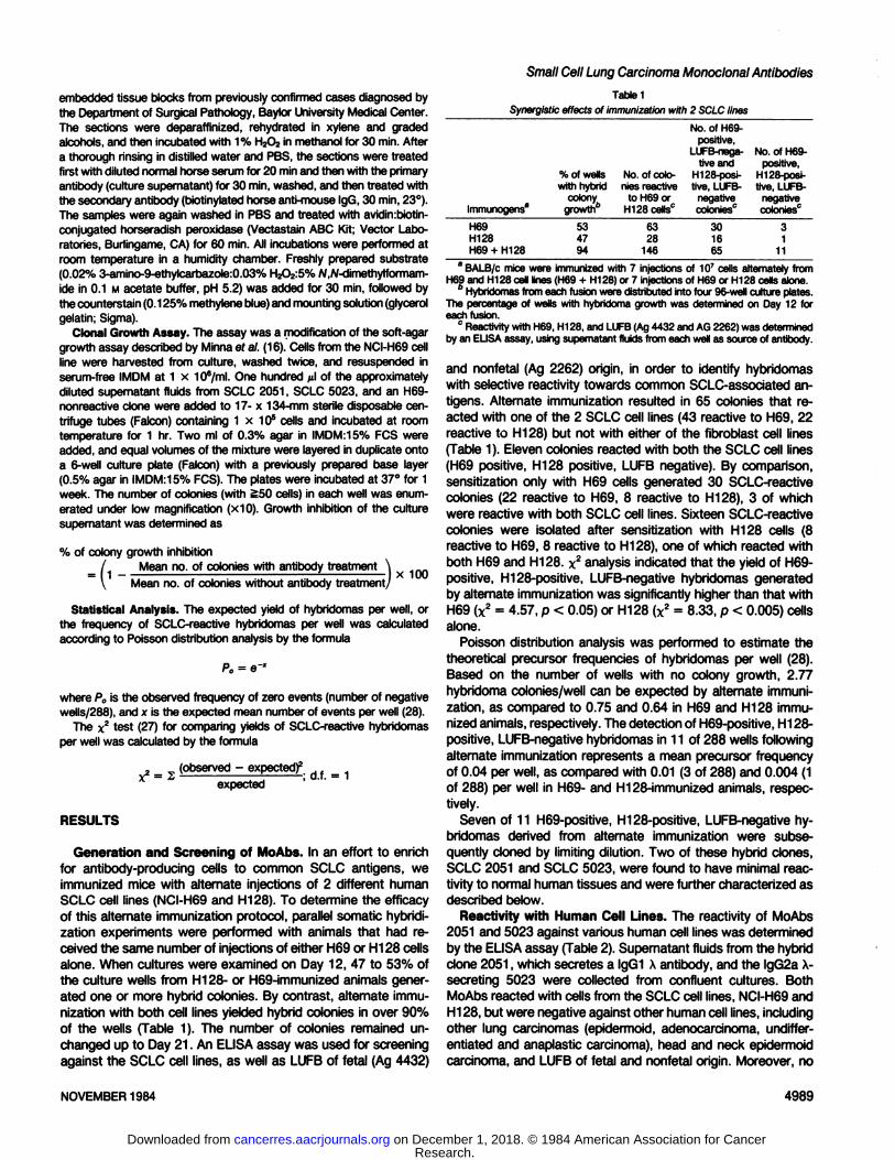
of this alternate immunization protocol, parallel somatic hybridization experiments were performed with animals that had received the same number of injections of either H69 or H128 cellsalone. When cultures were examined on Day 12, 47 to 53% ofthe culture wells from H128- or H69-immunized animals gener
ated one or more hybrid colonies. By contrast, alternate immunization with both cell lines yielded hybrid colonies in over 90%of the wells (Table 1). The number of colonies remained unchanged up to Day 21. An ELISA assay was used for screeningagainst the SCLC cell lines, as well as LUFB of fetal (Ag 4432)
Table 1
Synergistic effects of immunization with 2 SCLClinesImmunogens"H69
H128H69 + H128%
of wellswith hybrid
colonygrowth53
4794No.
of colonies reactive
to H69 orH128cells063
28146No.
of H69-positive,
LUFB-nega-tiveandH128-POSÕ-
tive, LUFB-negativecolonies'130
1665No.
of H69-positive,
H128-POSÌ-tive, LUFB-
negativecolonies03
111
" BALB/c mice were immunized with 7 injections of 10' cells alternately from
H69 and H128 cell lines (H69 + H128) or 7 injections of H69 or H128 cells alone.6 Hybridomas from each fusion were distributed into four 96-well culture plates.
The percentage of wells with hybridoma growth was determined on Day 12 foreach fusion.
0 Reactivity with H69, H128, and LUFB (Ag 4432 and AG 2262) was determined
by an ELISA assay, using supernatant fluids from each well as source of antibody.
and nonfetal (Ag 2262) origin, in order to identify hybridomaswith selective reactivity towards common SCLC-associated an
tigens. Alternate immunization resulted in 65 colonies that reacted with one of the 2 SCLC cell lines (43 reactive to H69, 22reactive to H128) but not with either of the fibroblast cell lines(Table 1). Eleven colonies reacted with both the SCLC cell lines(H69 positive, H128 positive, LUFB negative). By comparison,sensitization only with H69 cells generated 30 SCLC-reactive
colonies (22 reactive to H69, 8 reactive to H128), 3 of whichwere reactive with both SCLC cell lines. Sixteen SCLC-reactive
colonies were isolated after sensitization with H128 cells (8reactive to H69, 8 reactive to H128), one of which reacted withboth H69 and H128. x2 analysis indicated that the yield of Hog-
positive, H128-positive, LUFB-negative hybridomas generated
by alternate immunization was significantly higher than that withH69 (x2 = 4.57, p < 0.05) or H128 (x2 = 8.33, p < 0.005) cells
alone.Poisson distribution analysis was performed to estimate the
theoretical precursor frequencies of hybridomas per well (28).Based on the number of wells with no colony growth, 2.77hybridoma colonies/well can be expected by alternate immunization, as compared to 0.75 and 0.64 in H69 and H128 immunized animals, respectively. The detection of H69-positive, H128-positive, LUFB-negative hybridomas in 11 of 288 wells following
alternate immunization represents a mean precursor frequencyof 0.04 per well, as compared with 0.01 (3 of 288) and 0.004 (1of 288) per well in H69- and H128-immunized animals, respec
tively.Seven of 11 H69-positive, H128-positive, LUFB-negative hy
bridomas derived from alternate immunization were subsequently cloned by limiting dilution. Two of these hybrid clones,SCLC 2051 and SCLC 5023, were found to have minimal reactivity to normal human tissues and were further characterized asdescribed below.
Reactivity with Human Cell Lines. The reactivity of MoAbs2051 and 5023 against various human cell lines was determinedby the ELISA assay (Table 2). Supernatant fluids from the hybridclone 2051, which secretes a lgG1 X antibody, and the lgG2a X-secreting 5023 were collected from confluent cultures. BothMoAbs reacted with cells from the SCLC cell lines, NCI-H69 and
H128, but were negative against other human cell lines, includingother lung carcinomas (epidermoid, adenocarcinoma, undiffer-
entiated and anaplastic carcinoma), head and neck epidermoidcarcinoma, and LUFB of fetal and nonfetal origin. Moreover, no
NOVEMBER 1984 4989
Research. on December 1, 2018. © 1984 American Association for Cancercancerres.aacrjournals.org Downloaded from
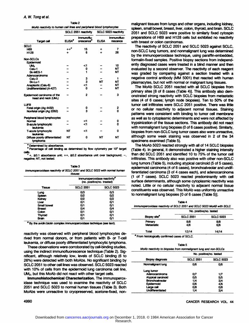
A IV.Tongetal.
Tabto2
MoAb reactivity to human cell lines and peripheral blood lymphocytes
Tar getcellSCLCH69H128Non-SCLCsEpidermoidUMjCalu-1Sk-MES-1AdenocarcinomaCalu-3Sk-Lu-1Anaplastia
(Calu-6)Undifferentiated(A-427)Epidermoid
carcinoma oftheheadand neck(UM,)LUFBFetal
origin (Ag4432)Nonfetalorigin (Ag2262)Peripheral
bloodlymphocytesNormalB-acute
lymphocyticleukemiaT-acute
lymphocyticleukemiaDiffuse
poorlydifferentiatedlymphomaSCLC
2051reactivityImmunoflu-
ELJSA*orescence"++°
15+
6<10133<10203<1<1NT
<1NT
0SCLC
5023reactivityImmunofluo-
EUSArescence+
7+2610NTNT1NT
NTNTNT320000NT
NT
8 Determined by absorbance.6 Percentage of cell binding as determined by flow cytometry per 10* target
cells.c +, £0.1 absorbance unit; ++, SO.2 absorbance unit over background; -,
negative; NT, not tested.
Table 3
immunoperoxidase reactivity of SCLC 2051 and SCLC 5023 with normal humantissues
Immunoperoxidase reactivity8
(no. positive/no,treated)TissueLungSpleenKidneyLiverSmall
bowelBreastColonThyroidBrainSCLC
20510/30/20/20/10/10/40/10/10/1SCLC50230/30/20/20/10/10/40/10/10/1
By the avidin:biotin complex Immunoperoxidase technique (see text).
reactivity was observed with peripheral blood lymphocytes derived from normal donors, or from patients with B- or T-cell
leukemia, or diffuse poorly differentiated lymphocytic lymphoma.These observations were corroborated by cell-binding studies,
using the indirect immunofluorescence technique (Table 2). Significant, although relatively low, levels of SCLC binding (6 to26%) were detected with both MoAbs. No significant binding bySCLC 2051 to other cell lines was observed. SCLC 5023 reactedwith 10% of cells from the epidermoid lung carcinoma cell line,UM2, but this MoAb did not react with other target cells.
Immunohistochemical Characterization. The immunoperox-
idase technique was used to examine the reactivity of SCLC2051 and SCLC 5023 to normal human tissues (Table 3). BothMoAbs were unreactive to cryopreserved, acetone-fixed, non-
malignant tissues from lungs and other organs, including kidney,spleen, small bowel, breast, liver, colon, thyroid, and brain. SCLC2051 and SCLC 5023 were positive to similarly fixed cytospinpreparations of H69 and H128 cells but exhibited no reactivitywith breast or colon carcinomas.
The reactivity of SCLC 2051 and SCLC 5023 against SCLC,non-SCLC lung tumors, and nonmalignant lung was determinedby the immunoperoxidase technique, using paraffin-embedded,formalin-fixed samples. Positive biopsy sections from independ
ently diagnosed cases were treated in a blind manner and thenevaluated by a second observer. The reactivity of each reagentwas graded by comparing against a section treated with anegative control antibody (MM 5001) that reacted with humanplasmacytes, but not with normal or malignant lung tissues.
The MoAb SCLC 2051 reacted with all SCLC biopsies fromprimary sites [8 of 8 cases (Table 4)]. This antibody also demonstrated strong reactivity with SCLC biopsies from metastaticsites (4 of 6 cases; lymph node biopsies). Ten to 50% of thetumor cell infiltrates were SCLC 2051 positive. There was littleor no cellular reactivity to adjacent normal tissues. Stainingpatterns were consistent with binding to tumor cell membraneas well as to cytoplasmic determinants and were not affected bytrypsinization of the tissue sections. This antibody did not reactwith nonmalignant lung biopsies (0 of 6 cases positive). Similarly,biopsies from non-SCLC lung tumor cases also were unreactive,
although some weak staining was observed in 2 of the 34specimens examined (Table 5).
The MoAb 5023 reacted strongly with all of 14 SCLC biopsies(Table 4). In general, it demonstrated a higher staining intensitythan did SCLC 2051 and identified 10 to 75% of the tumor cellinfiltrates. This antibody also was positive with other non-SCLC
lung tumors (Table 5), including atypical carcinoid (5 of 5 cases),epidermoid carcinoma (4 of 6 cases), bronchoalveolar and undif-
ferentiated carcinoma (3 of 4 cases each), and adenocarcinoma(1 of 7 cases). SCLC 5023 reacted predominantly with cellsurface determinants, although some cytoplasmic reactivity wasnoted. Little or no cellular reactivity to adjacent normal tissueconstituents was observed. This MoAb was uniformly unreactiveto nonmalignant lung biopsies [0 of 6 cases (Table 5)].
Table 4
Immunoperoxidase reactivity of SCLC 2051 and SCLC 5023 MoAB with SCLC
No. positive/no,testedBiopsy
site*Primary
MetastaticTotalSCLC
20518/84/612/14SCLC 50238/86/614/14
' From historically confirmed cases of SCLC.
TablesMoAb reactivity to biopsies from nonmalignant lung and non-SCLCs
BiopsydiagnosisNonmalignant
lungLung
tumorAdenocarcinomaAtypical
carcinoidBronchoalveolarEpidermoidLarge
cellUndifferentiatedNo.positive/no,
testedSCLC
2051 SCLC50230/60/70/51/40/60/81/40/61/75/53/44/60/83/4
4990 CANCER RESEARCH VOL. 44
Research. on December 1, 2018. © 1984 American Association for Cancercancerres.aacrjournals.org Downloaded from
Tabte6
MoAb inhibition of tumor cell colony growthColony growth inhibition"
MoAbSCLC
2051SCLC
5023Finsi
conc6ntration1:101:1001:10001:100001:101:1001:10001:10000H696351514671646269LJCR-MON-HMy20.60°-5„NT"000NT
8 By the soft-agar growth assay (see text).6 NT, not tested.
Inhibition of Colony Growth. The data described above indicated the presence of SCLC 2051- and SCLC 5021-reactive
antigens among SCLCs. In order to examine the biological effectof these antibodies on SCLC cells, we determined the colonygrowth of cells from the NCI-H69 cell line afer MoAb treatment,
using an in vitro semisolid agar growth assay (16).Compared to samples that were not exposed to antibody,
NCI-H69 cells treated with SCLC 2051 or SCLC 5023 demonstrated significant inhibition of donai growth over a wide rangeof dilutions (Table 6). SCLC 2051 inhibited 46% of colony formation at a final dilution of 1:10,000. Similarly, SCLC 5023 waseffective in inhibiting clonal growth (>60%) at various dilutions.Growth inhibition appeared related to specific antibody binding,since neither antibody inhibited clonal growth of an irrelevanthuman myeloma cell line (LICR-MON-HMy 2) within the same
dilution range.
DISCUSSION
Immunization with intact tumor cells has the theoretical advantage of presenting tumor-associated antigens in their native formbut is inefficient in generating tumor-reactive antibody-producingcells. Antibodies specific for tumor-associated antigens consti
tute only a portion of the humoral response, with the majority ofantibodies directed to other normally occurring surface antigens,such as histocompatibility antigens or blood group substances(24). Repeated boosts tend to result in suppression of the overallresponse, rather than having the desired effect of enhancingantibody production to "weak" tumor antigens (7,26).
In order to select for antibody-producing cells that were tumor
antigen specific, we immunized animals to alternate sources ofSCLC antigens expressed in 2 separate cell lines. Each of thesecell lines previously has been found to elicit SCLC-specific responses (3). Our thesis is similar to the concept of "originalantigenic sin" of Fazekas de St. Grotti, who described the
synergistic effect of a previous virus infection on the humoralresponse to a subsequent infection by another cross-reacting
strain (4,5,25). Antibodies that reacted equally with both viruseswere directed towards common determinants and had the highavidity of a secondary humoral response. Our observationssuggest that a similar mechanism may be operative in theantibody response to SCLC. In initial attempts to generate SCLC-
reactive hybridomas, we noticed that higher numbers of hybridcolonies resulted from boosting H128-immunized animals with
cells from the H69 cell line. Subsequently, animals were sensi
Small Cell Lung Carcinoma Monoclonal Antibodies
tized alternately with H69 and H128 cells to maximize thegeneration of SCLC-reactive hybridomas. Parallel fusion experi
ments using each cell line alone indicated that alternate immunization yielded significantly more hybridomas, with a proportionate increase in SCLC-reactive clones. Although the binding
avidities of these MoAbs remain to be determined, the majoritywere of the IgG isotype,4 a finding consistent with an anamnestic
response. This is in contrast to most other SCLC-reactive MoAbs
generated by hyperimmunization with a single SCLC cell line,which have been of the IgM isotype (1, 3, 23). Thus, alternateimmunization may be an effective approach in enhancing sensi-
tization to common antigenic determinants expressed by SCLCor other tumor cell lines.
The MoAbs 5023 and 2051 described in this report identifytumor antigens expressed both in SCLC cell lines, as well ashistological tissue sections. Determinations by ELISA, immunofluorescence, and immunoperoxidase techniques indicated thatthe SCLC 2051-reactive antigen was poorly expressed in normaltissues, most non-SCLC lung carcinomas, or non-lung carcinomas (Tables 3 and 5).4 By contrast, SCLC 5023 recognized an
antigen expressed by most lung carcinoma subtypes, includingsmall cell. Since SCLC 5023 was unreactive with nonrnalignantlung tissues and several non-lung tumor cell lines, its reactivityis probably directed to a common lung carcinoma-related antigen.Other investigators have produced SCLC-reactive MoAbs that
also react with other lung carcinoma cell types but not withnormal tissue constituents. MoAbs described by Cuttitta ef al.(3) and Rosen et al. (23) reacted with SCLC, as well as adeno-
carcinoma and squamous cell carcinoma of the lung. Alternatively, immunization with a large cell carcinoma cell line resultedin a MoAb that reacted with adenocarcinoma, epidermoid, andlarge cell lung carcinoma (19). Our SCLC 5023 MoAb reactedwith a lung tumor antigen that is expressed on small cell, as wellas bronchoalveolar and epidermoid carcinoma, but not on largecell and or most adenocarcinomas of the lung. Although each ofthese MoAbs reacts with multiple lung carcinoma subtypes, theirpattern of reactivity suggests that they probably recognize different tumor antigenic determinants.
MoAbs that are relatively subtype specific also have beenreported. These include 9.2.2, an epidermoid-carcinoma specificantibody (2), and the SCLC-specific antibody SM-1 described by
Bernal et al. (1), as well as SCLC 2051 described in this study.SM-1, an IgM antibody, and SCLC 2051, an lgG1 antibody, havesimilar reactivity largely restricted to SCLC. SM-1 reacted with
antigenic determinants with molecular weights of 50,000 and25,000, whereas SCLC 2051 immunoprecipitated antigenic determinants with molecular weights of approximately 30,000 and25.000.5 Further biochemical characterization will be required to
determine whether these 2 MoAbs identify the same SCLC-
related antigen.It is of biological interest that both SCLC 2051 and SCLC
5023 inhibited the clonal growth of H69 cells in vitro. This effectappeared to be related to specific antigen binding, since nogrowth inhibition of an unrelated myeloma cell line was observed.Several considerations may explain the significantly higher levelsof inhibition compared to the frequency of cell binding observedby indirect immunofluorescence. Immunofluorescence, a rela-
4A. W. Tong, J. Lee, and M. J. Stone. Identification of human small cell andnon-small cell lung carcinoma cell types using a panel of monoclonal antibodies,manuscript in preparation.
5Preliminary observations.
NOVEMBER 1984 4991
Research. on December 1, 2018. © 1984 American Association for Cancercancerres.aacrjournals.org Downloaded from
A. W. Tongetal.
lively insensitive technique, may fail to detect antibody bindingto cells with low antigenic densities on their surface. Alternatively,the expression of tumor antigens may be cell cycle related,accounting in part for the antigenic heterogeneity of most tumorcell populations (10). It is also possible that the antigens reactiveto SCLC 2051 and 5023 may be selectively expressed onsubpopulations required for donai growth.
The pattern of reactivity of SCLC 2051 and SCLC 5023suggests that each identified distinct antigenic determinants.However, certain evidence indicated that both antigens may beexpressed on the same cell subpopulation in SCLC. The 2antibodies when combined elicited a partially additive responsewith H69 or H128 cells, as quantified by the ELISA assay.However, the same treatment failed to result in an increase inthe percentage of reactive cells as measured by immunofluores-
cence or immunoperoxidase techniques. Further studies areunder way to characterize this putative subset that expressesboth common as well as subtype-specific tumor-associated an
tigens. The effect of these antibodies on SCLC tumor growth invivo also will be evaluated using a heterotransplant model.
Clinically, the ability to identify subtype-specific lung tumor
antigens may be of value for histopathological identification ofprimary tumors or métastases(8,11,20). SCLC 2051 and SCLC5023 may be useful for this purpose, since their reactive antigenswere preserved in paraffin-embedded, formalin-fixed biopsy sam
ples and were unaffected by trypsinization. The high incidenceof reactive SCLC cases observed with both MoAbs indicate thatthese antigens were expressed commonly. SCLC 2051 may beof value in differentiating SCLCs from non-SCLCs, including
atypical carcinoids which have similar histological and biochemical features but which carry a distinct prognosis (17). Althougheach MoAb reacted with only a fraction of the total tumor cellinfiltrate, sensitivity of detection can be enhanced by using apanel of several SCLC-reactive monoclonal reagents. The effi
cacy of such a panel in the in vitro diagnosis of small cell versusnon-SCLCs will be the subject of a separate report.4
ACKNOWLEDGMENTS
The authors wish to express their appreciation to Ors. F. Carson and W. Kingsleyand the staff of the Departments of Surgery and Pathology, Baylor UniversityMedical Center, for their assistance in procuring human tissue samples. We thankOrs. R. Reisfeld, L. Walker, D. Gray, and P. Yoshihara for their helpful advice anddiscussions relating to this study. Thanks also go to Deborah Rheaume for herassistance in the preparation of the manuscript.
REFERENCES
1. Bemal, S. D., and Speak, J. A. Membrane antigen in small cell carcinoma ofthe lung defined by monoclonal antibody SM1. Cancer Res., 44: 265-270,
1984.2. Brenner, B. G., Jothy, S., Shuster, J.. and Fuks, A. Monoclonal antibodies to
human lung tumor antigens demonstrated by immunofluorescence and im-munoprecipitation. Cancer Res., 42:3187-3192,1982.
3. Cuttitta, F., Rosen, S., Gazdar, A. F., and Minna, J. D. Monoclonal antibodiesthat demonstrate specificity for several types of human lung cancer. Proc.
Nati. Acad. Sci. USA, 78:4591-4595,1981.
4. Fazekas de St. Groth, S. Cross recognition and cross reactivity. Cold SpringHarbor Symp. Quant. Biol., 32:525-536,1967.
5. Fazekas de St. Groth, S., and Webster, R. G. Disquisitions on original antigenicsin. I: Evidence in man. J. Exp. Med., 124:331-345,1966.
6. Foon, K. A., Schroff, R. W., and Gale, P. R. Surface markers on leukemia andlymphoma cells: recent advances. Blood, 60:1-19,1982.
7. Foster, C. Lymphocyte hybridomas. Cancer Treat. Rev., 9:59-84,1982.8. Gatter, K. C., and Mason, D. Y. The use of monoclonal antibodies for histo-
pathologic diagnosis of human malignancy. Semin. Oncol., 9: 517-525,1982.9. Coding, J. W. Antibody production by hybridomas. J. Immunol. Methods, 39:
285-308,1980.10. Hand, P. H., Nuti, M., Coteher, D., and Schtorn, J. Definition of antigenic
heterogeneity and modulation among human mammary carcinoma cell populations using monoclonal antibodies to tumor associated antigens. CancerRes., 43: 728-735,1983.
11. Hirsch, F. R., Osterlind, K., and Hausen, H. H. The prognostic significance ofhistopathologic subtyping of small cell carcinoma of the lung according to theclassification of the World Health Organization. Cancer (Phila.), 52: 2144-2150,1983.
12. Kasai, M., Saxton, R. E., Holmes, E. C., Burk, M. W., and Morton, D. L.Hybridorna monoclonal antibody: use in defining surface antigens on humanlung carcinoma cells. Transplant. Proc., 73:1942-1946,1981.
13. Kohler, G., and Milstein, C. Continuous cultures of fused cells secretingantibody of predefined specificity. Nature (Lond.), 256:495-497,1975.
14. Levy, R., and Miller, R. A. Biological and clinical implications of lymphocytehybridomas: tumor therapy with monoclonal antibodies. Annu. Rev. Med., 34:107-116,1983.
15. Mazauric, T., Mitchell, K. F., Letchworth, G. J., Koprowski, H., and Steptewski,Z. Monoclonal antibody-defined human lung cell surface protein antigens.Cancer Res., 42:150-154,1982.
16. Minna, J. D., Cuttitta, F., Rosen, S., Bunn, P. A., Carney, D. N., Gazdar, A. F.,and Krasnow, S. Methods for production of monoclonal antibodies with specificity for human lung cancer cells. In Vitro (Rockville), 77:1058-1070,1981.
17. Minna, J. D., Higgins, G. A., and Glatstein, E. J. Cancer of the lung. In: V. T.DeVita, Jr., S. Hellman, and S. A. Rosenberg (eds), Cancer: Principles andPractice of Oncology, pp. 396-474. Philadelphia: Uppincott Co., 1982.
18. Morgan, A. C., Jr., Galloway, D. R. Wilson, B. S., and Reisfeld, R. A. Immu-nochemical delineation of an oncofetal antigen on normal and SV-40 transformed human fetal melanocytes. Proc. Nati. Acad. Sci. USA, 78:3834-3838,1981.
19. Mulshine, J. L., Cuttitta, F., Bibro, M., Fedorko, J., Fargion, S., Little, C.,Carney, D. N., Gazdar, A. F., and Minna, J. D. Monoclonal antibodies thatdistinguish non-small cell from small cell lung cancer. J. Immunol., 737: 497-502,1983.
20. Neville, A. M., Foster, C. S., Moshakis, V., and Gore, M. Monoclonal antibodiesand human tumor pathology. Hum. Palhol., 73:1067-1081,1982.
21. Oi, V. T., and Herzenberg, L. A. Immunoglobulin-producing hybrid cell lines.In: B. B. Mishell and S. M. Shiigi (eds.), Selected Methods in Cellular Immunology, pp. 351-372. San Francisco: W. H. Freeman Co., 1980.
22. Posner, M. R., Antoniou, D., Griffin, J., Schlossman, S. F., and Lazarus, H. Anenzyme-linked immunosorbent assay (ELISA) for the detection of monoclonal
antibodies to cell surface antigens on viable cells. J. Immunol. Methods, 48:23-31,1982.
23. Rosen, S. T., Mulshine, J. L., Cuttita, F. Fedorko, J., Carney, D. N., Gazdar,A. F., and Minna, J. D. Analysis of human small cell lung cancer differentiationantigens using a panel of rat monoclonal antibodies. Cancer Res., 44: 2052-2061,1984.
24. Sinkovics. J. G., and Dressman, G. Monoclonal antibodies of hybridomas. Rev.Infect. Dis., 5:9-34,1983.
25. Siskind, G. W., and Benacerraf, B. Cell selection by antigen in the immuneresponse. Adv. Immunol., 70:1-50,1969.
26. Stahli, C., Staehelin, T., Miggiano, V., Schmidt, J., and Haring, P. Highfrequencies of antigen-specific hybridomas: dependence on immunization pa
rameters and prediction by spleen cell analysis. J. Immunol. Methods, 32:297-304,1980.
27. Steel, R., and Torrie, J. Principles and Procedures of Statistics, pp. 366-371.New York: McGraw-Hill Book Co., Inc., 1960.
28. Steel, R., and Torrie, J. Principles and Procedures of Statistics, pp. 395-397.New York: McGraw-Hill Book Co., Inc., 1960.
29. Taylor, C. R. Immunoperoxidase techniques: practical and theoretical aspects.Arch. Pathol. Lab. Med., 702:113-121,1978.
4992 CANCER RESEARCH VOL. 44
Research. on December 1, 2018. © 1984 American Association for Cancercancerres.aacrjournals.org Downloaded from
1984;44:4987-4992. Cancer Res Alex W. Tong, Jennifer Lee and Marvin J. Stone Novel Immunization ApproachCarcinoma-reactive Monoclonal Antibodies Generated by a Characterization of Two Human Small Cell Lung
Updated version
http://cancerres.aacrjournals.org/content/44/11/4987
Access the most recent version of this article at:
E-mail alerts related to this article or journal.Sign up to receive free email-alerts
Subscriptions
Reprints and
To order reprints of this article or to subscribe to the journal, contact the AACR Publications
Permissions
Rightslink site. Click on "Request Permissions" which will take you to the Copyright Clearance Center's (CCC)
.http://cancerres.aacrjournals.org/content/44/11/4987To request permission to re-use all or part of this article, use this link
Research. on December 1, 2018. © 1984 American Association for Cancercancerres.aacrjournals.org Downloaded from


![Java High Performance Reactive Programmingiproduct.org/.../04/IPT_Reactive_Programming_Java.pdf · Reactive Programming. Functional Programing Reactive Programming [Wikipedia]: a](https://static.fdocuments.net/doc/165x107/5ec60814df097e0643499b13/java-high-performance-reactive-reactive-programming-functional-programing-reactive.jpg)


