Characterization of the TGF-beta signalosome and of TGF...
158
VVB LAUFERSWEILER VERLAG édition scientifique Ezekiel Onyonka Mecha Characterization of the TGF-beta Signalosome and of TGF-beta-dependent Endometrial Cell Proliferation INAUGURALDISSERTATION zur Erlangung des Grades eines Doktors der Humanbiologie des Fachbereichs Medizin der Justus-Liebig-Universität Gießen
Transcript of Characterization of the TGF-beta signalosome and of TGF...

VVBVVB LAUFERSWEILER VERLAG
édition scientifique
VVB LAUFERSWEILER VERLAGSTAUFENBERGRING 15D-35396 GIESSEN
Tel: 0641-5599888 Fax: [email protected]
VVB LAUFERSWEILER VERLAGédition scientifique
9 7 8 3 8 3 5 9 6 1 7 4 6
ISBN: 978-3-8359-6174-6
Photo cover:
Ezekie
l O
. M
ech
a TG
F-b
eta Sig
nalo
so
me an
d th
e En
do
metria
l C
ell P
ro
liferatio
n
Ezekiel Onyonka Mecha
Characterization of the TGF-beta Signalosome and
of TGF-beta-dependent Endometrial Cell Proliferation
INAUGURALDISSERTATION zur Erlangung des Grades eines Doktors der Humanbiologie
des Fachbereichs Medizin der Justus-Liebig-Universität Gießen
Ezekiel O. Mecha studied at the University of Nairobi obtaining a Bachelor of Science degree in Biochemistry/Chemistry in 2000 and a Master of Science in Biochemistry in 2007. He worked as a graduate assistant at the Department of Biochemistry, School of Medicine, University of Nairobi.
In 2010 he obtained a DAAD scholarship to study a Doctorate in Reproductive Bio-chemistry in Germany, working on characterization of TGF-beta signalosome and of TGF-beta-dependent endometrial cell proliferation with a view of identifying earlier diagnostic markers and discovery of appropriate intervention strategies of endometriosis.

Das Werk ist in allen seinen Teilen urheberrechtlich geschützt.
Die rechtliche Verantwortung für den gesamten Inhalt dieses Buches liegt ausschließlich bei dem Autor dieses Werkes.
Jede Verwertung ist ohne schriftliche Zustimmung des Autors oder des Verlages unzulässig. Das gilt insbesondere für Vervielfältigungen, Übersetzungen, Mikroverfilmungen
und die Einspeicherung in und Verarbeitung durch elektronische Systeme.
1. Auflage 2014
All rights reserved. No part of this publication may be reproduced, stored in a retrieval system, or transmitted,
in any form or by any means, electronic, mechanical, photocopying, recording, or otherwise, without the prior
written permission of the Author or the Publishers.
st1 Edition 2014
© 2014 by VVB LAUFERSWEILER VERLAG, GiessenPrinted in Germany
VVB LAUFERSWEILER VERLAG
STAUFENBERGRING 15, D-35396 GIESSENTel: 0641-5599888 Fax: 0641-5599890
email: [email protected]
www.doktorverlag.de
édition scientifique

Characterization of the TGF-beta Signalosome
and of TGF-beta-dependent Endometrial Cell
Proliferation
INAUGURAL-DISSERTATION
zur Erlangung des Grades eines Doktors der Humanbiologie
des Fachbereichs Medizin
der Justus-Liebig-Universität Gießen
vorgelegt von
Mecha Ezekiel Onyonka
aus Nairobi, Kenya
Gießen 2013

Aus dem
Zentrum für Frauenheilkunde
Universitätsklinikum Gießen und Marburg GmbH
Standort Gießen
Direktor: Prof. Dr. Dr. h. c. Hans-Rudolf Tinneberg
Gutachter: PD. Dr. Lutz Konrad
Gutachter: Prof. Dr. Ralph Schermuly
Tag der Disputation: 04-06-2014

i
Index
Index ........................................................................................................................... i Abbreviations........................................................................................................... vi List of Publications ................................................................................................. ix
Abstracts from Conferences and Scientific Meetings.......................................... ix
1 Introduction ....................................................................................................... 1
1.1 Endometriosis ................................................................................................. 1
1.1.1 Pathogenesis, Diagnosis, Classification and Therapy of Endometriosis .... 1
1.1.2 In vivo and in vitro Models of Endometriosis ............................................. 3
1.2 The Transforming Growth Factor Βetas and TGF-β Receptors .................. 4
1.2.1 The Smad-dependent and Smad-independent Pathways in TGF-β
Signaling.............................................................................................................. 5
1.2.2 TGF-βs in the Normal Endometrium and Endometriosis ............................ 7
1.3 TGF-β-induced Apoptosis in Human Endometrium and Endometriosis.... 9
1.3.1 Apoptosis.................................................................................................... 9
1.3.2 TGF-β-induced Apoptosis......................................................................... 10
1.3.3 TGF-β-induced Apoptosis in the Human Endometrium ............................ 10
1.3.4 TGF-β-induced Apoptosis in Endometriosis ............................................. 11
1.4 Crosslink of TGF-β and Bone Morphogenetic Proteins (BMPs) ............... 13
1.5 Plasminogen Activator Inhibitor-1............................................................... 14
1.6 Objectives...................................................................................................... 16
2 Materials and Methods.................................................................................... 17
2.1 Human Immortalized Endometrial, Endometriotic Cell Lines and Primary Endometrial Stromal cells .................................................................................. 17
2.1.1 Endometrial Epithelial and Stromal Cells.................................................. 17
2.1.2 Endometriotic Epithelial and Stromal Cells............................................... 17
2.1.3 Primary Endometrial Stromal Cells........................................................... 18
2.2 Cell Culture with Endometrial, Endometriotic Cell Lines and Primary Endometrial Stromal Cells ................................................................................. 19
2.2.1 Aseptic Techniques for Cell Culture ......................................................... 19
2.2.2 Cell Culture Media for Endometrial and Endometriotic Cells .................... 20
2.2.3 Changing Cell Culture Medium................................................................. 20
2.2.4 Passaging or Splitting of Cells .................................................................. 21

ii
2.2.5 Cryopreservation of Cells ......................................................................... 21
2.2.6 Thawing Frozen Cells............................................................................... 21
2.2.7 Counting and Seeding of Cells ................................................................ 22
2.2.8 Cell Starvation and Stimulation with Recombinant Human TGF-βs ......... 22
2.3 Analysis of PAI-1 and Inhibin B Secretion.................................................. 23
2.3.1 Culturing of Cells with Specific Intracellular Pathway Inhibitors................ 23
2.3.2 Supernatants Collection and Cell Quantification ...................................... 24
2.3.3 PAI-1 ELISAs ........................................................................................... 24
2.3.4 Inhibin B ELISAs....................................................................................... 26
2.4 Phospho-Smad3 ELISAs .............................................................................. 28
2.4.1 Collection of Cell Lysates for Phospho-Smad3 ELISAs............................ 29
2.4.2 Phospho-Smad3 ELISA Assay Procedure ............................................... 29
2.5 Cell Apoptosis Assays ................................................................................. 31
2.5.1 Stimulation of Cells with TGF-β1 or TGF-β2............................................. 31
2.5.2 Phosphatidylserine Apoptosis ELISA ....................................................... 32
2.5.3 Mitochondrial Membrane Potential Apoptosis ELISA ............................... 33
2.5.4 Caspase 3/7 Activity Apoptosis ELISA ..................................................... 34
2.6 Cell Surface ELISAs...................................................................................... 34
2.6.1 Preparation of Cells .................................................................................. 35
2.6.2 Cell Surface ELISA Procedure ................................................................. 35
2.7 TGF-β1 and TGF-β2 ELISA ........................................................................... 36
2.7.1 TGF-β1 or TGF-β2 ELISA Reagent Preparation ...................................... 37
2.7.2 TGF-β1 and TGF-β2 ELISAs Assay Procedure........................................ 40
2.8 Characterization of Endometrial and Endometriotic Cells ........................ 41
2.8.1 Characterization of Endometrial and Endometriotic Cells by
Immunofluorescence ......................................................................................... 41
2.8.1.1 Preparation of Cells for Immunofluorescence Assay .......................... 42
2.8.1.2 Cell Immunofluorescence Assay Procedure ....................................... 42
2.8.2 Characterization of Endometrial and Endometriotic Cells by Western
Blotting .............................................................................................................. 43
2.8.2.1 Preparation of Cells for Western Blot Assay ....................................... 43
2.8.2.2 Isolation of Proteins from Cells ........................................................... 44
2.8.2.3 Protein Quantification.......................................................................... 45
2.8.2.4 Preparation of Gels ............................................................................. 45

iii
2.8.2.5 SDS-polyacrylamide Gel Electrophoresis and Protein Transfer.......... 46
2.8.2.6 Immunoblotting ................................................................................... 48
2.9 Characterization of Endometrial and Endometriotic Tissues by Immunohistochemistry....................................................................................... 49
2.9.1 Patient Recruitment .................................................................................. 49
2.9.2 Preparation of Tissue Samples ................................................................ 49
2.9.3 Immunohistochemistry of Endometrial and Ovary Tissues....................... 50
2.10 Proximity Ligation Assay (PLA)................................................................. 51 2.10.1 Preparation of cells for PLA.................................................................... 51
2.10.2 Proximity Ligation Assay procedure ....................................................... 53
2.11 Statistical Methods ..................................................................................... 55 3 Results ............................................................................................................. 56
3.1 Characterization of Endometrial and Endometriotic Tissues and Cells .. 56 3.1.1 Characterization of Endometrial and Endometriotic Cells......................... 56
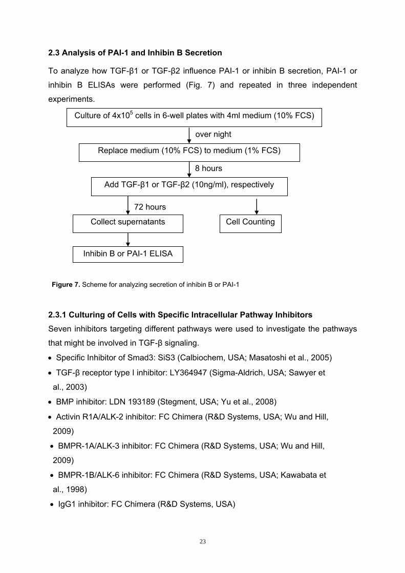
3.1.2 Characterization of Endometrial and Endometriotic Tissues .................... 57
3.2 Influence of TGF-β1 or TGF-β2 on Cell Numbers ....................................... 59 3.3 Analysis of the Smad Pathway in Endometrial and Endometriotic Cells. 61 3.4 Influence of TGF-β1 or TGF-β2 on Smad3 Phosphorylation of Endometrial and Endometriotic Cells in vitro ........................................................................ 63 3.5 Influence of TGF-β1 or TGF-β2 on Apoptosis of Endometrial and Endometriotic Cells in vitro ............................................................................... 69 3.6 Influence of TGF-β1 or TGF-β2 on Plasminogen Activator Inhibitor-1 Secretion by Endometrial and Endometriotic Cells in vitro ............................ 77
3.6.1 Analysis of TGF-β-induced PAI-1 Secretion by Endometrial and
Endometriotic Cells............................................................................................ 78
3.6.2 Analysis of the Smad Pathway in PAI-1 Secretion in Endometrial and
Endometriotic Cells............................................................................................ 80
3.6.3 Analysis of the BMP Pathway in PAI-1 Secretion..................................... 81
3.6.4 Analysis of BMP Receptors in PAI-1 Secretion ........................................ 83
3.7 Influence of TGF-β1 or TGF-β2 on Inhibin B Secretion by Endometrial and Endometriotic Cells in vitro ............................................................................... 85 3.8 Influence of TGF-β1 or TGF-β2 on TβRIII Expression of Endometrial and Endometriotic Cells in vitro ............................................................................... 87

iv
3.9 Influence of TGF-β1 or TGF-β2 on Secretion of TGF-β2 by Endometrial and Endometriotic Cells in vitro ........................................................................ 88 3.10 Influence of TGF-β1 or TGF-β2 on Secretion of TGF-β1 by Endometrial and Endometriotic Cells in vitro ........................................................................ 90 3.11 Influence of TGF-β1 or TGF-β2 on Interaction of TβRI, TβRII and TβRIII Receptors on Endometrial and Endometriotic Cells in vitro (Signalosome analysis)............................................................................................................... 92
3.11.1 Interaction of TβRII and TβRIII Receptors on HES Cells........................ 95
3.11.2 Interaction of TβRI and TβRII Receptors on HES Cells......................... 96
3.11.3 Interaction of TβRII and TβRIII Receptors on T-HESC Cells.................. 97
3.11.4 Interaction of TβRI and TβRII Receptors on T-HESC Cells................... 99
3.11.5 Interaction of TβRII and TβRIII Receptors on 12ZVK Cells ..................100
3.11.6 Interaction of TβRI and TβRII Receptors on 12ZVK Cells ....................101
3.11.7 Interaction of TβRII and TβRIII Receptors on 22B Cells........................103
3.11.8 Interaction of TβRI and TβRII Receptors on 22B Cells..........................104
4 Discussion ......................................................................................................106 4.1 Role of TGF-βs in Endometrial and Endometriotic Cells and Primary Endometrial Stromal Cells ................................................................................106
4.1.1 Influence of TGF-βs on Cell Numbers .....................................................106
4.1.2 Effects of TGF-βs on Apoptosis...............................................................107
4.1.3 The Role of TGF-βs in Phosphorylation of Smad3 ..................................108
4.1.4 The Role of TGF-βs in PAI-1 Secretion ...................................................109
4.1.5 Cross-talk of the TGF-β and BMP Pathways...........................................110
4.1.6 The Effect of the TGF-βs on Inhibin B Secretion .....................................111
4.1.7 Effects of TGF-βs on Interaction of TβRI, TβRII and TβRIII Receptors ...112
4.1.8 Characteristic Differences between Endometrial and Endometriotic Cells
and Tissues ......................................................................................................113
4.1.9 TGF-β Signaling in Endometrial Cells, Endometriotic Cells and Primary
Endometrial Stromal Cells ................................................................................114
4.1.10 The Smad-dependent Pathway in TGF-β Signaling in Endometrial Cells,
Endometriotic Cells and Primary Endometrial Stromal Cells ............................114
4.1.11 The Smad-independent Pathways in TGF-β Signaling in Endometrial and
Endometriotic Cell Lines and Primary Endometrial Stromal Cells ....................115
4.2 Conclusions .................................................................................................116

v
5 Summary .........................................................................................................118 6. Zusammenfassung.........................................................................................120 7. References ......................................................................................................122 8. Acknowledgements........................................................................................142

vi
Abbreviations
ActR activin receptor
ALK activin receptor-like kinase
Approx approximately
APS ammonium persulphate
BCA bicinchonic acid
Bcl-2 B-cell lymphoma/Leukemia-2 protein
BMP bone morphogenetic proteins
BMPR bone morphogenetic protein receptor
BSA bovine serum albumin
CA 125 cancer antigen 125
Caspase 3/7 cysteinyl-asparate-specific proteases 3/7
CD 10 cluster of differentiation
CK 18 cytokeratin18
cm2 square centimeter
CO2 carbon dioxide
Ctrl control
DAB 3,3’-diaminobenzidine tetrahydrochloride
DMEM Dulbecco’s modified eagle’s medium
DNA deoxyribonucleic acid
DTT Dithiothreitol
ECM extracellular matrix
EDTA ethylene diamine tetraacetic acid
ELISA enzyme-linked immunosorbent assay
FasL fas ligand
FBS fetal bovine serum
FC fragment crystallizable
FCS fetal calf serum
Fig figure
g gravitational force
GDF growth and differentiation factors
GnRH gonadotropin-releasing hormone
g/L grams/liter

vii
HEPES hydroxyethyl piperazineethanesulfonic acid
HRP horseradish peroxidise
IFN-γ interferon-gamma
IgG immunoglobulin G
IHC immunohistochemistry
IL interleukin
JNK c-Jun N-terminal kinases
LAP latency associated peptide
LLC large latent complex
LRPI low-density lipoprotein receptor-related protein-1
LTBP latent-TGF-β binding protein
Min minutes
ml mililiter
mM millimolar
MMP matrix metalloproteinases
mRNA messenger ribonucleic acid
MUC1 mucin-1
NBF neutral buffered formalin
ng nanogram
NK natural killer cells
nm nanometer
OD optical density
p38 MAPK p38 mitogen-activated protein kinase
PAI-1 plasminogen activator inhibitor-1
PBS Phosphate buffered saline
PDGF platelet-derived growth factor
pen/strep penicillin/streptomycin
PFA paraformaldehyde
pg picogram
PLA proximity ligation assay
PMSF phenylmethylsulfonyl fluoride
PVDF polyvinylidene difluoride
rAFS revised American Fertility Society System
rASRM revised American Society for

viii
Reproductive Medicine
RCA rolling cycle amplification
RFU relative fluorescence units
rh recombinant human
RT room temperature
SBE Smad binding elements
SERPIN serine proteinase inhibitor
SCID severe combined immune deficiency
SDS sodium dodecyl sulfate
SiS3 specific inhibitor for Smad3
α-SMA α-smooth muscle actin
SV40 Simian virus 40
TBST Tris buffered saline Tween-20
TEMED Tetramethylethyelenediamine
TGF-β transforming growth factor betas
TIMP tissue inhibitor of metalloproteinases
TMB 3,3’,5,5’-Tetramethylbenzidine
tPA tissue-type plasminogen activator
TβR receptor of transforming growth factor-beta
Tris tris (hydroxymethyl)-aminomethane
uPA urokinase-type plasminogen activator
uPAR urokinase receptor
UV light ultraviolet light
μg microgram
μl microliter
μM micromolar
v/v volume/volume

ix
List of Publications a) Mecha E, Omwandho CA, Tinneberg HR, Konrad L (2013). Characterization of the TGF-beta signalosome and of TGF-
beta dependent endometrial cell proliferation (2013). In preparation
b) Konrad L, Gronbach J, Mecha EO, Frank M, Berkes E, Gattenlöhner S, Omwandho COA, Oehmke F, Tinneberg HR
(2013). Endometriosis is one entity. Submitted.
Abstracts from Conferences and Scientific Meetings 1) Mecha E, Omwandho CA, Sui C, Tinneberg HR, Konrad L (2013). TGF-betas induce smad-dependent signaling and
apoptosis in endometrial and endometriotic cells. Abstract P005. 5th Dachverband Reproduktionsbiologie und medizin
Kongress. Műnster, Germany. Dec 4-7, 2013.
2) Mecha E, Sui C, Kloeppels K, Omwandho CA, Tinneberg HR, Konrad L (2013). The TGF-betas in human endometrial
and endometriotic cells. Abstract P45, 2nd European Congress on Endometriosis. Berlin, Germany. Nov 28-30, 2013.
3) Gronbach J, Kortum J, Mecha E, Berkes E, Omwandho CA, Tinneberg HR, Konrad L (2013). Endometriosis – What do the
neighbours think? Abstract SP11, 2nd European Congress on Endometriosis. Berlin, Germany. Nov 28-30, 2013.
4) Mecha E, Omwandho CA, Tinneberg HR, Konrad L (2013). The TGF-β signalosome in human endometrial and
endometriotic cells. Abstract P99, 6th International Giessen Graduate School for life Sciences Conference, Giessen,
Germany. Sept. 11-12, 2013.
5) Mecha E, Omwandho CA, Sui C, Tinneberg HR, Konrad L (2013). TGF-betas enhance secretion of MMP2/9 and PAI-1 of
human endometrial and endometriotic cells. Abstract 68, 2nd International Scientific Conference, University of
Nairobi/Kenyatta National Hospital, Nairobi, Kenya. June, 19-21. 2013.
6) Berkes E, Stammler A, Mecha EO, Tinneberg HR, Konrad L (2013). Sekretion von clusterin durch endometriale und
endometriotische Zellen. 10th Endometriosis Congress, Linz, Austria, April 25-27, 2013.
7) Gronbach J, Mecha E, Berkes E, Stammler A, Omwandho CA, Tinneberg HR, Konrad L (2013). Stromal cells in the
endometrium and endometriosis. 15th World Congress on Human Reproduction, Venezia, Italy, March 13-16, 2013.
8) Mecha E, Omwandho CA, Zoltan D, Tinneberg HR, Konrad L (2012). TGF-βs induce Smad-dependent signaling and
apoptosis in human endometrial and endometriotic cells lines. Abstract SP 04, 1st European Congress on Endometriosis,
Siena, Italy, 29th Nov- 1st Dec, 2012. J Endometriosis 4: 213-214.
9) Mecha E, Omwandho COA, Zoltan D, Tinneberg HR, Konrad L (2012). TGF-βs induce Smad-dependent signaling and
apoptosis in primary human endometrial stromal cells. Abstract P99, 5th International Giessen Graduate School for life
Sciences Conference, Giessen, Germany. Sept. 18-19, 2012
10) Mecha E, Omwandho CA, Zoltan D, Tinneberg HR, Konrad L (2011). Transforming growth factor betas strongly enhances
secretion of plasminogen activator inhibitor 1 protein by endometrial and endometriotic cell lines. Abstract P100, 4th
International Giessen Graduate School for life Sciences Conference, Giessen, Germany. August, 21-22. 2011.

.

1
1 Introduction
1.1 Endometriosis
Endometriosis is characterized by the presence of endometrial tissue outside the
uterus most commonly in the ovary and the peritoneum but may also occur in
pericardium, intestinal tract, lung, pleura or brain (Giudice and Kao, 2004). The
prevalence is approximately 10% in the general female population in their
reproductive age with 35-50% of the patients experiencing pain and infertility
(Houston, 1984; Cramer, 1987; Rothman and Greenland, 1998; Rogers et al., 2009).
Endometriosis is generally associated with inflammation, but, severe cases may
result in extensive pelvic adhesions and distortion of pelvic anatomy, which could
result in infertility (Giudice and Kao, 2004).
1.1.1 Pathogenesis, Diagnosis, Classification and Therapy of Endometriosis The pathogenesis of endometriosis is still poorly understood and remains
controversial. However, several theories including dissemination by retrograde
menstruation, induction, coelomic metaplasia, altered cellular immunity, lymphatic
and vascular metastasis, genetic causes and environmental causes have been
presented to understand the pathogenesis and etiology of endometriosis (Giudice
and Kao, 2004; Witz, 2005).
The most widely accepted hypothesis of the pathogenesis of endometriosis is
dissemination and implantation after retrograde menstruation first proposed by
Sampson (1922; 1927). According to Sampson, some of the endometrial tissue
fragments passes through the fallopian tubes during menstruation, then attach and
proliferate in the peritoneal cavity (Sampson, 1927). Recent data by Matsuzaki and
Darcha (2012) and Konrad et al. (2013, submitted) provide evidence that the
hypothesis by Sampson is probably correct.
However, Sampson's theory fails to explain why only a small percentage of women
experiencing retrograde menstruation develop endometriosis (Koninckx and Martin,
1992). Thus, other factors like the immune system might be involved in the
implantation of displaced endometrial cells which develop into endometriotic lesions
(Donald and Dmowski, 1998; Kyama et al., 2003). In women with endometriosis, the
immune system seems to be impaired, hence permitting endometrial cells to escape

2
immune surveillance and to grow in ectopic locations (Donald and Dmowski, 1998;
Herington et al., 2011). Diagnosis of endometriosis is by laparoscopy which is generally considered as the
“gold standard”, followed by histological confirmation of viable ectopic endometrial
glands and stroma (Brosens, 1997; Kennedy et al., 2005). Sometimes, a non-
invasive examination by auxiliary diagnostic methods such as pelvic ultrasound is
used before performing surgery (Brosens, 1997; Champaneria et al., 2010).
Peritoneal fluid, serum and tissue markers such as CD10 and CA125 have also been
tested in some clinics but none are used routinely (Mihalyi et al., 2010).
The revised American Society for Reproductive Medicine (rASRM) score is currently
the best known classification of endometriosis. According to rASRM, the staging is
based on the location, amount, depth and size of endometrial implants. Four stages
of endometriosis have been characterized
• Stage 1 (Minimal)
- Findings restricted to superficial lesions only and possibly a few filmy
adhesions
• Stage 2 (Mild)
- Presence of deep lesions in the rectouterine pouch in addition to
observations in stage1
• Stage 3 (Moderate)
- Presence of endometriomas on the ovary and more adhesions in addition to
observations in stage 2
• Stage 4 (Severe)
- Presence of large endometriomas and extensive adhesions in addition to
observations in stage 3
Treatment of endometriosis can be medical, surgical or a combination of both.
Treatment options depend on the severity of the symptoms, pregnancy, age and
therapeutic goals. Endometriosis is often treated surgically, but symptoms recur in
75% of cases within 2 years (Candiani et al., 1991). Medical treatment of
endometriosis involves contraceptive steroids, progestagens, agonists of
gonadotropin-releasing hormone (GnRH) analogues and non-steroidal anti-
inflammatory agents (Lessey, 2000; Kennedy et al., 2005; Valle and Sciarra, 2008;
Kappou et al., 2010). However, because of undesirable side-effects caused by most

3
of these drugs, they are useful only for a limited time period making it necessary to
change or use additional medication (Kuohung et al., 2002; Kennedy et al., 2005).
1.1.2 In vivo and in vitro Models of Endometriosis a) In vivo models The most widely accepted hypothesis for the development of endometriosis is
Sampson’s theory of retrograde menstruation which implies the implantation of
retrograde endometrial tissues in tissues outside the uterus. Thus, menstrual
shedding is a requirement for spontaneous development of endometriosis which has
been found to occur only in human, primates and some rats (Grümmer, 2006).
But owing to the species specificity, it is hard to compare human and primates. For
example, spontaneous and induced endometriosis has been found in baboons to be
as low as 4.8% and 27.6%, respectively, raising some doubts on the validity of the
baboon model for endometriosis (Dehoux et al., 2011). Also, the very high costs of
animal handling limit the use of baboons as an experimental model.
Recently, rodent models like severe combined immune deficiency (SCID) mice have
been used also to study endometriosis. In these models, human endometrial and
endometriotic tissue or cells were successfully engrafted into the peritoneal cavity of
immunodeficient mice (Liu et al., 2010; Becker et al., 2011). The grafted tissues or
cells were able to implant and develop into endometriotic lesions. The advantages of
such models are: (1) animals are easily available, (2) low costs and (3) the peritoneal
environment of the hosts can be altered to suit the study objectives. Mice
xenotransplanted with human eutopic endometrial tissue developed endometriosis-
like lesions in 63% and 68% in ovariectomized estrogen-supplemented TGF-β1-null
mutant mice and wild-type control, respectively. With the aid of this model it was
demonstrated that host derived TGF-β1 deficiency suppressed endometriotic lesion
development (Hull et al., 2012).
The in vivo models of endometriosis have many advantages, but the limitations of
such models is that they can hardly be used to study individual components and
some biological functions like the involvement of intracellular proteins and pathways,
These aspects of endometriosis can be addressed appropriately only by use of in
vitro models.

4
b) In vitro models In vitro models of endometriosis are build by obtaining endometrial and endometriotic
tissues from patients after surgical treatment. Primary epithelial or stromal cells are
isolated, cultured and then used for assays. Epithelial cells derived from
endometriotic lesions were found to be as invasive as metastatic cancer cells using
the matrigel invasion assay (Zeitvogel et al., 2001). Another model using both
invasion and angiogenesis within a 3D fibrin matrix demonstrated the ability of
ectopic endometrial fragments to proliferate and invade. The fragments generated in
vitro appeared histologically similar to endometriotic implants in vivo (Fasciani et al.,
2003). Therefore, in both models, there is a strong evidence of the possibility to study
endometriosis in vitro. The limitations of using primary cells are that the cells are
often heterogeneous and epithelial cells die at low passages (Starzinski-Powitz et al.,
1998).
This problem has been sorted out by establishment of immortalized endometrial and
endometriotic cells (Starzinski-Powitz et al. 1998; Boccellino et al. 2012). Konrad et
al. (2010) and Sui. (2012) showed that the above established immortalized
endometrial and endometriotic cell lines were able to secrete or to be stimulated by
TGF-βs. Thus, the immortalized cell lines are a suitable model for studying
endometriosis.
1.2 The Transforming Growth Factor Βetas and TGF-β Receptors
TGF-βs are a member of a large superfamily including activins, inhibins, nodals,
bone morphogenetic proteins (BMP), lefty A and B, growth and differentiation factors
(GDFs) and anti-Mullerian hormones (Chang et al., 2002; Peng, 2003). TGF-βs
comprise three isoforms namely TGF-β1, TGF-β2 and TGF-β3. Biologically, TGF-βs
regulate cell motility, proliferation, apoptosis, gene expression and differentiation. In
addition, TGF-βs tightly regulate production of the extracellular matrix (ECM) and are
involved in wound healing and immunosupression (Roberts and Sporn, 1993;
Roberts, 1998; Kaminska et al., 2005; Taylor, 2009). They are also involved in
tumorigenesis and inflammation (Padua and Massagué, 2009; Santibañez et al.,
2011).
Production and activation of the TGF-βs is triggered by cleavage of the inactive
dimeric TGF-β precursor called latency associated peptide (LAP). LAP is bound by
disulphide bonds to the latent-TGF-β binding protein (LTBP) resulting in the large

5
latent complex (LLC). The LLC is targeted either to the cell surface for activation or to
the extracellular matrix for storage (Munger et al., 1997; Rifkin, 2005).
Gene knockout techniques have revealed the physiological roles of each of the TGF-
β isoforms. TGF-β1 knockout mice die at gestation due to defective vasculature
(Shull et al., 1992; Chen et al., 1996). TGF-β2 knockout mice exhibit perinatal
mortality and a wide range of developmental defects (Sanford et al., 1997; Shi et al.,
1999). TGF-β3 knockout mice die within 24 hours after birth due to abnormal lung
development and feeding problems associated with a defective cleft palate
(Kaartinen et al., 1995; Proetzel et al., 1995).
The TGF-β family receptors are divided into three groups, namely type I, type II and
type III receptors. The three receptor types have distinct properties (Heldin et al.,
1997; Chang et al., 2002). TGF-β receptor type I (TβRI) and type II (TβRII) are
transmembrane serine/threonine kinases. The type III receptors, betaglycan (TβRIII)
and endoglin are accessory receptors and have high affinity to all three TGF-β
isoforms (Wrana et al., 1994; Gordon et al., 2008).
The general mechanism of TGF-β signaling starts by TGF-β binding either to TβRIII,
which presents it to TβRII, or binding to TβRII directly, which then binds to and
transphosphorylates TβRI. Then the activated TβRI phosphorylates Smad2 or
Smad3, which bind to Smad4 in the cytoplasm or the nucleus forming a Smad
complex. The Smad complex interacts with transcription factors in the nucleus to
regulate TGF-β responsive genes (Chen et al., 2003; Guglielmo et al., 2003; Biondi
et al., 2007; Wrighton et al., 2009).
1.2.1 The Smad-dependent and Smad-independent Pathways in TGF-β Signaling a) Smad-dependent Pathways Smads are intracellular proteins that transduce signals from the TGF-β receptors to
the nucleus where they activate downstream gene transcription (Miyazono al., 2000).
There are three classes of Smads, namely the receptor-regulated Smads (R-Smads)
including Smad1, 2, 3, 5 and 8/9 (Wu et al., 2001). The common-mediator Smad (co-
Smad) with only Smad4, which interacts with R-Smads to participate in signaling (Shi
et al.,1997). The inhibitory Smads (I-Smads), Smad6 and Smad7, block the activation
of R-Smads and co-Smads (Itoh et al., 2001). BMP type I receptor activates Smads1,

6
5 and 8 (Moustakas et al., 2001), while TβRI or activin receptors activate Smad2 and
Smad3 (Chen et al., 2003).
A general Smad-dependent signaling pathway activated by TGF-β is shown in
Figure 1 (Rebecca, 2000).
Figure 1. The TGF-β induced Smad signaling pathway (Rebecca, 2000). Binding of TGF-β results in
the formation of the TβRI/TβRII complex and then the TβRII kinase phosphorylates and activates
TβRI. The activated TβRI kinase phosphorylates Smad2/Smad3. The phospho-Smad2/Smad3 form
complexes with the co-Smad (Smad4) and move into the nucleus, where they interact with other
transcription factors to regulate transcription.
b) Smad-independent Pathways Smad-independent pathways which reinforce the signal transduction of the TGF-βs
were shown by Derynck and Zhang. (2003), Moustakas and Heldin. (2005) and
Zhang (2009). The Smad-independent pathways provide alternative TGF-β signal
transduction without the direct involvement of Smad proteins. For instance, the p38
substrate kinase, regulates the transcriptional activity of Smad3 by enhancing its
association with p300 (Abécassis et al., 2004) and TGF-β-activated JNK was shown
to phosphorylate Smad3 thus inducing its nuclear translocation (Engel et al. 1999).

7
1.2.2 TGF-βs in the Normal Endometrium and Endometriosis a) Normal Endometrium The TGF-βs are stage-specifically expressed in the human endometrium during the
menstrual cycle (Fig. 2). TGF-β1 and TGF-β3 are highly expressed in stromal and
glandular cells (Chegini et al., 1994). TGF-β2 was found to be strongly expressed in
stromal cells compared to glandular cells (Gold et al., 1994; Gaide Chevronnay et al.,
2008). This observation suggests the involvement of TGF-βs in the normal function of
the human endometrium (Omwandho et al., 2010).
Figure 2. Levels of TGF-βs during the menstrual cycle in human endometrium. Only the start and
endpoints of the strongest expression of the TGF-βs is shown. The three TGF-βs isoforms are
differentially and highly expressed during the menstrual cycle (Omwandho et al., 2010).
b) TGF-βs in Endometriosis TGF-βs were found to be expressed significantly higher in the serum and peritoneal
fluid in women with endometriosis (Pizzo et al., 2002). Also, TGF-βs levels are
enhanced markedly concomitant with the severity of the disease with high levels
observed in stages III and IV, suggesting a possible role in the pathogenesis of
endometriosis (Pizzo et al., 2002).
Endometriosis comprises six developmental stages namely cell shedding and
refluxing, cell survival, immune suppression, cell adhesion and invasion,
angiogenesis, and bleeding (Omwandho et al., 2010). TGF-βs have been shown to
0
2
4
6
8
10
12
14
1 2 3 4 5 6 7 8 9 10 11 12 13 14 15
Menses Proliferative Phase Secretory Phase
Estrogen Progesterone
β2 β1
β3
Con
cent
ratio
n

8
be directly or indirectly involved in most of these stages (Fig. 3).
Figure 3. The six developmental stages leading to endometriosis. The involvement of the TGF-βs
directly or indirectly in each stage has been emphasized (Omwandho et al., 2010).
Retrograde menstruation is the most widely accepted theory of pathogenesis of
endometriosis (Sampson, 1927), thus for establishment of endometriosis to occur,
shedding and refluxing of endometrial cells are prerequisites. Highest expression of
the TGF-βs was detected during menstruation, suggesting a possible involvement of
the TGF-βs in cell shedding (Gaide Chevronnay et al., 2008). Also, women with
endometriosis were found to experience abnormal myometrial contractions with
higher and a different frequency (Bulletti et al., 2002), which is possibly associated

9
with the dissemination of endometrial fragments.
TGF-βs regulate a wide variety of cellular responses including proliferation,
apoptosis, gene expression, immune responses, cell motility, tumorigenesis, immune
responses and extracellular matrix production (ECM) (Derynck et al., 2001).
Interestingly, TGF-β effects can be extremely variable depending on cell types and
stimulation context. For example, TGF-βs cause growth arrest in epithelial cells, but
induce activation of fibroblasts (Rahimi and Leof, 2007). In endometrial cells, TGF-β1
was found to stimulate DNA synthesis of epithelial cells with lower cell number, but
repressed it when the cell number was higher in women with and without
endometriosis (Meresman et al., 2003).
Escape of endometrial fragments from apoptosis and immune attack as they enter
and transit the peritoneal cavity is also very important for their survival. TGF-β1 can
induce expression of FasL mRNA in endometrial stromal cells (Garcia-Velasco et al.,
1999), possibly preventing apoptosis during transit of the peritoneal cavity. In
addition, TGF-β1 can inhibit IFN-γ and IL-10 secretion by uterine NK cells from
human endometrium but upon blocking of TGF-β1, secretion of the two cytokines
was increased (Eriksson et al., 2004). In general, the high levels of the TGF-βs might
have a direct or indirect effect on the immune escape by decreasing the response of
NK cells to ectopic endometrial fragments hence increasing their survival rate.
TGF-βs might be regulating cell adhesion to the ECM indirectly through regulating
the balance of matrix metalloproteinases (MMPs) and tissue inhibitor of
metalloproteinases (TIMPs) which are involved in the normal fibrolytic processes.
Also MMPs have been shown to convert the inactive membrane-bound FasL, to its
active soluble form which induces apoptosis (Otsuki, 2001). Taken together, TGF-βs
might be involved in most of the biological processes involved in the pathogenesis of
endometriosis, although more studies are needed to clarify their exact participation.
1.3 TGF-β-induced Apoptosis in Human Endometrium and Endometriosis
1.3.1 Apoptosis Apoptosis (programmed cell death) enables multicellular organisms to remove
excessive and potentially dangerous cells. Balance and coordination between
apoptosis and cell survival is important for homeostasis and development of
multicellular organisms (Green, 1998). A defect in the control of the balance may
cause diseases like cancer, neurodegenerative conditions and autoimmune diseases

10
(Kroemer et al., 2007). The morphological alteration of cells undergoing apoptosis is
caused mainly by cysteine protease proteins, called caspases, which are mainly
activated in cells undergoing apoptosis (Green, 1998).
The process of apoptosis is characterized by cell shrinkage, chromatin condensation,
DNA fragmentation and formation of cytoplasmic blebs (Elmore, 2007). Two major
apoptotic pathways exist in mammalian cells, the mitochondrial pathway (intrinsic)
and the death-receptor pathway (extrinsic). The two pathways are initiated by
different mechanisms but both cause activation of caspases which eventually result
in apoptosis.
1.3.2 TGF-β-induced Apoptosis Uterine cells have been shown to undergo apoptosis during blastocyst implantation,
oestrous cycle and decidualization. The control of the coordinated blastocyst
implantation processes presumably depends on secretion of various endometrial
factors, among them TGF-βs (Pollard, 1990).
TGF-β1 and TGF-β2 mRNAs have been detected in decidual cells after the first week
of pregnancy (Tamada et al., 1990; Manova et al., 1992). TGF-β1 has been reported
to increase apoptosis in primary cell cultures of uterine epithelial cells (Rotello et al.,
1989), also TGF-β1 and TGF-β2 stimulated the increase in nucleosome DNA
fragmentation in endometrial stromal cells indicating stimulation of apoptosis (Bruce,
1994). Furthermore, neutralization of TGF-βs secreted by endometrial stromal cells
completely inhibited apoptosis in vitro (Bruce, 1994). All above data strongly show
that TGF-βs are an important component in apoptosis of stromal and epithelial cells.
1.3.3 TGF-β-induced Apoptosis in the Human Endometrium Apoptosis occurs in normal endometrial tissue throughout the cycle and is important
in maintaining the homeostasis of cells during the menstrual cycle. Specific nuclear
DNA fragmentation related to apoptosis was reported in the human endometrium
(Hopwood and Levinson 1995). The B-cell lymphoma/Leukemia-2 protein (Bcl-2), an
anti-apoptotic protein, is expressed throughout the menstrual cycle with higher levels
in the glandular component of the human epithelium (Gombel et al., 1994, Otsuki et
al., 1994: McLaren et al., 1997).
The Bcl-2 immunoreactivity was maximal in the proliferative endometrium and the
levels decreased to a minimum in the secretory endometrium (Tao et al., 1997). In

11
contrast, levels of the pro-apoptotic proteins, Bax and Fas/FasL, were increased in
the secretory endometrium with minimal expression in the glandular epithelium
(Watanabe et al., 1997; Meresman et al., 2000). Furthermore, in Bcl-2-deficient mice,
many apoptotic cells and bodies were often observed in glands and myometrium
(Daikoku et al., 1998), implying that Bcl-2 may be essential in survival of both
endometrial glandular cells and myometrial smooth muscle cells (Meresman et al.,
2000).
Coincidently, TGF-βs have been shown to be stage-specifically and abundantly
expressed throughout the menstrual cycle with higher levels expressed in both
stromal and glandular cells (Chegini et al., 1994; Gaide Chevronnay et al., 2008).
The mRNA and protein expression of TGF-βs is high in the late secretory phase of
the menstrual cycle (Omwandho et al., 2010). Also the levels of the pro-apoptotic
proteins, Bax and Fas/FasL, are high in the secretory phase while the anti-apoptotic
protein, Bcl-2 expression, is low in the secretory phase of the menstrual cycle (Tao et
al., 1997; Watanabe et al., 1997; Garcia-Velasco et al., 1999; Meresman et al.,
2000). Other studies have shown that TGF-β1 increased the apoptotic rate of
endometrial stromal cells in vitro (Chatzaki et al., 2003).
We can only speculate that the stage-specific expression of TGF-βs, Bax, Fas/FasL
(direct correlation) and Bcl-2 protein (inverse correlation) throughout the menstrual
cycle indicates that TGF-βs might induce apoptosis of human endometrial stromal or
epithelial cells in human endometrium during the secretory phase of the menstrual
cycle. Further studies are needed to characterize the apoptotic effects of the TGF-βs
and which apoptotic pathways are involved in the normal endometrium and
endometriosis.
1.3.4 TGF-β-induced Apoptosis in Endometriosis The role of TGF-β-induced apoptosis in endometriosis has not been well studied,
however, the escape of endometrial fragments from apoptosis as they enter and
transit the peritoneal cavity is very important for their survival (Garcia-Velasco et al.,
1999). In addition, women who develop endometriosis showed greatly reduced
apoptosis in sloughed endometrial cells, implying that a high number of surviving
cells can enter the peritoneal cavity (Gebel et al., 1998).
Apoptosis was found to decrease as the severity of endometriosis increases
(Dmowski et al., 2001). Also, Bcl-2 protein expression was found to be increased in

12
proliferative eutopic endometrium in women with endometriosis, suggesting a
possible resistance to apoptosis (Meresman et al., 2000). Furthermore, FasL is highly
expressed in endometriotic tissues which possibly contributes to their survival and
thus to the development of endometriosis. Also higher levels of soluble FasL were
present in serum and peritoneal fluid of women from endometriosis (Garcia-Velasco
et al., 2002), which might contribute to increased apoptosis of Fas-expressing
immune cells. Garcia-Velasco et al. (1999) showed that platelet-derived growth factor
(PDGF) and TGF-βs are increased in peritoneal fluid of women with endometriosis.
Also, PDGF and TGF-βs induced FasL expression by endometrial stromal cells
(Garcia-Velasco et al.,1999). Increased FasL expression by the cells may protect the
stromal cells from cytotoxic T-cells, hence ectopic endometrial cells are able to
escape the immune system in the peritoneal cavity of women with endometriosis
hence possibly contributing to the pathogenesis and maintenance of the disease.
Furthermore, FasL expression by endometriotic cells was induced after adhesion of
the cells to ECM proteins of endometriotic patients (Selam et al., 2002), this possibly
induces apoptosis of activated T-lymphocytes thereby lowering their ability to attack
the endometrial cells. Thus, survival of endometriotic cells is promoted during initial
attachment at ectopic sites.
Increased expression of FasL, Bcl-2 and TGF-βs in serum, peritoneal fluid and
ectopic sites in women with endometriosis suggests the possible involvement of
TGF-βs in apoptosis during entry and transit of endometrial cells in the peritoneal
cavity, subsequent attachment of the cells at ectopic sites and eventual
establishment and maintenance of endometriosis.
Although, the role of TGF-β-induced apoptosis in endometriosis has not been fully
investigated, recently, Omwandho et al. (2010) showed that the stages of
endometriosis are similar to those of cancer. Since TGF-βs have been shown to play
an important role in suppression of apoptosis in various tumors (Lebrun, 2012),
together with their increased expression in serum, peritoneal fluid and at
endometriotic sites of women suffering from endometriosis, it will be of great
importance to find out if the TGF-βs induce apoptosis in endometriotic cells and by
which mechanisms.

13
1.4 Crosslink of TGF-β and Bone Morphogenetic Proteins (BMPs)
The BMPs are multifunctional proteins that regulate functions such as proliferation,
apoptosis and differentiation of a large variety of cell types (Reddi, 1997). BMPs
mediate their cellular functions through binding to a combination of type I and type II
receptor serine/threonine kinases (Kawabata et al., 1998). The BMP ligands can bind
to any of the three type II receptors (BMPRII, ActRIIa and ActRIIb) which then bind to
one of the three type 1 receptors (ALK-2, ALK-3 and ALK-6). Upon binding, the
constitutively active type II receptor phosphorylates type 1 receptor then
phosphorylates the BMP-responsive Smad proteins namely Smad1, Smad5 and
Smad 8. The activated Smads bind Smad4 either in the cytoplasm or in the nucleus
for signaling (Yu et al., 2008). In addition, BMP signals have been found to activate
other intracellular effectors like mitogen-activated protein kinase (MAPK) p38 via the
Smad pathway (Nohe et al., 2004).
Although TGF-βs transduce their signals through activation of Smad2 and Smad3
(Chen et al., 2003), recent studies have indicated that they can also strongly but only
transiently induce phosphorylation and activation of Smad1, Smad5 and Smad8
(BMP-responsive Smads) in endothelial cells, epithelial cells, fibroblasts and
epithelium derived cancer cells (Bharathy et al., 2008; Daly et al., 2008; Liu et al.,
2009). These observations have raised several questions of how the activation of
Smads1/5/8 by TGF-βs affect BMP responses (Grönroos et al., 2012).
Several hypothesis have been put forward to explain the possible crosstalk of
TGF-βs/BMP pathways, for example Grönroos et al., 2012 suggested the
involvement of ALK-5 and formation of pSmad3-pSmad1/5 complexes. Recently,
knockdown of Smad3 phosphorylation in mice abolished the ability of TGF-β to inhibit
BMP-induced transcription (Grönroos et al., 2012), further supporting a possible
crosstalk between TGF-β and BMP pathways.
Perturbations of both BMP and TGF-β signalling have been reported to cause distinct
bone diseases (Jansens et al., 2000). In endothelial cells, ALK-1 together with ALK-5
can activate TGF-β-responsive Smads (Smad2/3) and also phosphorylate BMP-
responsive Smads (Smad1/ 5/ 8; Miyazono and Kusanagi, 2001). Furthermore,
Smad5-deficient mice exhibit defects in vascular tissues (Chang et al., 1999), which
are similar to those observed in ALK-1 deficient mice. These observations suggest
that Smad5 is a downstream target of ALK-1 (Miyazono and Kusanagi, 2001). In
addition, molecules that repress both TGF-βs and BMPs have been found to be

14
involved in the pathogenesis of vascular diseases (Miyazono and Kusanagi, 2001),
hence indicating a possible crosstalk of both pathways. What remains to be
investigated is the exact location of cross-talk of the TGF-β and BMP pathways (e.g.
at the extracellular membrane, in the cytoplasm or in the nucleus) and which
molecules are involved (e.g. the individual BMP/TGF-β receptors or Plasminogen
Activator Inhibitor 1 (PAI-1) secretion among other proteins. These will be helpful in
determining the cross-talk of TGF-β and BMP pathways and understand their
possible role in the pathophysiology of endometriosis.
1.5 Plasminogen Activator Inhibitor-1
PAI-1 is the major inhibitor of tissue-type plasminogen activator (tPA) and urokinase-
type plasminogen activator (uPA) in vitro and is pivotal in fibrinolysis (Binder et al.,
2002; Czekay and Loskutoff, 2004). The promoter region of PAI-1 contains three
Smad binding elements (SBEs), which are the main response element of TGF-β
signals (Binder et al., 2002).
Endothelial and stromal cells are the main producers of PAI-1 which is then released
into the plasma (Bastelica et al., 2002; Vaughan, 2005). Expression of PAI-1 and
uPA was shown to be much higher in endometriotic and endometrial tissues of
women with endometriosis compared to tissue from women without endometriosis
(Bruce et al., 2004). PAI-1 is able to regulate levels of cell surface integrins through
triggering their internalization by binding to the low-density lipoprotein receptor-
related protein-1 (LRP1) resulting in detachment of cells from various substrates
(Akkawi et al., 2006; Pedroja et al., 2009; Czekay et al., 2011).
Recently, studies have shown that PAI-1 and TGF-βs are involved in fibrosis, wound
healing and metastasis and are capable of inducing ECM remodeling through
regulation of plasmin and MMP acitivity (Kortlever and Bernards, 2006). Furthermore,
activity of TGF-βs is involved in the conversion of fibroblasts to myofibroblasts
(Desmouliere et al., 2004), that are responsible for PAI-1 secretion at the edges of
stromal tissue during cancer invasion (Dublin et al., 2000; Offersen et al., 2003).
Given the fact that both TGF-β and PAI-1 are involved in fibrosis, wound healing,
metastasis, induction of ECM remodeling and regulation of MMP activity and that
their activities (TGF-β and PAI-1) are elevated in endometrial and ectopic tissues in
women with endometriosis, thus we suppose that TGF-β/PAI-1 might play an
important role in the pathogenesis of endometriosis. It will be interesting to

15
investigate whether or not, PAI-1 contributes to the pathophysiology of endometriosis
and which pathways are involved.

16
1.6 Objectives
Despite the fact that TGF-βs have been demonstrated to play important roles in the
pathogenesis of endometriosis, their precise function needs to be investigated in
more depth. TGF-βs have been shown to mediate their functions via Smad
molecules. Of note, cross-talk with other signaling pathways has been reported, but
the exact molecules involved have not been fully established. Thus, it will be
interesting to examine the distinct molecules involved (Smads and receptors) and the
cross-talk with other pathways like BMPs to better understand the pathogenesis of
endometriosis. Furthermore, PAI-1 expression has been found to be regulated by
TGF-β isoforms in many tissues, thus it will be of great importance to elucidate how
TGF-βs regulate the expression of this protein and the pathways utilized for its
regulation.
Endometriosis has been studied by the use of immortalized endometriotic and
endometrial cell lines (for comparison) both of which have been characterized well.
Thus both endometrial and endometriotic cell lines together with primary endometrial
stromal cells will enable us to establish an in vitro model which will allow a deeper
understanding of the basic aspects and provide comparisons of endometriotic cells to
normal endometrial cells.
The main objective of this study was to investigate the influence of TGF-βs on
endometrial, endometriotic cell lines and primary endometrial stromal cells in regard
to cell numbers, regulation of some proteins essential in endometriosis and TGF-β
receptor interactions. The study will further investigate the signaling pathways of
TGF-βs and possible cross-talks with other pathways in endometrial and
endometriotic cell lines and primary endometrial stromal cells in vitro. In addition, the
different characteristics of normal endometrial cells and endometriotic cells observed
in all the above aspects will be reported.

17
2 Materials and Methods 2.1 Human Immortalized Endometrial, Endometriotic Cell Lines and Primary Endometrial Stromal cells
2.1.1 Endometrial Epithelial and Stromal Cells The HES cells are a spontaneously immortalized human endometrial epithelial cell
line (Fig. 4). The cells express E-cadherin, vimentin and cytokeratin (Desai et al.,
1994) and estrogen receptor alpha (Banu et al., 2008).
The T-HESC cells (Fig. 4) are telomerase immortalized endometrial stromal cells
(Krikun et al., 2004). T-HESC exhibit considerable and constitutive expression of
progesterone receptor mRNA (Leila et al., 2011). The estrogen receptor beta and P-
450 aromatase are strongly expressed also in T-HESC (Banu et al., 2008). The cells
undergo decidualization after treatment with estrogen plus medroxyprogesterone 17-
acetate (Krikun et al., 2004).
Figure 4. Human endometrial epithelial (HES) and stromal (T-HESC) cells. The HES cells are
triangular, flat and strongly aggregate indicating epithelial cell-to-cell contacts. The T-HESC cells show
a spindle-shaped, fibroblastoid morphology and are larger in size compared to HES. They are often
scattered when their numbers are few, however, they often aggregate in parallel clusters when
crowded.
2.1.2 Endometriotic Epithelial and Stromal Cells 12ZVK are epithelial-like endometriotic cells and are immortalized with SV40 (Fig.
5). They express cytokeratins, E-Cadherin and vimentin usually present in
endometrial epithelial tissue (Zeitvogel et al., 2001). They express steroidogenic
acute regulatory protein, the estrogen receptor α and β, progesterone receptor and
steroidal stimulating factor-1 (Banu et al., 2008).
HES T-HESC

18
The 22B cells are stromal-like endometriotic cells and have been immortalized with
SV40 (Fig. 5). They express cytochrome P-450 mRNA abundantly and estrogen
receptor β. 22B are E-Cadherin-negative but they are vimentin-positive (Zeitvogel et
al., 2001).
Figure 5. Human endometriotic epithelial (12ZVK) and stromal (22B) cells. The 12ZVK cells are
triangular, flat and aggregate closely to each other suggestive of epithelial cell-to-cell contacts. The
22B cells show a spindle-shaped, elongated, fibroblastoid morphology and some of them have a
branched cytoplasm. 22B cells often aggregate in parallel clusters when crowded and are scattered
when their numbers are few.
2.1.3 Primary Endometrial Stromal Cells Primary endometrial stromal cells which have been isolated earlier from endometrial
tissue obtained from a fertile women in their reproductive age after laporoscopy were
characterized using antibodies against cytokeratin 18 and α-smooth muscle actin.
They are α-smooth muscle actin positive but cytokeratin 18 negative.
Morphologically, they show similar properties like those of the T-HESC cell line (Fig.
6).
T-HESC
12ZVK 22B

19
Primary Endometrial Stromal Cells Figure 6. Human endometrial stromal cells show a spindle-shaped, fibroblastoid morphology in vitro.
They are often scattered when their numbers are few, however, they often aggregate in parallel
clusters when crowded.
2.2 Cell Culture with Endometrial, Endometriotic Cell Lines and Primary Endometrial Stromal Cells
2.2.1 Aseptic Techniques for Cell Culture Equipments and reagents were either bought sterile or sterilized in our laboratory.
Heat-labile solutions were sterilized with filters (pore size 0.22, μm; Millipore). The
bench surface of the laminar flow was completely cleared and the surface was wiped
with 70% ethanol. The UV light in the laminar was turned on for 30 minutes. Then the
hood blower and lights were turned on and the air was allowed to circulate for 20
minutes before use. The items needed were wiped with 70% ethanol just before
introducing them into the laminar flow. Latex surgical gloves were worn to avoid
contaminations and desinfected with 70% ethanol before starting to work which was
repeated in regular intervals.
Cell culture media used were supplemented with 1% pen/strep (100X, PAA, Austria)
and 1% plasmocin (Amaxa) or mycokill (10μg/ml, PAA) in order to avoid microbial
contaminations like fungi, mycoplasms and bacteria. Transfer and preparations of cell
culture media in the laminar was done using sterile media flasks and disposable
pipettes to minimize contamination. When adding (or replacing) medium, care was
taken not to touch the neck of the culture flasks which were cleaned with sterile
cotton swabs after medium transfer before putting the flasks into the incubator.

20
2.2.2 Cell Culture Media for Endometrial and Endometriotic Cells Generally, media were supplemented with appropriate amounts of serum, glutamine,
antibiotics and other nutrients. All sterile media were prepared before starting to work
with cells to avoid contamination and cross-infection during cell culture. All
procedures for media preparations were carried out in the laminar to ensure maximal
sterility.
The purchased ready-to-use media were stored at 4°C and after reconstitution were
stored at 4°C for a maximum of one month. Before cell culture, appropriate amounts
of reconstituted media were pre-warmed to 37°C in a water bath for about 20
minutes.
• Cell culture medium for primary endometrial stromal cells, endometrial (HES) and
endometriotic (12ZVK) epithelial cells:
DMEM High Glucose (4.5 g/L) without phenol red (PAA, Austria)
+ 10% fetal calf serum (FCS) (PAA, Austria)
+ 1% 100x pen/strep (PAA, Austria) and 1% L-glutamine (PAA, Austria)
• Cell culture medium for endometrial stromal cells (T-HESC):
DMEM F12 with L-glutamine without phenol red (Invitrogen, U.S.A.)
+ 10% fetal calf serum (FCS) (PAA, Austria)
+ 1% 100x pen/strep (PAA, Austria) and 1% Insulin Transferrin Selenium X
(Invitrogen, U.S.A.)
• Cell culture medium for endometriotic stromal cells (22B):
DMEM High Glucose (4.5 g/L) without phenol red
+ 10% Charcoal/Dextran Treated FCS (Thermo Scientific, U.S.A.)
+ 1% 100x pen/strep (PAA, Austria) and 1% L-glutamine (PAA, Austria)
2.2.3 Changing Cell Culture Medium Cell culture flasks were removed from the CO2 incubator and placed directly in the
laminar flow. The medium in the cell culture flask was discarded completely and
replaced with fresh pre-warmed medium every 2-3 days depending upon cell
confluency. The cap was fixed loosely on the flask so that air can enter into the flask.
The date of medium change was recorded on the flask and then returned back to the
CO2 incubator.

21
2.2.4 Passaging or Splitting of Cells Cells were passaged regularly at 80% confluency to avoid senescence associated
with prolonged high cell density. All old medium was discarded, 10 ml pre-warmed
accutase added and the flask was rotated to achieve a complete coverage of the
accutase, then the cells were incubated (37°C, 5% CO2) for 5 to 10 minutes. After
microscopic inspection to ensure complete detachment of cells from the flask, it was
gently tapped at the sides to release remaining adherent cells. Cells were
resuspended in 10 ml fresh serum-containing medium to inactivate accutase, and
then transferred into a 50 ml falcon tube and centrifuged for 5 minutes at 1500 x g.
The supernatant was discarded and 5 ml of medium was added to re-suspend cell
pellets. 500 μl to 2000 μl of cell suspension was transferred into a new cell culture
flask with 25 ml fresh medium. The cell passage number and date of passage were
documented on the flask. The flask was shaken gently to achieve a uniform cell
suspension and then taken back into the incubator.
2.2.5 Cryopreservation of Cells Cell freezing is essential for long-term storage of cells. The cell pellet obtained by the
process of passaging (2.2.4) was resuspended in medium Filocethplus (Procryoptect,
Germany). The cryo-medium reduced the freezing point of media and also allowed a
slower cooling rate, greatly reducing the risk of crystal formation which can damage
the cells and cause cell death. Aliquots of 1 ml cell suspensions were transferred into
cryotubes which were then stored at -80°C overnight for slow cooling before storage
in liquid nitrogen.
2.2.6 Thawing Frozen Cells Proper thawing of cryopreserved cells is crucial to ensure viability and functionality of
the cells. The thawing process is done very quickly to ensure that a high proportion of
the cells survive the procedure.
Cryotubes were taken out of the liquid nitrogen and immediately placed into a 37°C
water bath. The contents of the cryotube were transferred to a 15 ml falcon tube with
9 ml pre-warmed fresh medium. The cell suspension was centrifuged (1500 x g, 5
min) to remove the Filoceth. After centrifugation, the liquid phase was aseptically
discarded without disturbing the cell pellet. The remaining pellet was gently
resuspended with 1 ml medium and then transferred into a 75 cm² or 150 cm² cell

22
culture flask containing 14 ml or 24 ml medium, respectively. Mycokill AB (PAA,
Austria) was added into the flasks to protect cells from contamination by
microorganisms. The cell culture flasks were placed in an incubator (37°C, 5% CO2).
2.2.7 Counting and Seeding of Cells Counting of cells was done by adding accutase to the plates (500 μl for 6-well plates,
200 μl for 24-well plates) and then incubated in the incubator for 5-10 minutes. After
complete detachment, equal volumes of fresh medium were added and cells were
resuspended thoroughly by pipetting up and down. 10 μl of the cell suspension was
transferred to a CASY tube with 10 ml CASY-ton solution and mixed well.
Measurement of the cells was done with the CASY-counter (Schaerfe System,
Germany). Cells were seeded onto cell culture plates. In my experiments, 2x10⁴ cells were
seeded into each well of a 96-well plate (Corning, USA) with 100 μl media containing
10% FCS; 4x10⁴ cells were seeded into each well of a 24-well cell culture plate (TPP,
Switzerland) with 1 ml medium containing 10% FCS, and 2x10⁵ to 4x10⁵ cells were
seeded into each well of a 6-well plate (TPP, Switzerland) with 4 ml medium
containing 10% FCS. The plates were incubated overnight (37°C, 5% CO2).
2.2.8 Cell Starvation and Stimulation with Recombinant Human TGF-βs After overnight culturing, the old medium was discarded from the plates. Fresh
medium containing 1% FCS was added to starve the cells for an appropriate time
(37°C, 5% CO2).
Cell stimulation was done with rh-TGF-β1 and rh-TGF-β2 (Promokine, Germany).
Media containing 1% FCS with 10 ng/ml TGF-β1 or TGF-β2, respectively, were
prepared. From the plates with the starved cells, the old medium was removed and
fresh medium with 10 ng/ml TGF-β1 or TGF-β2 was added into the appropriate wells,
respectively. PBS was added into the control well and then cultured for an
appropriate time (37°C, 5% CO2).
23
2.3 Analysis of PAI-1 and Inhibin B Secretion
To analyze how TGF-β1 or TGF-β2 influence PAI-1 or inhibin B secretion, PAI-1 or
inhibin B ELISAs were performed (Fig. 7) and repeated in three independent
experiments.
2.3.1 Culturing of Cells with Specific Intracellular Pathway Inhibitors Seven inhibitors targeting different pathways were used to investigate the pathways
that might be involved in TGF-β signaling.
• Specific Inhibitor of Smad3: SiS3 (Calbiochem, USA; Masatoshi et al., 2005)
• TGF-β receptor type I inhibitor: LY364947 (Sigma-Aldrich, USA; Sawyer et
al., 2003)
• BMP inhibitor: LDN 193189 (Stegment, USA; Yu et al., 2008)
• Activin R1A/ALK-2 inhibitor: FC Chimera (R&D Systems, USA; Wu and Hill,
2009)
• BMPR-1A/ALK-3 inhibitor: FC Chimera (R&D Systems, USA; Wu and Hill,
2009)
• BMPR-1B/ALK-6 inhibitor: FC Chimera (R&D Systems, USA; Kawabata et
al., 1998)
• IgG1 inhibitor: FC Chimera (R&D Systems, USA)
Culture of 4x10⁵ cells in 6-well plates with 4ml medium (10% FCS)
Add TGF-β1 or TGF-β2 (10ng/ml), respectively
Collect supernatants Cell Counting
Inhibin B or PAI-1 ELISA
8 hours
over night
Replace medium (10% FCS) to medium (1% FCS)
72 hours
Figure 7. Scheme for analyzing secretion of inhibin B or PAI-1

24
Culture media with 1% FCS containing either 5 μM LY364947, 2μM SiS3, 5 μM BMP
inhibitor, 10 μg/ml ALK-2 inhibitor, 4 μg/ml ALK-3 inhibitor, 6 μg/ml ALK-6 inhibitor
and 2 μg/ml IgG1 inhibitor, respectively, were prepared. Media in 6-well plates was
discarded. Then 2000 μl media with or without inhibitors was added into the
corresponding wells, respectively. After cells were incubated for 2 hours (37°C, 5%
CO2), they were stimulated with 10 ng/ml TGF-β1 or TGF-β2 (Fig. 8).
2.3.2 Supernatants Collection and Cell Quantification One ml of supernatant was collected from each well and 7 μl Protease Inhibitor
cocktail (Sigma-Aldrich, USA) was added to protect against proteases. After
vortexing and centrifugation (5000 x g for 10 min at 4°C), 750 μl of the supernatant
from each tube was collected and stored as aliquots at -20°C. Quantification of cells
was done as described (2.2.7).
2.3.3 PAI-1 ELISAs The PAI-1 levels in the supernatants were quantitated by use of the TECHNOZYM®
PAI-1 Antigen ELISA Reagent Kit (Technoclone, Germany).
The kit contains:
• Coating Antibody - a vial with 500μg lyophilized monoclonal anti-PAI-1
• Conjugate monoclonal anti-PAI-1 POX, dyed blue.
Seed 4x10⁵ cells in 6-well plates with 4 ml medium (10% FCS)
Add inhibitors
Add TGF-β1 or TGF-β2 (10ng/ml), respectively
Collect supernatants Cell Counting
PAI-1 ELISA
6 hours
over night
Replace medium (10% FCS) to medium (1% FCS)
2 hours
72 hours
Figure 8. Scheme for analyzing secretion of PAI-1

25
• Calibrator- 0.5ml lyophilized
Preparation of Reagents Reagents were brought to room temperature before use.
• Coating Buffer -2.93 g NaHCO3, 1.59 g Na2CO3 diluted with distilled water to a
final volume of 1 liter, pH=9.6
• Phosphate buffered saline (PBS, 1x) - 8.0 g NaCl, 0.2 g KH2PO4, 1.44 g
Na2HPO4·2H2O,0.2 g KCl, dissolved in distilled water to a final volume of 1 liter,
pH=7.4
• Washing Buffer - 0.5% Tween 20 (Sigma, Germany) in 1x PBS
• Incubation Buffer - 1% bovine serum albumin (BSA, Roth, Germany) +
0.01% thimerosal in PBS
• TMB substrate (Calbiochem, Germany)
• Stop Solution - 2M sulfuric acid
Test Preparation
• Coating Plate
After reconstitution of the coating antibody (500 μl distilled water), 100 μl were mixed
thoroughly with 10 ml coating buffer. Then 100 μl were pipetted into each well of a
96-well plate (Nunc Maxisorp, Denmark) and the sealed plate was incubated over
night at 4°C for at least 16 hours.
• Reconstituting Calibrator
The calibrator was reconstituted with 500 μl distilled water (stock solution of 132.7
ng/ml), mixed for 10 seconds and then incubated at room temperature for at least 15
minutes with gentle agitation. 5 microcentrifuge tubes were labeled and 100 μl
incubation buffer was added to each tube. Serial dilutions of the 100 μl stock solution
were prepared (66.35 ng/ml, 33.18 ng/ml, 16.59 ng/ml, 8.29 ng/ml and 4.15 ng/ml).
The last tube contained only incubation buffer which served as a blank (0 ng/ml).
• Supernatant Samples
Supernatants were removed from -20°C and put at room temperature. Supernatants
from HES, 12ZVK, THESC, 22B and primary endometrial stromal cells were diluted
25-fold, 50-fold, 75-fold, 100-fold and 75-fold with incubation buffer, respectively.

26
• Diluting Conjugate Solution
The conjugate solution was stored as a 50x concentrated solution. One part of the
conjugate solution (v/v) was diluted with 50 parts (v/v) of the incubation buffer.
Assay Procedure All samples, standards and controls were assayed in duplicate.
1. The samples and the reagents were prepared as described above.
2. 100 μl incubation buffer was added into each well and the plate incubated at
37°C for 1 hour.
3. The incubation buffer was discarded and the plate washed with 200 μl wash
buffer three times. The wash buffer was discarded.
4. 25 μl standard or sample diluted with 75 μl incubation buffer was added into
each well and the plate incubated at 37°C for 1 hour.
5. All liquids were discarded and 100 μl conjugate working solution was added
into each well. The plate was incubated at 37°C for 1 hour.
6. All wells were emptied thoroughly and washed with 200 μl wash buffer three
times.
7. 100 μl TMB substrate was added into each well and the plate incubated at
room temperature for 10 minutes protected from light.
8. 100 μl Stop Solution was added to each well.
9. OD values at 450 nm were measured on a microplate reader, with a reference
wavelength set at 620 nm.
2.3.4 Inhibin B ELISAs Inhibin B levels in the supernatant were measured by using inhibin B Enzyme
immunoassay ELISA kits (RayBiotech, USA) detecting inhibin beta B, activin B and
activin AB.
The kits contains:
• Inhibin B Microplate - A 96-well polystyrene microplate (12 strips of 8 wells)
coated with anti-rabbit secondary antibody against human inhibin B
• Anti-inhibin B polyclonal antibody - A polyclonal antibody against human inhibin
B with preservatives
• Standard Inhibin B Peptide - A buffered protein with preservatives
• Biotinylated Inhibin B peptide - A buffered protein with preservatives

27
• Positive control - A cell culture medium sample with an expected signal between
10% to 30% of total binding
• Wash Buffer Concentrate - A 20x solution of buffered surfactant with
preservatives
• Assay Diluent A - Contains 0.09% sodium azide as preservative. Diluent for
standards and serum or plasma samples
• Assay Diluent B - A 5x diluent buffer for standards, cell culture media or other
sample types
• HRP-Streptavidin Concentrate - 400x HRP-conjugated Streptavidin
• TMB One-Step Substrate Reagent - 3, 3’, 5,5’- tetramethylbenzidine (TMB) in
buffered solution
• Stop Solution - 0.2 M sulfuric acid
Reagent Preparation All kit reagents were kept on ice during reagent preparation. The plate was
equilibrated to room temperature.
• Assay Diluent B - 15 ml of assay diluent B concentrate was diluted with 60 ml
deionized water forming 1x assay diluent B.
• Anti-inhibin B Polyclonal antibody - 5 μl of anti-inhibin B polyclonal antibody was
added to 50 μl assay diluent B (1x), mixed gently and placed on ice.
• Biotinylated Inhibin B peptide - 5 μl of biotinylated inhibin B peptide was added to
5 ml assay diluent B (1x) to a final concentration of 100 pg/ml, mixed gently and
placed on ice.
- 10x biotinylated inhibin B peptide was prepared by adding 2 μl of biotinylated inhibin
B peptide to 18 μl of assay diluent B.
• Standards - Six microcentrifuge tubes were labeled (10,000 pg/ml, 100 pg/ml, 10
pg/ml, 1 pg/ml and 0 pg/ml). Then 450 μl 1x biotinylated inhibin B peptide solution
was added to each tube, except to the tube labeled 10,000 pg/ml. In this tube
(10,000 pg/ml) 8 μl of standard inhibin B peptide was added with 792 μl of 1x
biotinylated inhibin B peptide solution and mixed thoroughly. This was inhibin B stock
solution (10,000 pg/ml inhibin B, 100 pg/ml biotinylated inhibin B) and served as the
first standard. Then 50 μl of the first standard solution was used to produce dilution
series (100 pg/ml, 10 pg/ml and 1 pg/ml). The final tube (0 pg/ml inhibin B, 100 pg/ml
biotinylated inhibin B) served as the zero standard.

28
• Positive Control - 100 μl of the positive control was diluted with 101 μl of 1x Assay
Diluent B. Also 2 μl of 10x biotinylated inhibin B peptide was added and mixed
thoroughly.
• Wash Buffer Concentrate - 20 ml Wash Buffer Concentrate was diluted with 380
ml deionized water (1x Wash Buffer).
• HRP-Streptavidin Concentrate - The HRP-Streptavidin concentrate was diluted
400-fold with 1x Assay Diluent B.
• Supernatant Sample - Supernatants stored at -20°C were thawed at room
temperature. 247.5 μl of each sample was added to 2.5 μl of 10x diluted biotinylated
inhibin B peptide solution and mixed thoroughly before use.
Assay Procedure All reagents and samples were kept on ice during reagent preparation steps.
All samples, standards and controls were assayed in duplicate.
1. Reagents and samples were prepared as described above.
2. 100 μl anti-inhibin B antibody was added to each well and the plate incubated
overnight at 4°C.
3. Each well was aspirated and washed with 300 μl wash buffer four times.
4. 100 μl standard, control or sample was added to each well and the plate
incubated for 2.5 hours on an orbit shaker at RT.
5. Each well was aspirated and washed with 300 μl wash buffer four times.
6. 100 μl of the prepared Streptavidin Solution was added and the plate
incubated for 45 minutes on an orbit shaker at RT.
7. 100 μl TMB Substrate Solution was added to each well and the plate
incubated for 30 minutes on an orbit shaker at RT protected from light.
8. 50 μl Stop Solution was added to each well.
9. OD values at 450 nm were measured on a microplate reader.
2.4 Phospho-Smad3 ELISAs
To analyze the affect of TGF-β1 or TGF-β2 on phosphorylation of Smad3, cell lysates
were collected from all cell lines and primary endometrial stromal cells.

29
2.4.1 Collection of Cell Lysates for Phospho-Smad3 ELISAs 1. 4x10⁵ cells were seeded in 6-well plates with 4 ml medium (10% FCS) and the
plate incubated overnight at 37°C.
2. The medium (10% FCS) was replaced with medium (1% FCS) and the plate
incubated for 6 hours at 37°C.
3. SiS3 (3 μM) and Ly364947 (5 μM) inhibitors, respectively, were added and the
plate incubated for 2 hours at 37°C.
4. TGF-β1 or TGF-β2 (10ng/ml), respectively, were added and the plate
incubated for 30 minutes at 37°C.
5. Media were aspirated from each well and the plate washed once with 4 ml/well
ice cold PBS.
6. 400 μl 1x Cell Lysis Buffer was added to each well and the plate incubated for
10 minutes on an orbit shaker at RT.
7. The cell lysates were collected.
8. Phospho-Smad3 ELISA was performed.
2.4.2 Phospho-Smad3 ELISA Assay Procedure The phosphorylated human Smad3 (Ser423/425) in cell lysates was measured by
using The InstantOneTM ELISA Reagent Kit (eBioscience, USA).
The kit contains:
• InstantOneTM ELISA Assay Plate - A 96-well polystyrene microplate
• Phospho-Smad3 (Ser423/425) Capture Antibody Reagent - Capture antibody
reagent which will be mixed in equal parts with the Detection Antibody Reagent to
yield the Antibody Cocktail (ELISA antibody sandwich pair).
• Phospho-Smad3 (Ser423/425) Detection Antibody Reagent - Detection antibody
reagent which will be mixed in equal parts with the Capture Antibody Reagent to yield
the Antibody Cocktail (ELISA antibody sandwich pair).
• Positive Control Cell Lysate - The Positive Control from various cell types is
supplied lyophilized.
• Enhancer Solution - The Enhancer Solution is supplied as a concentrate.
• Cell Lysis Buffer - The Cell Lysis Buffer is a combination of the Cell Lysis Buffer
and Enhancer Solution to yield a versatile Cell Lysis Mix that can be applied to many
cells and tissues. The Cell lysis buffer (5x) contains a combination of detergents,
phosphatase inhibitors, salt and buffers.

30
• Wash Buffer - The Wash Buffer is supplied as a 10x concentrate.
• Detection Reagent (TMB substrate)
• Stop Solution - The acidic Stop Solution is used for stopping the HRP-mediated
colorimetric conversion.
Reagents Preparation All reagents were brought to room temperature before use.
• InstantOneTM ELISA Assay Plate - Rinse plate with double distilled water prior
performing the assay.
• Antibody Cocktail - The Antibody Cocktail was made by mixing 200 μl Capture
Antibody Reagent with 200 μl Detection Antibody Reagent. Then 50 μl was added to
each well.
• Positive Control Cell Lysate - The Positive Control Cell Lysate was reconstituted
with 250 μl of double distilled water.
• Wash Buffer - The Wash Buffer (10x) was diluted to 1x with double distilled water.
• Enhancer Solution - The Enhancer Solution was supplied as a concentrate. It was
warmed to 37°C and mixed prior to use.
• Cell Lysis Buffer - The Cell Lysis Buffer (5x) was prepared by mixing 900 μl Cell
Lysis Buffer (5x) with 100 μl Enhancer Solution. The resulting solution was diluted
with water to 1x Cell lysis buffer and was used to directly lyse the cells after
aspiration of the medium.
• Detection Reagent - Warm to room temperature before use.
• Sample preparation - Cell lysates were collected earlier as described.
Assay Procedure All reagents and samples were brought to room temperature before use. All samples
and controls were assayed in duplicate. The procedure of the assay is described
below
1. Reagents and samples were prepared as described earlier.
2. The desired number of InstantOne microplate strips was determined.
3. 50 μl sample lysates or controls were added into each well.
4. 50 μl of freshly prepared antibody cocktail was added to each of the test wells
and the plate incubated for 1 hour on an orbit shaker at RT.
5. The liquid phase was discarded and wells were washed with 200 μl wash

31
buffer three times.
6. 100 μl Detection Reagent was added into each well and the plate was
incubated for 30 minutes at RT protected from light.
7. 100 μl Stop Solution was added to each well.
8. The OD values at 450 nm were measured on a microplate reader, with the
correction wavelength at 570 nm.
2.5 Cell Apoptosis Assays
Cell apoptosis assays were done to investigate whether or not the effect of TGF-β1
or TGF-β2 on cell numbers is through apoptosis and which apoptotic pathways might
be involved.
2.5.1 Stimulation of Cells with TGF-β1 or TGF-β2 Cells were treated with or without TGF-β1 or TGF-β2 (10 ng/ml) and with or without
the SiS3 (3 μM) and Ly364947 (5 μM) inhibitors, respectively. Staurosporine (0.1 μM,
Promokine, USA) was used as a positive control while growth medium alone was
used as blank. Cells were treated for 24 hours and then apoptosis assays were
performed. The apoptotic parameters investigated included quantification of
phosphatidylserine on the outer surface of the cell membrane, measurement of the
inner mitochondrial membrane potential and quantification of Cysteinyl-asparate-
specific proteases 3/7 (Caspase 3/7) levels (Fig. 9).

32
2.5.2 Phosphatidylserine Apoptosis ELISA The effect of the TGF-βs on translocation of phosphatidylserine to the outer surface
of the cell membrane was measured by the Cell meterTM Phosphatidylserine
Apoptosis assay Kit (AAT Bioquest, USA).
The kit contains: ApopxinTM Orange(100X Stock solution); Assay Buffer (10ml).
Preparation of Reagents All reagents were brought to room temperature before use.
• ApopxinTM orange assay working solution –10 μl of ApopxinTM Orange was
added into 1 ml Assay Buffer and mixed well. The mixture was protected from light.
Always a fresh mixture was prepared prior to use.
a) 2x10⁴ cells in 96-well black plates with 100 μl medium (10% FCS) for phosphatidylserine apoptosis assay
OR b) 4x10⁴ cells in 96-well black plates with 100 μl medium (10% FCS) for
mitochondrial membrane potential assay OR
c) 2x10⁴ cells in 96-well black plates with 90 μl medium (10% FCS for Caspase 3/7 apoptotis assay
Add inhibitors, respectively - LY364947(5 μM) - SiS3 (3 μM)
Add TGF-β1 or TGF-β2 (10ng/ml) or staurosporine (0.1 μM)
Perform Apoptosis ELISAs
Incubate 6 hours at 37°C
Incubate overnight at 37°C
Starve cells with 1% FCS medium
Incubate 8 hours at 37°C
Incubate 2 hours at 37°C
Incubate 24 hours at 37°C
Figure 9. Scheme of treatment of cells for Apoptosis ELISAs

33
Assay Procedure
A 96-well plate (Corning, USA) with cells treated as indicated (2.5.1) was removed
from the incubator after 24 hours incubation. All samples and controls were assayed
in duplicate. The procedure is described below
1. Reagents and samples were prepared as described above.
2. The 96-well plate with cells treated was removed as indicated (2.5.1).
3. 100 μl of freshly prepared ApopxinTM orange assay working solution was
added to each of the test wells and the plate incubated for 1 hour at RT
protected from light.
4. The fluorescence intensity was measured at Excitation/Emission of 540/590
nm.
2.5.3 Mitochondrial Membrane Potential Apoptosis ELISA The effect of the TGF-βs on the loss of the mitochondrial membrane potential by the
cells was measured by the Cell meterTM Mitochondrial Membrane Potential Apoptosis
assay Kit (AAT Bioquest, USA).
The kit contains: MitoLiteTM Orange (200X Stock solution); Assay Buffer A (50 ml);
Assay Buffer B (25 ml).
Preparation of Reagents All reagents were brought to room temperature before use.
• MitoLiteTM Orange dye-loading solution – 50 μl of MitoLiteTM Orange was added
to 10 ml Assay Buffer A and mixed well. The unused 200x MitoLiteTM Orange was
aliquoted and stored at -20°C.
• Assay Buffer B - Ready to use.
Assay Procedure A 96-well plate (Corning, USA) with cells treated as indicated (2.5.1) was removed
from the incubator after 24 hours of incubation. All samples and controls were
assayed in duplicate. The procedure is described below
1. Reagents and samples were prepared as described above.
2. The 96-well plate with cells treated was removed as indicated (2.5.1).
3. The test wells were emptied and 100 μl of freshly prepared MitoLiteTM Orange
dye-loading solution was added to each of the test wells. The plate was
incubated for 30 minutes at RT protected from light.

34
4. 50 μl of Assay Buffer B was added to each of the test wells.
5. The fluorescence intensity was measured at Excitation/Emission= 540/590
nm.
2.5.4 Caspase 3/7 Activity Apoptosis ELISA The effect of the TGF-βs on Caspase 3/7 activation by the cells was measured by the
Cell meterTM Caspase 3/7 Activity Apoptosis assay Kit (AAT Bioquest, USA).
The kit contains: Caspase 3/7 Substrate (200x Stock solution); Assay Buffer
(20ml).
Preparation of Reagents All reagents were brought to room temperature before use.
• Caspase 3/7 assay loading solution - 50 μl of Caspase 3/7 Substrate was added
to 10 ml Assay Buffer and mixed well. The unused 200x Caspase 3/7 substrate and
assay buffer were aliquoted and stored at -20°C.
Assay Procedure The 96-well plate (Corning, USA) with cells treated as indicated (2.5.1) was removed
from the incubator after 24 hours of incubation. All samples and controls were
assayed in duplicate. The procedure is described below
1. Reagents and samples were prepared as described above.
2. The 96-well plate with cells treated was removed as indicated (2.5.1).
3. 100 μl of freshly prepared Caspase 3/7 assay loading solution was added to
each of the test wells. The plate was incubated for 1 hour at RT protected from
light.
4. 50 μl of Assay Buffer B was added to all wells.
5. The fluorescence intensity was measured at Excitation/Emission of 350/450
nm.
2.6 Cell Surface ELISAs
The Cell surface ELISA was used to investigate the effects of TGF-β1 or TGF-β2 on
cell surface expression of TβRIII in HES, T-HESC, 12ZVK, 22B and primary
endometrial stromal cells.

35
2.6.1 Preparation of Cells The cells were prepared as described below
1. A 24-well plate was coated with TGF-β1 or TGF-β2 (10 ng/ml), respectively
and the plate incubated overnight at 4°C.
2. PBS was aspirated from each well and the plate washed once with ice cold
PBS, 4 ml per well.
3. 4x10⁴ cells were seeded in a 24-well plate with 1 ml medium (10% FCS) and
incubated for 48 hours at 37°C, 5% CO2.
4. The Cell-Surface ELISA was performed.
2.6.2 Cell Surface ELISA Procedure Preparation of Reagents
• Phosphate buffered saline (PBS) - 8.0 g NaCl, 0.2 g KH2PO4, 1.44 g
Na2HPO4·2H2O, 0.2 g KCl, diluted with distilled water to a final volume of 1 liter,
pH=7.4
• 4% paraformaldehyde - 4.0 g paraformaldehyde (ROTH, Germany) were
dissolved in 10ml PBS (10X) and diluted with distilled water to a final volume of
100ml, pH=7.3
• Binding buffer - 2.86 g HEPES, 8.18 g NaCl and 0.37 g KCl, diluted with distilled
water to a final volume of 1 liter, pH=7.4
• 5% BSA in Binding buffer - 2.5 g BSA in 50ml Binding buffer prepared above.
Freshly made prior to the experiment.
• Primary antibody - Rabbit TβRIII monoclonal antibody (R&D Systems, USA).
Diluted with 5% BSA binding buffer (1:200)
• Secondary antibody - Rabbit Anti-Goat IgG/HRP (DAKO, USA). Diluted with 5%
BSA binding buffer (1:2000)
• TMB substrate (Calbiochem, Germany)
• Stop Solution - 0.18N sulfuric acid
Assay Procedure All samples and controls were assayed in duplicate. The procedure of the assay is
summarized below
1. Reagents and samples were prepared as described above.

36
2. A 24-well plate with cells treated as indicated (2.6.1) was put on ice.
3. The medium (10% FCS) was aspirated from each test well.
4. All wells were washed once with 1 ml ice cold binding buffer.
5. Cells were fixed with 500 μl ice cold 4% paraformaldehyde and the plate was
incubated for 15 minutes at RT.
6. Wells were washed with 1 ml ice cold binding buffer three times.
7. Wells were blocked with 500 μl of 5% BSA binding buffer. The plate was
incubated for 1 hour at RT.
8. 300 μl Primary antibody was added to each well. The plate was incubated for
1 hour at RT.
9. Wells were washed with 1 ml ice cold binding buffer three times.
10. 300 μl Secondary antibody was added to each well. The plate was incubated
for 1 hour at RT protected from light.
11. Wells were washed with 1 ml ice cold binding buffer three times.
12. 300 μl TMB Substrate was added to each well. The plate was incubated for 1
hour at RT.
13. 300 μl Stop Solution was added to each well.
14. OD values at 450 nm were measured on a microplate reader, with the
correction wavelength at 570 nm.
2.7 TGF-β1 and TGF-β2 ELISA
The TGF-β1 or TGF-β2 ELISA was performed to investigate the pathways after
treatment of cells with TGF-β1 or TGF-β2. All cell lines and primary endometrial
stromal cells were treated with TGF-β1 or TGF-β2 (10 ng/ml), respectively. Then cell
supernatants were collected after 48 hours upon TGF-β1 or TGF-β2 treatment to
quantitate TGF-β1 or TGF-β2 (Fig. 10).

37
2.7.1 TGF-β1 or TGF-β2 ELISA Reagent Preparation The TGF-β1 and TGF-β2 levels in the supernatants was measured by use of the
TGF-β1 and TGF-β2 DuoSet Development Kits (R&D Systems, USA).
a) TGF-β1 DuoSet Development Kit contains:
• Capture Antibody - 360 μg/ml of mouse anti-TGF-β1 when reconstituted with 1 ml
PBS
• Detection Antibody - 54 μg/ml of biotinylated chicken anti-human TGF-β1 when
reconstituted with 1ml Reagent Diluent
• Standard - 140 ng/ml of recombinant human TGF-β1 when reconstituted with 0.5
ml Reagent Diluent
• Streptavidin-HRP - 1.0 ml of streptavidin conjugated to horseradish-peroxidase
Culture 4x10⁵ cells in 6-well plates with 4 ml medium (10% FCS)
Add TGF-β1 or TGF-β2 (10ng/ml), respectively
Collect supernatants Cell Counting
TGF-β1 or TGF-β2 ELISAs
Incubate 8 hours at 37°C
Incubate over night at 37°C
Replace medium (10% FCS) to medium (1% FCS)
Incubate 24 hours at 37°C
Figure 10. Scheme for analyzing the collected supernatants for TGF-β1 or TGF-β2 ELISAs
Replace medium (1% FCS) with new medium (1% FCS)
Incubate 24 hours at 37°C
Incubate 24 hours at 37°C

38
Preparation of Reagents All reagents were brought to room temperature before use.
• Phosphate buffered saline (PBS) - 137mM NaCl, 1.5mM KH2PO4, 8.1mM
Na2HPO4·2H2O, 2.7mM KCl, diluted with distilled water to a final volume of 1 liter,
pH=7.4, 0.2μm filtered
• Wash Buffer - 0.05% Tween 20 (Sigma, Germany) in PBS, pH=7.2-7.4
• Block Buffer - 5% Tween 20 (Sigma, Germany) in PBS
• Reagent Diluent - 1.4% delipidized BSA, 0.05 % Tween 20 (Sigma, Germany) in
PBS pH=7.2-7.4, 0.2 μm filtered
• TMB substrate (Calbiochem, Germany), Stop Solution (2 N sulfuric acid), 1N HCl
(Roth, Germany), 1N NaOH (Roth, Germany) and 0.5M HEPES (Roth, Germany)
Test Preparation
• Reconstituting Capture Antibody
Reconstitution of the capture antibody was reconstituted with 1 ml PBS, aliquoted
and stored at -80°C. One aliquot was diluted to a working concentration of 2.0 μg/ml
in PBS and 100 μl of this dilution was pipetted into each well of a 96-well ELISA plate
(Nunc Maxisorp, Denmark). Then the plate was incubated over night (at least 16
hours) at 4°C.
• Reconstituting Detection Antibody
The detection antibody was reconstituted with 1 ml Reagent Diluent, aliquoted and
stored at -80°C. One aliquot was diluted to a working concentration of 300 ng/ml in
Reagent Diluent and 100 μl of this dilution was pipetted into each well of a 96-well
ELISA plate (Nunc Maxisorp, Denmark).
• Reconstituting Standard
The standard was reconstituted by mixing for 10 seconds with 0.5 ml Reagent Diluent
(stock solution of 140 ng/ml), and kept at room temperature for at least 15 minutes
with gentle agitation prior making dilutions. Aliquots were made and stored at -80°C.
One aliquot was diluted to a working concentration of 2000 pg/ml in Reagent Diluent.
Meanwhile, 7 microcentrifuge tubes were labeled and then 500 μl Reagent Diluent
was added to each tube. Then 500μl stock solution was used to make serial dilutions

39
(1000 pg/ml, 500 pg/ml, 250 pg/ml, 125 pg/ml, 62.5 pg/ml and 31.25 pg/ml). The last
tube contained only Reagent Diluent served as a standard of 0 pg/ml.
b) TGF-β2 DuoSet Development Kit contains:
• Capture Antibody - 360 μg/ml of mouse anti-TGF- β2 after reconstitution with 1 ml
PBS
• Detection Antibody - 27 μg/ml of biotinylated goat anti-human TGF-β2 when
reconstituted with 1 ml PBS
• Standard - 70 ng/ml of recombinant human TGF-β2 when reconstituted with 0.5 ml
Reagent Diluent
• Streptavidin-HRP - 1 ml streptavidin conjugated to horseradish-peroxidase
Preparation of Reagents All reagents were brought to room temperature before use.
• Phosphate buffered saline (PBS) - 137 mM NaCl, 1.5 mM KH2PO4, 8.1 mM
Na2HPO4·2H2O, 2.7 mM KCl, diluted with distilled water to a final volume of 1 liter,
pH=7.4, 0.2 μm filtered
• Wash Buffer - 0.05% Tween 20 (Sigma, Germany) in PBS, pH=7.2-7.4
• Block Buffer - 5% Tween 20 (Sigma, Germany) in PBS
• Reagent Diluent - 1% BSA in PBS, pH=7.2-7.4, 0.2 μm filtered
• TMB substrate (Calbiochem, Germany), Stop Solution (1N sulfuric acid), 1N HCl
(Roth, Germany), 1N NaoH (Roth, Germany) and 0.5M HEPES (Roth, Germany)
Test Preparation
• Reconstituting Capture Antibody
The capture antibody was reconstituted with 1 ml PBS, aliquoted and stored at -
80°C. One aliquot was diluted to a working concentration of 2.0 μg/ml in PBS and
100 μl of this dilution was pipetted into each well of a 96-well ELISA plate (Nunc
Maxisorp, Denmark). Then the plate was incubated over night (at least 16 hours) at
4°C.
• Reconstituting Detection Antibody
The detection antibody was reconstituted with 1 ml Reagent Diluent, aliquoted and
stored at -80°C. One aliquot was diluted to a working concentration of 150 ng/ml in

40
Reagent Diluent and 100 μl of this dilution was pipetted into each well of a 96-well
ELISA plate (Nunc Maxisorp, Denmark).
• Reconstituting Standard
The standard was reconstituted by mixing for 10 seconds with 0.5 ml Reagent Diluent
(stock solution of 70 ng/ml), and kept at room temperature for at least 15 minutes
with gentle agitation prior making dilutions. Aliquots were made and stored at -80°C.
One aliquot was diluted to a working concentration of 2000 pg/ml in reagent diluent.
Meanwhile, 7 microcentrifuge tubes were labeled and then 500 μl reagent diluent
was added to each tube. Then 500 μl stock solution was used to make serial dilutions
(1000 pg/ml, 500 pg/ml, 250 pg/ml, 125 pg/ml, 62.5 pg/ml and 31.25 pg/ml). The last
tube contained only reagent diluent served as a standard of 0 pg/ml.
• Reconstituting Streptavidin-HRP for TGF-β1 and TGF-β2 ELISAs
The Streptavidin-HRP was diluted in Reagent Diluent (1:200).
• Activation of latent TGF-βs for Both TGF-β1 and TGF-β2 ELISAs
Supernatants from HES, 12ZVK, T-HESC, 22B and primary endometrial stromal cells
(Fig. 10) were activated by HCl at room temperature. 100 μl supernatant sample and
3.5 μl HCl was added into each microcentrifuge tube, vortexed, placed on ice and
incubated for 1 hour at 4°C. Neutralization was done by adding 7 μl NaOH and 9 μl
HEPES, after vortexing thoroughly, 1 μl was taken from each tube for pH testing
using a pH paper.
2.7.2 TGF-β1 and TGF-β2 ELISAs Assay Procedure The same assay procedure was used for TGF-β1 and TGF-β2 ELISAs after
respective reagent preparations as described.
All samples, standards and controls were assayed in duplicate. The procedure of the
assay is summarized below
1. Reagents and samples were prepared as described above.
2. The Capture Antibody was also prepared as described above.
3. 100 μl diluted Capture Antibody was added into each well. The plate was
incubated overnight at 4°C.
4. Test wells were emptied and washed with 400 μl wash buffer three times.

41
5. 300 μl block buffer was added into each well. The plate was incubated for 1
hour at RT.
6. Test wells were emptied and washed with 400 μl wash buffer three times.
7. 100 μl standard or sample or controls was added and the plate incubated for 2
hours at RT.
8. Test wells were emptied and washed with 400 μl wash buffer three times.
9. 100 μl diluted Detection Antibody was added into each well. The plate was
incubated for 2 hours at RT.
10. Test wells were emptied and washed with 400 μl wash buffer three times.
11. 100 μl diluted Streptavidin-HRP was added into each well. The plate was
incubated for 20 minutes at RT protected from light.
12. Test wells were emptied and washed with 400 μl wash buffer three times.
13. 100 μl TMB substrate was added into each well. The plate was incubated for
20 minutes at RT protected from light.
14. 100 μl Stop Solution was added to each well.
15. OD values at 450 nm were measured on a microplate reader with the
reference wavelength at 570 nm.
2.8 Characterization of Endometrial and Endometriotic Cells
Purity of endometrial and endometriotic cells during the cell culture was assessed by
morphological analysis and by expression analysis with cytokeratin 18 and α-smooth
muscle actin monoclonal antibodies using immunofluorescence and Western blotting
assays. Characterization was done to assess the purity of stromal or epithelial cells
2.8.1 Characterization of Endometrial and Endometriotic Cells by Immunofluorescence The purity of endometrial and endometriotic epithelial cell cultures was characterized
by indirect immunofluorescence using cytokeratin18 monoclonal antibody (Epitomics;
Fig. 11).

42
2.8.1.1 Preparation of Cells for Immunofluorescence Assay After counting, 6x10⁴ cells were seeded into each well of a 24-well Imaging Plate FC
(Zell-Kontact, Germany), designed specially for immunofluorescence, with 1 ml
medium containing 10% FCS. After culturing overnight (37°C, 5% CO2), cells were
starved (2.2.9) for 48 hours.
2.8.1.2 Cell Immunofluorescence Assay Procedure After an incubation of 48 hours (2.8.1.1) medium was removed and the
immunofluorescence assay performed. Images were evaluated under a microscope
(Olympus, Germany).
Preparation of Reagents:
• Blocking buffer - 3% bovine serum albumin (BSA) in TBST
• Diluting buffer - 1% bovine serum albumin (BSA) in TBST
• Phosphate buffered saline (PBS) - 0.2 g KCl, 0.2 g KH2PO4, 8.0 g NaCl, 1.44 g
Na2HPO4·2H2O diluted with distilled water to a final volume of 1 liter, pH=7.4
• Fixation buffer - Ice cold acetone and methanol mixture (1:1)
• Tris buffered saline Tween-20 (TBST) - 8.0 g NaCl, 0.2 g KCl, 3 g Tris-base, 500
μl Tween-20 diluted with distilled water to a final volume of 1 liter, pH=7.4
• First antibody – Cytokeratin 18 (Epitomics) from rabbit, 1:200 diluted in diluting
buffer
• Second antibody - Goat anti-rabbit IgG labelled with Alexa Fluor® 546 dye
(Invitrogen), 1:200 diluted in diluting buffer
Culture 6x10⁴ cells with 1 ml medium (10% FCS) in each well of an Imaging Plate FC (24 wells)
Replace medium (10% FCS) with medium (1% FCS)
Cell immunofluorescence
Figure 11. Procedure of preparation of cells for immunofluorescence
Incubate over night at 37°C
Incubate 48 hours at 37°C

43
• Nuclei staining buffer - Hoechst dye (Invitrogen), 1:1000 diluted in PBS
Cell Immunofluorescence Assay
1. After 48 hours (2.8.1), all wells were emptied and washed with PBS for 2
times.
2. 200 μl fixation buffer was added to each well. The plate was incubated for 10
minutes on an orbit shaker at RT.
3. Wells were emptied and then washed with PBS for 3 times.
4. 200 μl blocking buffer was added into each well. The plate was incubated for 1
hour on an orbit shaker at RT.
5. The blocking buffer was removed from each well.
6. Immediately, 200 μl first antibody was added into each well. The plate was
incubated overnight at 4°C.
7. Wells were emptied and then washed with PBS for 3 times.
8. 200 μl second antibody was added into each well. The plate was incubated
for 1 hour at RT protected from light.
9. Wells were emptied and then washed with PBS for 3 times protected from
light.
10. 200 μl nuclei staining buffer was added into each well, protected from light and
incubated for 5 minutes on an orbit shaker at RT.
11. Wells were emptied and then washed with PBS for 3 times.
12. 500 μl PBS was added into each well and the plate was protected from light.
13. Observation was done under a confocal microscope.
2.8.2 Characterization of Endometrial and Endometriotic Cells by Western Blotting The CK18 and α-smooth muscle actin monoclonal antibodies were used to analyse
the purity of the lysates from the cell lines by Western blotting assay comparable to
the immunofluorescence analysis.
2.8.2.1 Preparation of Cells for Western Blot Assay The procedure for preparation of endometrial and endometriotic cells for Western
blotting assay is described below
1. Cells were cultured with 10% FCS medium and incubated overnight at 37°C,

44
5% CO2.
2. Medium was removed and cells washed once with 15 ml ice-cold PBS on ice.
3. The ice-cold PBS was removed and 10 ml ice-cold PBS added on ice.
4. Cells were scrapped and transferred to labeled and weighed 50 ml falcon
tube.
5. The cell culture culture flasks were rinsed with 5 ml ice cold PBS and cell
contents transferred into appropriate 50 ml falcon tube.
6. The falcon tube were centrifuged for 5 minutes at 1200x g at 4°C.
7. Supernatants were discarded and the 50 ml falcon tube together with cell
pellets was weighed.
8. The 50 ml falcon tube containing the pellets was transferred into the liquid
nitrogen tank for 5 minutes.
9. The 50ml falcon tube with the cell pellet was stored at -80°C.
2.8.2.2 Isolation of Proteins from Cells Cells stored in -80°C (2.8.2.1) were removed and proteins were isolated.
Reagents preparation:
• Homogenization Buffer - 7.88g Tris-HCl, 0.416 g EDTA, 1 ml of 10mM DTT, 1 ml
of 100 mM PMSF diluted with distilled water to a final volume of 1 liter, pH=7.4
Protein Isolation Procedure 1. Stored cells were removed from -80°C (2.8.2.1) and put on ice.
2. 0.1g of cells were resuspended in 1 ml ice-cold homogenization buffer.
3. The mixture was put in a Potter-Elvehjem Tissue Grinder (Homogenizer).
4. The pellet was crushed with a pestle and the mixture transferred into a 15 ml
falcon tube.
5. The Potter-Elvehjem Homogenizer was rinsed with 1 ml ice-cold
homogenization buffer and the cell contents transferred into a 15 ml falcon
tube.
6. Centrifugation was done for 8 minutes at 3000g at 4°C.
7. The liquid phase was collected and the pellet was discarded.
8. The proteins in the liquid phase were quantified using the BCA method.

45
2.8.2.3 Protein Quantification Protein quantification was performed to ensure equivalent loading of samples on the
gels. The proteins isolated from cells (2.8.2.2) were quantified with the Pierce
bicinchonic acid (BCA) protein assay (Thermo Scientific, USA).
The kit contains:
• BCA Reagent A - 500 ml of sodium carbonate, sodium bicarbonate, bicinchonic
acid and sodium tartrate in 0.1M NaOH.
• BCA Reagent B - 25 ml of 4% cupric sulfate
• BCA Working Reagent - 50 parts of BCA Reagent A with 1 part of BCA Reagent B
(50:1).
• Albumin Standard Ampules - 2 mg/ml in 1 ml ampules, containing bovine serum
albumin (BSA) at 2 mg/ml in 0.9% saline and 0.05% sodium azide
Preparation of reagents:
• Diluted Albumin (BSA) Standards - The BSA standards were prepared in BCA
working reagent to get serial dilutions (25-2,000 μg/ml). The BCA working Reagent
alone was used as a blank.
Assay procedure. 1. Reagents and samples were prepared as described above.
2. 25 μl standards or sample were added to each well of 96-well plate.
3. 200 μl working reagent was added to each well. The plate was mixed
thoroughly on a shaker for 30 seconds and incubated for 30 minutes at 37°C.
4. The OD values were measured at 562 nm on a microplate reader.
5. Aliquots of quantified samples were stored at -80 °C until use in Western blot
experiments.
2.8.2.4 Preparation of Gels 10% gels were prepared for separation. Preparation of reagents:
• Separating buffer - 1.5 M Tris Base, 0.4% SDS, diluted with double distilled water
to a final volume of 500 ml, the pH was adjusted to 8.8 with HCl and stored at 4 °C
until use.

46
• Stacking buffer - 1 M Tris Base, 0.4% SDS, diluted with double distilled water to a
final volume of 200 ml, the pH was adjusted to 6.8 with HCl and stored at 4 °C until
use.
• Ammonium persulphate (APS) - 10% APS in double distilled water, aliquoted and
stored at -20°C until use.
•TEMED (Roth, Germany)
• Acrylamide/ Bisacrylamide solution (AppliChem, Germany)
Assay Procedure (one gel) All reagents were brought to room temperature before use. The gel cast was
assembled with 1.5 mm spacers and tested with double distilled water for leaks. First
the separation gel (10%) was prepared with 3.1 ml separation buffer, 4.1 ml
acrylamide/bisacrylamide solution and 8.5 μl TEMED in 5.2 ml double distilled water.
85 μl ammonium persulphate was added to start the crosslinking reaction. This
solution was immediately transferred into the gel cast with a plastic Pasteur pipette
and overlaid with isobutanol, to stop interference of air with the crosslinking reaction.
The remaining solution was used to confirm polymerization, which takes
approximately 20 minutes. The isobutanol was poured off slowly, the top of the gel
rinsed with double distilled water and dried with a blotting paper.
The stacking gel (5%) was prepared with 1.25 ml stacking buffer, 825 μl
acrylamide/bisacrylamide solution and 5 μl TEMED in 2.85 ml double distilled water.
50 μl ammonium persulphate was added to start the crosslinking reaction. This
solution was immediately overlaid on the separating gel and the gel comb inserted
carefully to avoid bubbles. The remaining solution was used to confirm
polymerization, which takes approximately 20 minutes.
2.8.2.5 SDS-polyacrylamide Gel Electrophoresis and Protein Transfer
Preparation of reagents:
• 5x SDS running buffer - 0.125 M Tris Base, 1.25 M Glycine, 10% SDS, diluted
with double distilled water to a final volume of 2000 ml and stored at 4 °C until use.
• Blotting buffer - 1.0 M Tris Base, 1.25 M Glycine, 1000 ml methanol, diluted with
double distilled water to a final volume of 2000 ml. The pH was adjusted to 8.1-8.4
and the buffer stored at 4 °C until use.

47
• 4x Sample buffer - 12.5ml stacking buffer, 5% SDS, 40% Glycerin, 0.004%
bromophenol blue, diluted with double distilled water to a final volume of 50 ml. The
pH was adjusted to 6.8 with HCl and the buffer stored at room temperature until use.
• Human Uterus Protein Medley (Clontech, USA) - 10 mg/ml protein solution in
Laemmli buffer.
• Spectra Multicolour Broad Range Protein Ladder (Thermo Scientific, USA).
Assay Procedure All reagents were brought to room temperature before use. The gel assembly was
taken out of the casting apparatus and put into the gel tank. 5x SDS running buffer
was diluted to 1x and then poured into the tank so that the buffer level was above the
top of the gel (approximately 900 ml). The comb was removed and the wells rinsed
with the running buffer with the gel still in the gel tank. Aliquots of the cell lysates
(2.8.2.3) were removed from -80°C, appropriate volumes were transferred to
eppendorf tubes, sample buffer was added at 1/6th of the volume of the lysate and
then boiled (100°C) for 5 minutes. 30 μg of the lysates were loaded into the wells
using gel loading tips, run against a protein standard (Spectra Multicolour Broad
Range Protein Ladder) and a positive control (Human Uterus Protein Medley). Gels
were electrophoresed at 80V for two hours.
A PVDF membrane (Millipore, USA) was cut to the exact size of the gel and soaked
in methanol (ten seconds), double distilled water (two minutes) followed by full
immersion in blotting buffer. Blotting paper and sponges were also soaked in blotting
buffer. The gels were removed from the electrophoresis tank and carefully removed
from the glass plates. The gels were sandwiched in a transfer cassette (Fig. 12). The
transfer cassette was loaded into the gel tank and an ice pack was added before
adding blotting buffer. Electrophoresis was performed at 100V for 90 minutes.

48
White side of transfer cassette
Sponge
Blotting paper
PVDF membrane
Gel
Blotting paper
Sponge
Black side of cassette
2.8.2.6 Immunoblotting Immunoblotting of the PVDF membrane was performed after protein transfer.
Preparation of reagents: • Tris buffered saline Tween-20 (TBST) tablets – One tablet of TBST (Medicago,
Sweden) diluted with double distilled water to a final volume of 500 ml.
• Blocking buffer – 5 % Skim Milk Powder (Fluka BioChemika, Zwitzerland), in
TBST
• Primary antibodies – Cytokeratin 18 (Epitomics), from rabbit, 1:1000 diluted in
blocking buffer
– α-smooth muscle actin (DAKO), from mouse, 1:500 diluted in blocking buffer
• Secondary antibodies – Anti-rabbit Alkaline Phosphatase (Perkin Elmer, USA),
1:2000 diluted in blocking buffer for the Cytokeratin 18 antibody
– Anti-mouse Alkaline Phosphatase (Perkin Elmer, USA), 1:20000 diluted in blocking
buffer for α-smooth muscle actin
• Western LightningTM CDP-Star Chemiluminescence Reagent (Perkin Elmer,
USA).
Immunoblotting assay procedure
Following protein transfer, the membrane was blocked with blocking buffer (5 ml) in
a 50 ml falcon tube for one hour gently on a roller. After discarding the blocking
buffer, the membrane was incubated with Cytokeratin 18 or α-smooth muscle actin
primary antibody diluted in blocking buffer overnight at 4 °C on a roller. Thereafter,
membranes were washed 3x 5 min in TBST solution followed by incubation with
Figure 12. Scheme for assembly of SDS-PAGE transfer cassette

49
either Anti-rabbit Alkaline Phosphatase or Anti-mouse Alkaline Phosphatase
secondary antibody for Cytokeratin 18 or α-smooth muscle actin primary antibody,
respectively. Incubation was done gently for one hour at room temperature on a roller
protected from light. Membranes were washed 4x 10 min and then detection was
done in the dark by addition of Western LightningTM CDP-Star Chemiluminescence
Reagent to the membranes for two minutes before the membrane was sandwiched
between Saran wrap (Dow Company, Germany). The membrane was placed into an
X-ray cassette and an X-ray film (Fuji) was placed on the membrane. The X-ray
cassette was closed and then exposure done for various times. The X-ray film was
developed in a developer (AGFA Healthcare, Belgium) until a good signal was
observed. The film was rinsed once with distilled water and fixed in a rapid fixer
(AGFA Healthcare, Belgium) for 5 minutes. Then the film was rinsed in running tap
water and allowed to dry.
2.9 Characterization of Endometrial and Endometriotic Tissues by Immunohistochemistry
Endometrial and endometriotic glands were examined in vivo by
immunohistochemistry for cytokeratin 18 (CK 18) and Mucin-1 (MUC1) proteins.
2.9.1 Patient Recruitment The study included women undergoing laparoscopy who had provided a written
informed consent under the study protocol 95/09 approved by the Ethics Committee
of the Medical Faculty of the Justus-Liebig-University, Giessen, Germany.
Endometrial and ovarian tissues were obtained by laparoscopy from endometriotic
patients. Endometriosis was staged according to the revised American Fertility
Society System (rAFS). Also, tissues were obtained from patients who did not show
endometriosis after laparoscopy and were used as control.
2.9.2 Preparation of Tissue Samples Immediately after collecting the tissue samples, they were fixed in Bouins solution
and embedded in paraffin. The tissues were cut to a thickness of 5 µm.

50
2.9.3 Immunohistochemistry of Endometrial and Ovary Tissues 1. Slides were deparaffined and rehydrated with consecutive simmering in Neoclear
and ethanol in the following way:
• Three times for 20 min in Neoclear (Baker, Netherlands)
• Two times for 5 min in ethanol 100% (Baker, Netherlands)
• Two times for 5 min in ethanol 96% (Baker, Netherlands)
• Two times for 5 min in ethanol 70% (Baker, Netherlands)
2. Slides were washed for 5 min in distilled water on a shaker.
3. Unmasking of the antigens on the sections was done by heating the slides in
gourmetgarer (Braun, Germany) with citrate buffer, pH 6.0 at 100 °C for 20
minutes.
4. Slides were removed from citrate buffer and allowed to cool at room temperature
for 20 minutes.
5. Slides were washed three times for 5 min in PBS on a shaker.
6. To inhibit unspecific protein binding, the slides were incubated with 3% H2O2 in
methanol at room temperature for 30 minutes.
7. Slides were washed three times for 5 min in PBS on a shaker and then put in
1.5% BSA (Roth, USA) for 20 min on a shaker.
8. Primary CK 18 (Epitomics; USA) and MUC1 (Epitomics, USA) antibodies were
diluted in antibody diluent (Dako, USA) at a dilution of 1:300 and 1:200,
respectively and then applied to the tissue sections in a moist chamber and
incubated overnight at 4 °C.
9. Slides were washed three times for 5 min in PBS on a shaker.
10. Secondary antibody (Dako, USA) was added until it covered the tissue section
and slides incubated at room temperature for 30 minutes.
11. Slides were washed three times for 5 min in PBS on a shaker.
12. Slides were incubated with the substrate chromogen system solution DAB (Dako,
USA) and microscopically controled.
13. Slides were washed three times for 5 min in distilled water on a shaker.
14. The tissue sections were counterstained with haematoxylin (Waldeck, USA) for 45
seconds.
15. The haematoxylin was rinsed off by putting the slides in running tap water for at
least 10 minutes.
16. The slides were mounted with Eukitt quick-hardening mounting medium (Fluka

51
Analytical, Germany).
2.10 Proximity Ligation Assay (PLA)
The influence of TGF-β1 or TGF-β2 on interaction of TβRI, TβRII and TβRIII
receptors on endometrial and endometriotic cells in vitro (Signalosome analysis) was
investigated by use of DuolinkR In Situ kit (Olink Bioscience, Sweden).
The kit contains:
• PLA probe Anti-Goat PLUS (5x) - Donkey anti-goat secondary antibody
conjugated to oligonucleotide PLUS enough for 30 reactions. Each reaction is based
on 40 μl of the total reaction mixture covering 1cm2.
• PLA probe Anti-Rabbit MINUS (5x) - Donkey anti-rabbit secondary antibody
conjugated to oligonucleotide MINUS (30 reactions). Each reaction is based on 40 μl
of the total reaction mixture covering 1cm2.
• Blocking solution (4 ml) - for 30 reactions.
• Antibody Diluent (2.5 ml) - For dilution of PLA probes and primary antibodies (30
reactions).
• Ligation (5x) - Contains oligonucleotides that hybridize to the PLA probes
(30 reactions).
• Ligase (1 unit/μl) - Enough for 30 reactions.
• Amplification Red (5x) - Contains all components needed for Rolling Circle
Amplification (RCA). It also contains oligonucleotide probes labeled with a
fluorophore that hybridizes to the RCA product (30 reactions).
• Polymerase (10 units/μl) - Enough for 30 reactions.
• Wash buffer A - Contains 3 pouches. For washes after the ligation, incubation with
primary antibodies and PLA probes.
• Wash buffer B - Contains 1 pouch. For washes after incubation with the
amplification reagents.
• Mounting Medium with DAPI - For preserving fluorescence signals.
2.10.1 Preparation of cells for PLA Cells were cultured in 8-well chamber slides, treated with TGF-βs, fixed and then
permeabilized as described (Fig. 13).

52
Reagent Preparation
• Phosphate buffered saline (PBS) - 0.2 g KCl, 0.2 g KH2PO4, 8.0 g NaCl, 1.44 g
Na2HPO4·2H2O diluted with distilled water to a final volume of 1 liter, pH=7.4
• Tris buffered saline (TBS) (1x) - 3.025 g Tris Base, 4.38 g NaCl, diluted with
distilled water to a final volume of 500 ml, pH=7.6
• 10% Neutral Buffered Formalin - 2 g NaH2POH2O, 3.25g NaHPO4 50ml
Formaldehyde (37-40%) diluted with double distilled water to a final volume of 500ml,
pH=6.8
• 10% Triton X-100 -Triton X-100 stock diluted with double distilled water (1:10)
• 0.25% Triton X-100 - 10 % Triton X-100 in TBS (1:40)
Culture 2x10⁴ cells with 0.1ml medium (10% FCS) in 8-well chamber slide
Replace medium (10% FCS) with medium (1% FCS)
Stimulate cells with TGF-β1
Stimulate cells with TGF-β2
Incubate 8 hours at 37 °C
Incubate 30 min at 37 °C
Add PBS as negative control
Incubate overnight at 37 °C
Empty and wash all wells with PBS
Remove the chambers, cycle cells with hydrophobic barrier pen
Fix the cells with 50 μl of 10% Neutral Buffered Formalin to each well
Incubate 30 min at RT
Permeabilize the cells with 0.25% Triton X-100 in a Coplin jar
Proximity Ligation Assay
Figure 13. Scheme for preparation of cells for PLA
Incubate 10 min at RT

53
2.10.2 Proximity Ligation Assay procedure The fixed and permeabilized cells as described in 2.9.1 were used for PLA.
Preparation of Reagents
• PLA probe Anti-Goat PLUS (5x) - After vortexing, it was diluted 1:5 in antibody
diluent and vortexed again. Equal volumes of diluted PLA probe Anti-Goat PLUS and
PLA probe Anti-Rabbit MINUS were mixed and vortexed immediately before addition
to the slide.
• PLA probe Anti-Rabbit MINUS (5x) - After vortexing, it was diluted 1:5 in antibody
diluent and vortexed again. Equal volumes of diluted PLA probe Anti-Rabbit MINUS
and PLA probe Anti-Goat PLUS were mixed and vortexed immediately before
addition to the slide.
• Blocking solution (4 ml) - Vortexed before use. Ready-to-use.
• Antibody Diluent (2.5 ml) - Vortexed before use. Ready-to-use.
• Ligation (5x) - Thawed at room temperature and diluted 1:5 in double distilled
water immediately before use.
• Ligase (1 unit/μl) - The ligase was kept in a freezing block (-20°C) when diluted
1:40 in the ligation mixture prepared above. It was thoroughly mixed and then added
to the slide.
• Amplification Red (5x) - Thawed at room temperature and diluted 1:5 in double
distilled water immediately before use.
• Polymerase (10 units/μl) - The polymerase was kept in a freezing block (-20°C)
when diluted 1:80 in the amplification mixture prepared above. It was thoroughly
mixed and then added to the slide.
• Wash buffer A- To prepare 1x buffer, the content of 1 pouch was dissolved in
double distilled water to a final volume of 1L.
• Wash buffer B - To prepare 1x buffer, the content of 1 pouch was dissolved in
double distilled water to a final volume of 1L.
• Mounting Medium with DAPI - It was vortexed before use. Ready-to-use.
• Primary antibodies
• TGFβ RI antibody (Santa Cruz, USA) from goat, 1:200 diluted in
Duolink antibody diluent. Equal volumes of diluted TGFβRI and TGFβ

54
RII antibodies were mixed and vortexed immediately before addition to
the sample.
• TGFβ RII antibody (Abcam, UK) from rabbit, 1:350 diluted in
Duolink antibody diluent. Equal volumes of diluted TGFβRII and TGFβ
RI or TGFβRIII antibodies were mixed and vortexed immediately before
addition to the sample.
• TGFβRIII antibody (R&D Systems, USA) from goat, 1:200 diluted
in Duolink antibody diluent. Equal volumes of diluted TGFβRIII and
TGFβRII antibodies were mixed and vortexed immediately before
addition to the sample.
PLA Assay Procedure 1. Cells were washed with PBS for 3 x 5 minutes in a Coplin jar with agitation at
RT.
2. One drop of Duolink II blocking solution was added to each well and the slides
incubated for 1 hour at 37°C.
3. 40 μl of the antibody pair was added into each well. In some wells, the primary
antibody was omitted and instead 40 μl antibody diluent was added (Control).
The slides were incubated over night at 4°C.
4. The slides were washed with 1x wash buffer A for 3x5 minutes in a Coplin jar
with agitation at RT.
5. 40 μl of mixed and diluted PLA probes (MINUS+PLUS) were added into each
well followed by an incubation for 1 hour at 37°C.
6. The slides were washed with 1x wash buffer A for 2x5 minutes in a Coplin jar
with agitation at RT.
7. 40 μl of ligation-ligase solution was added to each well. The slides were
incubated for 30 minutes at 37°C.
8. The slides were washed with 1x wash buffer A for 2x2 minutes in a Coplin jar
with agitation at RT.
9. 40 μl of Amplification-Polymerase solution was added to each well, protected
from light, incubated for 100 minutes at 37°C.
10. The slides were washed with 1x wash buffer B for 2x10 minutes in a Coplin jar
with agitation at RT and protected from light.
11. The slides were dipped in 0.1x wash buffer B for 1 minute in a Coplin jar.

55
12. 40 μl mounting medium with DAPI was added on the cover slip and gently
placed over the slide.
13. After 15 minutes, the samples were analyzed under the olympus fluorescence
microscope.
14. The signals on the images were counted to obtain quantification of the signals.
2.11 Statistical Methods
Statistical analysis was performed to analyse experimental data. Each experiment
was done at least three times in duplicate. The experimental data are expressed as
means+SEM. The significance of the data was analysed by use of Instat Graphpad® statistics software and we performed the Kruskal-Wallis test which is a non-
parametric test for all experimental data.

56
3 Results 3.1 Characterization of Endometrial and Endometriotic Tissues and Cells
3.1.1 Characterization of Endometrial and Endometriotic Cells Endometrial and endometriotic cells were characterised by immunofluorescence and
Western blotting with cytokeratin 18 and α-smooth muscle actin monoclonal
antibodies to assess their purity. Cytokeratin 18 is a member of intermediate filament
proteins that are expressed preferentially as pairs mainly in epithelial tissue (Pekny
and Lane, 2007). Cytokeratins are critical in differentiation, tissue specialization and
function to maintain the overall structure and integrity of epithelial cells. In contrast,
stromal cells were characterised by antibodies against α-smooth muscle actin which
is expressed mainly in smooth muscle cells, myofibroblasts and myoepithelial cells
(Ogawa, 2003).
Cells were grown overnight and then fixed with ice-cold acetone/methanol (1:1). A
high affinity cytokeratin 18 antibody was used as primary antibody. A goat anti-rabbit
IgG labelled with Alexa Fluor® 546 dye was used as secondary antibody; nuclei were
counterstained with Hoechst dye (Fig. 14).
Figure 14. The epithelial HES (A) and 12ZVK (B) cells which represent endometrial and endometriotic
epithelial cells, respectively, were strongly positive for cytokeratin 18. No staining for cytokeratin 18
was observed in T-HESC (C) and 22B (D) cells which represent endometrial and endometriotic
stromal cells, respectively.
B (12ZVK) A (HES)
C (T-HESC) D (22B)

57
In addition to the immunofluorescence, the CK 18 and anti-α-smooth muscle actin
antibodies were further used to analyse the lysates from the cell lines by Western
blotting (Fig. 15).
Positive 49Z 12ZVK Primary 22B T-H St-T1b HUF HES Control Cells
49Z 12ZVk Primary 22B T-H St-T1b HUF HES Cells
Figure 15. Cytokeratin 18 (A) and α-smooth muscle actin (B) expression in endometrial and
endometriotic cell lysates. Cytokeratin 18 expression (42kDa) was found in HES cells and 12ZVK cells
which represent endometrial and endometriotic epithelial cells, respectively, but not in T- HESC (T-H)
cells and 22B cells, which represent endometrial and endometriotic stromal cells. In addition, there
was no cytokeratin 18 expression in primary endometrial stromal cells. Alpha-smooth muscle actin
(45kDa) was expressed in endometrial and endometriotic cell lysates of T- HESC (T-H) cells, 22B cells
and primary endometrial stromal cells but not in HES or 12ZVK cells.
3.1.2 Characterization of Endometrial and Endometriotic Tissues Endometrial and endometriotic glands were examined by immunohistochemistry to
assess their characteristics. Endometrial biopsies were obtained from patients with or
without endometriosis and then assessed for expression of cytokeratin 18 (CK 18)
and Mucin-1 (MUC1). MUC1 is a member of the mucin family, is normally expressed
on polarised epithelial cells and is also a component of glandular secretions
(Thathiah and Carson, 2004; Hattrup and Gendler, 2008). Abnormal MUC1
expression has been observed in over 80% of all cancers and is often associated
with poor prognosis (Hattrup and Gendler, 2008).
Both CK 18 and MUC1 were expressed in all endometrial glands of the endometrium.
There were no differences observed in staining in patients with endometriosis (Fig.
Cytokeratin 18
α-smooth muscle actin
A
B
42kDa
45kDa
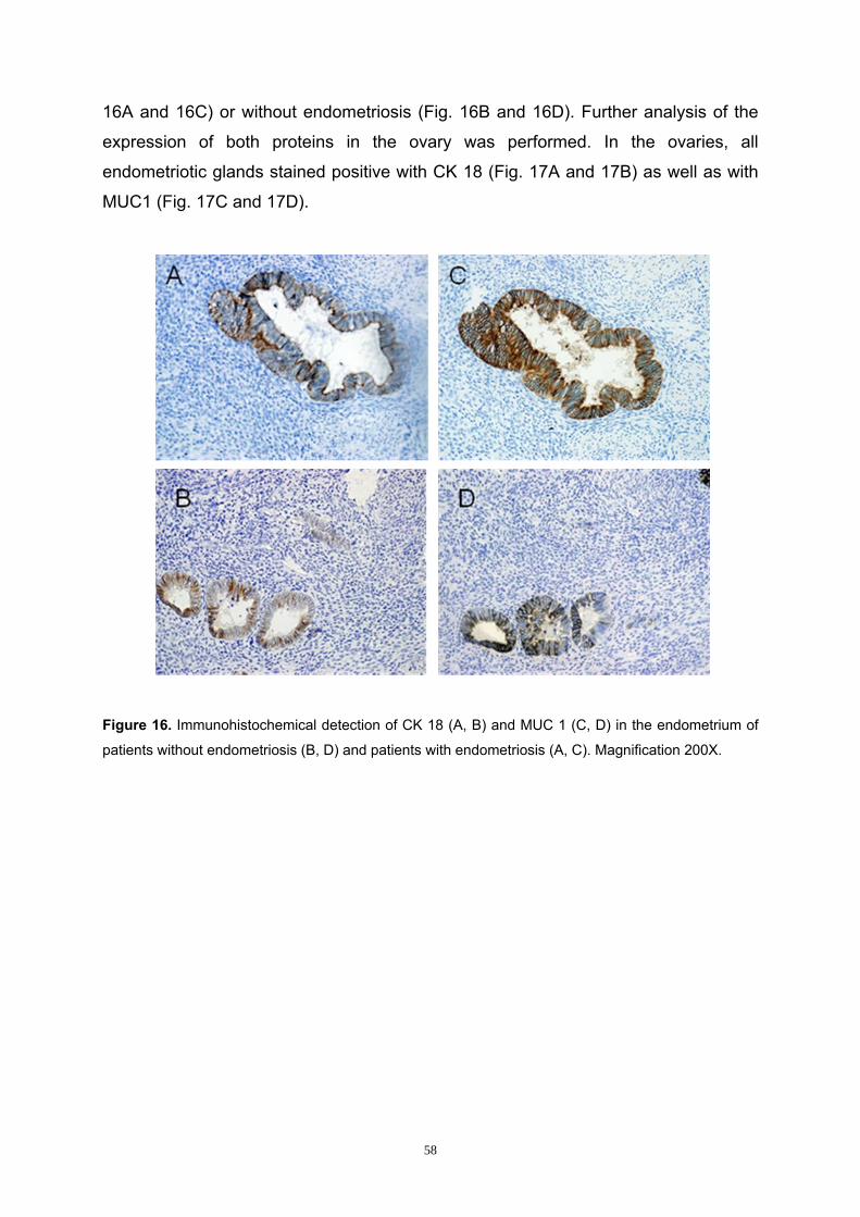
58
16A and 16C) or without endometriosis (Fig. 16B and 16D). Further analysis of the
expression of both proteins in the ovary was performed. In the ovaries, all
endometriotic glands stained positive with CK 18 (Fig. 17A and 17B) as well as with
MUC1 (Fig. 17C and 17D).
Figure 16. Immunohistochemical detection of CK 18 (A, B) and MUC 1 (C, D) in the endometrium of
patients without endometriosis (B, D) and patients with endometriosis (A, C). Magnification 200X.

59
Figure 17. Immunohistochemical detection of CK 18 (A, B) and MUC 1 (C, D) in the ovaries of
patients without endometriosis (B, D) and patients with endometriosis (A, C). Magnification 200X.
In summary, MUC 1 was expressed in all endometrial and endometriotic glands of all
tissues. CK 18 was expressed in endometrial and endometriotic glands of
endometrium and ovary. In addition, the surface epithelium of the ovary expressed
CK 18.
3.2 Influence of TGF-β1 or TGF-β2 on Cell Numbers
In order to find out how TGF-βs affect cell numbers, we treated endometrial,
endometriotic cell lines and primary endometrial stromal cells with TGF-β1 or TGF-β2
(10ng/ml), respectively.
The results showed that TGF-β1 or TGF-β2 reduced cell numbers of both
endometrial and endometriotic cell lines and primary endometrial stromal cells
within 72 hours (Fig. 18). Reduction was higher in endometrial cells in comparison
with endometriotic cells.

60
A
B C Figure 18. Quantification of cell numbers of endometrial cells (A), endometriotic cells (B) and primary
endometrial stromal cells (C) with or without treatment with TGF-βs (10ng/ml). The quantity of
endometrial epithelial cells (HES) was reduced by approximately 39% and that of endometrial stromal
cells (T-HESC) was reduced by approximately 51% (A). Endometriotic epithelial cell numbers (12ZVK) were reduced by approximately 21% and of endometriotic stromal cells (22B) by approximately 13%
(B). Primary endometrial stromal cell numbers were reduced by approximately 19% (C). (**=P<0.01,
*=P<0.05, n=9).
Cel
l num
bers
Ctrl TGF-ß1 (10ng/ml) TGF-ß2 (10ng/ml) Ctrl TGF-ß1 (10ng/ml) TGF-ß2 (10ng/ml)0
500000
1000000
1500000
2000000
2500000
** **
** **
HES T-HESC

61
3.3 Analysis of the Smad Pathway in Endometrial and Endometriotic Cells
The inhibitors LY364947 and SiS3 were used to investigate the role of the Smad-
dependent pathway in cell proliferation. LY364947 and SiS3 inhibitors block the
phosphorylation of Smad2/3 by selectively inhibiting TβR1 or Smad3, respectively.
LY364947 (5 μM) or SiS3 (2 μM) were added two hours before adding the TGF-βs
(10ng/ml). Cells were cultured for 72 hours after stimulation with the TGF-βs. The
appropriate negative controls were also performed.
The results showed that TGF-β1 and TGF-β2 reduced cell numbers in all cell lines
studied (Figs. 19-21). LY364947 was able to block completely the TGF-β-induced
reduction in cell numbers to control levels in all cell lines studied, whereas SiS3 had
only a partial but strongly reducing effect (70%) in blocking the TGF-β-induced
reduction in cell numbers.
B
A
Cel
l num
bers
Ctrl
TGF-ß1
TGF-ß2SiS3
TGF-ß1+
SiS3
TGF-ß2+
SiS3
TßR1 inh
.
TGF-ß1+
TßR1 inh
.
TGF-ß2+
TßR1 inh
.
Cel
l num
bers
Ctrl
TGF-ß1
TGF-ß2SiS3
TGF-ß1+
SiS3
TGF-ß2+
SiS3
TßR1 inh
.
TGF-ß1+
TßR1 inh
.
TGF-ß2+
TßR1 inh
.

62
Figure 19. The TβR1 inhibitor LY364947 completely inhibited the TGF-β1- or TGF-β2-induced
reduction of endometrial cell numbers of both endometrial epithelial cells, HES (A), and endometrial
stromal cells, T-HESC (B). The Smad3 inhibitor SiS3 partially inhibited the TGF-β1- or TGF-β2-
induced reduction of endometrial cell numbers of both endometrial epithelial cells, HES (A), and
endometrial stromal cells, T-HESC (B), by approximately 60% and 80%, respectively (*=P<0.05, n=9).
Figure 20. The TβR1 inhibitor (LY364947) completely inhibited the TGF-β1- or TGF-β2-induced
reduction of endometriotic cell numbers of both endometriotic epithelial cells, 12ZVK (A), and
endometriotic stromal cells, 22B (B). The Smad3 inhibitor SiS3 partially inhibited the TGF-β1- or TGF-
β2-induced reduction of endometriotic cell numbers of both endometriotic epithelial cells, 12ZVK (A), and endometriotic stromal cells, 22B (B), by approximately 70% and 90%, respectively (*=P<0.05,
n=9).
A
B
Cel
l num
bers
Ctrl
TGF-ß1
TGF-ß2SiS3
TGF-ß1+SiS3
TGF-ß2+SiS3
TßR1 inh
.
TGF-ß1+TßR1 i
nh.
TGF-ß2+TßR1 i
nh.

63
Figure 21. The TβR1 inhibitor (LY364947) completely inhibited the TGF-β1- or TGF-β2-induced
reduction of cell numbers, whereas the Smad3 inhibitor SiS3 only partially inhibited the TGF-β1- or
TGF-β2-induced reduction of cell numbers of primary endometrial stromal cells (*=P<0.05, n=9).
These results were comparable to the results obtained with the endometrial stromal cell line, T-HESC
(Fig. 19B).
3.4 Influence of TGF-β1 or TGF-β2 on Smad3 Phosphorylation of Endometrial and Endometriotic Cells in vitro
Smad3 is a member of a transcription factor family involved in mediating signaling of
growth factors such as TGF-βs and activins. Smad3 belongs to a class of SMAD
proteins that are activated via phosphorylation by specific receptors in response to
ligand binding (Derynk et al., 1998). Upon TGF-β binding, Smad3 is phosphorylated
by the TGF-β receptor then dissociates from the TGF-β receptor and interacts with
Smad4 either in the cytoplasm or nucleus. This Smad complex modulate the TGF-β-
induced transcription of genes (Attisano and Wrana, 2002). Smad3 is one of the
standard proteins for studying the Smad-dependent effects of TGF-βs.
In our experiments, endometrial, endometriotic cell lines and primary endometrial
stromal cells were treated with or without TGF-β1 or TGF-β2 (10ng/ml) for 30
minutes and then cell lysates were collected for the phospho-Smad3 ELISA to
quantitate phosphorylated human Smad3.
Treatment of cells with TGF-β1 or TGF-β2, respectively, stimulated phosphorylation
of Smad3 in endometrial, endometriotic cell lines (Fig. 22A and B), and primary
endometrial stromal cells (Fig. 22C). Stimulation was higher in stromal cells
compared to epithelial cells in both endometrial and endometriotic cells.
Primary endometrial stromal cells
Cel
l num
bers
Ctrl
TGF-ß1
TGF-ß2SiS3
TGF-ß1+
SiS3
TGF-ß2+
SiS3
TßR1 inh
.
TGF-ß1+TßR1 i
nh.
TGF-ß2+
TßR1 inh
.0
200000
400000
600000
800000
* *

64
Figure 22. Treatment of cells with TGF-β1 or TGF-β2, respectively, stimulated phosphorylation of
Smad3. Smad3 phosphorylation was 2-fold higher in endometrial stromal cells, T-HESC, compared to
endometrial epithelial cells, HES (A). In endometriotic stromal cells, 22B, 2-fold more Smad3
phosphorylation was observed compared to endometriotic epithelial cells, 12ZVK (B). Interestingly, in
endometrial cells more Smad3 phosphorylation occured compared to endometriotic cells. Primary
endometrial stromal cells showed similar Smad3 phosphorylation levels compared to endometrial
stromal cells, T-HESC (C) (**=P<0.01, *=P<0.05, n=9).
A
B
C
Abs
orba
nce
(450
nm)

65
Analysis of the Smad3 Pathway in Endometrial and Endometriotic Cells The inhibitors LY364947 and SiS3 were used to investigate the role of the Smad-
dependent pathway in Smad3 phosphorylation. The inhibitors block phosphorylation
of Smad2/3 by selectively inhibiting TβR1 and Smad3, respectively. LY364947 (5
μM) or SiS3 (2 μM) was added two hours before adding the TGF-βs (10ng/ml). Cells
were incubated for 30 minutes after stimulation with the TGF-βs. Then cell lysates
were collected to quantitate the amounts of phosphorylated Smad3 by phospho-
Smad3 ELISAs.
The results showed that treatment of cells with TGF-β1 or TGF-β2, respectively,
resulted in Smad3 phosphorylation in all cell lines. The LY364947 and SiS3 inhibitors
were able to block completely the TGF-β-induced Smad3 phosphorylation in all cell
lines studied (Figs. 23-25).

66
Figure 23. Treatment of endometrial cells with TGF-β1 or TGF-β2, respectively, stimulated
phosphorylation of Smad3. Smad3 phosphorylation was 2-fold higher in endometrial stromal cells, T-
HESC (B), compared to endometrial epithelial cells, HES (A). LY364947 and SiS3 inhibitors
completely blocked the TGF-β-induced Smad3 phosphorylation in endometrial cells (**=P<0.01, n=6).
A
B
Abs
orba
nce
(450
nm)
Ctrl
TGF-ß1SiS3
TGF-ß1+SiS3
TßR1 inh.
TGF-ß1+TßR1 i
nh.
TGF-ß2
TGF-ß2+SiS3
TGF-ß2+TßR1 i
nh.0.0
0.2
0.4
0.6
0.8**
**
****
****
pSmad3 levels
HES
Abs
orba
nce
(450
nm)
Ctrl
TGF-ß1SiS3
TGF-ß1+SiS3
TßR1 inh.
TGF-ß1+TßR1 i
nh.
TGF-ß2
TGF-ß2+SiS3
TGF-ß2+TßR1 i
nh.0.0
0.5
1.0
1.5
**
**
****
**
**
pSmad3 levels
T-HESC

67
Figure 24. Treatment of endometriotic cells with TGF-β1 or TGF-β2, respectively, stimulated
phosphorylation of Smad3. Smad3 phosphorylation was 2-fold higher in endometriotic stromal cells,
22B (B), compared to endometriotic epithelial cells, 12ZVK (A). LY364947 and SiS3 inhibitors
completely blocked the TGF-β-induced Smad3 phosphorylation in endometriotic cells (*=P<0.05,
**=P<0.01, n=6).
B
AA
bsor
banc
e (4
50nm
)
Ctrl
TGF-ß1SiS3
TGF-ß1+SiS3
TßR1 inh.
TGF-ß1+TßR1 i
nh.
TGF-ß2
TGF-ß2+SiS3
TGF-ß2+TßR1 i
nh.0.0
0.1
0.2
0.3
0.4**
*
****
****
pSmad3 levels
12ZVK

68
Figure 25. TGF-β1 or TGF-β2, respectively, stimulated phosphorylation of Smad3 in primary
endometrial stromal cells. Smad3 phosphorylation levels were very similar to the endometrial stromal
cells, T-HESC (Fig. 23B). LY364947 and SiS3 inhibitors completely blocked TGF-β-induced Smad3
phosphorylation in primary endometrial cells (**=P<0.01, n=6).
In summary, endometrial cells showed higher phosphorylation levels of Smad3
compared to endometriotic cells upon treatment with TGF-β1 or TGF-β2. Smad3
phosphorylation in endometrial epithelial cells, HES, was 2-fold higher compared to
endometriotic epithelial cells, 12ZVK. Similarly, Smad3 phosphorylation in
endometrial stromal cells, T-HESC, was 2-fold higher compared to endometriotic
stromal cells 22B. Primary endometrial stromal cells showed similar Smad3
phosphorylation compared to endometrial stromal cells, T-HESC. LY364947 and
SiS3 inhibitors completely blocked TGF-β-induced Smad3 phosphorylation in all cell
lines studied.
Abs
orba
nce
(450
nm)
Ctrl
TGF-ß1SiS3
TGF-ß1+SiS3
TßR1 inh.
TGF-ß1+TßR1 i
nh.
TGF-ß2
TGF-ß2+SiS3
TGF-ß2+TßR1 i
nh.

69
3.5 Influence of TGF-β1 or TGF-β2 on Apoptosis of Endometrial and Endometriotic Cells in vitro
Apoptosis is characterized by activation of an endogenous cell suicide programme
which involves chromatin condensation, nuclear fragmentation, cell shrinkage,
membrane blebbing and dissolution of cells into apoptotic bodies which are rapidly
phagocytosed by neighbouring cells or resident macrophages (Ruaidhri et al., 1998).
In our experiments, we wanted to investigate whether or not TGF-βs have an
apoptotic effect on human endometrial and endometriotic cells and if yes, which
apoptotic pathways are involved.
Endometrial, endometriotic cell lines and primary endometrial stromal cells were
treated with or without TGF-β1 or TGF-β2 (10ng/ml), respectively, for 24 hours and
then apoptosis experiments were carried out by use of ELISAs. Staurosporine
(0.1μM) was included in the assay as a positive control. Staurosporine is a potent
inhibitor of protein kinase C, most other kinases, and topoisomerase II by blocking
transfer of phosphodiester bonds from DNA to the active site of tyrosine hence
inducing apoptosis (Seynaeve et al., 1994).
The apoptotic parameters investigated included quantification of phosphatidylserine
on the outer membrane surface, of the inner mitochondrial membrane potential, and
of Cysteinyl-asparate-specific proteases 3/7 (Caspase 3/7) proteins. The three
parameters are important because they can be used to monitor necrosis and
apoptosis. In apoptotic cells, phosphatidylserine which is usually located on the inner
side of the cell membrane is translocated in apoptotic cells onto the outer surface of
the cell membrane due to loss of cell symmetry (Sims and Wiedmer, 2001). The
translocated phosphatidylserine can be quantified by ELISA. The inner mitochondrial
membrane integrity is intact but once the cell undergoes apoptosis, the integrity of
the inner mitochondrial potential is lost causing release of some ions among them
cytochrome C which initiates caspase cascades leading to apoptosis (Kroemer et al.,
2007).
The results showed an increase of phosphatidylserine, a decrease of the inner
mitochondrial membrane potential and an increase of Caspase 3/7 levels upon
treatment of cells with TGF-β1 or TGF-β2 (10ng/ml), respectively (Figs. 26-30).

70
Figure 26. Treatment with TGF-β1 or TGF-β2, respectively, induced apoptosis in endometrial
epithelial cells, HES. There was an increase in the amount of translocated phosphatidylserine (A), a
decrease of the mitochondrial membrane potential (B), and an increase of the amount of Caspase 3/7
(C). Staurosporine strongly induced apoptosis in HES cells (*=P<0.05, **=P<0.01, ***=P<0.001, n=6).
RFU; relative fluorescence units.
A
B
C
RFU
(540
nm)
RFU
(540
nm)
Ctrl TGF-ß1 (10ng/ml) TGF-ß2 (10ng/ml) Staurosporine0
5000
10000
15000
20000
*
Mitochondrial membrane potential
**
RFU
(350
nm)
Ctrl TGF-ß1 (10ng/ml) TGF-ß2 (10ng/ml) Staurosporine0
10000
20000
30000
40000
50000
** **
CASPASE 3/7 levels
***

71
Figure 27. Treatment with TGF-β1 or TGF-β2, respectively, induced apoptosis in endometrial stromal
cells, T-HESC. There was an increase in the amount of translocated phosphatidylserine (A), a
decrease of the mitochondrial membrane potential (B), and an increase of the amount of Caspase 3/7
(C). Staurosporine strongly induced apoptosis in T-HESC cells (*=P<0.05, **=P<0.01, ***=P<0.001,
n=6). RFU; relative fluorescence units.
A
B
C
RFU
(540
nm)
RFU
(540
nm)
Ctrl TGF-ß1 (10ng/ml) TGF-ß2 (10ng/ml) Staurosporine0
10000
20000
30000
40000
Mitochondrial membrane potential
***
****
RFU
(350
nm)
Ctrl TGF-ß1 (10ng/ml) TGF-ß2 (10ng/ml) Staurosporine0
10000
20000
30000
40000
50000 CASPASE 3/7 levels
***
*
**

72
.
Figure 28. Treatment with TGF-β1 or TGF-β2, respectively, induced apoptosis in endometriotic
epithelial cells, 12ZVK. There was an increase in the amount of translocated phosphatidylserine (A), a
decrease of the mitochondrial membrane potential (B), and an increase of the amount of Caspase 3/7
(C). Staurosporine strongly induced apoptosis in 12ZVK cells (**=P<0.01, ***=P<0.001, n=6). RFU;
relative fluorescence units.
B
C
A
RFU
(540
nm)
RFU
(540
nm)
Ctrl TGF-ß1 (10ng/ml) TGF-ß2 (10ng/ml) Staurosporine0
10000
20000
30000
40000 Mitochondrial membrane potential
**
*
RFU
(350
nm)
Ctrl TGF-ß1 (10ng/ml) TGF-ß2 (10ng/ml) Staurosporine0
10000
20000
30000
40000 CASPASE 3/7 levels
***
** **

73
Figure 29. Treatment with TGF-β1 or TGF-β2, respectively, induced apoptosis in endometriotic
stromal cells, 22B. There was an increase in the amount of translocated phosphatidylserine (A), a
decrease of the mitochondrial membrane potential (B), and an increase of the amount of Caspase 3/7
(C). Staurosporine strongly induced apoptosis in 22B cells (**=P<0.01, ***=P<0.001, n=6). RFU;
relative fluorescence units.
C
B
A
RFU
(540
nm)
RFU
(540
nm)
Ctrl TGF-ß1 (10ng/ml) TGF-ß2 (10ng/ml) Staurosporine0
5000
10000
15000
20000
25000 Mitochondrial membrane potential
**
***
**
RFU
(350
nm)
Ctrl TGF-ß1 (10ng/ml) TGF-ß2 (10ng/ml) Staurosporine0
10000
20000
30000 CASPASE 3/7 levels
**
*****

74
Figure 30. Treatment with TGF-β1 or TGF-β2, respectively, induced apoptosis in primary endometrial
stromal cells. There was an increase in the amount of translocated phosphatidylserine (A), a decrease
of the mitochondrial membrane potential (B), and an increase of the amount of Caspase 3/7 (C). Staurosporine strongly induced apoptosis in primary endometrial cells (**=P<0.01, ***=P<0.001, n=6).
RFU; relative fluorescence units.
A
C
B
RFU
(540
nm)
RFU
(540
nm)
Ctrl TGF-ß1 (10ng/ml) TGF-ß2 (10ng/ml) Staurosporine0
10000
20000
30000
40000 Mitochondrial membrane potential
**
***
**
RFU
(350
nm)
Ctrl TGF-ß1 (10ng/ml) TGF-ß2 (10ng/ml) Staurosporine0
10000
20000
30000
40000 CASPASE 3/7 levels
**
***
**

75
In summary, either TGF-β1 or TGF-β2 induced apoptosis in all cell lines studied.
There was an increase in the amount of phosphatidylserine translocated to the outer
surface of the cells, an increase of Caspase 3/7 levels and a decrease of the inner
mitochondrial membrane potential. There were no significant differences between
endometrial and endometriotic cell lines. Furthermore, the apoptotic effects of
TGF-β1 or TGF-β2 on primary endometrial stromal cells and endometrial stromal
cells were similar.
Analysis of the Smad Pathway in Apoptosis in Endometrial and Endometriotic Cells The inhibitors LY364947 and SiS3 were used to investigate the role of the Smad-
dependent pathway in apoptosis. The inhibitors block phosphorylation of Smad2/3 by
selectively inhibiting TβR1 and Smad3 phosphorylation, respectively.
LY364947 (5 μM) and SiS3 (2 μM) were added two hours before adding the TGF-βs
(10ng/ml) or staurosporine (0.1 μM), respectively, for 24 hours and then apoptosis
experiments were carried out by ELISAs. The apoptotic parameters tested included
quantification of phosphatidylserine, the mitochondrial membrane potential and
Caspase 3/7 levels.
The results showed that treatment of cells with TGF-β1 or TGF-β2, respectively,
increased phosphatidylserine on the outer surface of the cell membrane, decreased
the inner mitochondrial membrane potential and increased Caspase 3/7 in all cells.
LY364947 blocked TGF-β-mediated apoptosis in all cell lines completely, whereas
SiS3 blocked TGF-β-mediated apoptosis in all cell lines only partially (Fig. 31).

76
A
B
RFU
(540
nm)
Ctrl
TGF-ß1SiS3
TGF-ß1+SiS3
TßR1 inh
.
TGF-ß1+TßR1 i
nh.
TGF-ß2
TGF-ß2+SiS3
TGF-ß2+TßR1 i
nh.
Stauros
porin
e
77
Figure 31. Treatment of endometrial epithelial HES cells with TGF-β1 or TGF-β2, respectively,
induced apoptosis. There was an increase in the amount of translocated phosphatidylserine (A) (**=P<0.01, ***=P<0.001, n=6), a decrease of the mitochondrial membrane potential (B) (*=P<0.05,
**=P<0.01, n=6), and an increase of the amount of Caspase 3/7 (C) (*=P<0.05, **=P<0.01,
***=P<0.001, n=6). LY364947 blocked TGF-β-mediated apoptosis in HES cells completely, whereas
SiS3 blocked TGF-β-mediated apoptosis in cells only partially (A-C). Similar results were observed in
the other cell lines (data not shown). RFU; relative fluorescence units.
3.6 Influence of TGF-β1 or TGF-β2 on Plasminogen Activator Inhibitor-1 Secretion by Endometrial and Endometriotic Cells in vitro
Plasminogen Activator Inhibitor-1 (PAI-1) belongs to the serine protease inhibitor
family (SERPIN) and is a modifier of pathways that impact proliferative/migratory
events (Czekay et al., 2003), PAI-1 is one of the gold standards for studying the
effects of TGF-βs on gene expression. Moreover, PAI-1 disrupts integrin-containing
adhesions and actin stress fibers which might result in apoptosis or reduced cell
numbers (Czekay et al., 2003).
C
RFU
(350
nm)
Ctrl
TGF-ß1SiS3
TGF-ß1+
SiS3
TßR1 inh
.
TGF-ß1+TßR1 i
nh.
TGF-ß2
TGF-ß2+
SiS3
TGF-ß2+TßR1 i
nh.
Stauros
porin
e
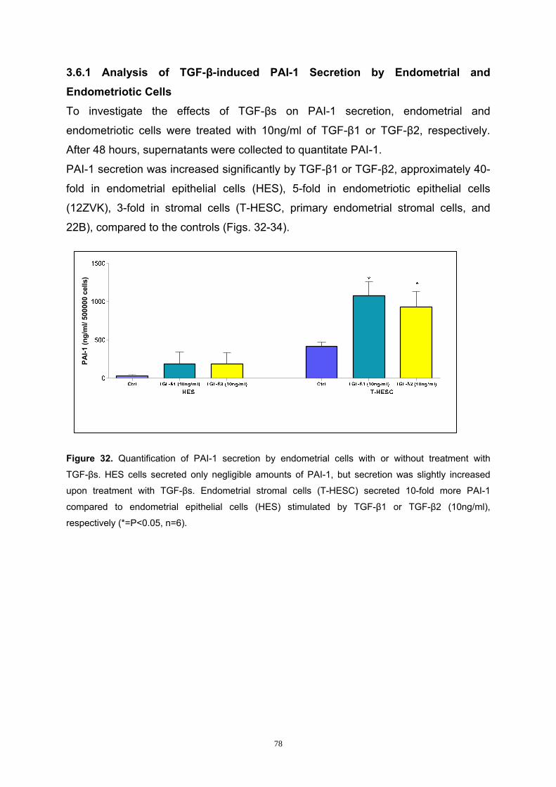
78
3.6.1 Analysis of TGF-β-induced PAI-1 Secretion by Endometrial and Endometriotic Cells To investigate the effects of TGF-βs on PAI-1 secretion, endometrial and
endometriotic cells were treated with 10ng/ml of TGF-β1 or TGF-β2, respectively.
After 48 hours, supernatants were collected to quantitate PAI-1.
PAI-1 secretion was increased significantly by TGF-β1 or TGF-β2, approximately 40-
fold in endometrial epithelial cells (HES), 5-fold in endometriotic epithelial cells
(12ZVK), 3-fold in stromal cells (T-HESC, primary endometrial stromal cells, and
22B), compared to the controls (Figs. 32-34).
Figure 32. Quantification of PAI-1 secretion by endometrial cells with or without treatment with
TGF-βs. HES cells secreted only negligible amounts of PAI-1, but secretion was slightly increased
upon treatment with TGF-βs. Endometrial stromal cells (T-HESC) secreted 10-fold more PAI-1
compared to endometrial epithelial cells (HES) stimulated by TGF-β1 or TGF-β2 (10ng/ml),
respectively (*=P<0.05, n=6).
PAI-1
(ng/
ml/
5000
00 c
ells
)
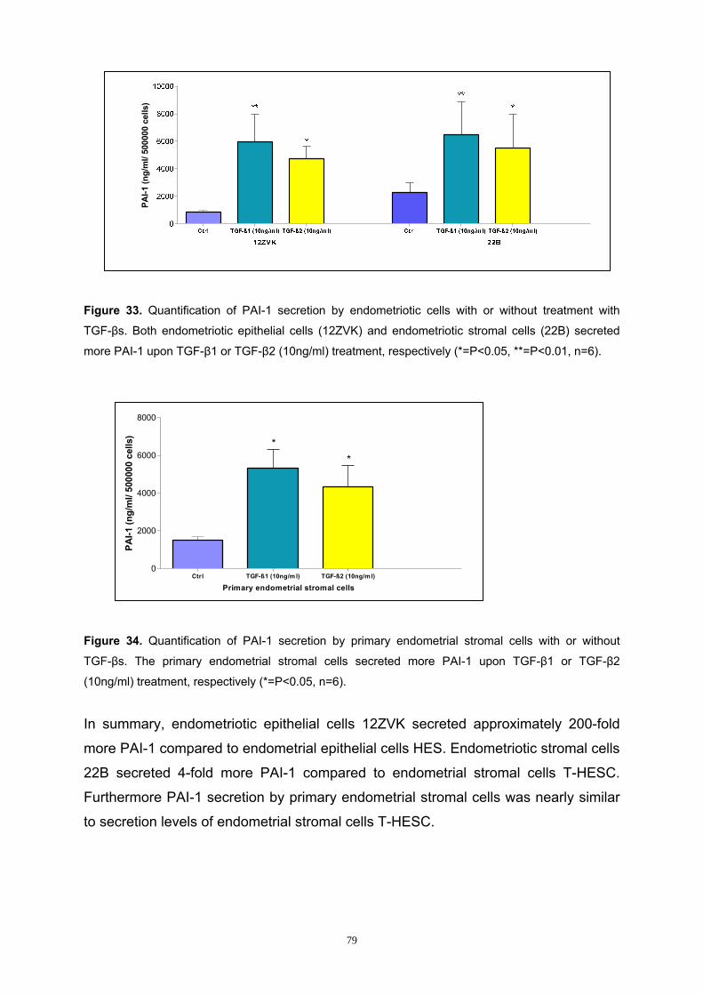
79
Figure 33. Quantification of PAI-1 secretion by endometriotic cells with or without treatment with
TGF-βs. Both endometriotic epithelial cells (12ZVK) and endometriotic stromal cells (22B) secreted
more PAI-1 upon TGF-β1 or TGF-β2 (10ng/ml) treatment, respectively (*=P<0.05, **=P<0.01, n=6).
Figure 34. Quantification of PAI-1 secretion by primary endometrial stromal cells with or without
TGF-βs. The primary endometrial stromal cells secreted more PAI-1 upon TGF-β1 or TGF-β2
(10ng/ml) treatment, respectively (*=P<0.05, n=6).
In summary, endometriotic epithelial cells 12ZVK secreted approximately 200-fold
more PAI-1 compared to endometrial epithelial cells HES. Endometriotic stromal cells
22B secreted 4-fold more PAI-1 compared to endometrial stromal cells T-HESC.
Furthermore PAI-1 secretion by primary endometrial stromal cells was nearly similar
to secretion levels of endometrial stromal cells T-HESC.
PAI-1
(ng/
ml/
5000
00 c
ells
)
PAI-1
(ng/
ml/
5000
00 c
ells
)
Ctrl TGF-ß1 (10ng/m l) TGF-ß2 (10ng/ml)0
2000
4000
6000
8000
**
Primary endometrial stromal cells

80
3.6.2 Analysis of the Smad Pathway in PAI-1 Secretion in Endometrial and Endometriotic Cells The inhibitors LY364947 and SiS3 were used to investigate the role of the Smad-
dependent pathway in PAI-1 secretion. The inhibitors block phosphorylation of
Smad2/3 by selectively inhibiting TβR1 and Smad3, respectively. LY364947 (5 μM)
and SiS3 (2 μM) were added two hours before adding the TGF-βs (10ng/ml). Cells
were cultured for 48 hours after stimulation with the TGF-βs. Then supernatants were
collected for quantification of PAI-1 secretion by PAI-1 ELISA.
The results showed that TGF-β1 and TGF-β2 both increased PAI-1 secretion in all
cell lines studied. LY364947 blocked TGF-β-mediated PAI-1 secretion in cells
completely to control levels, whereas SiS3 blocked TGF-β-mediated PAI-1 secretion
in all cells only partially (Fig. 35).
A
PAI-1
(ng/
ml/
5000
00 c
ells
)
Ctrl
TGF-ß1SiS3
TGF-ß1+SiS3
TßR1 inh
.
TGF-ß1+
TßR1 inh
.
TGF-ß2
TGF-ß2+SiS3
TGF-ß2+
TßR1 inh
.

81
Figure 35. Treatment of cells with TGF-β1 or TGF-β2 (10ng/ml), respectively, induced PAI-1 secretion
in endometrial epithelial cells, HES (A), and endometrial stromal cells, T-HESC (B). LY364947
blocked TGF-β-induced PAI-1 secretion of all cells completely to control levels while SiS3 blocked
TGF-β-induced PAI-1 secretion of cells in a range of 40-70%. Similar results were observed with the
other cell lines (data not shown).
3.6.3 Analysis of the BMP Pathway in PAI-1 Secretion The BMP inhibitor LDN-193189 was used to study the role of the BMP pathway in
PAI-1 secretion. The BMP inhibitor selectively inhibits the BMP type 1 receptors
ALK2, ALK3 and ALK 6 and thus blocks BMP-mediated phosphorylation of
Smad1/5/8.
The BMP inhibitor (5μM) was added two hours before adding the TGF-βs (10ng/ml).
Cells were cultured for 48 hours after stimulation with the TGF-βs. Then supernatants
were collected for quantification of PAI-1 secretion.
The results showed that TGF-β1 or TGF-β2, respectively, increased PAI-1 secretion
in all cell lines studied. The BMP inhibitor demonstrated a complete decrease (100%)
of TGF-β1 or TGF-β2 induced-PAI-1 secretion in all cell lines studied (Figs. 36-38).
B
PAI-1
(ng/
ml/
5000
00 c
ells
)
Ctrl
TGF-ß1SiS3
TGF-ß1+
SiS3
TßR1 inh
.
TGF-ß1+TßR1 i
nh.
TGF-ß2
TGF-ß2+
SiS3
TGF-ß2+TßR1 i
nh.
0
1000
2000
3000
4000
T-HESC
*** **
******
*****
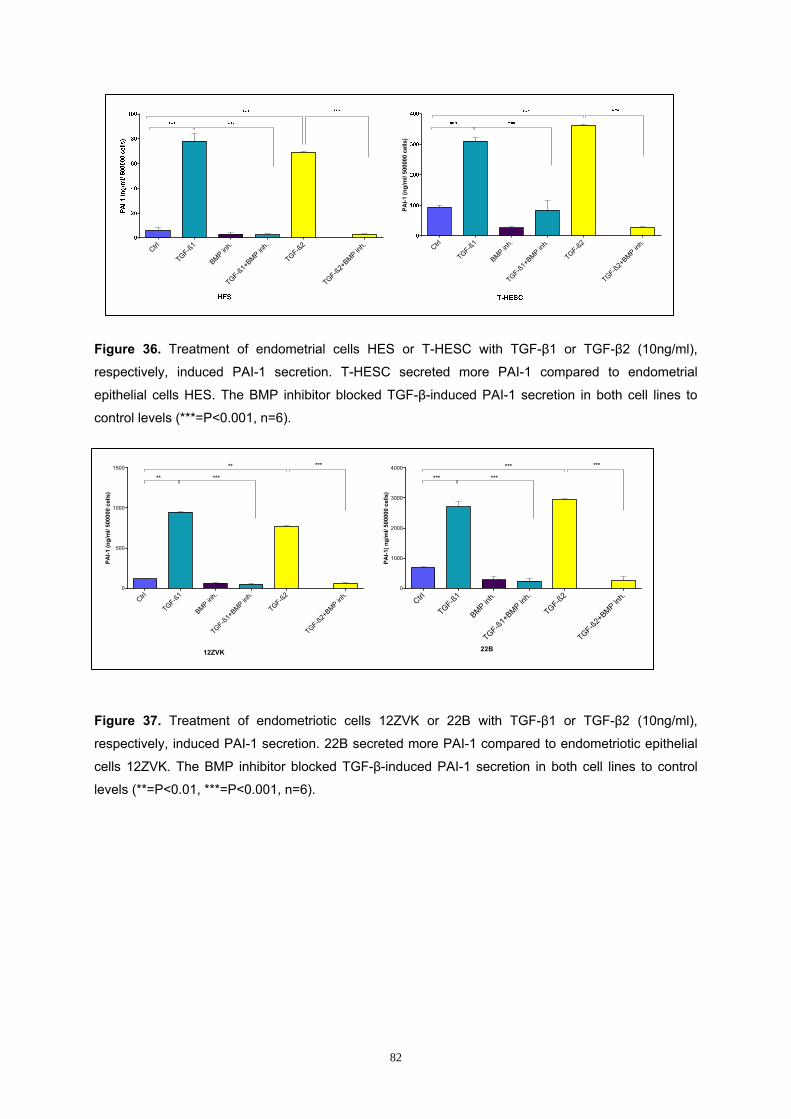
82
Figure 36. Treatment of endometrial cells HES or T-HESC with TGF-β1 or TGF-β2 (10ng/ml),
respectively, induced PAI-1 secretion. T-HESC secreted more PAI-1 compared to endometrial
epithelial cells HES. The BMP inhibitor blocked TGF-β-induced PAI-1 secretion in both cell lines to
control levels (***=P<0.001, n=6).
Figure 37. Treatment of endometriotic cells 12ZVK or 22B with TGF-β1 or TGF-β2 (10ng/ml),
respectively, induced PAI-1 secretion. 22B secreted more PAI-1 compared to endometriotic epithelial
cells 12ZVK. The BMP inhibitor blocked TGF-β-induced PAI-1 secretion in both cell lines to control
levels (**=P<0.01, ***=P<0.001, n=6).
PAI-1
(ng/
ml/
5000
00 c
ells
)
Ctrl
TGF-ß1
BMP inh.
TGF-ß1+BMP in
h.
TGF-ß2
TGF-ß2+BMP in
h.0
500
1000
1500
**
**
***
***
12ZVK
PAI-1
( ng/
ml/
5000
00 c
ells
)
Ctrl
TGF-ß1
BMP inh.
TGF-ß1+BMP in
h.
TGF-ß2
TGF-ß2+BMP in
h.0
1000
2000
3000
4000
***
***
***
***
22B
Ctrl
TGF-ß1
BMP inh.
TGF-ß1+
BMP inh.
TGF-ß2
TGF-ß2+
BMP inh.
PAI-1
(ng/
ml/
5000
00 c
ells
)
Ctrl
TGF-ß1
BMP inh.
TGF-ß1+
BMP inh.
TGF-ß2
TGF-ß2+
BMP inh.

83
Figure 38. Treatment of primary endometrial stromal cells with TGF-β1 or TGF-β2, (10ng/ml)
respectively, induced PAI-1 secretion. The BMP inhibitor blocked TGF-β-induced PAI-1 secretion of
primary endometrial cells to control levels (**=P<0.01, ***=P<0.001, n=6).
3.6.4 Analysis of BMP Receptors in PAI-1 Secretion Activin Receptor-Like Kinase (ALK), namely ALK-2, ALK-3 and ALK-6, inhibitors were
used separately to study the specific BMP receptor involved in the complete
decrease of TGF-β1 or TGF-β2 induced-PAI-1 secretion in cells as shown by the
BMP inhibitor LDN-193189 in section 3.6.3.
The ALK-2 (10μM), ALK-3 (4μM), ALK-6 (6μM) inhibitors and IgG1 (2μM) were
added two hours before adding the TGF-βs (10ng/ml). Cells were cultured for 48
hours after stimulation with the TGF-βs. Then supernatants were collected for
quantification of PAI-1 secretion.
The results showed that TGF-β1 or TGF-β2 increased PAI-1 secretion of all cell lines
studied. The ALK-2 inhibitor demonstrated a complete decrease (100%) of TGF-β1
or TGF-β2 induced-PAI-1 secretion in all cell lines, whereas the ALK-3 and ALK-6
inhibitors demonstrated only a partial effect of 40% and 25%, respectively. The IgG1
(control) had no affect on TGF-β1 or TGF-β2 induced-PAI-1 secretion (Figs. 39-41).
PAI-1
(ng/
ml/
5000
00 c
ells
)
Ctrl
TGF-ß1
BMP inh.
TGF-ß1+BMP in
h.
TGF-ß2
TGF-ß2+BMP in
h.0
100
200
300
400
**
**
***
***
Primary endometrial stromal cells
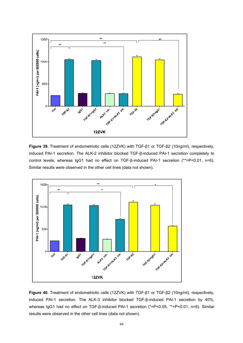
84
Figure 39. Treatment of endometriotic cells (12ZVK) with TGF-β1 or TGF-β2 (10ng/ml), respectively,
induced PAI-1 secretion. The ALK-2 inhibitor blocked TGF-β-induced PAI-1 secretion completely to
control levels, whereas IgG1 had no effect on TGF-β-induced PAI-1 secretion (**=P<0.01, n=6).
Similar results were observed in the other cell lines (data not shown).
Figure 40. Treatment of endometriotic cells (12ZVK) with TGF-β1 or TGF-β2 (10ng/ml), respectively,
induced PAI-1 secretion. The ALK-3 inhibitor blocked TGF-β-induced PAI-1 secretion by 40%,
whereas IgG1 had no effect on TGF-β-induced PAI-1 secretion (*=P<0.05, **=P<0.01, n=6). Similar
results were observed in the other cell lines (data not shown).
PAI-1
(ng/
ml)
per 5
0000
0 ce
lls)
Ctrl
TGF-ß1IgG1
TGF-ß1+IgG1
ALK3 inh.
TGF-ß1+ALK3
inh
TGF-ß2
TGF-ß2+IgG1
TGF-ß2+ALK3
inh

85
Figure 41. Treatment of endometriotic cells (12ZVK) with TGF-β1 or TGF-β2 (10ng/ml), respectively,
induced PAI-1 secretion. The ALK-6 inhibitor blocked TGF-β-induced PAI-1 secretion by 25%,
whereas IgG1 had no effect on TGF-β-induced PAI-1 secretion (**=P<0.05, **=P<0.01, n=6). Similar
results were observed in the other cell lines (data not shown).
3.7 Influence of TGF-β1 or TGF-β2 on Inhibin B Secretion by Endometrial and Endometriotic Cells in vitro
Inhibin B belongs to the TGF-β superfamily regulates reproduction. Recent studies
have shown that inhibins might be involved in tumour suppression (Gail et al., 2001).
Inhibin B secretion is one possible marker for the functionality of the BMP pathway.
Thus, by treating endometrial or endometriotic cells with TGF-β1 or TGF-β2, it might
be possible to elucidate whether TGF-βs might also use the BMP pathway-
dependent Smad1/5/8.
To investigate the effects of TGF-βs on inhibin B secretion, endometrial and
endometriotic cells were treated with TGF-β1 or TGF-β2 (10 ng/ml), respectively, for
48 hours and supernatants were collected for the inhibin B ELISA.
The results showed that TGF-β1 or TGF-β2 reduced inhibin B secretion of HES cells
and primary endometrial stromal cells while inhibin B secretion was not affected by
TGF-β1 or TGF-β2 in the other cell lines studied (Figs. 42-44).
PAI-1
(ng/
ml)
per 5
0000
0 ce
lls)
Ctrl
TGF-ß1IgG1
TGF-ß1+IgG1
ALK6 inh.
TGF-ß1+ALK6
inh
TGF-ß2
TGF-ß2+IgG1
TGF-ß2+ALK6
inh

86
Figure 42. Treatment of endometrial epithelial cells (HES) with TGF-β1 or TGF-β2 (10ng/ml),
respectively, reduced inhibin B secretion compared to control. There were no effects on inhibin B
levels in T-HESC cells (*=P<0.05, **=P<0.01 n=6).
Figure 43. Treatment of endometriotic cells (12ZVK and 22B) with TGF-β1 or TGF-β2 (10ng/ml),
respectively, did not show any effect on inhibin B secretion.
Figure 44. Treatment of primary endometrial stromal cells with TGF-β1 or TGF-β2 (10ng/ml),
respectively reduced inhibin B secretion compared to the control (*=P<0.05, n=6).
Inhi
bin
B (p
g/m
l) pe
r 500
000
cells
Ctrl TGF-ß1 (10ng/ml) TGF-ß2 (10ng/ml) Ctrl TGF-ß1 (10ng/ml) TGF-ß2 (10ng/m l)0
200
400
600
12ZVK 22B
Inhi
bin
B (p
g/m
l) pe
r 500
000
cells

87
3.8 Influence of TGF-β1 or TGF-β2 on TβRIII Expression of Endometrial and Endometriotic Cells in vitro
High affinity binding of TGF-β2 to Transforming growth factor beta receptor II (TβRII)
is best via TβRIII (Lopez et al., 1993). However, TGF-β1 as well as TGF-β3 also bind
to TβRIII.
In our experiments, 24-well plates were coated with TGF-β1 or TGF-β2 (10ng/ml),
respectively, and incubated for 48 hours. The cells were fixed with ice-cold 4%
paraformaldehyde (PFA). A high affinity TβRIII antibody (R&D) was used as a
primary antibody. An anti-goat peroxidase (DAKO) was used as a secondary
antibody. TMB substrate (Calbiochem, Germany) was added followed by addition of
0.18N H2SO4 to stop the reaction. Absorbance was read at 450nm.
Treatment of cells with TGF-β1 or TGF-β2 increased expression of TβRIII on stromal
cells whereas epithelial cells showed little or no increase (Figs. 45-47).
Figure 45. Treatment of endometrial cells with TGF-β1 or TGF-β2 (10ng/ml), respectively, increased
expression of TβRIII only on T-HESC cells with no effect on endometrial epithelial cells HES
(*=P<0.05, n=6).
OD
(450
nm) p
er 5
0000
0 ce
lls

88
Figure 46. Treatment of endometriotic cells with TGF-β1 or TGF-β2 (10ng/ml), respectively, increased
expression of TβRIII only on 22B cells with no effect on endometriotic epithelial cells 12ZVK
(*=P<0.05, n=6).
Figure 47. Treatment of primary endometrial stromal cells with TGF-β1 or TGF-β2 (10ng/ml)
increased expression of TβRIII on the cells (*=P<0.05, n=6). The TβRIII expression was similar to the
endometrial stromal cell line T-HESC (Fig. 45).
3.9 Influence of TGF-β1 or TGF-β2 on Secretion of TGF-β2 by Endometrial and Endometriotic Cells in vitro
To investigate the influence of TGF-β1 or TGF-β2 on secretion of TGF-β2 by
endometrial and endometriotic cells, we quantitated TGF-β2 secretion by cells.
The endometrial and endometriotic cells were treated with TGF-β1 or TGF-β2
(10ng/ml), respectively. Then cell supernatants were collected after 48 hours upon
TGF-β1 or TGF-β2 treatment to quantitate TGF-β1 or TGF-β2.
Treatment of cells with TGF-β1 or TGF-β2 increased secretion of TGF-β2 in
TGF-β1-treated cells. No effect on TGF-β2 levels was observed in controls and TGF-
β2-treated cells (Figs. 48-50).
OD
(450
nm) p
er 5
0000
0 ce
lls
OD
(450
nm) p
er 5
0000
0 ce
lls
Ctrl TGF-ß1 (10ng/m l) TGF-ß2 (10ng/m l)0.0
0.5
1.0
1.5
2.0
* *
Primary endometrial stromal cells

89
Figure 48. Treatment of endometrial cells (HES and T-HESC) with TGF-β1 or TGF-β2 (10ng/ml),
respectively, increased secretion of TGF-β2 in TGF-β1-treated cells. Endometrial stromal cells, T-
HESC, secreted more TGF-β2 compared to endometrial epithelial cells, HES. No effect on TGF-β2
levels was observed in controls and TGF-β2-treated endometrial cells (*=P<0.05, **=P<0.01, n=6).
Figure 49. Treatment of endometriotic cells (12ZVK and 22B) with TGF-β1 or TGF-β2 (10ng/ml),
respectively, increased secretion of TGF-β2 in TGF-β1-treated cells. Endometriotic stromal cells, 22B,
secreted more TGF-β2 compared to endometriotic epithelial cells, 12ZVK. No effect on TGF-β2 levels
was observed in controls and TGF-β2-treated cells in endometriotic cells (*=P<0.05, **=P<0.01, n=6).
TGF-
ß2 (p
g/m
l)pe
r 500
000
cells
TGF-
ß2 (p
g/m
l)pe
r 500
000
cells

90
Figure 50. Treatment of primary endometrial stromal cells with TGF-β1 or TGF-β2 (10ng/ml),
respectively, increased secretion of TGF-β2 in TGF-β1-treated endometrial stromal cells. No effect on
TGF-β2 levels was observed in controls and TGF-β2-treated cells (**=P<0.01, n=6).
In summary, in all cell lines studied, there was increased TGF-β2 secretion in
TGF-β1-treated cells. No effect was observed in control cells and cells treated with
TGF-β2. Endometriotic epithelial cells, 12ZVK, secreted 2-fold more TGF-β2
compared to endometrial epithelial cells, HES, after stimulation with TGF-β1.
Endometriotic stromal cells, 22B, secreted 3-fold more TGF-β2 compared to
endometrial stromal cells T-HESC. TGF-β1-treated T-HESC as well as TGF-β-
treated primary endometrial stromal cells secreted almost the same amount of TGF-
β2.
3.10 Influence of TGF-β1 or TGF-β2 on Secretion of TGF-β1 by Endometrial and Endometriotic Cells in vitro
To investigate the influence of TGF-β1 or TGF-β2 on secretion of TGF-β1 by
endometrial and endometriotic cells, we quantitated TGF-β1 secretion by cells.
The endometrial and endometriotic cells were treated with TGF-β1 or TGF-β2
(10ng/ml), respectively. Then cell supernatants were collected after 48 hours upon
TGF-β1 or TGF-β2 treatment to quantitate TGF-β1.
Treatment of cells with TGF-β1 or TGF-β2 increased secretion of TGF-β1 in
TGF-β1-treated cells. No effect on TGF-β1 levels was observed in controls and TGF-
β2-treated cells (Figs. 51-53).
TGF-
ß2 (p
g/m
l)pe
r 500
000
cells

91
Figure 51. Treatment of endometrial cells (HES and T-HESC) with TGF-β1 or TGF-β2 (10ng/ml),
respectively, increased secretion of TGF-β1 in TGF-β1-treated cells. Endometrial stromal cells, T-
HESC, secreted 2-fold more TGF-β1 compared to endometrial epithelial cells HES. No effect on TGF-
β1 levels was observed in controls and TGF-β2-treated cells in endometrial cells (*=P<0.05, n=6).
Figure 52. Treatment of endometriotic cells (12ZVK and 22B) with TGF-β1 or TGF-β2 (10ng/ml),
respectively, increased secretion of TGF-β1 in TGF-β1-treated cells. Endometriotic stromal cells, 22B,
secreted 1.5-fold more TGF-β1 compared to endometriotic epithelial cells 12ZVK. No effect on TGF-
β1 levels was observed in controls and TGF-β2-treated endometriotic cells (*=P<0.05, n=6).
TGF-
ß1 (p
g/m
l)pe
r 500
000
cells
TGF-
ß1 (p
g/m
l)pe
r 500
000
cells

92
Figure 53. Treatment of primary endometrial stromal cells with TGF-β1 or TGF-β2 (10ng/ml),
respectively, increased secretion of TGF-β1 in TGF-β1-treated endometrial stromal cells. No effect on
TGF-β1 levels was observed in controls and TGF-β2-treated cells (*=P<0.01, n=6).
3.11 Influence of TGF-β1 or TGF-β2 on Interaction of TβRI, TβRII and TβRIII Receptors on Endometrial and Endometriotic Cells in vitro (Signalosome analysis)
To quantify and analyze the interaction of the TβRI, TβRII and TβRIII receptors on
primary endometrial cells and other cell lines, we used the Proximity Ligation Assay
(PLA). PLA enables detection, visualization and quantification of protein interactions
in tissue and cell samples. The PLA principle is shown in Fig. 54
TGF-
ß1 (p
g/m
l)pe
r 500
000
cells

93
Figure 54. Principle of Duolink In Situ PLA assay (adapted from http//www.Olink.com)
The resulting fluorescence in each single-molecule amplification product is visible as
a distinct spot under a fluorescence microscope (Soderberg et al., 2006; Jurvious et
al., 2007).
In our experiments, we applied in situ PLA (Duolink) with a combination of antibodies
that allows the detection of TβRII/TβRIII and TβRI/TβRII interaction only when these
are in close proximity upon treatment of cells with TGF-β1 or TGF-β2. The details of
the antibodies used are indicated in Table 1.
Step 1: A pair of primary antibodies raised in different species
bind to the target antigens of interest
Step 2: A pair of species-specific secondary antibodies
called, PLA probes (PLUS and MINUS), bind to their respective primary antibody
Step 3: Two oligonucleotides are joined by a ligase to form a circular molecule only when the two PLA probes are at a close proximity
Step 4: A polymerase amplifies the circle forming several
hundred-fold replication of DNA circle
Step 5: Labeled complementary oligonucleotide probes bind
the synthesized DNA strand allowing the product to be visualized with a fluorescence microscope

94
For the PLA assay, 20,000 cells on 8-well chamber slides were treated with or
without TGF-β1 or TGF-β2 for 24 hours and afterwards fixed and permeabilized. A
pair of primary antibodies against TβRII and TβRIII or TβRI and TβRII was used to
detect the complexes (Table 1). Controls were performed by omissions of the primary
antibodies. Phase contrast images showed the exact localization of the signals.
Images were obtained with the inverse microscope FSX 100 (Olympus) using the
Olympus FSX-BSW software. Images were processed with Adobe Photoshop. The
quantity of PLA signals was counted and the average number of spots per cell was
presented graphically.
The results showed interaction of TβRII and TβRIII or TβRI and TβRII in all cell lines
as shown by the red fluorescent spots. The quantity of red fluorescent spots varied in
the cell lines depending on the intensity of TGF-βs/TβRs interaction. Control cells
and cells without primary antibodies showed no TβR interactions (Figs. 55-62).

95
3.11.1 Interaction of TβRII and TβRIII Receptors on HES Cells
E
Figure 55. Interaction of TβRII/III on HES cells after TGF-β1 or TGF-β2 (10ng/ml) stimulation. Cells
were treated with TGF-β1 (C, D) or TGF-β2 (E-G) for 30 minutes or left untreated (A, B). Antibodies
against TβRII and TβRIII were used to detect the TβRII/TβRIII receptor complexes (A, C, E and G).
The phase contrast image (G) showed the exact location of the receptor interaction on the cells.
Omission of the primary antibodies was used as controls (B, D, F). Signals were counted and the
average number of spots per cell is presented in the graph (H).
A
Control
+ TβRII and TβRIII no antibodies
+ TβRII and TβRIII
B
E
B
D
F G
x200
x200
x200
x200 x200
x200
x200
+ TGF-β1
+ TGF-β2
C
x200
F
D
A B
x200
x200 x200
x200
PL
A s
igna
ls/c
ell
H

96
Treatment of HES cells with TGF-β1 or TGF-β2 (10ng/ml) induced interaction of
TβRII and TβRIII receptors. The interaction was stronger in TGF-β2- compared to
TGF-β1-treated cells and the receptor complex interactions were localized mainly in
the proximity of the membranes. Untreated cells and cells without primary antibodies
showed no receptor interactions.
3.11.2 Interaction of TβRI and TβRII Receptors on HES Cells
Control
+ TβRI and TβRII no antibodies
+ TβRI and TβRII
B
E
D
G
+ TGF-β1
+ TGF-β2
x200
A B
x200
C
x200
D
x200
E F G
x200 x200x200
D
x200
E F G
C
A B
x200 x200 x200
x200
x200
x200
H
PLA
sig
nals
/cel
l

97
Figure 56. Interaction of TβRI/II on HES cells after TGF-β1 or TGF-β2 (10ng/ml) stimulation. Cells
were treated with TGF-β1 (C, D) or TGF-β2 (E-G) for 30 minutes or left untreated (A, B). Antibodies
against TβRI and TβRII were used to detect the TβRI/TβRII receptor complexes (A, C, E and G). The
phase contrast image (G) showed the exact location of the receptor interaction on the cells. Omission
of the primary antibodies was used as controls (B, D, F). Signals were counted and the average
number of spots per cell is presented in the graph (H).
Treatment of HES cells with TGF-β1 or TGF-β2 (10ng/ml) induced interaction of TβRI
and TβRII receptors. The interaction was moderate in both TGF-β1- and TGF-β2-
treated cells and the receptor complex interactions were localized mainly found in the
proximity of the membranes. Untreated cells and cells without primary antibodies
showed no receptor interaction. All experiments were performed only once.
3.11.3 Interaction of TβRII and TβRIII Receptors on T-HESC Cells
+ TβRII and TβRIII
no antibodies
+ TβRII and TβRIII
B
B
F G
+ TGF-β1
+ TGF-β2
A
x200
B
x200
FE
DC
G
x200 x200
x200
E
x200
F G
x200 x200 x200x200 x200
GFE
Control

98
Figure 57. Interaction of TβRII/III on T-HESC cells after TGF-β1 or TGF-β2 (10ng/ml) stimulation.
Cells were treated with TGF-β1 (C, D) or TGF-β2 (E-G) for 30 minutes or left untreated (A, B).
Antibodies against TβRII and TβRIII were used to detect the TβRII/TβRIII receptor complexes (A, C, E
and G). The phase contrast image (G) showed the exact location of the receptor interaction. Omission
of the primary antibodies was used as controls (B, D, F). Signals were counted and the average
number of spots per cell is presented in the graph (H). Similar results were observed with primary
endometrial stromal cells (data not shown).
Treatment of T-HESC cells with TGF-β1 or TGF-β2 (10ng/ml) induced interaction of
TβRII and TβRIII receptors. The interaction was stronger in TGF-β2- compared to
TGF-β1-treated cells and the receptor complex interactions were localized mainly in
the proximity of the membranes. Untreated cells and cells without primary antibodies
showed no receptor interaction.
H
PLA
sig
nals
/cel
l

99
3.11.4 Interaction of TβRI and TβRII Receptors on T-HESC Cells
Figure 58. Interaction of TβRI/II on T-HESC cells after TGF-β1 or TGF-β2 (10ng/ml) stimulation. Cells
were treated with TGF-β1 (C, D) or TGF-β2 (E-G) for 30 minutes or left untreated (A, B). Antibodies
against TβRI and TβRII were used to detect the TβRI/TβRII receptor complexes (A, C, E and G). The
phase contrast image (G) showed the exact location of the receptor interaction. Omission of the
primary antibodies was used as controls (B, D, F). Signals were counted and the average number of
spots per cell is presented in the graph (H). Similar results were observed with primary endometrial
stromal cells (data not shown).
Control
+ TβRI and TβRII no antibodies
+ TβRI and TβRII
B
E
B
F G
+ TGF-β1
+ TGF-β2
C
G
A
x200
B
x200
FE
DC
G
x200 x200
x200 x200 x200
A
C
E G
x200
x200
x200 x200
H
PLA
sig
nals
/cel
l

100
Treatment of T-HESC cells with TGF-β1 or TGF-β2 (10ng/ml) induced interaction of
TβRI and TβRII receptors. The interaction was moderate in both TGF-β1- and TGF-
β2-treated cells and the receptor complex interactions were mainly revealed in the
proximity of the membranes. Untreated cells and cells without primary antibodies
showed no receptor interaction. All experiments were performed only once.
3.11.5 Interaction of TβRII and TβRIII Receptors on 12ZVK Cells
x200
+ TβRII and TβRIII
no antibodies
+ TβRII and TβRIII
B
E
B
F G
+ TGF-β1
+ TGF-β2
A
F
C
G
x200
A
x200 DC
B
GFE x200
x200
x200
x200
x200 x200
Control
Gx200
BA
DC
E F
x200 x200x200
x200x200
x200
H
PLA
sig
nals
/cel
l

101
Figure 59. Interaction of TβRII/III on 12ZVK cells after TGF-β1 or TGF-β2 (10ng/ml) stimulation. Cells
were treated with TGF-β1 (C, D) or TGF-β2 (E-G) for 30 minutes or left untreated (A, B). Antibodies
against TβRII and TβRIII were used to detect the TβRII/TβRIII receptor complexes (A, C, E and G).
The phase contrast image (G) showed the exact location of the receptor interaction. Omission of the
primary antibodies was used as controls (B, D, F). Signals were counted and the average number of
spots per cell is presented in the graph (H).
In summary, treatment of 12ZVK cells with TGF-β1 or TGF-β2 (10ng/ml) induced
interaction of TβRII and TβRIII receptors. The interaction was stronger in TGF-β2-
compared to TGF-β1-treated cells and the receptor complex interactions were mainly
located in the proximity of the membranes. Untreated cells and cells without primary
antibodies showed no receptor interaction.
3.11.6 Interaction of TβRI and TβRII Receptors on 12ZVK Cells
Control
+ TβRI and TβRII
no antibodies
B
E
B
F G
+ TGF-β1
+ TGF-β2
C
G
FE
DC
BA
x200 x200
x200x200
x200x200 + TβRI and TβRII
G
x200
A B
DC
FE
x200
x200x200
x200x200
x200

102
Figure 60. Interaction of TβRI/II on 12ZVK cells after TGF-β1 or TGF-β2 (10ng/ml) stimulation. Cells
were treated with TGF-β1 (C, D) or TGF-β2 (E, F) for 30 minutes or left untreated (A, B). Antibodies
against TβRI and TβRII were used to detect the TβRI/TβRII receptor complexes (A, C ,E and G).
Omission of the primary antibodies was used as controls (B, D, F). The phase contrast image (G)
showed the exact location of the receptor interaction. Signals were counted and the average number
of spots per cell is presented in the graph (H).
In summary, treatment of 12ZVK cells with TGF-β1 or TGF-β2 (10ng/ml) induced
interaction of TβRI and TβRII receptors. The interaction was moderate in both TGF-
β1- and TGF-β2-treated cells. The receptor complex interactions were mainly in the
proximity of the membranes. Untreated cells and cells without primary antibodies
showed no receptor interaction.
H
PLA
sig
nals
/cel
l

103
3.11.7 Interaction of TβRII and TβRIII Receptors on 22B Cells
Figure 61. Interaction of TβRII/III on 22B cells after TGF-β1 or TGF-β2 (10ng/ml) stimulation. Cells
were treated with TGF-β1(C, D) or TGF-β2 (E-G) for 30 minutes or left untreated (A, B). Antibodies
against TβRII and TβRIII were used to detect the TβRII/TβRIII receptor complexes ( A, C, E and G).
The phase contrast image (G) showed the exact location of the receptor interaction. Omission of the
primary antibodies was used as controls (B, D, F). Signals were counted and the average number of
spots per cell is presented in the graph (H).
x200
Control
+ TβRII and TβRIII
no antibodies
+ TβRII and TβRIII
B
E
B
F G
+ TGF-β1
+ TGF-β2
A
F
C
G
x200
A
G
x200
A
x200
GFE
DC
B
x200
x200
x200x200 x200
A
x200
B
GFE
DC
x200x200
x200 x200x200
x200
H
PLA
sig
nals
/cel
l

104
In summary, treatment of 22B cells with TGF-β1 or TGF-β2 (10ng/ml) induced
interaction of TβRII and TβRIII receptors. The interaction was stronger in TGF-β2-
compared to TGF-β1-treated cells and the receptor complex interactions were mainly
found in the proximity of the membranes. Untreated cells and cells without primary
antibodies showed no receptor interaction.
3.11.8 Interaction of TβRI and TβRII Receptors on 22B Cells
Control
+ TβRI and TβRII
no antibodies
+ TβRI and TβRII
B
E
B
G
+ TGF-β1
+ TGF-β2
C
GF
A
x200
FE
DC
B
G
x200
x200
x200 x200 x200
x200
A
x200x200
x200 x200
x200 x200 x200
GFE
DC
B
H
PLA
sig
nals
/cel
l

105
Figure 62. Interaction of TβRI/II on 22B cells after TGF-β1 or TGF-β2 (10ng/ml) stimulation. Cells
were treated with TGF-β1 (C, D) or TGF-β2 (E-G) for 30 minutes or left untreated (A, B). Antibodies
against TβRI and TβRII were used to detect the TβRI/TβRII receptor complexes (A, C, E and G). The
phase contrast image (G) showed the exact location of the receptor interaction. Omission of the
primary antibodies was used as controls (B, D, F). Signals were counted and the average number of
spots per cell is presented in the graph (H).
Treatment of 22B cells with TGF-β1 or TGF-β2 (10ng/ml) induced interaction of TβRI
and TβRII receptors. The interaction was moderate in both TGF-β1- and TGF-β2-
treated cells and the receptor complex interactions were mainly located in the
proximity of the membranes. Untreated cells and cells without primary antibodies
showed no receptor interaction.

106
4 Discussion
TGF-βs are one of the most essential growth factors involved in cell differentiation,
proliferation, motility and apoptosis in various cell types. Given the great importance
of TGF-βs in the process of menstruation and endometriosis (Pizzo et al., 2002;
Gaide Chevronnay et al., 2008), TGF-βs and their receptors (TβRs) are suspected to
be involved in establishment and maintenance of endometriosis (Omwandho et al.,
2010).
In this study, we investigated the influence of TGF-βs on cell numbers, regulation of
some proteins essential in endometriosis and TGF-β receptor interactions in
endometrial, endometriotic cells and primary endometrial stromal cells. Furthermore,
we studied the signaling pathways of TGF-βs and possible cross-talks with other
pathways, especially the Smad-dependent and Smad-independent pathways were
investigated in this study. Also the possible cross-talk between TGF-βs and BMPs
and TGF-β/TβR in endometrial and endometriotic cells were investigated for the first
time.
4.1 Role of TGF-βs in Endometrial and Endometriotic Cells and Primary Endometrial Stromal Cells
4.1.1 Influence of TGF-βs on Cell Numbers Cell numbers in tissues are usually determined by the rate of proliferation and
apoptosis (Sommer and Rao, 2002). Up to date, it remains unclear how the TGF-β
pathways results in induction of apoptosis. Because the effects of TGF-βs on
endometrial and endometriotic cells numbers are controversial, we investigated the
effects of TGF-βs on endometrial and endometriotic cell numbers after treatment with
TGF-β1 and TGF-β2. TGF-βs decreased the cell numbers in all cell lines studied.
Our results agree with those of Meresman et al. (2003) and Sui. (2012) who showed
that the influence of TGF-βs on cell numbers decrease when the initial cell number is
high. Of note, in this study, the reduction of cell numbers was higher in endometrial
cells compared to endometriotic cells, suggesting that possibly the endometriotic
cells are less responsive to stimulation by TGF-βs as similarly shown for cancer cells
and will be discussed later.
Furthermore, we showed that the effect of TGF-β1 or TGF-β2 on cell numbers was
completely blocked with a TβR1 inhibitor, while a Smad3 inhibitor blocked TGF-βs

107
effects on cell numbers only partially. Our results further showed that the Smad-
dependent pathway is utilized in the TGF-β-dependent reduction of cell numbers in
endometrial and endometriotic cells as also shown by Sui. (2012).
Apart from cell proliferation, other factors like apoptosis or cell attachment might
influence the cell numbers as will be explained in detail in the following sections.
4.1.2 Effects of TGF-βs on Apoptosis According to Garcia-Velasco et al. (1999), the escape of endometrial fragments from
apoptosis as they enter/transit the peritoneal cavity is very important for their survival.
Gebel et al. (1998) showed greatly reduced apoptosis in sloughed endometrial cells
from women who develop endometriosis, implying that high numbers of surviving
cells can enter the peritoneal cavity. Similarly, apoptosis was found to decrease as
the severity of endometriosis increased (Dmowski et al., 2001). Also, Bcl-2 protein
expression was found to be increased in proliferative eutopic endometrium in women
with endometriosis and FasL was highly expressed by endometriotic tissues
(Meresman et al., 2000; Garcia-Velasco et al., 2002).
Up to date, the connection between the TGF-β pathway and the apoptotic pathways
still remains unclear. Since TGF-βs are increased in the serum and peritoneal fluid of
women with endometriosis and their levels are enhanced markedly with the severity
of the disease (Pizzo et al., 2002; Garcia-Velasco et al., 2002), we suspect that TGF-
βs possibly induce apoptosis which might play an important role in the pathogenesis
of endometriosis by increasing survival rate of endometriotic cells because less cells
die upon TGF-β treatment. In our experiments, we wanted to investigate whether or
not TGF-βs exert an apoptotic effect on human endometrial and endometriotic cells
and if yes, which apoptotic pathways are involved. We quantified phosphatidylserine
on the outer membrane surface, the inner mitochondrial membrane potential, and
activation of Caspase 3/7 proteins. The three parameters are important because they
can be used to discriminate between necrosis and apoptosis.
The results showed that TGF-β1 or TGF-β2 increased phosphatidylserine, decreased
the inner mitochondrial membrane potential and increased Caspase 3/7 levels in
endometrial and endometriotic cells. However, in our results, TGF-β1- or TGF-β2-
induced apoptosis did not show any significant differences among the four cell lines
and the primary endometrial stromal cells. This implies that concerning the aspect of

108
apoptosis, endometrial cells, endometriotic cells, as well as primary endometrial
stromal cells, response to the stimulation by TGF-βs in a similar manner.
Furthermore, we showed that the effect of TGF-β1 or TGF-β2 on apoptosis was
completely blocked by a TβR1 inhibitor, while a Smad3 inhibitor blocked TGF-β-
induced apoptosis in all cells only partially. Our results indicate that the Smad-
dependent pathway is mainly utilized in TGF-β-induced apoptosis in endometrial and
endometriotic cells. Thus, we showed that both the mitochondrial apoptotic pathway
(intrinsic) and the death receptor apoptotic pathway (extrinsic) are involved in TGF-β-
induced apoptosis in endometrial and endometriotic cells. Our results further agrees
with Sánchez-Capelo (2005) who showed that TGF-βs cooperate with the death
receptor apoptotic pathway (Fas), the mitochondrial apoptotic pathway (Bcl-2) and a
number of intracellular apoptotic modulators in mediating apoptosis.
Since, the effects of TGF-βs on apoptosis are similar in both endometrial and
endometriotic cell numbers, we suppose that other factors like the amount of Smad3
phosphorylation or PAI-1 levels might possibly affect cell numbers or proliferation as
will be discussed in the following sections.
4.1.3 The Role of TGF-βs in Phosphorylation of Smad3 Despite many studies on Smad proteins, there is so far no direct evidence implicating
Smad3 as an intermediate protein in TGF-β signaling pathway in endometriotic cells.
Luo et al. (2003) showed that TGF-β1 induced phosphorylation of Smad3 in
endometrial epithelial cell line (HES) and endometrial stromal cells (ESC) in a dose-
dependent manner. He quantified Smad3 phosphorylation in the HES cell line and
endometrial stromal cells using Western blot, IHC and semiquantitative Polymerase
Chain Reaction. The effect of TGF-β1 on pSmad3 induction was in part abrogated
upon treatment of HES and ESC cells with TGF-β type II receptor antisense (Luo et
al., 2003). Given the fact that the six developmental stages leading to endometriosis
are similar to tumourigenesis (Omwandho et al., 2010), also Smad proteins have
been found to be frequently mutated in cancer cells (Liu et al., 1997). Moreover,
Smad3 is frequently downregulated in cancer (Jones et al., 2008). Thus, we
hypothesized that the effects of TGF-βs on Smad3 phosphorylation in endometriosis
might be similar to those observed in tumour cells. We further hypothesized that the
effects of TGF-βs on Smad3 phosphorylation might have an indirect impact on cell
numbers in endometrial or endometriotic cells. In our experiments, we quantified

109
Smad3 phosphorylation with a phospho-Smad3 ELISA upon TGF-β1 or TGF-β2
stimulation.
Our results showed that TGF-βs stimulated phosphorylation of Smad3 in
endometrial, endometriotic cell lines and primary endometrial stromal cells. Our
results agree with those of Luo et al. (2003) who also observed phosphorylation of
Smad3 in HES and endometrial stromal cells upon stimulation with TGF-β1.
Interestingly, we also demonstrated that Smad3 phosphorylation was significantly
higher in endometrial cells compared to endometriotic cells upon stimulation with
TGF-βs, suggesting that possibly the endometriotic cells are somehow less
responsive to stimulation by TGF-βs. Furthermore, we showed that the effects of
TGF-β1 or TGF-β2 on pSmad3 induction were abrogated completely following
treatment of cells with Smad3 or TGF-β type I receptor inhibitors. Thus, our results
provide strong evidence that Smad3 is involved in TGF-β-dependent responses and
we suppose that its activity is downregulated in endometriosis. These observations
might explain the reduced responsiveness in reduction of cell numbers of
endometriotic cells upon stimulation with TGF-βs. However, expression levels of
Smad3 need to be determined in all cell lines studied.
Liu et al. (1997) showed that Smad3 phosphorylation might be involved in TGF-β-
dependent growth inhibition and activation of genes encoding PAI-1 protein in lung
epithelial cells. Also, constitutive phosphorylation of Smad3 was associated with
increased transcription of PAI-1 genes in activated hepatic stellate cells (Inagaki et
al., 2001). Czekay et al. (2011) observed that PAI-1 lowered attachment of cells to
ECM through inhibition of uPAR-vitronectin interaction. Based on these observations,
we hypothesised that PAI-1 might have an effect on endometrial and endometriotic
cell numbers since it is directly correlated to Smad3 phosphorylation. In our
experiments, we quantified secretion of PAI-1 by endometrial and endometriotic cells
as described in the next chapter.
4.1.4 The Role of TGF-βs in PAI-1 Secretion PAI-1 is the major inhibitor of tissue/urokinase plasminogen activator (tPA/ uPA) and
is important in extracellular matrix turnover (Vial and Longo, 2008). Expression of
PAI-1 and uPA was found to be higher in endometriotic and endometrial tissue of
women with endometriosis compared to tissue of women without endometriosis
(Bruce et al., 1998; 2004). Also, PAI-1 levels were higher in ectopic tissues compared

110
to eutopic tissues of women with endometriosis (Bruce et al., 2004) indicating that
PAI-1 is important in the pathogenesis of endometriosis.
In our experiments, we wanted to investigate whether or not PAI-1 is involved in
endometriosis and which pathways are concerned. We compared secretion of PAI-1
in endometrial and endometriotic cells upon stimulation with TGF-β1 or TGF-β2 in
vitro. We demonstrated that endometriotic cells secreted more PAI-1 than
endometrial cells. Since PAI-1 was found to favour detachment of cells from various
substrates and also to increase their motility (Akkawi et al., 2006; Czekay et al.,
2011), then increased secretion of PAI-1 in endometriotic cells compared to
endometrial cells observed in our experiments suggests that endometriotic cells are
more easily detached and thus more cells are possibly retrograded to ectopic sites
hence enhancing establishment of endometriosis.
Furthermore, we showed that the effects of TGF-β1 or TGF-β2 on PAI-1 secretion
were completely blocked by a TβR1 inhibitor, while a Smad3 inhibitor blocked PAI-1
only partially. Our results further affirm that the Smad-dependent pathway is mainly
utilized in TGF-β-dependent PA1-1 secretion by endometrial and endometriotic cells.
Since TGF-β-dependent PA1-1 secretion was observed to be mainly Smad-
dependent, we hypothesized that by the use of PAI-1 secretion, we might study
possible cross-talks between the TGF-βs and BMP pathways. Both pathways utilize
Smad proteins as their signal transducers from the cell surface to the nucleus. The
cross-talk between the two pathways is described in the following section.
4.1.5 Cross-talk of the TGF-β and BMP Pathways. The BMP ligands can bind to any of the three type II receptors (BMPRII, ActRIIa and
ActRIIb) and the three type 1 receptors (ALK-2, ALK-3 and ALK-6). Upon binding, the
constitutively active type II receptor phosphorylates type 1 receptor. The activated
type 1 receptor phosphorylates the BMP-responsive Smad proteins namely Smad1,
Smad5 and Smad8. The activated Smads bind Smad4 either in the cytoplasm or in
the nucleus to facilitate signaling (Yu et al., 2008).
Perturbations of both BMP and TGF-β signaling have been reported to cause distinct
and also overlapping phenotypic bone diseases (Jansens et al., 2000). Furthermore,
alterations of both TGF-β and BMP pathways have been associated with vascular
diseases like pulmonary hypertension (Boeck and Dijke, 2011). These two
observations clearly indicate that the two pathways might be cross-talking at some

111
point. Thus, it might be crucial to determine whether or not a cross-talk exists
between the two pathways in endometrial and endometriotic cells. This will enhance
further understanding of the roles of the two pathways in the pathogenesis of
endometriosis.
In our experiments, we used a general BMP inhibitor LDN-193189 selectively
inhibiting the BMP type 1 receptors ALK-2, ALK-3 and ALK-6 and thus blocking BMP-
mediated phosphorylation of Smad1/5/8. In addition, we used distinct inhibitors for
ALK-2, ALK-3 and ALK-6 separately to determine the possible cross-talk of the TGF-
β and BMP pathways upon TGF-β1 or TGF-β2 stimulation.
Our results showed that LDN-193189 as well as the ALK-2 inhibitor completely
inhibited the TGF-β-induced secretion of PAI-1 in endometrial and endometriotic
cells. Interestingly, both ALK-3- and ALK-6-inhibitors reduced the TGF-β-induced
secretion of PAI-1 only partially. Our results suggest a cross-talk between the TGF-β
and BMP pathways and also that the Smads are the most important intracellular
transducers of TGF-β and BMP signals from the receptors to the nucleus.
Furthermore, we also showed for the first time that an ALK-2 inhibitor completely
blocked the TGF-β-induced secretion of PAI-1 in endometrial and endometriotic cells.
Our results are in agreement with those of Chen et al. (2003) and Bharathy et al.
(2008) who showed that TGF-βs can also strongly but only transiently phosphorylate
Smad1, Smad5 and Smad8 in endothelial, epithelial, fibroblasts and cancer-derived
cells. Our results are further supported by Barnet et al. (2002) who showed that TGF-
βs signals via ALK-2 and ALK-5 in chick atrial cells and Olivey et al. (2006) who
implicated ALK-2 in the TGF-βs stimulated epithelial-mesenchymal transformation in
mammary glands of the mouse.
To further understand other cross-talks between the TGF-β and BMP pathways, we
measured secretion of inhibin B which is a marker for the functionality of the BMP
pathway as described in the following section.
4.1.6 The Effect of the TGF-βs on Inhibin B Secretion Inhibin B is a member of the TGF-β superfamily and regulates reproduction. Inhibin B
secretion is a marker for the functionality of the BMP pathway. Petraglia et al. (1998)
showed that inhibin B is found in peritoneal fluid and serum of women with or without
endometriosis. They showed that there was no significant difference in inhibin B
levels in women with and without endometriosis although inhibin B levels were higher

112
in the peritoneal fluid compared to serum in women with or without endometriosis.
Cultured endometrial stromal and epithelial cells also expressed inhibin B with no
significant differences between women with or without endometriosis (Petraglia et al.,
1998). Since inhibin B was expressed by both endometrial and endometriotic cell
cultures, we hypothesized that by treating endometrial or endometriotic cells with
TGF-β1 or TGF-β2, it might be possible to elucidate whether TGF-β cross-talks with
the BMP pathway in these cells with respect to inhibin B secretion.
In our experiments, we stimulated endometrial or endometriotic cells with TGF-β1 or
TGF-β2 and measured inhibin B secretion. Our results showed that TGF-β1 or TGF-
β2 slightly reduced inhibin B secretion by HES cells and primary endometrial stromal
cells with no effects observed in the other cell lines. There were no significant
differences between endometrial and endometriotic cells. Thus, the result shows that
inhibin B is not a common protein as PAI-1 in both BMP and TGF-β pathways.
4.1.7 Effects of TGF-βs on Interaction of TβRI, TβRII and TβRIII Receptors TGF-βs are secreted into the extracellular matrix as a latent protein complex and only
after activation they become biologically active (Rebecca, 2000). Once activated, the
TGF-βs bind to their high affinity cell surface receptors (TβRI, TβRII and TβRIII).
However, TβRI and TβRII were found to be inactivated in various human tumors
(Levy and Hill, 2006).
Given the fact that the stages for endometriosis resemble closely metastasis thus, we
suspect that the relevance of TGF-βs in metastasis might be similar in endometriosis.
In our experiments, we characterized the TGF-β signalosome in endometrial and
endometriotic cells. Upon stimulation with TGF-β1 or TGF-β2, TβRII and TβRIII or
TβRI and TβRII interaction were quantified by in situ Proximity Ligation Assay.
Expression of TβRIII by cell surface ELISA was also further investigated.
Our results showed that stimulation of endometrial and endometriotic cells with TGF-
β1 or TGF-β2 increased TβRIII expression on the cell surfaces. Of note, stromal cells
showed higher expression of TβRIII compared to epithelial cells in both endometrial
and endometriotic cells. We observed no significant differences in TβRIII receptor
expression between endometrial and endometriotic cells. Our results are in contrast
to the observations by Dong et al. (2007), Hempel et al. (2007), Turley et al. (2007)
and Gordon et al. (2008) who showed reduction of TβRIII receptor expression in
pancreatic, breast, prostrate and ovarian cancers. The increased TβRIII receptor

113
expression upon TGFβ stimulation in both endometrial and endometriotic cells in this
study might be attributed to the fact that unlike cancer cells, endometriotic cells still
form functional glands.
Stimulation of endometrial and endometriotic cells with TGF-β1 or TGF-β2 increased
TβRII and TβRIII interaction. Interestingly, the interaction was stronger in TGF-β2-
treated cells compared to TGF-β1-treated cells. This shows that TGF-β2 has a higher
sensitivity to TβRIII compared to TGF-β1. Also, endometrial cells showed stronger
interaction compared to endometriotic cells. Furthermore, we observed a moderate
TβRI/TβRII interaction in TGF-β1-treated as well as in TGF-β2-treated endometrial
cells which was slightly stronger compared to endometriotic cells. Overall our results
showed that the TGF-βs exert a stronger influence in endometrial cells compared to
endometriotic cells with respect to the interaction of the high-affinity TGF-β receptors
upon TGF-β treatment. This might explain the stronger reduction in cell numbers of
endometrial cells due to stronger receptor interactions on these cells. Thus we
predict that the weak interaction of TβRI, TβRII and TβRIII receptors expression in
endometriotic cells might be associated with the pathogenesis of endometriosis. Our
results agrees with those of Meng et al. (2011) who observed a decreased
expression of TβRII and TβRIII in oral squamous cell carcinoma and oral carcinoma-
associated fibroblasts upon TGF-β1 treatment. Also TβRIII expression decreased
during breast cancer and lung cancer progression (Dong et al., 2007; Malkoski et al.,
2012). In addition, Chen et al. (1997) and Kim et al. (1999) observed a decreased
expression of TβRI, TβRII and TβRIII in some human cancers. In summary, the
different expression levels of TβRs in endometrial cells compared to endometriotic
cells suggest an involvement in the pathogenesis of endometriosis.
4.1.8 Characteristic Differences between Endometrial and Endometriotic Cells and Tissues Several epithelial cell markers have been used in tumor biology by pathologists to
characterize metastatic cells; most investigators use a panel of epithelial markers
which are like fingerprints for certain tissues. In this study we characterized
endometrial and endometriotic tissues using antibodies for Cytokeratin 18 (CK18)
and Mucin-1 (MUC1) proteins which are expressed on epithelial cells and polarized
epithelial cells, respectively. We characterized endometrial biopsies obtained from
patients with or without endometriosis and we assessed endometrial glands from the

114
endometrium and the ovary. Our results showed that both proteins are expressed in
the endometrial and endometriotic glands of both the endometrium and the ovary.
The identical expression profile of MUC1 and CK 18 in both tissues examined
suggests a common origin. Thus, our data are consistent with Sampson’s hypothesis,
who postulated that all endometriotic lesions result from dissemination of endometrial
cells, however, more markers need to be tested to fully characterise endometrial and
endometriotic tissues.
4.1.9 TGF-β Signaling in Endometrial Cells, Endometriotic Cells and Primary Endometrial Stromal Cells The Smad-dependent pathway is the main pathway of TGF-β signaling and is
activated by phosphorylation of TβRI which in turn phosphorylates Smad2/3
(Massagué and Wotton, 2000).
In our experiments, we demonstrated a reduction in cell numbers, increased PAI-1
secretion, increased Smad3 phosphorylation and increased apoptosis upon
stimulation of endometrial, endometriotic cells and primary endometrial stromal cells
with TGF-βs. In addition, we studied both Smad-dependent and Smad-independent
pathways by using specific inhibitors that target downstream cascades of TGF-β
signaling in endometrial, endometriotic cells and primary endometrial stromal cells.
4.1.10 The Smad-dependent Pathway in TGF-β Signaling in Endometrial Cells, Endometriotic Cells and Primary Endometrial Stromal Cells TGF-βs can also activate Smad1, Smad5 and Smad8 (BMP-responsive Smads) in
endothelial cells, epithelial cells, fibroblasts and epithelium-derived cancer cells
(Bharathy et al., 2008; Liu et al., 2009).
In our study, we investigated the Smad-dependent pathway by using LY364947 and
SiS3 inhibitors that selectively block the kinase activity of TβR1 and Smad3
phosphorylation, respectively. Our results showed that LY364947 can completely
inhibit reduction in cell numbers, block increased PAI-1 secretion, inhibit increased
Smad3 phosphorylation and block increased apoptosis upon stimulation of
endometrial, endometriotic cells and primary endometrial stromal cells with TGF-βs.
The Smad3 inhibitor SiS3 completely blocked the TGF-β-induced Smad3
phosphorylation but only partially blocked reduction in cell numbers, increased PAI-1
secretion, and increased apoptosis, implying that Smad2 is also involved in TGF-β

115
signaling. These data further supports that Smads are the most important mediators
of TGF-β signaling from the receptors to the nucleus as published by Chen et al.
(2003).
4.1.11 The Smad-independent Pathways in TGF-β Signaling in Endometrial and Endometriotic Cell Lines and Primary Endometrial Stromal Cells A number of Smad-independent pathways which modulate the signal transduction by
TGF-βs have been described (Yue and Mulder, 2000; Massagué and Chen, 2000;
Moustakas and Heldin, 2005; Zhang et al., 2009). For example, Engel et al. (1999)
showed that TGF-β-activated JNK phosphorylated Smad3 inducing its nuclear
translocation. TGF-β1 mediated activation of TGF-β2 expression in human hepatic,
murine fibroblasts and keratinocytes cells (Bascom et al., 1989; Shimada et al.,
2011). Also, TGF-β2 is significantly upregulated in breast cancer cells (Dave et al.,
2011). The mechanisms used by TGF-β1 to activate TGF-β2 have not been fully
understood. In our experiments, we investigated the influence of TGF-β1 or TGF-β2 on secretion of TGF-β1 or TGF-β2 by endometrial and endometriotic cells.
Our results showed increased TGF-β2 secretion in TGF-β1-treated cells but no
effects upon treatment with TGF-β2. Of note, endometriotic epithelial cells as well as
endometriotic stromal cells secreted more TGF-β2 compared to endometrial
epithelial and stromal cells, respectively. Thus, the increased expression of TGF-β2
in endometriotic cells compared to endometrial cells suggests the possible TGF-β
involvement in pathogenesis of endometriosis. Our results concur with those of Dave
et al. (2011) who observed higher expression of TGF-β2 in advanced stages of
breast cancer. Also our results agree with the observations by Shimada et al. (2011)
who showed that TGF-β1 mediated activation of TGF-β2 in human hepatic cells. Of
note, TGF-β1 treated stromal T-HESC and primary endometrial stromal cells
secreted almost the same amount of TGF-β2.
Furthermore we analyzed the mechanisms used by TGF-β1 in mediating activation of
TGF-β2 by investigating several Smad-dependent and Smad-independent pathways
using specific inhibitors. Our results showed that only the JNK inhibitor blocked TGF-
β1-induced secretion of TGF-β2 partially by about 50% (data not shown). Our results
possibly suggest that TGF-β1 might stimulate secretion of TGF-β2 by the JNK
pathway. Our results concur with those of Li and Wicks. (2001) who observed that
JNK interacts with the retinoblastoma protein in stimulating TGF-β2.

116
Also, we observed increased TGF-β1 secretion in TGF-β1 treated cells indicating
autostimulation. However, no effect on TGF-β1 secretion was observed in both
controls and TGF-β2 treated cells. These results possibly indicate that TGF-β1
autostimulated itself through the JNK pathway. Our results are in agreement with
those of Pardouk and Derynck, (2004) and Ventura et al. (2004) who showed that
JNK regulates expression of TGF-β1 in fibroblasts. Further support for our results is
shown by Zhang et al. (2006) who found that TGF-β1 autoinduced itself through
JNK/p38 signaling in proximal tubular epithelial cells.
4.2 Conclusions
In this study, we investigated the influence of TGF-βs on endometrial, endometriotic
cell lines and primary endometrial stromal cells in vitro. The findings showed that
TGF-βs dramatically increased secretion of PAI-1, Smad3 phosphorylation, apoptosis
and TBRIII receptor expression on endometrial, endometriotic cell lines and primary
endometrial stromal cells. We also demonstrated reduction in cell numbers in all cells
studied. The TGF-β-induced strong increase in PAI-1 secretion in endometriotic cells
compared to endometrial cells suggest that PAI-1 might cause also an increased
breakdown of endometrial tissue during menstruation, possibly resulting in increased
invasiveness and implantation of endometrial tissues at ectopic sites after retrograde
menstruation. Our findings that reduced Smad3 phosphorylation and reduced TBRI,
TBRII and TBRIII interactions in endometriotic cells compared to endometrial cells
indicate that endometriotic cells might become somehow less responsive to TGF-β
signals. Furthermore, this might possibly also explain for example why there was a
lower reduction in cell numbers in endometriotic cells compared to endometrial cells
upon treatment with TGF-βs. Also, the results obtained with MUC1 and CK 18
expression in nearly all endometrial glands as well as ovarian cysts indicate that
endometrial cells are disseminated like tumor cells thus further corroborating the
importance of the results obtained with PAI-1 and the TGF-βs.
Our study also affirmed that the Smad pathway is the main pathway of TGF-β
signaling in endometrial, endometriotic cell lines and primary endometrial stromal
cells. Our results also showed for the first time that ALK-2 acts as a link between
BMP and TGF-β signaling in endometrial, endometriotic cell lines and primary
endometrial stromal cells. Furthermore, we reported for the first time that the JNK

117
pathway is utilized by TGF-β1 to stimulate TGF-β2 and TGF-β1 secretion in
endometrial, endometriotic cell lines and endometrial stromal cells.
We showed for the first time that TGF-β-induced apoptosis of endometrial and
endometriotic cells is Smad-dependent and both intrinsic and extrinsic pathways are
involved. In addition, we showed for the first time the TGF-βs and TBRs interaction
(Signalosome analysis) in endometrial, endometriotic cells and primary endometrial
stromal cells. It is tempting to speculate that the observed downregulation of
receptors on endometriotic cells might suggest an escape mechanism by which
endometriotic cells tries to evade the growth regulation by TGF-βs.
Interestingly, we were able to demonstrate ALK-2 as a possible point of cross-talk
between the BMP and TGF-β pathways besides ALK-3 and ALK-6. However, further
studies are required to clarify this connection between the two pathways. Finally, for
the first time, we showed that the endometrial stromal cell line T-HESC demonstrated
similar characteristics compared to primary endometrial stromal cells. Further
investigations are needed to analyse the characteristics of other primary cells
compared to cell lines as these will show a clear picture of what might happen in vivo.

118
5 Summary Endometriosis is characterized by the presence of endometrial-like cells outside the
uterus mostly in the ovary and peritoneum. TGF-βs are expressed significantly higher
in the serum and peritoneal fluid of patients with endometriosis. TGF-βs have been
also observed in endometriotic sites. Thus, TGF-βs might be involved in the
pathogenesis of endometriosis. The aim of this study was to investigate the signaling
pathways of the TGF-βs and possible cross-talks with other pathways. Also, we
investigated the interaction of the TGF-βs to their receptors.
In this study, we used four different cell lines including endometrial epithelial and
stromal cell lines, endometriotic epithelial and stromal cell lines and primary
endometrial stromal cells. Also, endometrial and ovarian tissues were used. Our
results showed that in all four cell lines and primary cells studied: (1) TGF-β1 or
TGF-β2 decreased cell numbers in all cells and the reduction was higher in
endometrial cells compared to endometriotic cells, (2) TGF-β1 or TGF-β2 induced
apoptosis in all cells with no significant differences between endometrial or
endometriotic cells, (3) TGF-β1 or TGF-β2 induced Smad3 phosphorylation in all
cells studied with higher phosphorylation levels observed in endometrial cells
compared to endometriotic cells, (4) a TβRI inhibitor completely blocked the TGF-β-
induced reduction in cell numbers, apoptosis, PAI-1 secretion and Smad3
phosphorylation. A Smad3 inhibitor only partly blocked it. (5) TGF-β1 or TGF-β2
increased TBRII and TBRIII or TBRI and TBRII interaction with a stronger interaction
observed in endometrial cells compared to endometriotic cells. (6) A BMP as well as
an ALK-2 inhibitor completely blocked the TGF-β-induced PAI-1 secretion. In
contrast, ALK-3 and ALK-6 inhibitors only partly blocked it. (7) A JNK inhibitor
blocked increased secretion of TGF-β2 and TGF-β1 in TGF-β1-treated cells. (8) Both
endometrial glands and ovarian endometriotic foci express CK 18 and MUC1
proteins.
From these results, we suppose that the reduced responsiveness upon TGF-β
treatment observed in endometriotic cells compared to endometrial cells in regard to
reduction in cell numbers, Smad3 phosphorylation and TBR receptor interaction
indicates that endometriotic cells are more resistant to TGF-β signals. This suggests
that endometriotic cells might acquire tumor-like characteristics which might
contribute to their survival, evasion of the immune system and subsequent
119
implantation during the pathogenesis of endometriosis. In addition, we provided
evidence that endometriotic cells have possibly the same origin from endometrial
cells and thus are disseminated like tumor cells. Furthermore, we demonstrated for
the first time that endometrial and endometriotic cells undergo apoptosis upon TGF-β
treatment and both intrinsic and extrinsic pathways are involved. In addition, we
demonstrated the participation of the JNK and BMP pathways in TGF-β signaling in
endometrial, endometriotic and primary endometrial stromal cells. These findings
might provide new insights into the roles of TGF-βs in the pathophysiology of
endometriosis. However, more studies are needed on BMP and JNK pathways in
TGF-β signaling in endometrial, endometriotic and primary cells to elucidate the
connection between BMP or JNK with TGF-βs since our study only gave a first
glimpse into involvement in TGF-β signaling in endometrial, endometriotic and
primary endometrial stromal cells.

120
6. Zusammenfassung Endometriose ist charakterisiert durch die Anwesenheit von endometrium-ähnlichen
Zellen ausserhalb des Uterus meistens im Ovar und im Peritoneum. TGF-βs sind
signifikant stärker exprimiert im Serum und Peritonealflüssigkeit von Patienten mit
Endometriose. TGF-βs wurden auch beobachtet an endometriotischen Stellen und
könnten deshalb in der Pathogenese der Endometriose involviert sein. Das Ziel der
Studie war die Untersuchung der Signalwege der TGF-βs und mögliche
Wechselwirkungen mit anderen Signalwegen. Ebenso analysierten wir die Interaktion
der TGF-βs mit ihren Rezeptoren.
In dieser Studie nutzten wir vier verschiedene Zelllinien eingeschlossen endometriale
epitheliale und stromale Zelllinien, endometriotische epitheliale and stromale
Zelllinien und primäre endometriale stromale Zellen. Ebenso wurden endometriale
und ovarielle Gewebe verwendet. Unsere Resultate zeigten in allen untersuchten vier
Zelllinien und den primären Zellen: (1) TGF-β1 oder TGF-β2 verminderten die
Zellzahlen in allen Zelllinien und die Reduktion war höher in den endometrialen
Zelllinien verglichen mit den endometriotischen Zelllinien, (2) TGF-β1 oder TGF-β2
induzierten die Apoptose in allen Zelllinien mit keinem signifikanten Unterschied
zwischen endometrialen oder endometriotischen Zellen, (3) TGF-β1 oder TGF-β2
induzierten die Phosphorylierung von Smad3 in allen untersuchten Zellen mit
höheren Phoshorylierungsspiegeln in endometrialen Zellen verglichen zu
endometriotischen Zellen, (4) ein TβRI Inhibitor blockierte komplett die TGF-β-
induzierte Reduktion der Zellzahlen, der Apoptose, der PAI-1 Sekretion und der
Smad3 Phosphorylierung. Ein Smad3 Inhibitor dagegen blockierte nur teilweise. (5)
TGF-β1 oder TGF-β2 erhöhten die Interaktion von TBRII mit TBRIII oder von TBRI
mit TBRII mit einer stärkeren Interaktion in den endometrialen Zellen verglichen mit
den endometriotischen Zellen. (6) Ein BMP als auch ein ALK-2 Inhibitor inhibierte
vollständig die TGF-β-induzierte PAI-1 Sekretion. Im Gegensatz dazu blockierten ein
ALK-3 oder ein ALK-6 Inhibitor nur teilweise. (7) Ein JNK Inhibitor blockierte die
erhöhte Sekretion von TGF-β2 und TGF-β1 in TGF-β1-behandelten Zellen. (8)
Sowohl endometriale Drüsen als auch ovarielle endometriotische Foci exprimieren
die Proteine CK 18 und MUC1.
Ausgehend von diesen Resultaten vermuten wir, dass die reduzierte Responsivität
nach TGF-β Stimulation beobachtet in endometriotischen Zellen im Vergleich zu
121
endometrialen Zellen bezüglich Reduktion der Zellzahlen, der Smad3
Phosphorylierung und TBR Rezeptor Interaktion darauf hinweist, dass
endometriotische Zellen resistenter gegenüber TGF-β Signalen sind. Das deutet
daraufhin, dass endometriotische Zellen möglicherweise Tumor-ähnliche
Eigenschaften erwerben, die beitragen zu ihrem Überleben, Umgehen des
Immunsystems und der nachfolgenden Implantation während der Pathogenese der
Endometriose. Zusätzlich zeigten wir, dass endometriotische Zellen möglicherweise
alle von endometrialen Zellen abstammen und sich deshalb wie Tumorzellen
ausbreiten. Des weiteren zeigten wir erstmalig, dass endometriale und
endometriotische Zellen die Apoptose einleiten nach Stimulation mit TGF-βs und
daran sowohl der intrinsische als auch der extrinsische Weg beteiligt sind. Ebenso
konnten wir zeigen, dass es eine Beteiligung des JNK und BMP Signalwegs beim
TGF-β Signalweg gibt in endometrialen, endometriotischen und primären
endometrialen stromalen Zellen. Diese Befunde erlauben möglichwerweise neue
Einblicke in die Rolle der TGF-βs in der Pathophysiologie der Endometriose.
Dennoch sind mehr Studien zu den BMP und JNK Signalwegen in endometrialen,
endometriotischen und primären Zellen nötig um die Zusammenhänge zwischen
BMP oder JNK mit den TGF-βs aufzuklären, weil unsere Studie nur einen ersten
Eindruck in die Beteiligung der TGF-β Signalwege in endometrialen,
endometriotischen und primären endometrialen stromalen Zellen vermitteln konnte.

122
7. References
Abécassis L, Rogier E, Vazquez A, Atfi A, Bourgeade MF (2004). Evidence for a role
of MSK1 in transforming growth factor-beta-mediated responses through p38 alpha
and Smad signaling pathways. J Biol Chem 279:30474-9.
Akkawi S, Nassar T, Tarshis M, Douglas B, Higazi A (2006). LRP and αvβ3 mediate
tPA activation of smooth muscle cells. Am J Physiol Heart Circ Physiol 291:1351-9.
Attisano L, Wrana J (2002). Signal transduction by the TGF-beta superfamily.
Science 296:1646-7.
Banu SK, Lee J, Starzinski-Powitz A, Arosh JA (2008). Gene expression profiles and
functional characterization of human immortalized endometriotic epithelial and
stromal cells. Fertil Steril 90:972-87.
Bascom C, Wolfshohl J, Coffey R, Madisen L (1989). Complex regulation of
transforming growth factor-β1, β2 and β3 mRNA expression in mouse fibroblasts and
keratinocytes by transforming growth factor-β1 and β2. Mol Cell Biol 270:5508-15.
Bastelica D, Morange P, Berthet B, Borghi G, Lacroix O, Grino M, Juhan-Vague I,
Alessi M (2002). Stromal cells are the main plasminogen activator inhibitor-1-
producing cells in human fat: Evidence of differences between visceral and
subcutaneous deposits. Arterioscler Thromb Vasc Biol 22:173-8.
Becker CM, Beaudry P, Funakoshi T, Benny O, Zaslavsky A, Zurakowski D, Folkman
J, D'Amato RJ, Ryeom S (2011). Circulating endothelial progenitor cells are up-
regulated in a mouse model of endometriosis. Am J Pathol 178:1782-91.
Bharathy S, Xie W, Yingling J, Reiss M (2008). Cancer-associated transforming
growth factor β type II receptor gene mutant causes activation of bone
morphogenetic protein-Smads and invasive phenotype. Cancer Res 68:1656-66.

123
Binder BR, Christ G, Gruber F, Grubic N, Hufnagl P, Krebs M, Mihaly J, Prager GW
(2002). Plasminogen activator inhibitor 1: physiological and pathophysiological roles.
News Physiol Sci 17:56-61.
Biondi CA, Das D, Howell M, Islam A, Bikoff EK, Hill CS, Robertson EJ (2007). Mice
develop normally in the absence of Smad4 nucleocytoplasmic shuttling. J Biochem
404: 235-45.
Boccellino M, Lucio Q, Annarita V, Raffaele P, Stefania C, Maria P, Antonio V,
Alfonso B, Pietro S (2012). In vitro model of stromal and epithelial immortalized
endometriotic cells. J Cell Biochem 113:1292-301.
Boeck M, Dijke P (2011). Deregulation of BMP signalling in the pathogenesis of
pulmonary hypertension. Pulmonary Hypertension-From Bench research to clinical
challenges. In: Tech, (Ed) ISBN: 835-9.
Bourboulia D, Stetler-Stevenson W (2010). Matrix metalloproteinases (MMPs) and
tissue inhibitors of metalloproteinases (TIMPs): Positive and negative regulators in
tumor cell adhesion. Semin Cancer Biol 20:161-8.
Brosens I (1997). Diagnosis of endometriosis. Semin Reprod Endocrinol 15:229-33.
Bruce CM (1994). Transforming growth factor-β stimulates endometrial stromal
apoptosis in vitro. The Endocrine Society 134:3.
Bruce C, Bergqvist A, Carlström K, Fianu-Jonasson A, Lecander I, Astedt B (1998).
Fibrinolytic factors in endometriotic tissue, endometrium, peritoneal fluid, and plasma
from women with endometriosis and in endometrium and peritoneal fluid from healthy
women. Fertil Steril 70:821-6.
Bruce C, Radu D, Bergqvist A (2004). In situ localization of mRNA for the fibrinolytic
factors uPA, PAI-1 and uPAR in endometriotic and endometrial tissue. Hum Reprod
10: 159-66.

124
Bulletti C, De Ziegler D, Polli V, Del Ferro E, Palini S, Flamigni C (2002).
Characteristics of uterine contractility during menses in women with mild to moderate
endometriosis. Fertil Steril 77:1156-61.
Candiani B, Fedele L, Vercellini P, Bianchi S, DiNola G (1991). Repetitive
conservative surgery for recurrence of endometriosis. Obstet Gynecol 77:421-4.
Champaneria R, Abedin P, Daniels J, Balogun M, Khan KS (2010). Ultrasound scan
and magnetic resonance imaging for the diagnosis of adenomyosis: systematic
review comparing test accuracy. Acta Obstet Gynecol Scand 89:1374-84.
Chang H, Huylebroeck D, Verschueren K, Guo Q, Matzuk M (1999). Smad5 knockout
mice die at mid-gestation due to multiple embryonic and extraembryonic defects.
Development 126:1631-42.
Chang H, Brown CW, Matzuk MM (2002). Genetic analysis of the mammalian
transforming growth factor-beta superfamily. Endocr Rev 23:787-823.
Chatzaki E, Kouimtzoglou E, Margioris A, Gravanis A (2003). Transforming growth
factor β1 exerts an autocrine regulatory effect on human endometrial stromal cells
apoptosis involving the FasL and Bcl-2 apoptotic pathways. Mol Hum Reprod 9:91-5.
Chegini N, Zhao Y, Williams R and Flanders C (1994). Human uterine tissue
throughout the menstrual cycle expresses transforming growth factor-b1 (TGFb1),
TGFb2, TGFb3, and TGFb type II receptor messenger ribonucleic acid and protein
and contains [125I] TGFb1-binding sites. Endocrinology 135:439-49.
Chen H, Triton T, Kenny N, Absher M, Chiu J (1996). Tamoxifen induces TGF-beta
activity and apoptosis of human MCF-7 breast cancer cells in vitro. J Cell Biochem
61:9-17.
Chen YG, Liu F, Massague J (1997). Mechanisms of TGFβ receptor inhibition by
FKBP12. EMBO J 16:3866-76.

125
Chen W, Jin W, Hardegen A (2003). Conversion of peripheral CD4+CD252 naïve T
Cells to CD4+CD25+ regulatory T cells by TGF-beta induction of transcription factor
Foxp3. J Exp Med 198:1875-86.
Cramer DW (1987). Endometriosis. Epidemiology of Endometriosis in Adolescents.
In: Wilson EA, ed. Endometriosis. New York: Alan Liss, 5-8.
Czekay RP, Aertgeerts K, Curriden SA, Loskutoff DJ (2003). Plasminogen activator
inhibitor-1 detaches cells from extracellular matrices by inactivating integrins. J Cell
Biol 160:781-91.
Czekay RP, Loskutoff DJ (2004). Unexpected role of plasminogen activator inhibitor
1 in cell adhesion and detachment. Exp Biol Med 229:1090-6.
Czekay R, Loskutoff D (2009). Plasminogen activator inhibitors regulate cell adhesion
through a uPAR-dependent mechanism. J Cell Physiol 220:655-63.
Czekay RP, Wilkins-Port CE, Higgins SP, Freytag J, Overstreet JM, Klein RM,
Higgins CE, Samarakoon R, Higgins PJ (2011). PAI-1: An integrator of cell signaling
and migration. J Cell Biol 231:462-81.
Daikoku E, Ito Y, Otsuki Y (1998). The induction of apoptosis in ovaries and uteri of
Bcl-2-deficient mice. Med Electron Microsc 31:68-76.
Daly A, Randall R, Hill C (2008). Transforming growth factor β-induced Smad1/5
phosphorylation in epithelial cells is mediated by novel receptor complexes and is
essential for anchorage-independent growth. Mol Cell Biol 28:6889-902.
Dave H, Trivedi S, Shah M, Shukla S (2011). Transforming growth factor β2. A
predictive marker for breast cancer. Indian J Exp Biol 49:879-87.
Dehoux JP, Defrère S, Squifflet J, Donnez O, Polet R, Mestdagt M, Foidart JM, Van
Langendonckt A, Donnez J (2011). Is the baboon model appropriate for
endometriosis studies? Fertil Steril 96:728-33.

126
Derynck R, Feng X (1997). TGF-beta receptor signaling. Biochem Biophys Acta
1333: 105-50.
Derynck R, Zhang Y, Feng X (1998). Smads transcriptional activators of TGF-βs
responses. Cell 94:695-8.
Derynck R, Akhurst R, Balmain A (2001). TGF-beta signaling in tumor suppression
and cancer progression. Nat Genet 29:117-29.
Derynck R, Zhang Y (2003). Smad-dependent and Smad-independent pathways in
TGF-beta family signalling. Nature 425:577-84.
Desai NN, Kennard EA, Kniss DA, Friedman CI (1994). Novel human endometrial
cell line promotes blastocyst development. Fertil Steril 61:760-6.
Desmouliere A, Guyot C, Gabbiani G (2004). The stromal reaction myofibroblast.
J Dev Biol 48:509-17.
Dmowski WP, Ding J, Shen J, Rana N. Fernandez BB, Braun DP (2001). Apoptosis
in endometrial glandular and stromal cells in women with and without endometriosis.
Hum Reprod 16:1802-8.
Donald P, Dmowski W (1998). Endometriosis: abnormal endometrium and
dysfunctional immune response and interractions in endometriosis. Potential
involvement of the immune system in the development of endometriosis. Curr Opin
Obstet and Gynecol 10:611-26.
Dong M, How T, Kirkbride KC, Gordon KJ, Lee JD, Hempel N, Kelly P, Moeller BJ,
Marks JR, Blobe GC (2007). The type III TGF-beta receptor suppresses breast
cancer progression. J Clin Invest 117:206-17.

127
Dublin E, Hanby A, Patel N, Liebman R, Barnes D (2000). Immunohistochemical
expression of uPA, uPAR and PAI-1 in breast carcinoma. Fibroblastic expression has
strong associations with tumor pathology. Am J Pathol 157:1219-27.
Elmore S (2007). A review of programmed cell death. Toxicol Pathol 35:495-516.
Engel ME, McDonnel MK, Law BK, Moses HL (1999). Interdependent SMAD and
JNK signalling in transforming growth factor-β-mediated transcription. J Biol Chem
274: 37415-20.
Eriksson M, Meadows SK, Wira CR, Sentman CL (2004). Unique phenotype of
human uterine NK cells and their regulation by endogenous TGF-β. J Leukoc Biol
76:667-75.
Fasciani A, Boscci G, Xu J, Bielecki R, Greenblatt E, Layland N (2003). Three
dimensional in vitro culture of endometrial explants mimics early stages of
endometriosis. Fertil Steril 5:1137-43.
Gaide Chevronnay HP, Cornet PB, Delvaux D, Lemoine P, Courtoy PJ, Henriet P,
Marbaix E (2008). Opposite regulation of transforming growth factors-beta2 and -
beta3 expression in the human endometrium. Endocrinology 149:1015-25.
Gail P, Risbridger J, Schmitt F, Robertson M (2001). Activins and inhibins in
endocrine and other tumors. Endocrine Rev 6:836-58.
Garcia-Velasco J, Arici A, Zreik T, Noftolin F, Mor G (1999). Macrophage derived
growth factors modulate Fas ligand expression in cultured endometrial stromal cells:
a role in endometriosis. Mol Hum Reprod 5:642-50.
Garcia-Velasco JA, Mulayim N, Kayisli U, Arici A (2002). Soluble Fas ligand
concentrations are elevated in serum and peritoneral fluid of women with
endometriosis. Fertil Steril 78:855-8.

128
Gebel H, Braun D, Tambur A, Frame D, Rana N, Dmowski W (1998). Sponteneous
apoptosis of endometrial tissue is impaired in women with endometriosis. Fertil Steril
69:1042-7.
Giudice LC, Kao LC (2004). Endometriosis. Lancet 364:1789-99.
Gold L, Saxena B, Mittal R, Marmor M, Goswami S, Nactigal L, Korc M, Demopoulos
R (1994). Increased expression of transforming growth factor beta isoforms and basic
fibroblast growth factor in complex hyperplasia and adenocarcinoma of the
endometrium: evidence for paracrine and autocrine action. Cancer Res 54:2347-58.
Gombel A, Sabourin JC, Martin A (1994). Bcl-2 expression in normal endometrium
during menstrual cycle. Am J Pathol 144:1195-202.
Gordon KJ, Dong M, Chislock EM, Fields TA, Blobe GC (2008). Loss of type III
transforming growth factor beta receptor expression increases motility and
invasiveness associated with epithelial to mesenchymal transition during pancreatic
cancer progression. Carcinogenesis 29:252-62.
Green DR (1998). Apoptotic pathways: The roads to ruin. Cell 94:695-8.
Grönroos E, Kingston I, Ramachandran A, Randall R, Visan P, Hill C (2012).
Transforming growth factor β inhibits bone morphogenetic protein-induced
transcription through novel phosphorylated Smad1/5-Smad complexes. Mol Cell Biol
32:2904.
Grümmer R (2006). Animal models in endometriosis research. Hum Reprod Update
12:641-9.
Guglielmo G, Roy C, Goodfellow A, Wrana J (2003). Distinct endocytic pathways
regulate TGF-beta receptor signaling and turnover. Nat Cell Biol 5:410-21.
Hattrup CL, Gendler SJ (2008). Structure and function of the cell surface (tethered)
mucins. Annu Rev Physiol 70:431-57.

129
Heldin C, Miyazano K, Tendijke P (1997). TGF-β signaling from cell membrane to
nucleus through smad proteins. Nature 390:465-71.
Herington JL, Bruner-Tran KL, Lucas JA, Osteen KG (2011). Immune interactions in
endometriosis. Expert Rev Clin Immunol 7:611-26.
Hempel N, How T, Dong M, Murphy S, Fields T, Blobe G (2007). Loss of betaglycan
expression in ovarian cancer: role in motility and invasion. Cancer Res 67:5231-8.
Hopwood D, Levinson DA (1995). Atrophy and apoptosis in the cyclical human
endometrium. J Pathol 119:159-66.
Houston DE (1984). Evidence for the risk of pelvic endometriosis by age, race and
socioeconomic status. Epidemiol Rev 6:167-91.
Hull ML, Johan MZ, Hodge WL, Robertson SA, Ingman WV (2012). Host derived
TGF-β1 deficiency suppresses lesion development in a mouse model of
endometriosis. Am J Pathol 180:880-7.
Inagaki Y, Mamura M, Kanamaru Y, Greenwell P, Nemoto T, Takehara K, Djike P,
Nakao A (2001). Constitutive phosphorylation and nuclear localization of Smad3
are.correlated with increased collagen gene transcription in activated hepatic stellate
cells. Cell Physiol 187:117-23.
Itoh F, Horonobu A, Kasua S, Carl H, Dijke P, Itoh S (2001). Promoting bone
morphogenetic protein signaling through negative regulation of inhibitory Smads.
EMBO J 15:4132-42.
Jansens K, Gershoni-Baruch R, Guanabens N, Migone N, Ralston S, Bonduelle M,
Lissens W, Van Maldergen L, Vanhoenacker F (2000). Mutations in the gene
encoding the latency-associated peptide of transforming growth factor β1 cause
Camurati-1 Engelmann disease. Nat Genet 26:273-5.

130
Jones S, Zhang X, Parsons DW, Lin JC, Leary JR, Angenendt P, Mankoo P, Carter
H, Kamiyama H, Jimeno A, Hong S, Fu B, Lin M, Calhoun ES, Kamiyama M, Walter
K, Nikolskaya T, Nikolsky Y, Hartigan J, Smith DR, Hidalgo M , Leach SD, Klein AP,
Jaffee EM, Goggins M, Maitra A, Iacobuzio-Donahue C, Eshleman JR, Kern SE,
Hruban RH, Karchin R, Papadopoulos N, Parmigiani G, Vogelstein B, Velculescu FE,
Kinzler KW (2008). Core signaling pathways in human pancreatic cancers revealed
by global genomic analysis. Science 321:1801-6.
Jurvious M, Paulsson J, Weibrecht I, Leuchowius K, Andersson A, Hiby C,
Gullberg M, Botling J, Blom T, Markova B, Stman A, Landergren U, Deberg AO
(2007). In situ detection of phosphorylated platelet derived growth factor receptor
using a generalized proximity ligation method. Mol Cell Proteomic 6:1500-9.
Kaartinen V, Voncken JW, Shuler C, Warburton D, Bu D, Heisterkamp N, Groffen J
(1995). Abnormal lung development and cleft palate in mice lacking TGF-β3 indicates
defects of epithelial mesenchymal interaction. Nat Gen 11:415-21.
Kaminska B, Wesolowska A, Danilkiewicz M (2005). TGF beta signalling and its role
in tumour pathogenesis. Acta Biochem Pol 52:329-37.
Kappou D, Matalliotakis M, Matalliotakis I (2010). Medical treatments for
endometriosis. Minerva Ginecol 5:415-32.
Kawabata M, Imamura T, Miyazono K (1998). Signal transduction by bone
morphogenetic proteins. Cytokine Growth Factor Rev 9:49-61.
Kennedy S, Berggyist A, Chapron C, D’Hooghe T, Dunselman G, Greb R,
Hummelshoi L, Prentice A, Sandogani E (2005). ESHRE guideline for the diagnosis
and treatment of endometriosis. Hum Reprod 10:2698-704.
Kim WS, Park C, Jung YS, Kim HS, Han J, Park CH, Kim K, Kim J, Shim YM, Park K
(1999). Reduced transforming growth factor-beta type II receptor expression in
adenocarcinoma of the lung. Anticancer Res 19:301-6.

131
Koninckx PR, Martin DC (1992). Deep endometriosis: a consequence of infiltration
or retraction or possibly adenomyosis externa? Fertil Steril 58(5):924-8.
Konrad L, Omwandho CA, Von Hobe A, Sui C, Kloeppels K, Hersemeyer K,
Tinneberg HR (2010). Are cell lines a suitable model for studying endometriosis? In:
The proceedings of the 14th World Congress of Gynecological Endocrinology, March
4-7, Firenza, Italy.
Konrad L, Gronbach J, Mecha EO, Berkes E, Frank M, Gattenlohner S, Omwandho
CA, Oehmke F, Tinneberg HR (2013). Endometriosis is one entity. Modern Pathol
(Submitted).
Kortlever R, Bernards R (2006). Senescence, wound healing and cancer. Cell Cycle
5:2697-2703.
Krikun G, Mor G, Alvero A, Guller S, Schatz F, Sapi E, Rahman M, Caze R,
Qumsiyeh M, Lockwood CJ (2004). A novel immortalized human endometrial stromal
cell line with normal progestational response. Endocrinology 145:2291-6.
Kroemer G, Galluzzi L, Brenner C (2007). Mitochondrial membrane permeabilization
in cell death. Physiol Rev 87:99-163.
Kuohung W, Jones G, Vitonis A, Cramer D, Kennedy S, Thomas D, Hornstein M
(2002). Characteristics of patients with endometriosis in the United States and the
United Kingdom. Fertil Steril 78:767-72.
Kyama C, Debrock S, Mwenda J, D’Hooghe T (2003). Potential involvement of the
immune system in the development of endometriosis. Reprod Biol Endocrinol 1:123.
Lebrun J (2012). The dual role of TGF-β in human cancer: From tumor suppression
to cancer metastasis. Mol Biol 1:3814-28.

132
Lee MK, Pardoux C, Hall MC, Lee PS, Warburton D, Qing J, Smith SM, Derynck R
(2007). TGF-beta activates Erk MAP kinase signalling through direct phosphorylation
of ShcA. EMBO 26:3957-67.
Leila S, Gerlinde O, Christian F, Pollheimer J, Martin K (2011). Evaluation of human
first trimester decidual and telomerase-transformed endometrial stromal cells as
model systems of invitro decidualization. Reprod Biol Endocrinol 9:155.
Lessey BA (2000). Medical management of endometriosis and infertility. Fertil Steril
73:1089-96.
Levy L, Hill CS (2006). Alterations in components of the TGF-beta superfamily
signaling pathways in human cancer. Cytokine Growth Factor Rev 17:41-58.
Li H, Wicks W (2001). Retinoblastoma protein interacts with ATF2 and JNK/p38 in
stimulating the transforming growth factor-beta2 promoter. Arch Biochem Biophys
394:1-12.
Liu X, Sun Y, Constantinescu NS, Karam E, Weinberg AR, Lodish HF (1997).
Transforming growth factor β-induced phosphorylation of Smad is required for growth
inhibition and transcriptional induction in epithelial cells. Proc Natl Acad Sci USA
94:10669-74.
Liu M, Schilling H, Knouse K, Choy L, Derynck R, Wang X (2009). Transforming
growth factor β-stimulated Smad1/5 phosphorylation requires the ALK-5L45 loop and
mediates the pro-migratory transforming growth factor β switch. EMBO 28:88-98.
Liu B, Wang NN, Wang ZL, Hong SS, Li JT, Ding HJ, Pan QH, Dong Y, Zhou CQ,
Zhuang GL (2010). Improved nude mouse models for green fluorescence human
endometriosis. J Obstet Gynaecol Res 36:1214-21.
Lopez-Casillas F, Wrana JL, Massague J (1993). Betaglycan presents ligand to the
TGF-beta signaling receptor. Cells 73:1435-44.

133
Luo X, Xu J, Chegini N (2003). The expression of Smads in human endometrium and
regulation and induction in endometrial and epithelial and stromal cells by
Transforming Growth Factor-B. J Clin Endocrinol Metab 88:4967-76.
Malkoski S, Haeger S, Cleaver T, Rodriquez K, Li H, Lu S, Feser W, Bar A, Merrick
D, Lighthall J, Ijichi H, Franklin W, Wang X (2012). Loss of transforming growth
factor beta type II receptor increases aggressive tumor behaviour and reduces
survival in lung adenocarcinoma and squamous cell carcinoma. Clin Cancer Res
18:2174-83.
Manova K, Paynton BV, Bachvarova RF (1992). Expression of activins and TGF-β1
and TGF-β2 RNAs in earlier postimplantation mouse embryos and uterine decidua.
Mech Dev 36:141-52.
Masatoshi J, Hironobu I, Kunuhiko T (2005). Characterization of SiS3, a novel
specific inhibitor of Smad3 and its effect on TGF-β1 induced extracellular matrix
expression. Molecular Pharm 69:597-607.
Massagué J, Chen YG (2000). Controlling TGF-beta signaling. Genes Dev 14:627-
44.
Massagué J, Wotton D (2000). Transcriptional control by the TGF-beta/Smad
signaling system. EMBO 19:1745-54.
Matsuzaki S, Darcha C (2012). Epithelial to mesenchymal transition-like and
mesenchymal to epithelial transition-like process might be involved in the
pathogenesis of pelvic endometriosis. Human Reprod 27:712-21.
McLaren J, Prentice A, Charnock-Jones AM (1997). Immunolocalization of the
apoptosis regulating proteins Bcl-2 and Bax in human endometrium and isolated
peritoneal fluid macrophages in endometriosis. Hum Reprod 12:146-52.
Meng W, Xia Q, Wu L, Chen S, He X, Zhang L, Gao Q, Zhou H (2012).
Downregulation of TGF-beta receptor types II and III in oral squamous cell carcinoma
and oral carcinoma-associated fibroblasts. BMC Cancer 8:1471.

134
Meresman GF, Vighi S, Buquet RA, Contreras-Ortis O, Tesone M, Rumi L (2000).
Apoptosis and expression of Bcl-2 and Bax in eutopic endometrium from women with
endometriosis. Fertil Steril 74:760-6.
Meresman GF, Bilotas M, Buquet RA, Baranao RI, Sueldo C, Tesone M (2003).
Gonadotropin-releasing hormone agonist induces apoptosis and reduces cell
proliferation in eutopic endometrial cultures from women with endometriosis. Fertil
Steril 80:702-7.
Mihalyi A, Gevaert O, Kyama CM, Simsa P, Pochet N, De Smet F, De Moor B,
Meulema C, Billen J, Blanckaert N, Vodolazkaia A, Fulop V, D'Hooghe TM (2010).
Non-invasive diagnosis of endometriosis based on a combined analysis of six plasma
biomarkers. Hum Reprod 25:654-64.
Miyazono K, Duke P, Heldin C (2000). TGF-β signaling by Smad proteins. Adv
Immunol 75:115-57.
Miyazono K, Kusanagi K (2001). Divergence and convergence of TGF-β/BMP
signaling. J Cell Physiol 187:265-76.
Moustakas A, Serhiy S, Heldin C (2001). Smad regulation in TGF-β Signal
transduction. J Cell Sci 114:4359-69.
Moustakas A, Heldin CH (2005). Non-Smad TGF-beta signals. J Cell Sci 118:3573-
84.
Munger J, Harpel J, Gleizes P, Mazzieri R, Nunes I, Rifkin D (1997). Latent
transforming growth factor-beta: structural features and mechanisms of activation.
Kidney Int 51:1376-82.
Murphy G, Stanton H, Cowell M (1999). Mechanisms for pro-matrix metalloproteinase
activation. APMIS 107:38-44.

135
Nohe A, Keating E, Knaus P, Peterson N (2004). Signal transduction of bone
morphogenetic protein receptors. Cell Signal 16:531-9.
Offersen B, Nielsen B, Hoyer-Hansen G, Rank F, Hamilton-Dutoit S, Overgaard J,
Andreasen P (2003). The myofibroblast is the predominant plasminogen activator
inhibitor-1-expressing cell type in human breast carcinomas. Am J Pathol 163:1887-
99.
Ogawa Y (2003). Immunocytochemistry of myoepithelial cells in the salivary glands.
Prog Histochem Cytochem 38:343-426.
Olivey H, Mundell N, Austin A, Barnet J (2006). Transforming growth factor-β
stimulates epithelial-mesenchymal transformation in the proepicardium. Dev Dyn
235:50-9.
Omwandho CO, Konrad L, Halis G, Oehmke F, Tinneberg HR (2010). Role of TGF-
betas in normal human endometrium and endometriosis. Hum Reprod 25:101-9.
Otsuki Y, Misaki O, Sugimoto O, Ito Y, Tsujimoto Y, Akao Y (1994). Cyclic Bcl-2 gene
expression during the menstrual cycle. Lancet 344:28-9.
Otsuki Y (2001). Apoptosis in human endometrium: apoptotic detection methods and
signaling. Med Electron Microsc 34:166-173.
Padua D, Massagué J (2009). Roles of TGF-beta in metastasis. Cell Res 19:89-102.
Pardoux C, Derynck R (2004). JNK regulates expression and autocrine signalling of
TGF-beta1. Mol Cell 2:290-5.
Pedroja B, Kang L, Imas A, Carmeliet P, Bernstein M (2009). Plasminogen activator
inhibitor-1 regulates integrin αvβ3 expression and autocrine transforming growth
factor-β signalling. J Biol Chem 284:20708-17.

136
Pekny E, Lane E (2007). Intermediate filaments and stress. Exp Cell Res 313:2244-
54.
Peng C (2003). The TGF-beta superfamily and its roles in the human ovary and
placenta. J Obstet Gynaecol 25:834-44.
Petraglia F, Florio P, Luisi S, Gallo R, Gadducci A, ViganÒ P, Di Blasio AM,
Genazzani AR, Valei W (1998). Expression and secretion of inhibin and activin in
normal and neoplastic uterine tissues. High levels of serum activin A in women with
endometrial and cervical carcinoma. J Clin Endocrinol Metab 83:1194-200.
Pizzo A, Salmeri FM, Ardita FV, Sofo V, Tripepi M, Marsico S (2002). Behaviour of
cytokine levels in serum and peritoneal fluid of women with endometriosis. Gynecol
Obstet Invest 54:82-7.
Pollard JW (1990). Regulation of polypeptide growth factor synthesis and growth
factor related gene expression in the rat and mouse before and after implantation. J
Reprod Fertil 88:721-31.
Proetzel G, Pawlowski SA, Wiles WV, Yin M. Borvin GP, Howles PN, Ding
J,Ferguson MW, Doetschman T (1995). Transforming growth factor-β3 is required for
secondary palate fusion. Nat Gen 11:409-14.
Rahimi RA, Leof BE (2007). TGF-β signaling: a tale of two responses. J Cell
Biochem 102:593-608.
Rebecca GW (2000). TGF-β signalling pathways. Am J Physiol Gastrointest Liver
Physiol 279:845-50.
Reddi AH (1997). Bone morphogenetic proteins: an unconventional approach to
isolation of first mammalian morphogens. Cytokine Growth Factor Rev 8:11-20.
Rifkin DB (2005). Latent transforming growth factor-β (TGF-β) binding proteins:
orchestrators of TGF-β availability. J Biol Chem 280:7409-12.

137
Roberts AB, Sporn MB (1993). A major advance in the use of growth factors to
enhance wound healing. J Clin Invest 92:2565-66.
Roberts AB (1998). Molecular and cell biology of TGF-beta. Miner Electrolyte Metab
94: 111-19.
Roberts AB (1999). TGF-beta signaling from receptors to the nucleus. Microbes
Infect 1:1265-73.
Rogers PA, D'Hooghe TM, Fazleabas A, Gargett CE, Giudice LC, Montgomery GW,
Rombauts L, Salamonsen LA, Zondervan KT (2009). Priorities for endometriosis
research: recommendations from an international consensus workshop. Reprod Sci
16:335-46.
Rotello RJ, Hocker MB, Gerschenson LE (1989). Biochemical evidence for
programmed cell death in rabbit uterine epithelium. Am J Pathol 134:491-5.
Rothman KJ, Greenland S (1998). Modern Epidemiology, Second Edition.
Philadelphia: Lippincott-Raven Publishers.
Ruaidhri J, Ana P, Sharon L, Cotter T (1998). Detection of molecular events during
apoptosis by flow cytometry. Methods Mol Med 10:71-83.
Sampson JA (1922). Ovarian hematomas of endometrial type (perforating
hemorrhagic cyst of the ovary) and implantation adenomas of endometrial type.
Boston Med Surg J 186:446.
Sampson JA (1927). Peritoneal endometriosis due to the menstrual dissemination of
endometrial tissue into the peritoneal cavity. Am J Obstet Gynecol 14:422-69.
Sánchez-Capelo A (2005). Dual role for TGF-beta1 in apoptosis. Cytokine Growth
Factor Rev 16:15-34.

138
Sanford L, Ormsby I, Gitternberger A, Sariola H, Friedman R, Boivin P, Lou C,
Doetschman T (1997). TGF-β2 knockout mice have multiple developmental defects
that are non-overlapping. Development 124:2659-2670.
Santibañez JF, Quintanilla M, Bernabeu C (2011). TGF-β/TGF-β receptor system
and its role in physiological and pathological conditions. Clin Sci 121:233-51.
Sawyer JS, Anderson BD, Beight DW, Campbell RM, Jones ML, Herron DK, Lampe
JW, McCowan JR, McMillen WT, Mort N, Parsons S, Smith EC, Vieth M, Weir LC,
Yan L, Zhang F, Yingling JM (2003). Synthesis and activity of new aryl- and
heteroaryl-substituted pyrazole inhibitors of the transforming growth factor-beta type I
receptor kinase domain. J Med Chem 46:3953-6.
Selam B, Kayisli UA, Garcia-Velasco JA, Arici A (2002). Extracellular matrix-
dependent regulation of fas ligand expression of human endometrial stromal cells.
Biol Reprod 66:1-5.
Seynaeve C, Kazanitz M, Blumberg P, Sausville E, Worland P (1994). Differential
inhibition of protein kinase isoenzymes by UCN-01; a staurosporine analogue. Mol
Pharmacol 45:1207-14.
Shi Y, Hata A, Massague J, Pavletich N (1997). A structural basis for mutational
inactivation of the tumor suppressor Smad4. Nature 388:87-93.
Shi W, Heisterkamp N, Groffen J, Zhao J, Warburton D, Kaartinen V (1999). TGF-
beta3-null mutation does not abrogate fetal lung maturation in vivo by
glucocorticoids. Am J Physiol 277:1205-13.
Shimada H, Staten N, Rajagopalan L (2011). TGF-beta1 activation of Rho kinase
induces TGF-beta2 and endothelin-1 expression in human hepatic stellate cells.
J Hepatol 54:521-28.
Sims P, Wiedmer T (2001). Unraveling the mysteries of phospholipid scrambling.
Thromb Haemost 86:266-75.

139
Soderberg O, Gullberg M, Jarvious M, Ridderstrale K, Leuchowius K, Jarvius J,
Wester K, Hydbring P, Bahram F, Larson L, Landergren U (2006). Direct observation
of individual endogeneous protein complexes in situ by proximity ligation. Nat
Methods 3:997-1000.
Sommer L, Rao M (2002). Neural stem cells and regulation of cell number. Prog
Neurobiol 66:1-18
Starzinski-Powitz A, Gaetje R, Zeitvogel A, Kotzian S, Handrow-Metzmacher H,
Herrmann G, Fanning E, Baumann R (1998). Tracing cellular and molecular
mechanisms involved in endometriosis. Hum Reprod Update 4:724-9.
Sui C (2012). Induction of Smad-dependent and -independent Pathways by TGF-βs
in Human Endometrial and Endometriotic Cells. Doctoral thesis.
Tamada H, McMaster M, Flandel K, Andrews G, Dey S (1990). Cell type-specific
expression of transforming growth factor-β1 in the mouse uterus during the
periimplantation period. Mol Endocrinol 4:965-72.
Tao XJ, Tilly KI, Maravei DV, Shifren JL. Krajewski S, Reed JC (1997). Differential
expression of members of the Bcl-2 gene family in the proliferative and secretory
human endometrium: glandular epithelial cell apoptosis is associated with increased
expression of Bax. J Clin Endoclinol Metab 82:2738-46.
Taylor AW (2009). Review of the activation of TGF-beta in immunity. J Leukoc Biol
85:29-33.
Thathiah A, Carson DD (2004). MTI-MMP mediates MUC1 shedding independent of
TACE/ADAM17. J Biochem 382:363-73.
Turley RS, Finger EC, Hempel N, How T, Fields TA, Blobe GC (2007). The type III
transforming growth factor-β receptor as a novel tumor suppressor gene in prostate
cancer. Cancer Res 67:1090-8.

140
Valle R, Sciarra J (2003). Endometriosis treatment strategies. Ann N Y Acad Sci 997:
229-39.
Vaughan DE (2005). PAI-1 and atherothrombosis. J Thromb Haemost 3:1879-83.
Ventura J, Kennedy N, Flavell R, Davis R (2004). JNK regulates autocrine expression
of TGF-beta1. Mol Cell 15:269-78.
Vial D, Longo P (2008). PAI-1 stimulates assembly of the fibronectin matrix in
osteosarcoma cells through crosstalk between the αvβ5 and α5β1 integrins. J Cell
Sci 121:1661-70.
Watanabe H, Kanzaki H, Narukawa S, Inoue T. Katsuragawa H, Kaneko Y (1997).
Bcl-2 and Fas expression in eutopic and ectopic endometrium during the menstrual
cycle in relation to endometrial cell apoptosis. Am J Obstet Gynecol 176:360-8.
Ward S, Desgrosellier J, Zhuang X, Barnet J, Galper J, (2002). Transforming growth
factor-β (TGF-β) signaling via differential activation of activin receptor-like kinase2
and 5 during cardiac development. Journal Biol Chem 51:50183-9.
Wipff P, Hinz B (2008). Integrins and the activation of latent transforming growth
factor b1 an intimate relationship. Eur J Cell Biol 87:601-615.
Witz CA, Cho S, Centonze VE, Montaya-Rodriguez IA, Schenken RS (2003). Time
series analysis of transmesothelial invasion by endometrial stromal and epithelial
cells using three dimensional confocal microscopy. Fertil Steril 79:770-8.
Witz CA (2005). Pathogenesis of endometriosis. Clinical Practice 61:77.
Wrana J, Attisano L, Wieser R, Ventura F, Massague J (1994). Mechanism of
activation of the TGF-beta receptor. Nature 370:341-7.
Wrighton KH, Lin X, Feng XH (2009). Phospho-control of TGF-beta superfamily
signaling. Cell Res 19:8-20.

141
Wu S, Hu M, Chai J, Seore J, Huse M, Li C, Rigotti D, Kyin S, Muur T, Fairman R,
Massague J (2001). Crystal structure of a phosphorylated Smad2. Recognition of
phosphoserine by the MH2 domain and insights on Smad function in TGF-β
signaling. Mol Cell 8:1277-89.
Wu M, Hill CS (2009). TGF-beta superfamily signaling in embryonic development and
homeostasis. Dev Cell 16:3294-43.
Xiong J, Stehle T, Diefenbach B, Zhang R, Dunker R, Scott D, Joachmiak A,
Goodman S, Arnaout M (2001). Crystal structure of the extracellular segment of
integrin αvβ3. Science 294:339-45.
Yeaman G, Collins J, Lang G (2002). Autoantibody responses to carbohydrate
epitopes in endometriosis. Ann N Y Acad Sci 955:174-82.
Yu P, Deng D, Lai C, Hong C. Cuny G, Bouxsein M, Hong D, McManus P, Katagiri
T, Sachidanandan C, Kamiya N, Fukuda T. Mishina Y, Peterson R, Bloch K
(2008). BMP type 1 receptor inhibition reduces heterotopic ossification. Nat Med
12:1363-9.
Yue J, Mulder KM (2000). Requirement of Ras/MAPK pathway activation by
transforming growth factor beta for transforming growth factor beta 1 production in a
Smad-dependent pathway. J Biol Chem 275:30765-73.
Zeitvogel A, Baumann R, Starzinski-Powitz A (2001). Identification of an invasive, N-
cadherin-expressing epithelial cell type in endometriosis using a new cell culture
model. Am J Pathol 159:1839-52.
Zhang M, Frazer D, Phillips A (2006). ERK, P38, and Smad signaling pathways
differentially regulate transforming growth factor-β1 autoinduction in proximal tubular
epithelial cells. Am J Pathol 169:1282.
Zhang YE (2009). Non-Smad pathways in TGF-beta signaling. Cell Res 19:128-39.

142
8. Acknowledgements
Firstly, I wish to express my sincere gratitude to my supervisor, PD. Dr. Lutz Konrad
for his invaluable guidance, encouragement and positive criticism without which I
would have not accomplished my objectives. He helped me to develop an
understanding of the subject and offered me opportunities of advancing my research
career. I wish to sincerely thank my co-supervisor Prof. Charles Omwandho of the
Department of Biochemistry, University of Nairobi for his constant advice, invaluable
assistance and encouragement throughout my PhD studies.
Secondly, special thanks go to Prof. Hans-Rudolf Tinneberg, the director of the
Department of Gynecology and Obstetrics of UKGM, who invited me to his laboratory
in Giessen and provided me with research facilities that enabled me to accomplish
my PhD studies. He also offered me constant advice and gave me a lot of help during
the entire period of my PhD studies here in Giessen.
Thirdly, I wish to acknowledge the “Deutscher Akademischer Austauschdienst”
(DAAD) for the generous financial support right from the Germany language course
and throughout the PhD programme at the Justus-Liebig University, Giessen.
Fourthly, I would like to thank in a special way Mrs. Zoltan, MTA of the Department of
Gynecology and Obstetrics who taught me cell culture techniques. Thanks a lot to Dr.
Hersemeyer who supported me with confocal microscopy experiments and Dr Assad
who I consulted during thesis write-up. Special thanks go to Mrs Hof, Mrs Lepper and
Mrs Neeb for their help and support during my PhD studies in the laboratory.
Last but not least, my heartfelt thanks go to my lovely wife Carolynne Kemunto
Ayienda who amidst despair on my side gave me an impetus to trudge on. I have
known the true meaning and value of patience through her. Her personal interest in
my daily experiments helped me a lot. Special thanks goes to my lovely son Steve-
Michael Onyonka, I dedicate to you this thesis to work hard for greater heights. To all
members of Mecha family, I say thank you very much for standing by me throughout
my studies.

143
Declaration
“I hereby declare that I have completed this work independently and without
inadmissible assistance or the use of other than the resources quoted. All texts that
have been quoted verbatim or by analogy from published and non‐published writings
and all details based on verbal information have been identified as such. In the
analyses that I have conducted and to which I refer in this thesis, I have followed the
principles of good scientific practice, as stated in the Statute of Justus Liebig
University Giessen for Ensuring Good Scientific Practice, as well as ethical principles
and those governing data protection and animal welfare. I give my assurance that
third parties have not received from me, either directly or indirectly, any financial
remuneration for work in connection with the content of this doctoral thesis and that
the work presented has not been submitted in the same or a similar form to another
assessment authority in Germany or elsewhere for the purpose of being awarded a
doctorate or another assessment procedure. All material taken from other sources
and other persons and used in this thesis or to which direct reference is made has
been identified as such. In particular, all those who took part directly and indirectly in
the production of this study have been named. I agree to my thesis being subjected
to scrutiny by plagiarism detection software or by an internet‐based software
programme.”
Place, Date Signature

VVBVVB LAUFERSWEILER VERLAG
édition scientifique
VVB LAUFERSWEILER VERLAGSTAUFENBERGRING 15D-35396 GIESSEN
Tel: 0641-5599888 Fax: [email protected]
VVB LAUFERSWEILER VERLAGédition scientifique
9 7 8 3 8 3 5 9 6 1 7 4 6
ISBN: 978-3-8359-6174-6
Photo cover:
Ezekie
l O
. M
ech
a TG
F-b
eta Sig
nalo
so
me an
d th
e En
do
metria
l C
ell P
ro
liferatio
n
Ezekiel Onyonka Mecha
Characterization of the TGF-beta Signalosome and
of TGF-beta-dependent Endometrial Cell Proliferation
INAUGURALDISSERTATION zur Erlangung des Grades eines Doktors der Humanbiologie
des Fachbereichs Medizin der Justus-Liebig-Universität Gießen
Ezekiel O. Mecha studied at the University of Nairobi obtaining a Bachelor of Science degree in Biochemistry/Chemistry in 2000 and a Master of Science in Biochemistry in 2007. He worked as a graduate assistant at the Department of Biochemistry, School of Medicine, University of Nairobi.
In 2010 he obtained a DAAD scholarship to study a Doctorate in Reproductive Bio-chemistry in Germany, working on characterization of TGF-beta signalosome and of TGF-beta-dependent endometrial cell proliferation with a view of identifying earlier diagnostic markers and discovery of appropriate intervention strategies of endometriosis.