
Characterization of the influence of polyol fatty acid esters on the permeation of diclofenac...
8
Journal of Controlled Release 73 (2001) 351–358 www.elsevier.com / locate / jconrel Characterization of the influence of polyol fatty acid esters on the permeation of diclofenac through rat skin Koichi Takahashi*, Hitomi Sakano, Maho Yoshida, Nanako Numata, Nobuyasu Mizuno Department of Pharmaceutics, School of Pharmaceutical Sciences, Mukogawa Women’ s University, 11-68 Koshien Kyuban-cho, Nishinomiya, Hyogo 663-8179, Japan Received 11 November 2000; accepted 11 April 2001 Abstract In this study, we investigated the effect of sefsols on the skin to clarify the mechanism of the sefsol enhancement effect. In vitro percutaneous absorption experiments were performed using Franz diffusion cells. Removal of the stratum corneum and delipidization of the skin increased the permeation of diclofenac from aqueous suspension, with the enhancement effects being similar for both treatments. Further enhancement effects of diclofenac permeation by sefsol through the stripped and delipidized skin were not observed. Attenuated total reflectance-Fourier transform infrared spectroscopy (ATR-FTIR) was employed to investigate the biophysical changes in the stratum corneum lipids by sefsols. One of the sefsols, propylene glycol mono caprylate (S-218), induced higher and broader absorbance shifts in both asymmetric and symmetric C–H bond stretching regions. However, no significant differences were observed among the sefsols with respect to peak heights and areas for both absorbances when compared with H O treatment. These results suggest that sefsol may change the lipid-chain 2 fluidity of the stratum corneum without lipid extraction. The accumulated amounts of diclofenac in the skin significantly increased in the presence of sefsol. Also, the amounts of diclofenac in the skin increased with the amount of sefsol in the skin. This sefsol enhancement effect was reversed at 12 h after treatment. Thus, enhancement of the diclofenac flux by sefsols is reversible and may be due to a change in the lipid-chain fluidity of the stratum corneum and improvement in drug partitioning to the stratum corneum. 2001 Elsevier Science B.V. All rights reserved. Keywords: Percutaneous absorption; Sodium diclofenac; Monoesters of polyol fatty acid; Sefsol; Penetration enhancer; FT-IR 1. Introduction blood is desired. Diclofenac sodium is a strong non-steroidal anti-inflammatory drug used in the Transdermal drug delivery is comparable to con- treatment of rheumatoid arthritis and other rheumatic tinuous intravenous infusion in some cases of sys- disorders. Because of its short biological half-life, temic medication, when a constant drug level in the the drug has to be given frequently. Thus, developing a transdermal delivery system would be beneficial. However, diclofenac sodium has been reported to be *Corresponding author. Tel.: 181-798-45-9943; fax: 181-798- poorly absorbed by transdermal application, with the 45-9943. primary barrier being the stratum corneum [1]. To E-mail address: [email protected] (K. Takahashi). overcome problems arising from skin impermeability 0168-3659 / 01 / $ – see front matter 2001 Elsevier Science B.V. All rights reserved. PII: S0168-3659(01)00359-5
-
Upload
koichi-takahashi -
Category
Documents
-
view
212 -
download
0
Transcript of Characterization of the influence of polyol fatty acid esters on the permeation of diclofenac...

Journal of Controlled Release 73 (2001) 351–358www.elsevier.com/ locate / jconrel
Characterization of the influence of polyol fatty acid esters onthe permeation of diclofenac through rat skin
Koichi Takahashi*, Hitomi Sakano, Maho Yoshida, Nanako Numata,Nobuyasu Mizuno
Department of Pharmaceutics, School of Pharmaceutical Sciences, Mukogawa Women’s University, 11-68 Koshien Kyuban-cho,Nishinomiya, Hyogo 663-8179, Japan
Received 11 November 2000; accepted 11 April 2001
Abstract
In this study, we investigated the effect of sefsols on the skin to clarify the mechanism of the sefsol enhancement effect. Invitro percutaneous absorption experiments were performed using Franz diffusion cells. Removal of the stratum corneum anddelipidization of the skin increased the permeation of diclofenac from aqueous suspension, with the enhancement effectsbeing similar for both treatments. Further enhancement effects of diclofenac permeation by sefsol through the stripped anddelipidized skin were not observed. Attenuated total reflectance-Fourier transform infrared spectroscopy (ATR-FTIR) wasemployed to investigate the biophysical changes in the stratum corneum lipids by sefsols. One of the sefsols, propyleneglycol mono caprylate (S-218), induced higher and broader absorbance shifts in both asymmetric and symmetric C–H bondstretching regions. However, no significant differences were observed among the sefsols with respect to peak heights andareas for both absorbances when compared with H O treatment. These results suggest that sefsol may change the lipid-chain2
fluidity of the stratum corneum without lipid extraction. The accumulated amounts of diclofenac in the skin significantlyincreased in the presence of sefsol. Also, the amounts of diclofenac in the skin increased with the amount of sefsol in theskin. This sefsol enhancement effect was reversed at 12 h after treatment. Thus, enhancement of the diclofenac flux bysefsols is reversible and may be due to a change in the lipid-chain fluidity of the stratum corneum and improvement in drugpartitioning to the stratum corneum. 2001 Elsevier Science B.V. All rights reserved.
Keywords: Percutaneous absorption; Sodium diclofenac; Monoesters of polyol fatty acid; Sefsol; Penetration enhancer; FT-IR
1. Introduction blood is desired. Diclofenac sodium is a strongnon-steroidal anti-inflammatory drug used in the
Transdermal drug delivery is comparable to con- treatment of rheumatoid arthritis and other rheumatictinuous intravenous infusion in some cases of sys- disorders. Because of its short biological half-life,temic medication, when a constant drug level in the the drug has to be given frequently. Thus, developing
a transdermal delivery system would be beneficial.However, diclofenac sodium has been reported to be
*Corresponding author. Tel.: 181-798-45-9943; fax: 181-798-poorly absorbed by transdermal application, with the45-9943.primary barrier being the stratum corneum [1]. ToE-mail address: [email protected] (K.
Takahashi). overcome problems arising from skin impermeability
0168-3659/01/$ – see front matter 2001 Elsevier Science B.V. All rights reserved.PI I : S0168-3659( 01 )00359-5

352 K. Takahashi et al. / Journal of Controlled Release 73 (2001) 351 –358
and biological variability, various ways to reversibly 2.2. Preparation of test vehicleremove barrier resistance have been investigated.Among these approaches is the use of penetration A slight excess amount of diclofenac sodium wasenhancers [2]. When enhancers are incorporated into suspended in a mixture of water and sefsol (95 anda transdermal formulation, they can partition into 5%, w/w). The mixture was allowed to stand at 328Cskin and interact with the constituents. Therefore, the under agitation for 12 h, and the resulting suspensionenhancers may reduce the resistance of the skin to was used for the experiments.drug diffusion [3,4].
Fatty acid–alcohol esters are commonly used as 2.3. In vitro permeation studyadjuvants for cosmetics and pharmaceuticals. Someesters have been used as a permeation enhancer with The abdominal hair was removed from maledrugs [5–7]. In a previous paper, we reported that Wistar rats (250 to 300 g) with an electric clipperthe monoesters of polyol fatty acid esters were and an electric razor 1 day before the penetrationsolubilized [8] in water in the presence of diclofenac study, and the abdominal skin was excised immedi-sodium and suggested that the mechanism of this ately before the experiment. Rats were used insolubilization may be hydrotropy not micelle forma- accordance with the Guidelines for Animal Ex-tion. We also reported that several monoesters in this perimentation of Mukogawa Women’s University,system enhance the permeation of diclofenac through which are based on the Guidelines for Animalthe skin [9]. The present study was undertaken in Experimentation of the Japanese Association fororder to clarify the mechanism involved in the Laboratory Animal Science. The excised skin wasenhancement of percutaneous absorption resulting mounted in a Franz-type diffusion cell. In this study,from this system. Using rat abdominal skin, we 10 ml of 0.1 M sodium phosphate buffer (pH 7.2)investigated the effect of monoesters on the in vitro was used as the receptor medium, and 1 g of the testpercutaneous absorption of diclofenac through strip- vehicle was placed on the donor side. The surface
2ped or delipidized skin, the accumulated amount of area exposed for diffusion was 0.785 cm (diameterdiclofenac and the monoesters in skin, and the 1.0 cm). The receptor medium was kept at 378C andbiophysical changes in the stratum corneum by stirred with a magnetic stirrer at 600 rpm. Aliquotsattenuated total reflectance-Fourier transform in- (0.1 ml) of the receptor medium were withdrawnfrared (ATR-FTIR) spectroscopy. We also examined periodically over 22 h. Immediately after collectionthe reversibility of the ester effect on percutaneous of the medium, 0.1 ml of fresh buffer was added.diclofenac absorption. The concentration of diclofenac in the sample was
determined by high performance liquid chromatog-raphy (HPLC).
2. Materials and methods Stripped skin was obtained by repeated stripping(25 times) of the stratum corneum with adhesive
2.1. Materials tape. Delipidized skin was prepared by treatmentwith a mixture of chloroform and methanol (2:1,
Diclofenac sodium was purchased from Wako v/v) [10] for 30 min. Any remaining solvent on thePure Chemical Industries (Osaka, Japan). Polyol skin was blotted with Kimwipe, and the skin wasfatty acid esters (sefsols) were supplied by Nikko mounted on a diffusion cell.Chemicals (Tokyo, Japan). Ethylene glycol mono The steady state flux, J, was obtained from thecaprylate (S-118), propylene glycol mono caprylate initial linear portion of the penetration curve which(S-218) and glyceryl mono caprylate (S-318) were was obtained by plotting the accumulated amount ofused in this study. N,O-bis(Trimethylsilyl)-acetamide diclofenac against time. The permeability coefficientand n-caproic acid were purchased from Nacalai was calculated fromTesque (Kyoto, Japan). Other reagents used were of J 5 Pe ? A ? Cd (1)analytical grade and were used without furtherpurification. where Pe is the permeability coefficient, A the

K. Takahashi et al. / Journal of Controlled Release 73 (2001) 351 –358 353
2surface area (0.785 cm for the diffusion cells in the mounted on a diffusion cell, and diclofenac sodiumpresent study), and Cd the solubility of diclofenac suspension was applied on the donor side.sodium in the applied suspension.
2.7. Analytical methods2.4. Measurement of solubility
Diclofenac assay was carried out by HPLC. TheAn excess amount of diclofenac sodium was HPLC system consisted of a pump (880-PU, Jasco,
added to the test vehicle. The mixture was then Tokyo, Japan), a detector (875-UV, Jasco), a 4.63
allowed to stand at 328C for 24 h under agitation. 250 mm column packed with Nucleosil 100-5C18The suspension was passed through a membrane (Macherey-Nagel, Germany), and an integrator (C-filter with a pore size of 0.45 mm to obtain a clear R3A, Shimadzu, Kyoto, Japan). The flow rate wasfluid, and the concentration of diclofenac was mea- 1.0 ml /min, and the separation was performed atsured by HPLC. ambient temperature. The mobile phase composition
was 8.7 mM H PO /CH OH (23:77) and UV3 4 3
2.5. Skin accumulation detection was performed at 282 nm.Sefsol measurement was done using gas chroma-
The amounts of diclofenac and sefsol that accumu- tography. The gas chromatograph (GC-7AG,lated in the skin tissue were measured. At the end of Shimadzu) was equipped with a flame ionizationa permeation period, the skin was removed from the detector. The carrier gas was nitrogen at a flow ratecell, washed with water and wiped with a cotton of 40 ml /min. The column was of coiled glass, 1wash soaked with acetone. After weighing, the skin m32 mm i.d., packed with Silicone OV-17 (Gasu-was homogenized with 2.6 ml ethyl acetate con- kuro Kogyo, Tokyo, Japan). The column temperaturetaining n-caproic acid as an internal standard for was set at 1008C for 1 min and programmed to reachsefsol. The homogenate was centrifuged for 10 min 1708C at a rate of 88C/min. Both the injector and theat 13,000 rpm, and the supernatant (1 ml) was detector temperatures were maintained at 2008C. Theevaporated to dryness. The residue to determine limits of detection for sefsol in the skin were 0.1diclofenac was redissolved in a mobile phase of mg/mg tissue. The calibration curves for sefsol wereHPLC, and this solution was injected into HPLC. To linear up to 2 mg/mg tissue.determine the amount of sefsol the residue wasredissolved in a mixture of ethyl acetate (0.1 ml) and 2.8. Preparation of skin for ATR-FTIRN,O-bis(trimethylsilyl)-acetamide (0.06 ml). After spectroscopy20 min for S-118 and S-218 and after 19 h for S-318,the solution was injected into a gas chromatograph. The abdominal hair of male Wistar rats (250 to
300 g) was removed carefully with electric clippers 12.6. Reversibility studies day before the experiment. The abdominal skin was
excised after the animals were sacrificed, and theThe recovery of the skin after exposure to S-218 skin was cut to 1.0 cm in diameter. The skin was
was studied by measuring the permeability of di- pretreated with test vehicle for 22 h at 328C. Theclofenac after pre-treatment of S-218. After removal surface of the stratum corneum was rinsed withof abdominal hair from male Wistar rats (250 to 300 water and then gently wiped with Kimwipe.g) with an electric clipper and an electric razor 1 daybefore the experiment, 0.5 ml of S-218 was applied 2.9. ATR-FTIR spectroscopic study
2to the abdominal skin (20 cm ) under in vivoconditions. After 12 h, the abdominal skin was The spectrum of the surface of the stratum cor-washed with 75% ethanol solution and buffer solu- neum subjected to sefsol treatment in vitro wastion. Any remaining solvent on the skin was blotted recorded at ambient temperature using a FT-IRwith Kimwipe. At 6 or 12 h after washing, the skin spectrophotometer (Spectrum 2000, Perkin-Elmer).was excised after sacrifice of the animal and The spectrophotometer was equipped with an ATR

354 K. Takahashi et al. / Journal of Controlled Release 73 (2001) 351 –358
accessory (Pike Technologies, WI, USA), which delipidized skin samples. The permeated amountssupported a zinc selenide crystal plate (83130.4 significantly increased in the presence of sefsols withcm, with a 458 incident angle). This ATR configura- no significant differences among the different typestion results in an IR beam penetration depth of (Fig. 1). The permeation parameters through theapproximately 2 mm. All spectra reported are aver- stripped and delipidized skin, and solubilities ofages of 10 scans. The location of the IR absorbance- diclofenac sodium are summarized in Table 1. In thepeak maximum was determined to an accuracy of 0.1 control, the flux (J) and permeability coefficient (Pe)
21cm using a center-of-gravity algorithm. increased with delipidization and stripping, and thelag time decreased. These results suggest that the
2.10. Statistical analysis main barrier to diclofenac permeation is the stratumcorneum, especially the lipid domain. Although the J
The results are expressed as the mean6standard values for sefsols, when compared with those fordeviation. Statistical significance was determined by control, significantly increased in all pre-treatments,Student’s t-test or analysis of variance (ANOVA) the values of Pe significantly decreased for all pre-(Bonferroni’s method was used to compare indi- treatments. The lag-times for the permeation ofvidual data when a significant F value was shown), diclofenac were not significantly different betweendepending on the design of the experiments. control and sefsols for both delipidized and stripped
skin samples. The flux was actually proportional tothe gradient of thermodynamic activity rather than
3. Results and discussion concentration. In this study, we used the drugsuspension as a donor solution. So, the above results
3.1. Effects of removal of the stratum corneum suggest that sefsols mainly affect diclofenac partitionand delipidization on skin permeation from the vehicle to the stripped and delipidized skin.
To clarify the mechanism of sefsol enhancement 3.2. Effect of sefsols on C–H symmetric andof drug permeation, we first investigated their effects asymmetric stretching absorbance using ATR-FTIRon the stratum corneum using stripped and delipid-ized skin samples. Fig. 1 shows the permeation FT-IR studies were performed to gain furtherprofiles of diclofenac through both stripped and insight into the effect of sefsols on the biophysical
Fig. 1. Effect of sefsols on the permeation of diclofenac through stripped (A) and delipidized (B) rat abdominal skin. (s) Control, (n)S-118, (m) S-218, (d) S-318. One gram of test vehicle was placed on the donor side. Each point represents the mean6S.D. (n 5 3–5).

K. Takahashi et al. / Journal of Controlled Release 73 (2001) 351 –358 355
Table 1Effect of delipidization and stripping on the permeation parameters of declofenac
a a b 2 cJ Lag time Pe (310 ) Solubility2(mmol/cm /h) (h) (cm/h) (mg/g)
ControlIntact skin 0.0160.00 7.1061.97 0.0260.00 16Delipidization 2.0560.17 0.7460.21 4.0760.33Stripping 1.8160.30 0.3260.04 3.8560.34
S-118Intact skin 0.2860.04* 9.2360.70 0.1660.02* 56Delipidization 3.9960.37* 0.9460.13 2.2760.21*Stripping 4.7760.44* 0.3860.07 2.7160.25*
S-218Intact skin 1.4360.14* 10.3460.95* 0.9160.09* 50Delipidization 4.2560.40* 0.9460.18 2.7560.25*Stripping 4.4060.30* 0.4260.13 2.8060.19
S-318Intact skin 0.0360.01* 10.9760.92* 0.0260.00 54Delipidization 3.8460.41* 0.8160.12 2.2660.24*Stripping 4.8960.16* 0.5760.23 2.8860.35*
*P , 0.05 vs. control. Five percent sefsol in water was used in this study. Each value represents the mean6S.D. (n 5 3 to 5).a The values were calculated from the straight lines of the permeated amount of diclofenac vs. time in Fig. 1.b Calculated from J and the solubility of diclofenac sodium in the donor compartment.c Measured at 328C.
properties of the stratum corneum. With respect to wavenumber shifts of the C–H asymmetric stretch-the stratum corneum, the most informative FT-IR ing vibration are associated with an increase in thelipid absorbances are those originating from the number of gauche conformers along the lipid hydro-hydrophobic alkyl chain [11,12]. Of these, the mostextensively studied are the carbon–hydrogen stretch-ing vibrations. The FT-IR spectra of the rat abdomi-nal skin surface treated for 22 h with 5% sefsolaqueous solution are presented in Fig. 2. The promi-
21nent peaks near 2850 and 2920 cm result fromasymmetric and symmetric stretching modes, respec-tively. Table 2 shows the absorbance peaks for bothsymmetric and asymmetric stretching absorbances.Compared with H O, treatment of the stratum cor-2
neum for 22 h with S-218 resulted in shifts to higherfrequency for the C–H symmetric and asymmetricstretching absorbance peaks. With S-118 and S-318,no significant differences were observed comparedwith H O except for the asymmetric peak absor-2
bance for S-118. Knutson et al. [13,14] reported thatincreases in the frequency of the C–H asymmetric
Fig. 2. ATR-FTIR spectra of the stratum corneum of rat abdomi-stretching peak and band widths are due to transi-nal skin showing asymmetric and symmetric C–H bond stretching
tions in stratum corneum lipids involving enhanced absorbances after enhancer treatment. The skin was pretreatedmotional freedom of the hydrocarbon chains (i.e., with test vehicle for 22 h at 328C. Water-treated skin was used asincreased lipid fluidity). Furthermore, the higher- the control in this experiment.

356 K. Takahashi et al. / Journal of Controlled Release 73 (2001) 351 –358
Table 2 shown). These results suggest that sefsols do notEffects of sefsols on the C–H symmetric and asymmetric stretch- affect the lipid extraction from the stratum corneum.ing absorbance shifts of the corneum lipids of the rat dorsal skinin vitro
3.3. Accumulation of sefsols and diclofenac in skin21C–H stretching absorbances (cm )
Symmetric Asymmetric Skin has been shown to have a reservoir effectwith many compounds [18]. A permeation studyH O 2849.8960.69 2917.8060.172
5% S-218 2854.8962.67 2924.1262.76 using control abdominal skin (Table 1) indicated that5% S-118 2849.6060.80 2920.2461.30 sefsols may affect the accumulation of diclofenac in5% S-318 2849.1760.06 2917.6160.47 the skin, because the lag times for S-218 and S-318
Each value represents the mean6S.D. of four samples. significantly increased compared with the control.Fig. 3A shows the time course for the amount of
carbon chains. Thus, S-218 may disrupt the stratum diclofenac accumulated in rat abdominal skin aftercorneum lipid packing and increase lipid-chain disor- application of test vehicle. The amounts of di-der resulting in a decrease in diffusional resistance to clofenac increased with time, and significant in-permeants. creases compared with those in the control were
There are several reports that some compounds observed from 6 h in both S-118 and S-218, andlead to lipid and/or protein extraction in the stratum from 18 h in S-318, respectively. The amounts ofcorneum and enhanced drug permeation [15–17]. sefsols that accumulated in the skin were alsoThese extractions can be evaluated by comparing the estimated. As shown in Fig. 3B, the accumulatedpeak heights and areas of the symmetric and amounts of S-318 in the skin were very low and theasymmetric C–H stretching absorbances before and amounts were almost constant from 12 h after theafter penetration enhancer treatment. No significant application. In other sefsols, however, the amountschanges were observed in the absorbance peak increased with time and the highest accumulationheights and areas due to sefsol treatments in com- was observed with S-218. Fig. 4 shows the relation-parison with the treatment with H O (data not ship between the accumulated amounts of diclofenac2
Fig. 3. Effect of sefsol on the accumulated amount of diclofenac (A) and sefsol (B) in rat abdominal skin. (s) Control, (n) S-118, (m)S-218, (d) S-318. One gram of test vehicle was placed on the donor side. Each point represents the mean6S.D. (n 5 3–5).
K. Takahashi et al. / Journal of Controlled Release 73 (2001) 351 –358 357
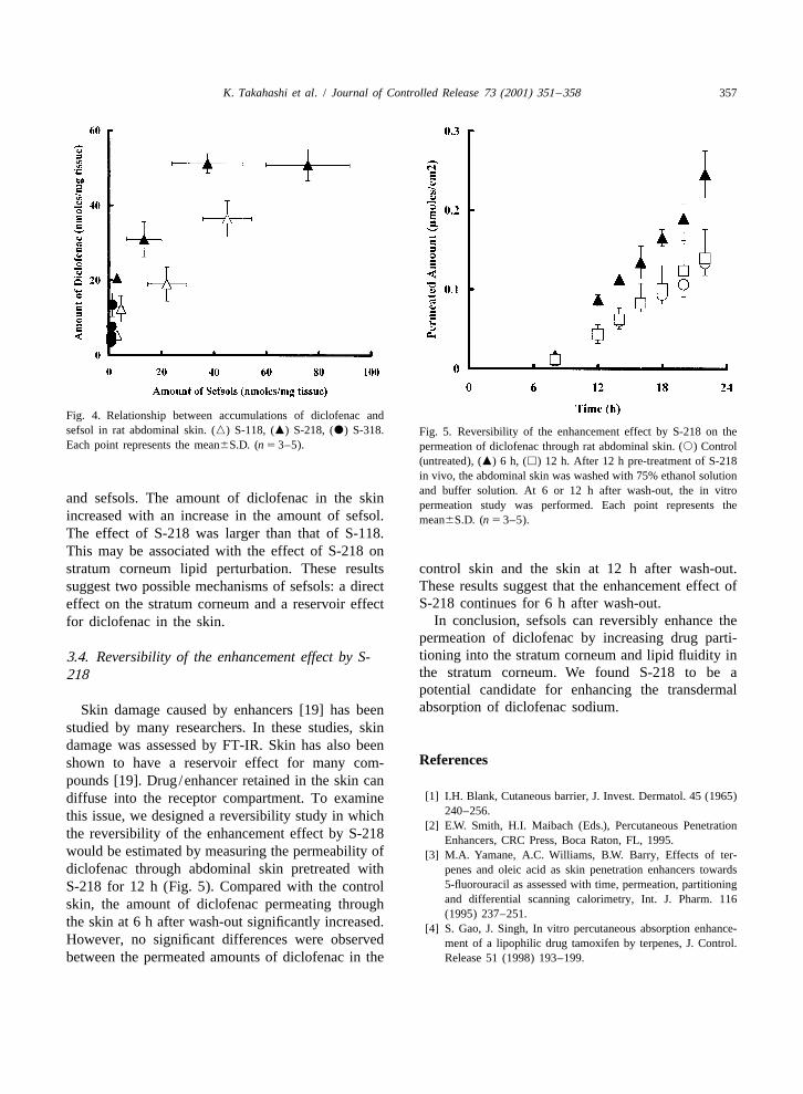
Fig. 4. Relationship between accumulations of diclofenac andsefsol in rat abdominal skin. (n) S-118, (m) S-218, (d) S-318. Fig. 5. Reversibility of the enhancement effect by S-218 on theEach point represents the mean6S.D. (n 5 3–5). permeation of diclofenac through rat abdominal skin. (s) Control
(untreated), (m) 6 h, (h) 12 h. After 12 h pre-treatment of S-218in vivo, the abdominal skin was washed with 75% ethanol solutionand buffer solution. At 6 or 12 h after wash-out, the in vitro
and sefsols. The amount of diclofenac in the skin permeation study was performed. Each point represents theincreased with an increase in the amount of sefsol. mean6S.D. (n 5 3–5).The effect of S-218 was larger than that of S-118.This may be associated with the effect of S-218 onstratum corneum lipid perturbation. These results control skin and the skin at 12 h after wash-out.
These results suggest that the enhancement effect ofsuggest two possible mechanisms of sefsols: a directS-218 continues for 6 h after wash-out.effect on the stratum corneum and a reservoir effect
In conclusion, sefsols can reversibly enhance thefor diclofenac in the skin.permeation of diclofenac by increasing drug parti-tioning into the stratum corneum and lipid fluidity in3.4. Reversibility of the enhancement effect by S-the stratum corneum. We found S-218 to be a218potential candidate for enhancing the transdermalabsorption of diclofenac sodium.Skin damage caused by enhancers [19] has been
studied by many researchers. In these studies, skindamage was assessed by FT-IR. Skin has also been
Referencesshown to have a reservoir effect for many com-pounds [19]. Drug/enhancer retained in the skin can
[1] I.H. Blank, Cutaneous barrier, J. Invest. Dermatol. 45 (1965)diffuse into the receptor compartment. To examine240–256.this issue, we designed a reversibility study in which
[2] E.W. Smith, H.I. Maibach (Eds.), Percutaneous Penetrationthe reversibility of the enhancement effect by S-218 Enhancers, CRC Press, Boca Raton, FL, 1995.would be estimated by measuring the permeability of [3] M.A. Yamane, A.C. Williams, B.W. Barry, Effects of ter-diclofenac through abdominal skin pretreated with penes and oleic acid as skin penetration enhancers towards
5-fluorouracil as assessed with time, permeation, partitioningS-218 for 12 h (Fig. 5). Compared with the controland differential scanning calorimetry, Int. J. Pharm. 116skin, the amount of diclofenac permeating through(1995) 237–251.
the skin at 6 h after wash-out significantly increased. [4] S. Gao, J. Singh, In vitro percutaneous absorption enhance-However, no significant differences were observed ment of a lipophilic drug tamoxifen by terpenes, J. Control.between the permeated amounts of diclofenac in the Release 51 (1998) 193–199.

358 K. Takahashi et al. / Journal of Controlled Release 73 (2001) 351 –358
[5] Y. Ozawa, T. Yamahira, H. Sunada, T. Nadai, Influence of [12] G.M. Golden, D.B. Guzek, A.H. Kennedy, J.E. Mckie, R.O.fatty acid–alcohol esters on percutaneous absorption of Potts, Stratum corneum lipids phase transitions and waterhydrocortisone butyrate propionate, Chem. Pharm. Bull. 36 barrier properties, Biochemistry 26 (1987) 2382–2388.(1988) 2145–2151. [13] K. Knutson, R.O. Potts, D.B. Guzek, G.M. Golden, J.E.
[6] T. Inagi, T. Muramatsu, H. Nagai, H. Terada, Influence of Mckie, W.J. Lambert, W.I. Higuchi, Macro- and molecularvehicle composition on the penetration of indomethacin physical-chemical considerations in understanding drugthrough guinea-pig skin, Chem. Pharm. Bull. 29 (1981) transport in the stratum corneum, J. Control. Release 21708–1714. (1985) 67–87.
[7] M. Okumura, K. Sugibayashi, Y. Morimoto, Effect of several [14] K. Knutson, S.L. Krill, W.J. Lambert, W.I. Higuchi, Physico-enhancers on the skin permeation of water-soluble drugs, chemical aspects of transdermal permeation, J. Control.Chem. Pharm. Bull. 37 (1989) 1375–1378. Release 6 (1987) 59–74.
[8] M. Ueno, H. Morijiri, K. Kojima, T. Kimura, T. Matsumoto, [15] H.L. Casal, H.H. Mantsch, Polymeric phase behavior ofK. Takahashi, Additional effect of polar solvents on the phospholipid membranes studied by infrared spectroscopy,dissolution of diclofenac sodium in aqueous solutions — The Biochim. Biophys. Acta 779 (1984) 381–401.effects of nonionic surfactant, J. Jpn. Med. Soc. Biol. [16] C.Y. Goates, K. Knutson, Enhanced permeation of polarInterface 25 (1994) 5–12. compounds through human epidermis. I. Permeability and
[9] K. Takahashi, T. Matsumoto, T. Kimura, H. Sakano, N. membrane structural change in the presence of short-chainMizuno, N. Yata, Effect of polyol fatty acid esters on alcohols, Biochim. Biophys. Acta 1195 (1994) 169–179.diclofenac permeation through rat skin, Biol. Pharm. Bull. 19 [17] K. Zhao, J. Singh, In vitro percutaneous absorption enhance-(1996) 893–896. ment of propranolol hydrochloride through porcine epidermis
[10] S. Kitagawa, H. Li, Effects of removal of stratum corneum, by terpenes /ethanol, J. Control. Release 62 (1999) 359–366.delipidization and addition of enhancers, ethanol and l- [18] R.J. Scheuplein, I.H. Blank, G.J. Brumer, D.J. MacFarlane,menthol, on skin permeation of benzoic acid and its 4-n-alkyl Percutaneous absorption of steroids, J. Invest. Dermatol. 52substituents in excised guinea pig dorsal skin, Chem. Pharm. (1969) 63–70.Bull. 47 (1999) 44–47. [19] K. Zhao, J. Singh, Mechanisms of percutaneous absorption
[11] G.M. Golden, J.E. Mckie, R.O. Potts, Role of stratum of tamoxifen by terpenes: eugenol, D-limonene and menth-corneum lipid fluidity in transdermal drug flux, J. Pharm. one, J. Control. Release 55 (1999) 253–260.Sci. 76 (1987) 25–28.