Characterization of sulfate-reducing bacteria isolated...
12
FEMS Microbiology Ecology 101 (1992) 217-228 O 1992 Federation of European Microbiological Societies 0168-6496/92/$05.00 Published by Elsevier FEMSEC 00406 ? Characterization of sulfate-reducing bacteria isolated from Senegal ricefields Aboubakar S. Ouattara and Vincent A. Jacq Laboratoire de Microbiologie ORSTQM, Université de Provence, Marseille, France Received 30 March 1992 Revision, received 1 , June 1992 Accepted 16 June 1992 Key words: Sulfate-reduction; . 1 Desulfouibrio; Ricefields; Sulfide toxicity; Ecology ( 1 --, l'< I 4- L' 1. SUMMARY I ? 5 . Sulfate-reducing bacteria were enumerated in soils and water samples from Senegal ricefields using lactate and sulfate as substrates. When rice plants were severely injured by sulfides, maxi- mum densities ranged from lo7 to lo9 bacteria 8-l of dry spermosphere or rhizosphere soil. Seven non-sporulating, mesophilic strains were isolated. The strains had motile curved cells and stained Gram-negative. Lactate, pyruvate, H, + CO,, malate, fumarate, or ethanol could serve as electron donors. Organic acids were incompletely oxidized to acetate. Alcohols were degraded to the corresponding fatty acids. Sulfate, sulfite, or thiosulfate could serve as electron acceptors and were reduced to sulfide. Vitamins, yeast extract, Biotrypcase, or additional NaCl were not re- quired for growth. On the basis of morphological and physiological properties, and the G + C mol % of the DNA, six isolates were identified as 211 Desulfouibrio vulgaris and one as Desulfovibrio desulfuricans. The comparison of their main phys- iological propertje: with the physico-chemical properties of sampling sites indicated that they were better adapted to conditions prevailing in the rice rhizosphere than to those prevailing in the bulk of soil. 2. INTRODUCTION Microbial reduction of sulfate is performed by the morphologically and physiologically versatile dissimilatory sulfate-reducing bacteria, an het- erogenous group of microorganisms exhibiting a large nutritional diversity [l-31. As in any natural environment, the activity of sulfate reducers in ricefields depends upon redox potential, sulfate, temperature, salinity, pH, and the availability of organic nutrients provided by rice exudates or derived from the metabolism of heterotrophic fermentative bacteria. Althsugh most rice soils in I the world are S-deficient, significant reduction of sulfate has been reported in a wide range of ricefields [41. Production of sulfides, at levels that cause rice injury, was obseked in Asia [5,6], USA Correspondence to: V.A. Jacq, Laboratoire de Microbiologie ORSTOM, case 87, Université de Provence, 3, Place Victor I -Hugo, F-13331 Marseille Gédex 3, France. 7-- - Il * 1 I
Transcript of Characterization of sulfate-reducing bacteria isolated...

FEMS Microbiology Ecology 101 (1992) 217-228 O 1992 Federation of European Microbiological Societies 0168-6496/92/$05.00 Published by Elsevier
FEMSEC 00406 ?
Characterization of sulfate-reducing bacteria isolated from Senegal ricefields
Aboubakar S. Ouattara and Vincent A. Jacq
Laboratoire de Microbiologie ORSTQM, Université de Provence, Marseille, France
Received 30 March 1992 Revision, received 1, June 1992
Accepted 16 June 1992
Key words: Sulfate-reduction; . 1 Desulfouibrio; Ricefields; Sulfide toxicity; Ecology ( 1
- - , l'< I
4 - L ' 1. SUMMARY I?
5 .
Sulfate-reducing bacteria were enumerated in soils and water samples from Senegal ricefields using lactate and sulfate as substrates. When rice plants were severely injured by sulfides, maxi- mum densities ranged from lo7 to l o9 bacteria 8-l of dry spermosphere or rhizosphere soil. Seven non-sporulating, mesophilic strains were isolated. The strains had motile curved cells and stained Gram-negative. Lactate, pyruvate, H, + CO,, malate, fumarate, or ethanol could serve as electron donors. Organic acids were incompletely oxidized to acetate. Alcohols were degraded to the corresponding fatty acids. Sulfate, sulfite, or thiosulfate could serve as electron acceptors and were reduced to sulfide. Vitamins, yeast extract, Biotrypcase, or additional NaCl were not re- quired for growth. On the basis of morphological and physiological properties, and the G + C mol % of the DNA, six isolates were identified as
211
Desulfouibrio vulgaris and one as Desulfovibrio desulfuricans. The comparison of their main phys- iological propertje: with the physico-chemical properties of sampling sites indicated that they were better adapted to conditions prevailing in the rice rhizosphere than to those prevailing in the bulk of soil.
2. INTRODUCTION
Microbial reduction of sulfate is performed by the morphologically and physiologically versatile dissimilatory sulfate-reducing bacteria, an het- erogenous group of microorganisms exhibiting a large nutritional diversity [l-31. As in any natural environment, the activity of sulfate reducers in ricefields depends upon redox potential, sulfate, temperature, salinity, pH, and the availability of organic nutrients provided by rice exudates or derived from the metabolism of heterotrophic fermentative bacteria. Althsugh most rice soils in
I the world are S-deficient, significant reduction of sulfate has been reported in a wide range of ricefields [41. Production of sulfides, at levels that cause rice injury, was obseked in Asia [5,6], USA
Correspondence to: V.A. Jacq, Laboratoire de Microbiologie ORSTOM, case 87, Université de Provence, 3, Place Victor
I -Hugo, F-13331 Marseille Gédex 3, France. 7-- -
Il * 1
I

21s
[7,8], and Senegal [9]. In lowland ricefields, strictly anaerobic bacteria, including sulfate reducers, be- come active when the soil becomes anoxic, be- cause 0,-consumption by facultative anaerobes is not balanced by 0,-diffusion from the floodwater and the oxidating power of rice roots [10,11].
Sulfate-reducers are common in flooded soils [3,12,13]. They are present in oxidized and re- duced soil layers, as colonies located in 0,-de- pleted sites such as iron-clay aggregates and or- ganic debris [14,15]. They are also found in the rhizosphere and the spermosphere of rice [9,13,16]. Reported densities "in rice soils range from lo3 to lo5 cells g-' dry soils in Asia [12]. Studies in Senegal ricefields showed the existence of a very active anaerobic microflora, including methanogens [13], sulfate reducers [9,13], sulfur reducers [17], and ferric iron reducers [18] lo- cated in the soil and the vicinity of the roots. Pot experiments with seedlings showed densities of sulfate reducers ranging from 6 X 10' to 7 x l o5 cells g-' of soil in rhizospheric samples [13], and microplot experiments showed that in situ densi- ties may reach lo9 cells 8-l of soil [16].
Although DesuIfouibrio strains were suspected to be dominant in Asian ricefields [12], there is only one report on the isolation of both sporulat- ing and non-sporulating sulfate reducers [19]. Si- multaneous enumeration of lactate-using sulfate reducers and Thiobacilli [20], suggested that the most numerous sulfate reducers in the spermo- sphere and the rhizosphere, and, because of their locations, the most potentially harmful for the plant, were mostly non-sporulating forms. This work follows several applied studies of the condi- tions inducing sulfide toxicity in Senegal ricefields [9,11,16,21]. It reports on the isolation and char- acterization of seven strains of DesuIfouibrio. Physiological properties of these strains are dis- cussed with regard to their growth conditions in the sampling sites and the rice rhizosphere.
3. MATERIALS AND METHODS
3.1. Sampling sites Surveys were done in 82 ricefields located in
the delta of the Senegal River and the Casamance,
the two rice producing areas of Senegal. In situ soil temperature varied from 28°C to 30°C at 5-10 cm depth under waterlogged conditions [21]. Soil pH ranged from 4.0 to 5.2 in newly rewetted soils and from 5.2 to 6.9 after four weeks of submersion [211.
Four sites with high intensity of microbial re- ducing processes and marked detrimental effects on rice growth, were chosen for detailed surveys during three rice cropping seasons [11,18,21]. Soils were sampled at time intervals in the root vicinity (rhizosphere) and around germinating seeds (spermosphere). Sites from which the strains were isolated have been described in previous papers [9,16,18,21,22]. Their main characteristics are summarized in Table 1. These soils differed widely in texture and salt content. A lower variability was observed for carbon content and soil pH measured on newly rewetted soils. Senegal River delta soils became less acidic than Casamance soils after 4 weeks of submersion. Despite a low organic matter content, they were favourable to chemical and microbial reducing processes when waterlogged, because of their high clay content [22,231. Tilène 3 soil was a moderately saline, acidic and sulfated soil [16,23]. Pont-Gendarme 9 soil is highly sulfated and acidic, with a sulfuric horizon and jarosite mottles, and 2%0 w/w min- eral sulfur in surface layer [23]. In the Casamance, Djibélor soil, after 30 years of rice cultivation, lost some properties of the original mangrove soil: in soil surface part of soluble sulfates was leached and pH became almost neutral, but jarosite mottles were still encountered under the plough layer [22]. Loudia-Ouoloff soils were acidic mangrove-type soils, in which the anaerobic bac- terial process most potentially harmful1 to rice was Fe2+ production by ferric iron reducers [22].
3.2. Field enumerations In situ enumerations of sulfate reducers were
performed on 3000 soil or water samples (5-10 g) collected in fields exhibiting clear sulfide toxicity symptoms (Fig. 1). Soil samples were collected anoxically at 3-15 cm depth in the spermosphere or the rhizosphere of rice, as described by Prade et al. [22]. Millipore filters were used for the enumeration procedure, as described by
li

219
Baldensperger and Mouraret [24] for Thiobacilli, and by Traoré and Jacq [17] for sulfur-reducing bacteria. Samples were diluted in sterile 0.6%
and immediately deposited on Millipore filters (HAWP 04700; Millipore Corp., Bedford, MA; 0.45 pm, 6 filters/sample). Each filter was then placed in a screw-cap tube (22 ml; 125 X 16 mm) containing 21 ml of Medium 1. Tubes were incu- bated at 35°C until the appearance of a black Fes-precipitate on the filter. The time necessary for total blackening of the filter was correlated with the initial numbers of viable cells [17]. Cali- bration curves were established with pure cul- tures of sulfate reducers.
Medium 1 was used for enumeration studies. Solution A: KH,PO,, 0.5 g; MgSO, .7H,O, 2.0 g; Na,SO,~lOH,O, 1.0 g; CaC1,*2H20, 0.1 g; modified Pfennig’s element trace solution [25], (1 ml); distilled water, 750 ml. Solution B: NH,CI, 1.0 g; 60% sodium lactate, 6 ml; FeSO,(NH,), SO,. 6H,O, 6.0 g; yeast extract (Difco) 1.0 g;
? MgCl, solution, homogenized by a rotary shaker,
r
Table 1
Some isolation site and strain growth characteristics
Site characteristics Senegal River delta
distilled water, 235 ml. Solution C: NaOH, 10 ml of 0.43 M solution. Solutions A and 8 C were autoclaved for 20 min at 120°C. Medium 1 was prepared by mixing cooled solutions A and C to B, sterilized through Millipore filters (0.45 ‘,um) and 21 ml of the final medium were aseptically dispensed into sterile Kimax test-tubes containing 50 mg of finely ground Fes and 1 ml of water. The final pH was 7.0. During the last field experi- ment on Casamance (Loudia-Oualoff) a modifica- tion of Medium 1 was tested, in which yeast extract was replaced by 1 ml 1-’ of 0.4% (w/v) biotin solution and 1 ml 1-l 0.02% (w/v) vitamin BI, solution [17]. Modified media, with lactate replaced by Na-acetate (10 mM) or Na-palmitate (2 mM), were used to enumerate other types of sulfate-reducing bacteria.
3.3. Enrichment cultures Enumeration tubes in which calculated num-
bers of sulfate-reducing bacteria were higher than 10’ cells g-’ of dry soil were kept for enrich-
Casamance Sampling zone Tiltne 3 Pont-Gendarme 9 Loudia Oualoff Djibélor 4
Soil Average temperature (“Cl a pH (dry soil) pH (soil solution) SO;- M.kg-’) Na* M.kg-l) CI- ( lob2 M.k Clay contents (%) b)
Carbon (w%)
29-31 4.0-4.4 6.1-6.5 3.2-4.3 - 9.0 16.4 55-58 1.1-1.2
28-32 3.8-4.7 5.6-6.1 7.7-9.3 n.d. n.d. 59-65 0.85-1.1
28-30 4.1-4.4 4.3-6.4 0.73-1.26 0.9-1.57 9.0-15.0 15-36 1.9-2.8
26-28 3.7-5.5 4.2-6.8 0.55-2.2 0.11-0.65 3.4-4.1 16-31 2.1
Strain growth characteristics
Strain FO F3 F1 F2W F2X F2Y G1
Temperature range (“C) 16-42 16-46 16-43 20-43 16-44 20-45 20-48 Optimum temperature 33-35 35-37 30-31 30-32 30-34 36-39 38-42 pH range 5.8-8.7 5.8-8.65 5.7-8.7 6.3-8.6 5.8-8.7 5.2-8.6 5.7-8.5 Optimum pH 6.8 6.85 6.9 7.0 6.95 6.75 6.9 NaCl range (g.1-l) 0-15 0-15 0-17 0-12 o15 0-10 0-15 Optimum NaCl 5-7 5-10 10-15 3-7 7-14 1-7 1-7
o.,
a At 10-15 cm depth, at sampling. At 0-10 cm depth, on dry soil before sowing or transplanting of rice. At 10 cm depth, after 4 weeks of waterlogging. Moderately salted soil before first irrigation, then progessively desalting; n.d.: not determined.
j

220
ment-cultures on various electron donors (Fig. 1). Higher in vitro densities were obtained on lac- tate, after four or five successive transfers on the freshwater medium of Widdel and Pfennig [26], supplemented with 1 ml of trace-element solution SL7 [27] and reduced with 2 ml of a 0.5 M Na2S solution (Medium 2). General techniques for the cultivation of strict anaerobes [28-311 were used for media preparations and during experiments. Substrates or vitamins were supplied from auto- claved or filter-sterilized stock solutions.
3.4. Isolation and purity control Thirty fast-growing subcultures with a cell den-
sity higher than lo9 were chosen for isolation tests (Fig. l), using repeated deep agar (0.8-0.9%) dilutions adapted from Pfennig et al. [321. Strain purity was checked in Medium 2 supplemented with 0.25% yeast extract (Difco), 0.25% peptone,
0.25% Biotrypcase (BioMCrieux, Craponne, France), 0.25% nutrient broth, 0.25% glucose (or 0.25% fructose). Cultures were examined micro- scopically for purity after three weeks of incuba- tion at 35°C.
3.5. Growth studies Unless otherwise indicated, experiments were
conducted in Medium 2 with 20 mM lactate and 20 mM sulfate, at 35°C and pH 7.2, and with 0.1% NaCI. Growth was monitored by measuring optical density at 580 nm with a Bausch and Lomb Spectronic 21 spectrophotometer. Total non-precipitated sulfides (H,S, S2-, and HS-) were measured spectrophotometrically as col- loical CuS [331.
The pH range of growth was estimated in bicarbonate buffered mineral Medium 2, where pH was adjusted with sterile Na,CO,, NaOH, or
82 locatlons in 2 areas of Senegal
ENUMERATIONS on4media (3.000 soiland water samples)
I I I I
I I I Lactate Modified Acetate Palmitate Medium 1 lactate Medium Medium
Medlum 1
Selection of enumeration-tubes containing more than 1 O* sulfate-reducers g-1 of soil (14 soils) -
504 70 58
Total: 568tubes~thdbnoidcell~, 505tubeswthmotiledbnior
ENRiCHMENTS
Media -1 2
isolation of major - iXmkAil'irm
Selection of 30 tubes wlth densltles of sulfate-reducers > 1 O 9 cells mi-' contaminant .?c&A?tm
I I lSOLATIONS on pyruvate 7 on acetate on propionate
on lactate
2 slow-grdwing and sporulating sulfate-reducers
lost after subcuiturinas
& Second purificatlon
Fig. 1. Schematic representation of the isolation process of studied strains.

221
HC1 solutions. The temperature range for growth was tested from 16 to 55°C. A possible salt re- quirement in addition to the concentration used in Medium 2 was tested by adding NaCl concen- trations ranging from 0.1 to 3%.
Ability of strains to use various substrates as electron donor was tested in presence of 20 mM sulfate. When the growth failed, substrate oxida- tion ability was tested in presence of vitamins [32] and 2 g 1-1 of yeast extract. Sulfite, thiosulfate, elemental sulfur, nitrate, fumarate, and malate were tested as electron acceptors in presence of 20 mM lactate.
9
J
3.6. Analytical techniques Organic acids, alcohols, and fatty acids were
assayed by high-performance liquid chromatogra- phy (HPLC): pump, Analprep 93 (Touzart et Matignon, Vitry-sur-Seine, France); flow rate, 0.6 ml/min; injection loop 20 pl; column ORH-801, 300 X 6.5 mm (Interaction Chemicals, Mountain View, CA); eluent: H,SO, 0.01N; H t ions ex- change column, 8-pm pore size; elution according to the pK, and/or the molecular mass for the non-ionic compounds; column temperature, 35°C; detection, differential refractometer (Knauer, Berlin, FRG); recorder, Chromatopak C-R3A (Shimadzu, Kyoto, Japan).
For cell pigment determination, 3 g of wet cells, suspended in 10 ml of 20 mM Tris.HC1 buffer (pH 7.6), were sonicated and the suspen- sion was centrifuged at 30000 X g for 20 min. The resulting cell-free extract was centrifuged at 140000 X g for 2 h. The pellet was resuspended in the previous buffer. Cytochromes were identi- fied by recording air-oxidized and dithionite-re- duced spectra, as well as the redox difference spectra of the dithionite-reduced minus air- oxidized of each fraction, using a Shimadzu UV 300 spectrophotometer (Shimadzu, Kyoto, Japan).
Gram staining was done using a standard method with a coloration kit (Sigma, St Louis, MO). Escherichia coli and Micrococcus luteus were used as controls.
The guanine-plus-cytosine content of the DNA (G 3- C%) was determined at the German Collec- tion of Microorganisms (DSM, Braunschweig, FRG). DNA was isolated by chromatography on
,
I
A: log SRB g-1 Of Soil 10
E
6
4
2
O
Days after sowing
6 : log SRB g-1 of soil 10
8
6
4
2 -200 - 1 0 0 O 1 O0
Days after transplanting
Fig. 2. (A) Lactate-using bacteria enumerated in the spermo- sphere and the rhizosphere of direct-seeded rice in 2 fields of the Senegal River delta (mean of 6 measurements). (B) Lac- tate-using bacteria enumerated before transplanting and in the rhizosphere of transplanted rice in 2 fields situated in
Casamance/Senegal (mean of 6 measurements).
hydroxyapatite 1341. The G -F C% was determined by HPLC; non-methylated Lambda virus DNA was used for calibration [35].
4. RESULTS
4.1. Field results Densities of lactate-utilizing sulfate reducers
were measured from sowing (spermospherical samples: 0-14 days) to harvest (rhizospherical samples: 15-110 or 120 days) in the two Senegal River delta soils (Fig. 2a). In the two Casamance soils, they were enumerated before the first use- ful rain (unplanted soil samples), and on rhizo-

222
spherical samples, from transplanting to harvest 4.2. Microscopical observations on enumeration and (Fig. 2b). Higher densities were observed on the enrichment cultures days after seed germination, from transplanting The enumeration-tubes chosen for enrich- to tillering, and at flowering. ment-cultures (Fig. 1) contained two carbon
Table 2
Electron donors and acceptors, G + C mol %, pigments and morphology
Strain FO F1 F2W F2X F2Y F3 G1 D. oulgaris D. desirlfLiricans
Electron donors (in mM) on Medium 2 Lactate (15) + + + + + + + + + Pyruvate (15) a t + + + + + + + +
Electron donors (in mM) oxidized on Medium 2, acetate (2 mM) and vitamins added H, +CO, (2 bars) + + + + + + + Fumarate (10) + + + + + + Malate (10) - + + + - + + Ethanol (5) + + + + + + + Propanol (5) + t + + + + + Butanol (5) - - + + + + + Glycerol (5) ( + ) d - ( + I - ( + I (+) - Serine (10) ( t ) - ( + I ( + I - Choline (5) - - -
-
- - - - - -
+ + + + nr + i- -
+ + + + + + nr + +
Carbon substrates (in mM) fermented in sulfate-free Medium 2 e
+ - - Pyruvate (15) + + + + + + - + - - - - - - - Malate (10)
Electron acceptors (in mM), lactate (20 mM) as electron donor Sulfate (20) + + + + t + + Sulfite (5) + + + + + + + Thiosulfate (10) + + + + + + + Elemental sulfur (10) - - - - - - - Nitrate (5) - - - - - - - Fumarate (10) - - - - - - -
G + C mol % and pigments G t C m o l % 62.5k0.6 59.0 60.420.2 62.8 56.3 60.1 60.2
Desulfoviridin + + + + + + + Cytochromes c3 c3 c3 c3 c3 c3 c3
t + + -
61.0 c3 +
55.0 c3 +
Morphology Form vibrio vibrio vibrio vibrio vibrio vibrio short rod vibrio vibrio Averagelength(ym) 3-6 4-7 3-7 2-6 3-6 4-7 2-4(4-6) 1.5-4 1.5-4 Average width (pm) 0.5-0.8 0.5-1.0 0.4-0.7 0.5-0.8 0.5-0.8 0.5-0.7 0.6-0.8 0.5-0.8 0.5-0.8
(0.8-1.0)
a Without vitamins or yeast extract. Substrates tested (mM), either in presence or absence of acetate or vitamins, and not supporting the growth of any of the 7 isolates: formate (lo), oxalate (5), succinate (lo), methanol (5), iso-propanol (5), iso-butanol (lo), pentano1 (lo), acetate (lo), propionate (10); n- or iso-butyrate (lo), 2-methylbutyrate (lo), n-benzoate (lo), valerate (5) palmitate (2), fructose (15), glucose (lo), xylose (5), ribose (51, and cysteine (10). nr = not reported. (+) Slow degradation without significant growth when yeast extpct added (0.1%).
(lo), fructose (15), glucose (lo), and ethanol (10). Size and motility were different on lactate or on pyruvate (data in brackets).
e No fermentative process observed, in sulfate medium without yeast extract on: lactate (151, succinate (lo), fumarate (lo), oxalate

223
sources, the electron donor (lactate, acetate, or palmitate) and yeast extract. Microscopical obser- vations showed mixed cultures of one or more
motile or non-motile rods, sometimes observed in their sporulating stage. Vibrio-shaped cells (dif- fering by motility and sizes) were significantly dominant in more than 90% of the tubes ob- served during the hours following the blackening of filter, whatever the site or the electron donor. Most of cultures (98%) did not show a modifica- tion of cell forms when subcultured. on Media 1 or 2. But vibrios died when subcultured 1 to 3 times on acetate or palmitate-sulfate media. Four or five rapid subculturings on Medium 2 (free from yeast extract), with lactate or with pyruvate reduced the relative number of sporulating bacte- ria (that might be other sulfate reducers or con- taminants, such as anaerobic bacteria using the yeast extract).
Enrichment-tests on propionate, acetate, n- and iso-butyrate, n-benzoate, palmitate, glucose and fructose failed, whereas those with propi- onate or palmitate progressively resulted in the selection of two sporulating sulfate-reducing bac- teria (slighly curved rods or oval cells with round spores). They grew very slowly (2-6 months of incubation were necessary to obtain cell densities around lo5), and could not be subcultured for more than three transfers and identified.
b motile sulfate-reducing bacteria, and weakly
- C
4.3. Isolation Four strains (FO, F2, F3 and G1) were isolated
on lactate (15 mM) as electron donor, while strain F1 was obtained on pyruvate (15 mM). They developed after 3-9 days of incubation at 35°C as brownish, lens-shaped colonies in deep agar tubes. Because the growth characteristics of strain F2 were not constant, this strain was repurified on deep-agar lactate medium, leading to the isola- tion of three strains (F2W, F2X, and F2Y).
o
,1 4.4. Strain description 4.4.1 Morpliology. The seven isolates were
motile vibrios, 1-3 p m in length and 0.5-0.8 pm
in width (Table 2). They lost motility and became spirilloid in old cultures. Strain G1 exhibited a slight variability (both in size and motility) when cultured on lactate or pyruvate. Cells stained Gram-negative and did not sporulate.
4.4.2. Physiology. Growth characteristics of the strains on Medium 2 are reported in Table 1. The range of temperature for growth was 16-48"6, optima were between 30 to 42°C. The range of pH was 5.2-8.7, optimum pH were between 6.75-7.0. Strains could grow in Medium 2 whitliout additional NaCl. But NaCl improved growth, with optima observed when added con- centrations varied from 1 to 5 g 1-l.
Data on electron acceptors are presented in Table 2. The strains were fast-growing bacteria with minimum doubling times between 1.5 and 4 h on Medium 2 with lactate or pyruvate. Lactate and pyruvate were incompletely oxidized to ac- etate, and growth occurred without addition of vitamins, yeast extract, peptone, or Biotrypcase. Fumarate (except for strain F2Y), malate, H, + CO,, ethanol, propanol, and butanol (except for strains FO and F1) could serve as electron donors only when vitamins and yeast extract were pro- vided. Acetate was the end product of fumarate and malate degradation. Alcohols were degraded to the corresponding volatile fatty acids. Except strain G1, all isolates could ferment pyruvate. Substrates tested and not utilized are listed in Table 2.
Sulfate, sulfite, and thiosulfate were used as electron acceptors by all isolates (Table 2) and sulfide was the product of reduction. No isolate was able to use elemental sulfur, fumarate, malate, or nitrate as electron acceptor.
44.3, Cytoclzroines, pigments and DNA base composition. When reduced with sodium dithion- ite, the soluble extracts of all isolates exhibited absorption bands characteristic of c,-type cy- tochrome, with maxima at 419, 522, and 552 nm. The oxidized extracts showed the cytochrome Soret peak at 408 nm. In addition, the spectra showed a characteristic absorption band at 628 nm indicating the presence of desulfoviridin [36,371.
The G + 6% varied from 56.3 to 62.8 k 0.3% (Table 2).

224
5. DISCUSSION
5.1. Taxonomy The seven isolates are strictly anaerobic,
mesophilic, Gram-negative, non-sporulating sul- fate-reducing bacteria. They perform incomplete oxidation of lactate and pyruvate to acetate and CO,. They are unable to oxidize acetate, propi- onate, butyrate, or palmitate. Cells contain cg- type cytochromes and desulfoviridin. Based on the above characteristics, they belong to genus Desrilfouibrio [1,3,36,38]. Morphological and phys- iological characteristics are similar to those of D. desulfuricans or D. vulgaris [1,31. The G i- C% of strain F2Y (56.3%) is close to that of D. desrtlfu- ricans (55%) whereas the G + C % of the other six strains (59-62.8%) is close to that of D. vulgaris (61%). The six strains similar to D. uul- garis differ from the type strain only by the use of glycerol (Table 2). Strain F2Y differs from D. desulfiiricans by its inability to oxidize choline and to utilize elemental sulfur and nitrate as electron acceptors. Likely, strain F2Y is a strain of D. desulfiiricans and other isolates are strains of D. vulgaris.
5.2. Low variability of isolated strains Results show a limited strain diversity (two
species of non-sporulating sulfate reducers) whereas we surveyed the rhizosphere of at least a dozen of rice cultivars growing in soils exhibiting a wide range of physico-chemical properties (Ta- ble l) and situated at about 800 km distance. The dominance' of non-sporulating sulfate reducers was also reported by Bak and Pfennig, in the sediment of Lake Constance, where fast-growing H,- and lactate-consumer strains were mostly found [39].
However, reported field enumerations and subsequent isolations might be insufficient to conclude that rice spermosphere and rhizosphere are colonized only by the described fast-growing, mesophilic, neutrophilic, and non-sporulating strains.
Populations of acetate-utilizing sulfate reduc- ers might be underestimated because of the short incubation time required to obtain positive re-
sults (blackening of the filter). Experiments by Jargensen and Bak [40] on agar media with ma- rine sediments showed that 12 weeks of incuba- tion were necessary to obtain the maximum cell numbers of acetate-using sulfate reducers, whereas 1-3 weeks was enough for the develop- ment of all colonies of fast-growing lactate- (or H,)-utilizing sulfate-reducing bacteria. When ac- etate- or palmitate-utilizing sporulating sulfate reducers were enumerated on modified Medium 1 (lactate replaced by 15 mM acetate, or 1 mM palmitate), 2-4 weeks of incubation were needed to obtain the blackening of the filters.
The low strain diversity recorded could also be attributed to low salt content of the media used for enumeration, enrichment and isolation. In particular, fatty acid-oxidizing sulfate-reducing bacteria (Desulfobacter or Desulfobacterium spp.) require a relatively high level of NaCI. Laanbroek and Pfennig [41], comparing the anaerobic oxida- tion of short-chain fatty acids in freshwater and in marine sediments, established that acetate- oxidizing strains did not grow in freshwater medium. They were isolated from brackish or marine samples only, and their densities were significantly lower than those of Desulfouibrio desulfuricans.
Vitamins requirement could have limited the growth of some strains. However, during the last survey in Loudia Oualoff, the use of the modified Medium 1 (yeast extract replaced by biotin and vitamin B,,), did not result in the growth of morphologically different sulfate reducers, as compared with samples collected during the pre- ceding cropping season. But it reduced the rela- tive number of non-sulfate-reducing bacteria- probably those able to use yeast extract as carbon source-and increased the number of vibrioid sulfate reducers.
Acetate- or palmitate-utilizing sporulating sul- fate reducers, found as minor component in less than 10% of fast-growing enumeration tubes, were different from Desulfobacter postgatei or Desulfo- tomaculum acetoxidans, as shown by the very slow growth observed on the specific acetate-sulfate media established for these two species by Pfen- nig et al. [26,42]. Enrichment-tests with various times of incubation, carbon sources, salt content,
b

225 , A : tog SRB 6’ of root
li
A 10
J 9
8
7
E
---5 Acetate
Palmitate
Hawest
20 40 60 80 1 O0
Days after transplanting
B : log SRB CJ‘ of root 11
10
9
0
7
6
5 O 20 40 60 80 1 O 0
Days after transplanting
Fig. 3. (A) Lactate-, acetate-, and palmitate-using bacteria enumerated on the rhizoplane of transplanted IR8 rice in Loudia-Oualoff fields, Casamance/Senegal (mean of 6 meas- urements). (B) Lactate-, acetate-, and palmitate-using bacteria enumerated on the rhizoplane of I-Kong-Pao transplanted rice in Loudia-Oualoff fields, Casamance/Senegal (mean of 6
measurements).
and pH, did not permit to isolate these species
In a field experiment [11,18], it was shown that densities of acetate- or palmitate-utilizing sulfate reducers, were 102-103 times less abundant than fast-growing lactate-utilizing sulfate reducers on the root surface (Figs. 3a,b). Because of their low populations and very low growth rate, it was assumed that they were not significantly impli- cated in rice plant intoxication. Other sulfate
B11.
E
.1
reducers resembling Desulfobulbus (oval or lemon shape), or Desulfonenta (filamentous cells) were also observed as rare contaminants [lll. Their isolation was not assayed: they could not be re- sponsible for the damages on rice because of their very low occurrence in the rhizosphere.
Despite possible methodological reservations, the dominance of non-sporulating sulfate reduc- ers observed in this study is consistent with a previous work reporting that the detrimental ef- fects of sulfides produced in ricefieds was due, whatever the soil and the cultivar, to lactate- utilizing sulfate reducers, and may be indirectly correlated with their number. Jacq and Roger [20] determined that sulfide toxicity was the result of the antagonistic action of sulfate-reducing, lac- tate-oxidizing bacteria versus sulfide-oxidizing bacteria. Major injuries to and death of the rice plant occurred only when the density of lactate- oxidizing sulfate reducers, enumerated on Medi- um 1, was at least 100 times higher than that of Thiobacillus denitrificans in the same sample.
It was found that non-sporulating sulfate re- ducers were associated in the rhizosphere of dis- eased rice plant with 0,-consuming facultative ferric iron-reducing bacteria, including Clostridia [11,21]. We also found that major contaminants on lactate-sulfate were mainly Clostridia. Clostridium aceticum was evidenced as the domi- nant species [21]. As it did not use lactate, it was supposed that its presence did not significantly modify the composition of lactate-Medium 1 dur- ing a short incubation time.
5.3. Strains and their environment Isolated strains grow in rice rhizosphere on the
small number of carbon substrates (Table 2) that can be provided by germinating seed and root exudates [9,13,43], or/and resulting from the ac- tivity of heterotrophs. Lactate and pyruvate exu- dated by rice roots or germinating seeds are the main electron donors for all strains and can en- sure their growth. Experiments with hydroponic cultures of l-2-week-old seedlings of IR8 rice [43] showed that lactate + pyruvate comprised about 15% of excreted carbon compounds and only 25-30% of substrates were usable by most Desulfovibrio strains. Average exudation was esti-
226
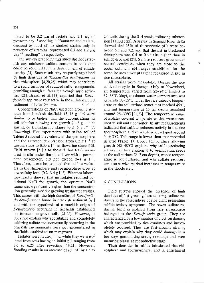
mated to be 3.2 p g of lactate and 2.1 p g of pyruvate day-' seedling-'. Fumarate and malate, oxidized by most of the studied strains only in presence of vitamins, represented 8.3 and 1.2 p g day-' seedling-', respectively.
The surveys preceding this study did not estab- lish any minimum sulfate content in soils that could be required for the development of sulfide toxicity [21]. Such result may be partly explained by high densities of Thiobncillus denitrificans in rice rhizosphere [4,20,261, which may contribute to a rapid turnover of reduced sulfur compounds, providing enough sulfates for .Desulfovibrio activi- ties 1211. Brandl et a h P441 'reported that Desul- fouibrio spp. were very active in the sulfate-limited sediment of Lake Geneva. 4 "
Concentrations of NaCl used for growing iso- lates from brackish ricefields (5-15 g I-') were similar to or higher than the concentrations in soil solution allowing rice growth (1-2 g 1-I at sowing or transplanting stages to 5-6 g 1-' at flowering). Plot experiments with saline soil of Tilène 3 showed that salinity in the spermosphere and the rhizosphere decreased from 0.2 g I-' at sowing stage to 0.09 g I-' at flowering stage [16]. Field surveys [21] also showed that NaCl meas- ured in situ under the plow layer with a perma- nent piezometer, did not exceed 3-4 g 1-'. Therefore, it can be assumed that sulfate reduc- ers in the rhizosphere and spermosphere grow at low salinity level (0.2-3-4 g 1-l). Whereas labora- tory results showed that no isolates required ad- ditional NaCl for growth, the optimum NaCl range was significantly higher than the concentra- tion generally used for growing freshwater strains. This agrees with the high densities of Desulfovib- rio desulfuricans found in brackish sediment [41] and with the hypothesis of a brackish origin of Desulfovibrio occurring in ricefields established on former mangrove soils [21,22]. However, it does not explain why sporulating and completely oxidizing sulfate reducers usually occurring in the brackish environments were not encountered in ricefields established on mangroves.
Isolates were neutrophilic, while they were iso- lated from soils having an initial pH ranging from 3.6 to 6.25 after rewetting [13,211. However, flooding results in an increase of soil pH by 1.5 to
2.0 units during the 3-4 weeks following submer- sion [11,13,16,231. A survey in Senegal River delta showed that 95% of rhizospheric pHs were be-
rhizosphere was 0.4 to 0.6 units higher than in sulfide-free soil [21]. Sulfate reducers grow under
roots: optimum pH ranges established for the seven isolates cover pH range measured in situ in rice rhizosphere.
All strains were mesophilic. During the rice cultivation cycle in Senegal (July to November), air temperature varied from 23-24°C (night) to 37-38°C (day), maximum water temperature was generally 30-32°C under the rice canopy, temper- ature at the soil surface sometimes reached 45"C, and soil temperature at 15 cm depth remained around 28-30°C [21,23]. The temperature ran.ge of isolates covered temperatures that were meas- ured in soil and floodwater. In situ measurements indicated that sulfate reducers activity in the rice spermosphere and rhizosphere developed around 30 5 2°C. This range is lower than that recorded in vitro (Table 1). Upper temperature allowing growth (42-48°C) explains why sulfate-reducing activity can be detrimental to germinating seeds at the soil surface (2-3 cm depth), where temper- ature is not buffered, and why sulfate reducers can also survive marked increases in temperature in the floodwater.
tween 6.5 and 7.2, and that the pH in blackened
neutral conditions when they are close to the
r
L
6. CONCLUSIONS
Field surveys showed the presence of high densities of fast-growing, lactate-using, sulfate re- ducers in the rhizosphere of rice plant presenting sulfide-toxicity symptoms. The seven sulfate-re- ducing bacteria isolated from rice rhizosphere belonged to the Desulfovibrio group. They are characterized by a low number of electron donors, which are provided by rice exudates and incom- pletely oxidized. They are fast-growing strains, which may explain why they could damage in a few days germinating seeds, seedlings and even maturing plants at reproductive stage.
Their densities in sulfide-intoxicated rice rhi- zosphere and spermosphere, and in enrichment
k'

227
cultures, were significantly higher than those of sporulating sulfate reducers which could not be identified. Therefore, described Desulfovibrio are
development of sulfide toxicity. In the studied area, environmental conditions
in +ice rhizosphere appear to be quite similar in terms of available substrates, temperature, salin- ity and pH. Desulfovibrio seems to be the genus best adapted to conditions prevailing in this envi- ronment.
The seven strains have been deposited with the D.S.M. under DSM numbers: 6617 (FO), 6618 (FU, 6619 (F2W), 6620 (F2X), 6621 (F2Y), 6622 (F3) and 6623 (Gl).
6 to be considered as mostly responsible for the
1
ACKNOWLEDGEMENTS
The authors thank J.-M. Chianea, C. Mi- maudo, and E. Dupont for technical assistance during laboratory experiments, and Dr.-Ing. K. Prade for technical assistance during field work. Thanks are due to Dr. J.-L. Garcia for helpful discussions and Dr. P.A. Roger for valuable com- ments and revision of the manuscript.
REFERENCES
[l] Postgate, J.R. (1984) The sulphate-reducing bacteria, 2nd Edn., Cambridge University Press, London.
[2] Hansen, T.A. (1988) Physiology of sulfate-reducing bacte- ria. Microbiol. Sci. 5, 81-84.
[3] Widdel, F. (1988) Microbiology and ecology of sulfate- and sulfur-reducing bacteria. In: Biology of anaerobic microorganisms (Zehnder, A.J.B., Ed.), pp. 469-585. John Wiley, New York.
[4] Freney, J.R., Jacq, V.A. and Baldensperger, J.F. (1982) The significance of the biological sulfur cycle in rice production. In: Microbiology of tropical soils and plant productivity, (Dommergues, Y.R. and Diem, H.G., Eds.), pp. 271-317. Nijhoff/Junk, The Hague, The Nether- lands.
[5] Baba, I. (1958) Methods of diagnosing Akiochi, iron and hydrogen sulphide toxicity in the wet zone rice fields in Ceylon. Tropic. Agriculturist 114, 231-236.
[6] Furusaka, C. (1968) Studies on the activity of sulfate reducers in paddy soils. Bull Inst. Agric. Res, Tohoku Univ. 19, 101-184 (in Japanese).
e,
I
[7] Hollis, J.P., Allam, A.I., Pitts, G., Joshi, M.M. and Ibrahim, I.K.A. (1975) Sulfide disease of rice on iron-ex- cess soils. Acta Phytopathol. Acad. Sci. Hung. 10, 329- 341.
[81 Joshi, M.M.,'Ibrahim, I.K.A. and Hollis, J.P. (1975) Hy- drogen sulfide: effects on the physiology on rice plant and relation to straighthead disease. Phytopathol. 65,
[9] Jacq, V.A. (1973) Biological sulphate reduction in the spermosphere and the rhizosphere of rice in some acid sulphate soils of Senegal. In: Proc. Int. Acid Sulphate Soils, (Dost, H., Ed.), pp. 82-98, I.L.R.I. Reports 18 (Vol. II), Wageningen, The Nethetlands.
[lo] Armstrong, W. (1969) Rhizosphere in rice: an analysis of intervarietal difference in oxygen flux from the roots. Physiol. Plant. 22, 296-303.
1111 Jacq, V.A, Prade, K. and Ottow, J.C.G. (1991) ITCIII sulphide accumulation in the rhizosphere of wetland rice (Oryza satiua L.) as the result of microbial activities. In: Developements in Geochemistry (Fyfe, W.S., Adv. Ed.) Vol. 6, Diversity of Environmental Biogeochemistry (Berthelin, J., Ed.), pp. 453-468. Elsevier, The Nether- lands.
[12] Watanabe, I. and Furusaka, C. (1980) Microbial ecology of flooded rice soils. In: Advances in Microbial Ecology (Alexander, M., Ed.), Vol. '4, pp. 125-168. Plenum, New York.
[13] Garcia, J.-L., Raimbault, M., Jaca, V., Rinaudo, G. et
1165-1170.
Roger, P. (1974) Activités microbiennes dans les sols de rizières du Sénégal: relations avec les propriétés physic- ochimiques et influence de la rhizosphère. Rev. Ecol. Biol. Sol 11 169-185. Furusaka, C., Nagatsuka, Y . and Ishikuri, H. (1991) Sur- vival of sulphate-reducing bacteria in oxic layers of paddy soils. In: Developements in geochemistry, (Fyfe, W.S., Adv. Ed.), Vol. 6, Diversity of Environmental Biogeo- chemistry (Berthelin, J., Ed.), pp. 259-266. Elsevier, The Netherlands. Wakao, N. and Furusaka, C. (1976) Presence of micro- aggregates containing sulfate-reducing bacteria in paddy-field soil. Soil Biol. Biochem. 8, 157-159. Loyer, J.-Y., Jacq, V.A. and Reynaud, P.A. (1982) Varia- tions physico-chimiques dans un sol de rizière inondée et évolution de la biomasse algale et des populations micro- biennes du cycle du soufre. Cah. ORSTOM, s&. Biol. 45, 53-72. Traoré, S.A. and Jacq, V.A. (1991) A simple membrane- filter technique for the enumeration of S-reducing bacte- ria in soil and water samples. J. Microbiol. Methods 14,
Prade, K. (1987) Einfluss der Nahrstoffversorgung auf die Eisenvergiftungvon Nassreis (O. saliva L.) in der Basse Casamance/ Senegal. Agr. Thesis, Univ. Stuttgart- Hohenheim, FR. Durbin, K.J. and Watanabe, I. (1980) Sulfate-reducing bacteria and nitrogen fixation in flooded rice soil. Soil Biol. Biochem. 12, 11-14.
1-9.

228
[20] Jacq, V.A. et Roger, P.A. (1978) Evaluation des risques de sulfato-réduction en rizière par un critère microbi- ologique mesurable in situ. Cah. ORSTOM, SBr. Biol. 13,
[21] Jacq, V.A. (1989) Participation des bactéries sulfato- réductrices aux processus microbiens de certaines mal- adies physiologiques du riz inondé: exemple du Sénégal. PhD. Thesis, Univ. of Provence, Marseille, France.
[22] Prade, R, Ottow, J.C.G., Jacq, V.A., Malouf, G. et Loyer, J.-Y. (1990) Relations entre les propriétés des sols de rizières inondées et la toxicité ferreuse en basse Casamance: études, revue et synthèse de travaux antérieurs. Cah. ORSTOM, SBr. Pédol. 25, 453-474.
[23] Loyer, J.-Y. (1989) Les sols salés de la basse vallée du Fleuve Sénégal: caractérisation, distribution et 6volutions sous cultures. Etudes et Thèses, ORSTOM, Paris.
[24] Mouraret, M. and Baldensperger, J. (1979) Use of mem- brane filters for the enumeration of autotrophic Thlobacilli. Microbiol. Ecol. 3, 345-357.
[25] Pfennig, N. (1965) Anreicherungs kulturen fur rote und grüne Schevofel bakterien. In: Anreicherungs Kultur und Mutantenausless (H.G. Schegel, Hrsg.) pp. 179-189, Zbl. Bakt. I. Abt. Orig. Suppl. 1.
[26] Widdel, F. and Pfennig, N. (1981) Studies on dissimila- tory sulfate-reducing bacteria that decompose fatty acids. I. Isolation of new sulfate-reducing enriched with acetate from saline environments. Description of Desulfobacter posgatez gen. nov., sp. nov.. Arch. Microbiol. 129,395-400.
[27] Widdel, F. (1983) Methods for enrichments and pure culture isolation of filamentous gliding sulfate-reducing bacteria. Arch. Microbiol. 134, 282-285.
[28] Hungate, R.E. (1969) A roll tube method for the cultiva- tion of strict anaerobes. In: Methods in microbiology (Norris, J.R. and Ribbons, D.W., Eds.), Vol 3 B, pp. 117-132. Academic Press, New York.
[29] Macy, J.M., Snellen, J.E. and Hungate, R.E. (1972) Use of syringe methods for anaerobiosis. Am. J. Clin. Nutr.
[30] Miller, T.L. and Wollin, M.J. (1974) A serum bottle modification of the Hungate technique for cultivating obligate anaerobes. Appl. Microbiol. 27, 985-987.
[31] Balch, W.E., Fox, G.E., Magrum L.J. and Wolfe, R.S. (1979) Methanogens: reevaluation of a unique biological group. Microbiol. Rev. 43, 260-296.
[32] Pfennig, N., Widdel, F. and Trüper, H.G. (1981) The dissimilatory sulfate-reducing bacteria. In: The Prokary-
137-142.
25, 1318-1323.
otes, (Starr, M.P., Stolp, H., Triiger, H.G., Balows, A. and Schlegel, H.G., Eds.), pp. 926-940. Springer, Berlin, Heidelberg, New York.
[33] Cord-Ruwisch, R. (1985) A quick method for the deter- mination of dissolved and precipitated sulfides in cul- tures of sulfate-reducing bacteria. J. Microbiol. Methods
[34] Cashion, P., Holder-Franklin, M.A, McCully, J., Franklin, M. (1977) A rapid method for the base ratio determina- tion of bacterial DNA. Anal. Biochem. 81, 461-466.
[35] Meshbah, M., Premachandran, U. and Withman, W. (1989) Precise measurement of the G + C content of deoxyribonucleic acid by high-performance liquid chro- matography. Int. J. Syst. Bacteriol. 39, 159-167.
[36] Postgate, J.R. (1956) Cytochrome cg and desulphoviridin pigments of the anaerobic Desidphouibrio desulphuricans. J. Gen. Microbiol. 14, 545-572.
[37] Lee, J.P. and Peck, H.D. (1971) Purification of the en- zyme reducing bisulfite to trithionate from Desulfovibrio gigas and its identification as desulfoviridin. Biochem. Biophys. Res. Commun. 45, 583-589.
[38] Postgate, J.R. and Campbell, L.L. (1966) Classification of Desiclfouibrio species, the non sporulating sulfate-reduc- ing bacteria. Bacteriol. Rev. 30, 732-738.
[39] Bak, F. and Pfennig, N. (1991) Sulfate-reducing bacteria in littoral sediment of Lake Constance. FEMS Microbiol.
[40] Jergensen, B.B. and Bak, F. (1991) Pathways and micro- biology of thiosulfate transformations and sulfate reduc- tion in a marine sediment (Kattegat, Denmark). Appl. Environ. Microbiol. 57, 847-856.
[41]' Laanbroek, H.J. and Pfennig, N. (1981) Oxidation of short-chain fatty acids by sulfate-reducing bacteria in fresh and in marine sediments. Arch. Microbiol. 128,
[42] Widdel, F. and Pfennig, N. (1977) A new anaerobic, sporing, acetate-oxidizing, sulfate-reducing bacterium, Desulfotomacuhrm (emend.) acetoxidans. Arch. Micro- biol. 112, 119-122.
[43] Jacq, V. (1975) La sulfato-réduction en relation avec l'excrétion racinaire. Soc. Bot. Fr., Coll. Rhizosphbre
[44] Brandl, H., Hanselmann, K.W. and Bachofen, R. (1990) In situ stimulation of bacterial sulfate reduction in sul- fate-limited freshwater lake sediments. FEMS Microbiol.
4, 33-36.
Ecol. 85, 43-52.
330-335.
122, 169-181.
Ecol. 74, 21-32.
. _ I .
. .