
Characterization of silk fibroin based films loaded with ... · By Philips X’pert Pro X-ray...
13
ORIGINAL ARTICLE Characterization of silk fibroin based films loaded with rutin–b- cyclodextrin inclusion complexes Merve S ¸ amlı • Oguz Bayraktar • Figen Korel Received: 15 November 2013 / Accepted: 18 February 2014 / Published online: 27 February 2014 Ó Springer Science+Business Media Dordrecht 2014 Abstract In this study, cyclodextrin inclusion complexes with rutin were prepared via co-precipitation method. Stability constant and solubility energy of beta-cyclodex- trin complex were calculated as 262 M -1 and 1,737 kJ mol -1 , respectively. Aqueous solubility of rutin was increased with inclusion complex of beta-cyclodextrin. The effect of temperature on both aqueous solubility of free rutin, and its inclusion complex was also studied. Charac- terization of cyclodextrin complexes were conducted with UV–Vis spectrophotometry, Fourier transform infrared spectroscopy, X-ray diffractometry, differential scanning calorimetry, thermal gravimetric analysis, nuclear mag- netic resonance spectroscopy and scanning electron microscopy techniques. Characterization results supported formation of inclusion complexes. Dissolution profiles of rutin, physical mixture and inclusion complex of rutin were observed at 37 °C. Dissolution results proved the effect of cyclodextrin addition on solubility rate of rutin. After loading rutin and its complexes into silk fibroin based films, release tests were performed at 37 °C in neutral pH conditions for 24 h. Most of the rutin were released from silk fibroin films within the first 5 h and the rest of it was released slowly (sustained release). Electron microscope analyses showed that films had homogenous and dense morphologies. These results revealed that silk fibroin is useful for preparing bioactive films loaded with natural compounds and for modifying their release behaviour at physiological conditions. Keywords Rutin Beta-cyclodextrin Inclusion complex Silk fibroin Film Introduction Host–guest complexes of cyclodextrins (CDs) with natural compounds have been studied extensively via researchers from different disciplines [1–5]. Rutin (Rt) is a naturally occurring phenolic flavonol glycoside, which is named as vitamin P and has been to be an activating factor for vitamin C in the nutrition literature [6]. Rutin (2-3,4-dihydroxyphenyl-3,5,7-trihydroxy-4-oxo- chromen-3yl rutinoside) is an o-glycoside composed of aglycon of quercetin and rutinose. Rt present in many of the plants. Beside the beneficial effects of Rt, its low aqueous solubility causes this substance to have lower in vivo antioxidant and/or anticarcinogenic activities compared with its in vitro activities. Also, this compound is very sensitive to temperature, light, oxygen; it can easily degrade via these factors [7]. At this point utilization of CDs would provide a practical solution to these problems. CDs are cyclic molecules composed of glucopyranose ring units to form truncated cone type, doughnut structures. The exterior of the CDs are hydrophilic while the interior is hydrophobic, and the different CDs possess different cavity sizes according to the number of glucopyranose rings present [8]. This structure provides CDs to form complex compounds with the molecules having ability to locate the cavity. The most common are the a, b, and c-CDs (alpha-, beta- and gama-cyclodextrins) which are composed of six, seven, and eight sugar units respectively [9]. Inclusion M. S ¸ amlı F. Korel Department of Food Engineering, Izmir Institute of Technology, Gu ¨lbahc ¸e Ko ¨yu ¨, 35430 Izmir, Turkey O. Bayraktar (&) Department of Chemical Engineering, Izmir Institute of Technology, Gu ¨lbahc ¸e Ko ¨yu ¨, 35430 Izmir, Turkey e-mail: [email protected] 123 J Incl Phenom Macrocycl Chem (2014) 80:37–49 DOI 10.1007/s10847-014-0396-4
Transcript of Characterization of silk fibroin based films loaded with ... · By Philips X’pert Pro X-ray...

ORIGINAL ARTICLE
Characterization of silk fibroin based films loaded with rutin–b-cyclodextrin inclusion complexes
Merve Samlı • Oguz Bayraktar • Figen Korel
Received: 15 November 2013 / Accepted: 18 February 2014 / Published online: 27 February 2014
� Springer Science+Business Media Dordrecht 2014
Abstract In this study, cyclodextrin inclusion complexes
with rutin were prepared via co-precipitation method.
Stability constant and solubility energy of beta-cyclodex-
trin complex were calculated as 262 M-1 and
1,737 kJ mol-1, respectively. Aqueous solubility of rutin
was increased with inclusion complex of beta-cyclodextrin.
The effect of temperature on both aqueous solubility of free
rutin, and its inclusion complex was also studied. Charac-
terization of cyclodextrin complexes were conducted with
UV–Vis spectrophotometry, Fourier transform infrared
spectroscopy, X-ray diffractometry, differential scanning
calorimetry, thermal gravimetric analysis, nuclear mag-
netic resonance spectroscopy and scanning electron
microscopy techniques. Characterization results supported
formation of inclusion complexes. Dissolution profiles of
rutin, physical mixture and inclusion complex of rutin were
observed at 37 �C. Dissolution results proved the effect of
cyclodextrin addition on solubility rate of rutin. After
loading rutin and its complexes into silk fibroin based
films, release tests were performed at 37 �C in neutral pH
conditions for 24 h. Most of the rutin were released from
silk fibroin films within the first 5 h and the rest of it was
released slowly (sustained release). Electron microscope
analyses showed that films had homogenous and dense
morphologies. These results revealed that silk fibroin is
useful for preparing bioactive films loaded with natural
compounds and for modifying their release behaviour at
physiological conditions.
Keywords Rutin � Beta-cyclodextrin � Inclusion
complex � Silk fibroin Film
Introduction
Host–guest complexes of cyclodextrins (CDs) with natural
compounds have been studied extensively via researchers
from different disciplines [1–5]. Rutin (Rt) is a naturally
occurring phenolic flavonol glycoside, which is named as
vitamin P and has been to be an activating factor for
vitamin C in the nutrition literature [6].
Rutin (2-3,4-dihydroxyphenyl-3,5,7-trihydroxy-4-oxo-
chromen-3yl rutinoside) is an o-glycoside composed of
aglycon of quercetin and rutinose. Rt present in many of
the plants. Beside the beneficial effects of Rt, its low
aqueous solubility causes this substance to have lower
in vivo antioxidant and/or anticarcinogenic activities
compared with its in vitro activities. Also, this compound is
very sensitive to temperature, light, oxygen; it can easily
degrade via these factors [7]. At this point utilization of
CDs would provide a practical solution to these problems.
CDs are cyclic molecules composed of glucopyranose ring
units to form truncated cone type, doughnut structures. The
exterior of the CDs are hydrophilic while the interior is
hydrophobic, and the different CDs possess different cavity
sizes according to the number of glucopyranose rings
present [8]. This structure provides CDs to form complex
compounds with the molecules having ability to locate the
cavity. The most common are the a, b, and c-CDs (alpha-,
beta- and gama-cyclodextrins) which are composed of six,
seven, and eight sugar units respectively [9]. Inclusion
M. Samlı � F. Korel
Department of Food Engineering, Izmir Institute of Technology,
Gulbahce Koyu, 35430 Izmir, Turkey
O. Bayraktar (&)
Department of Chemical Engineering, Izmir Institute of
Technology, Gulbahce Koyu, 35430 Izmir, Turkey
e-mail: [email protected]
123
J Incl Phenom Macrocycl Chem (2014) 80:37–49
DOI 10.1007/s10847-014-0396-4

complexes have been used in food and drug industry for
many years; for example, they have been used in order to
control the activity and/or dose of flavonoids in the body
[7]. As a general consideration in most of the researches b-
CD has been preferred as the host molecule mainly because
of availability and practical reasons. Also, some specific
properties of b-CD (i.e. water solubility, pseudo polymor-
phism, etc.) is more preferable when compared to other
CDs [10]. The host cavity size is critical for the drug’s
locating ability into CD [11]. The interactions responsible
for the association of guest molecules with b-CD are
mainly van der Waals interactions, hydrogen bonds and
hydrophobic interactions [12]. For obtaining complexes
that have higher shelf life, b-CD is preferred because of its
optimal internal cavity size, so that the ‘best fit’ arrange-
ment between guest and host could be achieved [13].
The stability constant and stoichiometry of the inclusion
complexes, depending on the guest/host concentration
employed are useful indices for estimating the binding
strength of the complex and changes in the physicochem-
ical properties of the guest molecule in the complex.
Moreover, attention should be directed towards various
environmental factors, such as dilution, temperature, pH,
and additives, in the design of practical formulations and
routes of administration [14]. In the literature, highest
stability constants were obtained with b-CD and its
hydrophilic derivative hydroxypropyl-b-CD [15, 16].
Longer time and more harsh conditions needed to degrade
complexes that have higher stability constant values [14].
The physicochemical properties of guest molecules can
easily be modified as a result of the interactions between
guest molecules and CD’s host cavity [17]. CDs have been
found to be safe enough to be used in food and drug systems.
Therefore, cyclodextrins and their derivatives have found a
place in a number of consumer products and in many
industrial processes [8]. Usage of CD in drug formulations
enhance solubility [18]. Complete complex formation can be
confirmed by the good dissolution and in vitro diffusion
ability of the products hold within the cavity [18].
CDs can be used as a protective layer for sensitive com-
pounds like flavonoids, but sometimes an additional carrier
material is needed to modify release properties of flavonoids in
advance. Silk fibroin (SF) has been selected as water insoluble
carrier material in many studies, since it is a biodegradable and
biocompatible biopolymer [7]. Fibroin is a fibrous silk protein,
which is produced by silkworm Bombyx mori. SF has capability
to form pure or blended films with different polymer solutions
in aqueous or formic acid based solutions [19].
The aim of this paper is to produce the inclusion com-
plex of Rt with b-CD and to investigate whether com-
pounds can effectively be loaded into SF films. The release
behavior of Rt from SF films was also evaluated. There are
a few study in literature, that are interested in loading CD
complexes into films/membranes [20–22] and there is no
study on usage of SF films in literature and this point
makes our study unique.
Experimental
Materials
Dialysis tubes were obtained from Milipore Chem. Co. (St.
Louis, MO, USA). Formic acid (98–100 %) supplied from
Riedel–de Haen. Hydrochloric acid and sodium hydroxide
were obtained from Riedel. Phosphate buffered saline
(PBS) tablets were purchased from OXOID (Dulbecco’s
PBS tablets). Rutin (98.5 ? %; MW: 664.58) was obtained
from Merck Co. (Darmstadt, Germany). b-CD (MW: 1135)
obtained from Sigma Aldrich. Silk fibroin was purchased
from KOZA BIRLIK (Bursa, Turkey).
Preparation of materials
The inclusion complexes of Rt were prepared with a sim-
ilar procedure reported in the literature [1, 2, 16, 23]. The
solid Rt complexes with b-CD in a molar ratio of 1:1 were
obtained by suspending adequate amounts of both mole-
cules in 40 ml deionized water. Stirring was carried out for
144 h, under controlled temperature (25 ± 0.01 �C) and
then solutions were centrifuged in order to eliminate the
uncomplexed Rt which did not react with b-CD. Samples
were frozen at -18 �C and then freeze dried at -45 �C
yielding the solid complex as a pale yellow powder.
To form a SF film; the sericin was removed from raw silk
fibers. Silk fibers free from sericin were dissolved in Aji-
sewa’s solution [24].This solution was then dialyzed to
obtain aqueous silk fibroin solution. To obtain regenerated
silk fibroin (RSF), aqueous SF solution was quickly put in a
shell freezer and then freeze dried until foam like dry sub-
stance was formed. RSF was slowly dissolved in formic acid:
water (1:1) binary solvent system. In case of active agent
loading, Rt, PM and IC were individually added into film
solution and stirred for 2 h. Film solutions were poured into
Polystyrene sterile petri dishes and casted under 25 �C
within fume hood for 48 h. The dried films were stored in
desiccators at 20 �C until they are used in experiments. The
solubility of the films was controlled by immersing them in
deionised water and buffer solutions.
Measurements
For determination of experimental solubilities; aqueous solu-
tions of Rt between 0 and 0.1 mM concentrations were pre-
pared. The solution was stirred till the Rt was mostly dissolved.
The supernatants’ absorbances were measured at 255 nm in
38 J Incl Phenom Macrocycl Chem (2014) 80:37–49
123

Thermo Multiscan UV–Vis spectrophotometer. Absorbance
values were used in calculation of experimental solubility
values of the samples. Calculation of the solubility of the
inclusion complexes were done via spectrophotometric
method.
For calculation of dissolution energy; Free Rutin (Rt),
inclusion complex (IC) and physical mixture (PM) con-
taining the same amount of rutin were suspended in 50 ml
deionized water. These suspensions were placed in water
bath at 25, 45, 65 �C and shaked for 6 h. Samples were
taken at every one time interval and active constituent
concentrations were determined spectrophotometrically.
Then, solubility energies were determined with Clasius–
Clapeyron equation given below (Eq. 1)
logc1
c2
¼ Q sol
4:573� T1 � T2
T1 � T2
ð1Þ
where dQ sol dissolution energy (kJ mol-1), T1, T2 absolute
temperatures (K), c1 and c2 solubilities (mM) at T1 - T2
temperatures.
Phase solubility analyses were also performed by adding
excess amount of Rt into b-CD solutions prepared with
deionized water at different concentrations (0,
0.01,….,0.30 M). The suspensions were vortexed and soni-
cated for 1 h and kept on a horizontal rotary shaker (200 rpm)
for 3 days. The suspension was filtered in order to obtain a
clear solution. All samples were prepared in triplicate. Final
clear solutions were diluted properly with deionized water to
the calibration curve concentration range, the concentrations
of Rt in b-CD solutions were measured with a UV-spectro-
photometer at 255 nm. The apparent inclusion rate constant
(K1:1) was calculated from the slope and intercept of phase
solubility curve using the following equation (Eq. 2).
K1:1 ¼slope
S0ð1� slopeÞ ; ð2Þ
where S0 is the intrinsic solubility of active compound or
drug.
The dissolution profiles of Rt, PM and IC were also eval-
uated. Rt in excess amount of its aqueous solubility was
carefully weighted into erlen mayer flasks, including deion-
ized water and then slowly stirred at 37�C. Aliquots were
taken from this solution for every 15 min time intervals and
the same volume of fresh medium at the same temperature as
that of the tested medium was added immediately. The sam-
ples were centrifuged. The concentration of rutin from the
supernatant was measured spectrophotometrically at 255 nm
with Thermo Multiskan Spectrophotometer. The same pro-
cedure was repeated for each sample from PM and IC. All
analyses were conducted in triplicate, and the average values
were plotted in the graphs.
Characterization
Fourier Transform Infrared Spectroscopy (FTIR) analyses
were done using the standard KBr pellet technique; sam-
ples were scanned in the wavenumber range between 4,000
and 400 cm-1 at room temperature under vacuum with
Digilab FTS 3000 Mx FTIR spectrophotometer.
By Philips X’pert Pro X-ray diffractometer (XRD)—
equipped with Ni filtered CuKa radiation source- the
changes in the crystalline state were monitored over 2hrange of 5�–50�. Measurements with XRD were applied to
Rt, b-CD, PM and IC. X-ray diffractograms were used to
determine whether crystallinity varied due to different
forms of Rt (free, physically mixed or complexed). Rela-
tive crystallinity of samples was calculated via area under
curve method with ACDLABS 6.0 software. Segal’s
method were adjusted in order to handle data effectively
[25]. See Eq. 3 below.
Relative Crystallinity %ð Þ ¼ 100 � 1�Atotal � Acrypeak
� �
Atotal
� �;
ð3Þ
where Acrypeak summation of area of individual crystalline
peaks (amorphous region was substracted), Atotal total area
of crystalline and amophous regions.
The variation in thermal stability of Rutin due to its form
(free, PM or IC) were also analyzed with Shimadzu TGA-51,
thermal gravimetric analyzer (TGA), using stainless-steel
pans in the 40–800 �C temperature range at a heating rate of
10�C min-1, under 40 ml min-1 nitrogen flow.
The derivative of data were taken and analyzed in order
to find the degradation temperatures of samples; derivative
thermal gravimetry (dTG or DTG).
Thermal treatment were done for Rt, b-CD, PM and IC,
respectively, in the 50–280 �C temperature range at a
10�C min-1 heating rate, using aluminium pans under
50.0 ml min-1 nitrogen flow with differential scanning
calorimetry (DSC) Q10 V9.4 Build 287 (TA Instruments,
USA) instrument.
Proton (H-NMR) and carbon nuclear magnetic reso-
nance spectroscopy (C-NMR) analyses were conducted for
b-CD, Rt and IC. 5 mg sample was dissolved in Deuterium
Dimethyl sulfoxide and analyzed at 21�C with varian NMR
spectrophotometer. Fourier transformation of the raw data
and the analyses of results were carried out with Mest-
ReNova 6.0.2 and ACDLabs 6.0. software.
Scanning electron microscopy (SEM) analyses were
performed with Philips XL 30S FEG to detect the mor-
phological structure of the films at 1,0009 magnification
and the structure of Rt, b-CD, PM and IC at 5,0009
magnification. Films were cut into 1 9 1 cm pieces using a
J Incl Phenom Macrocycl Chem (2014) 80:37–49 39
123
sharp razor for the cross-section observation. The surfaces
of samples were coated with gold–palladium (100–200 A
thickness) with sputter coating equipment (Emitech
K550X) prior to imaging.
Release of rutin from silk fibroin based films
The release of Rt and IC from SF based films were ana-
lyzed using 12 well plates and perforated cartridges within
each wells containing release medium (PBS at pH 7.3 or
Acetate buffer at pH 4.0), with Thermo Varioskan Spec-
trophotometer. The well plate was placed in spectropho-
tometer with horizontal shaking at 100 rpm and thermo
stated at 37 �C. At set time intervals, spectroscopic read-
ings were performed for each well plates including blank
sample solutions at 255 nm. All experiments were carried
out with three samples, and the average values were plotted
with standard error bars. The effect of release medium pH
[isoelectric point of SF (4.0) and physiological pH (7.3)] on
the release behavior of Rt from SF films were investigated
at constant temperature (37 �C).
Results and discussion
Solubility and dissolution energy
Initially, aqueous solubility of the free and complexed rutin
was determined. Then, the wavelengths, at which maxi-
mum absorbances occur (kmax), were determined as 255
and 351 nm for Rt and these values shifted to 260 and
355 nm when it is in the complex form. Maximum solu-
bility of rutin at 25�C was found to be 0.0462 g L-1 which
was in accordance with the results reported in literature [1,
2, 4, 15, 16, 26]. The maximum solubility of its IC was
determined as 15.252 g L-1. Aqueous solubility of rutin at
room conditions increased *330-fold after inclusion
complexation.
The changes in dissolution energies with respect to
temperature were also observed. These results are tabulated
in Table 1. Dissolution energy values has supported the
fact that free rutin needs more energy for dissolution as
compared to the IC and PM, which proves the occurrence
of inclusion complex formation. Results were compatible
with the findings of Taneri et al. [27] who used the same
method for calculation of solubility energies. As seen in
Table 1, at constant temperature range dissolution energies
decrease significantly for inclusion complexed form of Rt.
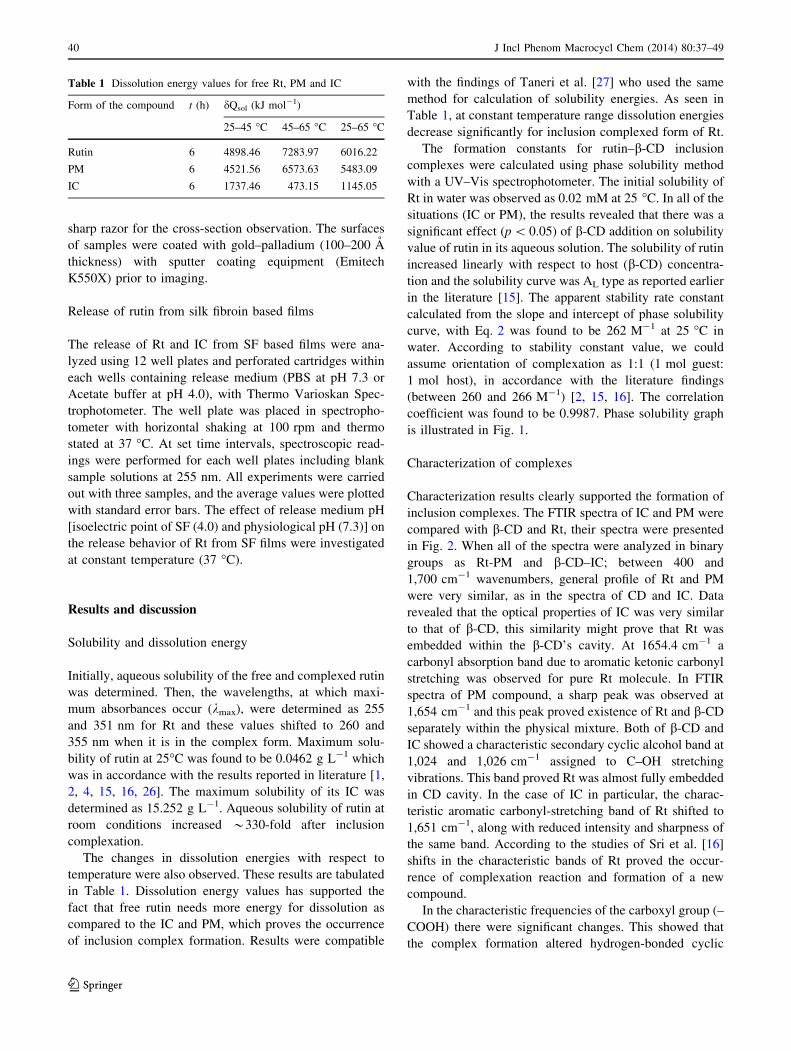
The formation constants for rutin–b-CD inclusion
complexes were calculated using phase solubility method
with a UV–Vis spectrophotometer. The initial solubility of
Rt in water was observed as 0.02 mM at 25 �C. In all of the
situations (IC or PM), the results revealed that there was a
significant effect (p \ 0.05) of b-CD addition on solubility
value of rutin in its aqueous solution. The solubility of rutin
increased linearly with respect to host (b-CD) concentra-
tion and the solubility curve was AL type as reported earlier
in the literature [15]. The apparent stability rate constant
calculated from the slope and intercept of phase solubility
curve, with Eq. 2 was found to be 262 M-1 at 25 �C in
water. According to stability constant value, we could
assume orientation of complexation as 1:1 (1 mol guest:
1 mol host), in accordance with the literature findings
(between 260 and 266 M-1) [2, 15, 16]. The correlation
coefficient was found to be 0.9987. Phase solubility graph
is illustrated in Fig. 1.
Characterization of complexes
Characterization results clearly supported the formation of
inclusion complexes. The FTIR spectra of IC and PM were
compared with b-CD and Rt, their spectra were presented
in Fig. 2. When all of the spectra were analyzed in binary
groups as Rt-PM and b-CD–IC; between 400 and
1,700 cm-1 wavenumbers, general profile of Rt and PM
were very similar, as in the spectra of CD and IC. Data
revealed that the optical properties of IC was very similar
to that of b-CD, this similarity might prove that Rt was
embedded within the b-CD’s cavity. At 1654.4 cm-1 a
carbonyl absorption band due to aromatic ketonic carbonyl
stretching was observed for pure Rt molecule. In FTIR
spectra of PM compound, a sharp peak was observed at
1,654 cm-1 and this peak proved existence of Rt and b-CD
separately within the physical mixture. Both of b-CD and
IC showed a characteristic secondary cyclic alcohol band at
1,024 and 1,026 cm-1 assigned to C–OH stretching
vibrations. This band proved Rt was almost fully embedded
in CD cavity. In the case of IC in particular, the charac-
teristic aromatic carbonyl-stretching band of Rt shifted to
1,651 cm-1, along with reduced intensity and sharpness of
the same band. According to the studies of Sri et al. [16]
shifts in the characteristic bands of Rt proved the occur-
rence of complexation reaction and formation of a new
compound.
In the characteristic frequencies of the carboxyl group (–
COOH) there were significant changes. This showed that
the complex formation altered hydrogen-bonded cyclic
Table 1 Dissolution energy values for free Rt, PM and IC
Form of the compound t (h) dQsol (kJ mol-1)
25–45 �C 45–65 �C 25–65 �C
Rutin 6 4898.46 7283.97 6016.22
PM 6 4521.56 6573.63 5483.09
IC 6 1737.46 473.15 1145.05
40 J Incl Phenom Macrocycl Chem (2014) 80:37–49
123
dimer structure of the carboxyl-group. Characteristic bond
types and their wavenumbers that were used in detection of
complex formation are tabulated in Table 2.
With XRD analysis it was possible to detect whether
inclusion complexation was formed or the compounds were
only physically mixed [28]. The samples were analyzed in
β-CD (mM)0 2 4 6 8 10 12 14 16 18
Com
plex
ed R
t (
mM
)
0.00
0.02
0.04
0.06
0.08
0.10
0.12Fig. 1 Phase solubility chart
for rutin–b-CD complexes
Fig. 2 FTIR spectra of b-CD, IC, PM, Rt
Table 2 FTIR Analysis for Detecting Presence of Complexation Reaction
Type of bond Wavenumber (cm-1) Shifted to Vibrational assignments Reason
C=O 1,709 1,730 cm-1 C=O stretching band (intense) suggesting a less strong or no
H-bonding interaction
C–C–O–H 1,403 1,396 cm-1 C–C–O–H stretching the strength of the H-bonds decreased
and complexation occurred through
the carboxyl groupC–O–H 1,271 1,265 cm-1 C–O–H in-plane bending modes
J Incl Phenom Macrocycl Chem (2014) 80:37–49 41
123
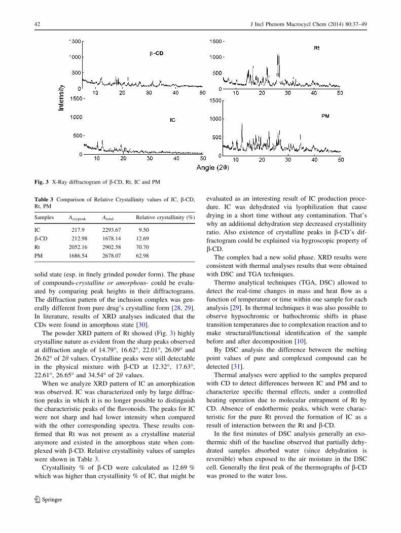
solid state (esp. in finely grinded powder form). The phase
of compounds-crystalline or amorphous- could be evalu-
ated by comparing peak heights in their diffractograms.
The diffraction pattern of the inclusion complex was gen-
erally different from pure drug’s crystalline form [28, 29].
In literature, results of XRD analyses indicated that the
CDs were found in amorphous state [30].
The powder XRD pattern of Rt showed (Fig. 3) highly
crystalline nature as evident from the sharp peaks observed
at diffraction angle of 14.79�, 16.62�, 22.01�, 26.09� and
26.62� of 2h values. Crystalline peaks were still detectable
in the physical mixture with b-CD at 12.32�, 17.63�,
22.61�, 26.65� and 34.54� of 2h values.
When we analyze XRD pattern of IC an amorphization
was observed. IC was characterized only by large diffrac-
tion peaks in which it is no longer possible to distinguish
the characteristic peaks of the flavonoids. The peaks for IC
were not sharp and had lower intensity when compared
with the other corresponding spectra. These results con-
firmed that Rt was not present as a crystalline material
anymore and existed in the amorphous state when com-
plexed with b-CD. Relative crystallinity values of samples
were shown in Table 3.
Crystallinity % of b-CD were calculated as 12.69 %
which was higher than crystallinity % of IC, that might be
evaluated as an interesting result of IC production proce-
dure. IC was dehydrated via lyophilization that cause
drying in a short time without any contamination. That’s
why an additional dehydration step decreased crystallinity
ratio. Also existence of crystalline peaks in b-CD’s dif-
fractogram could be explained via hygroscopic property of
b-CD.
The complex had a new solid phase. XRD results were
consistent with thermal analyses results that were obtained
with DSC and TGA techniques.
Thermo analytical techniques (TGA, DSC) allowed to
detect the real-time changes in mass and heat flow as a
function of temperature or time within one sample for each
analysis [29]. In thermal techniques it was also possible to
observe hypsochromic or bathochromic shifts in phase
transition temperatures due to complexation reaction and to
make structural/functional identification of the sample
before and after decomposition [10].
By DSC analysis the difference between the melting
point values of pure and complexed compound can be
detected [31].
Thermal analyses were applied to the samples prepared
with CD to detect differences between IC and PM and to
characterize specific thermal effects, under a controlled
heating operation due to molecular entrapment of Rt by
CD. Absence of endothermic peaks, which were charac-
teristic for the pure Rt proved the formation of IC as a
result of interaction between the Rt and b-CD.
In the first minutes of DSC analysis generally an exo-
thermic shift of the baseline observed that partially dehy-
drated samples absorbed water (since dehydration is
reversible) when exposed to the air moisture in the DSC
cell. Generally the first peak of the thermographs of b-CD
was proned to the water loss.
Fig. 3 X-Ray diffractogram of b-CD, Rt, IC and PM
Table 3 Comparison of Relative Crystallinity values of IC, b-CD,
Rt, PM
Samples Acrypeak Atotal Relative crystallinity (%)
IC 217.9 2293.67 9.50
b-CD 212.98 1678.14 12.69
Rt 2052.16 2902.58 70.70
PM 1686.54 2678.07 62.98
42 J Incl Phenom Macrocycl Chem (2014) 80:37–49
123
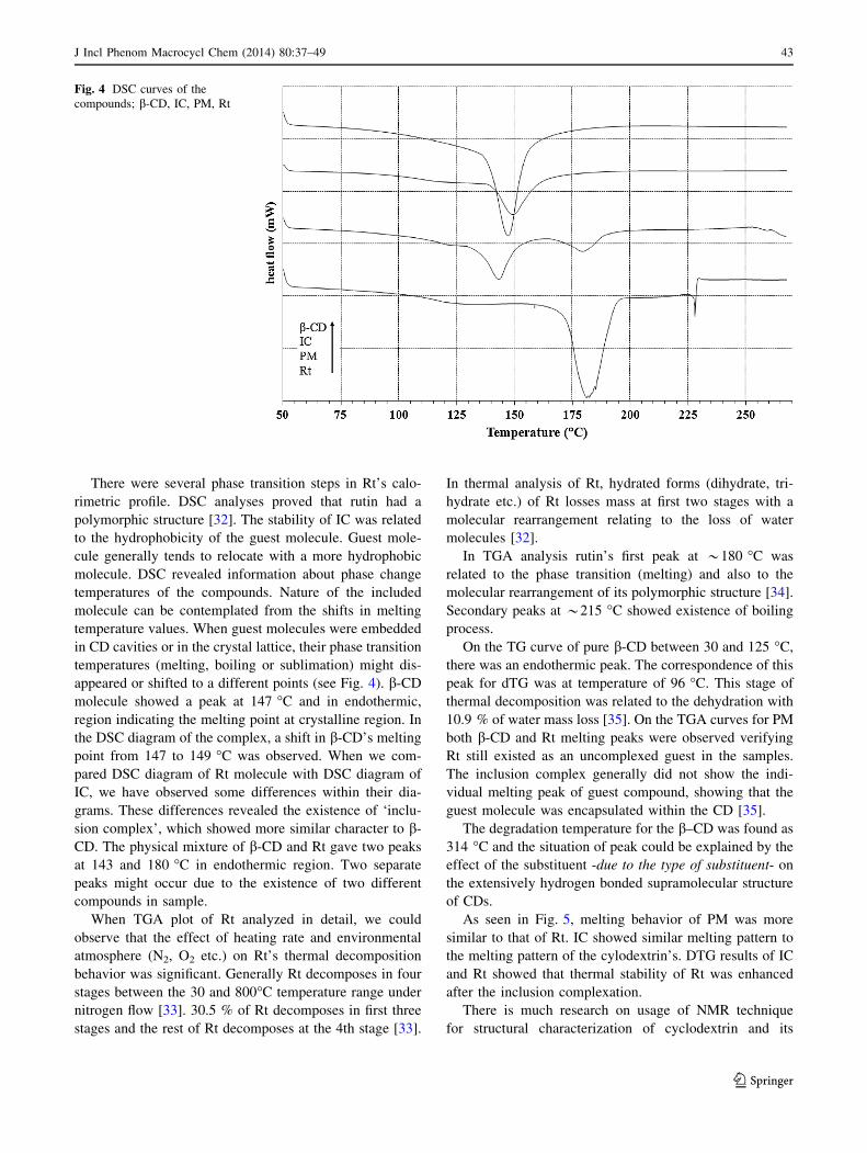
There were several phase transition steps in Rt’s calo-
rimetric profile. DSC analyses proved that rutin had a
polymorphic structure [32]. The stability of IC was related
to the hydrophobicity of the guest molecule. Guest mole-
cule generally tends to relocate with a more hydrophobic
molecule. DSC revealed information about phase change
temperatures of the compounds. Nature of the included
molecule can be contemplated from the shifts in melting
temperature values. When guest molecules were embedded
in CD cavities or in the crystal lattice, their phase transition
temperatures (melting, boiling or sublimation) might dis-
appeared or shifted to a different points (see Fig. 4). b-CD
molecule showed a peak at 147 �C and in endothermic,
region indicating the melting point at crystalline region. In
the DSC diagram of the complex, a shift in b-CD’s melting
point from 147 to 149 �C was observed. When we com-
pared DSC diagram of Rt molecule with DSC diagram of
IC, we have observed some differences within their dia-
grams. These differences revealed the existence of ‘inclu-
sion complex’, which showed more similar character to b-
CD. The physical mixture of b-CD and Rt gave two peaks
at 143 and 180 �C in endothermic region. Two separate
peaks might occur due to the existence of two different
compounds in sample.
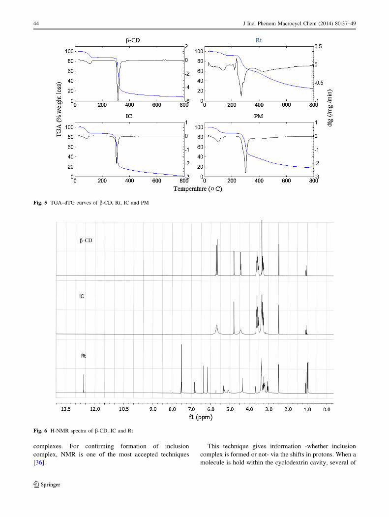
When TGA plot of Rt analyzed in detail, we could
observe that the effect of heating rate and environmental
atmosphere (N2, O2 etc.) on Rt’s thermal decomposition
behavior was significant. Generally Rt decomposes in four
stages between the 30 and 800�C temperature range under
nitrogen flow [33]. 30.5 % of Rt decomposes in first three
stages and the rest of Rt decomposes at the 4th stage [33].
In thermal analysis of Rt, hydrated forms (dihydrate, tri-
hydrate etc.) of Rt losses mass at first two stages with a
molecular rearrangement relating to the loss of water
molecules [32].
In TGA analysis rutin’s first peak at *180 �C was
related to the phase transition (melting) and also to the
molecular rearrangement of its polymorphic structure [34].
Secondary peaks at *215 �C showed existence of boiling
process.
On the TG curve of pure b-CD between 30 and 125 �C,
there was an endothermic peak. The correspondence of this
peak for dTG was at temperature of 96 �C. This stage of
thermal decomposition was related to the dehydration with
10.9 % of water mass loss [35]. On the TGA curves for PM
both b-CD and Rt melting peaks were observed verifying
Rt still existed as an uncomplexed guest in the samples.
The inclusion complex generally did not show the indi-
vidual melting peak of guest compound, showing that the
guest molecule was encapsulated within the CD [35].
The degradation temperature for the b–CD was found as
314 �C and the situation of peak could be explained by the
effect of the substituent -due to the type of substituent- on
the extensively hydrogen bonded supramolecular structure
of CDs.
As seen in Fig. 5, melting behavior of PM was more
similar to that of Rt. IC showed similar melting pattern to
the melting pattern of the cylodextrin’s. DTG results of IC
and Rt showed that thermal stability of Rt was enhanced
after the inclusion complexation.
There is much research on usage of NMR technique
for structural characterization of cyclodextrin and its
Fig. 4 DSC curves of the
compounds; b-CD, IC, PM, Rt
J Incl Phenom Macrocycl Chem (2014) 80:37–49 43
123
complexes. For confirming formation of inclusion
complex, NMR is one of the most accepted techniques
[36].
This technique gives information -whether inclusion
complex is formed or not- via the shifts in protons. When a
molecule is hold within the cyclodextrin cavity, several of
Fig. 5 TGA–dTG curves of b-CD, Rt, IC and PM
Fig. 6 H-NMR spectra of b-CD, IC and Rt
44 J Incl Phenom Macrocycl Chem (2014) 80:37–49
123

the protons on the interior of the host and in the protons of
the guest-the part reacting with the cavity- shifts are
observed. According to previous studies, relatively large
shift in the interior protons proved formation of inclusion
complex [36–38].
The results of NMR analyses are shown in Figs. 6 and 7.
H-NMR and C-NMR were applied for Rt, IC and b-CD
samples. In both of H-NMR and C-NMR spectra, the
characteristic peaks for rutin were observed in spectrum of
inclusion complex and its spectrum contained peaks at
similar regions with b-CDs. These results showed the
formation of inclusion complex between Rt and b-CD.
Electron microscope images of the samples could be
seen from Fig. 8. Rt molecules were visualized in the form
of column like crystals and b-CD particles showed a dia-
mond shape morphology. In microstructure image of PM,
Rt molecules covered surface of b-CD particles and there
was not a definite interaction between Rt and b-CD. In
SEM image of PM, some part of Rt particles embedded in
b-CD and a comparable morphology with pure compounds
taken separately, also. In contrast, a profound change was
observed in the morphology and shape of IC particles,
revealing an apparent interaction in the solid-state. Also, IC
presented a homogenous morphology with rectangular
prism shaped particles.
Morphology of films
Surface images of SF films (a) Control SF film, (b) Rt,
(c) IC and (d) PM loaded film were obtained by SEM
magnified at 1,0009 see Fig. 9. Also cross section images
of the SF films obtained by SEM magnified at 1,0009 are
given in (Fig. 9). From SEM observations, it could be
concluded that at high concentrations of Rt, semi-uniform
distribution was observed in SF films while totally uniform
distribution was observed for IC loaded SF films. Micro-
scopic phase separation and cracks were not observed for
these films. Cross section images of SF films revealed the
dense film structures.
Dissolution profile and rutin release
The dissolution characteristics of free Rt and IC of Rt are
given in Table 1. More than 70 % of Rt was dissolved at
first 20 min when Rt formed complex with b-CD. Disso-
lution rate of IC was much higher than dissolution rates of
physical mixtures and free Rt. Dissolution efficiency per-
cent at 40 min (DE40 (%)) were calculated for Rt, IC and
PM. The DE40 (%) values supported the dissolution char-
acteristics of each compound that were tabulated in
Table 1. IC of rutin–b-CD in 1:1 molar ratio showed higher
Fig. 7 C-NMR spectra of b-CD, IC and Rt
J Incl Phenom Macrocycl Chem (2014) 80:37–49 45
123

dissolution efficiency (Fig. 10; Table 4) than the physical
mixture and free rutin.
As seen in Figs. 11, 12, 13 almost 50 % of Rt was
released from SF film within the first 5 h. The rest of Rt
was released within next 19 h completely at a sustained
manner. Release behaviour of Rt was mostly the same in
both acidic and neutral medium (acetic acid buffer at pH
4.0 and PBS at pH 7.3; 10 mM; 37�C; 100 rpm shaking).
As seen in Fig. 11 burst release occurred for IC loaded SF
film at the beginning in physiological conditions due to
Fig. 8 5,0009 magnified SEM images of compounds; b-CD (a), Rt (b), IC (c), PM (d)
Fig. 9 Surface image of a Control SF film, b IC film, c PM film, d Rt film; cross section image of e Control SF film f IC film g PM film h Rt film
taken with SEM
46 J Incl Phenom Macrocycl Chem (2014) 80:37–49
123

high dissolution rates of IC in both neutral and acidic
medium. Jug et al. [22] related geometry of the polymer
matrix (presence large surface cause fast swelling of rutin)
for burst release from films. During release tests, the whole
structure of films were preserved both in acidic and neutral
release medium, only slight differences in colour of the
films were observed. Also, films become crispy and brittle
after release period.
Conclusion
Among all of cyclodextrins, b-CD is preferred because of
its suitable cavity sizes and low price. Hence, in our present
Fig. 10 Percent dissolution
profiles of IC, PM, Rt
Table 4 DE 40 (%) for Rt and
Rt-CD binary systems
(mean ± SD values; n = 3)
Product DE40 (%)
Rt 48.65 ± 0.037
PM of Rt 65.33 ± 0.144
IC of Rt 71.90 ± 0.068
time (h)0 5 10 15 20 25
% R
utin
Rel
ease
0
20
40
60
80
100
IC at pH= 4.0IC at pH= 7.3
Fig. 11 Release profile for IC of Rt from SF film
time (h)0 5 10 15 20 25
% R
utin
Rel
ease
0
20
40
60
80
100
PM at pH = 4.0 PM at pH = 7.3
Fig. 12 Release profile for PM of Rt from SF film
time (h)0 5 10 15 20 25
% R
utin
Rel
ease
0
20
40
60
80
100
Rt at pH= 4.0Rt at pH= 7.3
Fig. 13 Release profile for Rt from SF film
J Incl Phenom Macrocycl Chem (2014) 80:37–49 47
123

work, rutin–b-CD solid complex was successfully pre-
pared. Aqueous solubility of Rt was increased as a result of
the inclusion complex formation. Improvement in the
hydrophilicity of Rt by CD caused to the enhancement of
dissolution rate. The calculated solubility energies showed
the energy needed to solubilise in water was highest in free
Rt and lowest for IC. Analysis of the complexes by X-ray
diffraction, DSC and FT-IR methods showed considerable
interaction of b-CD with Rt. Results of dissolution profile
of Rt, PM and IC showed that addition of CD had an
increasing effect on solubility rate and amount. Solid
inclusion complexes exhibited higher dissolution efficien-
cies than their corresponding physical mixtures. Silk
fibroin based films were prepared and loading of Rt, PM
and IC into silk fibroin based films was carried out suc-
cessfully. By release tests it was investigated that most of
the Rt-independent from the form, whether free or com-
plexed-released from SF films within the first 5 h (burst
release occurs) and the rest of it released slowly within
24 h. Electron microscope analyses showed that films have
a homogenous and dense morphology. Consequently, silk
fibroin can be used to load natural compounds in the form
of films.
References
1. Alvarez-Parrilla, E., Rosa, L.D., Torres-Rivas, F., Rodrigo-Gar-
cia, J., Gonzalez-Aguilar, G.: Complexation of apple antioxi-
dants: chlorogenic acid, quercetin and rutin by b-cyclodextrin (b-
CD). J. Incl. Phenom. Macrocycl. Chem. 53(1–2), 121–129
(2005)
2. Haiyun, D., Jianbin, C., Guomei, Z., Shaomin, S., Jinhao, P.:
Preparation and spectral investigation on inclusion complex of
[beta]-cyclodextrin with rutin. Spectrochim. Acta Part A 59(14),
3421–3429 (2003)
3. Kacso, I., Borodi, G., Farcas, S., Hernanz, A., Bratu, I.: Host–
guest system of Vitamin B10 in b-cyclodextrin: characterization
of the interaction in solution and in solid state. J. Incl. Phenom.
Macrocycl. Chem. 68(1), 175–182 (2010)
4. Nguyen, T.A., Liu, B., Zhao, J., Thomas, D.S., Hook, J.M.: An
investigation into the supramolecular structure, solubility, sta-
bility and antioxidant activity of rutin/cyclodextrin inclusion
complex. Food Chem. 136(1), 186–192 (2013)
5. Zhao, J., Lin, D.-Q., Yao, S.-J.: Adsorption of rutin with a novel
b-cyclodextrin polymer adsorbent: thermodynamic and kinetic
study. Carbohydr. Polym. 90(4), 1764–1770 (2012)
6. Song, Z., Wang, L.: Chemiluminescence investigation of detec-
tion of rutin in medicine and human urine using controlled-
reagent-release technology. J. Agric. Food Chem. 49(12),
5697–5701 (2001)
7. Samlı, M.: Preparation and Characterization of Silk Fibroin
Based Materials Loaded With Natural Compounds. MSc Thesis,
Izmir Institute of Technology (2010)
8. Madison, P.H.: Carbohydrate Mediation of Aqueous Polymer-
izations: Cyclodextrin Mediation of Aqueous Polymerizations of
Methacrylates. (2001)
9. Fromming, K.H., Szejtli, J.: Cyclodextrins in Pharmacy. Topics
in Inclusion Science, vol. 5. Kluwer Academic, London (1993)
10. Giordano, F., Novak, C., Moyano, J.R.: Thermal analysis of
cyclodextrins and their inclusion compounds. Thermochim. Acta
380(2), 123–151 (2001)
11. Mura, P., Maestrelli, F., Cirri, M., Furlanetto, S., Pinzauti, S.:
Differential scanning calorimetry as an analytical tool in the
study of drug-cyclodextrin interactions. J. Therm. Anal. Calorim.
73(2), 635–646 (2003)
12. Agostiano, A., Catucci, L., Castagnolo, M., Colangelo, D.,
Cosma, P., Fini, P., Della Monica, M.: Interaction between
chlorophyll a and b-cyclodextrin derivatives in aqueous solutions.
J. Therm. Anal. Calorim. 70(1), 115–122 (2002)
13. Novak, C., Ehen, Z., Fodor, M., Jicsinszky, L., Orgovanyi, J.:
Application of combined thermoanalytical techniques in the
investigation of cyclodextrin inclusion complexes. J. Therm.
Anal. Calorim. 84(3), 693–701 (2006)
14. Dodziuk, H.: Cyclodextrins and their Complexes. WILEY-VCH,
Weinheim (2006)
15. Miyake, K., Arima, H., Hirayama, F., Yamamoto, M., Horikawa,
T., Sumiyoshi, H., Noda, S., Uekama, K.: Improvement of sol-
ubility and oral bioavailability of rutin by complexation with
2-hydroxypropyl-b-cyclodextrin. Pharm. Dev. Technol. 5(3),
399–407 (2000)
16. Sri, K.V., Kondaiah, A., Ratna, J.V., Annapurna, A.: Preparation
and characterization of quercetin and rutin cyclodextrin inclusion
complexes. Drug Dev. Ind. Pharm. 33(3), 245–253 (2007)
17. Singh, M., Sharma, R., Banerjee, U.C.: Research review paper:
biotechnological applications of cyclodextrins. Biotechnol. Adv.
20, 341–359 (2002)
18. Hassan, H.B., Kata, M., Er}os, I., Aigner, Z.: Preparation and
investigation of inclusion complexes containing gemfibrozil and
DIMEB. J. Incl. Phenom. Macrocycl. Chem. 50(3), 219–225
(2004)
19. Altman, G.H., Diaz, F., Jakuba, C., Calabro, T., Horan, R.L.,
Chen, J., Lu, H., Richmond, J., Kaplan, D.L.: Silk-based bio-
materials. Biomaterials 24(3), 401–416 (2003)
20. Fenyvesi, E., Balogh, K., Siro, I., Orgovanyi, J., Senyi, J., Otta,
K., Szente, L.: Permeability and release properties of cyclodex-
trin-containing poly(vinyl chloride) and polyethylene films.
J. Incl. Phenom. Macrocycl. Chem. 57(1–4), 371–374 (2007)
21. Fontananova, E., Di Profio, G., Curcio, E., Giorno, L., Drioli, E.:
Functionalization of polymeric membranes by impregnation and
in situ cross-linking of a PDMS/b-cyclodextrin network. J. Incl.
Phenom. Macrocycl. Chem. 57(1–4), 537–543 (2007)
22. Jug, M., Maestrelli, F., Mura, P.: Native and polymeric b-
cyclodextrins in performance improvement of chitosan films
aimed for buccal delivery of poorly soluble drugs. J. Incl. Phe-
nom. Macrocycl. Chem. 74(1–4), 87–97 (2012)
23. Peng, B., Li, R., Yan, W.: Solubility of rutin in ethanol ? water
at (273.15–323.15) K. J. Chem. Eng. Data 54(4), 1378–1381
(2009)
24. Ajisawa, A.: Dissolution of silk fibroin with calciumchloride/
ethanol aqueous solution. J Seric Sci Jpn 67(2), 91–94 (1998)
25. Bansal, P., Hall, M., Realff, M.J., Lee, J.H., Bommarius, A.S.:
Multivariate statistical analysis of X-ray data from cellulose: A
new method to determine degree of crystallinity and predict
hydrolysis rates. Bioresour. Technol. 101(12), 4461–4471 (2010)
26. Calabro, M.L., Tommasini, S., Donato, P., Stancanelli, R., Ra-
neri, D., Catania, S., Costa, C., Villari, V., Ficarra, P., Ficarra, R.:
The rutin/b-cyclodextrin interactions in fully aqueous solution:
spectroscopic studies and biological assays. J. Pharm. Biomed.
Anal. 36(5), 1019–1027 (2005)
27. Taneri, F., Guneri, T., Aigner, Z., Berkesi, O., Kata, M.: Ther-
moanalytical studies on complexes of ketoconazole with
48 J Incl Phenom Macrocycl Chem (2014) 80:37–49
123

cyclodextrin derivatives. J. Therm. Anal. Calorim. 74(3),
769–777 (2003)
28. Nalluri, B.N., Chowdary, K.P.R., Murthy, K.V.R., Hayman, A.R.,
Becket, G.: Physicochemical characterization and dissolution
properties of nimesulide–cyclodextrin binary systems. AAPS
Pharmaceutica 4(1), 6–17 (2005). article 2
29. Bettinetti, G., Novak, C., Sorrenti, M.: Thermal and structural
characterization of commercial a-, b-, and c-cyclodextrins.
J. Therm. Anal. Calorim. 68(2), 517–529 (2002)
30. Aigner, Z., Hassan, H.B., Berkesi, O., Kata, M., Er}os, I.: Ther-
moanalytical, FTIR and X-ray studies of gemfibrozil–cyclodex-
trin complexes. J. Therm. Anal. Calorim. 81(2), 267–272 (2005)
31. Patyi, G., Bodis, A., Antal, I., Vajna, B., Nagy, Z., Marosi, G.:
Thermal and spectroscopic analysis of inclusion complex of
spironolactone prepared by evaporation and hot melt methods.
J. Therm. Anal. Calorim. 102, 1–7 (2010)
32. Budavari, S.: The Merck Index, An Encyclopedia of Chemicals
Drugs and Biologicals. In. Merck & CO Inc., Whitehouse Station,
NJ (1996)
33. da Costa, E.M., Filho, J.M.B., do Nascimento, T.G., Macedo,
R.O.: Thermal characterization of the quercetin and rutin flavo-
noids. Thermochim. Acta 392–393, 79–84 (2002)
34. Madhusudanan, P.M., Krishnan, K., Ninan, K.N.: New equations
for kinetic analysis of non-isothermal reactions. Thermochim.
Acta 221(1), 13–21 (1993)
35. Orgovanyi, J., Poppl, L., Otta, K.H., Lovas, G.A.: Thermoana-
lytical method for studying the guest content in cyclodextrin
inclusion complexes. J. Therm. Anal. Calorim. 81(2), 261–266
(2005)
36. Schneider, H.-J., Hacket, F., Rudiger, V., Ikeda, H.: NMR studies
of cyclodextrins and cyclodextrin complexes. Chem. Rev. 98(5),
1755–1786 (1998)
37. Bender, M.L., Komiyama, M.: Cyclodextrin chemistry. Reactiv-
ity and Structure Concepts in Organic Chemistry, vol. 6.
Springer, Berlin Heidelberg (1978)
38. Szejtli, J.: Cyclodextrin Technology. Topics in Inclusion Science.
Kluwer Academic, Budapest (1988)
J Incl Phenom Macrocycl Chem (2014) 80:37–49 49
123


![filtered correlation-network approach · 2014. 10. 22. · arXiv:1410.5621v1 [q-fin.PM] 21 Oct 2014 Risk diversification: a study of persistence with a filtered correlation-network](https://static.fdocuments.net/doc/165x107/6141e4682035ff3bc76251c1/iltered-correlation-network-approach-2014-10-22-arxiv14105621v1-q-finpm.jpg)

