Characterization of Cytochrome 579, an Unusual Cytochrome … · were purified from serum by using...
9
APPLIED AND ENVIRONMENTAL MICROBIOLOGY, July 2008, p. 4454–4462 Vol. 74, No. 14 0099-2240/08/$08.000 doi:10.1128/AEM.02799-07 Copyright © 2008, American Society for Microbiology. All Rights Reserved. Characterization of Cytochrome 579, an Unusual Cytochrome Isolated from an Iron-Oxidizing Microbial Community Steven W. Singer, 1 Clara S. Chan, 2 † Adam Zemla, 3 Nathan C. VerBerkmoes, 4 Mona Hwang, 1 Robert L. Hettich, 4 Jillian F. Banfield, 2 and Michael P. Thelen 1 * Chemistry Directorate 1 and Computations Directorate, 3 Lawrence Livermore National Laboratory, Livermore, California 94550; Department of Earth and Planetary Sciences, University of California, Berkeley, Berkeley, California 94720 2 ; and Chemical Sciences Division, Oak Ridge National Laboratory, Oak Ridge, Tennessee 37831 4 Received 11 December 2007/Accepted 26 March 2008 A novel, soluble cytochrome with an unusual visible spectral signature at 579 nm (Cyt 579 ) has been characterized after isolation from several different microbial biofilms collected in an extremely acidic ecosys- tem. Previous proteogenomic studies of an Fe(II)-oxidizing community indicated that this abundant red cytochrome could be extracted from the biofilms with dilute sulfuric acid. Here, we found that the Fe(II)- dependent reduction of Cyt 579 was thermodynamically favorable at a pH of >3, raising the possibility that Cyt 579 acts as an accessory protein for electron transfer. The results of transmission electron microscopy of immunogold-labeled biofilm indicated that Cyt 579 is localized near the bacterial cell surface, consistent with periplasmic localization. The results of further protein analysis of Cyt 579 , using preparative chromatofocusing and sodium dodecyl sulfate-polyacrylamide gel electrophoresis, revealed three forms of the protein that correspond to different N-terminal truncations of the amino acid sequence. The results of intact-protein analysis corroborated the posttranslational modifications of these forms and identified a genomically unchar- acterized Cyt 579 variant. Homology modeling was used to predict the overall cytochrome structure and heme binding site; the positions of nine amino acid substitutions found in three Cyt 579 variants all map to the surface of the protein and away from the heme group. Based on this detailed characterization of Cyt 579 , we propose that Cyt 579 acts as an electron transfer protein, shuttling electrons derived from Fe(II) oxidation to support critical metabolic functions in the acidophilic microbial community. Biological oxidation of Fe(II) by acidophilic microbial com- munities found in mines with exposed pyrite ore accelerates the dissolution of FeS 2 and acidification of the mine water, resulting in acid mine drainage (AMD), a global environmen- tal problem (8). One of the most-intensively studied AMD sites is the Richmond Mine at Iron Mountain, CA, where copious biofilm communities are found in extremely low-pH (0.5 to 1.0) solutions (2). Most of these communities are pink biofilms dominated by Leptospirillum group II bacteria, with lower abundances of Leptospirillum group III bacteria and several archaeal species (4). A Leptospirillum group II bacte- rium-dominated biofilm was collected at the “5-way” site at the Richmond Mine (11) and analyzed by metagenomic sequenc- ing (5-way community genomics data set [24]). Proteomic char- acterization by mass spectrometry (MS) of a similar biofilm isolated from the “AB end” site of the Richmond Mine iden- tified an abundant extracellular protein from Leptospirillum group II bacteria, encoded by gene 20 on sequencing scaffold 20 (gene 14-20), that has a CXXCH heme binding motif com- mon to c-type cytochromes but otherwise insignificant se- quence similarity to known proteins (17). The results of gel electrophoresis and N-terminal sequencing confirmed that this protein contained heme and was abundant in the extracellular fraction. The first 40 amino acids deduced from the environ- mental genomic sequence were nearly identical to the N-ter- minal sequence deduced for the Fe(II)-oxidizing cytochrome 579 (Cyt 579 ) purified from an isolate of Leptospirillum ferriphi- lum. The reduction potential of L. ferriphilum Cyt 579 was esti- mated to be 660 mV, and the cytochrome was fully reduced in the presence of excess Fe(II) at pH 2.0 (17). A cytochrome with very similar spectral and pH-dependent-redox properties had also been isolated from Leptospirillum ferrooxidans (10). The ability of L. ferriphilum and L. ferrooxidans Cyt 579 to oxi- dize Fe(II) at low pH led to the hypothesis that this novel cytochrome identified in the biofilm acted as the primary Fe(II) oxidant for Leptospirillum group II bacteria. Here we report the purification and characterization of Cyt 579 from a Leptospirillum group II bacterium-dominated biofilm collected at Richmond Mine. The results of detailed biochemical and MS studies of Cyt 579 from the biofilm suggest that it functions as a periplasmic electron transfer protein. MATERIALS AND METHODS Isolation of extracellular proteins. Richmond Mine biofilm samples were collected in 50-ml conical Falcon tubes (BD Biosciences, San Jose, CA), frozen at the site on dry ice, and later stored at 80°C. Biofilm samples were collected from the AB end site (near the junction of the “A drift” and B drift) in January 2004; from the C drift site (15 m beyond the AMD dam) in November 2005; and from the “UBA” site (in the A drift) in November 2005. A map describing the field site can be found in online supplementary information of reference 11. To obtain the extracellular fraction, the biofilm was thawed, suspended in 110 ml 0.2 MH 2 SO 4 (pH 1.1), and homogenized in a glass tube by using several vigorous strokes of a tight-fitting, round, glass pestle. The resulting homogeneous cell suspension was stirred for 2 h at 4°C and then centrifuged at 24,000 g for 12 * Corresponding author. Mailing address: Lawrence Livermore Na- tional Laboratory, L-452, P.O. Box 808, Livermore, CA 94551-0808. Phone: (925) 422-6547. Fax: (925) 422-2282. E-mail: [email protected]. † Present address: Woods Hole Oceanographic Institution, Woods Hole, MA 02543. Published ahead of print on 9 May 2008. 4454 on April 6, 2020 by guest http://aem.asm.org/ Downloaded from
Transcript of Characterization of Cytochrome 579, an Unusual Cytochrome … · were purified from serum by using...

APPLIED AND ENVIRONMENTAL MICROBIOLOGY, July 2008, p. 4454–4462 Vol. 74, No. 140099-2240/08/$08.00�0 doi:10.1128/AEM.02799-07Copyright © 2008, American Society for Microbiology. All Rights Reserved.
Characterization of Cytochrome 579, an Unusual Cytochrome Isolatedfrom an Iron-Oxidizing Microbial Community�
Steven W. Singer,1 Clara S. Chan,2† Adam Zemla,3 Nathan C. VerBerkmoes,4 Mona Hwang,1Robert L. Hettich,4 Jillian F. Banfield,2 and Michael P. Thelen1*
Chemistry Directorate1 and Computations Directorate,3 Lawrence Livermore National Laboratory, Livermore, California 94550;Department of Earth and Planetary Sciences, University of California, Berkeley, Berkeley, California 947202; and
Chemical Sciences Division, Oak Ridge National Laboratory, Oak Ridge, Tennessee 378314
Received 11 December 2007/Accepted 26 March 2008
A novel, soluble cytochrome with an unusual visible spectral signature at 579 nm (Cyt579) has beencharacterized after isolation from several different microbial biofilms collected in an extremely acidic ecosys-tem. Previous proteogenomic studies of an Fe(II)-oxidizing community indicated that this abundant redcytochrome could be extracted from the biofilms with dilute sulfuric acid. Here, we found that the Fe(II)-dependent reduction of Cyt579 was thermodynamically favorable at a pH of >3, raising the possibility thatCyt579 acts as an accessory protein for electron transfer. The results of transmission electron microscopy ofimmunogold-labeled biofilm indicated that Cyt579 is localized near the bacterial cell surface, consistent withperiplasmic localization. The results of further protein analysis of Cyt579, using preparative chromatofocusingand sodium dodecyl sulfate-polyacrylamide gel electrophoresis, revealed three forms of the protein thatcorrespond to different N-terminal truncations of the amino acid sequence. The results of intact-proteinanalysis corroborated the posttranslational modifications of these forms and identified a genomically unchar-acterized Cyt579 variant. Homology modeling was used to predict the overall cytochrome structure and hemebinding site; the positions of nine amino acid substitutions found in three Cyt579 variants all map to the surfaceof the protein and away from the heme group. Based on this detailed characterization of Cyt579, we propose thatCyt579 acts as an electron transfer protein, shuttling electrons derived from Fe(II) oxidation to support criticalmetabolic functions in the acidophilic microbial community.
Biological oxidation of Fe(II) by acidophilic microbial com-munities found in mines with exposed pyrite ore acceleratesthe dissolution of FeS2 and acidification of the mine water,resulting in acid mine drainage (AMD), a global environmen-tal problem (8). One of the most-intensively studied AMDsites is the Richmond Mine at Iron Mountain, CA, wherecopious biofilm communities are found in extremely low-pH(0.5 to 1.0) solutions (2). Most of these communities are pinkbiofilms dominated by Leptospirillum group II bacteria, withlower abundances of Leptospirillum group III bacteria andseveral archaeal species (4). A Leptospirillum group II bacte-rium-dominated biofilm was collected at the “5-way” site at theRichmond Mine (11) and analyzed by metagenomic sequenc-ing (5-way community genomics data set [24]). Proteomic char-acterization by mass spectrometry (MS) of a similar biofilmisolated from the “AB end” site of the Richmond Mine iden-tified an abundant extracellular protein from Leptospirillumgroup II bacteria, encoded by gene 20 on sequencing scaffold20 (gene 14-20), that has a CXXCH heme binding motif com-mon to c-type cytochromes but otherwise insignificant se-quence similarity to known proteins (17). The results of gelelectrophoresis and N-terminal sequencing confirmed that thisprotein contained heme and was abundant in the extracellular
fraction. The first 40 amino acids deduced from the environ-mental genomic sequence were nearly identical to the N-ter-minal sequence deduced for the Fe(II)-oxidizing cytochrome579 (Cyt579) purified from an isolate of Leptospirillum ferriphi-lum. The reduction potential of L. ferriphilum Cyt579 was esti-mated to be �660 mV, and the cytochrome was fully reducedin the presence of excess Fe(II) at pH 2.0 (17). A cytochromewith very similar spectral and pH-dependent-redox propertieshad also been isolated from Leptospirillum ferrooxidans (10).The ability of L. ferriphilum and L. ferrooxidans Cyt579 to oxi-dize Fe(II) at low pH led to the hypothesis that this novelcytochrome identified in the biofilm acted as the primaryFe(II) oxidant for Leptospirillum group II bacteria.
Here we report the purification and characterization ofCyt579 from a Leptospirillum group II bacterium-dominatedbiofilm collected at Richmond Mine. The results of detailedbiochemical and MS studies of Cyt579 from the biofilm suggestthat it functions as a periplasmic electron transfer protein.
MATERIALS AND METHODS
Isolation of extracellular proteins. Richmond Mine biofilm samples werecollected in 50-ml conical Falcon tubes (BD Biosciences, San Jose, CA), frozenat the site on dry ice, and later stored at �80°C. Biofilm samples were collectedfrom the AB end site (near the junction of the “A drift” and B drift) in January2004; from the C drift site (15 m beyond the AMD dam) in November 2005; andfrom the “UBA” site (in the A drift) in November 2005. A map describing thefield site can be found in online supplementary information of reference 11. Toobtain the extracellular fraction, the biofilm was thawed, suspended in 110 ml 0.2M H2SO4 (pH 1.1), and homogenized in a glass tube by using several vigorousstrokes of a tight-fitting, round, glass pestle. The resulting homogeneous cellsuspension was stirred for 2 h at 4°C and then centrifuged at 24,000 � g for 12
* Corresponding author. Mailing address: Lawrence Livermore Na-tional Laboratory, L-452, P.O. Box 808, Livermore, CA 94551-0808.Phone: (925) 422-6547. Fax: (925) 422-2282. E-mail: [email protected].
† Present address: Woods Hole Oceanographic Institution, WoodsHole, MA 02543.
� Published ahead of print on 9 May 2008.
4454
on April 6, 2020 by guest
http://aem.asm
.org/D
ownloaded from

min. The supernatant is the extracellular fraction used for cytochrome purifica-tion. For proteomic analysis, proteins from a 10-ml sample of the extracellularfraction of the biofilm from the C drift were precipitated with 10% trichloro-acetic acid and the precipitate was collected by centrifugation, rinsed twice withcold methanol, and air dried.
Purification of Cyt579. Proteins in the extracellular fraction (150 ml) wereprecipitated with (NH4)2SO4 and redissolved in �5 ml sample buffer (SB)containing 20 mM H2SO4 and 100 mM NH4(SO4)2 at pH 2.2. A light redprecipitate at 45% NH4(SO4)2 saturation was gelatinous, indicating the presenceof exopolysaccharides. A deeper red precipitate at 95% NH4(SO4)2 contained 75to 80% of the protein found in the extracellular fraction. This precipitate wasdialyzed for 16 h at 4°C against 1 liter SB. The dialysate was loaded onto anSP-Sepharose FF column (5 ml) preequilibrated in SB. The column was washedwith 2 column volumes of SB, and the red fraction (9 ml; 4 mg total protein)eluted with 100 mM sodium acetate (NaOAc), pH 5.0. Visible spectroscopyindicated that the pH 5.0 fraction was highly enriched in Cyt579. The remainingprotein was removed with a 0 to 2 M NaCl gradient (30 ml) in pH 5.0 buffer.Between 1.2 M and 2.0 M NaCl, light yellow fractions (3 ml each; 2 mg totalprotein) eluted that had visible spectra consistent with the presence of c-typecytochromes (�-band at 552 nm for reduced samples).
Immunogold labeling of Cyt579 and transmission electron microscopy (TEM)of biofilms. Polyclonal antibodies were produced in rabbits (Covance, Denver,PA) by using the cation-exchange fraction of Cyt579 as the antigen. Prior toimmunization, the antigen was concentrated by using MicroCon spin filters(10-kDa-molecular-mass cutoff; Millipore, Billerica, MA) and resuspended inphosphate-buffered saline. Immunoblotting of a biofilm lysate indicated a highspecificity of the antibody preparation for Cyt579 (data not shown). Antibodieswere purified from serum by using a Melon gel antibody purification kit (Pierce,Rockville, IL).
A biofilm sample was frozen under high pressure (Bal-tec HPM 010) andfreeze substituted in 0.2% glutaraldehyde and 0.1% uranyl acetate in acetone.The sample was then rinsed in acetone and embedded in LR White resin.Microtomed sections (�70 nm thick) were mounted on carbon-coated, Formvarfilm-covered nickel grids and blocked with bovine serum albumin and cold-water-fish gelatin (Sigma Aldrich, St. Louis, MO). The anti-Cyt579 antibody was used asthe primary antibody, and goat anti-rabbit antibody conjugated with 10-nm goldparticles was used as the secondary antibody. After being labeled, samples werefixed in 0.5% glutaraldehyde. Prior to analysis, all samples were stained withuranyl acetate and lead citrate. Samples were observed with an FEI Tecnai 12TEM operated at 120 kV. Images were recorded on film, and the negatives werescanned and digitally processed to optimize contrast by using Adobe Photoshop.
Separation of forms of Cyt579. The fraction enriched in Cyt579 (3 mg) from theC drift biofilm in 100 mM NaOAc, pH 5.0, was concentrated (as described above)to �1 ml and dialyzed for 16 h against 1 liter of 25 mM L-histidine–HCl, pH 6.2.The dialyzed Cyt579 fraction was loaded onto a 1- by 30-cm chromatofocusingcolumn (PBE 94 Polybuffer exchange; Amersham Biosciences, Piscataway, NJ)preequilibrated with 2 column volumes of pH 6.2 buffer and eluted with PBE 74
Polybuffer, pH 5.0. Two red fractions eluted from the column at pH 5.5 (0.3 mg)and pH 5.1 (1.0 mg), and a third fraction was eluted with 1 M NaCl in 100 mMNaOAc, pH 5.0 (1.5 mg).
pH-dependent Fe(II) oxidation of Cyt579. The C drift site Cyt579 fraction (1.5mg/ml) was diluted 1:10 in 100 mM glycine–200 mM SO4
2�, pH 2.0, and oxidizedwith a small amount of Fe2(SO4)3 in pH 2.0 buffer. The oxidized Cyt579 was thendiluted 1:10 further in buffer that contained 30 mM FeSO4–200 mM SO4
2� in a1.5-ml quartz cuvette, and the visible spectrum was obtained after 1 min. Low-pHbuffers (pH 1.2 to 4.0) were prepared according to the method described bySchnaitman et al. using glycine and �-alanine (19). The spectrum was retakenafter 10 min to ensure that the reaction had reached equilibrium. In all cases, thereaction was �95% equilibrated after 1 min.
Protein MS. Intact-protein characterization was performed by high-resolutionFourier transform ion cyclotron resonance (FTICR)-MS analysis. All FTICRmass spectra were acquired with a Varian 9.4-Tesla HiRes electrospray FTICR-MS. Micromolar solutions of the purified Cyt579 proteins were prepared in 50:50water-acetonitrile (with �0.1% acetic acid added). Using a syringe pump (flowrate of 1.75 �l/min), the analyte was directly infused into a Z-type electrosprayion source. After generation, ions were accumulated in an external hexapole for1 s and then transferred into the high-vacuum region with a quadrupole lenssystem. Detection then followed in the cylindrical analyzer cell of the MS.Calibration of the MS was accomplished externally with the various charge statesof the protein ubiquitin, resulting in a mass accuracy of plus or minus 3 to 5 ppmand mass resolutions of 50,000 to 160,000 Da (full width at half maximum), aspreviously described (7). Ion dissociation was accomplished by infrared mul-tiphoton dissociation (IRMPD) with a Synrad carbon dioxide laser (75-W max-imum power and 10.2-�m wavelength). For this experiment, the desired parention was isolated by ejecting all other ons from the analyzer cell and dissociatedwith infrared laser irradiation (30% maximum laser power for 1.5 s), and theresulting fragment ions were measured at high resolution in the FTICR analyzercell.
To verify amino acid differences in Cyt579 variants, purified samples weredenatured, reduced, and digested with trypsin (sequencing grade; Promega,Madison, WI). Peptides were analyzed by using one-dimensional liquid chroma-tography-tandem MS (LC–MS-MS) on a Thermo Fisher linear-trapping quadru-pole instrument. All MS-MS spectra were searched with DBDigger (22) againsta database of all proteins predicted by genomic sequencing of biofilm samples(15, 24), as well as all potential amino acid variants of Cyt579. The output datafiles were then filtered and sorted with the DTA Select algorithm (21) using thefollowing parameters: fully tryptic peptides only; delta correlation value of atleast 0.08; cross-correlation scores of at least 25 (�1 ions), 30 (�2 ions), and 45(�3 ions); and at least two unique peptides per protein.
Amino acid variants were also verified from crude extracellular fractions ofbiofilms from the A bend, C drift, and UBA sites. Extracellular proteins weredenatured, reduced, trypsin digested, and analyzed by using two-dimensionalLC–MS-MS on a linear-trapping quadrupole instrument as previously described
FIG. 1. Alignment of amino acid sequences of Cyt579 from the 5-way and UBA genomic datasets. One gene from the 5-way site (5wayCG14-20)and two from the UBA site (UBA8062-147 and UBA8062-372) encode variants of Cyt579. Three N-terminal start sites observed by sequencingisolated proteins are indicated in gray, as are the predicted heme binding residues Cys68, Cys71, His72, and Met121 (see model in Fig. 8B). Theline above the alignment indicates that portion of Cyt579 used for structural modeling. Arrows indicate the N-terminal signal cleavage site and theobserved C terminus.
VOL. 74, 2008 CHARACTERIZATION OF CYTOCHROME 579 4455
on April 6, 2020 by guest
http://aem.asm
.org/D
ownloaded from

(11, 15, 17). The MS-MS spectra were searched and filtered by using the samemethod as described above for the purified protein.
Structural modeling of Cyt579. For the best possible results of homologymodeling, several different techniques were combined (9) with our high-through-put computational system, AS2TS (29). Pairwise sequence alignments using bothSmith-Waterman (20) and FASTA (16) and multiple sequence alignments usingPSI-BLAST (1) and CLUSTALW (23) were carried out. PSI-BLAST analyseswere performed on the nonredundant set of protein sequences in the NCBIdatabase, with an E-value threshold of 0.001. After five iterations on NR se-quences, the final PSI-BLAST run was restricted to sequences corresponding toPDB structures.
Secondary structure predictions were tested by using PSIPRED (12) and PHD(18). Structural alignments between all identified templates and preliminarymodels were calculated by LGA (28), and these results were used to furtherguide the process of three-dimensional (3D) model construction. Regions ofinsertion-deletion and uncertain sequence-structure alignments were built asloops. These regions were modeled using LGA (28) by “grafting” in suitablefragments from related structures in PDB. Finally, SCWRL (5) was used to addcoordinates for missing side chain atoms.
General methods. Sodium dodecyl sulfate-polyacrylamide gel electrophoresis(SDS-PAGE) was performed according to the method of Laemmli (14). Theprotein concentration was estimated according to the method of Bradford (6).Trypsin digestion and N-terminal sequencing of proteins were performed asdescribed previously (17). Gel filtration was performed on a 1- by 30-cm Super-dex 75 column (Amersham Biosciences, Piscataway, NJ) equilibrated with 100mM NaOAc, pH 5.0, containing 150 mM NaCl. Bovine serum albumin (67 kDa),ovalbumin (43 kDa), chymotrypsin (25 kDa), and RNase A (13 kDa) were usedas molecular-mass standards.
RESULTS
Environmental genomic data indicate three distinct Cyt579
genes. In addition to the previous metagenomics data for the5-way site, we examined a second genomic data set obtainedfrom a biofilm collected at the UBA site, which was dominatedby a Leptospirillum group II species closely related to the char-acterized species from the 5-way site (15). Two homologs ofCyt579 were identified. One is encoded by gene 8062-147, withan amino acid sequence 99% identical to the amino acid se-quence encoded by gene 14-20 from the AB end site; theamino acid sequence encoded by a paralog of this gene, 8062-372, is 83% identical to that encoded by 14-20 (Fig. 1).
Cyt579 purification from biofilms. Cyt579 was purified fromthe acidic wash of the C drift biofilm by using ammoniumsulfate precipitation and cation-exchange chromatography atlow pH. Visible spectroscopy of the deep red band that elutedat pH 5.0 confirmed the characteristic absorption peak at 579nm, consistent with the assignment of this cytochrome asCyt579. Examination of the purified protein by circular dichro-ism (CD) spectroscopy indicated a structure that is 70% �-he-lical, 3% �-strand, 8% turn, and 20% disordered when com-pared with the structures indicated by reference CD spectra(data not shown). These results were distinctly different fromthose of similar analyses of a purified membrane cytochrome,Cyt572, which consists largely of �-strands (11).
The visible spectrum of purified Cyt579 oxidized with Fe(III)at pH 2.0 exhibited a Soret band at 427 nm. In addition, a weakabsorption band at 695 nm characteristic of an axial methio-nine ligand was observed in concentrated solutions (�0.2 mM)of oxidized Cyt579 (data not shown). Upon reduction of iso-lated Cyt579 with 500 �M sodium ascorbate, the Soret bandshifted to 441 nm and � (539 nm) and � (579 nm) bands wereobserved (Fig. 2A). The Soret band of the reduced spectrumalso had a distinct shoulder at 419 nm, a feature absent in thespectrum of reduced Cyt579 isolated from L. ferriphilum (17).
The alkaline pyridine hemochrome spectrum had a Soret bandat 443 nm and an � band at 587 nm (Fig. 2B). The results ofSDS-PAGE of this fraction revealed two closely spaced proteinbands at �16 kDa (Fig. 3). Since MS proteomics of this frac-
FIG. 2. Visible spectroscopy of Cyt579. (A) Cyt579 (0.015 mg/ml)isolated from the C drift biofilm in 100 mM glycine–200 mM SO4
2�,pH 2.0, was treated separately with 5 �l of 10% Fe2(SO4)3 [23%Fe(III)] (gray line) and 5 �l of 1 mM sodium ascorbate (black line) inquartz cuvettes. The spectra were compared to those of the samesolutions lacking Cyt579. (B) Cyt579 (1.5 mg/ml) was diluted by adding50 �l into 450 �l of 0.2 M NaOH, 500 �M sodium ferricyanide (grayline) or 2 mM sodium dithionite (black line), and 500 �l of pyridinewas added. Abs., absorbance.
4456 SINGER ET AL. APPL. ENVIRON. MICROBIOL.
on April 6, 2020 by guest
http://aem.asm
.org/D
ownloaded from

tion digested with trypsin indicated that �98% of the peptideswere from Cyt579, we concluded that two protein species rep-resented different forms of Cyt579 (data not shown). The re-sults of Edman degradation identified two N-terminal se-quences of Cyt579 from the C drift biofilm (AELDILKPRVand ILKPRVPAD) that corresponded to the predicted aminoacid sequence for all the Cyt579 variants. Identical N-terminalsequences were obtained for a Cyt579 preparation from the ABend site, the original proteomic sample (data not shown). Thepredicted N-terminal cleavage site to give the N-terminal se-quence AELDILKPRV of signal peptidase I is between resi-dues 23 and 24 for the variant sequences of Cyt579. The Cyt579
fraction eluted as a single band at an apparent molecular massof 20 kDa from a Superdex 75 gel filtration column, consistentwith the assignment of Cyt579 as a monomer.
Cyt579 was localized in Leptospirillum group II cells by TEMimaging of a thin section of the C drift biofilm that had beentreated with polyclonal antibodies raised against Cyt579 and asecondary gold-labeled antibody. Visualization of the anti-body-treated thin section by TEM indicated that Cyt579 waslocalized on the exterior of the Leptospirillum group II cellsand was not distributed throughout the biofilm (Fig. 4). SinceCyt579 contains a signal peptide and has no other hydrophobicregions in its amino acid sequence, we hypothesize that it islocated in the periplasm of Leptospirillum group II cells.
Multiple forms of Cyt579 separated by chromatofocusing. Asmentioned above, the results of SDS-PAGE indicated thatmultiple forms of Cyt579 were present in the purified fraction.The forms were too close in molecular weight to separatesuccessfully by gel filtration. However, the forms of Cyt579 wereseparated by using a preparative chromatofocusing column.Two red bands were eluted at pH 5.5 (C1) and pH 5.1 (C2) ina pH gradient of 6.2 to 5.0. The red fraction remaining on thecolumn was eluted with pH 5.0 1 M NaCl buffer (C3). All threered fractions had nearly identical visible spectra; however, C1had a Soret band for the oxidized Cyt579 that was shifted to 425nm, compared to 428 nm for C2 and C3. The results of SDS-PAGE of the separated Cyt579 fractions confirmed that the pH5.5 and pH 5.1 fractions represented the higher band in thecrude Cyt579 fraction, while the pH 5.0 1 M NaCl fractionrepresented the lower band (Fig. 3). N-terminal sequencing of
the individual bands revealed different start sites for each (Ta-ble 1). Cyt579-specific polyclonal antibodies detected all threeforms of the protein.
Mass spectrometry of separated Cyt579 forms. To determinethe accurate molecular masses and fragmentation products forthe individual forms of C drift biofilm Cyt579, the separatedproteins were examined by FTICR-MS. The measured averagemolecular masses of the peaks in each Cyt579 fraction are givenin Table 1.
The amino acid sequences of each of these proteins wereexamined by MS-based fragmentation techniques. Isolationand IRMPD fragmentation of the (M � 13H)13� ion for the16,060-Da species revealed a variety of fragment ions, includ-ing a sequence tag, MVWVVSNGS, which is representative ofthe 8062-147-encoded sequence (Fig. 5, upper panel). The
FIG. 3. Separation of different forms of Cyt579. Chromatofocusingwas used to fractionate a Cyt579 sample, and proteins were analyzed ona 10 to 20% acrylamide gel using SDS-PAGE. First lane, C driftbiofilm Cyt579 fraction; second lane, C1 fraction; third lane, C2 frac-tion; and fourth lane, C3 fraction.
FIG. 4. TEM images of immunogold-labeled biofilm. Ultrathin sec-tion of biofilm showing Cyt579 distribution on the edges of cells, pos-sibly in the periplasm, and along the exterior of cells. Two represen-tative fields are shown. Black arrows show gold particles; scale barsshow 500 nm.
TABLE 1. Forms of Cyt579 identified by N-terminal sequence andintact mass
Cyt579fraction
N-terminalsequencea
Avg molecular mass(Da) of species Sequence start
and endMajor Minorb
C1 AELDILKPRV 16,058.97 16,045.60 AELD . . . LKPEC2 ILKPRVPAD 15,691.10 15,705.87 ILKP . . . . LKPEC3 AKAMPPFV 14,316.57 14,332.22 AKAM . . . LKPE
14,571.63 � LAAK . . . LKPE
a N-terminal sequences determined in each fraction by Edman degradation.Overlap in sequences is indicated in bold type.
b �, species not detected by MS.
VOL. 74, 2008 CHARACTERIZATION OF CYTOCHROME 579 4457
on April 6, 2020 by guest
http://aem.asm
.org/D
ownloaded from

larger b-type fragment ions verified the presence of a truncatedN terminus, supporting the experimentally determined N ter-minus, AELDILKPRV, and provided sequence informationfor the first 110 amino acids of the mature protein. Interest-
ingly, some of the smaller y-type fragment ions revealed trun-cation of the C terminus, indicating that this form of Cyt579
corresponds to the sequence AELD . . . . LKPE of the productof gene 8062-147 lacking the C-terminal eight amino acids. The
FIG. 5. Sequence tags of Cyt579 obtained by IRMPD dissociation of the molecular species. The upper panel shows C1, 16,060 Da, and the lower panel showsC2, 15,690 Da. Amino acids are presented in the single-letter code above the spectra, and these indicate the difference in sequence between the two variants.
4458 SINGER ET AL. APPL. ENVIRON. MICROBIOL.
on April 6, 2020 by guest
http://aem.asm
.org/D
ownloaded from

observed mass is also consistent with removal of the hemegroup from the protein. However, the predicted average mo-lecular mass of this species at 16,075.26 Da is 16 Da heavierthan the measured value stated above. Further studies willdetermine if the discrepancy between the observed and calcu-lated molecular masses of C1 is due to posttranslational mod-ification or is an artifact of purification and mass spectrometryanalysis.
The 15,691 Da (C2) and 14,317 Da (C3) species most closelycorresponded to the 8062-372-encoded sequences ILKPR . . . .LKPE and AKAMP . . . . LKPE, respectively, based on theobserved N-terminal sequences (see Table 1). The additionalsatellite peak at 14,572 Da in C3 was assigned to the sequenceLAAAK . . . . LKPE, although this N-terminal sequence wasnot observed by Edman degradation. Based on the measuredmolecular masses, the C-terminal truncations are identical inthe C1 to C3 samples. In each of these cases, the predictedaverage molecular mass based on the predicted sequence of8062-372 was 32 Da heavier than the observed mass. To de-termine if an amino acid variation could account for this dif-ference, the relevant ions from these species were isolated andfragmented by IRMPD as described above. In both C2 and C3,the fragmentation revealed a sequence tag corresponding tothe amino acid sequence MFWVVANGS (Fig. 5, lower panel).This sequence was identical to the sequence encoded by gene8062-372, MFWVVSNGS, except for the Ser to Ala (S112A)variation (in bold), which accounts for a difference of 16 Da.The S112A variation was confirmed by PCR amplification andsequencing of the 8062-372 gene from the C drift biofilm (datanot shown). The amino acid variant was also confirmed by theresults of two-dimensional LC–MS-MS analyses of the crudeextracellular fraction of the C drift biofilm (Table 1). TheS112A variation accounts for the observation of the minorspecies at 15,706 Da (C2) and 14,332 Da (C3). The majorspecies in C2 (15,691 Da) and C3 (14,317 Da and 14,572 Da)may arise from the same posttranslational process as the C1species.
The S112A variation of the 8062-372 sequence was notfound in the genomic data set for the 5-way or UBA genome.However, reexamination of the LC–MS-MS peptide data ob-tained for the AB end and UBA biofilm extracellular pro-teomes identified tryptic peptides corresponding to this se-quence (Table 2).
Fe(II) oxidation by Cyt579 forms. Previous work on Cyt579
purified from L. ferriphilum and L. ferrooxidans demonstrated that
the oxidized form was fully reduced with excess Fe(II) at pH 2.0(10, 17). When subjected to the same conditions as L. ferriphilumCyt579 (30 mM FeSO4, 0.2 M total SO4
2�), the C drift Cyt579
fraction before separation by chromatofocusing was �30% re-duced at pH 2.0, as determined by measuring the amplitude of the579-nm band, in comparison to reduction with sodium ascorbate(data not shown). Studies of the pH dependence of Fe(II) oxida-tion by Cyt579 indicated that minimal oxidation occurred at pH 1to 2, but the equilibrium shifted to reduced Cyt579 at a pH of �3,and Cyt579 was almost fully reduced in the presence of 30 mMFe(II) at pH 4 (Fig. 6). A nearly identical pH dependence ofFe(II) oxidation was observed for the crude Cyt579 fraction ob-tained from the AB end biofilm, as well as the separated Cyt579
forms (C1 to C3) obtained by chromatofocusing (data notshown).
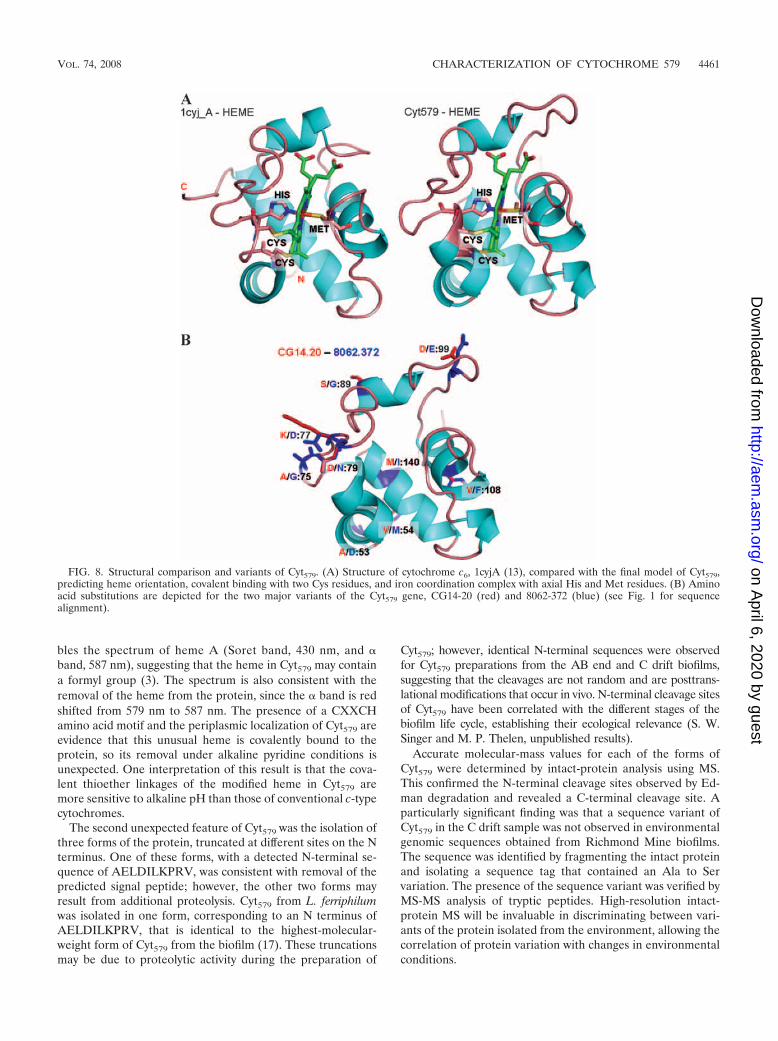
Structural model of Cyt579. Although no significant homol-ogy to Cyt579 was found in protein database searches, over 100candidate structural templates for modeling Cyt579 were de-tected, ranging from 7% to 25% sequence identity. Secondary-structure predictions, along with high levels of structural sim-ilarities observed between the analyzed templates, narrowedthe candidates to 25. An initial 3D model was constructedbased on an alignment of the Cyt579 sequence with that ofcytochrome c6, 1cyjA (Fig. 7A).
Based on calculated alignments to several structural tem-plates (including RCSB Protein Data Bank accession no. 1cyj,2dge, 1w5c, 1ls9, 1h1o, 1jdl, 1kv9, and 1nir), the final 3D modelwas created, including the position of a c-type heme groupfrom cytochrome c6 (13). The heme in Cyt579 is likely to bedifferent, as discussed above, due to the unique spectral char-acter of the cytochrome (see also Discussion). This model was
FIG. 6. pH-dependent Fe(II) oxidation by Cyt579. The results ofredox experiments are shown as follows: pH 1.2 (red), pH 2.0 (gray),pH 3.0 (green), and pH 4.0 (blue). Abs, absorbance.
TABLE 2. Spectral counts obtained for MXWVVXN sequences ofCyt579 from extracellular proteomes
Cyt579 gene SequenceaSpectral countb of sample from:
AB end UBA C drift
8062-147 (14-20) MVWVVSN 28 48 578062-372 MFWVVSN 85 144 1488062-372 C drift
(S112A)MFWVVAN 73 7 128
a Spectral counts are derived from peptide R.TAGEMXWVVXNGSPLQPMVGFVSAGQITDK.Q. Amino acid substitutions are indicated in bold type.
b Spectral counts refer to the total number of MS-MS spectra taken for thepeptide as an indicator of overall abundance. Each count is the average of theresults for three technical replicates.
VOL. 74, 2008 CHARACTERIZATION OF CYTOCHROME 579 4459
on April 6, 2020 by guest
http://aem.asm
.org/D
ownloaded from

compared by sequence to structure alignments and in 3D plotswith selected structural templates (Fig. 7B and C). The hemeorientation and structural elements were compared with thecytochrome c6 structure, 1cyj_A (13) (Fig. 8A). Models for thetwo major genetic variants of Cyt579 were then superimposed toindicate the positions of all nine side chain substitutions, thioetherlinkages between heme and Cys68 and Cys71, and heme-Fe com-plex with axial ligands His72 and Met121 (Fig. 8B).
DISCUSSION
In this study, we have purified the abundant, novel bacterialcytochrome first identified by proteogenomic studies in theacidic-wash fraction of biofilms collected at the RichmondMine in Iron Mountain, CA. We have confirmed the predictionthat this protein is Cyt579, a modified c-type cytochrome thathas been implicated as the Fe(II) oxidase in biochemicaland physiological studies of Leptospirillum isolates (17). In the
initial genomic data set obtained from a biofilm at the Rich-mond Mine, only one gene was sequenced that coded for Cyt579;however, two paralogs of Cyt579 were sequenced in a genomicdata set from a second biofilm (15, 24). The amino acid substitu-tions observed in these genetic variants can be predicted in a 3Drendering of the protein structure based on homology modeling(Fig. 8B). It is noteworthy that the predicted variant residues areall located on the surface of the protein and in contact withsolvent and thus do not appear to impose any perturbance tostructural elements or to the putative interactions with heme.Modeling also predicts a His-Met axial ligation for Cyt579 that isconsistent with the observation of an absorption band at 695 nmand a mostly helical protein structure that is corroborated by CDspectroscopy.
Detailed biochemical studies of Cyt579 isolated from thebiofilms have revealed some unexpected features of Cyt579.The alkaline pyridine hemochrome spectrum closely resem-
FIG. 7. Modeling of Cyt579. (A) The initial structural model of Cyt579 was constructed based on sequence alignment with the structure ofcytochrome c6, 1cyjA (13). In the alignment, amino acids repeated on the first and second lines are identical, and residues that are chemicallysimilar to those of Cyt579 are indicated by plus symbols. Dashes indicate gaps in the alignment. Highlighted residues Cys68, Cys71, His72, andMet121 form the direct interactions with the heme. (B) Regions in the model having structures similar to those of corresponding regions in thestructural templates analyzed are aligned in a schematic bar plot. Structural similarity with these templates is indicated as good (green),intermediate (yellow), and nonhomologous (red). Black boxes (R1 to R6) mark regions of structural deviation, or insertions/deletions, observedin structural templates. The region between R1 and R2 corresponds to the conserved CXXCH heme-binding motif. In Cyt579, the regions R1 toR6 correspond to the following fragments: R1, 65-AGT-67; R2, 73-GV-74; R3, 78-GDGPGA-83; R4, 93-FTNHQFDQ-100; R5, 115-SPLQPA-120; andR6, 126-SAGQI-130. (C) Residue-to-residue correspondences extracted from structurally conserved regions that were identified within a set of the closeststructural templates. The results of the analysis of these regions increased confidence in the calculated sequence alignments used in modeling. The resultsfrom calculation of sequence identities between the templates and the model in structurally conserved regions are given in the column labeled “Seq_ID”;in most cases these values are higher than the corresponding Seq_IDs calculated for entire structural alignments shown in panel B.
4460 SINGER ET AL. APPL. ENVIRON. MICROBIOL.
on April 6, 2020 by guest
http://aem.asm
.org/D
ownloaded from
bles the spectrum of heme A (Soret band, 430 nm, and �band, 587 nm), suggesting that the heme in Cyt579 may containa formyl group (3). The spectrum is also consistent with theremoval of the heme from the protein, since the � band is redshifted from 579 nm to 587 nm. The presence of a CXXCHamino acid motif and the periplasmic localization of Cyt579 areevidence that this unusual heme is covalently bound to theprotein, so its removal under alkaline pyridine conditions isunexpected. One interpretation of this result is that the cova-lent thioether linkages of the modified heme in Cyt579 aremore sensitive to alkaline pH than those of conventional c-typecytochromes.
The second unexpected feature of Cyt579 was the isolation ofthree forms of the protein, truncated at different sites on the Nterminus. One of these forms, with a detected N-terminal se-quence of AELDILKPRV, was consistent with removal of thepredicted signal peptide; however, the other two forms mayresult from additional proteolysis. Cyt579 from L. ferriphilumwas isolated in one form, corresponding to an N terminus ofAELDILKPRV, that is identical to the highest-molecular-weight form of Cyt579 from the biofilm (17). These truncationsmay be due to proteolytic activity during the preparation of
Cyt579; however, identical N-terminal sequences were observedfor Cyt579 preparations from the AB end and C drift biofilms,suggesting that the cleavages are not random and are posttrans-lational modifications that occur in vivo. N-terminal cleavage sitesof Cyt579 have been correlated with the different stages of thebiofilm life cycle, establishing their ecological relevance (S. W.Singer and M. P. Thelen, unpublished results).
Accurate molecular-mass values for each of the forms ofCyt579 were determined by intact-protein analysis using MS.This confirmed the N-terminal cleavage sites observed by Ed-man degradation and revealed a C-terminal cleavage site. Aparticularly significant finding was that a sequence variant ofCyt579 in the C drift sample was not observed in environmentalgenomic sequences obtained from Richmond Mine biofilms.The sequence was identified by fragmenting the intact proteinand isolating a sequence tag that contained an Ala to Servariation. The presence of the sequence variant was verified byMS-MS analysis of tryptic peptides. High-resolution intact-protein MS will be invaluable in discriminating between vari-ants of the protein isolated from the environment, allowing thecorrelation of protein variation with changes in environmentalconditions.
FIG. 8. Structural comparison and variants of Cyt579. (A) Structure of cytochrome c6, 1cyjA (13), compared with the final model of Cyt579,predicting heme orientation, covalent binding with two Cys residues, and iron coordination complex with axial His and Met residues. (B) Aminoacid substitutions are depicted for the two major variants of the Cyt579 gene, CG14-20 (red) and 8062-372 (blue) (see Fig. 1 for sequencealignment).
VOL. 74, 2008 CHARACTERIZATION OF CYTOCHROME 579 4461
on April 6, 2020 by guest
http://aem.asm
.org/D
ownloaded from

The third unexpected feature of Cyt579 was that Fe(II) oxi-dation was not favored thermodynamically at a pH of 3. Thisresult is inconsistent with the results of previous studies withLeptospirillum isolates, where complete reduction of Cyt579 inthe presence of 30 mM Fe(II) was observed at pH 2, and castsdoubt on the proposed role of Cyt579 as the Fe(II) oxidase forLeptospirillum group II bacteria (10, 17).
The properties of Cyt579 from Leptospirillum group II bac-teria are analogous to those of rusticyanin, a periplasmic Cu-containing protein expressed by Acidithiobacillus ferrooxidans,an acidophilic Fe(II)-oxidizing bacterium found in environ-ments similar to those where members of Leptospirillum groupII are found. Biochemical and transcriptomic evidence hasimplicated rusticyanin as the initial electron acceptor for anouter membrane-bound c-type cytochrome, Cyc2, which is theproposed Fe(II) oxidase for A. ferrooxidans (25–27). In supportof this analogy, we have recently purified a novel membranecytochrome, Cyt572, that is expressed by Leptospirillum group IIin the Richmond biofilms (11). In contrast to Cyt579, Cyt572
oxidizes Fe(II) at low pH and may donate electrons to Cyt579.Efforts to reconstruct the Fe(II)-dependent electron transferpathway in Leptospirillum group II bacteria and clarify the roleof Cyt579 in this pathway are currently under way.
ACKNOWLEDGMENTS
Funding was provided by the U.S. Department of Energy, Office ofScience, from the Genomics: GTL Program, grant DE-FG02-05ER64134, to J.F.B., R.L.H., and M.P.T. Work at LLNL was per-formed under the auspices of the U.S. Department of Energy undercontract DE-AC52-07NA27344.
We are grateful to the Banfield lab members for obtaining biofilmsamples and to T. W. Arman, President, Iron Mountain Mines, R.Sugarek, EPA, and R. Carver for site access and on-site assistance. Wealso thank Chris Jeans and Anna Siebers for CD spectroscopy andassistance in biochemical studies on biofilm proteins at LLNL; KentMacDonald and Reena Zalpuri for assistance with sample preparationat the Electron Microscope Laboratory, University of California,Berkeley; Mary Ann Gawinowicz at the Columbia University ProteinCore Facility for protein sequence analyses; and Brian Erickson forassistance in acquiring IRMPD mass spectra at ORNL.
REFERENCES
1. Altschul, S. F., T. L. Madden, A. A. Schaffer, J. H. Zhang, Z. Zhang, W.Miller, and D. J. Lipman. 1997. Gapped BLAST and PSI-BLAST: a newgeneration of protein database search programs. Nucleic Acids Res. 25:3389–3402.
2. Baker, B. J., and J. F. Banfield. 2003. Microbial communities in acid minedrainage. FEMS Microbiol. Ecol. 44:139–152.
3. Berry, E. A., and B. L. Trumpower. 1987. Simultaneous determination ofhemes-a, b and c from pyridine hemochrome spectra. Anal. Biochem. 161:1–15.
4. Bond, P. L., S. P. Smriga, and J. F. Banfield. 2000. Phylogeny of microor-ganisms populating a thick, subaerial, predominantly lithotrophic biofilm atan extreme acid mine drainage site. Appl. Environ Microbiol. 66:3842–3849.
5. Bower, M. J., F. E. Cohen, and R. L. Dunbrack. 1997. Prediction of proteinside-chain rotamers from a backbone-dependent rotamer library: a newhomology modeling tool. J. Mol. Biol. 267:1268–1282.
6. Bradford, M. M. 1976. Rapid and sensitive method for the quantitation ofmicrogram quantities of protein utilizing principle of protein-dye binding.Anal. Biochem. 72:248–254.
7. Connelly, H. M., D. A. Pelletier, T. Y. Lu, P. K. Lankford, and R. L. Hettich.2006. Characterization of pII family (GlnK1, GlnK2, and GlnB) proteinuridylylation in response to nitrogen availability for Rhodopseudomonaspalustris. Anal. Biochem. 357:93–104.
8. Druschel, G. K., B. J. Baker, T. M. Gihring, and J. F. Banfield. 2004. Acidmine drainage biogeochemistry at Iron Mountain, California. Geochem.Trans. 5:13–32.
9. Ginalski, K., N. V. Grishin, A. Godzik, and L. Rychlewski. 2005. Practicallessons from protein structure prediction. Nucleic Acids Res. 33:1874–1891.
10. Hart, A., J. C. Murrell, R. K. Poole, and P. R. Norris. 1991. An acid-stablecytochrome in iron-oxidizing Leptospirillum ferrooxidans. FEMS Microbiol.Lett. 81:89–94.
11. Jeans, C., S. W. Singer, C. S. Chan, N. C. VerBerkmoes, M. Shah, R. L.Hettich, J. F. Banfield, and M. P. Thelen. 2008. Cytochrome 572 is a con-spicuous membrane protein with iron oxidation activity purified directlyfrom a natural acidophilic microbial community. ISME J. 2:542–550.
12. Jones, D. T. 1999. Protein secondary structure prediction based on position-specific scoring matrices. J. Mol. Biol. 292:195–202.
13. Kerfeld, C. A., H. P. Anwar, R. Interrante, S. Merchant, and T. O. Yeates.1995. The structure of chloroplast cytochrome-c6 at 1.9 angstrom resolution:evidence for functional oligomerization. J. Mol. Biol. 250:627–647.
14. Laemmli, U. K. 1970. Cleavage of structural proteins during the assembly ofthe head of bacteriophage T4. Nature 227:680–685.
15. Lo, I., V. J. Denef, N. C. VerBerkmoes, M. B. Shah, D. Goltsman, G.DiBartolo, G. W. Tyson, E. E. Allen, R. J. Ram, J. C. Detter, P. Richardson,M. P. Thelen, R. L. Hettich, and J. F. Banfield. 2007. Strain-resolved com-munity proteomics reveals recombining genomes of acidophilic bacteria.Nature 446:537–541.
16. Pearson, W. R. 1991. Searching protein-sequence libraries: comparison ofthe sensitivity and selectivity of the Smith-Waterman and FASTA algo-rithms. Genomics 11:635–650.
17. Ram, R. J., N. C. VerBerkmoes, M. P. Thelen, G. W. Tyson, B. J. Baker, R. C.Blake, M. Shah, R. L. Hettich, and J. F. Banfield. 2005. Community pro-teomics of a natural microbial biofilm. Science 308:1915–1920.
18. Rost, B., and C. Sander. 1993. Prediction of protein secondary structure atbetter than 70-percent accuracy. J. Mol. Biol. 232:584–599.
19. Schnaitman, C. A., M. S. Korczynski, and D. G. Lundgren. 1969. Kineticstudies of iron oxidation by whole cells of Ferrobacillus ferrooxidans. J. Bac-teriol. 99:552–557.
20. Smith, T. F., and M. S. Waterman. 1981. Identification of common molec-ular subsequences. J. Mol. Biol. 147:195–197.
21. Tabb, D. L., W. H. McDonald, and J. R. Yates. 2002. DTASelect andContrast: tools for assembling and comparing protein identifications fromshotgun proteomics. J. Proteome Res. 1:21–26.
22. Tabb, D. L., C. Narasimhan, M. B. Strader, and R. L. Hettich. 2005.DBDigger: reorganized proteomic database identification that improvesflexibility and speed. Anal. Chem. 77:2464–2474.
23. Thompson, J. D., D. G. Higgins, and T. J. Gibson. 1994. Clustal-W: improv-ing the sensitivity of progressive multiple sequence alignment through se-quence weighting, position-specific gap penalties and weight matrix choice.Nucleic Acids Res. 22:4673–4680.
24. Tyson, G. W., J. Chapman, P. Hugenholtz, E. E. Allen, R. J. Ram, P. M.Richardson, V. V. Solovyev, E. M. Rubin, D. S. Rokhsar, and J. F. Banfield.2004. Community structure and metabolism through reconstruction of mi-crobial genomes from the environment. Nature 428:37–43.
25. Yarzabal, A., C. Appia-Ayme, J. Ratouchniak, and V. Bonnefoy. 2004. Reg-ulation of the expression of the Acidithiobacillus ferrooxidans rus operonencoding two cytochromes c, a cytochrome oxidase and rusticyanin. Micro-biology 150:2113–2123.
26. Yarzabal, A., G. Brasseur, and V. Bonnefoy. 2002. Cytochromes c of Acidi-thiobacillus ferrooxidans. FEMS Microbiol. Lett. 209:189–195.
27. Yarzabal, A., G. Brasseur, J. Ratouchniak, K. Lund, D. Lemesle-Meunier,J. A. DeMoss, and V. Bonnefoy. 2002. The high-molecular-weight cyto-chrome c Cyc2 of Acidithiobacillus ferrooxidans is an outer membrane pro-tein. J. Bacteriol. 184:313–317.
28. Zemla, A. 2003. LGA: a method for finding 3D similarities in protein struc-tures. Nucleic Acids Res. 31:3370–3374.
29. Zemla, A., C. E. Zhou, T. Slezak, T. Kuczmarski, D. Rama, C. Torres, D.Sawicka, and D. Barsky. 2005. AS2TS system for protein structure modelingand analysis. Nucleic Acids Res. 33:W111–W115.
4462 SINGER ET AL. APPL. ENVIRON. MICROBIOL.
on April 6, 2020 by guest
http://aem.asm
.org/D
ownloaded from