Characterization of Acetic Acid Bacteria in Traditional...
5
APPLIED AND ENVIRONMENTAL MICROBIOLOGY, 0099-2240/01/$04.0010 DOI: 10.1128/AEM.67.2.986–990.2001 Feb. 2001, p. 986–990 Vol. 67, No. 2 Copyright © 2001, American Society for Microbiology. All Rights Reserved. Characterization of Acetic Acid Bacteria in Traditional Acetic Acid Fermentation of Rice Vinegar (Komesu) and Unpolished Rice Vinegar (Kurosu) Produced in Japan KUMIKO NANDA, 1 MARIKO TANIGUCHI, 1 SATOSHI UJIKE, 1 NOBUHIRO ISHIHARA, 1 HIROTAKA MORI, 1 HISAYO ONO, 2 AND YOSHIKATSU MUROOKA 2 * Research Center, Tamanoi Vinegar Co., Ltd., 100, Nishimachi, Yamatokoriyama, Nara 639-1038, 1 and Department of Biotechnology, Graduate School of Engineering, Osaka University, Yamadaoka, Suita, Osaka 565-0871, 2 Japan Received 26 June 2000/Accepted 16 November 2000 Bacterial strains were isolated from samples of Japanese rice vinegar (komesu) and unpolished rice vinegar (kurosu) fermented by the traditional static method. Fermentations have never been inoculated with a pure culture since they were started in 1907. A total of 178 isolates were divided into groups A and B on the basis of enterobacterial repetitive intergenic consensus-PCR and random amplified polymorphic DNA fingerprinting analyses. The 16S ribosomal DNA sequences of strains belonging to each group showed similarities of more than 99% with Acetobacter pasteurianus. Group A strains overwhelmingly dominated all stages of fermentation of both types of vinegar. Our results indicate that appropriate strains of acetic acid bacteria have spontane- ously established almost pure cultures during nearly a century of komesu and kurosu fermentation. The rice vinegars komesu and kurosu are produced from polished and unpolished rice, respectively, by the same process (saccharification of rice, alcohol fermentation, and oxidation of ethanol to acetic acid). Both of these vinegars are traditional seasonings that have long been used in Japan, China, and Asian countries. Komesu is colorless and has a plain taste and thus is used for sushi cooking, while kurosu is black and con- tains more amino acids and vitamins than komesu and thus is used as a healthy drink. With increasing public interest in health, the effective health-related elements of the traditional vinegars have come under scrutiny. Vinegar is produced industrially by two main methods, a slow process involving static surface acetic acid fermentation and a fast submerged fermentation process. Generally, static fermentation is employed in traditional vinegar production. This technique is not costly in terms of plant investment, and the quality of the product is good, although a rather long time is required to complete the fermentation. An alcoholic liquid with vinegar (moromi) is fermented in appropriate containers fitted with covers, and this is considered a good way to prevent bacterial contamination during static fermentation. In a few days, a crepe pellicle of acetic acid bacteria covers the surface of the moromi, after which the fermentation proceeds and finishes about 1 month later. In this process, no strict steriliza- tion measures are used. No purified strain is inoculated after the start of vinegar fermentation, which in some cases has been continued without inoculation of a pure culture for more than 100 years. The acetic acid bacteria consist of two genera, Acetobacter and Gluconobacter. Strains of Acetobacter are generally in- volved in vinegar production (12). Identification of the species and characterization of the dominant strains in static acetic acid fermentation are desirable in order to stabilize the fer- mentation and improve the strain (7). In recent years the enterobacterial repetitive intergenic consensus (ERIC)-PCR method and random amplified polymorphic DNA (RAPD) fingerprinting have been applied to taxonomic grouping of bacteria, including enterobacteria (3, 16), Acetobacter sp. (15), lactic acid bacteria (17), and rhizobia (4, 6). In this study, we investigated acetic acid bacterial strains isolated from samples obtained during commercial production of komesu and kurosu by using the ERIC-PCR and RAPD methods. The isolates were identified and characterized genetically and physiologi- cally. Changes in the flora during the manufacturing process were also examined. All of the TN strains isolated in this study and Acetobacter pasteurianus ATCC 33445 T were cultivated aerobically at 28°C in potato medium containing (per liter) 20.0 g of glycerol, 10 g of Polypeptone, 10 g of yeast extract, 5 g of glucose, and 100 ml of potato extract. Escherichia coli JM109 (Takara Shuzo Co., Ltd., Ohtsu, Shiga, Japan) was grown in Luria-Bertani me- dium. Samples used for isolation of bacteria were taken from komesu and kurosu commercially produced by Tamanoi Vin- egar Co., Ltd. (Nara, Japan). The samples were obtained from microbial films on the moromi surface in the early, middle, and late phases of acetic acid fermentation (and in the last fermen- tation period in the case of komesu). The initial moromi con- tained about 3 to 3.5% acetic acid and 4 to 4.5% ethyl alcohol and had a pH of 3.3. Acetic acid bacteria were isolated on isolation medium agar (1% glucose, 1% glycerol, 0.2% yeast extract, 0.2% Polypeptone, 10% potato extract, 1% acetic acid, 2% ethanol, 0.9% agar) and were grown at 30°C for 4 days. Total DNA was extracted by the method of Ohmori et al. (10). DNA for the ERIC-PCR template was prepared by the rapid method described by Nuswantara et al. (9). The PCR conditions used with oligonucleotide primers ERIC1R (39-CA CTTAGGGGTCCTCGAATGTA-59) and ERIC2 (59-AAGT AAGTGACTGGGGTGAGCG-39) were those described by * Corresponding author. Mailing address: Department of Biotech- nology, Graduate School of Engineering, Osaka University, Yamada- oka 2-1, Suita, Osaka 565-0871, Japan. Phone: 81 6 6879 7418. Fax: 81-6-6879-7418. E-mail: [email protected]. 986 on May 13, 2018 by guest http://aem.asm.org/ Downloaded from
Transcript of Characterization of Acetic Acid Bacteria in Traditional...
APPLIED AND ENVIRONMENTAL MICROBIOLOGY,0099-2240/01/$04.0010 DOI: 10.1128/AEM.67.2.986–990.2001
Feb. 2001, p. 986–990 Vol. 67, No. 2
Copyright © 2001, American Society for Microbiology. All Rights Reserved.
Characterization of Acetic Acid Bacteria in Traditional AceticAcid Fermentation of Rice Vinegar (Komesu) and Unpolished
Rice Vinegar (Kurosu) Produced in JapanKUMIKO NANDA,1 MARIKO TANIGUCHI,1 SATOSHI UJIKE,1 NOBUHIRO ISHIHARA,1
HIROTAKA MORI,1 HISAYO ONO,2 AND YOSHIKATSU MUROOKA2*
Research Center, Tamanoi Vinegar Co., Ltd., 100, Nishimachi, Yamatokoriyama, Nara 639-1038,1 and Department ofBiotechnology, Graduate School of Engineering, Osaka University, Yamadaoka, Suita, Osaka 565-0871,2 Japan
Received 26 June 2000/Accepted 16 November 2000
Bacterial strains were isolated from samples of Japanese rice vinegar (komesu) and unpolished rice vinegar(kurosu) fermented by the traditional static method. Fermentations have never been inoculated with a pureculture since they were started in 1907. A total of 178 isolates were divided into groups A and B on the basisof enterobacterial repetitive intergenic consensus-PCR and random amplified polymorphic DNA fingerprintinganalyses. The 16S ribosomal DNA sequences of strains belonging to each group showed similarities of morethan 99% with Acetobacter pasteurianus. Group A strains overwhelmingly dominated all stages of fermentationof both types of vinegar. Our results indicate that appropriate strains of acetic acid bacteria have spontane-ously established almost pure cultures during nearly a century of komesu and kurosu fermentation.
The rice vinegars komesu and kurosu are produced frompolished and unpolished rice, respectively, by the same process(saccharification of rice, alcohol fermentation, and oxidation ofethanol to acetic acid). Both of these vinegars are traditionalseasonings that have long been used in Japan, China, andAsian countries. Komesu is colorless and has a plain taste andthus is used for sushi cooking, while kurosu is black and con-tains more amino acids and vitamins than komesu and thus isused as a healthy drink. With increasing public interest inhealth, the effective health-related elements of the traditionalvinegars have come under scrutiny.
Vinegar is produced industrially by two main methods, aslow process involving static surface acetic acid fermentationand a fast submerged fermentation process. Generally, staticfermentation is employed in traditional vinegar production.This technique is not costly in terms of plant investment, andthe quality of the product is good, although a rather long timeis required to complete the fermentation. An alcoholic liquidwith vinegar (moromi) is fermented in appropriate containersfitted with covers, and this is considered a good way to preventbacterial contamination during static fermentation. In a fewdays, a crepe pellicle of acetic acid bacteria covers the surfaceof the moromi, after which the fermentation proceeds andfinishes about 1 month later. In this process, no strict steriliza-tion measures are used. No purified strain is inoculated afterthe start of vinegar fermentation, which in some cases has beencontinued without inoculation of a pure culture for more than100 years.
The acetic acid bacteria consist of two genera, Acetobacterand Gluconobacter. Strains of Acetobacter are generally in-volved in vinegar production (12). Identification of the speciesand characterization of the dominant strains in static acetic
acid fermentation are desirable in order to stabilize the fer-mentation and improve the strain (7). In recent years theenterobacterial repetitive intergenic consensus (ERIC)-PCRmethod and random amplified polymorphic DNA (RAPD)fingerprinting have been applied to taxonomic grouping ofbacteria, including enterobacteria (3, 16), Acetobacter sp. (15),lactic acid bacteria (17), and rhizobia (4, 6). In this study, weinvestigated acetic acid bacterial strains isolated from samplesobtained during commercial production of komesu and kurosuby using the ERIC-PCR and RAPD methods. The isolateswere identified and characterized genetically and physiologi-cally. Changes in the flora during the manufacturing processwere also examined.
All of the TN strains isolated in this study and Acetobacterpasteurianus ATCC 33445T were cultivated aerobically at 28°Cin potato medium containing (per liter) 20.0 g of glycerol, 10 gof Polypeptone, 10 g of yeast extract, 5 g of glucose, and 100 mlof potato extract. Escherichia coli JM109 (Takara Shuzo Co.,Ltd., Ohtsu, Shiga, Japan) was grown in Luria-Bertani me-dium. Samples used for isolation of bacteria were taken fromkomesu and kurosu commercially produced by Tamanoi Vin-egar Co., Ltd. (Nara, Japan). The samples were obtained frommicrobial films on the moromi surface in the early, middle, andlate phases of acetic acid fermentation (and in the last fermen-tation period in the case of komesu). The initial moromi con-tained about 3 to 3.5% acetic acid and 4 to 4.5% ethyl alcoholand had a pH of 3.3. Acetic acid bacteria were isolated onisolation medium agar (1% glucose, 1% glycerol, 0.2% yeastextract, 0.2% Polypeptone, 10% potato extract, 1% acetic acid,2% ethanol, 0.9% agar) and were grown at 30°C for 4 days.
Total DNA was extracted by the method of Ohmori et al.(10). DNA for the ERIC-PCR template was prepared by therapid method described by Nuswantara et al. (9). The PCRconditions used with oligonucleotide primers ERIC1R (39-CACTTAGGGGTCCTCGAATGTA-59) and ERIC2 (59-AAGTAAGTGACTGGGGTGAGCG-39) were those described by
* Corresponding author. Mailing address: Department of Biotech-nology, Graduate School of Engineering, Osaka University, Yamada-oka 2-1, Suita, Osaka 565-0871, Japan. Phone: 81 6 6879 7418. Fax:81-6-6879-7418. E-mail: [email protected].
986
on May 13, 2018 by guest
http://aem.asm
.org/D
ownloaded from
Versalovic et al. (16). PCR were carried out by using a pro-grammable temperature control system (PC-700; Astec,Fukuoka, Japan). RAPD Analysis Beads and Primers werepurchased from Amersham Pharmacia Biotech (Uppsala, Swe-den). Electrophoresis was carried out on a horizontal 0.7, 1.5,or 2% agarose gel (SeaKem GTG agarose; FMC BioProducts,Rockland, Maine) in electrophoresis buffer (0.04 M Tris-ace-tate, 0.001 M EDTA) at a constant voltage of 100 V. The gelwas stained with ethidium bromide and photographed with aUV transilluminator.
16S ribosomal DNA (rDNA) was amplified in vitro (PCR)by using the method of Both et al. (1) in combination witholigonucleotide primers complementary to highly conservedregions of bacterial rRNA genes. The 59- and 39-terminal prim-ers used were GAGTTTGAT(C/T)(C/A)TGGCTCA (posi-tions 9 to 26, according to the E. coli numbering system [2])and CA(G/T)AAAGGAGGTGATCC (positions 1545 to1529), respectively (11). Double-stranded PCR products wereligated at the HincII site of pUC19. The DNA was transferredinto E. coli JM109 competent cells (Takara Shuzo). Sequenc-ing was performed with an ABI PRISM 310 genetic analyzer(PE Biosystems Japan) according to the manufacturer’s in-structions. DNA sequences were processed with GENETYX-MAC, version 10.1 (Software Development Co., Tokyo, Japan)for multialignment analysis.
The G1C contents of DNA were determined by high-per-formance liquid chromatography as described previously (8,14). The nucleotides obtained by treatment with P1 nucleaseand alkaline phosphatase were applied to a high-performanceliquid chromatograph; a Hitachi L7000 analyzer equipped witha Cosmosil 5C18-AR column (4.6 by 150 mm; Nacalai Teque,Inc., Kyoto, Japan) was used for this analysis.
Gram staining, catalase production tests, oxidase tests, testsfor acid production from glucose under aerobic or anaerobicconditions, and tests for acetate and lactate oxidation to CO2
were conducted with all isolates. Tests for assimilation of am-moniacal nitrogen, growth on carbon sources, acid productionfrom carbon sources, formation of ketogluconic acids fromD-glucose (13), and determination of the ubiquinone system
were conducted with representative isolates as described inBergey’s Manual of Systematic Bacteriology (5). Ubiquinone 10was obtained from Sigma Chemical Co. (St. Louis, Mo.).Ubiquinone 9 was isolated and purified from Acetobacter acetiIFO3281. Acetobacter strains were observed with a HitachiS-3500N scanning electron microscope equipped with a fieldemission gun and operated at 10 kV. In acetic acid fermenta-tion tests, acetic acid bacterial mats were floated on 50 ml of amedium containing 1.0% D-glucose, 1.0% glycerol, 0.2%Polypeptone, 0.2% yeast extract, 10% potato extract, 1.0%acetic acid, and 4.0% ethanol in 100-ml vials, and surfacefermentation was continued at 30°C for 2 weeks. Acetic acidcontents were determined by titration with 0.1 N NaOHagainst phenolphthalein.
A total of 178 bacterial strains were obtained from themoromi of komesu and kurosu in static surface acetic acidfermentations. Surface bacterial mats of moromi were sus-pended in sterile water, and bacteria were isolated on isolationmedium plates incubated at 30°C for a few days. In staticfermentations, a portion of the surface mat bacteria from theearly phase of fermentation is used as a starter for the nextfermentation batch. Both komesu and kurosu are fermentedfor about 1 month at room temperature, which can range from10 to 30°C. The final concentration of acetic acid is about 6 to6.5%, and the pH is 3.1. The residual alcohol content is usually0.1%.
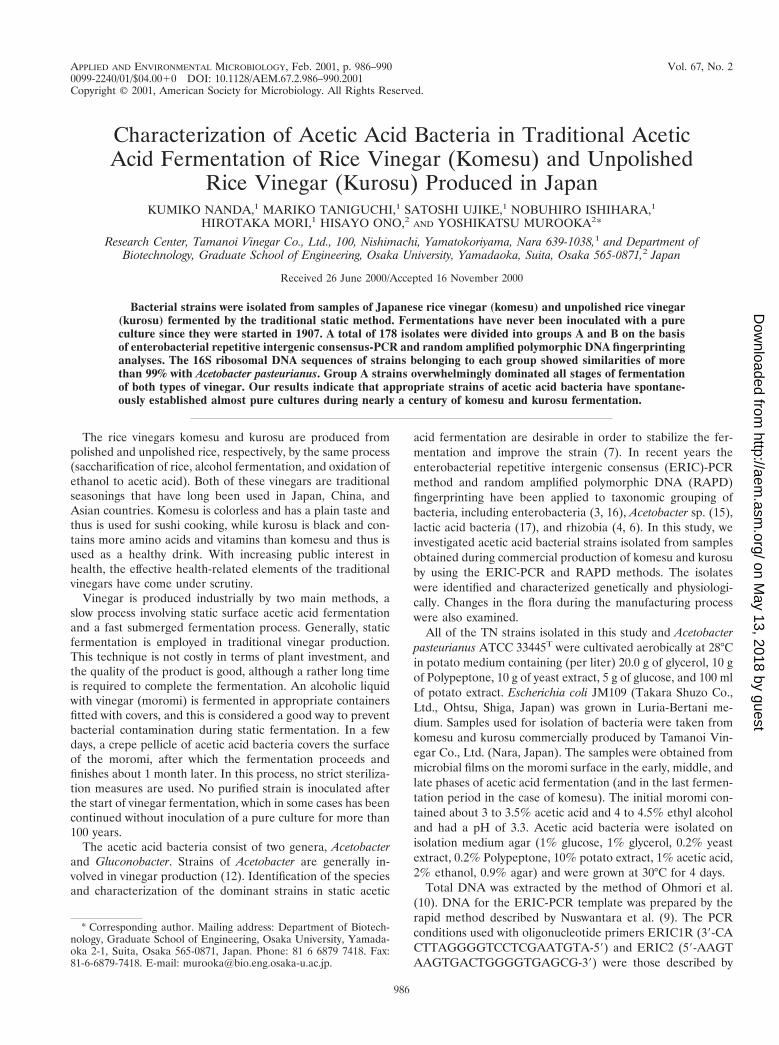
DNA prepared from pure cultures of all isolates obtainedfrom samples taken at different stages of fermentation wereamplified by ERIC-PCR (Fig. 1). On the basis of the amplifi-cation profiles, the bacterial strains were divided into twogroups, groups A and B. Most of the isolates produced ERICbands at 1,350, 1,150, 860, 770, 390, and 220 bp, and theseisolates were placed in group A. The rest of the isolates, in-cluding TN-1 and TN-2, produced ERIC bands at 1,150, 710,640, 500, 390, and 310 bp and were placed in group B. Thesestrains were isolated mainly after fermentation had finished.All of the isolates from kurosu had the same profiles as groupA isolates (data not shown).
Intraspecific variation was demonstrated by differential am-
FIG. 1. ERIC profiles of selected acetic acid bacterial strains from rice vinegar. Lanes 1 and 20, PCR markers (Novagen); Lanes 2 to 19, strainsTN-106, TN-6, TN-9, TN-12, TN-70, TN-71, TN-21, TN-23, TN-27, TN-117, TN-118, TN-119, TN-96, TN-98, TN-193, TN-195, TN-1, and TN-2,respectively.
VOL. 67, 2001 ACETIC ACID BACTERIA IN VINEGAR FERMENTATION 987
on May 13, 2018 by guest
http://aem.asm
.org/D
ownloaded from
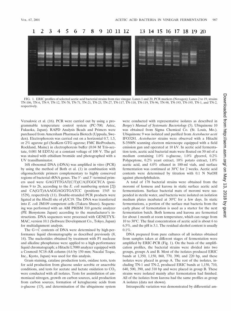
plification of certain DNA fragments displaying polymorphismin the following strains: TN-1, TN-2, TN-6, TN-9, TN-12, TN-21, TN-23, TN-27, TN-70, and TN-71 (Fig. 2). These strainswere arbitrarily selected from groups A and B. On average, thePCR products obtained with RAPD analysis primer 6 gener-ated fingerprints consisting seven or eight fragments that var-ied in length from 150 bp to 2.0 kbp. These strains all producedRAPD bands at 810, 600, and 430 bp. However, TN-1 andTN-2 produced an additional band at about 1.3 kb, whereas theother strains produced an additional band at about 340 bp.This result shows that strains TN-1 and TN-2 (group B) aredifferent from other strains (group A) and is consistent withthe ERIC-PCR findings.
The 16S rDNA of strain TN-27 in group A and strains TN-1and TN-2 in group B were sequenced. The level of similaritybetween the sequence of strain TN-27 and the sequence of thetype strain of A. pasteurianus, LMD 22.1 (EMBL data libraryaccession no. X71863), was 99.5%, and the difference corre-sponded to 8 base changes. The 16S rDNA sequences ofstrains TN-1 and TN-2 were also very similar (99.5%) to that ofA. pasteurianus LMD 22.1. The TN-1 and TN-2 sequencesdiffered at 7 and 8 bases, respectively, from the sequence of theA. pasteurianus strain. Levels of similarity between group Aand B strains of 99.9% were found. Since the level of similaritybetween each TN strain and A. aceti NCIB 8621 (EMBL datalibrary accession no. X74066) was only 96.9%, the TN strainsapparently belong to A. pasteurianus.
The taxonomic characteristics of two strains belonging togroup A, two strains belonging to group B, and the type strainof A. pasteurianus, ATCC 33445, were examined. All of thestrains were gram-negative rods that were catalase positive andoxidase negative. The results of the oxidation-fermentationtest showed oxidation. The strains were able to oxidize lactateand acetate. The G1C contents of the chromosomal DNAranged from 53.6 to 54.3 mol%, values which are within therange of values for A. pasteurianus according to Bergey’s Man-ual of Systematic Bacteriology (5). No formation of ketogluconicacids was detected by thin-layer chromatography analysis. Thestrains produced ubiquinone 9. No assimilation of ammoniacalnitrogen was detected. All isolates utilized D-glucose, galac-
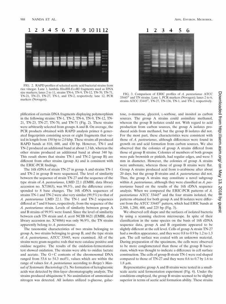
tose, D-mannose, glycerol, L-sorbose, and inositol as carbonsources. The group A strains could assimilate methanol,whereas the group B isolates could not. With regard to acidproduction from carbon sources, the group A isolates pro-duced acids from methanol, but the group B isolates did not.For the most part, these characteristics were consistent withthose of A. pasteurianus, although differences were found ingrowth on and acid formation from carbon sources. We alsoobserved that the colonies of group A strains differed fromthose of group B strains. Colonies of members of both groupswere pale brownish or pinkish, had regular edges, and were 5mm in diameter. However, the colonies of group A strainswere umbonate, whereas those of group B strains were flat.Group A strains produced acid from D-arabinose after about20 days, but the group B strains and A. pasterurianus did not.Thus, the group A strains may constitute a novel subgroupwithin A. pasteurianus, although they were classified as A. pas-teurianus based on the results of the 16S rDNA sequenceanalysis. When we compared the ERIC-PCR patterns of A.pasteurianus ATCC 33445T and the four strains isolated, thepatterns obtained for both group A and B isolates were differ-ent from the ATCC 33445T pattern, which had ERIC bands at2,300, 1,200, 400, and 225 bp (Fig. 3).
We observed cell shape and the surfaces of isolated bacteriaby using a scanning electron microscope. In spite of theirclassification in the same species on the basis of 16S rDNAsequence data, group A and B organisms appeared to beslightly different at the cell level. Cells of group A strain TN-27had a swollen appearance, and they were 0.8 to 0.9 by 1.2 to 1.3mm. The cell surface was coated with an unknown material.During preparation of the specimens, the cells were observedto be more conglomerated than those of the group B bacte-rium, which was thought to indicate a difference in cell surfaceconstruction. The cells of group B strain TN-1 were rod shapedcompared to those of TN-27 and they were 0.6 to 0.7 by 1.6 to1.8 mm.
Representative group A and B strains were tested in a small-scale acetic acid fermentation experiment (Fig. 4). Under theconditions employed, the group B strains seemed to be slightlysuperior in terms of acetic acid formation ability. These strains
FIG. 2. RAPD profiles of selected acetic acid bacterial strains fromrice vinegar. Lane 1, lambda HindIII-EcoRI fragments used as DNAsize markers; lanes 2 to 11, strains TN-6, TN-9, TN-12, TN-70, TN-71,TN-21, TN-23, TN-27, TN-1, and TN-2, respectively; lane 12, PCRmarkers (Novagen).
FIG. 3. Comparison of ERIC profiles of A. pasteurianus ATCC33445T and TN strains. Lane 1, PCR markers (Novagen); lanes 2 to 6,strains ATCC 33445T, TN-27, TN-136, TN-1, and TN-2, respectively.
988 NANDA ET AL. APPL. ENVIRON. MICROBIOL.
on May 13, 2018 by guest
http://aem.asm
.org/D
ownloaded from
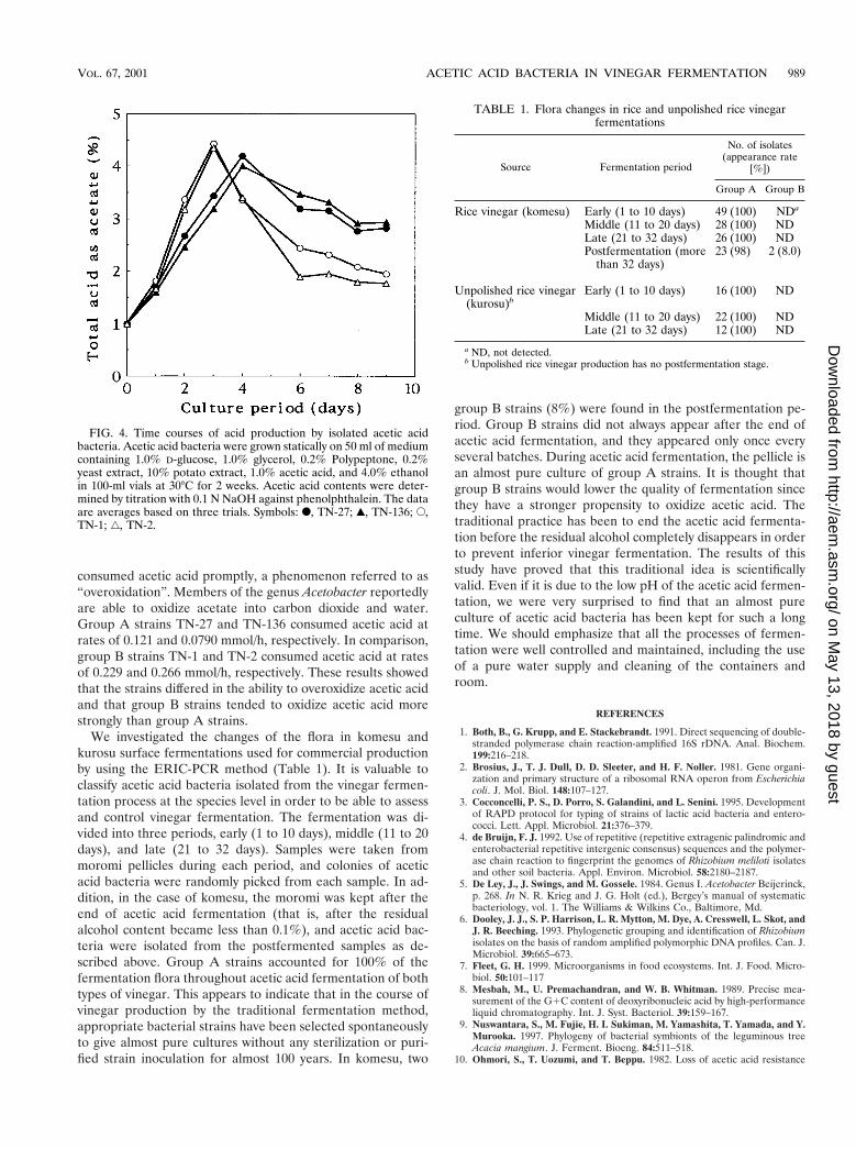
consumed acetic acid promptly, a phenomenon referred to as“overoxidation”. Members of the genus Acetobacter reportedlyare able to oxidize acetate into carbon dioxide and water.Group A strains TN-27 and TN-136 consumed acetic acid atrates of 0.121 and 0.0790 mmol/h, respectively. In comparison,group B strains TN-1 and TN-2 consumed acetic acid at ratesof 0.229 and 0.266 mmol/h, respectively. These results showedthat the strains differed in the ability to overoxidize acetic acidand that group B strains tended to oxidize acetic acid morestrongly than group A strains.
We investigated the changes of the flora in komesu andkurosu surface fermentations used for commercial productionby using the ERIC-PCR method (Table 1). It is valuable toclassify acetic acid bacteria isolated from the vinegar fermen-tation process at the species level in order to be able to assessand control vinegar fermentation. The fermentation was di-vided into three periods, early (1 to 10 days), middle (11 to 20days), and late (21 to 32 days). Samples were taken frommoromi pellicles during each period, and colonies of aceticacid bacteria were randomly picked from each sample. In ad-dition, in the case of komesu, the moromi was kept after theend of acetic acid fermentation (that is, after the residualalcohol content became less than 0.1%), and acetic acid bac-teria were isolated from the postfermented samples as de-scribed above. Group A strains accounted for 100% of thefermentation flora throughout acetic acid fermentation of bothtypes of vinegar. This appears to indicate that in the course ofvinegar production by the traditional fermentation method,appropriate bacterial strains have been selected spontaneouslyto give almost pure cultures without any sterilization or puri-fied strain inoculation for almost 100 years. In komesu, two
group B strains (8%) were found in the postfermentation pe-riod. Group B strains did not always appear after the end ofacetic acid fermentation, and they appeared only once everyseveral batches. During acetic acid fermentation, the pellicle isan almost pure culture of group A strains. It is thought thatgroup B strains would lower the quality of fermentation sincethey have a stronger propensity to oxidize acetic acid. Thetraditional practice has been to end the acetic acid fermenta-tion before the residual alcohol completely disappears in orderto prevent inferior vinegar fermentation. The results of thisstudy have proved that this traditional idea is scientificallyvalid. Even if it is due to the low pH of the acetic acid fermen-tation, we were very surprised to find that an almost pureculture of acetic acid bacteria has been kept for such a longtime. We should emphasize that all the processes of fermen-tation were well controlled and maintained, including the useof a pure water supply and cleaning of the containers androom.
REFERENCES
1. Both, B., G. Krupp, and E. Stackebrandt. 1991. Direct sequencing of double-stranded polymerase chain reaction-amplified 16S rDNA. Anal. Biochem.199:216–218.
2. Brosius, J., T. J. Dull, D. D. Sleeter, and H. F. Noller. 1981. Gene organi-zation and primary structure of a ribosomal RNA operon from Escherichiacoli. J. Mol. Biol. 148:107–127.
3. Cocconcelli, P. S., D. Porro, S. Galandini, and L. Senini. 1995. Developmentof RAPD protocol for typing of strains of lactic acid bacteria and entero-cocci. Lett. Appl. Microbiol. 21:376–379.
4. de Bruijn, F. J. 1992. Use of repetitive (repetitive extragenic palindromic andenterobacterial repetitive intergenic consensus) sequences and the polymer-ase chain reaction to fingerprint the genomes of Rhizobium meliloti isolatesand other soil bacteria. Appl. Environ. Microbiol. 58:2180–2187.
5. De Ley, J., J. Swings, and M. Gossele. 1984. Genus I. Acetobacter Beijerinck,p. 268. In N. R. Krieg and J. G. Holt (ed.), Bergey’s manual of systematicbacteriology, vol. 1. The Williams & Wilkins Co., Baltimore, Md.
6. Dooley, J. J., S. P. Harrison, L. R. Mytton, M. Dye, A. Cresswell, L. Skot, andJ. R. Beeching. 1993. Phylogenetic grouping and identification of Rhizobiumisolates on the basis of random amplified polymorphic DNA profiles. Can. J.Microbiol. 39:665–673.
7. Fleet, G. H. 1999. Microorganisms in food ecosystems. Int. J. Food. Micro-biol. 50:101–117
8. Mesbah, M., U. Premachandran, and W. B. Whitman. 1989. Precise mea-surement of the G1C content of deoxyribonucleic acid by high-performanceliquid chromatography. Int. J. Syst. Bacteriol. 39:159–167.
9. Nuswantara, S., M. Fujie, H. I. Sukiman, M. Yamashita, T. Yamada, and Y.Murooka. 1997. Phylogeny of bacterial symbionts of the leguminous treeAcacia mangium. J. Ferment. Bioeng. 84:511–518.
10. Ohmori, S., T. Uozumi, and T. Beppu. 1982. Loss of acetic acid resistance
FIG. 4. Time courses of acid production by isolated acetic acidbacteria. Acetic acid bacteria were grown statically on 50 ml of mediumcontaining 1.0% D-glucose, 1.0% glycerol, 0.2% Polypeptone, 0.2%yeast extract, 10% potato extract, 1.0% acetic acid, and 4.0% ethanolin 100-ml vials at 30°C for 2 weeks. Acetic acid contents were deter-mined by titration with 0.1 N NaOH against phenolphthalein. The dataare averages based on three trials. Symbols: F, TN-27; Œ, TN-136; E,TN-1; ‚, TN-2.
TABLE 1. Flora changes in rice and unpolished rice vinegarfermentations
Source Fermentation period
No. of isolates(appearance rate
[%])
Group A Group B
Rice vinegar (komesu) Early (1 to 10 days) 49 (100) NDa
Middle (11 to 20 days) 28 (100) NDLate (21 to 32 days) 26 (100) NDPostfermentation (more
than 32 days)23 (98) 2 (8.0)
Unpolished rice vinegar(kurosu)b
Early (1 to 10 days) 16 (100) ND
Middle (11 to 20 days) 22 (100) NDLate (21 to 32 days) 12 (100) ND
a ND, not detected.b Unpolished rice vinegar production has no postfermentation stage.
VOL. 67, 2001 ACETIC ACID BACTERIA IN VINEGAR FERMENTATION 989
on May 13, 2018 by guest
http://aem.asm
.org/D
ownloaded from
and ethanol oxidizing ability in an Acetobacter strain. Agric. Biol. Chem.46:381–389.
11. Sievers, M., L. Wolfgang, and M. Teuber. 1994. Phylogenetic positioning ofAcetobacter, Gluconobacter, Rhodopila and Acidiphilium species as a branchof acidophilic bacteria in the a-subclass of Proteobacteria based on 16Sribosomal DNA sequences. Syst. Appl. Microbiol. 17:189–196.
12. Sokollek, S. J., C. Hertel, and W. P. Hammes. 1998. Cultivation and pres-ervation of vinegar bacteria. J. Biotechnol. 60:195–206.
13. Sugisawa, T., T. Hoshino, S. Masuda, S. Nomura, Y. Setoguchi, M. Tazoe, M.Shinjoh, S. Someha, and A. Fujiwara. 1990. Microbial production of 2-keto-L-gulonic acid from L-sorbose and D-sorbitol by Gluconobacter melanogenus.Agric. Biol. Chem. 54:1201–1209.
14. Tamaoka, J., and K. Komagata. 1984. Determination of DNA base compo-sition by reversed-phase high-performance liquid chromatogrophy. FEMSMicrobiol. Lett. 25:125–128.
15. Trcek, J., J. Ramus, and P. Raspor. 1997. Phenotypic characterization andRAPD-PCR profiling of Acetobacteri sp. isolated from spirit vinegar produc-tion. Food Technol. Biotechnol. 35:63–67.
16. Versalovic, J., T. Koeuth, and J. R. Lupski. 1991. Distribution of repetitiveDNA sequences in eubacteria and application to fingerprinting of bacterialgenomes. Nucleic Acids Res. 19:6823–6831.
17. Ward, L. J., and M. J. Timmins. 1999. Differentiation of Lactobacillus casei,Lactobacillus paracasei and Lactobacillus rhamnosus by polymerase chainreaction. Lett. Appl. Microbiol. 29:90–92.
990 NANDA ET AL. APPL. ENVIRON. MICROBIOL.
on May 13, 2018 by guest
http://aem.asm
.org/D
ownloaded from