Characterization of a UL49-Null Mutant: VP22 of Herpes...
12
JOURNAL OF VIROLOGY, Sept. 2006, p. 8664–8675 Vol. 80, No. 17 0022-538X/06/$08.000 doi:10.1128/JVI.00498-06 Copyright © 2006, American Society for Microbiology. All Rights Reserved. Characterization of a U L 49-Null Mutant: VP22 of Herpes Simplex Virus Type 1 Facilitates Viral Spread in Cultured Cells and the Mouse Cornea Carol Duffy, 1 Jennifer H. LaVail, 2 Andrew N. Tauscher, 2 Elizabeth G. Wills, 1 John A. Blaho, 3 and Joel D. Baines 1 * Department of Microbiology and Immunology, Cornell University, Ithaca, New York 14853 1 ; Departments of Anatomy and Ophthalmology, University of California San Francisco, San Francisco, California 94143-0452 2 ; and Department of Microbiology, Mount Sinai School of Medicine, New York, New York 10029 Received 9 March 2006/Accepted 12 June 2006 Herpes simplex virus type 1 (HSV-1) virions, like those of all herpesviruses, contain a proteinaceous layer termed the tegument that lies between the nucleocapsid and viral envelope. The HSV-1 tegument is composed of at least 20 different viral proteins of various stoichiometries. VP22, the product of the U L 49 gene, is one of the most abundant tegument proteins and is conserved among the alphaherpesviruses. Although a number of interesting biological properties have been attributed to VP22, its role in HSV-1 infection is not well under- stood. In the present study we have generated both a U L 49-null virus and its genetic repair and characterized their growth in both cultured cells and the mouse cornea. While single-step growth analyses indicated that VP22 is dispensable for virus replication at high multiplicities of infection (MOIs), analyses of plaque morphology and intra- and extracellular multistep growth identified a role for VP22 in viral spread during HSV-1 infection at low MOIs. Specifically, VP22 was not required for either virion infectivity or cell-cell spread but was required for accumulation of extracellular virus to wild-type levels. We found that the absence of VP22 also affected virion composition. Intracellular virions generated by the U L 49-null virus contained reduced amounts of ICP0 and glycoproteins E and D compared to those generated by the wild-type and U L 49-repaired viruses. In addition, viral spread in the mouse cornea was significantly reduced upon infection with the U L 49-null virus compared to infection with the wild-type and U L 49-repaired viruses, identifying a role for VP22 in viral spread in vivo as well as in vitro. Herpes simplex virus type 1 (HSV-1) virions, like those of all herpesviruses, are composed of a nucleocapsid harboring the double-stranded linear DNA genome, a proteinaceous layer surrounding the nucleocapsid termed the tegument, and a host-derived lipid membrane envelope that contains viral gly- coproteins. The tegument layer is a unique feature among herpesviruses and is composed of at least 20 different viral proteins of various stoichiometries. Tegument proteins have been shown to play a variety of roles in infection including the regulation of viral and host gene expression and the promotion of virus assembly and egress (6, 19, 34, 39). Because tegument proteins enter the cell upon fusion of the viral envelope with the host cell membrane, they can potentially exert their activ- ities prior to viral gene expression to provide herpesviruses an advantage early in infection. VP22, encoded by the U L 49 gene, is one of the most abun- dant HSV-1 tegument proteins, with an average of 2,000 copies present in each virion (18, 22). VP22 is conserved among the alphaherpesviruses and has been studied in HSV-1, herpes simplex virus type 2 (HSV-2), bovine herpesvirus, and pseudo- rabies virus (PRV). Localization studies suggest that this pro- tein is dynamically trafficked, as VP22 is present within both the cytoplasm and nucleus at different times in infection (16, 36). Although VP22 is present in infected cells in both phos- phorylated and nonphosphorylated isoforms, only hypophos- phorylated VP22 is incorporated into virions (4, 17, 21, 24, 30, 35, 36). In addition, VP22 has been shown to induce the sta- bilization and hyperacetylation of microtubules (15) and to interact with a number of proteins including histones H1 and H4 (40), the viral glycoproteins gE, gM, and gD (7, 20), and the viral transactivator of immediate-early gene expres- sion, VP16 (14). Despite these interesting observations, the role of VP22 in HSV-1 infection remains unclear. An HSV-1 recombinant vi- rus that expresses a truncated form of VP22 (HSV-1 RF177) expresses low levels of the amino-terminal 212 residues of the 301-residue VP22 (37). The truncated form of VP22 expressed by RF177 showed normal cellular localization and was incor- porated into extracellular virions. In addition, single-step growth kinetics, capsid assembly, and viral egress of RF177 were nearly indistinguishable from those of wild-type HSV-1. However, RF177 viral plaques were reduced in size by nearly 40% at 48 h postinfection compared to plaques produced by the wild-type virus, suggesting a role for VP22 in HSV-1 cell- cell spread. Recently, an HSV-1 U L 49-null mutant was con- structed and described (13). Although VP22 was found to alter the expression, localization, and virion incorporation of the viral transactivator ICP0, no difference in plaque size was re- ported. Thus, the role(s) of VP22 in HSV-1 infection, and specifically in viral spread, requires further study. To determine the contribution of VP22 to HSV-1 infection, * Corresponding author. Mailing address: Department of Microbi- ology and Immunology, Cornell University, C5143 Veterinary Educa- tion Center, Ithaca, NY 14853. Phone: (607) 253-3391. Fax: (607) 253-3384. E-mail: [email protected]. 8664 on May 26, 2018 by guest http://jvi.asm.org/ Downloaded from
Transcript of Characterization of a UL49-Null Mutant: VP22 of Herpes...

JOURNAL OF VIROLOGY, Sept. 2006, p. 8664–8675 Vol. 80, No. 170022-538X/06/$08.00�0 doi:10.1128/JVI.00498-06Copyright © 2006, American Society for Microbiology. All Rights Reserved.
Characterization of a UL49-Null Mutant: VP22 of Herpes SimplexVirus Type 1 Facilitates Viral Spread in Cultured Cells and
the Mouse CorneaCarol Duffy,1 Jennifer H. LaVail,2 Andrew N. Tauscher,2 Elizabeth G. Wills,1
John A. Blaho,3 and Joel D. Baines1*Department of Microbiology and Immunology, Cornell University, Ithaca, New York 148531; Departments of Anatomy and
Ophthalmology, University of California San Francisco, San Francisco, California 94143-04522; and Department ofMicrobiology, Mount Sinai School of Medicine, New York, New York 10029
Received 9 March 2006/Accepted 12 June 2006
Herpes simplex virus type 1 (HSV-1) virions, like those of all herpesviruses, contain a proteinaceous layertermed the tegument that lies between the nucleocapsid and viral envelope. The HSV-1 tegument is composedof at least 20 different viral proteins of various stoichiometries. VP22, the product of the UL49 gene, is one ofthe most abundant tegument proteins and is conserved among the alphaherpesviruses. Although a number ofinteresting biological properties have been attributed to VP22, its role in HSV-1 infection is not well under-stood. In the present study we have generated both a UL49-null virus and its genetic repair and characterizedtheir growth in both cultured cells and the mouse cornea. While single-step growth analyses indicated thatVP22 is dispensable for virus replication at high multiplicities of infection (MOIs), analyses of plaquemorphology and intra- and extracellular multistep growth identified a role for VP22 in viral spread duringHSV-1 infection at low MOIs. Specifically, VP22 was not required for either virion infectivity or cell-cell spreadbut was required for accumulation of extracellular virus to wild-type levels. We found that the absence of VP22also affected virion composition. Intracellular virions generated by the UL49-null virus contained reducedamounts of ICP0 and glycoproteins E and D compared to those generated by the wild-type and UL49-repairedviruses. In addition, viral spread in the mouse cornea was significantly reduced upon infection with theUL49-null virus compared to infection with the wild-type and UL49-repaired viruses, identifying a role for VP22in viral spread in vivo as well as in vitro.
Herpes simplex virus type 1 (HSV-1) virions, like those of allherpesviruses, are composed of a nucleocapsid harboring thedouble-stranded linear DNA genome, a proteinaceous layersurrounding the nucleocapsid termed the tegument, and ahost-derived lipid membrane envelope that contains viral gly-coproteins. The tegument layer is a unique feature amongherpesviruses and is composed of at least 20 different viralproteins of various stoichiometries. Tegument proteins havebeen shown to play a variety of roles in infection including theregulation of viral and host gene expression and the promotionof virus assembly and egress (6, 19, 34, 39). Because tegumentproteins enter the cell upon fusion of the viral envelope withthe host cell membrane, they can potentially exert their activ-ities prior to viral gene expression to provide herpesviruses anadvantage early in infection.
VP22, encoded by the UL49 gene, is one of the most abun-dant HSV-1 tegument proteins, with an average of 2,000 copiespresent in each virion (18, 22). VP22 is conserved among thealphaherpesviruses and has been studied in HSV-1, herpessimplex virus type 2 (HSV-2), bovine herpesvirus, and pseudo-rabies virus (PRV). Localization studies suggest that this pro-tein is dynamically trafficked, as VP22 is present within boththe cytoplasm and nucleus at different times in infection (16,
36). Although VP22 is present in infected cells in both phos-phorylated and nonphosphorylated isoforms, only hypophos-phorylated VP22 is incorporated into virions (4, 17, 21, 24, 30,35, 36). In addition, VP22 has been shown to induce the sta-bilization and hyperacetylation of microtubules (15) and tointeract with a number of proteins including histones H1and H4 (40), the viral glycoproteins gE, gM, and gD (7, 20),and the viral transactivator of immediate-early gene expres-sion, VP16 (14).
Despite these interesting observations, the role of VP22 inHSV-1 infection remains unclear. An HSV-1 recombinant vi-rus that expresses a truncated form of VP22 (HSV-1 RF177)expresses low levels of the amino-terminal 212 residues of the301-residue VP22 (37). The truncated form of VP22 expressedby RF177 showed normal cellular localization and was incor-porated into extracellular virions. In addition, single-stepgrowth kinetics, capsid assembly, and viral egress of RF177were nearly indistinguishable from those of wild-type HSV-1.However, RF177 viral plaques were reduced in size by nearly40% at 48 h postinfection compared to plaques produced bythe wild-type virus, suggesting a role for VP22 in HSV-1 cell-cell spread. Recently, an HSV-1 UL49-null mutant was con-structed and described (13). Although VP22 was found to alterthe expression, localization, and virion incorporation of theviral transactivator ICP0, no difference in plaque size was re-ported. Thus, the role(s) of VP22 in HSV-1 infection, andspecifically in viral spread, requires further study.
To determine the contribution of VP22 to HSV-1 infection,
* Corresponding author. Mailing address: Department of Microbi-ology and Immunology, Cornell University, C5143 Veterinary Educa-tion Center, Ithaca, NY 14853. Phone: (607) 253-3391. Fax: (607)253-3384. E-mail: [email protected].
8664
on May 26, 2018 by guest
http://jvi.asm.org/
Dow
nloaded from

we generated HSV-1 UL49-null and UL49-repaired viruses andcharacterized their growth relative to the wild-type parentalvirus in both cultured cells and the mouse cornea. Single-stepgrowth analyses indicated that VP22 was not required for ef-ficient virus assembly while multistep growth analyses, plaquemeasurements, and mouse corneal spread assays showed thatVP22 was required for efficient viral spread. Interestingly, theabsence of VP22 did not appear to affect either intracellularvirus production or cell-cell spread but decreased extracellularvirus at least 90%. Thus, the defect in viral spread associatedwith the UL49-null virus is due to decreased secondary infec-tion by an extracellular route.
MATERIALS AND METHODS
Viruses and cells. Viral stocks of wild-type HSV-1F, the UL49 deletion virus,and the UL49 repair virus were propagated exclusively on V49 cells, a Vero-derived cell line that constitutively expresses VP22 (37). Vero and V49 cells weremaintained in Dulbecco’s modified Eagle’s medium supplemented with 4.0 mML-glutamine, 4.5 g/liter glucose, 125 U/ml penicillin, 0.125 mg/ml streptomycin,and either 10% newborn calf serum or 10% fetal bovine serum, respectively.
Plasmid construction. The recombinant plasmid used to generate the UL49Rvirus by shuttle mutagenesis was constructed as follows. First, the ApoI-BamHIsegment (HSV-1 bp 104768 to 107022) of the BamHI F-fragment of HSV-1F wassubcloned from pRB128 (a gift from Bernard Roizman) into pUC19. Next, theApoI site was replaced with a BamHI site using the ApoI/BamHI linker 5�-AATTGGATCC-3� and standard cloning methods. This fragment was then releasedvia BamHI digest and cloned into the BamHI site of pST76K_SR. The shuttlemutagenesis plasmid pST76K_SR contains a Kanr gene for positive selectionupon transformation, a recA gene for recombination in Escherichia coli, a tem-perature-sensitive origin of replication for selection of cointegrates, and an sacBsucrose-sensitivity gene for selection of cointegrate resolution (1, 5). The result-ing pST76K_SR-derived shuttle mutagenesis plasmid carrying the UL49 genewith flanking arms for recombination was named pJB386.
Construction of WT, UL49 deletion, and UL49 repair viruses. HSV-1(F) (wildtype [WT]), the UL49 deletion virus (UL49�), the UL49 repair virus (UL49R),and the UL49.5 deletion virus (UL49.5�) were constructed using the HSV-1Fbacterial artificial chromosome (BAC) pYEbac102 (42) (Fig. 1; see also Fig. 3).The UL49� BAC, in which the entire UL49 open reading frame (ORF; HSV-1bp 105480 to 106396) (29) was replaced with an FRT-BamHI site, was generatedaccording to the method of Lee et al. (25) as follows. E. coli EL250 cells, whichharbor (i) a defective � prophage in which the � exo, bet, and gam recombinationand nuclease-inhibition genes are under the control of the � cI857 temperature-sensitive repressor and (ii) an arabinose-inducible flpe recombinase gene, weretransformed with pYEbac102. A chloramphenicol-resistant (Cmr) clone thatmaintained the full-length HSV-1(F) genome as a BAC was identified and usedfor the mutagenesis. A linear recombination fragment containing a Kanr geneflanked by Flp recognition target (FRT) sites, one BamHI site, and short se-quences homologous to regions up- and downstream of the UL49 ORF wasgenerated by PCR and gel purified. pKD13, a plasmid containing a FRT-Kanr-FRT-BamHI cassette (9) was used as a template for the PCR. The primers usedfor the PCR were 5�-ACGCAACGCCAACACCGAATGAACCCCTGTTGGTGCTTTATTGTCTGGGTACGGAATTCCGGGGATCCGTCGAC-3� and 5�-ACCCAGGCCTAATTGTCCGCGCATCCGACCCTAGCGTGTTCGTGCGTGTAGGCTGGAGCTGCTTC-3� (Integrated DNA Technologies, Coralville, IA);nucleotides in italics are homologous to the BAC target sequences up- anddownstream, respectively, of UL49; nucleotides in Roman type are homologousto the FRT-Kanr-FRT-BamHI template cassette of pKD13; and nucleotides inboldface indicate the BamHI site. To prepare E. coli EL250 cells carryingpYEbac102 for electroporation, liquid cultures were grown at 30°C in Luria-Bertani (LB) medium containing 30 �g/ml chloramphenicol (CHL) to an opticaldensity at 600 nm of 0.6; cultures were temperature shifted to 42°C for 15 min toinduce Exo, Beta, and Gam expression and then chilled on ice, washed threetimes with 10% ice-cold glycerol, and concentrated 500-fold. A total of 75 �l ofcompetent cells was transformed via electroporation (2.3 kV, 200 �, and 25�F)with �500 ng of the gel-purified recombination fragment. Following growth at30°C on LB agar plates containing 30 �g/ml CHL and 50 �g/ml kanamycin(KAN), Cmr Kanr recombinants were genotypically verified by restriction frag-ment length polymorphism (RFLP) of alkaline lysis-extracted BAC DNA. Next,the Kanr gene was excised through Flp-mediated recombination between the
flanking FRT sites after induction of Flp recombinase by growth in 0.1% arabi-nose overnight at 30°C. Kanamycin-sensitive clones were identified throughreplica plating on LB agar plates containing either CHL (30 �g/ml) or KAN (50�g/ml), and the BACs were again genotypically verified by RFLP of alkalinelysis-extracted BAC DNA. The resulting BAC was named pYEbac102-UL49�.
The UL49R BAC was constructed by reintroducing the UL49 ORF intopYEbac102-UL49� in its native position using a RecA-based method of shuttlemutagenesis (5, 31). First, pYEbac102-UL49� DNA was electroporated into
FIG. 1. Genetic analysis of WT, UL49�, and UL49R viral DNAs.(A) Schematic representation of the BamHI F fragment in WT andUL49R (upper) and UL49� (lower) viral genomes. Replacement of theUL49 gene with a FRT-BamHI cassette in the UL49� viral genomeyielded 6,512-bp and 724-bp BamHI fragments in place of the 8,054-bpBamHI F-fragment present in the WT and UL49R viral genomes.(B) Negative scanned image of an agarose gel containing BamHI-digested WT, UL49�, and UL49R viral DNAs visualized with ethidiumbromide staining. (C) Scanned autoradiograph of Southern DNA hy-bridization of the same gel in which [�-32P]dCTP-labeled HSV-1BamHI F-fragments were used as probes.
VOL. 80, 2006 HSV-1 VP22 PROMOTES VIRAL SPREAD 8665
on May 26, 2018 by guest
http://jvi.asm.org/
Dow
nloaded from
E. coli DH10B cells as above, and a Cmr clone was verified genotypically byRFLP. Next, E. coli DH10B cells harboring pYEbac102-UL49� were trans-formed with 200 ng of the shuttle plasmid pJB386 (see above) and incubated at30°C on a bacterial shaker in LB medium. Cells were plated on LB agar con-taining CHL (30 �g/ml) and KAN (50 �g/ml) and incubated overnight at 42°C toselect for integration of the shuttle plasmid into pYEbac102-UL49� via one ofthe UL49 flanking regions. Cointegrates were genotypically verified via RFLP ofBAC DNA extracted from liquid cultures grown at 42°C in LB containing 30�g/ml CHL and 50 �g/ml KAN. To resolve the cointegrates, positive clones weregrown in LB plus CHL (30 �g/ml) for 4 h at 37°C, sucrose was then added to afinal concentration of 10% (wt/vol), and the cultures were shaken for another 4 hat 37°C. Cells were plated on LB agar containing 30 �g/ml CHL and 10% sucroseand incubated overnight at 30°C to select for loss of the shuttle plasmid. Kana-mycin-sensitive, chloramphenicol-resistant clones identified through replica plat-ing on LB agar plates containing either 50 �g/ml KAN or 30 �g/ml CHL weregenotypically verified through RFLP of alkaline lysis-extracted BAC DNA. Theresulting BAC was named pYEbac102-UL49R.
The UL49.5� BAC, in which HSV-1 bp 106746 to 106928 were deleted, wasgenerated according to the method of Tischer et al. (43) as follows. A linearrecombination fragment containing a Kanr gene, an I-SceI endonuclease site,and short sequences homologous to regions up- and downstream of HSV-1F bp106746 and 106928 was generated by PCR and gel purified. The primers used forthe PCR were 5�-ACACAGGGCGGGTTCAGGCGTGCCCGGCAGCCAGTAGCCTGCCGCTAAGGCGACGAGCAATAGGGATAACAGGGTAATCGATTT-3� and 5�-GGGCGGGCCTGTTGTTTGTCTTGCTCGTCGCCTTAGCGGCAGGCTACTGGCTGCCGGGCAGCCAGTGTTACAACCAATTAACC-3� (Integrated DNA Technologies, Coralville, IA); nucleotides in italics arehomologous to the BAC target sequences upstream of HSV-1(F) bp 106746,nucleotides in Roman type are homologous to the BAC target sequences down-stream of HSV-1F bp 106928, and nucleotides in boldface are homologous to theKanr-I-SceI template cassette. E. coli EL250 cells carrying pYEbac102 weretransformed via electroporation (2.3 kV, 200 �, 25�F) with �200 ng of thegel-purified recombination fragment. Following growth at 30°C on LB agarplates containing 30 �g/ml CHL and 50 �g/ml KAN, Cmr Kanr recombinantswere genotypically verified by RFLP of alkaline lysis-extracted BAC DNA. Agenotypically correct clone was transformed with pBAD (43) which encodes anarabinose-inducible I-SceI restriction endonuclease, and transformants were se-lected via growth at 30°C on LB agar plates containing 30 �g/ml CHL, 50 �g/mlKAN, and 100 �g/ml ampicillin. Next, the Kanr gene was excised through Red-mediated recombination following induction of I-SceI with 0.5% arabinose.Kanamycin-sensitive clones were identified through replica plating on LB agarplates containing either CHL (30 �g/ml) or KAN (50 �g/ml), and the BACs wereagain genotypically verified by RFLP of alkaline lysis-extracted BAC DNA. Theresulting BAC was named pYEbac102-UL49.5�.
WT, UL49�, UL49R, and UL49.5� viruses were generated from pYEbac102,pYEbac102-UL49�, pYEbac102-UL49R, and pYEbac102-UL49.5�, respectively,as follows. Flasks (25 cm2) of V49 cells (for the WT, UL49�, and UL49R viruses)or Vero cells (for the UL49.5� virus) at �80% confluence were cotransfectedwith �300 ng of alkaline lysis-extracted BAC DNA and �2.5 �g of a Crerecombinase expression vector (pCAGGS-nlsCre; a gift from Michael Kotlikoff,Cornell University) using Superfect transfection reagent (QIAGEN, Valencia,CA). Cre recombinase expressed in BAC-transfected cells mediated excision ofthe BAC vector sequences via recombination between the flanking loxP sites.Viral plaques were purified and propagated on either V49 cells for the WT,UL49�, and UL49R viruses or Vero cells for the UL49.5� virus.
Viral DNA analyses. Viral DNAs were purified from filled capsids as follows.Vero cells in a 175-cm2 flask were infected with WT, UL49�, or the UL49R virusat a multiplicity of infection (MOI) of 5 and held at 34°C until a cytopathic effectwas visible in �80% of the cells. Cells were washed with phosphate-bufferedsaline (PBS), pelleted, resuspended in 2.5 ml of Tris-EDTA buffer (10 mMTris-HCl [pH 8.0], 1 mM EDTA), and allowed to swell on ice. Cells were lysedby the addition of NP-40 to 1% and spun to pellet cellular debris. The superna-tant containing DNA-filled capsids was transferred to a new tube, and viral DNAwas released by incubation at 37°C for 30 min in the presence of 0.5 mg/mlproteinase K and 1% sodium dodecyl sulfate (SDS). Viral DNA was then ex-tracted four times with phenol-chloroform-isoamyl alcohol (48:48:4), precipi-tated with sodium acetate and ethanol, and resuspended in water.
The genetically manipulated regions of the UL49� and UL49R genomes wereamplified by PCR from purified viral DNA, and nucleotide sequencing wasperformed to confirm that the desired mutations had been introduced.
Purified viral DNAs were digested with BamHI or EcoRV, separated byelectrophoresis through a 1% agarose gel, visualized with ethidium bromide, andtransferred to Hybond-N� nylon membrane (Amersham Biosciences, Piscataway,
NJ). Probes used in Southern hybridization were generated from the HSV-1 BamHIF-fragment (HSV-1 bp 98967 to 107022) or the ApoI-BamHI segment of the HSV-1BamHI F-fragment (HSV-1 bp 104768 to 107022) by [�-32P]dCTP nick translationlabeling. Southern hybridizations were performed according to standard methods.
35S-labeled protein analysis. 35S-labeled cell lysates were prepared by infec-tion of 25-cm2 flasks of Vero cells with WT, UL49�, UL49R, or UL49.5� virusesat an MOI of 5 at 37°C. From 8 to 12 h postinfection (hpi), the medium overlyingeach infected flask was replaced with Dulbecco’s modified Eagle’s medium (highglucose, plus pyridoxine hydrochloride, no L-glutamine, no L-methionine, and noL-cysteine) supplemented with 200 �Ci of Trans-35S label ([35S]methionine-cysteine). At 12 hpi cells were collected, washed with PBS, resuspended inSDS-polyacrylamide gel electrophoresis (PAGE) sample buffer (50 mM Tris-Cl[pH 6.8], 100 mM dithiothreitol, 2% SDS, 0.1% bromophenol blue, 10% glyc-erol), heated at 56°C for 10 min, and sonicated briefly. Labeled lysates wereseparated by electrophoresis through a Tricine-SDS–16% polyacrylamide geland transferred to a 0.2-�m-pore-size polyvinylidene difluoride membrane (Bio-Rad Laboratories, Hercules, CA). Labeled proteins were visualized by autora-diography on Pierce CL-X Posure film (Pierce Biotechnology, Rockford, IL).
Virion preparations. Extracellular virions were prepared from clarified over-lying medium of Vero cells infected with WT, UL49�, or UL49R viruses at anMOI of 0.1 at 37°C for 48 h. Virions were pelleted through a 30% sucrosecushion, resuspended in 0.5 ml of 10 mM Tris-Cl (pH 7.4), and separatedthrough a 14% to 26% Dextran T10 (Amersham Pharmacia Biotech, Uppsala,Sweden) gradient. Gradients were fractionated and titrated on Vero cells todetermine the PFU/ml of each fraction and probed with an anti-VP5 antibody viaimmunoblotting (see below) to determine the relative amount of VP5 present ineach fraction.
Intracellular virions were prepared from Vero cells infected with WT, UL49�,or UL49R viruses at an MOI of 0.1 at 37°C for 36 h. Cells were pelleted, washedwith PBS, and frozen at �80°C. Cells were later thawed, resuspended in 1.0 mMPO4(Na) (pH 7.4), and lysed by dounce homogenization. Sucrose [1.25 M in 1.0mM PO4(Na), pH 7.4] was added to stabilize nuclei, which were then pelleted bycentrifugation. Virions present in the supernatant were pelleted through a su-crose cushion and separated through a Dextran T10 gradient as for extracellularvirions. Virions observed as a light-scattering band were removed by needle andsyringe, resuspended in 10 mM Tris-Cl (pH 7.4), and pelleted by centrifugation.The virion pellet was resuspended in SDS-PAGE sample buffer and boiled for5 min.
Immunoblot analyses. Cell lysates were prepared by infection of 25-cm2 flasksof Vero cells with WT, UL49�, or UL49R viruses at an MOI of 5 at 37°C. At 18hpi, cells were collected, washed with PBS, boiled in SDS-PAGE sample buffer,and sonicated briefly. Cell lysates and virion preparations were separated bySDS–12% PAGE, and the proteins were transferred to nitrocellulose or poly-vinylidene difluoride membranes. Free binding sites on the membranes wereblocked by 10% skim milk in PBS before the addition of either anti-VP22 (4)(diluted 1:5,000), anti-VP16 (sc-17547; Santa Cruz Biotechnology, Santa Cruz,CA) (diluted 1:500), anti-pUL28 (3) (diluted 1:1,000), anti-VP5 (8) (diluted1:3,000), anti-ICP0 (Abcam, Cambridge, MA) (diluted 1:5,000), anti-gE (23)(diluted 1:2,500), anti-gD (Rumbaugh-Goodwin Institute for Cancer Research,Plantation, FL) (diluted 1:1,000), or anti--actin (Santa Cruz Biotechnology,Santa Cruz, CA) (diluted to 0.5 �g/ml) antibodies. After extensive washing toremove unbound antibody, bound antibodies were detected with anti-rabbit(Amersham Biosciences, Piscataway, NJ) or anti-goat (Jackson ImmunoresearchLaboratories, West Grove, PA) immunoglobulin G conjugated to horseradishperoxidase and visualized by ECL (Amersham Biosciences, Piscataway, NJ)chemiluminescence on Pierce CL-X Posure film (Pierce Biotechnology, Rock-ford, IL).
Plaque area determination in cultured cells. Vero cells were grown in 35-cm2
dishes, infected with WT, UL49�, or UL49R viruses at 10 PFU/dish, overlaid withmedium 199 containing 1% newborn calf serum (medium 199V), and held at37°C for 44 h. Cells were fixed with 90% acetone and analyzed by indirectimmunofluorescence using a rabbit polyclonal anti-gM primary antibody (2) anda fluorescein isothiocyanate-conjugated anti-rabbit secondary antibody (JacksonImmunoresearch Laboratories, West Grove, PA). Plaques were visualized undera fluorescence microscope (Zeiss Axiovert 25), and 70 plaques from each viruswere photographed with a digital camera (Zeiss AxioCam HRc). Plaque areaswere determined using ImageJ software available from the National Institutes ofHealth (http://rsb.info.nih.gov/ij/index.html). Statistical analyses (two-tailed ttests) were performed using Microsoft Excel software.
Single-step and multistep growth analyses. To examine replication kinetics,Vero cells were infected with WT, UL49�, or UL49R viruses at an MOI of 5(single-step growth analyses) or 0.001 (multistep growth analyses) and incubatedat 37°C for 1 h to allow for virus adsorption. Cells were then washed twice with
8666 DUFFY ET AL. J. VIROL.
on May 26, 2018 by guest
http://jvi.asm.org/
Dow
nloaded from

citrate buffer (135 mM NaCl, 10 mM KCl, 40 mM citric acid [pH 3.0]) and oncewith an excess of medium 199V to neutralize and remove unbound virus. Freshmedium 199V was added to the cells, and the infections were held at 37°C. At theindicated times postinfection, the medium containing extracellular virus wasremoved, clarified, and titrated on Vero cells. Infected cells were frozen imme-diately at �80°C. At a later date, the cells were thawed, scraped into 0.5-mlthrice-autoclaved milk, and briefly sonicated; virus was titrated on Vero cells todetermine intracellular growth kinetics.
To study the kinetics of intracellular multistep growth in the absence ofsecondary infections initiated by virus released to the medium, 199V mediumcontaining 0.3% human immunoglobulin (Sigma-Aldrich, St. Louis, MO) wasadded to cells following infection and citrate buffer-199V medium washes. At theindicated times postinfection, cells were scraped, pelleted, and washed threetimes with PBS to remove the neutralizing human immunoglobulins. Cell pelletswere resuspended in 0.5 ml of thrice-autoclaved skim milk, briefly sonicated, andfrozen at �80°C. At a later date, virus in the resuspended pellets was titrated onVero cells.
Particle-PFU analyses. Extracellular virions were prepared from clarifiedoverlying medium of Vero cells infected with WT, UL49�, or UL49R viruses atan MOI of 0.1 at 37°C for 48 h. Virions were pelleted through a 30% sucrosecushion, resuspended in TBSal (10 mM Tris-HCl, pH 7.5, 200 mM NaCl, 2.6 mMKCl, 20 mM MgCl, 1.8 mM CaCl), and briefly sonicated. Part of each virionpreparation was titrated on Vero cells to determine the PFU/ml. Part of eachvirion preparation was diluted in ultrapure H2O, mixed with a known quantity of0.3-�m polystyrene latex beads (Ladd Research, Williston, VT), and spotted anddried on Formvar and carbon-coated nickel grids. After drying, the samples werestained with 2% aqueous uranyl-acetate and viewed with a Philips 201 transmis-sion electron microscope. Viral particles and latex beads were counted, with thebeads serving as an internal control, and the number of viral particles/ml ofpreparation was calculated. The number of viral particles/PFU was calculatedfrom the number of viral particles/ml and number of PFU/ml of each virionpreparation.
Corneal viral spread analyses. All procedures involving animals adhered tothe Society for Neuroscience Guidelines for the Use of Animals in Research andthe guidelines of the University of California San Francisco Committee onAnimal Research. Male BALB/c mice 6 to 8 weeks old were anesthetized with anintraperitoneal injection of Avertin (28), followed by topical corneal administra-tion of 0.05% proparacaine and 1% atropine in the eye. The eyes were blottedwith sterile cotton, and immediately the corneas were scarified in a grid patternof 10 horizontal and 10 vertical strokes with a 27-gauge needle. A 5-�l drop ofvirus in minimal essential medium containing 106 PFU of virus was applied to thecornea. Anesthesia was maintained for 20 to 30 min following inoculation toallow for viral adsorption. The animals were monitored daily and showed nobehavioral signs of infection.
The mice were allowed to survive 24 (n 3), 48 (n 3), or 72 (n 3) h afterinfection. They were reanesthetized with Avertin and killed by intracardiacperfusion with normal saline. The corneas were dissected and prepared forimmunohistochemistry according to standard procedure (33). In brief, the cor-neas were first incubated overnight in blocking solution composed of 3% normalgoat serum, 0.1% Triton-100 in PBS (pH 7.2). They were incubated overnight inprimary antiserum (horseradish peroxidase-conjugated polyclonal antiserumraised in rabbits against human HSV-1; 1:100 dilution) (Accurate Chemical andScientific Corp., Westbury, N.Y.), and the presence of HSV antigens was deter-mined with nickel-enhanced diaminobenzine. The corneas were flat mounted onglass slides, and viral spread was estimated using digitized photographs of cor-neal whole mounts at �25 magnification and ImageJ software. For each mea-surement the distance from the edge of the scratch to the farthest boundary ofcontiguous infected cells was traced. Measurements were collected from at leastfive fields for each eye.
RESULTS
Generation of UL49 deletion and repair viruses. To identifythe role(s) of VP22 during HSV-1 infection, we generated botha UL49-null virus (UL49�) that lacks the entire UL49 ORF anda repair virus (UL49R) in which the UL49 ORF is restoredusing an HSV-1F BAC system (42) as described in Materialsand Methods. The deletion in the UL49-null virus spanned bp105480 to 106396, a region from 5 bp before the start codon to
5 bp after the stop codon of the UL49 ORF. This entire se-quence was restored to its original location in the UL49R virus.
When propagated on V49 cells and analyzed on Vero cells,the UL49� plaque phenotype (see below) remained consistent.However, when the UL49� virus was propagated on Vero orrabbit skin cells, some of the plaques became dramaticallylarger within the first two to three passages (http://www.vet.cornell.edu/labs/baines/). Therefore, all of the present studieswere performed with virus stocks propagated on the comple-menting V49 cell line.
To verify the expected genotypes of the recombinant viruses,viral DNA was purified, digested with BamHI, and electro-phoretically separated through a 1% agarose gel. The digestedDNAs were transferred to a nitrocellulose membrane andprobed with [�-32P]dCTP-labeled BamHI F-fragment derivedfrom WT HSV-1(F). Figure 1A shows the expected size of theUL49-containing BamHI F-fragment of the WT and UL49Rviruses compared to that of the UL49� virus in which the UL49ORF was replaced by an FRT-BamHI site. Figures 1B and 1Cshow the ethidium bromide-stained agarose gel and Southernhybridization, respectively, of BamHI-digested viral DNA. Asexpected, the BamHI F-fragment (predicted size of 8,054 bp)was present in the digested WT and UL49R viral DNAs andhybridized with the BamHI F-fragment probe (Fig. 1B and C,asterisks). In contrast, BamHI fragments of approximately6,500 bp (filled circle) and 700 bp (filled square) present in thedigested UL49� viral DNA hybridized with the BamHI F-fragment probe. These results were consistent with the ex-pected BamHI fragment sizes (6,512 bp and 724 bp) of theUL49 deletion as designed. To ensure that the nucleotide se-quences of the UL49� and UL49R viruses were correct, thegenetically manipulated regions of the genomes were amplifiedfrom purified viral DNA by PCR and sequenced. All nucleo-tide sequences were found to be correct as designed.
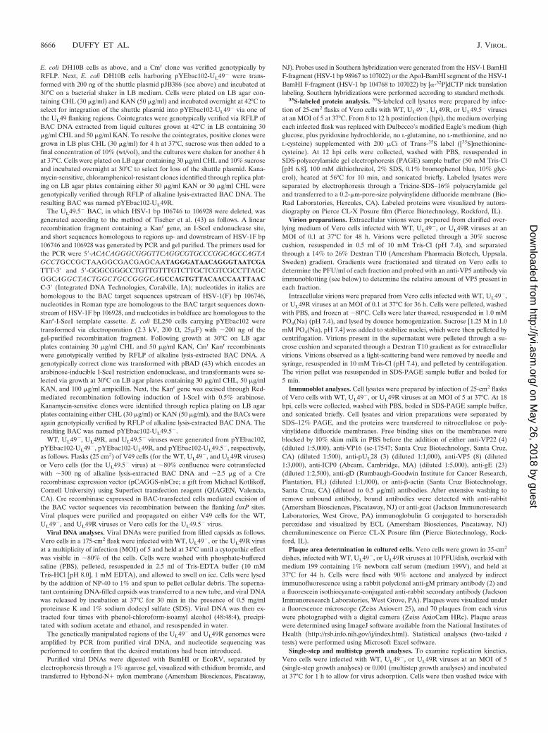
To confirm the deletion and repair of the UL49 ORF, theexpression of VP22 in cells infected with the recombinantviruses was examined. Vero cells were infected at an MOI of 5with WT, UL49�, or UL49R virus and then collected at 18 hpiand lysed. Proteins within the lysates were analyzed by immu-noblotting with an anti-VP22 polyclonal antibody (4). As ex-pected, VP22 was present in lysates of WT- and UL49R-in-fected cells but absent from lysates of UL49�-infected cells(Fig. 2, top).
To ensure that the deletion of UL49 did not affect expressionof the UL48 gene product VP16, immunoblots of the abovelysates were probed with an anti-VP16 polyclonal antibody(Fig. 2, middle). As a loading control, these immunoblots werealso probed with a polyclonal antibody directed against an-other late protein encoded by the UL28 gene (Fig. 2, bottom).VP16 was expressed in approximately equimolar amounts incells infected with the WT, UL49�, and UL49R viruses, confirm-ing that UL48 expression was unaffected by the deletion of UL49.
Due to the lack of a UL49.5 antibody, we used Tricine-SDS-PAGE separation of 35S-labeled infected cell lysates to ensurethat the deletion of UL49 did not affect expression of theUL49.5 gene product. To determine the position of the UL49.5gene product on the gel, we constructed a UL49.5 deletionmutant and compared 35S-labeled proteins present in lysatesderived from cells infected with this mutant to those infectedwith the WT, UL49�, and UL49R viruses. Construction of the
VOL. 80, 2006 HSV-1 VP22 PROMOTES VIRAL SPREAD 8667
on May 26, 2018 by guest
http://jvi.asm.org/
Dow
nloaded from
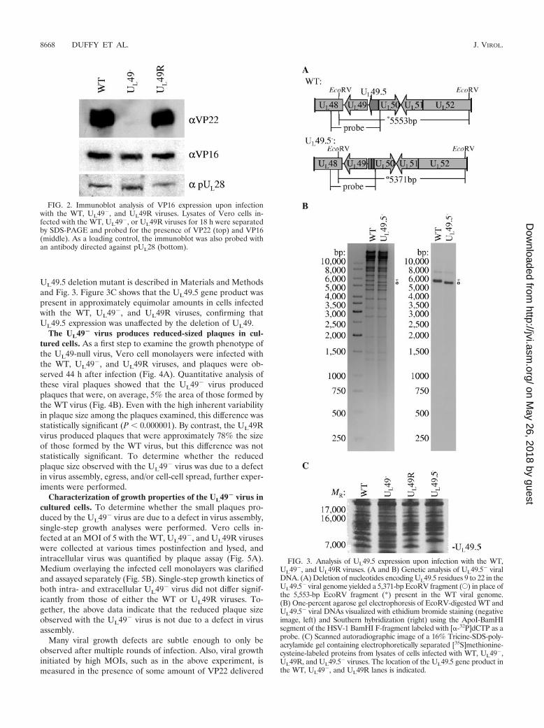
UL49.5 deletion mutant is described in Materials and Methodsand Fig. 3. Figure 3C shows that the UL49.5 gene product waspresent in approximately equimolar amounts in cells infectedwith the WT, UL49�, and UL49R viruses, confirming thatUL49.5 expression was unaffected by the deletion of UL49.
The UL49� virus produces reduced-sized plaques in cul-tured cells. As a first step to examine the growth phenotype ofthe UL49-null virus, Vero cell monolayers were infected withthe WT, UL49�, and UL49R viruses, and plaques were ob-served 44 h after infection (Fig. 4A). Quantitative analysis ofthese viral plaques showed that the UL49� virus producedplaques that were, on average, 5% the area of those formed bythe WT virus (Fig. 4B). Even with the high inherent variabilityin plaque size among the plaques examined, this difference wasstatistically significant (P � 0.000001). By contrast, the UL49Rvirus produced plaques that were approximately 78% the sizeof those formed by the WT virus, but this difference was notstatistically significant. To determine whether the reducedplaque size observed with the UL49� virus was due to a defectin virus assembly, egress, and/or cell-cell spread, further exper-iments were performed.
Characterization of growth properties of the UL49� virus incultured cells. To determine whether the small plaques pro-duced by the UL49� virus are due to a defect in virus assembly,single-step growth analyses were performed. Vero cells in-fected at an MOI of 5 with the WT, UL49�, and UL49R viruseswere collected at various times postinfection and lysed, andintracellular virus was quantified by plaque assay (Fig. 5A).Medium overlaying the infected cell monolayers was clarifiedand assayed separately (Fig. 5B). Single-step growth kinetics ofboth intra- and extracellular UL49� virus did not differ signif-icantly from those of either the WT or UL49R viruses. To-gether, the above data indicate that the reduced plaque sizeobserved with the UL49� virus is not due to a defect in virusassembly.
Many viral growth defects are subtle enough to only beobserved after multiple rounds of infection. Also, viral growthinitiated by high MOIs, such as in the above experiment, ismeasured in the presence of some amount of VP22 delivered
FIG. 3. Analysis of UL49.5 expression upon infection with the WT,UL49�, and UL49R viruses. (A and B) Genetic analysis of UL49.5� viralDNA. (A) Deletion of nucleotides encoding UL49.5 residues 9 to 22 in theUL49.5� viral genome yielded a 5,371-bp EcoRV fragment (E) in place ofthe 5,553-bp EcoRV fragment (*) present in the WT viral genome.(B) One-percent agarose gel electrophoresis of EcoRV-digested WT andUL49.5� viral DNAs visualized with ethidium bromide staining (negativeimage, left) and Southern hybridization (right) using the ApoI-BamHIsegment of the HSV-1 BamHI F-fragment labeled with [�-32P]dCTP as aprobe. (C) Scanned autoradiographic image of a 16% Tricine-SDS-poly-acrylamide gel containing electrophoretically separated [35S]methionine-cysteine-labeled proteins from lysates of cells infected with WT, UL49�,UL49R, and UL49.5� viruses. The location of the UL49.5 gene product inthe WT, UL49�, and UL49R lanes is indicated.
FIG. 2. Immunoblot analysis of VP16 expression upon infectionwith the WT, UL49�, and UL49R viruses. Lysates of Vero cells in-fected with the WT, UL49�, or UL49R viruses for 18 h were separatedby SDS-PAGE and probed for the presence of VP22 (top) and VP16(middle). As a loading control, the immunoblot was also probed withan antibody directed against pUL28 (bottom).
8668 DUFFY ET AL. J. VIROL.
on May 26, 2018 by guest
http://jvi.asm.org/
Dow
nloaded from

from the infecting virions that were propagated on a comple-menting cell line. To minimize such effects and further studythe role of VP22 in viral growth, we performed multistepgrowth analyses in cells infected with 0.001 PFU/cell. To studyextracellular virus as a component of viral spread, we titratedthe overlying medium and infected cell monolayers separately.Figure 6A shows that intracellular UL49� virus titers weredecreased, up to �11-fold at 48 hpi, compared to both the WTand UL49R viruses. Extracellular UL49� virus titers were alsodecreased, again up to �11-fold at 48 hpi, compared to the WTand UL49R viruses as shown in Fig. 6B.
Virion release is decreased in the absence of VP22. Thedecrease in UL49� intra- and extracellular multistep growthcould be due to one or more of the following mechanisms: (i)decreased release of UL49� virions into the medium, resultingin decreased secondary infection and spread through the cellmonolayer; (ii) decreased infectivity of individual extracellularUL49� virions, resulting in decreased secondary infection; (iii)decreased or defective virus assembly within UL49�-infectedcells, resulting in fewer intra- and extracellular infectious par-ticles; and (iv) decreased spread from infected cells to adjacentcells (cell-cell infection), resulting in decreased spread throughthe monolayer. To differentiate between these possibilities, weperformed multistep intracellular growth analyses in the pres-ence of neutralizing antibody in the overlaying medium. Thistreatment dramatically reduces extracellular virus and there-
fore focuses the analysis onto viral spread in the absence ofsecondary infections from extracellular particles. As shown inFig. 6C, when extracellular virus was eliminated from multistepgrowth, the UL49� virus generated intracellular titers approx-imately equivalent to those of the WT and UL49R viruses.Thus, the above data show the viral spread defect observedupon UL49� virus infection was primarily due to decreasedviral release and/or decreased infectivity of extracellular
FIG. 4. Analysis of plaques produced by the WT, UL49�, andUL49R viruses in cultured cells. Vero cell monolayers were infectedwith the WT, UL49�, or UL49R viruses at an MOI of �1 � 10�5
PFU/cell for 44 h. Cells were then fixed and prepared for immunoflu-orescence microscopy using an antibody directed against the viralprotein gM. Plaques were visualized under a fluorescence microscopeand photographed with a digital camera. (A) Representative photo-graphs of WT, UL49�, and UL49R viral plaques. Scale bar, 500 �m.(B) Mean areas of 70 UL49� and 70 UL49R plaques relative to themean area of 70 WT plaques. Error bars represent 1 standarddeviation.
FIG. 5. Single-step growth analyses of the WT, UL49�, and UL49Rviruses. Vero cell monolayers were infected with the WT, UL49�, andUL49R viruses at an MOI of 5 PFU/cell for 1 h to allow virus adsorp-tion. The cells were then washed extensively with citrate buffer toneutralize and remove unbound virus. The cells were overlaid withmedium and held at 37°C. At the indicated times postinfection, theinfected cells (A) and the overlaying medium (B) were analyzed sep-arately by plaque assay to determine intracellular and extracellularviral yields, respectively.
VOL. 80, 2006 HSV-1 VP22 PROMOTES VIRAL SPREAD 8669
on May 26, 2018 by guest
http://jvi.asm.org/
Dow
nloaded from

UL49� particles rather than to decreased virus assembly orcell-cell spread.
To determine whether the viral spread defect associatedwith the UL49� virus was due to decreased viral release, de-creased infectivity of extracellular UL49� particles, or both, we
compared the infectivity of purified WT, UL49�, and UL49Rextracellular particles. As shown in Fig. 7A, fractions fromgradients containing virions purified from the clarified overly-ing medium of infected cells contained substantially fewerUL49� PFU than either WT or UL49R PFU. Immunoblot
FIG. 6. Multistep growth analyses of the WT, UL49�, and UL49R viruses in the presence and absence of neutralizing antibody. Vero cellmonolayers were infected with the WT, UL49�, and UL49R viruses at an MOI of 0.001 PFU/cell for 1 h to allow virus adsorption. The cells werethen washed extensively with citrate buffer to neutralize and remove unbound virus, overlaid with medium, and held at 37°C. Cells infected forintracellular multistep growth analysis in the presence of neutralizing antibody were overlaid with medium containing 0.3% human gammaglobulins. At the indicated times postinfection, these cells were pelleted, washed three times with PBS to remove the neutralizing antibody, andlysed. Also at the indicated times postinfection, medium was removed from cells grown in the absence of neutralizing antibody and clarified.Intracellular virus was titrated from the cell lysates and extracellular virus was titrated from the clarified medium. The growth curves shownrepresent the means and standard deviations (error bars) of three independent experiments. Growth curves in the upper panels were performedat different times using different passages of Vero cells than those of the lower panel. Therefore, titers between upper and lower panels are notcomparable.
8670 DUFFY ET AL. J. VIROL.
on May 26, 2018 by guest
http://jvi.asm.org/
Dow
nloaded from

analyses of these gradient fractions using an antibody againstthe major capsid protein, VP5, showed that the UL49� gradi-ent fractions also contained less VP5 (Fig. 7B), indicating thatUL49� viral infections produce fewer extracellular particles aswell as fewer extracellular PFU compared to WT or UL49Rinfections. To further compare the infectivity of extracellularUL49� virions to extracellular wild-type and UL49R virions, weperformed particle-PFU analyses. The particle-to-PFU ratiosof purified WT, UL49�, and UL49R extracellular virions were234:1, 550:1, and 418:1, respectively; these ratios were notreadily distinguishable within the high inherent variability ofthis technique. These findings indicate that extracellularUL49� particles are not measurably less infectious than WT orUL49R extracellular particles and, together with the abovedata on multistep viral growth in the presence and absence ofneutralizing antibody, show that the spread defect associatedwith the UL49� virus is largely attributable to decreased viralrelease.
To correlate the decreased viral release of the UL49-nullvirus with the effect of extracellular virus on WT HSV-1spread, we examined the effect of neutralizing antibody on WTplaque size. We found that in the presence of 1% humanimmunoglobulin, WT HSV-1 produces plaques that are 15%the size of those produced in the absence of neutralizing an-tibody (data not shown), showing that extracellular virus con-tributes greatly to HSV-1 plaque size and viral spread.
Protein composition of UL49� virions. The defective viralrelease observed upon infection with the UL49� virus could bea direct or indirect consequence of the absence of VP22. Dif-ferences in intracellular virion composition could contribute todifferences in viral release. Therefore, we investigated whetherVP22 contributes indirectly to virion release, through a possi-ble role in virion assembly, by examining the protein compo-sition of WT, UL49�, and UL49R intracellular virions. The leftpanel of Fig. 8 shows a Coomassie-stained SDS-polyacrylamidegel separation of purified intracellular virions, whereas theright panels show immunoblot analyses of the same samples.The locations of the probed proteins are aligned with theircounterparts in the Coomassie-stained gel. The major capsidprotein, VP5, was probed as a loading control. Recently, Elliottand others showed that VP22 is absolutely required for theincorporation of ICP0 into virions (13, 38). We found thatalthough ICP0 was decreased in UL49� virions, VP22 was notabsolutely required for its incorporation. In HSV-1, VP22 hasbeen shown to interact with both VP16 and gD (7, 14). Aspreviously reported (13), we found that the absence of VP22had no effect on incorporation of VP16, whereas levels of gDwere decreased in UL49� virions compared to WT and UL49Rvirions. Although PRV VP22 was found to interact with PRVgE, virion incorporation of gE was not affected in a PRV UL49deletion virus (20). In contrast, we found that levels of gE weredramatically decreased in HSV-1 UL49� virions compared to
FIG. 7. Analysis of extracellular WT, UL49�, and UL49R virions. Extracellular virions were purified from the clarified medium overlying cellsinfected with either the WT, UL49�, or UL49R viruses by gradient centrifugation. (A) The number of PFU/ml in gradient fractions was determinedby plaque assay on Vero cells. Fraction 1 represents the bottom of the gradient, and fraction 9 represents the top of the gradient. (B) Immunoblotsof the same gradient fractions probed with an antibody against the major capsid protein VP5. Each lane is aligned with the corresponding fractionin panel A.
VOL. 80, 2006 HSV-1 VP22 PROMOTES VIRAL SPREAD 8671
on May 26, 2018 by guest
http://jvi.asm.org/
Dow
nloaded from

WT and UL49R HSV-1 virions. Michael et al. (32) recentlyreported extensive variability in levels of gE present in differ-ent PRV virion preparations. However, we found both thelevels of gE present in WT and UL49R virions and the relativedecrease in gE levels present in UL49� virions to be fairlyconsistent from virion preparation to preparation (data notshown). Therefore, the relative decrease of gE incorporationinto UL49� virions shown in Fig. 8 is a consistent and repro-ducible aspect of the HSV-1 UL49-null virus phenotype. Fi-nally, virion incorporation of actin was increased in PRVUL49� virions (10, 32) but was not affected in our HSV-1UL49� virus. The above data indicate that VP22 is involved in,but not required for, incorporation of ICP0, gD, and gE intoHSV-1 virions. In addition, the protein composition of VP22�
virions varies between HSV-1 and PRV, suggesting that therole(s) VP22 plays in virion assembly varies between the twosystems.
VP22 is required for efficient viral spread in the mousecornea. Many HSV-1 proteins exhibit functional redundancyand are therefore deemed nonessential when studied in cul-tured cells, but they exhibit more dramatic phenotypes in liveanimals. To determine whether VP22 is essential for viralgrowth in vivo, we analyzed viral spread in the mouse cornea.Corneas in live mice were scarified and infected with �100PFU of WT, UL49�, or UL49R virus. At 24 or 48 hpi, the micewere sacrificed, the corneas were dissected, and the presenceof infectious virus was revealed by immunohistochemistry withan HSV-specific antibody. At both time points the UL49� virusspread only �60% of the distance spread by the WT andUL49R viruses (Fig. 9 and Table 1). The far-right panels of Fig.9 show whole flat-mounted corneas photographed at lowermagnification and illustrate the decreased size of corneal le-sions produced upon infection with the UL49-null virus (Fig.9C) compared to the lesions produced by the WT and UL49R
viruses (Fig. 9I and F, respectively). The UL49� virus formedisolated rosettes of infected cells, whereas the WT and UL49Rviruses formed extensive borders of infected cells along thelength of the scarification. These data identify a role for VP22in HSV-1 viral spread in vivo as well as in cultured cells.
DISCUSSION
Although many studies have focused on identifying the bio-logical properties of HSV-1 VP22 and its homologs in otherherpesviruses, the role this protein plays in HSV-1 infection isunclear. To address this problem, we generated two recombi-nant viruses, one lacking the entire UL49 gene and one con-taining a restored UL49 gene, and characterized their growth.In this report we show that VP22 is required for efficient viralspread during infection of both cultured cells and the mousecornea.
Role of VP22 in viral spread in cultured cells. While single-step intracellular and extracellular growth analyses did notindicate a defect in virus assembly for the UL49-null virus,plaques produced by this virus in cultured cells were reduced insize by an average of 95% compared to the WT and UL49-repaired viruses. A similar, though less dramatic, phenotypewas previously observed with an HSV-1 UL49 truncation mu-tant that expresses low levels of the amino-terminal 212 resi-dues of VP22 (37). This virus, HSV-1 RF177, producedplaques in cultured cells that were reduced in size by nearly40% compared to those produced by the parental WT virus. Itis currently not known whether the reduced plaque size ob-served with this virus is due to the lack of the carboxy-terminal89 residues of VP22 or to the low expression level of thetruncated protein, and, therefore, we cannot attribute the func-tion of VP22 in viral spread to a particular domain of theprotein. In any case, the two studies are in agreement in de-fining a role for VP22 in viral spread of HSV-1.
Recently, an HSV-1 recombinant virus in which the UL49gene was replaced with the gene encoding green fluorescentprotein was reported (13). Elliott et al. constructed their UL49-null virus on a VP22-complementing cell line because the re-combinant virus could not be generated on noncomplementingcells, and it was therefore suggested that the WT virus main-tained a growth advantage over the UL49-null virus. However,once the UL49-null virus was generated, the authors observedthat VP22 was not necessary for virus growth on noncomple-menting Vero cells and, thus, performed all further experi-ments with virus stocks propagated on noncomplementingcells. A decrease in plaque size was not reported for this virus.
We have also observed that VP22 is dispensable for growthon Vero cells. However, we have noted with interest that whileUL49� stocks propagated on VP22-complementing cells con-sistently produced plaques �5% the size of those produced bythe WT and UL49R viruses, some plaques produced by theUL49-null virus were comparable in size to those produced bythe wild-type virus (http://www.vet.cornell.edu/labs/baines/) af-ter as few as two to three passages on noncomplementing cells.The reproducibility of these observations suggests that theUL49-null virus somehow compensates for the lack of VP22when propagated on noncomplementing cells. The mechanismof this phenotypic change is unknown but may reflect theacquisition of secondary mutations that compensate for the
FIG. 8. Analysis of the protein composition of WT, UL49�, andUL49R virions. Intracellular virions were purified from cells infectedwith either the WT, UL49�, or UL49R viruses. Virions proteins wereelectrophoretically separated on a 12% SDS-polyacrylamide gel andvisualized by either Coomassie staining (left panel) or immunoblotanalyses using antibodies against VP5, ICP0, gE, VP16, gD, -actin,and VP22 (right panels). The immunoblots are aligned with the posi-tion of each probed protein in the Coomassie-stained gel.
8672 DUFFY ET AL. J. VIROL.
on May 26, 2018 by guest
http://jvi.asm.org/
Dow
nloaded from

lack of VP22. Because phenotypic change may further definethe function of VP22 during virus infection, as well as identifythe basic mechanisms of HSV-1 spread, we are intrigued bythis phenotypic morphogenesis, and studies are under way toidentify its cause.
In the present work, studies were undertaken to determinethe mechanism by which VP22 promotes viral spread. Duringmultistep growth, both intracellular and extracellular UL49�
PFU were reduced 11-fold compared to the WT and UL49Rviruses. Multistep growth experiments performed in the pres-ence and absence of neutralizing antibody together with VP5immunoblot-PFU and particle-PFU analyses of UL49� extra-cellular particle infectivity showed that this reduction waslargely attributable to decreased levels of extracellular UL49�
virus rather than to a defect in either UL49� virus assembly,cell-cell spread, or particle infectivity. At this time it is unclearwhether the decreased extracellular virus observed duringUL49� infection is due to a defect in viral exit from infectedcells or decreased release of extracellular UL49� virions fromintercellular spaces. The answer to this question and the role ofVP22 in the mechanism(s) of viral exit and/or intercellularrelease will be the topics of future studies. Our present workshows that at least in some culture systems and tissue types,extracellular virus contributes greatly to viral spread, and VP22plays a role in the accumulation of extracellular virus.
VP22 may promote extracellular virus accumulation andspread through a direct or indirect mode of action, and severalpossibilities exist for either scenario. For example, VP22 mayaffect the cytoskeleton of infected cells and promote efficienttransport of virion-containing vesicles to the cell surfacethrough its ability to stabilize microtubules (15). Alternatively,VP22 may function indirectly through its interactions withand/or optimal incorporation of other viral proteins such asICP0, gE, or gD into virions (7, 13, 20). Elliott et al. havereported that in the absence of VP22 there is both a delay inthe synthesis of the transactivator ICP0 and a complete loss ofICP0 virion incorporation (13). If the decreased viral spreadobserved upon UL49� infection was due solely to these effects
FIG. 9. Analysis of UL49�, UL49R, and WT HSV-1 viral spread in the mouse cornea. The corneas of live mice were scarified and infected with�1,000 PFU of either UL49� (A to C), UL49R (D to F), or WT HSV-1 (G to I) virus. At 24 or 48 hpi, the mice were sacrificed, and the corneaswere dissected and prepared for immunohistochemistry using an antibody directed against HSV-1 proteins. Photographs of corneas flat mountedon glass slides are shown with the left and middle panels at the same magnification (bar 100 �m) and at lower magnification in the right panels(bar 1 mm).
TABLE 1. Spread of the WT, UL49-null, and UL49R virusesin corneal epithelium
Time(hpi)
Mean distance spread SD (�m)a
WT (HSV-1F) UL49� UL49R
24 179 81.7 (81) 105.9 75.6 (78) 171.7 112.2 (70)48 193.8 79.3 (64) 128.4 81.9 (77) 206.4 78.0 (64)
a The number of measurements is given in parentheses.
VOL. 80, 2006 HSV-1 VP22 PROMOTES VIRAL SPREAD 8673
on May 26, 2018 by guest
http://jvi.asm.org/
Dow
nloaded from

on ICP0, one would expect to observe decreased production ofintracellular virions, especially over several rounds of infectionas measured by multistep growth, and/or decreased infectivityof UL49� virions. However, we did not observe either of thesephenomena (Fig. 6, lower panel). Another possibility is thatVP22 mediates viral spread through incorporation of gD intovirions. However, because HSV-1 gD is required for virus entryand our particle-PFU analyses did not show a measurabledecrease in UL49� virion infectivity, it is unlikely that thedecreased incorporation of gD into UL49� virions can fullyexplain the UL49-null phenotype. Likewise, we observed de-creased incorporation of gE into UL49� virions. However, gEis required for normal cell-cell spread (12) which was notdecreased upon infection with the UL49� virus. Thus, whileboth gE and VP22 contribute to HSV-1 viral spread, VP22appears to contribute through extracellular virus, whereas gEfacilitates cell-cell spread.
Contribution of VP22 to herpetic disease. The spread ofHSV between corneal epithelial cells is critical for two phasesof herpetic eye disease. In the initial, primary phase, aerosol-ized HSV that has been released from one infected individualmay enter a break in the corneal epithelium of a second indi-vidual. The virus replicates and spreads between epithelial cellsof the second host, and it ultimately reaches the basal celllayer, where it can invade the fine axon endings of trigeminalganglion cells. Within these axons HSV is transported retro-grade to the ganglion cell bodies, where it can establish a latentinfection. In the second phase following reactivation, new virusis transported anterograde within the nerve endings to thecorneal epithelium (33). The corneal epithelial cells again be-come infected, and the resulting humoral and cellular immu-nological responses may result in scarring and recurrent in-flammation in the epithelium and deeper tissues (for review,see reference 41).
Understanding the pathogenesis of both phases of herpeticepithelial disease requires knowledge of the role(s) that viralgenes play in viral spread. Because productive infection inanimals may require specific viral proteins that are dispensablefor replication in cultured cells, we considered it important tostudy the contribution of VP22 to HSV-1 infection in thecontext of the whole-animal model. Corneal lesions producedby the UL49-null virus were dramatically reduced in size com-pared to those produced by the WT and UL49R viruses. Thus,we have identified a role for VP22 in HSV-1 corneal viralspread in vivo.
Studies on the contribution of VP22 to infection by otherherpesviruses have shown variable importance for VP22 inviral growth and virulence. While a bovine herpesvirus UL49deletion virus exhibited a significant delay in single-step growthanalyses and reduced virulence in cattle (26, 27), a PRV UL49deletion virus showed single-step growth profiles identical toWT PRV and did not promote virulence or neuroinvasivenessin the rat eye infection model (11). Together, these data indi-cate that the presence of VP22 is of variable importance todifferent herpesviruses and experimental systems.
In summary, we have characterized the growth of an HSV-1UL49-null virus and have identified a role for VP22 in viralspread during HSV-1 infection both in vitro and in vivo. Futurestudies will identify how this protein functions to promote viralspread and determine whether VP22 plays a role in virion exit
and/or intercellular release. In addition, the use of a UL49-nullvirus with its genetically repaired partner should greatly facil-itate investigations of the involvement of VP22 in HSV-1 teg-ument formation, pathogenesis, and virion egress.
ACKNOWLEDGMENTS
These studies were supported by NIH R01 grants GM 50740 and AI52341 to J.D.B. and AI 38873 to J.A.B., NIH NRSA F32 grantGM067519 to C.D., PHS grant EY 13867, and funds from That ManMay See, Inc., to J.H.L.
We thank Yasuchi Kawaguchi for the gift of the HSV-1F BAC. Wethank Klaus Osterrieder, B. Karsten Tischer, Jens von Einem, DanielSchumacher, Laura Goodman, and Cristina Rosas for their generoussharing of reagents, techniques, and helpful advice. We thank DavidJohnson for the gift of the anti-gE antibody and Elisabeth Schlegel(Mount Sinai School of Medicine) for assistance with the V49 cells.
REFERENCES
1. Adler, H., M. Messerle, M. Wagner, and U. H. Koszinowski. 2000. Cloningand mutagenesis of the murine gammaherpesvirus 68 genome as an infec-tious bacterial artificial chromosome. J. Virol. 74:6964–6974.
2. Baines, J. D., and B. Roizman. 1993. The UL10 gene of herpes simplex virus1 encodes a novel glycoprotein, gM, which is present in the virion and in theplasma membrane of infected cells. J. Virol. 67:1441–1452.
3. Beard, P. M., N. S. Taus, and J. D. Baines. 2002. The DNA cleavage andpackaging proteins encoded by genes UL28, UL15, and UL33 of herpessimplex virus 1 form a complex in infected cells. J. Virol. 76:4785–4791.
4. Blaho, J. A., C. Mitchell, and B. Roizman. 1994. An amino acid sequenceshared by the herpes simplex virus 1 alpha regulatory proteins 0, 4, 22, and27 predicts the nucleotidylylation of the UL21, UL31, UL47, and UL49 geneproducts. J. Biol. Chem. 269:17401–17410.
5. Borst, E. M., G. Hahn, U. H. Koszinowski, and M. Messerli. 1999. Cloningof the human cytomegalovirus (HCMV) genome as an infectious bacterialartificial chromosome in Eschericia coli: a new approach for construction ofHCMV mutants. J. Virol. 73:8320–8329.
6. Campbell, M. E. M., J. W. Palfreyman, and C. M. Preston. 1984. Identifi-cation of herpes simplex virus DNA sequences which encode a trans-actingpolypeptide responsible for stimulation of immediate early transcription. J.Mol. Biol. 180:1–19.
7. Chi, J. H., C. A. Harley, A. Mukhopadhyay, and D. W. Wilson. 2005. Thecytoplasmic tail of herpes simplex virus envelope glycoprotein D binds to thetegument protein VP22 and to capsids. J. Gen.Virol. 86:253–261.
8. Cohen, G. H., M. Ponce de Leon, H. Diggelmann, W. C. Lawrence, S. K.Vernon, and R. Eisenberg. 1980. Structural analysis of the capsid polypep-tides of herpes simplex virus types 1 and 2. J. Virol. 34:521–531.
9. Datsenko, K. A., and B. L. Wanner. 2000. One-step inactivation of chromo-somal genes in Escherichia coli K-12 using PCR products. Proc. Natl. Acad.Sci. USA 97:6640–6645.
10. del Rio, T., C. J. DeCoste, and L. W. Enquist. 2005. Actin is a component ofthe compensation mechanism in pseudorabies virus virions lacking the majortegument protein VP22. J. Virol. 79:8614–8619.
11. del Rio, T., H. C. Werner, and L. W. Enquist. 2002. The pseudorabies virusVP22 homologue (UL49) is dispensable for virus growth in vitro and has noeffect on virulence and neuronal spread in rodents. J. Virol. 76:774–782.
12. Dingwell, K. S., C. R. Brunetti, R. L. Hendricks, O. Tang, M. Tang, A. J.Rainbow, and D. C. Johnson. 1994. Herpes simplex virus glycoproteins E andI facilitate cell-to-cell spread in vivo and across junctions of cultured cells.J. Virol. 68:834–845.
13. Elliott, G., W. Hafezi, A. Whiteley, and E. Bernard. 2005. Deletion of theherpes simplex virus VP22-encoding gene (UL49) alters the expression,localization, and virion incorporation of ICP0. J. Virol. 79:9735–9745.
14. Elliott, G., G. Mouzakitis, and P. O’Hare. 1995. VP16 interacts via itsactivation domain with VP22, a tegument protein of herpes simplex virus,and is relocated to a novel macromolecular assembly in coexpressing cells.J. Virol. 69:7932–7941.
15. Elliott, G., and P. O’Hare. 1998. Herpes simplex virus type 1 tegumentprotein VP22 induces the stabilization and hyperacetylation of microtubules.J. Virol. 72:6448–6455.
16. Elliott, G., and P. O’Hare. 1999. Live-cell analysis of a green fluorescentprotein-tagged herpes simplex virus infection. J. Virol. 73:4110–4119.
17. Elliott, G., D. O’Reilly, and P. O’Hare. 1996. Phosphorylation of the herpessimplex virus type 1 tegument protein VP22. Virology 226:140–145.
18. Elliott, G. D., and D. M. Meredith. 1992. The herpes simplex virus type 1tegument protein VP22 is encoded by gene UL49. J. Gen. Virol. 73:723–726.
19. Fuchs, W., H. Granzow, B. G. Klupp, M. Kopp, and T. C. Mettenleiter. 2002.The UL48 tegument protein of pseudorabies virus is critical for intracyto-plasmic assembly of infectious virions. J. Virol. 76:6729–6742.
20. Fuchs, W., B. G. Klupp, H. Granzow, C. Hengartner, A. Brack, A. Mundt,
8674 DUFFY ET AL. J. VIROL.
on May 26, 2018 by guest
http://jvi.asm.org/
Dow
nloaded from

L. W. Enquist, and T. C. Mettenleiter. 2002. Physical interaction betweenenvelope glycoproteins E and M of pseudorabies virus and the major tegu-ment protein UL49. J. Virol. 76:8208–8217.
21. Geiss, B. J., J. E. Tavis, L. M. Metzger, D. A. Leib, and L. A. Morrison. 2001.Temporal regulation of herpes simplex virus type 2 VP22 expression andphosphorylation. J. Virol. 75:10721–10729.
22. Heine, J. W., R. W. Honess, E. Cassai, and B. Roizman. 1974. Proteinsspecified by herpes simplex virus. XII. The virion polypeptides of type 1strains. J. Virol. 14:640–651.
23. Johnson, D. C., M. C. Frame, M. W. Ligas, A. m. Cross, and N. D. Stow.1988. Herpes simplex virus immunoglobulin G Fc receptor activity dependson a complex of two viral glycoproteins, gE and gI. J. Virol. 62:1347–1354.
24. Knopf, K. W., and H. C. Kaerner. 1980. Virus-specific basic phosphoproteinsassociated with herpes simplex virus type a (HSV-1) particles and the chro-matin of HSV-1-infected cells. J. Gen. Virol. 46:405–414.
25. Lee, E. C., D. Yu, J. Martinez De Velasco, L. Tessarollo, D. A. Swing, D. L.Court, N. A. Jenkins, and N. G. Copeland. 2001. A highly efficient Esche-richia coli-based chromosome engineering system adapted for recombino-genic targeting and subcloning of BAC DNA. Genomics 73:56–65.
26. Liang, X., B. Chow, and L. A. Babiuk. 1997. Study of immunogenicity andvirulence of bovine herpesvirus 1 mutants deficient in the UL49 homolog,UL49.5 homolog and dUTPase genes in cattle. Vaccine 15:1057–1064.
27. Liang, X., B. Chow, Y. Li, C. Raggo, D. Yoo, S. Attah-Poku, and L. A. Babiuk.1995. Characterization of bovine herpesvirus 1 UL49 homolog gene andproduct: bovine herpesvirus 1 UL49 homolog is dispensable for virus growth.J. Virol. 69:3863–3867.
28. Lumb, W. V. 1963. Small animal anesthesia. Lea and Febiger, Philadelphia, Pa.29. McGeoch, D. J., M. A. Dalrymple, A. J. Davison, A. Dolan, M. C. Frame, D.
McNab, L. J. Perry, J. E. Scott, and P. Taylor. 1988. The complete DNAsequence of the long unique region in the genome of herpes simplex virustype 1. J. Gen. Virol. 69:1531–1574.
30. Meredith, D. M., J. A. Lindsay, I. W. Halliburton, and G. R. Whittaker. 1991.Post-translational modification of the tegument proteins (VP13 and VP14)of herpes simplex virus type 1 by glycosylation and phosphorylation. J. Gen.Virol. 72:2771–2775.
31. Messerle, M., I. Crnkovic, W. Hammerschmidt, H. Ziegler, and U. H.Koszinowski. 1997. Cloning and mutagenesis of a herpesvirus genome as aninfectious bacterial artificial chromosome. Proc. Natl. Acad. Sci. USA 94:14759–14763.
32. Michael, K., B. G. Klupp, T. C. Mettenleiter, and A. Karger. 2006. Compo-
sition of pseudorabies virus particles lacking tegument protein US3, UL47,or UL49 or envelope glycoprotein E. J. Virol. 80:1332–1339.
33. Ohara, P. T., M. S. Chin, and J. H. LaVail. 2000. The spread of herpessimplex virus type 1 from trigeminal neurons to the murine cornea: animmunoelectron microscopy study. J. Virol. 74:4776–4786.
34. Pellett, P. E., J. L. McKnight, F. J. Jenkins, and B. Roizman. 1985. Nucle-otide sequence and predicted amino acid sequence of a protein encoded ina small herpes simplex virus DNA fragment capable of trans-inducing alphagenes. Proc. Natl. Acad. Sci. USA 82:5870–5874.
35. Pinard, M. F., R. Simard, and V. Bibor-Hardy. 1987. DNA-binding proteinsof herpes simplex virus type 1-infected BHK cell nuclear matrices. J. Gen.Virol. 68:727–735.
36. Pomeranz, L. E., and J. A. Blaho. 1999. Modified VP22 localizes to the cellnucleus during synchronized herpes simplex virus type 1 infection. J. Virol.73:6769–6781.
37. Pomeranz, L. E., and J. A. Blaho. 2000. Assembly of infectious Herpessimplex virus type 1 virions in the absence of full-length VP22. J. Virol.74:10041–10054.
38. Potel, C., and G. Elliott. 2005. Phosphorylation of the herpes simplex virustegument protein VP22 has no effect on incorporation of VP22 into the virusbut is involved in optimal expression and virion packaging of ICP0. J. Virol.79:14057–14068.
39. Read, G. S., B. M. Karr, and K. Knight. 1993. Isolation of a herpes simplexvirus type 1 mutant with a deletion in the virion host shutoff gene andidentification of multiple forms of the vhs (UL41) polypeptide. J. Virol.67:7149–7160.
40. Ren, X., J. S. Harms, and G. A. Splitter. 2001. Bovine herpesvirus 1 tegu-ment protein VP22 interacts with histones, and the carboxyl terminus ofVP22 is required for nuclear localization. J. Virol. 75:8251–8258.
41. Ritterband, D. C., and D. N. Friedberg. 1998. Virus infections of the eye.Rev. Med. Virol. 8:187–201.
42. Tanaka, M., H. Kagawa, Y. Yamanashi, T. Sata, and Y. Kawaguchi. 2003.Construction of an excisable bacterial artificial chromosome containing afull-length infectious clone of herpes simplex virus type 1: viruses reconsti-tuted from the clone exhibit wild-type properties in vitro and in vivo. J. Virol.77:1382–1391.
43. Tischer, B. K., J. von Einem, B. Kaufer, and N. Osterrieder. 2006. Two-stepred-mediated recombination for versatile high-efficiency markerless DNAmanipulation in Escherichia coli. BioTechniques 40:191–197.
VOL. 80, 2006 HSV-1 VP22 PROMOTES VIRAL SPREAD 8675
on May 26, 2018 by guest
http://jvi.asm.org/
Dow
nloaded from